
Submitted:
23 January 2026
Posted:
23 January 2026
You are already at the latest version
Abstract
Anxiety disorders are the most prevalent mental health conditions worldwide, yet current treatments remain suboptimal, with benzodiazepines carrying risks of tolerance and dependence. These limitations motivate the search for novel anxiolytics. Tropical marine macroalgae represents a promising source of neuroactive metabolites. Here, we investigate the anxiolytic potential of Stypopodium zonale using a neuroproteomics-based approach in Drosophila melanogaster. Crude organic extracts were prepared via ultrasonic-assisted extraction and administered acutely to adult flies for six hours. Proteins from fly heads were quantified and analyzed using LC-MS/MS, revealing 66 significantly differentially abundant proteins (fold change ≥ |1.5|, p ≤ 0.05), 72.7% of which were less abundant in the extract-treated group. Principal Component Analysis demonstrated clear separation between control and experimental samples. Ingenuity Pathway Analysis (IPA) mapped 33 of the differentially abundant proteins to human orthologs and identified significant predicted inhibition of the Protein Kinase A (PKA) signaling pathway. An IPA Interaction Network enabled the construction of a preliminary working model, suggesting that the extract may antagonize Drosophila’s Dop1R2 (DAMB). Overall, this study integrates natural product drug discovery with neuroproteomics in an invertebrate model system, providing a foundation for future behavioral validation and isolation of bioactive compounds from S. zonale.
Keywords:
Drosophila melanogaster
; neuroproteomics
; behavior
; marine algae
; TMT quantitative proteomics
1. Introduction
Proteins are the workhorses of the cell, playing crucial roles in virtually all biological processes. In neurons, they function as both the molecular machinery and messengers of the nervous system, shaping neural architecture, enabling synaptic communication, and supporting neurodevelopment, learning, memory, and behavior. Under this premise, the field of neuroproteomics emerged. As a subdiscipline of proteomics, it is dedicated to studying the structure, function, and interactions of proteins within the nervous system to uncover the molecular basis of neural function and dysfunction [1,2].
Proteomics, a field formally conceptualized by Wilkins et al. in 1995, refers to the large-scale study of the complete protein complement, or proteome, expressed by a genome, tissue, or cell type. Unlike the static nature of the genome, the proteome is highly dynamic, varying across tissues of a singular organism and between organisms. It is also condition and time-dependent, as the entire set of proteins expressed by a cell continually changes in response to internal and external factors. This unique feature has made proteomics an essential tool in molecular biology, offering a functional layer of biological insight beyond what genomics can provide. As Wilkins and colleagues argued, the strength of proteomics lies not only in identifying which proteins are present but also in elucidating how they functionally interact within biological systems [3]. This versatility has led proteomics to become central in biomedical research, particularly in two major domains: biomarker identification and drug discovery.
In biomarker identification, comparative proteomics is employed to detect proteins whose abundance levels differ significantly between healthy and diseased states. Then, these proteins can serve as biological indicators, or biomarkers, that can inform the molecular mechanisms underlying diseases [4]. Neuroproteomics, in particular, has been instrumental in identifying potential biomarkers and molecular targets for neurological disorders such as Parkinson’s disease [5], Alzheimer’s disease [6], and psychiatric conditions like anxiety and depression [7].
In drug discovery, proteomics provides an equally important function. By enabling the detection of proteins whose abundance changes in response to drug treatment, proteomics offers mechanistic insight into how a compound exerts its effects at the molecular level [4]. Within this context, our study introduces a novel application of neuroproteomics for anxiolytic drug discovery. Specifically, we aim to investigate whether extracts from tropical marine macroalgae, namely Stypopodium zonale (a brown macroalga), exert anxiolytic effects in Drosophila melanogaster. Multiple layers of novelty drive this research question.
A central innovation lies in the use of tropical marine macroalgae as a source of potential anxiolytic compounds. These algae produce a chemically diverse array of bioactive metabolites, including phlorotannins, alkaloids, terpenoids, carotenoids, phytosterols, and polysaccharides, which have demonstrated neuroprotective effects in both in vitro and in vivo vertebrate models. Reported effects include protection against amyloid-β toxicity, oxidative stress, and glutamate-induced excitotoxicity; enhancement of antioxidant defenses; reduction of neuroinflammation; and promotion of neuronal survival and synaptic plasticity, among others [8].
Beyond these neuroprotective effects, what is known about the anxiolytic potential of marine macroalgae remains limited. For example, Ecklonia cava, a phlorotannin-rich brown macroalga, has been shown to exert central nervous system (CNS) depressant effects, including sedative, anticonvulsant, and, critically, anxiolytic-like effects [9]. These effects are attributed to phlorotannins binding to the benzodiazepine site on GABA A receptors, where they act as positive allosteric modulators. This enhances GABAergic inhibition, a mechanism well-established in the pharmacology of conventional anxiolytics such as benzodiazepines. Notably, Stypopodium zonale, the focus of our investigation, is also a brown macroalga [10], further supporting its potential as a source of anxiolytic compounds. Given this pharmacological profile, tropical marine macroalgae emerge as promising candidates for natural product-based drug discovery in the fields of neurology and psychiatry [8].
The therapeutic relevance of our work is further underscored by its focus on anxiety. As of 2021, an estimated 359.2 million people worldwide were affected by anxiety disorders, making them the most prevalent mental health condition globally [11]. Anxiety, unlike fear, is conceptualized as a sustained and diffuse response to uncertain or future threats. It involves a complex interplay of emotional, behavioral, and physiological responses. In animal models, researchers rely on observable behaviors-such as increased avoidance, reduced exploration, or heightened vigilance-to infer anxiety-like states. These behaviors serve as valid proxies for anxiety, particularly when paired with neurobiological correlates [12,13].
Despite the high global burden of anxiety disorders, current pharmacological treatments remain suboptimal. Benzodiazepines, although widely prescribed, present well-documented limitations associated with long-term use-including the development of tolerance and a significant risk of both physical and psychological dependence. These limitations highlight a space for continued progress in the discovery and development of novel anxiolytics, which serves as the rationale of our research [14]. In addition to the pharmacological gap, there are also methodological limitations in anxiety-related proteomics research. Much of this work has been predominantly conducted using rodent models, leaving a gap for the use of complementary animal models [1,7,15]. Our use of Drosophila melanogaster introduces a significant innovation in this regard. Drosophila offers a genetically tractable, cost-effective, and evolutionarily conserved system for studying anxiety-like behavior at the molecular level. Importantly, key anxiety-relevant signaling pathways and gene networks are conserved between flies and mammals, reinforcing the model’s translational value [16].
To guide our investigation into anxiolytic drug discovery, we adopted a two-tiered research approach. First, we asked whether exposure to the extract of Stypopodium zonale would lead to differential protein abundance in the heads of Drosophila melanogaster compared to untreated controls. We hypothesized that such exposure would elicit broad proteomic alterations, reflecting a systemic biological response to the treatment. Building on these findings, our second research question examined whether the observed proteomic changes were associated with a potential anxiolytic effect; specifically, whether exposure to the extract alters the abundance of proteins involved in anxiety-related cell signaling pathways. We hypothesized that modulation of these proteins may influence molecular mechanisms underlying reduced anxiety-like behavior.
This dual-level approach not only enables the identification of molecular changes following exposure to Stypopodium zonale but also establishes a critical foundation for constructing a mechanism of action and assessing whether these molecular alterations translate into behavioral effects. In this sense, the proteomics data will function as a "spearhead”, guiding future behavioral investigations. Should subsequently testing in Drosophila melanogaster reveal a reduction in anxiety-like behavior, these molecular findings would support the hypothesis that Stypopodium zonale possesses anxiolytic properties. In this way, our research will operate at the intersection of neuroproteomics and behavioral neuroscience, bridging molecular insights with functional outcomes.
2. Results
This section may be divided by subheadings. It should provide a concise and precise description of the experimental results, their interpretation, as well as the experimental conclusions that can be drawn.
2.1. Differential Protein Abundance Between Control and Stypopodium zonale-Exposed Samples
We applied a quantitative proteomics approach to analyze Drosophila melanogaster head proteins following acute exposure to Stypopodium zonale. Across four biological replicates per condition, a total of 951 proteins were identified. Although five samples were initially processed, only four were included in the final analysis because statistical testing showed a higher number of protein identifications in these four samples. Principal component analysis (PCA) of protein abundance profiles (Figure 1A) revealed clear separation between control (n = 4) and S. zonale-exposed samples (n = 4), with minimal within-group variability. PERMANOVA confirmed that this separation was statistically significant (p = 0.021). The non-overlapping clusters indicate that treatment-induced proteomic changes exceeded natural biological variation and were highly reproducible across replicates, consistent with a robust and widespread effect of acute S. zonale exposure on the Drosophila head proteome.
2.2. Sixty Six (66) Significantly Differentially Abundant Proteins Were Identified Between the Control and Stypopodium zonale-Exposed Samples
One-way analysis of variance (ANOVA) comparing control and Stypopodium zonale-exposed samples identified 66 differentially abundant proteins. Proteins were considered significant based on a fold-change threshold of |FC| ≥ 1.5 and a p-value ≤ 0.05. This criterion captures both decreases in abundance (-FC) and increases in abundance (+FC). Of these, 48 proteins (72.7%) were significantly less abundant (Table 1), whereas 18 proteins (27.3%) were significantly more abundant (Table 2) in S. zonale-exposed samples. As illustrated in the volcano plot (Figure 1B), proteins are distributed according to both the magnitude of change (log₂ fold change) and statistical significance (−log₁₀ p-value). Vertical dashed lines denote the fold-change thresholds
corresponding to |FC| ≥ 1.5, while the horizontal dashed line represents the significance cutoff (p ≤ 0.05). Proteins meeting both criteria appear in the upper left and upper right regions of the plot, corresponding to significantly less abundant and more abundant proteins, respectively. Notably, a greater density of statistically significant proteins is observed on the negative log₂ fold-change side, visually reinforcing the predominance of downregulated proteins following S. zonale exposure. Together, the asymmetric distribution of significant proteins supports a treatment-associated shift toward decreased global protein abundance in S. zonale-exposed Drosophila head samples.
2.3. Acute Exposure to S. zonale Led to Broad and Consistent Reduced Protein Abundance Relative to Controls
A heatmap of the 66 differentially abundant proteins (Figure 2A) revealed clear clustering between control and Stypopodium zonale-exposed samples, with strong consistency among biological replicates within each group. Proteins in the S. zonale-exposed samples clustered predominantly toward lower abundance values, reflected by a pronounced enrichment of blue coloration across the four experimental replicates. This pattern indicates a consistent reduction in protein abundance following acute exposure to the extract. This global trend is concordant with the differential abundance analysis, in which most proteins (72.7%) were significantly less abundant after treatment. In contrast, the same proteins exhibited higher abundance in control samples, as indicated by green to yellow coloration in the heatmap, reflecting a higher baseline proteomics state under control conditions.
2.4. Protein Localization
To further characterize the cellular context of the differentially abundant proteins, we examined their subcellular localizations using UniProt and Gene Ontology annotations (Figure 2B). Most proteins (45.45%) localized to the cytoplasm, consistent with the predominance of cytosolic enzymes, signaling components, and synaptic regulators identified in our dataset. A substantial proportion (22.73%) corresponded to secreted proteins, including several members of the odorant-binding protein family, which play key roles in chemosensory communication and environmental sensing. Additional groups included proteins localized to the cell membrane (10.61%) and nucleus (10.61%), reflecting components involved in receptor signaling, transcriptional regulation, and intracellular communication. Smaller fractions mapped to cell projections (6.06%), such as axons and dendrites, and to the mitochondrion (4.55%), the latter aligning with metabolic enzymes found to be differentially abundant. Collectively, this distribution highlights that acute exposure to S. zonale affects proteins across multiple cellular compartments, with a strong representation of cytoplasmic, membrane-associated, and secreted proteins that contribute to neuronal signaling and sensory processing.
2.5. Ingenuity Pathway Analysis (IPA)
Of the 66 differentially abundant proteins identified in our proteomic screen, 33 were successfully mapped to human orthologs within the IPA Knowledge Base. IPA identified several canonical pathways associated with these proteins and predicted significant functional inhibition in two of them: the Protein Kinase A (PKA) signaling pathway (z-score = -1.342) and the Oxytocin signaling pathway (z-score = -2). Proteins contributing to the predicted inhibition of the PKA signaling pathway included GNB1 (Gβ1 subunit), GNA13 (α-subunit homolog), PPP3R1 (calcineurin subunit B type 1), PYGM (glycogen phosphorylase), and YWHAE (14-3-3 protein epsilon). Proteins contributing to the predicted inhibition of the Oxytocin signaling pathway were GAD1 (glutamate decarboxylase), GNA13 (α-subunit homolog), GNB1 (Gβ1 subunit), and PPP3R1 (calcineurin subunit B type 1). In parentheses are the proteins identified in Drosophila. The specific proteins mapped to each canonical pathway, along with their corresponding z-scores, are presented in Table S4. Gene identifiers are used for all proteins.
3. Discussion
Our central research question is: Do extracts from tropical marine macroalgae, specifically Stypopodium zonale, exert anxiolytic effects? To address this question, we employed a quantitative proteomics-based approach to identify differentially abundant proteins. Our working hypotheses were: (1) exposure to the S. zonale extract would lead to significant alterations in protein abundance relative to controls, and (2) these molecular changes could contribute to an anxiolytic-like effect in Drosophila. In support of our first hypothesis, the proteomics analysis revealed a distinct and consistent shift in protein abundance following acute exposure to S. zonale. We identified 66 significantly differentially abundant proteins, of which 48 (72.7%) were less abundant and 18 (23.7%) were more abundant in the experimental group compared to controls. This widespread reduction in protein levels clearly separated the treatment and control groups in a 2D PCA Plot and a hierarchically clustered heatmap, demonstrating a robust and reproducible effect of the extract.
To address our second hypothesis, we sought to understand the neurobiological mechanisms through which these protein-level changes might underlie an anxiolytic-like effect in Drosophila. For this purpose, we performed IPA. In this discussion, we aim to elaborate on our second hypothesis by integrating these molecular insights to propose a working model through which the crude organic extract of S. zonale may exert an anxiolytic-like effect in Drosophila.
For assembling this hypothesized working model, we will draw from two resources: the list of 66 differentially abundant proteins presented in the Results section and the IPA Interaction Network shown in Figure 3. In this network, IPA identifies clusters of proteins that participate in specific neurobiological processes and represents each process as a functional node. Guidance on how to interpret this network is provided in the figure legend. To start, we must consider the proteins that act upstream in signal-transduction cascades, namely, the guanine nucleotide-binding proteins or G proteins that couple to G-protein-coupled receptors (GPCRs). Notably, 6 of our 66 differentially abundant proteins were G-proteins: α-subunit homolog, Gαq, Gβ1, Gβ2, Gγe, and Gγ1. Because G-proteins are activated by GPCRs, it is plausible that a bioactive compound in the extract interacts with a GPCR. Many major neurotransmitters and neurohormones signal through GPCRs, including dopamine, serotonin, acetylcholine, norepinephrine, epinephrine, glutamate, vasopressin, and oxytocin [23]. This diversity makes it difficult to infer which specific GPCR, or which neurotransmitter system, the extract may be modulating. However, we can turn to the IPA Interaction Network for additional insight. IPA mapped Gβ1 to its human ortholog, GNB1, and predicted that this protein indirectly inhibits the PKA signaling pathway, as illustrated by the blue dashed line in the IPA Interaction Network.
Given that the Gβ and Gγ subunits exist as a tightly bound heterodimer (the Gβγ dimer) within the heterotrimeric G-protein complex, it becomes necessary to consider the downstream actions of this dimer when evaluating its potential role in inhibiting the PKA signaling pathway. In Drosophila, Dop1R2 (also known as DAMB) is one of the two D1-like dopamine receptors and is a GPCR through which the Gβγ dimer can independently participate in downstream signaling. Upon dopamine binding, Dop1R2 promotes dissociation of Gα from the Gβγ dimer. The liberated Gβγ complex is thought to activate specific isoforms of adenylyl cyclase (AC), such as AC2, AC4, and AC7, leading to increased cAMP production, a function typically carried by an α subunit. Elevated cAMP activates PKA; thus, Dop1R2 stimulation would be expected to enhance PKA pathway activity. Because elevated cAMP activates PKA, Dop1R2 stimulation should increase PKA activity. Yet, in our data, the pathway is predicted to be downregulated, not stimulated [24].
Given that PKA plays a critical role in gene expression by acting as a primary regulator of the cAMP-responsive element-binding protein (CREB), downregulation of PKA would be expected to reduce CREB-mediated gene expression and protein synthesis. This effect could be further supported by the differential lower abundance of the RNA polymerase II transcriptional coactivator in our dataset, a protein essential for facilitating RNA Pol II-dependent transcription. It is relevant to note that genes regulated through the cAMP-CREB element are ubiquitous, respond to environmental stimuli, and support the activity-dependent synaptic plasticity required for long-term memory (LTM) formation, an aspect that will become important later [25,26].
To continue assembling our working model, we must discuss the atypical signaling mechanisms of the Dop1R2 receptor. First, instead of coupling to the canonical stimulatory Gαs protein used by most D1-like receptors, Dop1R2 predominantly couples to the Gαo subunit, a member of the Gαi/o family whose typical function is to inhibit adenylyl cyclase. Second, Dop1R2 demonstrates promiscuous coupling, also described as functional selectivity or biased agonism, meaning it can engage distinct heterotrimeric G-protein complexes to drive different downstream signaling pathways. For example, Dop1R2 is also capable of activating Gαq-containing heterotrimers. Mechanistically, the activated receptor functions much like a molecular catalyst: upon dopamine binding, it rapidly activates one G-protein heterotrimer, releases it, and becomes immediately available to engage and activate the next available G protein, whether Gαo or Gαq-coupled [24]. This explanation is relevant to our proposed mechanism of action because, as noted earlier, the Gαq subunit was differentially abundant in our dataset. Its involvement could account for why the IPA Interaction Network highlights calcium mobilization and calcium-dependent processes as key functional nodes. Canonically, the Gαq/11 family activates phospholipase Cβ (PLCβ), which leads to IP₃-mediated Ca²⁺ release from intracellular stores and DAG-dependent activation of protein kinase C (PKC). PKC, in turn, phosphorylates a range of ion channels, including AMPA and NMDA receptors, thereby modulating neuronal excitability and influencing long-term synaptic plasticity [27].
Although Ca²⁺ regulation is classically associated with Gαq, it is also known, primarily from mammalian systems, that the liberated Gβγ dimer from the Gαq-coupled heterotrimer can directly regulate PLCβ isoforms, particularly PLCβ2 and PLCβ3. Thus, the indirect influence of GNB1 (Gβ1) on calcium mobilization and quantity, as indicated by the blue dashed lines in the IPA Interaction Network, is consistent with established mechanisms in which Gβγ cooperates with Gαq to enhance PLCβ activation and further release of Ca² from intracellular stores [28]. However, to the best of our knowledge, the literature does not document this specific Gβγ-mediated regulation of PLCβ in Drosophila neurons.
A consistent pattern emerges from the IPA Interaction Network and our list of 56 differentially abundant proteins: calcium-related signaling appears broadly downregulated after exposure to the S. zonale extract. Several Ca²⁺-binding or Ca²⁺-responsive proteins essential for neurotransmission and synaptic regulation: neuronal synaptobrevin, SNAP-25, calmodulin, calcineurin subunit B type 1, and calbindin-32, were significantly less abundant. Neuronal synaptobrevin and SNAP-25 are core components of the SNARE machinery that mediates synaptic vesicle fusion and neurotransmitter release [29]. Calmodulin is a ubiquitous Ca²⁺ sensor that converts intracellular Ca²⁺ changes into downstream regulatory actions on kinases, phosphatases, ion channels, and transcriptional pathways important for vesicle mobilization [30,31]. Calcineurin subunit B type 1 is the Ca²⁺-binding regulatory subunit of calcineurin, which then regulates synaptic protein phosphorylation [32]. Calbindin-32 serves as a cytosolic Ca²⁺ buffer that shapes intracellular Ca²⁺ dynamics that drive exocytosis [33]. As a group, these proteins coordinate Ca²⁺-dependent synaptic vesicle fusion, Ca²⁺ sensing, Ca²⁺ buffering, and Ca²⁺-regulated phosphatase signaling, processes essential for efficient neurotransmitter release.
Consistent with this profile, neurotransmission itself appeared downregulated in our dataset, with calcineurin subunit B type 1 (mapped through its human ortholog PPP3R1), having an indirect influence in inhibiting neurotransmission. This finding is relevant because calcineurin activity is strictly dependent on Ca²⁺: the regulatory B subunit (CnB) must bind Ca²⁺ to prime the enzyme, and Ca²⁺-bound calmodulin must bind the catalytic subunit (CnA) to fully activate it. When CnA and CnB are properly engaged, calcineurin dephosphorylates numerous substrates, including ion channels, cytoskeletal regulators, mitochondrial proteins, and transcription factors, linking intracellular Ca²⁺ dynamics to synaptic plasticity, vesicle cycling, and neuronal structural stability [32]. Thus, the reduced abundance of CnB, combined with broad downregulation of other Ca²⁺-responsive proteins, suggests impaired Ca²⁺-dependent signaling pathways and provides a mechanistic explanation for reduced neurotransmission observed in the proteomic profile. This pattern also raises the possibility that canonical Ca²⁺-modulatory pathways, such as Gαq-mediated signaling, may indirectly contribute to the overall mechanism of action, given their central role in regulating intracellular Ca²⁺ availability and downstream cascades. Another important observation emerging from the IPA Interaction Network is the predicted inhibition of neurotransmission associated with GAD1, the gene that encodes for glutamate decarboxylase (specifically, GAD67), the enzyme that converts glutamate into γ-aminobutyric acid (GABA) [34]. In our proteomic data, this enzyme was differentially less abundant, and one possible reason for this decrease could stem from reduced gene expression and protein synthesis driven by a downregulated cAMP-PKA-CREB pathway. A reduction in its abundance would be expected to result in less GABA available for release. GABA is evolutionarily conserved in Drosophila and serves as a major inhibitory neurotransmitter in the fly brain [35]. Although our proteomic data alone cannot establish causality, the association of GAD1 with inhibited neurotransmission suggests that GABAergic signaling may be among the pathways influenced by the S. zonale extract. Therefore, our working model must take into account the potential involvement of GABAergic neurons.
In Drosophila, one GABAergic population of relevance is the MVP2 mushroom body output neurons (MBONs). Importantly, these neurons express the dopamine receptor Dop1R2 (DAMB), as they form part of a feedback loop with MP1 dopaminergic neurons. In this feedback loop, MVP2 neurons release GABA onto MP1 pre-synaptic dopaminergic neurons to control their over-activation. The general function of this MP1-MVP2 circuit has been studied in Drosophila in the context of appetitive-memory consolidation or how the fly forms a stable, long-term memory of a reward, in this case, a sugar source paired with an odorant. In this context, this circuit operates as follows: during early consolidation, sustained oscillatory activity in MP1 dopaminergic neurons is required to initiate and maintain plasticity in α/β mushroom body neurons. To permit these oscillations, MP1 neurons inhibit MVP2 through the inhibitory receptor dD2R, preventing premature GABAergic suppression of MP1. After ~30 minutes, activation of DAMB on MVP2 neurons enables MVP2 to re-engage and restore inhibition onto MP1. This feedback termination phase is essential: prolonged MP1 oscillations beyond this window impair long-term memory formation. Thus, this MP1-MVP2 circuit provides a temporally precise inhibitory gate over dopaminergic signals to allow proper LTM formation [36]. Although this circuit has primarily been studied in appetitive memory, its logic suggests that a similar regulatory principle may also contribute to the formation of aversive memories associated with anxiety-like behavior. To the best of our knowledge, no established neurobiological model currently explains the mechanisms underlying anxiety-like behavior in Drosophila. This gap presents both a challenge and an opportunity: it allows us to propose future directions grounded in a working model. Therefore, cautiously extending this framework to anxiety-like behavior in Drosophila, we hypothesize that antagonizing the DAMB receptor on MVP2 neurons could decrease GABA release from these neurons onto MP1 dopaminergic neurons. In turn, insufficient inhibitory control over MP1 could allow prolonged dopaminergic activity (which has been shown to impair LTM formation) contributing to, in this case, impairing the formation of aversive memories instead of appetitive memories. This hypothesis remains speculative but aligns with the IPA-predicted inhibition of GAD1 and the predicted activation of long-term potentiation (LTP) as noted in the IPA Interaction Network, which together may reflect shifts in the balance between inhibitory and dopaminergic transmission, and, therefore, in general synaptic plasticity.
Additional support for altered synaptic plasticity between our dataset comes from the downregulation of SAP47, a synaptic vesicle-associated protein essential for sustaining neurotransmitter release during repeated stimulation. Among all our 66 differentially abundant proteins, SAP47 stood out as the protein with the lowest abundance following exposure to Stypopodium zonale, with a fold change of –2.8322 and a p-value of 0.000000225. Previous studies have shown that SAP47 is essential for associative learning in Drosophila, as SAP47 loss-of-function mutants exhibit impaired learning due to disrupted rapid vesicle recruitment and consequent reductions in synaptic strengthening [37]. Given that SAP47 is an important synaptic plasticity protein and that CREB drives the synthesis of synaptic proteins, the reduction in SAP47 could be related to changes in gene expression and protein synthesis, ultimately weakening synaptic strength. However, how this diminished synaptic strength relates to the IPA-predicted activation of LTP remains unclear. Still, SAP47’s reduced abundance could be integrated into our working model through its potential role in weakening the formation of aversive memory associations.
Importantly, another pattern in our data, the differential abundance of five odorant-binding proteins (OBP28a, OBP19d, OBP83a, OBP-A10, and pheromone-binding protein-related protein 6), may indicate altered sensory processing. OBPs solubilize and deliver odorants and pheromones to receptors in olfactory and gustatory neurons and are essential for detecting cues related to foraging, mating, and predator or threat avoidance [38]. In our working model, their downregulation may reduce sensory sensitivity to aversive or arousing stimuli, thereby weakening the perceptual inputs that normally engage threat-detection circuits.
Taken together, these findings allow us to outline a working model: (1) in our design, flies were left without any other source of food than the extract and a dextrose solution for 6 hours. This could induce starvation in flies, which is a stressor; (2) a bioactive compound in the S. zonale extract could antagonize the DAMB receptor in MVP2 neurons; (3) this could impair the Gβγ dimer from activating certain isoforms of adenylyl cyclase, such as AC2, AC4, and AC7, leading to decreased cAMP production and reduced PKA activation, indirectly inhibiting this pathway; (4) reduced PKA activation would lead to decreased phosphorylation and activation of CREB; this would, in turn, reduces the expression of genes regulated by the CRE element, including those required for synaptic plasticity, and consequently diminish protein synthesis. Among the proteins whose reduced expression we hypothesize are glutamate decarboxylase and SAP47; (5) receptor antagonism could also prevent Gαq activation and dissociation, thereby preventing phospholipase Cβ (PLCβ) activation, IP₃-mediated Ca²⁺ release from intracellular stores, and DAG-dependent activation of protein kinase C (PKC). PLCβ could also fail to be activated by the Gβγ subunit. Given PKC’s role in phosphorylating ion channels, including AMPA and NMDA receptors, this could lead to decreased neuronal excitability and changes in synaptic plasticity; (6) the reduction in intracellular Ca²⁺ release from intracellular stores could diminish the activation of Ca²⁺-dependent proteins such as synaptobrevin, calmodulin, calcineurin subunit B type 1, and calbindin-52, important for vesicle fusion and neurotransmitter release which would also be decreased; (6) in parallel, reduced GAD1 abundance may further lower GABAergic neurotransmission, allowing MP1 neurons to release more dopamine, altering the formation and association of starvation stress into an aversive memory; (7) synaptic changes mediated by lower SAP47 abundance, also acting through its role in vesicle fusion, could further contribute to weakening the formation of aversive memory associations; (8) finally, if the combined outcome of these changes weakens the neural dynamics that consolidates aversive-related information into memory, the consequence could manifest as a dampening of Drosophila’s threat-detection systems. One such system is the olfactory system, which may help explain why so many odorant-binding proteins were differentially abundant in our dataset. Such a framework would be consistent with reduced indicators of anxiety-like behavior, although empirical testing is needed to evaluate this model.
Our study provides evidence supporting our first working hypothesis: exposure to the Stypopodium zonale extract leads to differential protein abundance compared to controls. Regarding our second working hypothesis, we were able to translate these proteomic shifts into a working model that may explain how the extract reduces anxiety-like behavior in Drosophila. The translational relevance of this model is grounded in the fact that aversive associative learning is central to pathological anxiety in humans. Importantly, anxiety is characterized by maladaptive learning processes, particularly the overgeneralization of fear and impaired discrimination between threat and safety cues. These exaggerated associations drive avoidance behavior, which is negatively reinforced and prevents extinction, allowing anxiety to persist [39,40,41].
Neuroscience findings highlight dysregulated interactions among the amygdala, hippocampus, bed nucleus of the stria terminalis (BNST), and prefrontal cortex, circuits that detect threat, encode context, sustain vigilance under uncertainty, and regulate defensive responses. Hyperexcitability within these structures, combined with weakened inhibitory control and impaired top-down regulation, strengthens pathological threat associations and broadens fear generalization. In summary, anxiety disorders arise from learning errors within threat-processing circuits, driven by excessive or dysregulated synaptic plasticity. [42].
Our model proposes that dopaminergic and GABAergic interactions within mushroom body circuits, an established locus for learning and memory in Drosophila [43], may contribute to assigning aversive valence to stressors. If the extract disrupts negative valence encoding within these circuits, the stressor may not be encoded as behaviorally relevant, reducing the likelihood of avoidance. Although speculative, this provides a conceptual bridge between Drosophila’s neurobiology and human anxiety mechanisms. Additional support for this model comes from literature on brown algae extracts: compounds such as dieckol and phlorofucofuroeckol-A (PFF-A), specific phlorotannins, found in brown algae, have been identified as potent full antagonists of D1 receptors [44]. Notably, our receptor of interest, Dop1R2 (DAMB), is a D1-like dopamine receptor in Drosophila, strengthening the plausibility of receptor antagonism as part of the mechanism.
Furthermore, the fact that Ingenuity Pathway Analysis identified that the PKA pathway is predicted to be inhibited supports that our data is related to anxiety-associated phenomena, as this pathway has been implicated in anxiety; specifically, its excessive activation can produce anxiogenic effects. Mice with downregulation of the regulatory subunit of PKA (Prkar1a), which results in heightened PKA activity, exhibit increased anxiety-like behavior. Targeted activation of cAMP–PKA signaling in the lateral amygdala has been shown to generate generalized fear in rats. Likewise, studies on transgenic mice with increased Gsα signaling (which increases cAMP–PKA signaling) report an anxiety-like phenotype. Similarly, mice deficient in Phosphodiesterase 4B (PDE4B), the enzyme that degrades cAMP and whose inhibition increases PKA activity, display anxiogenic behavior. Moreover, activation of cAMP during re-retrieval processes of contextual fear memory significantly enhances contextual fear in individuals with PTSD [45,46]. Therefore, inhibition, like the one predicted in our data, could be pointing toward an anxiolytic effect.
Several limitations warrant consideration. First, our design likely introduced starvation stress; however, how starvation interacts with aversive associative processes in Drosophila during the 6-hour exposure requires further investigation and empirical validation. Second, the absence of an established anxiety model in Drosophila makes it difficult to draw a definitive mechanism of action. Nevertheless, our findings contribute to the growing effort to expand Drosophila research on anxiety-like behavior and highlight promising directions for future mechanistic studies. Third, IPA predicted the inhibition of the oxytocin pathway. This result is difficult to interpret within our Drosophila model because flies do not possess oxytocin as a neurotransmitter; instead, they rely on other neuropeptides, such as Adipokinetic Hormone (AKH) and Corazonin (CRZ), that also signal through GPCRs [47]. Notably, because GNB1, GNA13, and PPP3R1 were common to both the PKA and Oxytocin signaling pathways, this overlap suggests that the extract may be acting on a shared signaling element, possibly at the level of a GPCR-related mechanism that contributes to both pathways. Alternatively, the convergence of these proteins may indicate that a neuropeptide acting upstream or downstream of the dopaminergic-GABAergic circuit is being modulated, leading IPA to associate the same proteins with both pathways.
Behavioral assays should be incorporated into future studies to assess both anxiety-like behavior and associative learning, allowing us to directly link proteomic changes with validated behavioral phenotypes. These assays should include pre- and post-exposure measurements to clearly establish whether the extract is responsible for reducing anxiety-like behavior. Because our model centers on disrupted aversive processing, future work should demonstrate reductions in aversive or avoidance behaviors following extract exposure.
Chemical characterization of S. zonale is also needed to identify and elucidate the structure of specific bioactive metabolites that may underlie the extract’s potential anxiolytic effects. Determining how these metabolites interact with known receptors and neural circuits involved in anxiety signaling, beginning with our proposed target receptor, will provide a more mechanistic and comprehensive understanding of their action.
Finally, validation through Western blotting is an essential next step to confirm the differential abundance of key proteins identified in our proteomic analysis, particularly those discussed earlier that are central to our proposed model, provided we find the appropriate antibodies that could work for Drosophila model.
4. Materials and Methods
Adult Drosophila melanogaster flies (Oregon R strain, both sexes) were used as the animal model in this study. Flies were maintained in a Shel Lab incubator from Sheldon Manufacturing (Oregon, United States) at a constant temperature of 25 °C. They were cultured on Formula 4-24® Instant Drosophila Medium (Plain) from Carolina Biological Supply Company (Carolina, Puerto Rico). For each vial, 5 g of medium were combined with 10 mL of distilled water, 8 drops of vinegar, and a few drops of active dry yeast to support optimal development.
The tropical marine macroalga selected for this study was Stypopodium zonale, a species of brown macroalga taxonomically classified within the class Phaeophyceae, order Dictyotales, and family Dictyotaceae. Specimens were collected underwater at shallow depths, approximately 1 to 3 feet from the shoreline, along coastal beaches in Puerto Rico, specifically at Pozuelo Beach (Guayama) and Flamenco Beach (Culebra), during the year 2020.
Following collection, the biomass underwent an extraction procedure using an ultrasonic bath. The S. zonale biomass was first lyophilized and grounded. Two individual portions of 14.5 g of S. zonale were weighed and placed in a 500 mL Erlenmeyer flask containing 250 mL of a dichloromethane: methanol (1:1) solvent mixture. The flasks were then submerged in the ultrasonic bath. Extractions were performed at room temperature for a total duration of 3 hours. After sonication, the extracts were filtered to remove solid residues, and the solvents were subsequently evaporated using rotary evaporation.
The second part of the protocol consisted of preparing the crude extract solutions for acute exposures at a final concentration of 1 μg of crude extract per μL of 95% ethanol as a vehicle. To achieve this, an appropriate aliquot was calculated and withdrawn from the stock solution, which had been prepared at a concentration of 10 mg/mL in 95% ethanol. This aliquot was then transferred to a 0.5 mL microtube, and the necessary volume of 95% ethanol was added to dilute the solution to the target concentration of 1 μg/μL.
Drosophila melanogaster flies were acutely exposed to the Stypopodium zonale crude organic extract for a 6-hour period under both experimental and control conditions. Ten vials, each containing twelve adult flies per group, were acutely exposed to the extract, resulting in a total sample size of 120 flies per condition. For the experimental condition, 2.38 microliters (µL) of the organic extract diluted in 95% ethanol were pipetted into a circular filter paper disc. The disc was left untouched for approximately 15 minutes to allow complete evaporation of the ethanol. Subsequently, 107.62 µL of a solution containing 5% dextrose and 0.5% vegetable coloring were pipetted to the disc. This solution was used to attract flies to the disc and encourage extract consumption, while the vegetable coloring acted as a visual indicator of consumption. The treated filter paper disc was placed at the bottom of an empty vial, into which 12 adult Drosophila melanogaster flies were introduced and acutely exposed to the treatment for six hours. Vials were positioned at a 70-degree angle on a rocking platform and inverted during exposure to account for Drosophila’s negative gravitropic behavior, which drives them toward the top of the vial, where the treated filter paper was placed, thereby increasing the likelihood of contact. In the control condition, flies were only exposed to a filter paper disc containing 98 µL of the 5% dextrose and 0.5% vegetable coloring solution. Control vials were positioned in the same manner as those of the experimental condition (Figure 4).
After the acute exposure, 120 flies were frozen at -20˚C for at least 24 hours and divided into 24 microtubes with six flies each. Drosophila heads were obtained by decapitation. Six frozen flies were placed on top of a petri dish under a dissecting microscope, and the heads were separated from the thorax with a scalpel. The heads were placed on a microtube that was pre-cooled with ice. The process was repeated until 120 heads were obtained. These were separated into groups of 30 and stored in microtubes at -80 °C until the protein extraction process.
Proteins from Drosophila heads were extracted using the Minute™ Total Protein Extraction Kit for Insects (Invent Biotechnologies, Minnesota, United States). The kit combines mechanical extraction and chemical lysis to properly homogenize the tissue. Thirty Drosophila heads were placed on the filter cartridge, which was inside the collection tube, and 80 mg of protein extraction powder were added. After adding 102 μL of denaturing buffer with protease inhibitor cocktail (Cell Signaling, Massachusetts, United States), heads were mashed for 4 minutes with a plastic rod. This was followed by another round of extraction with 102 μL of denaturing buffer with protease inhibitor cocktail, and the heads were further mashed with a plastic rod for an additional two minutes. Finally, the collection tube with the filter cartridge was centrifuged for 2 minutes to obtain a supernatant containing Drosophila heads proteins. This process was repeated four times, for a total of 120 Drosophila heads. The supernatants were stored at -20˚C for eventual quantification.
The extracted proteins were quantified with Bicinchoninic acid (BCA) Protein Assay Kit (Millipore Sigma, Missouri, United States). Bovine serum albumin (BSA) standards were prepared to obtain different protein concentrations for the construction of a standard curve. Afterwards, the working reagent, consisting of 140μL 4% cupric sulphate and 1mL BCA solution, was added to 50μL of each standard or experimental sample. The standard and experimental samples were incubated with the working reagent at 37 °C for 30 minutes. Once the samples reached room temperature, the content of each microtube was transferred to plastic light spectrophotometry cuvettes. The wavelength of the spectrophotometer was set to 562 nm, and the absorbance of each sample was measured. Finally, the protein concentration in the extracts (control and experimental) was determined by means of a linear regression curve obtained with the BSA protein standards using Excel. To assess the structural integrity of the proteins in the extracts we performed discontinuous SDS-PAGE. Ten, 20, and 35 ug of control and experimental protein samples were separated in a Mini-Protean TGX Precast gel (4% stacking gel/15% separating gel; from Bio-Rad, La Jolla, CA). The control group consisted of proteins obtained from flies unexposed to the S. zonale crude organic extract, while the experimental group consisted of proteins obtained from flies exposed to a S. zonale extract. As loading buffer, we used one microliter of Laemmli 1X per microliter of sample. SDS-PAGE was performed for 40 minutes at 160V. The gel was stained with Imperial Protein Stain Solution (Thermo Fisher Scientific, Illinois, United States) for 1.5 hours with gentle agitation and destained with distilled water, followed by two 1-minute washes and an overnight wash with gentle agitation (Figure 1).
Quantified and normalized samples (n = 10) were delivered to the Translational Proteomics Center core facilities, located at the University of Puerto Rico Medical Sciences Campus for proteomics analysis. Of these, five samples corresponded to the experimental condition and five to the control condition. Proteins (100ug) were concentrated by acetone precipitation and resuspended in sample buffer for a short-run SDS-PAGE. Gels were Coomassie stained, and proteome bands were cut out (Figure S1). Gel pieces were destained by incubation with 50 mM ammonium bicarbonate, 50% acetonitrile solution at 37° C for 2 to 3 hrs. Proteins in gel pieces were reduced by incubation with DL-Dithiothreitol (DTT) (25 mM DTT in 50 mM ammonium bicarbonate) at 55° C, alkylated with Iodoacetamide (IAA) (10 mM IAA in 50 mM ammonium bicarbonate) at room temperature in the dark as performed in previous studies [17,18,19]. Gel pieces are washed in between incubations to remove traces of previous reagents. Thereafter, proteins were digested with trypsin (Promega) overnight at 37° C at a trypsin/protein ratio of 1:50. The next day, digested peptides were extracted out of the gel pieces using a mixture of 50% acetonitrile, 2.5% formic acid in water. Extracted peptides were dried and stored at -80°C.
Tandem Mass Tag (TMT) labeling was performed following manufacturer’s instructions for the following kit: TMT10plex Mass Tag Labeling Kits and Reagents (REF: A34808, Thermo Fisher Scientific, Massachusetts, United States). Briefly, TMT reagents are reconstituted in acetonitrile (41 μL for 0.8 mg) the same day of use and dried digests were reconstituted in 100 mM Triethyl ammonium bicarbonate (TEAB). TMT labels were added according to the experimental design (Table S1), followed by one-hour incubation with gentle shaking and a quenching step of 15 min. Finally, equal amounts of each labelled sample were mixed to generate a final pool that was dried and later submitted to fractionation. This method was performed using the Pierce High pH Reversed-Phase Peptide Fractionation Kit (REF: 89875, Thermo Scientific) following manufacturer’s instructions. [17–19Briefly, the column was conditioned twice using 300 μL of acetonitrile, centrifuged at 5,000 x g for 2 min, and the steps were repeated using 0.1 % trifluoroacetic acid (TFA). The TMT labeled pool was reconstituted in 300 μL of 0.1% TFA, loaded onto the column, washed, and then eluted 8 times using a series of elution solutions with different acetonitrile/0.1% triethylamine percentages and centrifugation of 3,000 g for 2 min. Eight (8) fractions were recovered, dried, and stored at -80°C until LC-MS/MS analysis.
Sample Preparation: Fractions were reconstituted in 0.1 % formic acid in water (Buffer A) and a small portion was transferred to autosampler vials for injection using the Thermo Nano Easy-nLCl200 (Thermo Fisher Scientific). The remaining volumes were stored at -80° C, until LC-MS/MS analyses. For peptide separation, a PicoChip chromatographic column (New Objective, Massachusetts, United States) was used with the following specifications: H354 REPROSIL-Pur C18-AQ 3-5 μm, 120-300 Å, and 105mm bed length. The separation was obtained using a gradient of 7-25% of 0.1% of formic acid in acetonitrile (Buffer B) for 102 minutes, 25-60% of Buffer B for 20 minutes, and 60-95% Buffer B for 6 minutes. Making a total gradient time of 128 minutes at a flow rate of 300 nl/min, with an injection volume of 2 μL per sample. The samples were injected into the Q-Exactive Plus Hybrid Quadrupole-Orbitrap (Thermo Fisher Scientific), operated in positive polarity and data-dependent modes. The full scan (MS1) was measured over the range of 375 to 1400 at a resolution of 70,000. The MS2 (MS/MS) analysis was configured to select the ten (10) most intense ions (Top10) for HCD fragmentation with a resolution of 35,000. A dynamic exclusion parameter was set for 30 seconds.
Mass spectrometric raw data files were analyzed using Proteome Discoverer (PD) software, version 2.5 (Thermo Fisher Scientific). Files were searched against a Drosophila melanogaster database downloaded using the PD Protein Center tool (tax ID = 7227). The modifications included were the following: a dynamic modification for oxidation +15.995 Da (M), a static modification of +57.021 Da (C), and static modifications from the TMT reagents +229.163 Da (Any N Terminal end, K). The false discovery rate was set at 0.01 (strict) and 0.05 (relaxed). Values in the TMT certificate of analysis (Lot: WJ323734) were included to correct for reporter ions isotopic impurities. A series of rigorous filters were applied to the PD result file to ensure high quality data. Those were High level of confidence, only consider proteins with two or more #Unique Peptides, remove keratins, and show only master proteins. Protein hits were 3,417 proteins and 1,922 proteins after filters were applied. Filtered results are exported to Microsoft Excel Program 2016 (California, United States) for further bioinformatic analyses [17].
Statistical evaluation of differential protein abundance was carried out using MetaboAnalyst 5.0, a user-friendly web-based platform primarily developed for metabolomics but widely adopted for proteomics data analysis as well (https://www.metaboanalyst.ca/home.xhtml) [20,21,22]. Proteins were considered significantly differentially abundant based on a fold change threshold of ≥ |1.5| and a p-value ≤ 0.05. Comparative analyses were conducted between two groups: samples exposed to the crude organic extract of Stypopodium zonale (experimental group) and unexposed samples (control group). One-way analysis of variance (ANOVA) was performed across experimental groups to detect significantly dysregulated proteins, while PERMANOVA (Permutational Multivariate Analysis of Variance) was applied to evaluate global differences in the proteomics profiles and to validate overall group separation observed in the Principal Component Analysis (PCA). PCA plots, Volcano plots, and hierarchical clustered heatmaps were generated to visualize protein-level changes and sample clustering.
For protein identification and functional annotation, UniProt accession numbers were used to retrieve corresponding protein names. Proteins that met the differential abundance criteria were further analyzed for biological significance through Ingenuity Pathway Analysis (IPA) software (version 22.0.2, QIAGEN Digital Insights). Core analyses were conducted to identify significantly enriched canonical pathways, biological functions, and disease associations. Canonical pathways were considered significant if they met a -log₁₀(p-value) threshold of ≥ 1.30, corresponding to p ≤ 0.05.
5. Conclusions
The greatest strength of our study lies in its novelty and innovation. To the best of our knowledge, this is the first study to use the Drosophila model to investigate anxiolytic-like effects by integrating proteomics profiling with behavioral neuroscience. Our findings help address a gap in the literature and open new avenues for drug discovery using marine-derived natural products and invertebrate models. In the long term, this project could also have an environmental positive impact, as it will stimulate Puerto Rico’s economy by laying the groundwork for a new marine biopharmaceutical industry centered on the sustainable use of native brown seaweeds. The overgrowth of these native algae has a negative environmental impact. By identifying and developing novel anxiolytic compounds from Stypopodium zonale and other local species, the project could attract investment in drug discovery, biotechnology, and aquaculture. This, in turn, may generate high-skilled jobs in scientific research, marine harvesting, and pharmaceutical manufacturing. The cultivation and sustainable harvesting of these algae could provide economic opportunities for coastal communities and promote environmentally responsible resource use. Additionally, the commercialization of natural anxiolytics derived from Puerto Rican seaweeds could enhance the island’s global visibility in the nutraceutical and mental health sectors. Overall, this initiative has the potential to create a new value chain that links biodiversity conservation with economic growth.
Supplementary Materials
The following supporting information can be downloaded at the website of this paper posted on preprints.org
Author Contributions
Conceptualization, L.A.; Methodology, R.C. and X.P.; Software, E.T. and A.R.-L.; Validation, A.R. and Y.C.; Formal analysis, L.A. and X.P.; Investigation, R.C., L.M., A.R.-L., E.T., L.A. and X.P.; Resources, R.C., L.M., A.R.-L., E.T., L.A. and X.P.; Data curation, X.P.; Writing—original draft preparation, L.A.; Writing—review and editing, X.P., R.C. and L.M.; Visualization, E.T. and A.R.-L.; Supervision, L.M. and R.C.; Project administration, L.M. and R.C.; Funding acquisition, L.M. and R.C. All authors have read and agreed to the published version of the manuscript.
Funding
This research was supported in part by grants from Institutional Development Award (IDeA) from the National Institute of General Medical Sciences of the National Institutes of Health under grant number P20GM103475. Research infrastructure support and services in proteomics were provided, in part, by the grant U54MD007600 from the National Institute on Minority Health and Health Disparities and by the PR-INBRE program Supported by an IDeA from the National Institute of General Medical Sciences of the National Institutes of Health under grant number P20GM103475. Research Infrastructure for this publication was supported in part by the Comprehensive Cancer Center of the UPR (a public corporation of the Government of Puerto Rico created in virtue of Law 230 of August 26, 2004, as amended). The content is entirely the responsibility of the authors and does not necessarily represent the official views of NIH or the Comprehensive Cancer Center UPR.
Data Availability Statement
The data are available upon reasonable request from the corresponding author.
Acknowledgments
Eduardo Caro and his research team for their work on the recollection of biomass and preparation of the extract. María F. Acevedo Kury, Paola N. Guzmán Torres, and Geraldine M. Ortiz Sosa for their work on the proteomic experimental procedures that made the generation of these data possible.
Conflicts of Interest
The authors declare no conflicts of interest.
References
- Rodriguez-Ribeiro, L.; et al. Neuroproteomics: Unveiling the Molecular Insights of Psychiatric Disorders with a Focus on Anxiety Disorder and Depression. In Mass Spectrometry-Based Approaches for Treating Human Diseases and Diagnostics; Verano-Braga, T., Ed.; Springer: Cham, 2024; vol 1443. [Google Scholar] [CrossRef]
- Bayés, A.; Grant, S. Neuroproteomics: Understanding the molecular organization and complexity of the brain. Nature Reviews Neuroscience 2009, 10, 635–646. [Google Scholar] [CrossRef]
- Wilkins, M. R.; Sanchez, J. C.; Gooley, A. A.; Appel, R. D.; Humphery-Smith, I.; Hochstrasser, D. F.; Williams, K. L. Progress with Proteome Projects: Why all Proteins Expressed by a Genome Should be Identified and How To Do It. Biotechnology and Genetic Engineering Reviews 1996, 13(1), 19–50. [Google Scholar] [CrossRef] [PubMed]
- Al-Amrani, S.; Al-Jabri, Z.; Al-Zaabi, A.; Alshekaili, J.; Al-Khabori, M. Proteomics: Concepts and applications in human medicine. World Journal of Biological Chemistry 2021, 12(5), 57–69. [Google Scholar] [CrossRef] [PubMed]
- Khan, M. R.; Rahman, R. A.; Rashid, M. H.; Islam, M. S. Mass spectrometry-based proteomics for biomarker discovery in the Drosophila model of Parkinson's disease. Neuroprotection 2024, 2, 276–287. [Google Scholar] [CrossRef] [PubMed]
- Deolankar, S. C.; Najar, M. A.; Raghu, S. V.; Prasad, T. S. K. Aβ42 Expressing Drosophila melanogaster Model for Alzheimer's Disease: Quantitative Proteomics Identifies Altered Protein Dynamics of Relevance to Neurodegeneration. OMICS: A Journal of Integrative Biology 2022, 26, 51–63. [Google Scholar] [CrossRef]
- Gong, W.; et al. Analysis of Chronic Mild Stress-Induced Hypothalamic Proteome: Identification of Protein Dysregulations Associated With Vulnerability and Resiliency to Depression or Anxiety. Front. Mol. Neurosci. 2021, 14. [Google Scholar] [CrossRef]
- Hannan, M. A.; et al. Neuroprotective Potentials of Marine Algae and Their Bioactive Metabolites: Pharmacological Insights and Therapeutic Advances. Marine Drugs 2020, 18, 347. [Google Scholar] [CrossRef]
- Cho, S.; et al. Depressive effects on the central nervous system and underlying mechanism of the enzymatic extract and its phlorotannin-rich fraction from Ecklonia cava edible brown seaweed. Biosci. Biotechnol. Biochem. 2012, 76, 163–168. [Google Scholar] [CrossRef]
- Dorta, E.; Cueto, M.; Brito, I.; Darias, J. New Terpenoids from the Brown Alga Stypopodium zonale. J. Nat. Prod. 2002, 65, 1727–1730. [Google Scholar] [CrossRef]
- Wu, Y.; et al. Trends in the epidemiology of anxiety disorders from 1990 to 2021: A global, regional, and national analysis with a focus on the sociodemographic index. J. Affect. Disord. 2025, 373, 166–174. [Google Scholar] [CrossRef]
- Grogans, S. E.; et al. The nature and neurobiology of fear and anxiety: State of the science and opportunities for accelerating discovery. Neurosci. Biobehav. Rev. 2023, 151, 105237. [Google Scholar] [CrossRef]
- LeDoux, J. E.; Pine, D. S. Using Neuroscience to Help Understand Fear and Anxiety: A Two-System Framework. Am. J. Psychiatry. 2016, 173, 1083–1093. [Google Scholar] [CrossRef] [PubMed]
- Edinoff, A. N.; et al. Benzodiazepines: Uses, Dangers, and Clinical Considerations. Neurol. Int. 2021, 13, 594–607. [Google Scholar] [CrossRef] [PubMed]
- Tang, M.; et al. Hippocampal proteomic changes of susceptibility and resilience to depression or anxiety in a rat model of chronic mild stress. Transl Psychiatry 2019, 9, 260. [Google Scholar] [CrossRef] [PubMed]
- Mohammad, F.; et al. Ancient Anxiety Pathways Influence Drosophila Defense Behaviors. Curr. Biol. 2016, 26, 981–986. [Google Scholar] [CrossRef]
- Borges-Vélez, G.; et al. Zika virus infection of the placenta alters extracellular matrix proteome. Journal of Molecular Histology 2021. [Google Scholar] [CrossRef]
- Rosario-Rodríguez, L. J.; et al. Quantitative proteomics reveal that CB2R agonist JWH-133 downregulates NF-κB activation, oxidative stress, and lysosomal exocytosis from HIV-infected macrophages. International Journal of Molecular Sciences 2024, 25(6), 3246. [Google Scholar]
- Vélez-López, O.; et al. Analysis of Sigma-1 receptor antagonist BD1047 effect on upregulating proteins in HIV-1-infected macrophages. Biomedicines 2024, 12(9), 1934. [Google Scholar] [CrossRef]
- Costanzo, M.; et al. Proteome data of neuroblastoma cells overexpressing neuroglobin. Data in Brief 2022, 41, 107843. [Google Scholar] [CrossRef]
- Tripathi, G.; et al. Protocol for global proteome, virome, and metaproteome profiling of respiratory specimens by LC-MS/MS. STAR Protocols 2022, 3(1), 101045. [Google Scholar]
- Pang, Z.; et al. Using MetaboAnalyst 5.0 for LC-HRMS spectra processing and multi-omics integration. In Nature Protocols; 2022. [Google Scholar] [CrossRef]
- Yang, D.; et al. G protein-coupled receptors: Structure- and function-based drug discovery. Signal Transduction and Targeted Therapy 2021, 6, 7. [Google Scholar] [CrossRef] [PubMed]
- Karam, C. S.; Jones, S. K.; Javitch, J. A. Come fly with me: An overview of dopamine receptors in Drosophila. Basic & Clinical Pharmacology & Toxicology 2020, 126(S6), 56–65. [Google Scholar]
- Sakamoto, K.; Karelina, K.; Obrietan, K. CREB: A multifaceted regulator of neuronal plasticity. Journal of Neurochemistry 2011, 116(1), 1–9. [Google Scholar] [CrossRef] [PubMed]
- Keil, M. F.; et al. Protein kinase A and anxiety-related behaviors. Frontiers in Endocrinology 2016, 7, 83. [Google Scholar] [CrossRef]
- Callender, J. A.; Newton, A. C. Conventional PKC in the brain: 40 years later. Neuronal Signaling 2017, 1(2), NS20160005. [Google Scholar] [CrossRef]
- Smrcka, A. V.; Fisher, I. G-protein βγ subunits as scaffolds in GPCR signaling. Cellular and Molecular Life Sciences 2019, 76(22), 4447–4459. [Google Scholar] [CrossRef]
- Monteggia, L. M.; et al. Behavioral analysis of SNAP-25 and synaptobrevin-2 haploinsufficiency in mice. Neuroscience 2019, 420, 129–135. [Google Scholar] [CrossRef]
- Sasaki, Y.; Hidaka, H. Calmodulin and cell proliferation. Biochemical and Biophysical Research Communications 1982, 104(2), 451–456. [Google Scholar] [CrossRef]
- Timofeeva, Y.; Volynski, K. E. Calmodulin as a major calcium buffer shaping synaptic plasticity. Frontiers in Cellular Neuroscience 2015, 9, 239. [Google Scholar] [CrossRef]
- Creamer, T. P. Calcineurin. Cell Communication and Signaling 2020, 18, 137. [Google Scholar] [CrossRef]
- Reifegerste, R.; et al. An invertebrate calcium-binding protein of the calbindin subfamily. Journal of Neuroscience 1993, 13(5), 2186–2198. [Google Scholar] [CrossRef] [PubMed]
- Grone, B. P.; Maruska, K. P. Three distinct glutamate decarboxylase genes in vertebrates. In Scientific Reports; 2016. [Google Scholar] [CrossRef]
- Gad1 (Glutamic acid decarboxylase 1) NCBI Gene. 2025. Available online: https://www.ncbi.nlm.nih.gov/gene/38484.
- Pavlowsky, A.; et al. A GABAergic feedback shapes dopaminergic input on the Drosophila mushroom body. Current Biology 2018, 28(11), 1783–1793.e4. [Google Scholar] [CrossRef] [PubMed]
- Saumweber, T.; et al. Behavioral and synaptic plasticity are impaired upon lack of SAP47. Journal of Neuroscience 2011, 31, 3508–3518. [Google Scholar] [CrossRef] [PubMed]
- Rihani, K.; Ferveur, J. F.; Briand, L. The 40-year mystery of insect odorant-binding proteins. Biomolecules 2021, 11(4), 509. [Google Scholar] [CrossRef]
- McEwen, B. S.; et al. Stress and anxiety: Structural plasticity and epigenetic regulation. Neuropharmacology 2012, 62, 3–12. [Google Scholar] [CrossRef]
- Pittig, A.; et al. The role of associative fear and avoidance learning in anxiety disorders. Neuroscience & Biobehavioral Reviews 2018, 88, 117–140. [Google Scholar] [CrossRef]
- Rosen, J. B.; Schulkin, J. Hyperexcitability: From normal fear to pathological anxiety and trauma. Frontiers in Systems Neuroscience 2022, 16, 727054. [Google Scholar] [CrossRef]
- LeDoux, J. E.; Pine, D. S. Using neuroscience to help understand fear and anxiety: A two-system framework. American Journal of Psychiatry 2016. [Google Scholar] [CrossRef]
- Adel, M.; Griffith, L. C. The role of dopamine in associative learning in Drosophila. Neuroscience Bulletin 2021, 37(6), 831–852. [Google Scholar] [CrossRef]
- Seong, S. H.; et al. Multi-target action of phlorotannins as MAO inhibitors and dopaminergic modulators. Marine Drugs 2019, 17(6), 377. [Google Scholar] [CrossRef]
- Chen, D.; et al. cAMP–PKA signaling pathway and anxiety. Cell Signalling 2024, 122, 111311. [Google Scholar] [CrossRef]
- Chen, D.; Wang, J.; Cao, J.; Zhu, G. cAMP–PKA signaling pathway and anxiety: Where do we go next? Cellular Signaling 2024, 122, 111311. [Google Scholar] [CrossRef]
- Ben-Menahem, D. GnRH-related neurohormones in Drosophila. International Journal of Molecular Sciences 2021, 22(9), 5035. [Google Scholar] [CrossRef]
- Deutsch, E. W.; et al. The ProteomeXchange consortium in 2020. Nucleic Acids Research 2020, 48(D1), D1145–D1152. [Google Scholar]
- Perez-Riverol, Y.; et al. The PRIDE database and related tools in 2019. Nucleic Acids Research 2019, 47(D1), D442–D450. [Google Scholar] [CrossRef]
Figure 1A.
Principal component analysis (PCA) of protein abundance profiles. Three-dimensional principal component analysis (PCA) of protein abundance values demonstrates clear separation between control samples (n = 4) and S. zonale-exposed samples (n = 4), indicating treatment-associated changes in the head proteome. Each point represents an individual biological replicate, and ellipses denote 95 % confidence intervals.
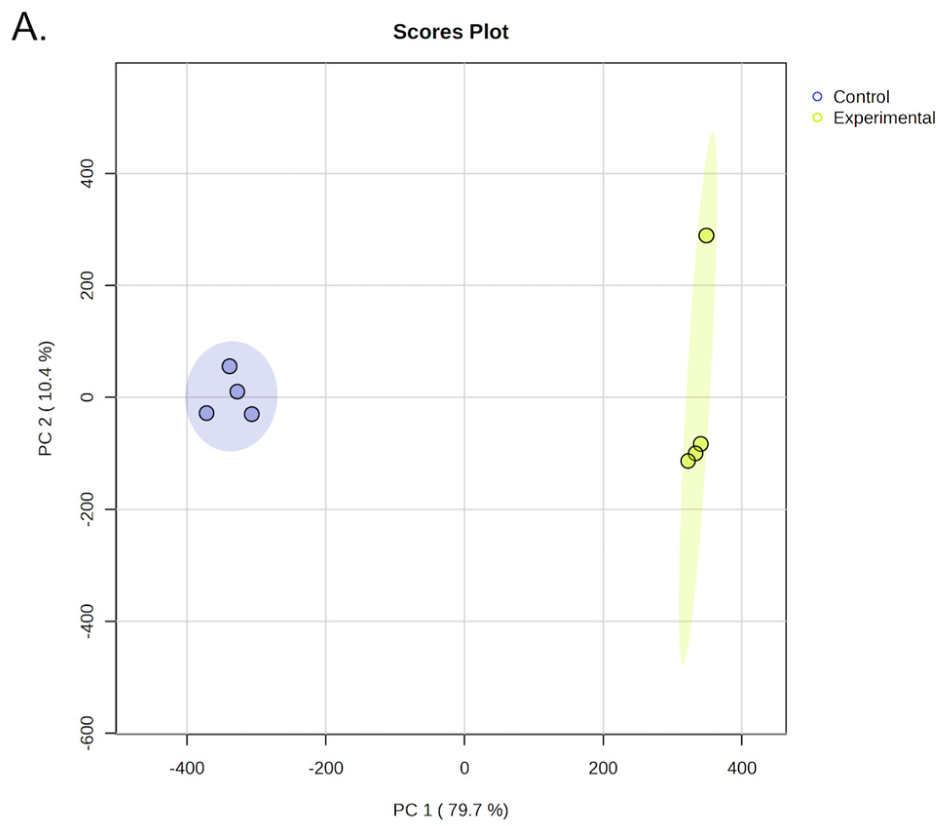
Figure 1B.
Volcano plot depicting differential protein abundance between experimental conditions, with proteins plotted according to log₂ fold change and -log₁₀(p-value). Proteins meeting the statistical significance threshold are highlighted.
Figure 1B.
Volcano plot depicting differential protein abundance between experimental conditions, with proteins plotted according to log₂ fold change and -log₁₀(p-value). Proteins meeting the statistical significance threshold are highlighted.
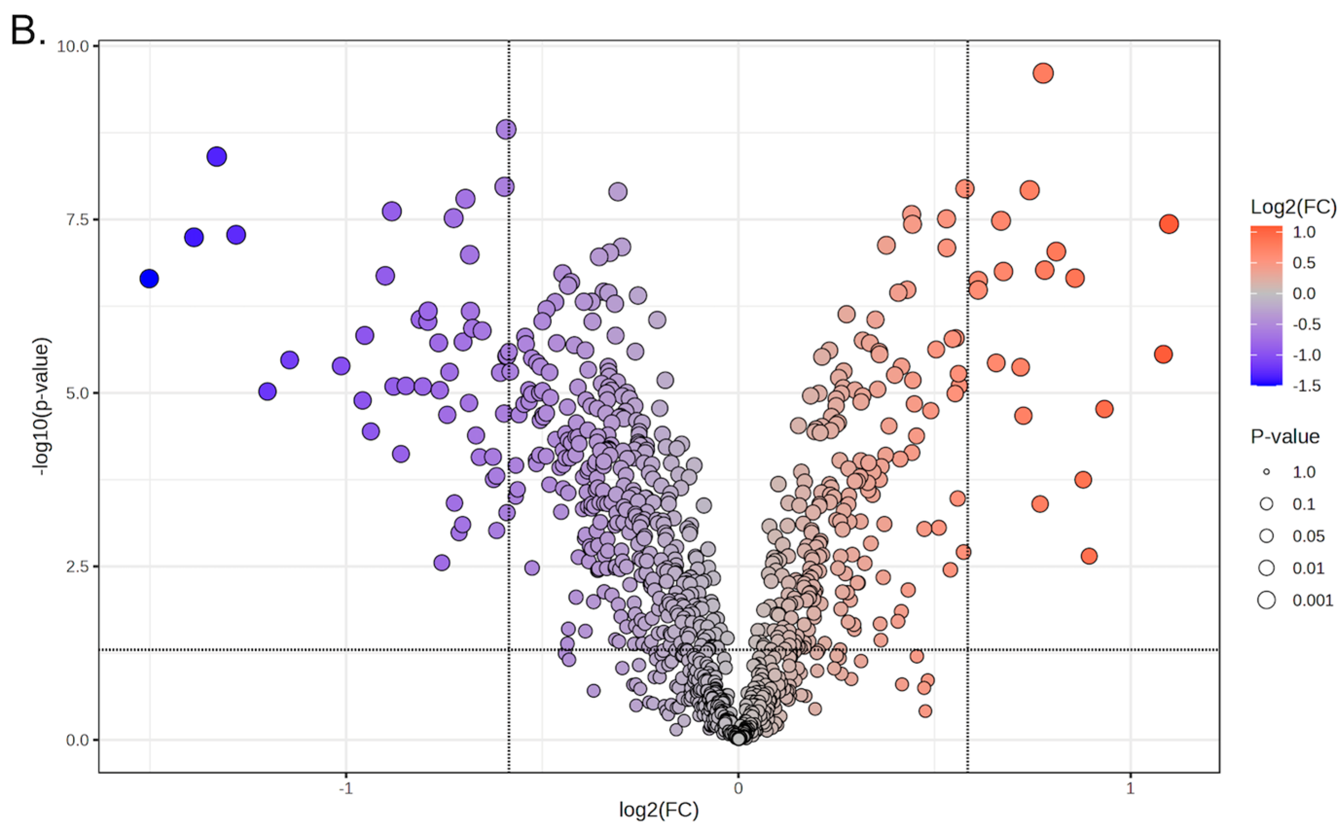
Figure 2A.
Heatmap. Heatmap of hierarchically clustered proteins based on log₂-transformed abundance values across control and Stypopodium zonale-treated Drosophila melanogaster head protein samples. The right columns correspond to four biological replicates of fly head samples exposed to S. zonale, whereas the left columns correspond to four unexposed control samples. Color intensity represents relative protein abundance, with blue indicating lower abundance and green indicating higher abundance. The heatmap illustrates that, following exposure to S. zonale, 48 proteins were less abundant (blue) and 18 proteins were more abundant (green).
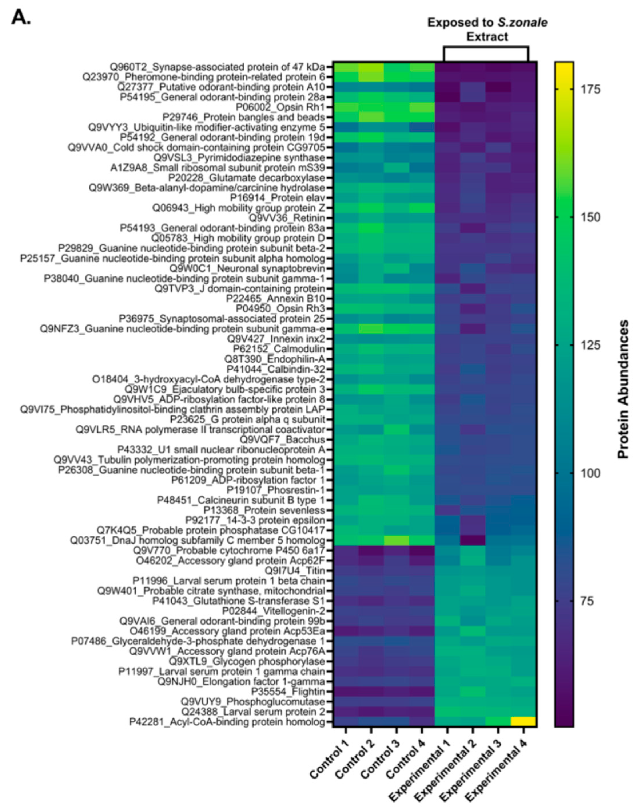
Figure 2B.
Pie Chart. Pie chart summarizing the predicted subcellular localization of differentially abundant proteins, grouped into major cellular compartments.
Figure 2B.
Pie Chart. Pie chart summarizing the predicted subcellular localization of differentially abundant proteins, grouped into major cellular compartments.
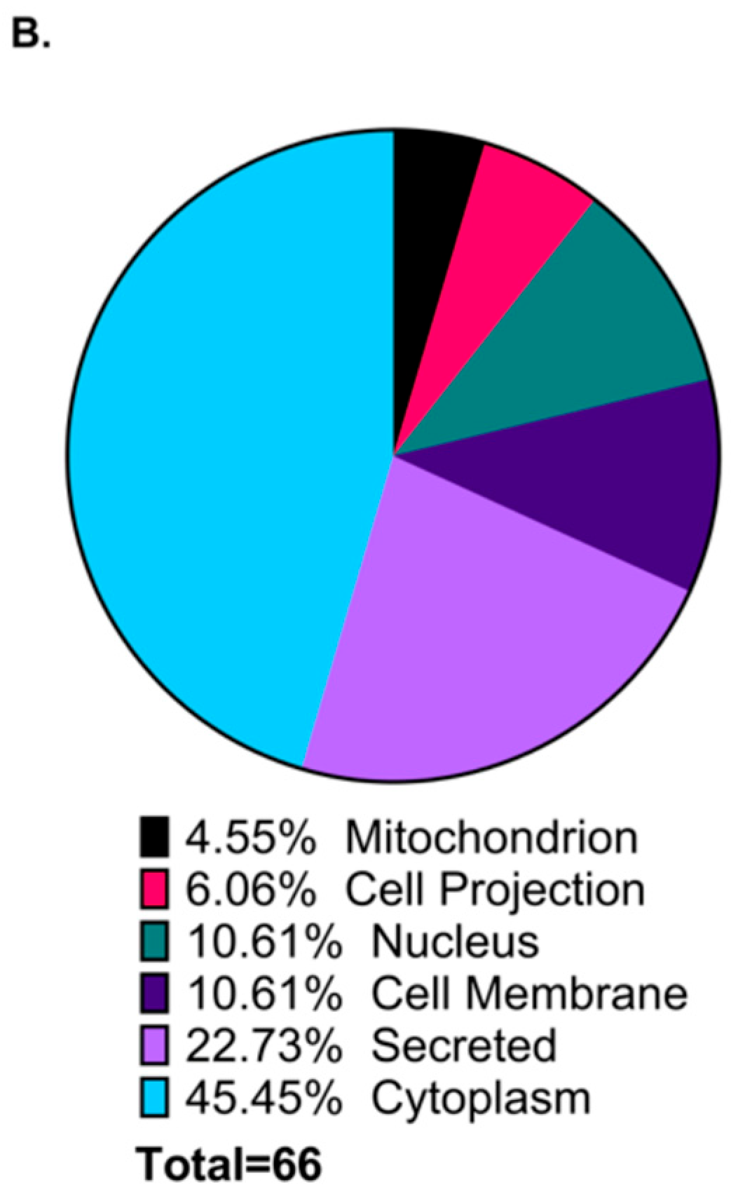
Figure 3.
Ingenuity Pathway Analysis (IPA) Interaction Network. In the IPA Interaction Network, IPA identifies clusters of proteins that participate in a given neurobiological process and displays that process as a functional node. Each protein is color-coded according to its expression change: red indicates upregulation and green indicates downregulation. Functional nodes themselves carry directionality predictions: orange functions are predicted to be activated, blue functions are predicted to be inhibited, and grey functions lack sufficient evidence for directional prediction. The lines connecting proteins to these functions specify the type of relationship: solid lines represent direct interactions, dashed lines represent indirect interactions, and yellow lines indicate relationships with inconsistent or contradictory supporting evidence. Directional arrows (→) denote activation, and inhibitory symbols (┤) denote suppression.
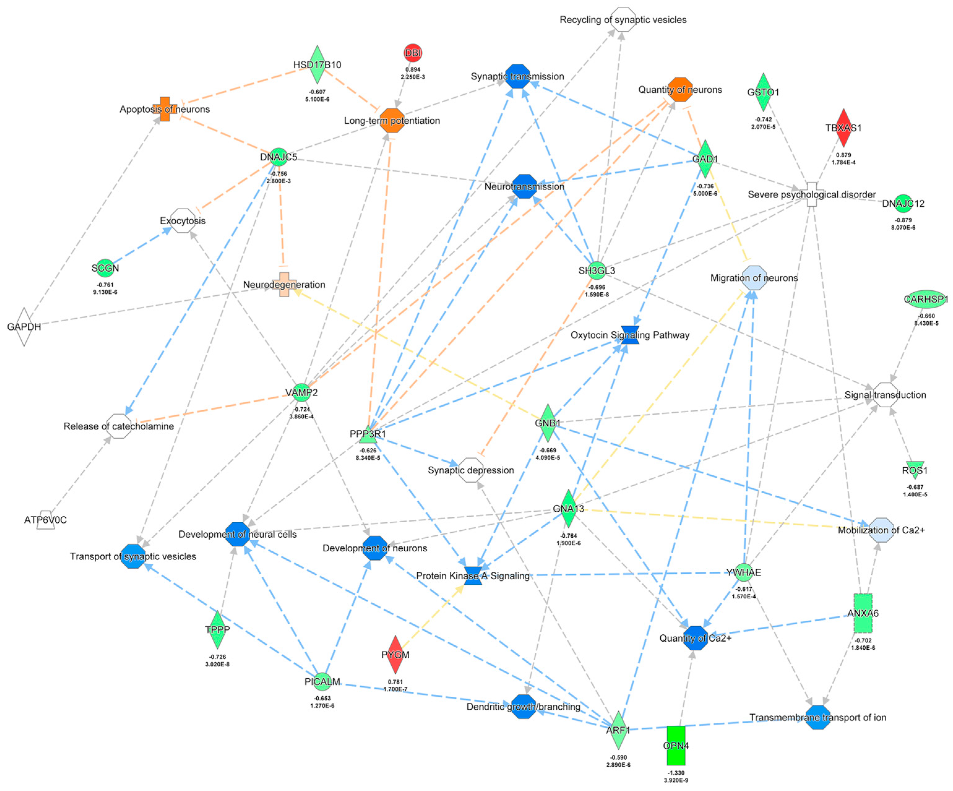
Figure 4.
Overview of the experimental workflow. (1) Stypopodium zonale biomass was collected from shallow coastal waters (1–3 ft from the shoreline) in Puerto Rico. (2) Crude organic extracts were prepared from lyophilized algal biomass using a dichloromethane:methanol (1:1) ultrasonic extraction protocol. (3) Adult Drosophila melanogaster were acutely exposed for 6 h to the crude extract dissolved in 107.62 µL of a 5% dextrose solution containing 0.5% vegetable coloring, in the absence of solid food. (4) Fly heads were dissected immediately following exposure. (5) Proteins were extracted from D. melanogaster heads. (6) Protein concentration was quantified using the BCA Protein Assay Kit. (7) Protein integrity was assessed by SDS-PAGE. (8) Protein samples were processed at the Translational Proteomics Center, where experimental and control samples were prepared for mass spectrometry analysis. (9) LC–MS/MS was performed to identify and quantify proteins based on peptide spectra. (10) Bioinformatic analyses were conducted to identify differentially abundant proteins, and Ingenuity Pathway Analysis (IPA) was used to map Drosophila proteins to their human functional orthologs and to identify their participation in canonical signaling pathways.
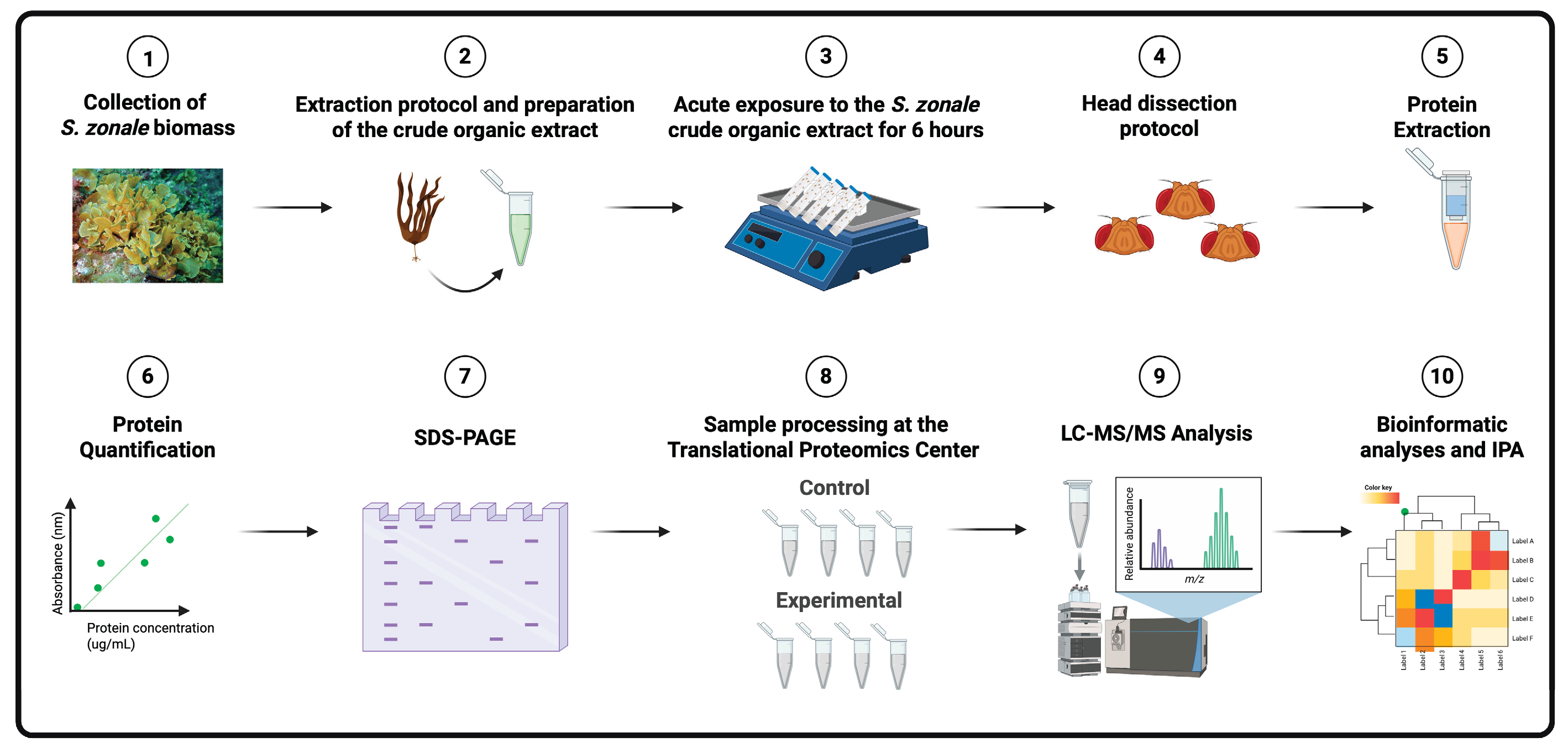

Table 1.
Significantly less abundant Drosophila head proteins after exposure to the crude organic extract of Stypopodium zonale compared to unexposed controls.
Table 1.
Significantly less abundant Drosophila head proteins after exposure to the crude organic extract of Stypopodium zonale compared to unexposed controls.
| Protein | FC | P value | Protein | FC | P value |
|---|---|---|---|---|---|
| Synapse-associated protein of 47 kda | -2.8322 | 2.25 × 10⁻⁷ | Tubulin polymerization-promoting protein homolog | -1.6539 | 3.02 × 10⁻⁸ |
| Pheromone-binding protein-related protein 6 | -2.6168 | 5.72 × 10⁻⁸ | Neuronal synaptobrevin | -1.6516 | 3.86 × 10⁻⁴ |
| Opsin Rh1 | -2.5137 | 3.92 × 10⁻⁹ | Ubiquitin-like modifier-activating enzyme 5 | -1.6381 | 1.03 × 10⁻³ |
| Protein bangles and beads | -2.4291 | 5.26 × 10⁻⁸ | Small ribosomal subunit protein ms39 | -1.628 | 7.90 × 10⁻⁴ |
| General odorant-binding protein 28a | -2.2982 | 9.53 × 10⁻⁶ | Annexin B10 | -1.6268 | 1.84 × 10⁻⁶ |
| General odorant-binding protein 19d | -2.2099 | 3.36 × 10⁻⁶ | Endophilin-A | -1.6198 | 1.59 × 10⁻⁸ |
| High mobility group protein Z | -2.0177 | 4.09 × 10⁻⁶ | Protein sevenless | -1.6097 | 1.40 × 10⁻⁵ |
| Guanine nucleotide-binding protein subunit gamma-e | -1.9425 | 1.28 × 10⁻⁵ |
G protein alpha q subunit | -1.6075 | 1.02 × 10⁻⁷ |
| Beta-alanyl-dopamine/carcinine hydrolase | -1.9352 | 1.48 × 10⁻⁶ | Bacchus | -1.6057 | 6.66 × 10⁻⁷ |
| General odorant-binding protein 83a | -1.9145 | 3.59 × 10⁻⁵ | U1 small nuclear ribonucleoprotein A | -1.5996 | 1.17 × 10⁻⁶ |
| High mobility group protein D | -1.8663 | 2.05 × 10⁻⁷ | Guanine nucleotide-binding protein subunit beta-1 | -1.5898 | 4.09 × 10⁻⁵ |
| Guanine nucleotide-binding protein subunit beta-2 | -1.8448 | 2.41 × 10⁻⁸ | Cold shock domain-containing protein CG9705 | -1.5798 | 8.43 × 10⁻⁵ |
| J domain-containing protein | -1.839 | 8.07 × 10⁻⁶ | Phosphatidylinositol-binding clathrin assembly protein LAP | -1.5727 | 1.27 × 10⁻⁶ |
| Putative odorant-binding protein A10 | -1.8154 | 7.58 × 10⁻5 | Calcineurin subunit B type 1 | -1.5429 | 8.34 × 10⁻⁵ |
| Opsin Rh3 | -1.7997 | 7.92 × 10⁻⁶ | Guanine nucleotide-binding protein subunit gamma-1 | -1.5411 | 1.75 × 10⁻⁴ |
| Ejaculatory bulb-specific protein 3 | -1.7547 | 8.71 × 10⁻⁷ | 14-3-3 protein epsilon | -1.5332 | 1.57 × 10⁻⁴ |
| Protein elav | -1.7463 | 8.08 × 10⁻⁶ | RNA polymerase II transcriptional coactivator | -1.5327 | 9.61 × 10⁻⁴ |
| Calmodulin | -1.7311 | 9.30 × 10⁻⁷ | 3-hydroxyacyl-coa dehydrogenase type-2 | -1.523 | 5.10 × 10⁻⁶ |
| Retinin | -1.7302 | 6.61 × 10⁻⁷ | ADP-ribosylation factor-like protein 8 | -1.5129 | 1.98 × 10⁻⁵ |
| Guanine nucleotide-binding protein subunit alpha homolog | -1.6983 | 1.90 × 10⁻⁶ | Innexin inx2 | -1.5117 | 1.07 × 10⁻⁸ |
| Calbindin-32 | -1.6952 | 9.13 × 10⁻⁶ | Phosrestin-1 | -1.5074 | 1.59 × 10⁻⁹ |
| Dnaj homolog subfamily C member 5 homolog | -1.6888 | 2.80 × 10⁻³ | Synaptosomal-associated protein 25 | -1.5061 | 2.96 × 10⁻⁶ |
| Pyrimidodiazepine synthase | -1.6728 | 2.07 × 10⁻⁵ | Probable protein phosphatase CG10417 | -1.5051 | 5.32 × 10⁻⁴ |
| Glutamate decarboxylase | -1.6658 | 5.00 × 10⁻⁶ | ADP-ribosylation factor 1 | -1.5051 | 2.89 × 10⁻⁶ |
Significantly less abundant proteins (n = 48) met the criteria of fold change ≥ |1.5| and p-value ≤ 0.05. Blue background denotes cytoplasmic, membrane-associated, and secreted proteins that contribute to neuronal signaling and sensory processing.
Table 2.
Significantly more abundant Drosophila head proteins after exposure to the crude organic extract of Stypopodium zonale compared to unexposed controls.
Table 2.
Significantly more abundant Drosophila head proteins after exposure to the crude organic extract of Stypopodium zonale compared to unexposed controls.
| Protein | FC | p value |
|---|---|---|
| Larval serum protein 1 beta chain | 1.5275 | 3.29 × 10⁻⁷ |
| Glyceraldehyde-3-phosphate dehydrogenase 1 | 1.5277 | 2.40 × 10⁻⁷ |
| Probable citrate synthase, mitochondrial | 1.5771 | 3.69 × 10⁻⁶ |
| Titin | 1.5905 | 3.30 × 10⁻⁸ |
| Vitellogenin-2 | 1.5973 | 1.78 × 10⁻⁷ |
| Accessory gland protein Acp76A | 1.6465 | 4.26 × 10⁻⁶ |
| General odorant-binding protein 99b | 1.6546 | 2.13 × 10⁻⁵ |
| Elongation factor 1-gamma | 1.6731 | 1.20 × 10⁻⁸ |
| Accessory gland protein Acp62F | 1.7042 | 3.99 × 10⁻⁴ |
| Phosphoglucomutase | 1.7137 | 2.46 × 10⁻¹⁰ |
| Glycogen phosphorylase | 1.7182 | 1.70 × 10⁻⁷ |
| Glutathione S-transferase S1 | 1.7537 | 9.17 × 10⁻⁸ |
| Larval serum protein 1 gamma chain | 1.8127 | 2.22 × 10⁻⁷ |
| Probable cytochrome P450 6a17 | 1.8392 | 1.78 × 10⁻⁴ |
| Acyl-CoA-binding protein homolog | 1.8587 | 2.25 × 10⁻³ |
| Accessory gland protein Acp53Ea | 1.9095 | 1.70 × 10⁻⁵ |
| Flightin | 2.1192 | 2.78 × 10⁻⁶ |
| Larval serum protein 2 | 2.1403 | 3.68 × 10⁻⁸ |
Significantly more abundant proteins (n=18) met the criteria of fold change ≥ |1.5| and p-value ≤ 0.05.
Disclaimer/Publisher’s Note: The statements, opinions and data contained in all publications are solely those of the individual author(s) and contributor(s) and not of MDPI and/or the editor(s). MDPI and/or the editor(s) disclaim responsibility for any injury to people or property resulting from any ideas, methods, instructions or products referred to in the content. |
© 2026 by the authors. Licensee MDPI, Basel, Switzerland. This article is an open access article distributed under the terms and conditions of the Creative Commons Attribution (CC BY) license (http://creativecommons.org/licenses/by/4.0/).
Copyright: This open access article is published under a Creative Commons CC BY 4.0 license, which permit the free download, distribution, and reuse, provided that the author and preprint are cited in any reuse.


