
Submitted:
22 January 2026
Posted:
22 January 2026
You are already at the latest version
Abstract
Paradoxical insomnia is characterized by a discrepancy between subjective sleep com-plaints and objectively preserved sleep, yet its autonomic mechanisms remain poorly un-derstood. This study examined stage-specific autonomic characteristics of paradoxical insomnia using heart rate variability (HRV)–based statistical, multivariate, and machine learning analyses in a large population-based cohort. HRV features were extracted from non-overlapping five-minute windows across non–rapid eye movement (NREM) sleep, rapid eye movement (REM) sleep, and wake after sleep onset (WASO), and compared among paradoxical insomnia, objective insomnia, and normal sleep groups. Whole-night and consolidated sleep–stage HRV features showed substantial overlap among groups. In contrast, consistent stage-dependent differences emerged selectively during WASO, dur-ing which paradoxical insomnia exhibited distinct autonomic patterns relative to both comparison groups. Multivariate analysis showed the greatest group displacement dur-ing WASO, with UMAP centroid distances exceeding those observed during NREM and REM sleep. Supervised models trained on WASO-specific features achieved the highest classification performance, yielding an accuracy of 0.61 and an F1-score of 0.69 for para-doxical insomnia versus normal sleep, although overall discriminability remained mod-est. These findings indicate that autonomic alterations in paradoxical insomnia are pref-erentially expressed during post–sleep-onset wakefulness. Stage-resolved analysis identi-fies WASO as a physiologically informative window for objective phenotyping and for characterizing heterogeneity in insomnia subtypes.
Keywords:
paradoxical insomnia
; heart rate variability
; wake after sleep onset
; autonomic nervous system
; stage-specific analysis
; sleep phenotyping
; machine learning
; digital phenotyping
1. Introduction
Sleep is a fundamental biological process essential for maintaining physiological homeostasis, cognitive performance, emotional regulation, and long-term health. Disruption of sleep has been associated with impaired cognitive function, mood disturbances, metabolic dysregulation, and increased risk of cardiovascular and neurodegenerative diseases [1,2,3,4,5,6]. Insomnia is one of the most prevalent sleep disorders, affecting approximately 10–25% of adults [7], and is characterized by difficulties in sleep initiation, maintenance, or early morning awakening despite adequate opportunity for sleep, accompanied by daytime impairment [8]. Chronic insomnia is strongly linked to psychological distress, cognitive decline, and elevated cardiometabolic risk [9,10,11,12,13].
Among insomnia phenotypes, paradoxical insomnia (PI), also referred to as sleep state misperception, represents a particularly challenging and poorly understood subtype. PI is defined by a marked discrepancy between severe subjective sleep complaints and objectively preserved sleep as measured by polysomnography (PSG) [14,15,16]. Individuals with PI often report poor sleep quality despite normal total sleep time and sleep efficiency, complicating diagnosis and frequently leading to inappropriate or prolonged pharmacological treatment [17,18]. Although PI has historically been regarded as a primarily perceptual or cognitive phenomenon, accumulating evidence suggests that it is accompanied by measurable neurophysiological alterations. Electroencephalographic studies have reported cortical hyperarousal during sleep, including increased beta, alpha, and sigma activity during NREM sleep [19,20,21,22], while neuroimaging studies have demonstrated structural alterations in subcortical regions involved in arousal regulation and emotional processing [23]. In addition, changes in sleep microarchitecture, such as reduced sleep spindle activity and increased cyclic alternating pattern frequency, have been reported, indicating heightened sleep instability [24,25]. However, despite growing evidence for central nervous system involvement, the peripheral autonomic correlates of paradoxical insomnia remain insufficiently characterized.
The autonomic nervous system (ANS) plays a critical role in sleep regulation and is tightly coupled with central arousal systems through hypothalamic–brainstem pathways. Heart rate variability (HRV), derived from beat-to-beat fluctuations in cardiac intervals, provides a non-invasive index of autonomic regulation and has been widely used to investigate autonomic activity in insomnia [26,27,28,29]. Prior studies have reported altered HRV profiles in insomnia, often interpreted within a hyperarousal framework. However, most HRV studies in insomnia have relied on whole-night or averaged metrics, which may obscure transient autonomic alterations associated with dynamic sleep–wake transitions.
Autonomic regulation during sleep is highly stage dependent, with parasympathetic predominance during non–rapid eye movement (NREM) sleep, increased sympathetic instability during rapid eye movement (REM) sleep [30]. Wake after sleep onset (WASO) represents a transitional and physiologically unstable state characterized by frequent arousals and repeated shifts between sleep and wakefulness. If paradoxical insomnia is characterized by state-dependent rather than sustained hyperarousal, autonomic alterations may preferentially emerge during such transitional states. However, sleep-stage–resolved HRV studies specifically targeting paradoxical insomnia are scarce, and no prior work has systematically examined whether autonomic differentiation in PI clusters during WASO rather than during consolidated sleep stages.
Based on these considerations, the present study aimed to characterize stage-specific autonomic features of paradoxical insomnia using sleep-stage–resolved heart rate variability analysis in a large population-based cohort. We hypothesized that (1) whole-night HRV metrics would show substantial overlap among paradoxical insomnia, objective insomnia, and normal sleep groups, whereas (2) HRV features derived specifically from wake after sleep onset (WASO) would demonstrate greater autonomic differentiation than those derived from NREM or REM sleep. To test these hypotheses, we integrated univariate statistical analysis, multivariate pattern analysis, and supervised machine learning to systematically evaluate whether autonomic alterations in paradoxical insomnia are globally expressed or preferentially emerge during specific sleep–wake states.
2. Materials and Methods
2.1. Dataset and Exclusion Criteria
This study utilized data from the Sleep Heart Health Study (SHHS) [31], a large-scale multicenter cohort including overnight in-home PSG, electrocardiography (ECG), and standardized sleep questionnaires. All available recordings from SHHS1 and SHHS2 (n = 10,115) were initially screened.
To ensure reliable autonomic analysis, a multi-step exclusion procedure was applied (Figure 1). Recordings with less than 6 h of scorable PSG data or low-quality ECG signals were excluded, and only recordings belonging to the highest three PSG quality levels were retained to minimize artifacts and segmentation errors affecting HRV estimation. Participants exhibiting extreme physiological values (heart rate > 150 bpm or < 30 bpm, or SpO2 < 75% for more than 10% of total sleep time) were excluded to avoid non-physiological and non-stationary RR interval dynamics.
Individuals with moderate-to-severe sleep-disordered breathing (apnea–hypopnea index ≥ 10 events/h) or frequent periodic limb movements were removed to minimize confounding autonomic fluctuations associated with comorbid sleep disorders. In addition, participants taking medications known to affect autonomic regulation, including beta-blockers, sympathomimetics, antidepressants, and benzodiazepines, were excluded. Recordings with missing PSG-derived sleep metrics required for phenotype classification were also discarded. After applying all exclusion criteria, 1,970 participants remained eligible for phenotype classification and subsequent analysis.
2.2. Insomnia Phenotypes Classification
Participants were classified into paradoxical insomnia (PI), objective insomnia (OI), and normal sleep (N) groups using a two-stage procedure integrating subjective insomnia complaints and objective polysomnographic (PSG) criteria (Table 1).
In the first stage, subjective insomnia complaints were assessed using the Sleep Heart Health Study Sleep Habits Questionnaire. Participants reporting insomnia symptoms as “often” or “almost always” for at least one item were classified as having clinically significant insomnia complaints.
In the second stage, objective sleep parameters derived from PSG were applied to define phenotypes. OI was defined as the presence of insomnia complaints in combination with at least one abnormal PSG criterion, including total sleep time (TST) < 6 h, sleep efficiency (SE) < 85%, wake after sleep onset (WASO) > 30 min, or sleep latency (SL) > 20 min, consistent with established insomnia definitions [14,32,33]. PI was defined as the presence of insomnia complaints despite preserved objective sleep (TST ≥ 6 h, SE ≥ 85%, WASO ≤ 30 min, and SL ≤ 20 min) or a marked discrepancy between objective and subjective total sleep time (oTST − sTST ≥ 60 min), reflecting sleep state misperception [14]. Normal sleep was defined as the absence of insomnia complaints with all PSG parameters within normal ranges.
Participants not meeting any phenotype criteria were categorized as unclassified and excluded from subsequent analyses. According to these criteria, 625 participants were classified as OI, 261 as PI, and 201 as N, while 883 were unclassified. To ensure balanced group sizes and to minimize potential bias associated with unequal class distributions, a random subset of 200 participants from each phenotype group was selected for subsequent analyses, which also improved computational efficiency and facilitated fair cross-group comparisons.
2.3. ECG Signal Preprocessing and R-Peak Detection
ECG signals were extracted from overnight polysomnography recordings and processed using a unified preprocessing and R-peak detection pipeline (Figure 2). To ensure consistency across recordings, all ECG signals were resampled to a uniform sampling rate of 250 Hz, as SHHS1 recordings were originally acquired at 125 Hz and SHHS2 recordings at 250 or 256 Hz. Analyses were restricted to the interval between sleep onset and final awakening to focus on sleep-related autonomic activity.
R-peaks were detected using the XQRS algorithm implemented in the Waveform Database (WFDB) toolkit [34], which is based on an enhanced Pan–Tompkins framework [35]. The algorithm employs bandpass filtering, signal differentiation, squaring, and adaptive thresholding to robustly identify QRS complexes under varying signal conditions. Detected beats were subjected to refractory period constraints to reduce false detections.
R–R interval time series were constructed from consecutive R-peaks. Intervals shorter than 0.3 s or longer than 2.5 s were considered physiologically implausible and removed. The proportion of excluded R–R intervals was low across all groups (OI: 0.30%, PI: 0.21%, N: 0.30%), indicating minimal artifact contamination. The resulting cleaned R–R interval series served as the basis for subsequent HRV feature extraction.
2.4. HRV Feature Extraction
HRV features were extracted from the cleaned R–R interval time series to characterize autonomic nervous system dynamics during sleep. HRV analysis was conducted using a short-term, window-based approach to capture transient and stage-dependent autonomic fluctuations that may not be reflected in whole-night averages.
2.4.1. Windowing Strategy and Stage-Wise Segmentation
HRV features were computed using non-overlapping 5-minute windows in accordance with established guidelines for short-term HRV analysis. This window length was selected based on the Task Force guidelines of the European Society of Cardiology and the North American Society of Pacing and Electrophysiology, which recommend 5-minute segments for reliable short-term HRV estimation [36]. Only windows fully contained within a single sleep stage were included to prevent contamination across stage boundaries.
WASO was defined as all epochs scored as wake occurring between sleep onset and final awakening based on standard polysomnographic scoring criteria. Each valid window was assigned a sleep stage label (NREM, REM, or WASO) according to polysomnographic annotations. For each participant, HRV features were aggregated within each sleep stage using the median value across all valid windows, providing robust stage-specific representations of autonomic activity while minimizing the influence of outliers and transient artifacts
2.4.2. HRV Metrics
Standard HRV metrics were extracted to characterize autonomic regulation from complementary perspectives. Time-domain metrics were computed to quantify overall variability and short-term beat-to-beat fluctuations, reflecting both global autonomic modulation and parasympathetic activity. Instantaneous heart rate (IHR) was also derived from NN intervals as IHR = 60 / RR (RR in seconds) and is reported in beats per minute (bpm). Frequency-domain metrics were derived to assess oscillatory components of heart rate dynamics associated with sympathetic and parasympathetic influences. In addition, non-linear HRV metrics were calculated to capture the complexity, irregularity, and fractal properties of heart rate time series that are not adequately described by linear measures alone. A comprehensive list of all HRV features, including their definitions and units, is provided in Table 2.
2.5. Statistical Analysis
All statistical analyses were performed to evaluate stage-specific differences in HRV features among insomnia phenotypes. Analyses were conducted separately for NREM sleep, REM sleep, and WASO to avoid dilution of transient autonomic effects in whole-night averages.
The distribution of HRV features was assessed using visual inspection and normality tests. As most features did not satisfy normality assumptions, non-parametric statistical methods were employed. Group-wise comparisons were performed using the Mann–Whitney U test for PI vs. N and PI vs. OI comparisons within each sleep stage.
Statistical significance was defined as a two-tailed p-value < 0.05. In addition to p-values, effect sizes were calculated to quantify the magnitude and direction of group differences. Effect sizes were reported using the common language effect size (CLES) and rank-biserial correlation coefficients, which provide interpretable estimates of stochastic dominance between groups.
To account for multiple HRV features and to facilitate comparison across sleep stages, statistical results were interpreted in conjunction with effect size estimates rather than relying solely on statistical significance. Given the multiple comparisons across HRV features and sleep stages, no formal multiple-comparison correction (e.g., Bonferroni or false discovery rate control) was applied. Instead, findings were interpreted cautiously by emphasizing consistent effect-size patterns and stage-wise reproducibility rather than relying solely on nominal p-values. HRV features demonstrating consistent stage-specific effects were subsequently used for unsupervised pattern analysis and machine learning–based classification. All statistical analyses were implemented using Python-based scientific computing libraries.
2.6. Unsupervised Pattern Analysis
Unsupervised pattern analysis was performed to explore the intrinsic structure of HRV features and to assess whether insomnia phenotypes exhibited separable autonomic patterns without imposing prior class labels. Analyses were conducted separately for each sleep stage (NREM, REM, and WASO) to preserve stage-specific autonomic characteristics.
Uniform Manifold Approximation and Projection (UMAP) was employed for non-linear dimensionality reduction of high-dimensional HRV feature spaces. UMAP was selected due to its ability to preserve both local neighborhood relationships and global data structure, making it well suited for visualizing complex physiological patterns. Prior to dimensionality reduction, HRV features were standardized to zero mean and unit variance to ensure equal contribution of features.
Two-dimensional UMAP embeddings were generated independently for PI vs. N and PI vs. OI comparisons within each sleep stage. To quantify relative multivariate group differences in the embedded space, Euclidean distances between group centroids were computed for each comparison and stage.
UMAP-based analyses were used exclusively for exploratory visualization and descriptive assessment of stage-dependent multivariate displacement, and were not intended for formal clustering or statistical inference. The results were interpreted in conjunction with univariate statistical analyses and supervised machine learning models.
2.7. Machine Learning Analysis
Supervised machine learning analysis was conducted to evaluate the discriminability of PI from OI and N based on stage-specific HRV features. Machine learning models were developed independently for each sleep stage (NREM, REM, WASO) to assess whether autonomic signatures distinguishing PI were stage dependent.
To ensure reproducibility and methodological consistency, all data preprocessing and model training steps were implemented using a unified pipeline constructed with the Scikit-learn framework. First, feature values were standardized using z-score normalization (zero mean and unit variance) via standard scaling to prevent dominance of features with larger numerical ranges. Second, to address class imbalance, a dual strategy was applied at both the data and algorithmic levels. At the data level, the Synthetic Minority Over-sampling Technique (SMOTE) was applied to the training set to synthetically augment minority class samples and achieve balanced class distributions. In parallel, class weights were incorporated into the learning algorithm to impose higher penalties for misclassification of minority classes, encouraging the model to prioritize minority class representation during training.
The dataset was randomly partitioned into training (70%) and testing (30%) subsets using stratified sampling to preserve class proportions. To optimize model generalizability and identify optimal hyperparameters, grid search was performed over key model parameters. Model selection and hyperparameter tuning were conducted within the training set using 10-fold stratified cross-validation. Given the relatively limited sample size, a 10-fold scheme was adopted to maximize the proportion of data used for training in each iteration (90%) while reducing variance and bias in performance estimation through repeated evaluation.
Model performance was evaluated on the held-out test set using accuracy, precision, recall, F1-score, and average precision (AP; area under the precision–recall curve) metrics. Machine learning results were interpreted in conjunction with statistical and unsupervised analyses to provide a comprehensive assessment of stage-specific autonomic differentiation among insomnia phenotypes.
3. Results
3.1. Overall HRV Differences Across the Entire Sleep Period
Stage-agnostic comparisons of HRV features were first performed across the entire sleep period to examine whether global nocturnal autonomic profiles differed among insomnia phenotypes. HRV features averaged over the whole night were compared between PI and N, as well as between PI and OI.
Across all time-domain, frequency-domain, and non-linear HRV metrics, no statistically significant differences were observed between groups in either comparison. Effect size estimates were consistently small, indicating substantial overlap in whole-night HRV distributions among insomnia phenotypes.
These findings suggest that autonomic characteristics distinguishing paradoxical insomnia are not adequately captured by whole-night HRV averages. Consequently, subsequent analyses focused on stage-specific HRV patterns to investigate whether transient autonomic alterations emerge within specific sleep–wake states.
3.2. Stage-Specific HRV Differences (Mann–Whitney U Test)
3.2.1. Stage-Specific HRV Differences Between Paradoxical Insomnia and Normal Sleep
Stage-specific comparisons of HRV features between paradoxical insomnia (PI) and normal sleep (N) were performed for NREM sleep, REM sleep, and wake after sleep onset (WASO) using the Mann–Whitney U test. During consolidated sleep stages, autonomic profiles of the two groups showed substantial overlap. In NREM sleep, no HRV feature demonstrated consistent group differences across time-domain, frequency-domain, or non-linear metrics. During REM sleep, only DFA α1 (median) showed a nominally significant difference between groups (PI: 1.218 vs. N: 1.265; p = 0.047; CLES = 0.56), with a small effect size.
In contrast, marked group differences emerged selectively during WASO, predominantly reflected by greater variability and dispersion of HRV measures in the PI group. Short-term variability indices showed larger dispersion in PI, as evidenced by a higher interquartile range (IQR) of RMSSD (PI: 19.59 ms vs. N: 11.69 ms; p < 0.0001; CLES = 0.37) and SDNN (PI: 27.39 ms vs. N: 18.26 ms; p < 0.0001; CLES = 0.36). Similarly, frequency-domain measures demonstrated increased dispersion in PI during WASO, with larger IQRs for HF power (PI: 445.80 ms2 vs. N: 172.73 ms2; p < 0.0001; CLES = 0.39) and LF power (PI: 499.18 ms2 vs. N: 278.60 ms2; p < 0.0001; CLES = 0.39).
Non-linear HRV metrics further highlighted this pattern. Measures of complexity and long-term regulation, including sample entropy (IQR; PI: 0.276 vs. N: 0.127; p < 0.0001; CLES = 0.34) and DFA α2 (IQR; PI: 0.204 vs. N: 0.120; p < 0.0001; CLES = 0.36), showed significantly greater dispersion in PI during WASO. Collectively, these results indicate that autonomic differentiation between PI and normal sleep is not expressed during stable sleep stages, but is concentrated during WASO as increased variability and complexity of autonomic regulation. A summary of stage-specific statistical results, including median values, p-values, CLES, and rank-biserial correlation coefficients for each HRV feature, is provided in Table 3.
3.2.2. Stage-Specific HRV Differences Between Paradoxical Insomnia and Objective Insomnia
Stage-specific comparisons between paradoxical insomnia (PI) and objective insomnia (OI) revealed minimal differences during consolidated sleep. In both NREM and REM sleep, HRV features showed substantial overlap between the two insomnia subtypes, with no consistent or robust group differences observed across HRV domains.
In contrast, distinct differences between PI and OI were evident during WASO. Compared with OI, PI exhibited larger central tendency values for several indices reflecting overall autonomic response magnitude. Specifically, SD2 (median) was higher in PI (125.71 ms) than in OI (112.32 ms; p = 0.010; CLES = 0.58), and total power was also greater in PI (6057.06 ms2 vs. 4936.61 ms2; p = 0.012; CLES = 0.58). In addition, VLF power (median) was higher in PI than in OI during WASO (4098.17 ms2 vs. 2628.26 ms2; p = 0.001; CLES = 0.60).
At the same time, several measures of response dispersion and irregularity were greater in OI than in PI. The IQRs of DFA α1 (OI: 0.247 vs. PI: 0.160; p < 0.0001; CLES = 0.36) and DFA α2 (OI: 0.275 vs. PI: 0.204; p < 0.0001; CLES = 0.36) were larger in OI, indicating greater variability in fractal scaling properties. Similar patterns were observed for RMSSD IQR (OI: 27.84 ms vs. PI: 19.59 ms; p = 0.002; CLES = 0.41) and SDNN IQR (OI: 36.91 ms vs. PI: 27.39 ms; p = 0.002; CLES = 0.41).
Overall, these findings indicate that paradoxical insomnia and objective insomnia diverge primarily during WASO. While PI is characterized by larger autonomic response magnitudes during post–sleep-onset wakefulness, OI exhibits greater dispersion and irregularity of autonomic responses, highlighting distinct patterns of WASO-specific autonomic dysregulation between the two insomnia subtypes. Detailed statistical results for stage-specific comparisons between PI and OI, including median values, p-values, CLES, and rank-biserial correlation coefficients for each HRV feature, are summarized in Table 4.
3.3. Multivariate Pattern Separation Using UMAP
To explore multivariate autonomic patterns across sleep stages, UMAP was applied as an exploratory visualization tool using standardized HRV feature sets. During NREM and REM sleep, UMAP projections revealed substantial overlap between paradoxical insomnia, objective insomnia, and normal sleep groups, indicating weak overall multivariate differentiation during consolidated sleep stages (Figure 3, Figure 4, Figure 5 and Figure 6).
In contrast, during WASO, embedded distributions showed modest shifts in the spatial location of paradoxical insomnia samples relative to comparison groups, although group distributions remained largely overlapping (Figure 7 and Figure 8).
To quantify these stage-dependent multivariate differences, centroid distances between phenotype groups were computed in the UMAP-embedded space. Across all stages, centroid distances were small, indicating weak overall separation; however, centroid distances were consistently larger during WASO than during NREM or REM sleep for both PI vs. N and PI vs. OI comparisons (Table 5). These results suggest that relative multivariate displacement between insomnia phenotypes is greatest during transient post-sleep-onset wakefulness, despite the absence of well-separated clusters.
3.4. Machine Learning Classification Performance
Supervised machine learning analysis was conducted to evaluate the discriminability of PI from N and OI based on stage-specific HRV features. Random Forest classifiers were trained and evaluated separately for each sleep stage (NREM, REM, and WASO) to assess the stage dependency of autonomic information for phenotype classification.
Across both classification tasks (PI vs. N and PI vs. OI), model performance varied substantially by sleep stage (Table 6). Classifiers trained on HRV features derived from NREM and REM sleep demonstrated limited discriminative ability, with accuracy and F1-scores remaining close to chance level, indicating minimal classification value from autonomic features during consolidated sleep stages.
In contrast, models trained on WASO-derived HRV features achieved consistently higher performance across multiple evaluation metrics. For the PI vs. N comparison, the WASO-based model yielded an accuracy of 0.610 with an F1-score of 0.687 for the PI class, outperforming models trained on NREM and REM features. Similarly, for the PI vs. OI comparison, WASO-based models achieved improved classification performance (accuracy = 0.574, F1-score was 0.542 for PI and 0.602 for OI, respectively).
Despite the improvement observed during WASO, classification performance did not reach levels indicative of strong clinical discrimination. These results suggest that autonomic features during post-sleep-onset wakefulness provide comparatively more informative signals for distinguishing paradoxical insomnia than features derived from stable sleep stages, while overall discriminability remains limited. However, given the modest performance metrics, the present machine learning results should be interpreted as proof-of-concept evidence supporting stage-dependent autonomic differentiation rather than as a definitive diagnostic classifier.
4. Discussion
4.1. Principal Findings
In this study, we investigated sleep-stage–resolved autonomic characteristics of paradoxical insomnia using heart rate variability analysis in a large population-based cohort. The principal finding is that autonomic differentiation in paradoxical insomnia does not manifest as a sustained nocturnal abnormality but becomes apparent specifically during wake after sleep onset (WASO). Whole-night HRV averages and features derived from consolidated sleep stages (NREM and REM) showed substantial overlap among paradoxical insomnia, objective insomnia, and normal sleep groups, indicating limited autonomic distinction during stable sleep.
In contrast, convergent evidence from univariate statistics, multivariate embedding, and supervised machine learning consistently identified WASO as the sleep–wake state in which group differences were most pronounced. Importantly, these differences were characterized not by uniform shifts in autonomic tone, but by increased variability, dispersion, and complexity of HRV measures, suggesting dysregulated autonomic responses during post–sleep-onset wakefulness.
Taken together, these findings argue against a model of generalized or persistent autonomic hyperarousal across the night in paradoxical insomnia. Instead, they indicate that autonomic abnormalities are context dependent, emerging preferentially when the sleep–wake regulatory system is challenged by transient awakenings. This observation provides a unifying framework for interpreting the results and motivates a mechanistic focus on post–sleep-onset wakefulness, which is further elaborated in the following section.
4.2. Autonomic Characteristics of Paradoxical Insomnia During Wake After Sleep Onset
Wake after sleep onset represents a transitional state characterized by repeated shifts between sleep and wakefulness, during which cortical arousal systems and downstream autonomic control must be rapidly disengaged and re-engaged. The present results indicate that this transition process constitutes a critical point of vulnerability in paradoxical insomnia. While autonomic regulation during consolidated NREM and REM sleep appears largely preserved, marked alterations emerge when the system is required to repeatedly re-establish homeostasis following brief awakenings.
Importantly, the WASO-specific HRV patterns observed in paradoxical insomnia should not be interpreted as evidence of reduced physiological arousal or enhanced autonomic calm. Although higher HRV indices are often associated with parasympathetic dominance under stable conditions, the concurrent increase in variability and complexity observed here more plausibly reflects inefficient or unstable autonomic regulation during sleep–wake transitions. Indeed, previous studies have shown that during arousal from sleep and sleep–wake transitions, increases in HRV and cardiovascular variability may reflect transient autonomic instability and nonstationary regulatory dynamics rather than reduced physiological arousal or enhanced vagal dominance [37,38]. Rather than maintaining a consistent regulatory strategy, the autonomic system in paradoxical insomnia appears to respond to awakenings in a fluctuating and poorly constrained manner.
One possible mechanistic interpretation is that paradoxical insomnia involves impaired coordination of arousal system re-engagement during sleep–wake transitions. Brief awakenings may trigger exaggerated or inconsistent activation of central arousal networks, which then propagate to peripheral autonomic outputs in a variable and energetically inefficient fashion. Such dysregulated re-engagement could amplify interoceptive signals associated with wakefulness, thereby contributing to the subjective perception of being awake despite objectively preserved sleep structure.
This transition-focused interpretation helps reconcile the apparent discrepancy between normal polysomnographic sleep and pronounced subjective sleep disturbance in paradoxical insomnia. Rather than reflecting a trait-like elevation of arousal throughout the night, the condition may be better understood as a disorder of dynamic regulation, in which autonomic control becomes unstable at specific moments when sleep continuity is disrupted. WASO thus emerges not merely as a descriptive sleep metric, but as a physiologically informative window for probing the mechanisms underlying sleep state misperception.
4.3. Implications for Stage-Aware Phenotyping and Digital Biomarker Development
The present findings have important implications for objective phenotyping of insomnia subtypes and the development of physiology-based digital biomarkers. A key contribution of this study is the demonstration that autonomic features relevant to paradoxical insomnia are state dependent, emerging preferentially during wake after sleep onset (WASO) rather than being uniformly expressed across the night. This observation highlights a fundamental limitation of conventional whole-night summary metrics and underscores the need for temporally resolved analytical approaches to capture transient but informative physiological dynamics.
Within this framework, the machine learning analysis was not intended to produce a clinically deployable diagnostic classifier, but rather to serve as a computational probe for evaluating the relative information content of stage-specific autonomic features. Although models trained on WASO-derived HRV features consistently outperformed those trained on NREM or REM features, overall classification performance remained modest. Specifically, an accuracy of approximately 0.60–0.65 and an F1-score below 0.70, while exceeding chance level, are generally considered insufficient for standalone clinical screening or diagnostic applications, which typically require substantially higher sensitivity and specificity to ensure reliable individual-level decision making. Accordingly, the present machine learning results should be interpreted as indicative of relative discriminative information across sleep stages, rather than as evidence of immediate clinical utility.
From a technological perspective, these results emphasize the value of stage-aware computational analysis pipelines for physiological signal interpretation. By integrating sleep staging with short-term HRV windowing and multilevel analysis, this study demonstrates a scalable framework for extracting physiologically meaningful information from large-scale biosignal data. Although HRV alone is unlikely to serve as a standalone biomarker for paradoxical insomnia, stage-resolved autonomic features—particularly those derived from WASO—may represent a valuable component within future multimodal digital phenotyping systems that integrate autonomic, behavioral, and central nervous system measures.
4.4. Limitations and Future Directions
Several limitations of the present study should be acknowledged. First, the analysis was based on cross-sectional polysomnography and electrocardiographic recordings obtained from a single-night assessment, which precludes evaluation of intra-individual variability and the longitudinal stability of the observed autonomic patterns. Although the large sample size enhances statistical robustness, single-night measurements may not fully capture night-to-night fluctuations in sleep–wake dynamics, particularly in individuals with insomnia.
Second, the present findings were derived from the Sleep Heart Health Study, which predominantly consists of middle-aged and older adults. Given that paradoxical insomnia is also prevalent in younger populations and may manifest differently across the lifespan, caution is warranted when generalizing these results to younger individuals or clinical samples with different demographic and psychosocial characteristics. Future studies involving age-diverse cohorts and clinical populations will be necessary to assess the developmental and age-related generalizability of WASO-specific autonomic dysregulation.
Third, the analysis focused exclusively on heart rate variability as an index of autonomic regulation. While HRV provides a valuable non-invasive window into peripheral autonomic dynamics, it does not directly capture central nervous system activity or subjective sleep experience. Consequently, HRV alone is unlikely to fully characterize the complex mechanisms underlying sleep state misperception. Future work should adopt multimodal phenotyping approaches that integrate HRV with electroencephalography-derived markers of cortical arousal and instability, as well as subjective and behavioral measures such as ecological momentary assessment of sleep perception and nocturnal awakenings. Such integrative frameworks may provide a more comprehensive understanding of how central arousal, peripheral autonomic regulation, and subjective experience interact during sleep–wake transitions.
Finally, the present study was conducted under laboratory-controlled conditions using in-home polysomnography. Extension of this stage-resolved analytical framework to ambulatory and wearable sensing environments will be essential to evaluate its feasibility and translational potential in real-world settings. Despite these limitations, the current findings provide a quantitative foundation for investigating state-dependent autonomic dysregulation in paradoxical insomnia and motivate future efforts toward integrated, multimodal, and longitudinal characterization of insomnia subtypes.
5. Conclusions
In conclusion, this study demonstrates that autonomic differentiation in paradoxical insomnia is not globally expressed throughout the night but emerges selectively during wake after sleep onset (WASO). By applying stage-resolved heart rate variability analysis in a large population-based cohort, we identify WASO as a physiologically informative window in which state-dependent autonomic dysregulation becomes detectable despite preserved polysomnographic sleep architecture. These findings provide quantitative evidence that sleep state misperception in paradoxical insomnia may be rooted in unstable autonomic regulation during sleep–wake transitions rather than in sustained nocturnal hyperarousal.
From a technological perspective, these results encourage the development of stage-aware, wearable-based monitoring pipelines that selectively emphasize HRV dynamics during nocturnal awakenings. Such approaches may improve the sensitivity of digital phenotyping systems for capturing transient autonomic instability and support the future design of scalable, physiology-driven tools for characterizing heterogeneity in insomnia subtypes.
Author Contributions
Conceptualization, Y.E.K. and S.D.M.; methodology, Y.E.K. and S.D.M.; software, Y.E.K; validation, Y.E.K. and S.D.M.; formal analysis, Y.E.K.; investigation, Y.E.K. and A.H.J; data curation, Y.E.K. and A.H.J; writing—original draft preparation, Y.E.K.; writing—review and editing, S.D.M.; visualization, Y.E.K.; supervision, S.D.M.; All authors have read and agreed to the published version of the manuscript.
Funding
This work was funded by BK21 FOUR (Fostering Outstanding Universities for Research) (No.: 5199990914048), the Korea Medical Device Development Fund (Project Number: 1711196787, RS-2023-00255005), and the Soonchunhyang University Research Fund.
Institutional Review Board Statement
Ethical review and approval were waived for this study because the analysis was conducted using de-identified publicly available data from the Sleep Heart Health Study.
Informed Consent Statement
Patient consent was waived because this study used de-identified publicly available data.
Data Availability Statement
The data used in this study are available from the National Sleep Research Resource (NSRR) Sleep Heart Health Study repository (https://sleepdata.org), subject to data use agreement and access approval.
Acknowledgments
The authors thank the National Sleep Research Resource and the Sleep Heart Health Study investigators for providing access to the dataset used in this study. During the preparation of this manuscript, the authors used ChatGPT (OpenAI, GPT-5.2) for language editing and manuscript refinement. The authors have reviewed and edited the output and take full responsibility for the content of this publication.
Conflicts of Interest
The authors declare no conflicts of interest.
References
- Van Cauter, E.; Spiegel, K.; Tasali, E.; Leproult, R. Metabolic Consequences of Sleep and Sleep Loss. Sleep Med. 2008, 9, S23–S28. [Google Scholar] [CrossRef]
- Lange, T.; Dimitrov, S.; Born, J. Effects of Sleep and Circadian Rhythm on the Human Immune System. Ann. N. Y. Acad. Sci. 2010, 1193, 48–59. [Google Scholar] [CrossRef]
- Rasch, B.; Born, J. About Sleep’s Role in Memory. Physiol. Rev. 2013, 93, 681–766. [Google Scholar] [CrossRef] [PubMed]
- Tomaso, C.C.; Johnson, A.B.; Nelson, T.D. The Effect of Sleep Deprivation and Restriction on Mood, Emotion, and Emotion Regulation: Three Meta-Analyses in One. Sleep 2021, 44, zsaa289. [Google Scholar] [CrossRef] [PubMed]
- Vandekerckhove, M.; Cluydts, R. The Emotional Brain and Sleep: An Intimate Relationship. Sleep Med. Rev. 2010, 14, 219–226. [Google Scholar] [CrossRef] [PubMed]
- Nedergaard, M.; Goldman, S.A. Glymphatic Failure as a Final Common Pathway to Dementia. Science 2020, 370, 50–56. [Google Scholar] [CrossRef]
- Sateia, M.J. International Classification of Sleep Disorders-Third Edition. Chest 2014, 146, 1387–1394. [Google Scholar] [CrossRef]
- Ohayon, M.M. Epidemiology of Insomnia: What We Know and What We Still Need to Learn. Sleep Med. Rev. 2002, 6, 97–111. [Google Scholar] [CrossRef]
- Riemann, D.; Dressle, R.J.; Benz, F.; Spiegelhalder, K.; Johann, A.F.; Nissen, C.; Hertenstein, E.; Baglioni, C.; Palagini, L.; Krone, L.; et al. Chronic Insomnia, REM Sleep Instability and Emotional Dysregulation: A Pathway to Anxiety and Depression? J. Sleep Res. 2025, 34, e14252. [Google Scholar] [CrossRef]
- Fortier-Brochu, É.; Beaulieu-Bonneau, S.; Ivers, H.; Morin, C.M. Insomnia and Daytime Cognitive Performance: A Meta-Analysis. Sleep Med. Rev. 2012, 16, 83–94. [Google Scholar] [CrossRef]
- Li, L.; Gan, Y.; Zhou, X.; Jiang, H.; Zhao, Y.; Tian, Q.; He, Y.; Liu, Q.; Mei, Q.; Wu, C.; et al. Insomnia and the Risk of Hypertension: A Meta-Analysis of Prospective Cohort Studies. Sleep Med. Rev. 2021, 56, 101403. [Google Scholar] [CrossRef] [PubMed]
- Shan, Z.; Ma, H.; Xie, M.; Yan, P.; Guo, Y.; Bao, W.; Rong, Y.; Jackson, C.L.; Hu, F.B.; Liu, L. Sleep Duration and Risk of Type 2 Diabetes: A Meta-Analysis of Prospective Studies. Diabetes Care 2015, 38, 529–537. [Google Scholar] [CrossRef] [PubMed]
- Sofi, F.; Cesari, F.; Casini, A.; Macchi, C.; Abbate, R.; Gensini, G.F. Insomnia and Risk of Cardiovascular Disease: A Meta-Analysis. J. Cardiovasc. Risk 2014, 21, 57–64. [Google Scholar] [CrossRef] [PubMed]
- Rezaie, L.; Fobian, A.D.; McCall, W.V.; Khazaie, H. Paradoxical Insomnia and Subjective–Objective Sleep Discrepancy: A Review. Sleep Med. Rev. 2018, 40, 196–202. [Google Scholar] [CrossRef]
- Edinger, J.D.; Krystal, A.D. Subtyping Primary Insomnia: Is Sleep State Misperception a Distinct Clinical Entity? Sleep Med. Rev. 2003, 7, 203–214. [Google Scholar] [CrossRef]
- Castelnovo, A.; Ferri, R.; Punjabi, N.M.; Castronovo, V.; Garbazza, C.; Zucconi, M.; Ferini-Strambi, L.; Manconi, M. The Paradox of Paradoxical Insomnia: A Theoretical Review towards a Unifying Evidence-Based Definition. Sleep Med. Rev. 2019, 44, 70–82. [Google Scholar] [CrossRef]
- Harvey, A.G.; Tang, N.K.Y. (Mis)Perception of Sleep in Insomnia: A Puzzle and a Resolution. Psychol. Bull. 2012, 138, 77–101. [Google Scholar] [CrossRef]
- Wasey, W.; Saleh, S.; Abernathy, K.; Sapra, A.; Bhandari, P. Paradoxical Insomnia in a Frustrated Patient Treated With Hypnotics for Ten Years. Cureus 13 e16234. [CrossRef]
- Van Someren, E.J.W. Brain Mechanisms of Insomnia: New Perspectives on Causes and Consequences. Physiol. Rev. 2021, 101, 995–1046. [Google Scholar] [CrossRef]
- Stephan, A.M.; Lecci, S.; Cataldi, J.; Siclari, F. Conscious Experiences and High-Density EEG Patterns Predicting Subjective Sleep Depth. Curr. Biol. 2021, 31, 5487–5500.e3. [Google Scholar] [CrossRef]
- Fasiello, E.; Gorgoni, M.; Galbiati, A.; Sforza, M.; Berra, F.; Scarpelli, S.; Alfonsi, V.; Annarumma, L.; Casoni, F.; Zucconi, M.; et al. Decreased Delta/Beta Ratio Index as the Sleep State-Independent Electrophysiological Signature of Sleep State Misperception in Insomnia Disorder: A Focus on the Sleep Onset and the Whole Night. NeuroImage 2024, 298, 120782. [Google Scholar] [CrossRef]
- St-Jean, G.; Turcotte, I.; Pérusse, A.D.; Bastien, C.H. REM and NREM Power Spectral Analysis on Two Consecutive Nights in Psychophysiological and Paradoxical Insomnia Sufferers. Int. J. Psychophysiol. 2013, 89, 181–194. [Google Scholar] [CrossRef] [PubMed]
- Emamian, F.; Mahdipour, M.; Noori, K.; Rostampour, M.; Mousavi, S.B.; Khazaie, H.; Khodaie-Ardakani, M.; Tahmasian, M.; Zarei, M. Alterations of Subcortical Brain Structures in Paradoxical and Psychophysiological Insomnia Disorder. Front. Psychiatry 2021, 12. [Google Scholar] [CrossRef] [PubMed]
- Normand, M.-P.; St-Hilaire, P.; Bastien, C.H. Sleep Spindles Characteristics in Insomnia Sufferers and Their Relationship with Sleep Misperception. Neural Plast. 2016, 2016, 6413473. [Google Scholar] [CrossRef] [PubMed]
- Parrino, L.; Milioli, G.; De Paolis, F.; Grassi, A.; Terzano, M.G. Paradoxical Insomnia: The Role of CAP and Arousals in Sleep Misperception. Sleep Med. 2009, 10, 1139–1145. [Google Scholar] [CrossRef]
- Dressle, R.J.; Riemann, D. Hyperarousal in Insomnia Disorder: Current Evidence and Potential Mechanisms. J. Sleep Res. 2023, 32, e13928. [Google Scholar] [CrossRef]
- McCall, W.V.; Looney, S.W.; Zulfiqar, M.; Ketcham, E.; Jones, M.; Mixson, C.; McCloud, L.; Miller, B.J.; Rosenquist, P.B. Daytime Autonomic Nervous System Functions Differ among Adults with and without Insomnia Symptoms. J. Clin. Sleep Med. JCSM Off. Publ. Am. Acad. Sleep Med. 2023, 19, 1885–1893. [Google Scholar] [CrossRef]
- Jarrin, D.C.; Ivers, H.; Lamy, M.; Chen, I.Y.; Harvey, A.G.; Morin, C.M. Cardiovascular Autonomic Dysfunction in Insomnia Patients with Objective Short Sleep Duration. J. Sleep Res. 2018, 27, e12663. [Google Scholar] [CrossRef]
- Fink, A.M.; Bronas, U.G.; Calik, M.W. Autonomic Regulation during Sleep and Wakefulness: A Review with Implications for Defining the Pathophysiology of Neurological Disorders. Clin. Auton. Res. Off. J. Clin. Auton. Res. Soc. 2018, 28, 509–518. [Google Scholar] [CrossRef]
- Shaffer, F.; Ginsberg, J.P. An Overview of Heart Rate Variability Metrics and Norms. Front. Public Health 2017, 5. [Google Scholar] [CrossRef]
- Quan, S.F.; Howard, B.V.; Iber, C.; Kiley, J.P.; Nieto, F.J.; O’Connor, G.T.; Rapoport, D.M.; Redline, S.; Robbins, J.; Samet, J.M.; et al. The Sleep Heart Health Study: Design, Rationale, and Methods. Sleep 1997, 20, 1077–1085. [Google Scholar] [CrossRef]
- Vgontzas, A.N.; Fernandez-Mendoza, J.; Liao, D.; Bixler, E.O. Insomnia with Objective Short Sleep Duration: The Most Biologically Severe Phenotype of the Disorder. Sleep Med. Rev. 2013, 17, 241–254. [Google Scholar] [CrossRef] [PubMed]
- Fernandez-Mendoza, J.; Calhoun, S.; Bixler, E.O.; Pejovic, S.; Karataraki, M.; Liao, D.; Vela-Bueno, A.; Ramos-Platon, M.J.; Sauder, K.A.; Vgontzas, A.N. Insomnia with Objective Short Sleep Duration Is Associated with Deficits in Neuropsychological Performance: A General Population Study. Sleep 2010, 33, 459–465. [Google Scholar] [CrossRef]
- Silva, I.; Moody, G.B. An Open-Source Toolbox for Analysing and Processing PhysioNet Databases in MATLAB and Octave. J. Open Res. Softw. 2014, 2, e27. [Google Scholar] [CrossRef]
- Pan, J.; Tompkins, W.J. A Real-Time QRS Detection Algorithm. IEEE Trans. Biomed. Eng. 1985, BME-32, 230–236. [Google Scholar] [CrossRef]
- Malik, M.; Bigger, J.T.; Camm, A.J.; Kleiger, R.E.; Malliani, A.; Moss, A.J.; Schwartz, P.J. Heart Rate Variability: Standards of Measurement, Physiological Interpretation, and Clinical Use. Eur. Heart J. 1996, 17, 354–381. [Google Scholar] [CrossRef]
- Blasi, A.; Jo, J.; Valladares, E.; Morgan, B.J.; Skatrud, J.B.; Khoo, M.C.K. Cardiovascular Variability after Arousal from Sleep: Time-Varying Spectral Analysis. J. Appl. Physiol. 2003, 95, 1394–1404. [Google Scholar] [CrossRef] [PubMed]
- Fink, A.M.; Bronas, U.G.; Calik, M.W. Autonomic Regulation during Sleep and Wakefulness: A Review with Implications for Defining the Pathophysiology of Neurological Disorders. Clin. Auton. Res. 2018, 28, 509–518. [Google Scholar] [CrossRef]
Figure 1.
Flowchart of participant selection and exclusion criteria. The diagram illustrates the stepwise exclusion process based on PSG quality, physiological outliers, sleep-disordered, medication use, and missing PSG metrics.
Figure 1.
Flowchart of participant selection and exclusion criteria. The diagram illustrates the stepwise exclusion process based on PSG quality, physiological outliers, sleep-disordered, medication use, and missing PSG metrics.
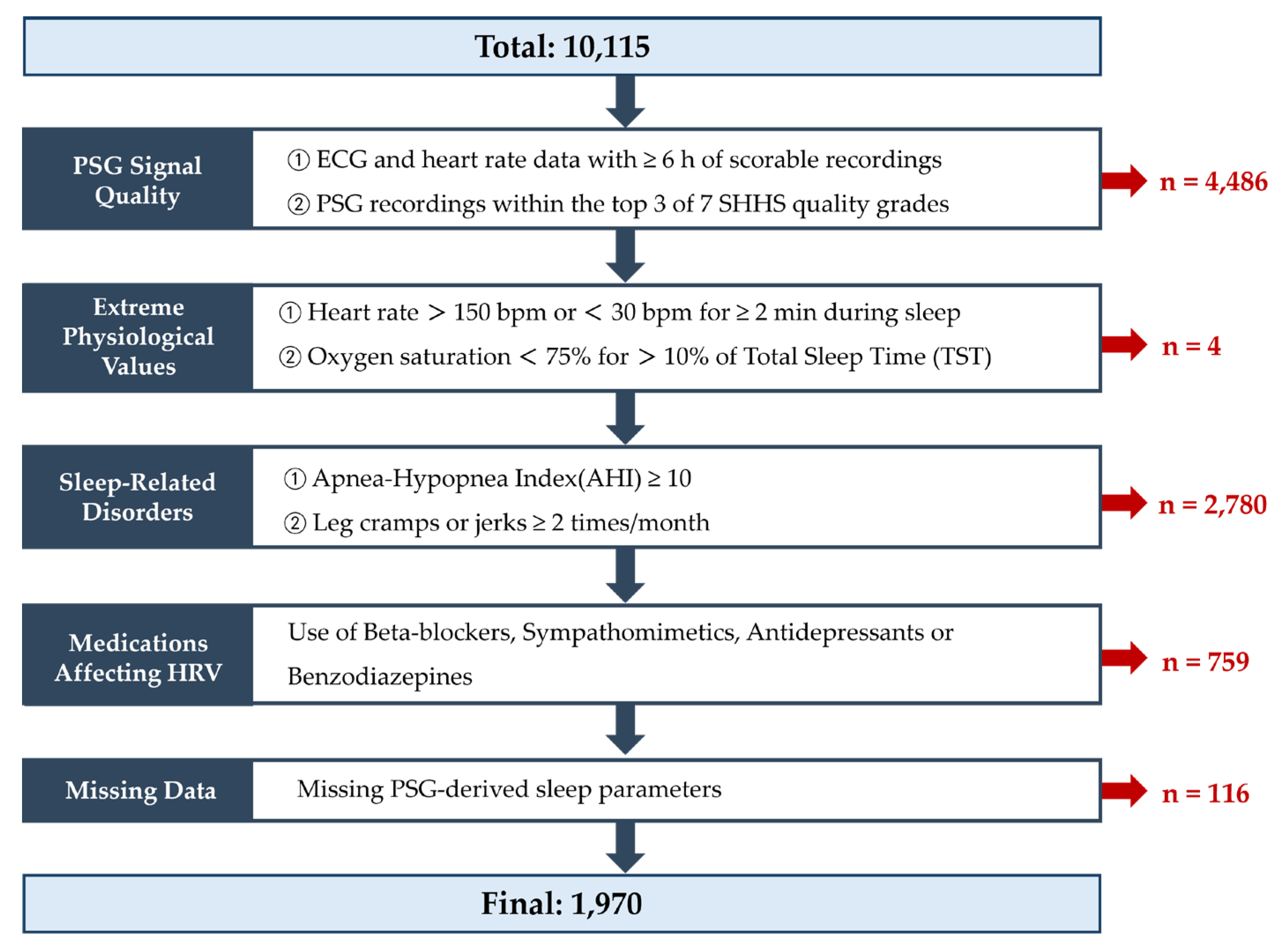
Figure 2.
ECG preprocessing and R-peak detection pipeline. The diagram illustrates the preprocessing steps applied to raw ECG signals, including filtering, artifact removal, R-peak detection, and construction of R–R interval time series for HRV analysis.
Figure 2.
ECG preprocessing and R-peak detection pipeline. The diagram illustrates the preprocessing steps applied to raw ECG signals, including filtering, artifact removal, R-peak detection, and construction of R–R interval time series for HRV analysis.
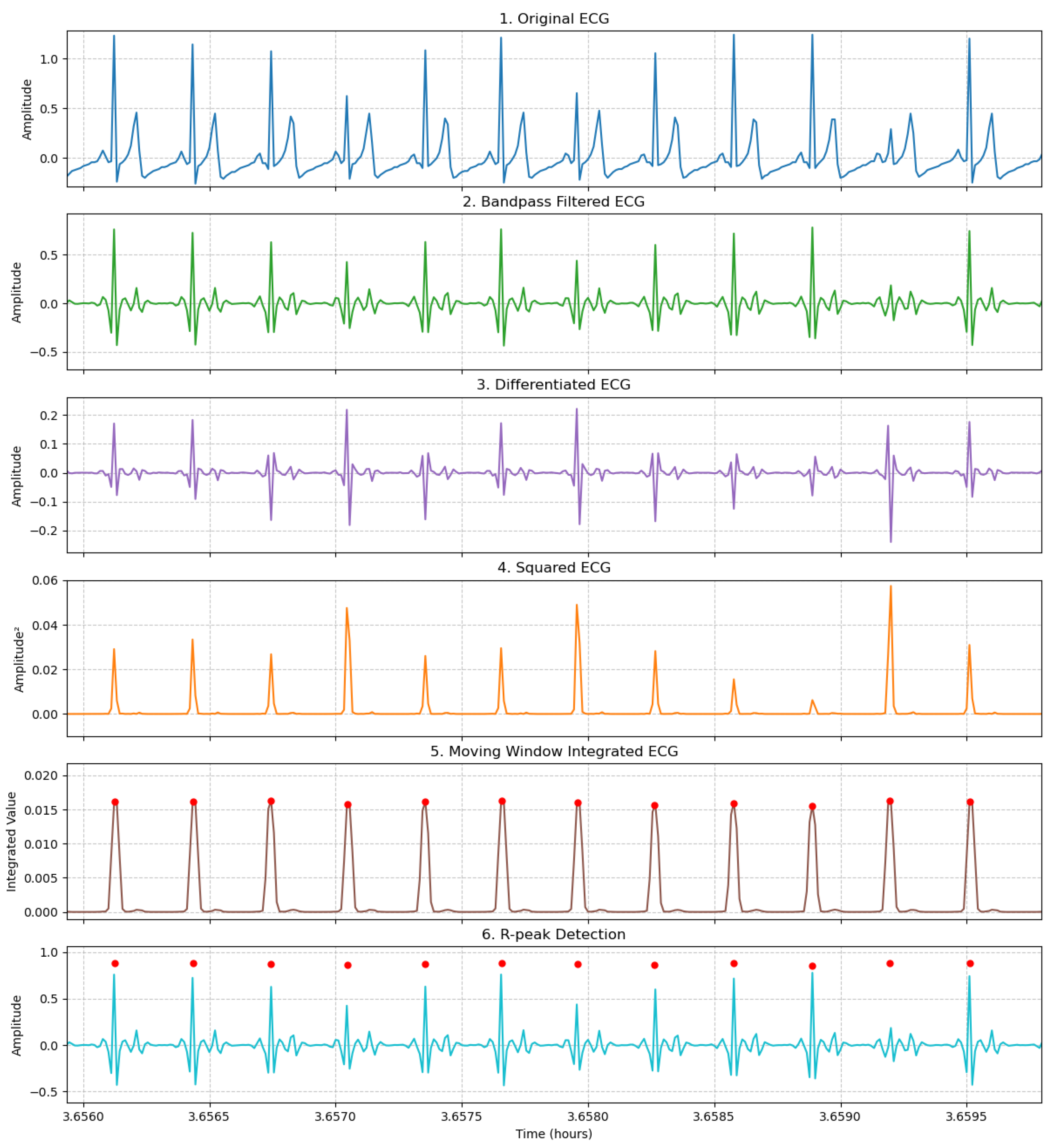
Figure 3.
UMAP embedding of heart rate variability (HRV) features during non–rapid eye movement (NREM) sleep. Each point represents an individual participant. Colors indicate phenotype group (paradoxical insomnia vs. normal sleep).
Figure 3.
UMAP embedding of heart rate variability (HRV) features during non–rapid eye movement (NREM) sleep. Each point represents an individual participant. Colors indicate phenotype group (paradoxical insomnia vs. normal sleep).
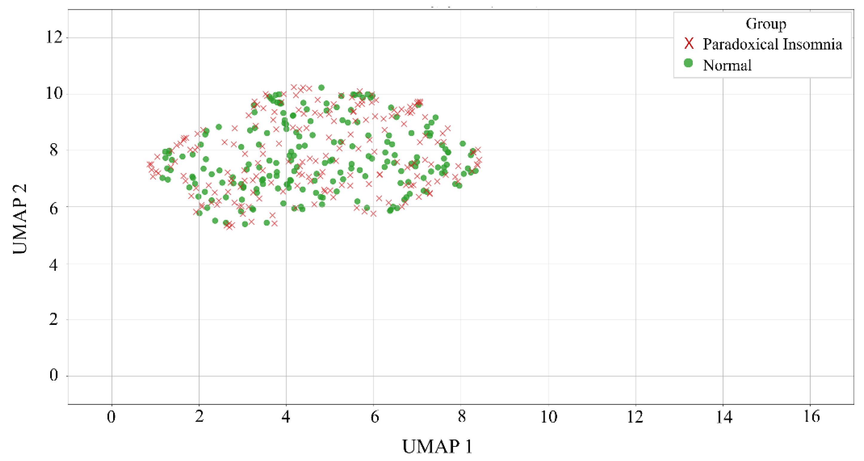
Figure 4.
UMAP embedding of heart rate variability (HRV) features during non–rapid eye movement (NREM) sleep. Each point represents an individual participant. Colors indicate phenotype group (paradoxical insomnia vs. objective insomnia).
Figure 4.
UMAP embedding of heart rate variability (HRV) features during non–rapid eye movement (NREM) sleep. Each point represents an individual participant. Colors indicate phenotype group (paradoxical insomnia vs. objective insomnia).
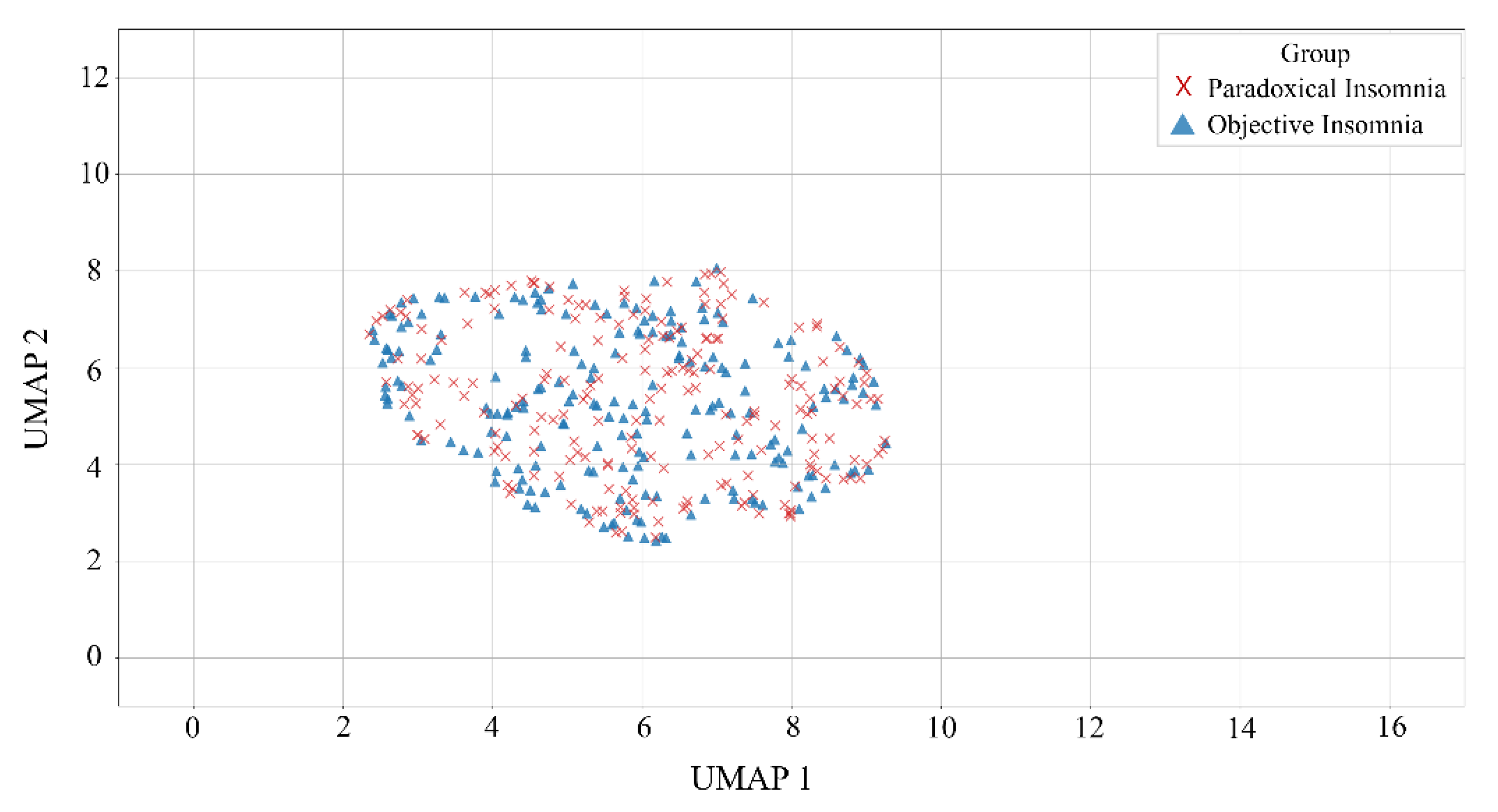
Figure 5.
UMAP embedding of heart rate variability (HRV) features during rapid eye movement (REM) sleep. Each point represents an individual participant. Colors indicate phenotype group (paradoxical insomnia vs. normal sleep).
Figure 5.
UMAP embedding of heart rate variability (HRV) features during rapid eye movement (REM) sleep. Each point represents an individual participant. Colors indicate phenotype group (paradoxical insomnia vs. normal sleep).
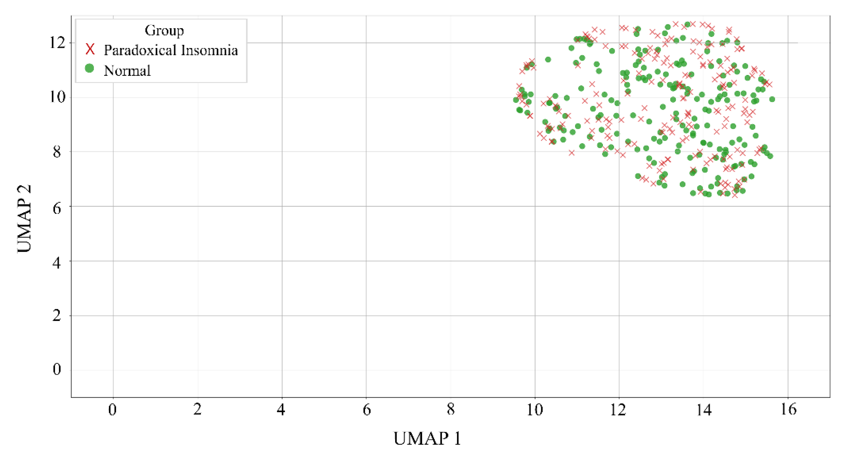
Figure 6.
UMAP embedding of heart rate variability (HRV) features during rapid eye movement (REM) sleep. Each point represents an individual participant. Colors indicate phenotype group (paradoxical insomnia vs. objective insomnia).
Figure 6.
UMAP embedding of heart rate variability (HRV) features during rapid eye movement (REM) sleep. Each point represents an individual participant. Colors indicate phenotype group (paradoxical insomnia vs. objective insomnia).
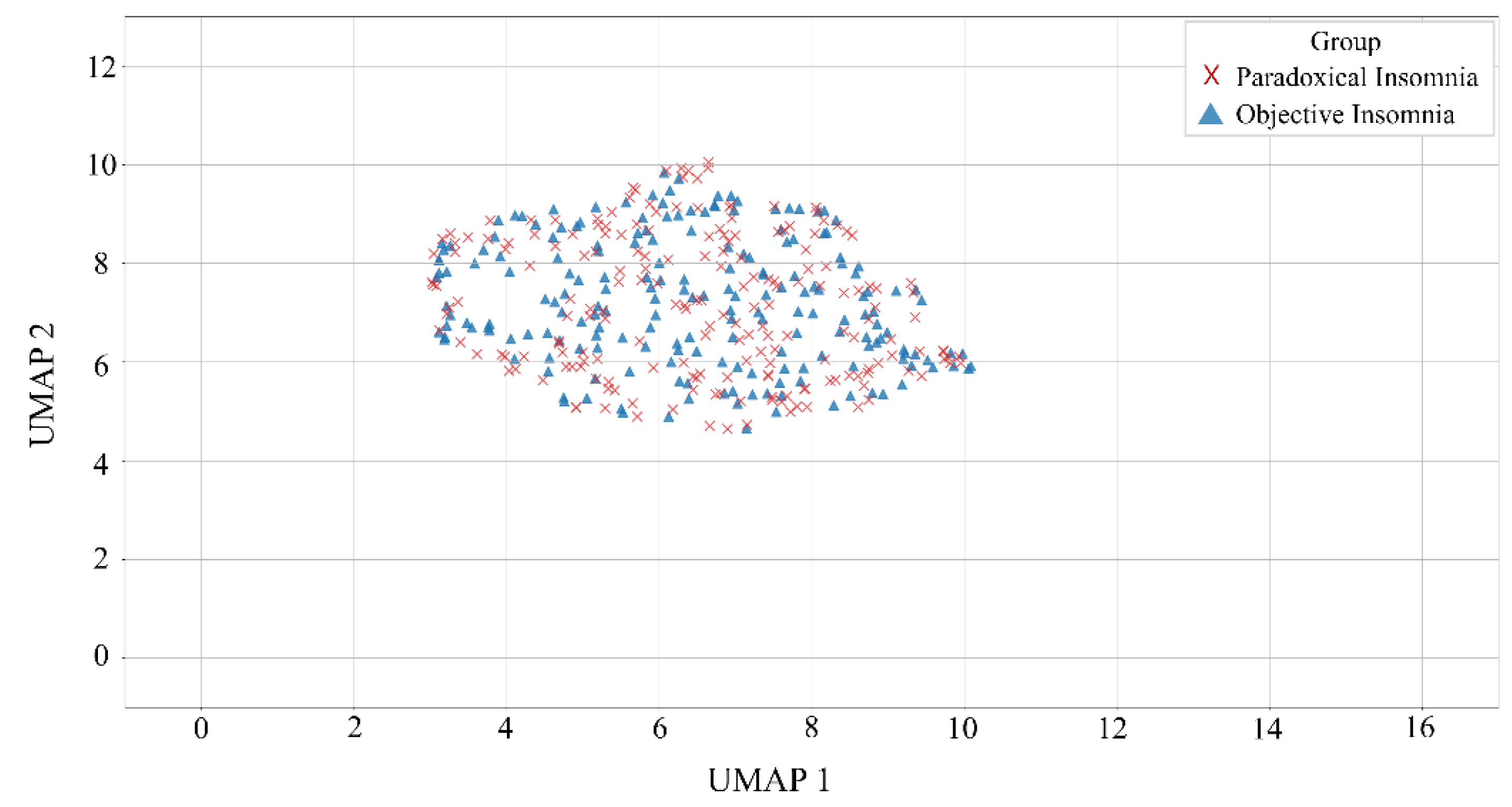
Figure 7.
UMAP embedding of heart rate variability (HRV) features during wake after sleep onset (WASO). Each point represents an individual participant. Colors indicate phenotype group (paradoxical insomnia vs. normal sleep).
Figure 7.
UMAP embedding of heart rate variability (HRV) features during wake after sleep onset (WASO). Each point represents an individual participant. Colors indicate phenotype group (paradoxical insomnia vs. normal sleep).
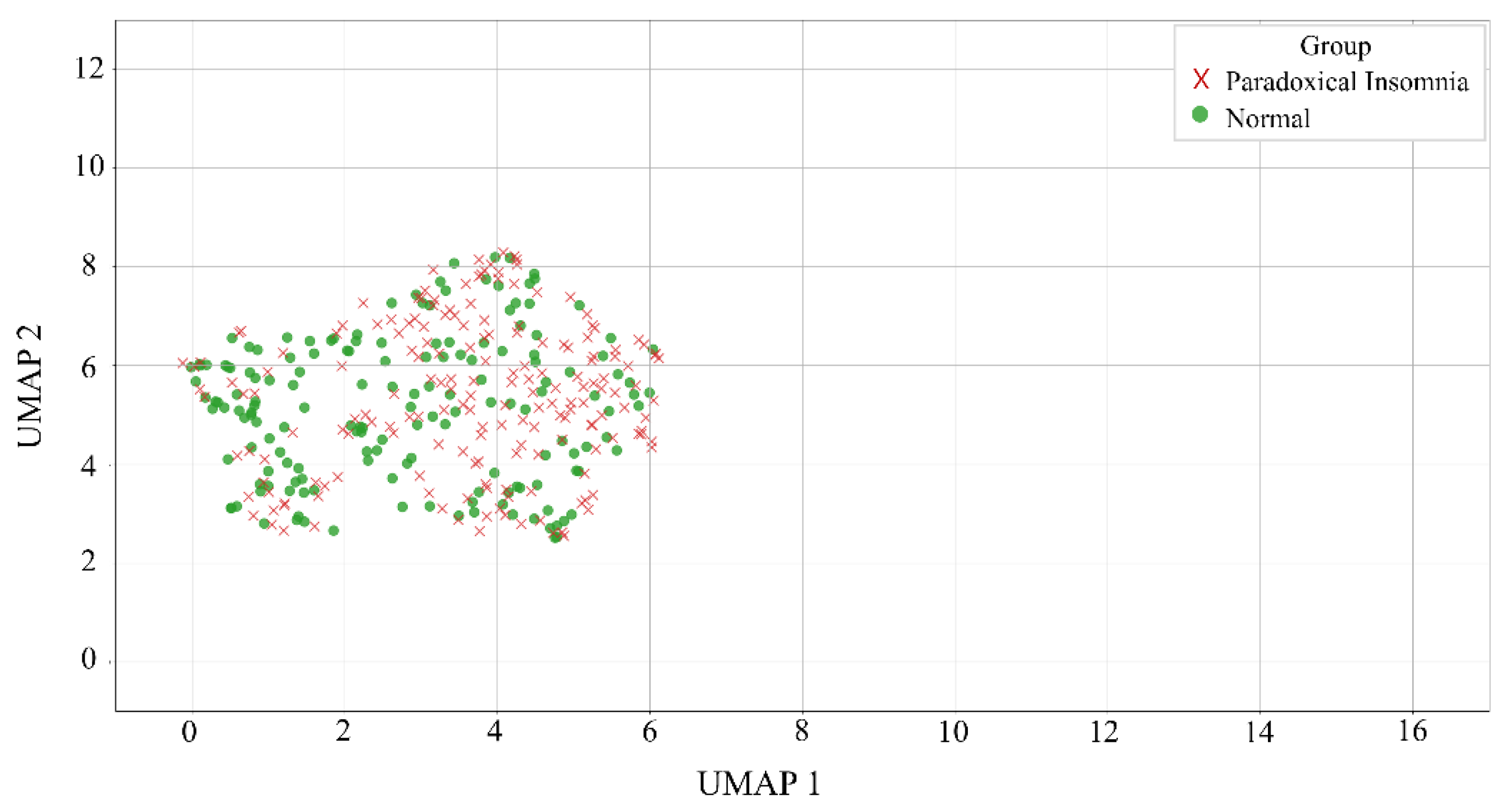
Figure 8.
UMAP embedding of heart rate variability (HRV) features during wake after sleep onset (WASO). Each point represents an individual participant. Colors indicate phenotype group (paradoxical insomnia vs. objective insomnia).
Figure 8.
UMAP embedding of heart rate variability (HRV) features during wake after sleep onset (WASO). Each point represents an individual participant. Colors indicate phenotype group (paradoxical insomnia vs. objective insomnia).
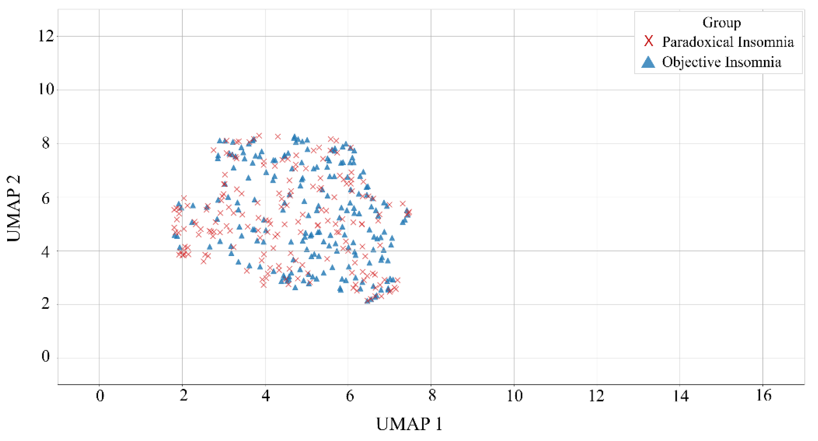

Table 1.
Operational criteria for classification of insomnia phenotypes based on subjective complaints and polysomnographic parameters.
Table 1.
Operational criteria for classification of insomnia phenotypes based on subjective complaints and polysomnographic parameters.
| Group | Classification criteria | N=1,970 |
| OI (Objective Insomnia) |
Subjective complaint of insomnia and objective insomnia indicated by PSG PSG criteria: - TST < 6 h or SE < 85% or WASO > 30 min or SL > 20 min [14,32,33] |
625 |
| PI (Paradoxical Insomnia) |
① Subjective complaint of insomnia with normal PSG findings PSG criteria: - TST ≥ 6 h and SE ≥ 85% and WASO ≤ 30 min and SL ≤ 20 min ② (oTST-sTST) ≥ 1 h [14] |
261 |
| N (Normal Sleep) |
No subjective complaint of insomnia and normal PSG findings PSG criteria: - TST ≥ 6 h and SE ≥ 85% and WASO ≤ 30 min and SL ≤ 20 min |
201 |
| Unclassified | Participants not meeting the criteria for any of the above groups | 883 |
Table 2.
Summary of heart rate variability features used in this study.
| Category | Feature | Description | Unit |
|---|---|---|---|
| Time-domain | AVNN | Mean of normal-to-normal (NN) intervals | ms |
| SDNN | Standard deviation of all NN intervals, reflecting overall HRV | ms | |
| RMSSD | Root mean square of successive NN interval differences, reflecting short-term vagal modulation | ms | |
| pNN50 | Percentage of successive NN intervals differing by more than 50 ms | % | |
| Frequency- domain |
Total power | Total spectral power across all frequency bands | ms2 |
| VLF power | Power in the very-low-frequency band (0.003–0.04 Hz) | ms2 | |
| LF power | Power in the low-frequency band (0.04–0.15 Hz) | ms2 | |
| HF power | Power in the high-frequency band (0.15–0.40 Hz) | ms2 | |
| LF/HF ratio | Ratio of low- to high-frequency power, reflecting sympathovagal balance | - | |
| Non-linear | SD1 | Poincaré plot index of short-term variability | ms |
| SD2 | Poincaré plot index of long-term variability | ms | |
| SD1/SD2 ratio | Ratio of short- to long-term variability | - | |
| Sample Entropy | Quantifying signal complexity and irregularity | - | |
| Approximate Entropy | Measuring regularity of time-series patterns | - | |
| DFA α1 | Short-term fractal scaling exponent from detrended fluctuation analysis | - | |
| DFA α2 | Long-term fractal scaling exponent from detrended fluctuation analysis | - |
Table 3.
Stage-specific HRV differences between paradoxical insomnia and normal sleep.
| Sleep Stage | HRV features | PI (Median) | N (Median) | p-value | CLES | Rank-biserial r |
| REM | DFA α1 median | 1.218 | 1.265 | 0.047 | 0.557 | 0.115 |
| WASO | SampEn IQR | 0.276 | 0.127 | < 0.0001 | 0.339 | -0.323 |
| ApEn IQR | 0.163 | 0.087 | < 0.0001 | 0.352 | -0.296 | |
| DFA α2 IQR | 0.204 | 0.120 | < 0.0001 | 0.357 | -0.287 | |
| SDNN IQR | 27.385 | 18.263 | < 0.0001 | 0.362 | -0.276 | |
| pNN50 IQR | 4.919 | 2.185 | < 0.0001 | 0.362 | -0.275 | |
| SD2 IQR | 38.119 | 22.141 | < 0.0001 | 0.367 | -0.265 | |
| SD1 IQR | 13.852 | 8.268 | < 0.0001 | 0.372 | -0.256 | |
| RMSSD IQR | 19.590 | 11.692 | < 0.0001 | 0.372 | -0.256 | |
| AVNN IQR | 57.743 | 32.086 | < 0.0001 | 0.373 | -0.253 | |
| IHR IQR | 4.652 | 2.835 | < 0.0001 | 0.375 | -0.251 | |
| SD1/SD2 IQR | 0.840 | 0.520 | < 0.0001 | 0.385 | -0.229 | |
| HF IQR | 445.802 | 172.727 | < 0.0001 | 0.389 | -0.222 | |
| LF IQR | 499.178 | 278.598 | < 0.0001 | 0.392 | -0.216 | |
| DFA α1 IQR | 0.160 | 0.106 | 0.001 | 0.393 | -0.213 | |
| DFA α2 median | 1.082 | 1.165 | 0.001 | 0.604 | 0.208 | |
| Total Power IQR | 4427.589 | 2599.252 | 0.001 | 0.398 | -0.205 | |
| VLF median | 4098.166 | 5603.812 | 0.001 | 0.601 | 0.203 | |
| LF/HF IQR | 0.749 | 0.382 | 0.003 | 0.407 | -0.187 | |
| SD1/SD2 median | 3.023 | 3.533 | 0.004 | 0.588 | 0.177 | |
| VLF IQR | 2981.896 | 2152.514 | 0.022 | 0.429 | -0.142 | |
| Total Power median | 6057.062 | 7767.248 | 0.034 | 0.566 | 0.131 |
* Only HRV features showing statistically significant differences (p < 0.05) are presented in this table; features without significant group differences were omitted for clarity.
Table 4.
Stage-specific HRV differences between paradoxical and objective insomnia.
| Sleep Stage | HRV features | OI (Median) | PI (Median) | p-value | CLES | Rank-biserial r |
| WASO | DFA α1 IQR | 0.247 | 0.160 | < 0.0001 | 0.357 | -0.286 |
| DFA α2 IQR | 0.275 | 0.204 | < 0.0001 | 0.359 | -0.281 | |
| SD1/SD2 IQR | 1.228 | 0.840 | < 0.0001 | 0.396 | -0.209 | |
| SampEn IQR | 0.359 | 0.276 | 0.001 | 0.397 | -0.205 | |
| SD2 IQR | 49.357 | 38.119 | 0.001 | 0.399 | -0.202 | |
| VLF median | 2628.257 | 4098.166 | 0.001 | 0.595 | 0.190 | |
| SDNN IQR | 36.906 | 27.385 | 0.002 | 0.407 | -0.186 | |
| ApEn IQR | 0.199 | 0.163 | 0.002 | 0.407 | -0.185 | |
| RMSSD IQR | 27.836 | 19.590 | 0.002 | 0.408 | -0.184 | |
| SD1 IQR | 19.686 | 13.852 | 0.002 | 0.408 | -0.184 | |
| SD2 median | 112.321 | 125.705 | 0.010 | 0.576 | 0.152 | |
| Total Power median | 4936.610 | 6057.062 | 0.012 | 0.575 | 0.150 | |
| SampEn median | 0.834 | 0.776 | 0.014 | 0.427 | -0.145 | |
| ApEn median | 0.838 | 0.792 | 0.014 | 0.427 | -0.145 | |
| AVNN IQR | 65.905 | 57.743 | 0.015 | 0.428 | -0.145 | |
| SDNN median | 86.379 | 95.636 | 0.024 | 0.567 | 0.134 | |
| pNN50 IQR | 6.439 | 4.919 | 0.026 | 0.434 | -0.132 | |
| DFA α2 median | 1.049 | 1.082 | 0.031 | 0.564 | 0.128 | |
| LF/HF IQR | 1.084 | 0.749 | 0.045 | 0.441 | -0.119 |
* Only HRV features showing statistically significant differences (p < 0.05) are presented in this table; features without significant group differences were omitted for clarity.
Table 5.
Stage-specific centroid distances between insomnia phenotypes in UMAP-embedded space.
| Sleep Stage | Group | Centroid Distance |
| NREM | PI vs. N | 0.534 |
| PI vs. OI | 0.420 | |
| REM | PI vs. N | 0.594 |
| PI vs. OI | 0.412 | |
| WASO | PI vs. N | 1.659 |
| PI vs. OI | 1.307 |
Table 6.
Machine learning classification performance using stage-specific HRV features.
| Sleep Stage | Group | Precision | Recall | F1-score | Accuracy | 10-Fold CV AP |
| NREM | PI | 0.567 | 0.567 | 0.567 | 0.567 | 0.578 ± 0.081 |
| N | 0.567 | 0.567 | 0.567 | |||
| PI | 0.540 | 0.783 | 0.639 | 0.558 | 0.534 ± 0.081 | |
| OI | 0.606 | 0.333 | 0.430 | |||
| REM | PI | 0.463 | 0.417 | 0.439 | 0.467 | 0.541 ± 0.065 |
| N | 0.470 | 0.517 | 0.492 | |||
| PI | 0.533 | 0.533 | 0.533 | 0.533 | 0.547 ± 0.078 | |
| OI | 0.533 | 0.533 | 0.533 | |||
| WASO | PI | 0.600 | 0.804 | 0.687 | 0.610 | 0.737 ± 0.077 |
| N | 0.633 | 0.388 | 0.481 | |||
| PI | 0.569 | 0.518 | 0.542 | 0.574 | 0.631 ± 0.078 | |
| OI | 0.578 | 0.627 | 0.602 |
Disclaimer/Publisher’s Note: The statements, opinions and data contained in all publications are solely those of the individual author(s) and contributor(s) and not of MDPI and/or the editor(s). MDPI and/or the editor(s) disclaim responsibility for any injury to people or property resulting from any ideas, methods, instructions or products referred to in the content. |
© 2026 by the authors. Licensee MDPI, Basel, Switzerland. This article is an open access article distributed under the terms and conditions of the Creative Commons Attribution (CC BY) license (http://creativecommons.org/licenses/by/4.0/).
Copyright: This open access article is published under a Creative Commons CC BY 4.0 license, which permit the free download, distribution, and reuse, provided that the author and preprint are cited in any reuse.


