Preprint
Article

This version is not peer-reviewed.

Molecular Identification of Toxoplasma gondii and Other Coccidia in Fecal Samples from Shelter Cats

Submitted:

21 January 2026

Posted:

22 January 2026

You are already at the latest version

Abstract

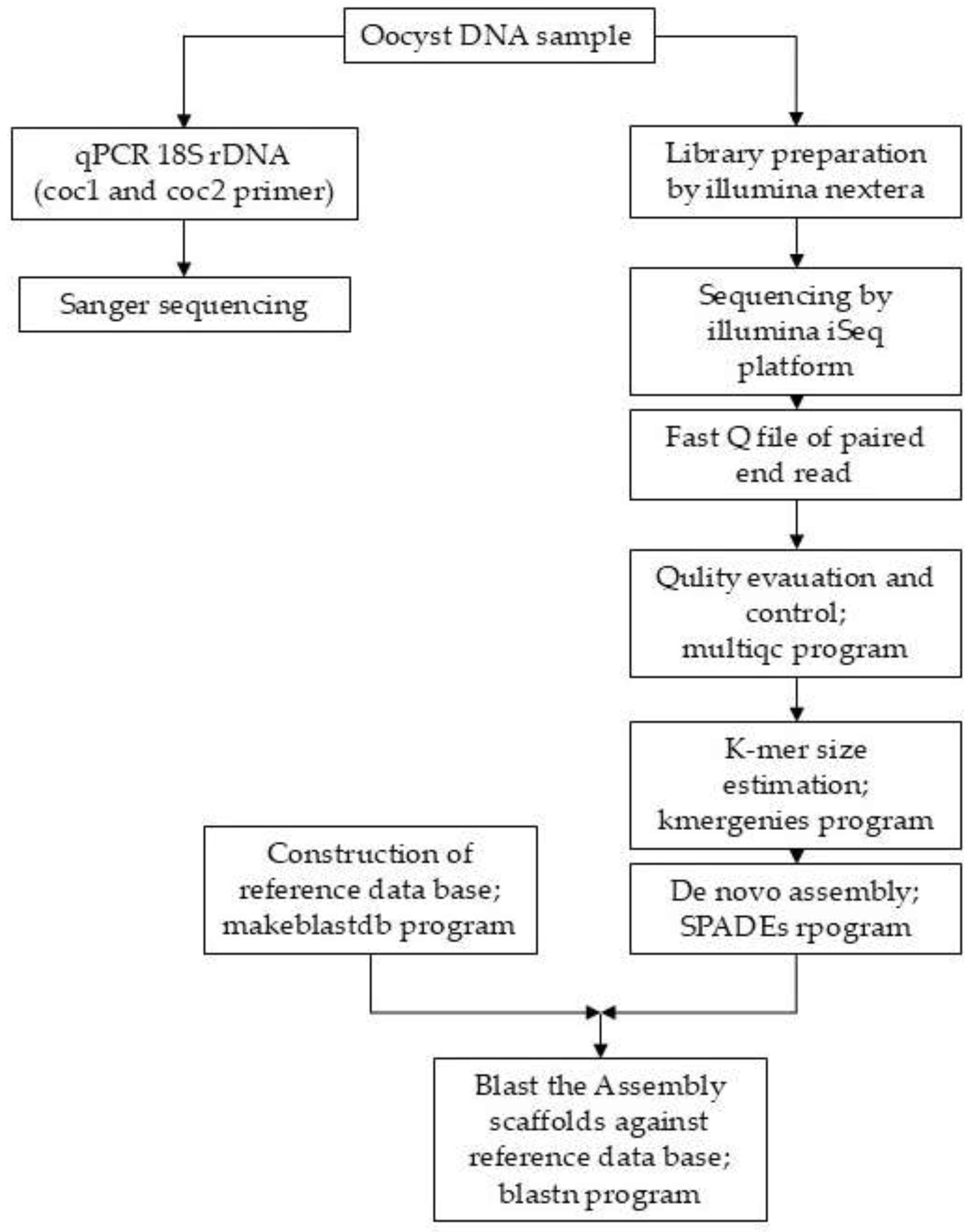
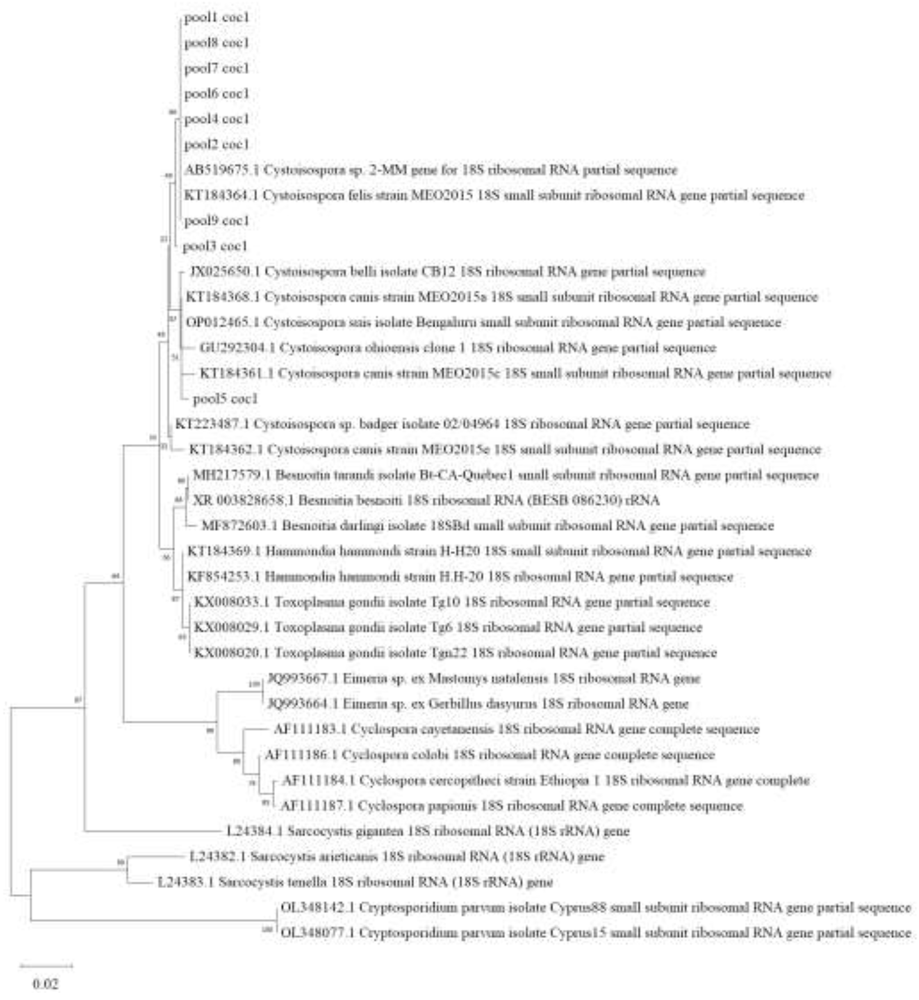
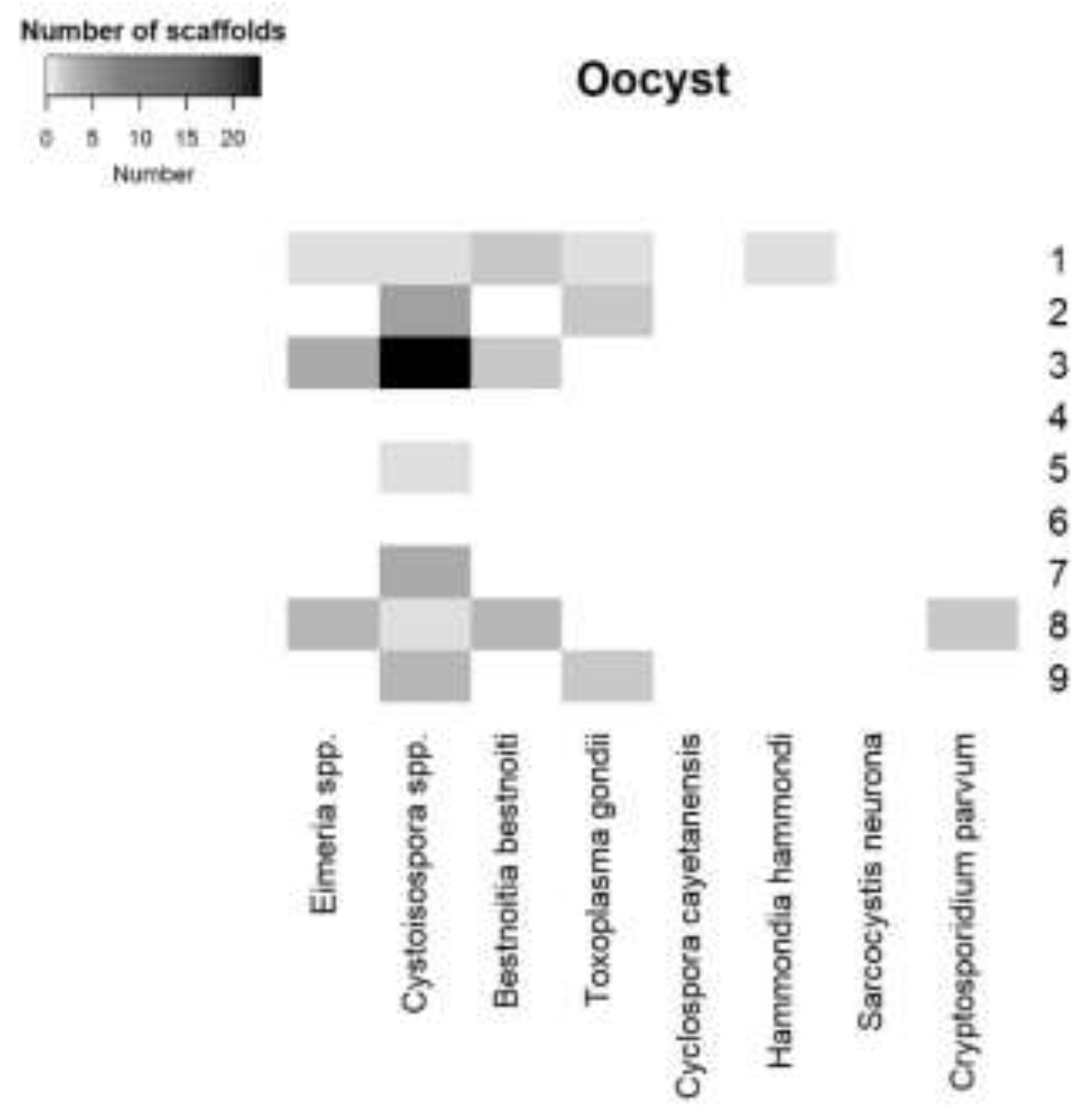
The objective was to detect T. gondii and various protozoan oocysts in the feces of sheltered cats in Thailand using two molecular approaches, Sanger sequencing and un-targeted Next-generation sequencing (NGS). A total of 166 fecal samples from shelter cats, in 26 samples, Toxoplasma gondii oocyst–like structures were detected. The harvested oocysts were grouped into nine pooled samples. DNA was extracted from all pooled samples and tested using quantitative PCR (qPCR), employing coc1 and coc2 primers, which are commonly used to amplify Apicomplexan DNA. The sequenced of the qPCR products were analyzed by Sanger sequencing. Sequence from all pooled samples had similarity to Cystoisospora spp. To further characterize the oocyst species, NGS was performed. Bioinformatic analysis was conducted using a de novo assembler to generate scaffolds, which were then aligned against a custom database of coccidian whole-genome references. This analysis revealed the presence of T. gondii DNA in three pooled samples. In addition, DNA from other protozoan parasites—including Eimeria spp., Cystoisospora spp., Besnoitia besnoiti, Hammondia hammondi, and Cryptosporidium parvum—was also detected. These findings indicate that T. gondii is circulating among shelter cats, many of which were formerly stray. Moreover, NGS sequencing provided more comprehensive information on the diversity of coccidian species in cat feces compared with Sanger sequencing of PCR-amplified targets.

Keywords: 
;  ;  ;  ;  

1. Introduction

Toxoplasma gondii is a protozoan parasite classified within the phylum Apicomplexa. It is capable of infecting both animals and humans. Transmission occurs through both vertical and horizontal routes. Vertical transmission involves the transfer of the parasite from the mother to the fetus via the placenta during parasitemia, which may result in placentitis, fetal encephalitis, and carditis, potentially leading to abortion [1]. Horizontal transmission occurs when a host ingests infected tissues or oocysts present in contaminated feed or water. T. gondii oocysts are highly resilient and can remain viable in soil or freshwater for more than one year under a wide range of environmental conditions. This resilience is a key factor contributing to the parasite’s widespread distribution and its ability to infect a broad range of hosts [2].
Felids are the definitive hosts of Toxoplasma gondii and are the only animal capable of producing oocysts in their gastrointestinal tract and excreting them in feces. In cats, shedding of un-sporulated oocysts begins 3–18 d post-infection following the ingestion of T. gondii tissue cysts [3]. Sporulation of the oocysts occurs 1–5 d after shedding when they are exposed to favorable environmental conditions [1]. A single cat can shed more than 100 million oocysts into the environment [1,3]. The most common method for detecting T. gondii oocysts in feces is centrifugal flotation followed by identification using light microscopy [4]. The coccidian oocysts are typically oval in shape, measuring approximately 6–48 μm, with a thin wall and usually containing one or two sporocysts per oocyst [5]. However, oocysts with similar morphological characteristics can also be produced by other protozoa, including Cystoisospora spp., Eimeria spp., Hammondia hammondi, and Cryptoisospora spp. Consequently, microscopic examination alone is mostly insufficient for accurate species identification and DNA-based methods are therefore required to confirm oocyst identity [6]. In Thailand, only a limited number of studies have investigated the presence of T. gondii oocysts in cat feces. A study conducted in the southern region reported that 6.7% of cat fecal samples were positive for T. gondii DNA using internal transcribed spacer-1 (ITS-1) gene detection [5]. In the northern region, another study reported a prevalence of 37% for T. gondii DNA in owned cats (n = 124) based on B1 gene detection. Furthermore, only 8% of these samples were positive for the GRA6 gene and were classified as genotype I [7].
Identifying Toxoplasma gondii oocysts in cats is challenging because infected cats often exhibit no clinical signs. In addition, the short duration of oocyst shedding further complicates detection [8]. Molecular techniques are therefore valuable for detecting T. gondii oocysts in both fecal and environmental samples. T. gondii DNA can be detected in fecal samples even when oocysts are not visually observed, likely due to low levels or intermittent shedding [9]. A variety of molecular methods are used for the detection of T. gondii, including conventional PCR, nested PCR, quantitative PCR (qPCR), and melting curve analysis [10]. More recently, next-generation sequencing (NGS) has been applied to obtain more comprehensive information on protozoan DNA present in fecal samples [11]. Targeted NGS approaches are commonly used to identify protozoa in fecal and environmental samples, with the 18S rRNA gene frequently employed as a target in metagenomic studies [12]
However, PCR primers may not amplify all protozoan species with equal efficiency, potentially resulting in biased outcomes that favor certain species while failing to detect others [11,13]. Targeted PCR combined with Sanger sequencing has been widely used for protozoan detection; however, the relatively limited amount of sequence data generated—typically ranging from 300 to 1,000 base pairs—may lead to an underestimation of parasite diversity and prevalence in samples. In addition, analysis based on chromatogram characteristics can further restrict the depth of information obtained [14,15]. Samples containing multi- protozoan oocyst often produce mixed chromatograms when analyzed by Sanger sequencing, making accurate species identification difficult or impossible. Conversely, in some cases of co-infection, Sanger sequencing may yield apparently clean chromatograms that identify only a single species. This phenomenon likely occurs because a dominant DNA template in a mixed sample can outcompete minority templates during PCR amplification, resulting in a biased or incomplete representation of the true diversity present in the sample [11]. In contrast, in un-targeted NGS all the genomic material is analyzed, reducing primer-associated amplification bias and providing a more comprehensive view of microbial diversity [11,15]. Bioinformatic analysis typically begins with quality filtering of raw reads, followed by assembly into contigs or scaffolds. These assembled sequences are subsequently identified or classified through sequence alignment against reference databases [15,16,17]. Consequently, NGS represents a valuable alternative for obtaining more comprehensive data, offering broader insights into the genetic composition of complex samples.
Cat shelters in Thailand continuously admit new residents, most of which are former stray cats from surrounding areas. These cats may harbor a variety of coccidian protozoa, including Cystoisospora spp., Besnoitia besnoiti, Hammondia hammondi, Toxoplasma gondii, Cryptosporidium parvum, and Sarcocystis hirsuta., [18,19,20]. Furthermore, environmental contaminants such as Eimeria spp. and Cyclospora cayetanensis can be detected in cat feces, either through external contamination or by passing directly through the gastrointestinal tract. [21,22] At present, information regarding protozoan infections in shelter cats in Thailand is limited, although previous research has indicated that Cystoisospora felis is the most prevalent protozoan species among stray cats in the country [23]. The continual introduction of new cats into shelters poses a risk of maintaining and disseminating these pathogens within the shelter environment and presents a potential zoonotic threat to shelter workers, particularly with respect to T. gondii. This concern is supported by studies reporting that indicate a 4.7% prevalence of oocyst shedding in companion cats with outdoor access [5], data from Thailand shows seroprevalence of 4–10% in stray populations [24,25], suggesting widespread exposure. Considering that a single infected cat can shed millions of environmentally resilient oocysts [2,3]. Given the critical role of cats in the transmission of T. gondii to humans and other animals through oocyst excretion, investigation of T. gondii oocysts in feline feces is essential for understanding the epidemiology of this parasite. Therefore, the aim of this study was to identify T. gondii and other protozoan oocysts present in fecal samples from shelter cats in Thailand using two sequencing approaches: Sanger sequencing and un-targeted NGS.

2. Materials and Methods

2.1. Fecal Sample and Oocyst Collection

A total of 166 feline fecal samples were collected from six animal shelters located in the western and central regions of Thailand, including Pathum Thani, Nakhon Pathom, Nonthaburi, Suphan Buri, and Kanchanaburi. Samples were obtained from both newly admitted cats and long-term shelter residents. Fecal material was collected from litter boxes and floors within the cats’ living areas. Each sample was placed in a zip-lock bag, stored at 4 °C, and processed within 24 h using the simple flotation method.

2.2. Simple Floatation Technique

Oocyst examination was performed after oocyst enrichment using the simple flotation method with Sheather’s sugar solution. Approximately 5 g of each fecal sample was mixed with 25 mL of sugar solution and filtered through gauze. The filtrate was centrifuged at 1,200 × g for 10 min. A coverslip was then placed on top of the sample tube and left undisturbed for 20 min. Oocyst-like structures resembling T. gondii were identified under a light microscope. A fecal sample was considered positive for T. gondii oocysts based on the following morphological criteria [5]: (1) oocyst diameter of 6–48 µm, (2) oval or ellipsoidal shape, and (3) presence of one or two internal sporocysts per oocyst. Positive T. gondii-like oocysts were harvested after a sedimentation technique. Briefly, 5 mL of the upper layer of the sugar solution was collected and diluted with 45 mL of tap water. The mixture was centrifuged at 1,200 × g for 10 minutes, after which the supernatant was discarded. The sediment containing the oocysts was stored at 4 °C until DNA extraction was performed [26].

2.3. DNA Extraction from Oocyst

The oocyst-like samples were centrifuged, and the resulting sediments were pooled, with 4–5 individual samples combined per pool. A total of nine pooled samples were subjected to DNA extraction using the Zymo® DNA Extraction Kit for Feces or Soil (Zymo Research Europe GmbH, Freiburg, Germany). Due to the difficulty in lysing the resilient oocyst wall, an additional pre-treatment step was incorporated prior to following the standard kit protocol. Specifically, samples were treated with Triton X-100 and 14% bleach to remove the oocyst outer wall [27]. Following this pre-treatment, DNA extraction proceeded according to the manufacturer’s instructions. As a positive control, T. gondii DNA was extracted from tachyzoites of the T. gondii ME49 strain, obtained from the Institute of Parasitology, Vetsuisse Faculty, University of Zurich. This control was included in the quantitative PCR (qPCR) assay alongside the test samples.

2.4. qPCR and Sanger Sequencing

The quantitative PCR (qPCR) assay targeted the 18S rDNA gene, a widely used region for detecting members of the phylum Apicomplexa. The two primers,including COC-1 (5′-AAGTATAAGCTTTTATACGGCT-3′) and COC-2 (5′-CACTGCCACGGTAGTCCAATAC-3′) were employed [28]. Each 20 µL reaction was prepared using the iTaq™ Universal SYBR® Green Supermix (Bio-Rad Laboratories Inc., Waltham, MA, USA) following the manufacturer’s instructions. Thermal cycling consisted of an initial pre-denaturation step at 95 °C for 5 min, followed by 35 cycles of amplification, each comprising denaturation at 95 °C for 15 sec, annealing at 55 °C for 1 min, and extension at 60 °C for 1 min. A melting curve analysis was subsequently performed from 60 °C to 95 °C at a ramp rate of 0.05 °C/sec, with an initial step at 95 °C for 15 sec. qPCR amplification and melting curve data were analyzed using QuantStudio™ Real-Time PCR Software v1.1. Following amplification, PCR products were resolved on a 1.5% agarose gel stained with RedSafe™ nucleic acid stain. The gel was run at 100 V for 20 min and visualized using the GelDoc Go Imaging System (Bio-Rad Laboratories Inc.). The target band (~297 bp) was excised using a sterile scalpel and purified with the NucleoSpin® Gel and PCR Clean-Up Kit. Purified products were submitted for Sanger sequencing using the COC-1 primer at Microsynth AG (Balgach, Switzerland). The resulting nucleotide sequences and chromatograms were aligned against the 18S rRNA gene sequences of common coccidian protozoa using BioEdit (version 7.2.5). A phylogenetic tree was subsequently constructed using MEGA11 (version 11.0.13) to determine the specific coccidian clustering of the sequences based on their genetic relationships.

2.5. DNA Library Preparation and Illumina Sequencing

The concentration and quality of DNA extracted from oocysts were assessed using the D500 ScreenTape Assay on the Agilent TapeStation System (Agilent Technologies Inc., Germany). Library preparation and next-generation sequencing (NGS) were performed at the Functional Genomics Center Zurich (FGCZ). DNA libraries were prepared using the Illumina Nextera Kit and subsequently sequenced on the Illumina iSeq platform.

2.6. Bioinformatics Workflow

The analysis pipeline for Illumina sequencing data is summarized in Figure 1. All analyses were performed on a 64-bit Ubuntu 22.04.4 LTS operating system using the high-performance computing (HPC) server at the Center for Agricultural Biotechnology, Kasetsart university, Kamphaeng Saen Campus, Thailand. The workflow began with quality control using MultiQC v1.11, with reads exhibiting Phred quality scores below 30 removed as low quality. K-mer size estimation, tailored to each paired-end dataset, was conducted using KmerGenie, and the optimal k-mer sizes are provided in Supplementary Data S2. De novo Assembly was then performed using SPAdes v3.14.0, generating contigs and scaffolds. Assembly statistics were visualized and evaluated using Bandage v0.8.1. A custom reference database for coccidian oocyst identification in cat feces was constructed using the makeblastdb tool (BLAST+ version 2.5.0). Whole-genome sequences were downloaded from the ToxoDB database (Supplementary Table S1) and included genomes from eight coccidian genera: Cyclospora cayetanensis, Cystoisospora suis, Cryptosporidium parvum, Sarcocystis neurona, Besnoitia besnoiti, Hammondia hammondi, Toxoplasma gondii, and Eimeria spp., which are potential for environmental contamination or pseudoparasitism in fecal samples. The Python script used in this study is provided in Supplementary Table S2. All assembled scaffolds from each pooled sample were queried against the custom coccidian database using BLASTn. Scaffolds with E-values < 1 × 10⁻⁵⁰ and percent identity above 98% were considered positive for the corresponding coccidian genus. A heatmap illustrating the number of scaffolds identified as positive for coccidian DNA in each pooled sample was generated using RStudio v4.3.2 (R Core Team, 2023).
To ensure taxonomic specificity and eliminate potential ambiguities, a stringent filtering pipeline was implemented for the assembled scaffolds. Although a sequence might meet the initial blast criteria (E-values < 1 × 10⁻⁵⁰ and percent identity above 98%), any scaffold aligning with multiple coccidian species was excluded. Consequently, only scaffolds demonstrating unique matches to a single coccidian species were retained for further analysis.

2.7. The Prevalence of T. gondii in Shelter Cats

To estimate prevalence at the level of individual cat fecal samples, a frequentist approach was employed using Maximum Likelihood (ML) analysis via the EpiTools online epidemiological calculator [29]. This estimation assumed perfect sensitivity and specificity of the NGS assay, in accordance with previously established methodologies [30,31].

3. Results

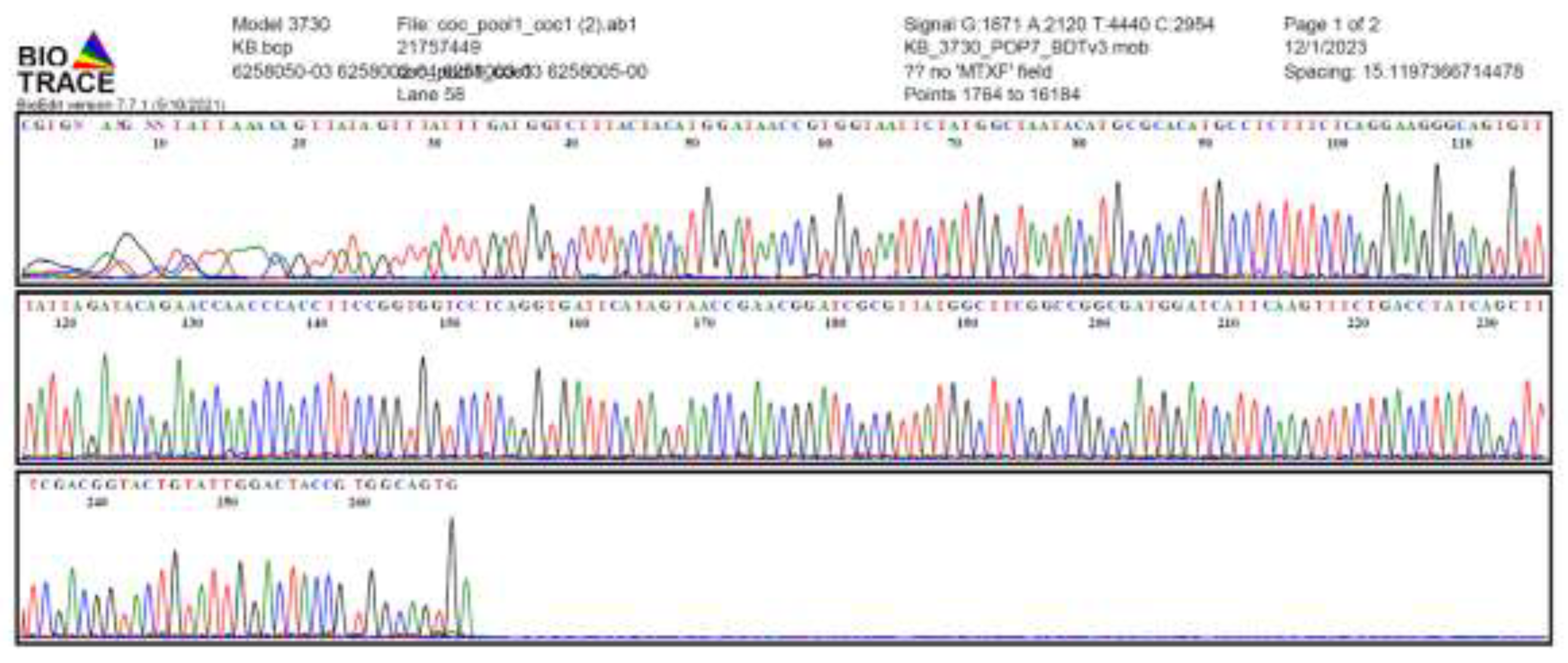
3.1. qPCR and Sanger Sequencing

A total of 26 (15.6%) T. gondii oocyst–like samples from four shelters were combined into nine pooled samples. All pooled samples tested positive for the coccidia -specific 18S rDNA gene. Melting curve analysis revealed no differences between the pooled samples and the T. gondii positive control (Supplementary Figure S1), indicating that melting curve analysis alone is insufficient to discriminate among different protozoan oocyst species.
The qPCR products from all pooled samples were analyzed by agarose gel electrophoresis, revealing bands corresponding to the expected amplicon size of 297 bp (Supplementary Figure S1). These PCR products were then purified using a commercial DNA purification kit and submitted for Sanger sequencing.
Chromatograms obtained from Sanger sequencing of pool1 samples exhibited clean peaks with minimal background noise and no overlapping signals, indicating the presence of a single predominant sequence per pooled sample (Figure 2). Additionally, other chromatograms of pool2 -9 samples from Sanger sequencing are provided in Supplementary Figure S2. Low-quality ends were trimmed prior to multiple sequence alignment. Phylogenetic analysis based on the 18S rRNA gene region amplified by the COC-1 and COC-2 primers, together with reference sequences from coccidian species, revealed that all pooled samples clustered with reference sequences of Cystoisospora spp. (Figure 3). Detailed sequence alignments are provided in Supplementary Figure S3.

3.2. Un-Targeted Next Generation Sequencing

The DNA concentration from oocysts samples was measured prior to library preparation, with results summarized in Supplementary Table S3. Short-read sequencing was performed on the Illumina iSeq platform, generating paired-end FASTQ files containing forward (R1) and reverse (R2) reads. All reads were filtered to retain sequences with Phred scores above 30 (Supplementary Figure S4). Prior to assembly, the filtered reads were used to estimate the optimal k-mer size for de novo assembly, and the selected k-mer sizes are provided in Supplementary Table S3. High-quality reads were subsequently assembled using the SPAdes assembler, and statistics for the assembled scaffolds are reported in Supplementary Table S3.
NGS sequencing generated total read lengths ranging from 34,536 bp to 2,704,357 bp across the pooled samples. Following assembly, the number of scaffolds varied among pools, with Pool 3 yielding the highest number of scaffolds (38,873) and an N50 of 112. In contrast, the remaining pools exhibited scaffold counts ranging from 155 to 1,761, with N50 values generally between 208 and 473 (Table 1).
All assembled scaffolds were annotated using BLASTn against a custom reference database. Scaffolds with BLAST hits meeting the criteria of E-value ≤ 1 × 10⁻⁵⁰ and percent identity ≥ 98% were classified as positive protozoan scaffolds. The BLAST alignment details for all scaffolds are available in Supplementary Table S4.
T. gondii DNA was detected in three of the nine pooled samples, all of which were positive for oocyst detection. Cystoisospora spp. was the most prevalent protozoan, appearing in seven of the nine pooled samples. Additionally, other coccidia—including Eimeria spp., Besnoitia besnoiti, Hammondia hammondi, and Cryptosporidium parvum—were identified in the pooled oocyst samples from cat feces. Notably, two pooled samples did not yield identifiable coccidian sequences (Table1). The distribution of positive protozoan scaffolds across pooled samples is illustrated in a heatmap (Figure 4).
Figure 5 presents the geographical distribution of the participating animal shelters in Thailand, with the two shelters where molecular evidence of T. gondii was detected indicated by dark dots. This map provides a clear visual representation of the parasite’s occurrence within the sampled regions.

4. Discussion

In this study, T. gondii-like oocyst structures were identified in 26 (15.6%) of 166 individual cat fecal samples based on morphological examination. The proportion of T. gondii-like oocyst–positive samples observed here is consistent with previous findings from southern Thailand, where 19.3% (49/254) of cat fecal samples contained T. gondii-like oocysts, of which 19 samples were confirmed by molecular detection [5]. In Germany reported a low prevalence (0.57%) of T. gondii-like oocysts by microscopy, with 44% of those samples subsequently confirmed by PCR [32]. These observations highlight that, while microscopy provides a preliminary indication of oocyst presence, molecular methods offer higher specificity and sensitivity, making them essential for definitive identification.
The low prevalence of T. gondii oocyst detection can be explained by the short shedding window in cats—of typically 1–2 weeks. In addition, most cats shed oocysts only once in their lifetime as immunity develops following primary infection [8]. Low oocyst numbers further complicate detection via microscopy; the detection limit for Sheather’s sugar flotation combined with light microscopy has been reported as 1,828 oocysts per gram of feces [26]. To overcome these limitations, this study employed molecular techniques, including Sanger sequencing and NGS, to enhance detection and identification of coccidian oocysts.
Sanger sequencing using the COC-1 and COC-2 primers detected only Cystoisospora spp. DNA in all pooled samples, with no detection of other coccidian DNA. This discrepancy may result from competitive inhibition during PCR, whereby the higher abundance of Cystoisospora spp. DNA in pooled samples likely outcompeted T. gondii and other coccidia during amplification. More precisely, the species with higher 18S rDNA copy numbers can dominate PCR amplification, thereby suppressing the detection of protozoa present at lower abundance. This occurs because a high concentration of the dominant template competes more effectively for limited primers and reagents within the reaction [18,33]. These observations indicate that Sanger sequencing may underestimate species diversity in samples containing mixed infections due to preferential amplification of dominant templates.
Moreover, the primers used in this study targeted a variable region of the 18S rDNA, which allowed clear differentiation of coccidian genera. This taxonomic resolution is supported by phylogenetic analysis, in which sequences from the same genus consistently clustered into distinct clades.
Illumina NGS, as a high-throughput sequencing technology, provided deeper insights into protozoan diversity within the pooled samples, enabling the detection of species present at low abundance. Assembled scaffolds were aligned against a custom reference database of coccidian genomes, including T. gondii, Eimeria spp., Cystoisospora spp., Besnoitia besnoiti, Hammondia hammondi, and Cryptosporidium parvum. Scaffolds with ambiguous matches were excluded from heatmap visualization. The non-targeted NGS approach revealed greater protozoan diversity than Sanger sequencing and minimized the biases associated with targeted PCR amplification [11,15].
This study detected T. gondii DNA in 13.2% of pooled samples containing T. gondii-like oocysts, a prevalence higher than the 6.7% previously reported in southern Thailand [5]. The difference likely reflects variation in cat populations, as shelter cats may have lower exposure than free-roaming companion cats. Predisposing factors for T. gondii infection in cats include outdoor access, hunting behavior, and a history of being stray [34]. Additionally, feeding practices, such as providing homemade food or offal, have been associated with seropositivity for T. gondii. The shelters in this study provided a combination of leftover homemade food and commercial feed, and the living areas were not fully enclosed, allowing access to small mammals or birds. Cats were housed together rather than individually, which may facilitate the circulation of T. gondii and other protozoa within the shelter environment.
After NGS, most assembled scaffolds identified by NGS corresponded to Cystoisospora spp., consistent with prior reports that Cystoisospora is the most commonly detected coccidian protozoan in cats in Thailand and Myanmar [35,36]. These NGS results corroborate the Sanger sequencing findings, which also identified Cystoisospora spp. as the dominant coccidian genus in cat fecal samples. Additionally, the study provides a comprehensive overview of the coccidian community, including taxa present at low abundance, such as Besnoitia besnoiti, Hammondia hammondi, and Cryptosporidium parvum. Eimeria spp. were also detected; however, these are likely spurious parasites passing through the gastrointestinal tract following ingestion of prey [22].
Two pooled samples (Pool 4 and Pool 6) did not yield scaffolds matching the protozoan reference database. This finding suggests a potential underestimation of protozoan diversity, likely due to the limited scope of the reference database, which primarily focused on coccidia relevant to cat feces and excluded broader protozoan taxa, as well as bacteria, viruses, and host DNA [13,18]. Expanding the database could enhance detection sensitivity and accuracy in future studies.
Notably, although Pools 4 and 6 were positive for Cystoisospora spp. by Sanger sequencing, no identifiable coccidian scaffolds were recovered in the Illumina dataset. This discrepancy is likely attributable to the high stringency of the bioinformatics pipeline, which applied an E-value threshold of ≤ 1 × 10⁻⁵⁰, prioritizing high-confidence assignments over low-coverage or highly fragmented sequences. To maximize specificity, stringent cut-offs (E-value ≤ 1 × 10⁻⁵⁰ and ≥ 98% identity) were applied to minimize false positives, an important consideration when distinguishing closely related coccidian protozoa [37,38]. Adjusting these criteria in future analyses may allow detection of a broader range of protozoan sequences.
Using an untargeted NGS approach, T. gondii DNA was detected in three of the nine pooled fecal samples, whereas Sanger sequencing failed to detect T. gondii in any pooled samples. This discrepancy underscores differences in sensitivity between the two methods, with NGS demonstrating greater sensitivity for detecting low-abundance targets in pooled samples. The detection of T. gondii in shelter cats, many of which were formerly stray, indicates ongoing environmental circulation of the parasite. Environmental contamination with T. gondii oocysts in soil, freshwater, and seawater represents a potential infection risk for both humans and animals [39]. Future studies should incorporate environmental surveillance of T. gondii oocysts in high-risk areas such as around cat shelters to better assess contamination levels and inform control strategies, thereby supporting public health interventions aimed at reducing zoonotic transmission.

5. Conclusions

This study represents the first molecular survey of coccidian oocysts in shelter cats in Thailand. Untargeted NGS of pooled oocyst DNA revealed the presence of Cystoisospora spp., Eimeria spp., Besnoitia besnoiti, Toxoplasma gondii, Hammondia hammondi, and Cryptosporidium parvum. The combination of coprological examination with NGS proved effective for detecting and characterizing diverse coccidian species in feline feces. Species-level identification was also achievable through targeted PCR amplification of conserved genes, such as 18S rDNA. These molecular approaches offer valuable tools for advancing our understanding of coccidian diversity and epidemiology in animal shelters and contribute to broader parasite surveillance and control efforts

Supplementary Materials

The following supporting information can be downloaded at the website of this paper posted on Preprints.org. Table S1. List of coccidian protozoa reference sequences and their GenBank accession numbers used for the construction of the custom database. Table S2. Python scripting utilized in this study. Table S3 The DNA concentrations of each pooled samples and summary of statistic from de novo assembly (SPADEs program). Table S4 Identification of genomic scaffolds based on the highest-scoring BLASTn alignments with the custom reference data base. Figure S1. The results of melting curve of coc1 and coc2 primer from SYBR green-base qPCR and gel electrophoresis image of qPCR product, showing the expected targeted size of 297 bp. Figure S2 Chromatograms obtained from Sanger sequencing of pool2 -pool9 samples. Figure S3. The alignment of sanger sequencing of T. gondii oocyst – like pooled sample compared to 18S gene reference sequence. Figure S4. The summarized output of quality control of paired read data, above 30 of Phred score. .

Author Contributions

Conceptualization, N.W., S.S. and T.R.; methodology, N.W, P.L.; software, N.W.; validation, T.R., P.L.; formal analysis, N.W. and T.R..; investigation, N.W., C.R., A.B.H.; resources, T.R., S.S.; data curation, N.W.; writing—original draft preparation, N.W.; writing—review and editing, T.R.; visualization, N.W.; supervision, T.R., P.L., C.R. and A.B.H.; project administration, N.W.; funding acquisition, T.R. and A.D.H. All authors have read and agreed to the published version of the manuscript.

Funding

Financial support for this study was provided by Kasetsart University and the Thailand Research Fund through the Royal Golden Jubilee PhD Program under Grant No. NRCT5-RGJ63002-034.

Institutional Review Board Statement

This research was conducted under the approval of the Kasetsart University Committee for Animal Care and Use for Scientific Research (ACKU64-VET-039), with additional authorization granted by the supervisor of the involved animal shelter.

Data Availability Statement

The data presented in this study are available on request from the corresponding author.

Acknowledgments

we are grateful to the Institute of Parasitology, University of Zurich, for providing the necessary administrative and laboratory facilities. Additionally, we would like to extend our appreciation to the Faculty of Veterinary Medicine Technology, Kasetsart University, Kamphaeng Saen Campus for providing access to laboratory facilities, equipment, and data analysis which greatly facilitated my research. Furthermore, we sincerely thank Dr. Marc Schmid for his expertise and guidance in NGS analysis, which were essential to the advancement of this work. Finally, we express our deepest gratitude to Dr. Pumipat Tongyoo; his support and mentorship have significantly enriched our bioinformatics knowledge and provided the critical HPC server resources required for our NGS analysis.

Conflicts of Interest

The authors declare no conflict of interest.

References

  1. Robert-Gangneux, F.; Darde, M. L. Epidemiology of and diagnostic strategies for toxoplasmosis. Clin. Microbiol. Rev. 2012, 25, 264–296. [Google Scholar] [CrossRef]
  2. Torrey, E. F.; Yolken, R. H. Toxoplasma oocysts as a public health problem. Trends Parasitol. 2013, 29, 380–384. [Google Scholar] [CrossRef]
  3. Dubey, J. P. The history and life cycle of Toxoplasma gondii. In Toxoplasma gondii, 3rd ed.; Elsevier: Amsterdam, The Netherlands, 2020; pp. 1–19. [Google Scholar]
  4. Sroka, J.; Karamon, J.; Dutkiewicz, J.; Wojcik-Fatla, A; Cencek, T. Optimization of flotation, DNA extraction and PCR methods for detection of Toxoplasma gondii oocysts in cat faeces. Ann. Agric. Environ. Med. 2018, 25, 680–685. [Google Scholar] [CrossRef]
  5. Chemoh, W.; Sawangjaroen, N.; Nissapatorn, V.; Sermwittayawong, N. Molecular investigation on the occurrence of Toxoplasma gondii oocysts in cat feces using TOX-element and ITS-1 region targets. Vet. J. 2016, 215, 118–122. [Google Scholar] [CrossRef] [PubMed]
  6. Lalonde, L. F.; Gajadhar, A. A. Detection and differentiation of coccidian oocysts by real-time PCR and melting curve analysis. J. Parasitol. 2011, 97, 725–730. [Google Scholar] [CrossRef]
  7. Suwancharoen, C.; Phuangsri, C.; Prakhammin, K.; Japa, O. Molecular detection and dense granule antigen 6 genotyping of feline Toxoplasma gondii in Phayao, Thailand. Vet. World 2022, 15, 2309–2314. [Google Scholar] [CrossRef] [PubMed]
  8. Zulpo, D. L.; Sammi, A. S.; Dos Santos, J. R.; Sasse, J. P.; Martins, T. A.; Minutti, A. F.; Cardim, S. T.; de Barros, L. D.; Navarro, I. T.; Garcia, J. L. Toxoplasma gondii: A study of oocyst re-shedding in domestic cats. Vet. Parasitol. 2018, 249, 17–20. [Google Scholar] [CrossRef] [PubMed]
  9. Miura, A. C.; Barros, L. D.; Minutti, A. F.; Martins, T. A.; Sasse, J. P.; Nino, B. S. L.; Garcia, J. L. Evaluation of quantitative polymerase chain reaction for the detection of Toxoplasma gondii oocysts shed by cats. Rev. Bras. Parasitol. Vet. 2021, 30, e016621. [Google Scholar] [CrossRef]
  10. Amouei, A.; Sarvi, S.; Sharif, M.; Aghayan, S. A.; Javidnia, J.; Mizani, A.; Moosazadeh, M.; Shams, N.; Hosseini, S. A.; Hosseininejad, Z.; Nayeri Chegeni, T.; Badali, H.; Daryani, A. A systematic review of Toxoplasma gondii genotypes and feline: Geographical distribution trends. Transbound. Emerg. Dis. 2020, 67, 46–64. [Google Scholar] [CrossRef] [PubMed]
  11. Paparini, A.; Gofton, A.; Yang, R.; White, N.; Bunce, M.; Ryan, U. M. Comparison of Sanger and next generation sequencing performance for genotyping Cryptosporidium isolates at the 18S rRNA and actin loci. Exp. Parasitol. 2015, 151, 21–27. [Google Scholar] [CrossRef]
  12. Gad, M.; Fawzy, M. E.; Al-Herrawy, A. Z.; Abdo, S. M.; Nabet, N.; Hu, A. PacBio next-generation sequencing uncovers Apicomplexa diversity in different habitats. Sci. Rep. 2023, 13, 15063. [Google Scholar] [CrossRef]
  13. Moreno, Y.; Moreno-Mesonero, L.; Amorós, I.; Pérez, R.; Morillo, J. A.; Alonso, J. L. Multiple identification of most important waterborne protozoa in surface water used for irrigation purposes by 18S rRNA amplicon-based metagenomics. Int. J. Hyg. Environ. Health. 2018, 221, 102–111. [Google Scholar] [CrossRef]
  14. Dulanto Chiang, A.; Dekker, J. P. From the pipeline to the bedside: advances and challenges in clinical metagenomics. J. Infect. Dis. 2020, 221, S331–S340. [Google Scholar] [CrossRef]
  15. Mthethwa, N. P.; Amoah, I. D.; Reddy, P.; Bux, F.; Kumari, S. A review on application of next-generation sequencing methods for profiling of protozoan parasites in water: Current methodologies, challenges, and perspectives. J. Microbiol. Methods 2021, 187, 106269. [Google Scholar] [CrossRef] [PubMed]
  16. Feldmeyer, B.; Wheat, C. W.; Krezdorn, N.; Rotter, B.; Pfenninger, M. Short read Illumina data for the de novo assembly of a non-model snail species transcriptome (Radix balthica, Basommatophora, Pulmonata), and a comparison of assembler performance. BMC Genomics 2011, 12, 317. [Google Scholar] [CrossRef]
  17. Wylezich, C.; Belka, A.; Hanke, D.; Beer, M.; Blome, S.; Höper, D. Metagenomics for broad and improved parasite detection: a proof-of-concept study using swine faecal samples. Int. J. Parasitol. 2019, 49, 769–777. [Google Scholar] [CrossRef] [PubMed]
  18. Lalonde, L. F.; Gajadhar, A. A. Detection and differentiation of coccidian oocysts by real-time PCR and melting curve analysis. J. Parasitol. 2011, 97, 725–730. [Google Scholar] [CrossRef] [PubMed]
  19. Dubey, J. P.; Lindsay, D. S.; Lappin, M. R. Toxoplasmosis and other intestinal coccidial infections in cats and dogs. Vet. Clin. North Am. Small Anim. Pract. 2009, 39, 1009–1034. [Google Scholar] [CrossRef]
  20. Lindsay, D. S.; Dubey, J. P. Neosporosis, toxoplasmosis, and sarcocystosis in ruminants: an update. Vet. Clin. North Am. Food Anim. Pract. 2020, 36, 205–222. [Google Scholar] [CrossRef]
  21. Almeria, S.; Cinar, H. N.; Dubey, J. P. Cyclospora cayetanensis and cyclosporiasis: an update. Microorganisms 2019, 7, 317. [Google Scholar] [CrossRef]
  22. Abbas, I.; Al-Araby, M.; Elmishmishy, B.; El-Alfy, E. S. Gastrointestinal parasites of cats in Egypt: high prevalence high zoonotic risk. BMC Vet. Res. 2022, 18, 420. [Google Scholar] [CrossRef]
  23. Jittapalapong, S.; Inparnkaew, T.; Pinyopanuwat, N.; Kengradomkij, C.; Sangvaranond, A.; Wongnakphet, S. Gastrointestinal parasites of stray cats in Bangkok metropolitan areas, Thailand. Agric. Nat. Resour. 2007, 41, 69–73. [Google Scholar]
  24. Jittapalapong, S.; Inpankaew, T.; Pinyopanuwat, N.; Chimnoi, W.; Kengradomkij, C.; Wongnarkpet, S.; Maruyama, S; Lekkla, A.; Sukthana, Y. Epidemiology of Toxoplasma gondii infection of stray cats in Bangkok, Thailand. Southeast Asian J. Trop. Med. Pub. Health. 2010, 41, 13–18. [Google Scholar]
  25. Kengradomkij, C.; Kamyingkird, K.; Pinyopanuwat, N.; Chimnoi, W.; Jittapalapong, S.; Inpankaew, T. Seroprevalence of Toxoplasma gondii from stray cats residing in temples, Bangkok, Thailand. J. Trop. Med. Parasitol. 2018, 41, 8–14. [Google Scholar]
  26. Miura, A. C.; Barros, L. D.; Minutti, A. F.; Martins, T. A.; Sasse, J. P.; Nino, B. S. L.; Garcia, J. L. Evaluation of quantitative polymerase chain reaction for the detection of Toxoplasma gondii oocysts shed by cats. Rev. Bras. Parasitol. Vet. 2021, 30, e016621. [Google Scholar] [CrossRef]
  27. Ramakrishnan, C.; Maier, S.; Walker, R. A.; Rehrauer, H.; Joekel, D. E.; Winiger, R. R.; Basso, W.U.; Grigg, M.E.; Hehl, A. B.; Deplazes, P.; Smith, N. C. An experimental genetically attenuated live vaccine to prevent transmission of Toxoplasma gondii by cats. Sci. Rep. 2019, 9, 1474. [Google Scholar] [CrossRef]
  28. Ho, M. S.; Barr, B. C.; Marsh, A. E.; Anderson, M. L.; Rowe, J. D.; Tarantal, A. F.; Hendrickx, A. G.; Sverlow, K.; Dubey, J. P.; Conrad, P. A. Identification of bovine Neospora parasites by PCR amplification and specific small-subunit rRNA sequence probe hybridization. J. Clin. Microbiol. 1996, 34, 1203–1208. [Google Scholar] [CrossRef]
  29. EpiTools Online Epidemiological Calculator. Available online: https://epitools.ausvet.com.au/ppvariablepoolsize (accessed on 13 October 2024).
  30. Mosquera, J. D.; Valle, C. A.; Nieto-Claudin, A.; Fessl, B.; Lewbart, G. A.; Deresienski, D.; Bouazzi, L.; Zapata, S; Villena, I; Poulle, M. L. Prevalence of Toxoplasma gondii in Galapagos birds: Inference of risk factors associated with diet. Plos One 2023, 18, e0287403. [Google Scholar] [CrossRef]
  31. Williams, C. J.; Moffitt, C. M. A critique of methods of sampling and reporting pathogens in populations of fish. J. Aquat. Anim. Health 2001, 13, 300–309. [Google Scholar] [CrossRef]
  32. Herrmann, D. C.; Pantchev, N.; Vrhovec, M. G.; Barutzki, D.; Wilking, H.; Frohlich, A.; Luder, C. G.; Conraths, F. J.; Schares, G. Atypical Toxoplasma gondii genotypes identified in oocysts shed by cats in Germany. Int. J. Parasitol. 2010, 40, 285–292. [Google Scholar] [CrossRef]
  33. Fehlberg, H. F.; Maciel, B. M.; Albuquerque, G. R. Identification and discrimination of Toxoplasma gondii, Sarcocystis spp., Neospora spp., and Cryptosporidium spp. by right-resolution melting analysis. Plos One 2017, 12, e0174168. [Google Scholar] [CrossRef]
  34. Udonsom, R.; Buddhirongawatr, R.; Nishikawa, Y.; Fereig, R. M.; Jirapattharasate, C. Toxoplasma gondii prevalence and risk factors in owned domestic cats from Nakhon Pathom Province, Thailand. Vet. Integr. Sci. 2021, 19, 557–566. [Google Scholar] [CrossRef]
  35. Jitsamai, W.; Khrutkham, N.; Hunprasit, V.; Chandrashekar, R.; Bowman, D.; Sukhumavasi, W. Prevalence of endoparasitic and viral infections in client-owned cats in metropolitan Bangkok, Thailand, and the risk factors associated with feline hookworm infections. Vet. Parasitol. Reg. Stud. Reports 2021, 25, 100584. [Google Scholar] [CrossRef]
  36. Soe, B. K.; Hlaing, K. S.; Naing, T. W.; Thaw, Z. H.; Myint, A. W. The first study on the prevalence of gastrointestinal parasites in owned and shelter cats in Yangon, Myanmar. Vet. World 2023, 16, 414–420. [Google Scholar] [CrossRef] [PubMed]
  37. Blake, D. P.; Alias, H.; Billington, K. J.; Clark, E. L.; Mat-Isa, M. N.; Mohamad, A. F. H.; Mohd-Amin, M.R.; Tay, Y.L.; Smithe, A.L.; Tomley, F.M.; Wan, K. L. EmaxDB: Availability of a first draft genome sequence for the apicomplexan Eimeria maxima. Mol. Biochem. Parasitol. 2012, 184, 48–51. [Google Scholar] [CrossRef] [PubMed]
  38. Wallner, B.; Fang, H.; Ohlson, T.; Frey-Skött, J.; Elofsson, A. Using evolutionary information for the query and target improves fold recognition. Proteins: Struct., Funct., Bioinf. 2004, 54, 342–350. [Google Scholar] [CrossRef] [PubMed]
  39. Shapiro, K.; Bahia-Oliveira, L.; Dixon, B.; Dumètre, A.; de Wit, L. A.; VanWormer, E.; Villena, I. Environmental transmission of Toxoplasma gondii: oocysts in water, soil and food. Food Waterborne Parasitol. 2019, 15, e00049. [Google Scholar] [CrossRef]
Figure 1. The diagram of bioinformatics analysis pipeline.
Preprints 195340 g001
Figure 2. Chromatogram from Sanger sequencing of Pool 1 samples using the COC1 primer.
Preprints 195340 g002
Figure 3. Phylogenetic analysis based on the 18S rRNA gene region amplified by COC-1 and COC-2 primers, including reference sequences from various coccidian species.
Preprints 195340 g003
Figure 4. Heatmap showing protozoa identified via BLASTn analysis against a custom reference database of coccidian species.
Preprints 195340 g004
Figure 5. Geographic distribution of participating animal shelters for cat fecal sample collection in Central and Western Thailand. Dark dots indicate samples positive for T. gondii DNA.
Preprints 195340 g005
Table 1. Results of coccidian protozoa detection: A comparison between un-targeted NGS and Sanger sequencing of nine fecal pools.
Sample Number of fecal samples Location Sanger
sequencing
Coccidia
identification
Un – targeted NGS (Illumina sequencing)
Total reads
(bp)
Total
scaffolds (N50)
Coccidia
identification
Pool 1 3 Muang district,
Kanchanaburi
province
Cystoisospora spp. 129,493 604
(230)
Cystoisospora spp,
Besnoitia besnoiti,
Hammondia hammondi,
Toxoplasma gondii,
Eimeria spp.
Pool 2 3 Muang district,
Kanchanaburi
province
Cystoisospora spp. 88,157 191
(473)
Cystoisospora spp.,
Toxoplasma gondii,
data
Pool 3 3 Nakhon Chai Sri district,
Nakhon Pathom province
Cystoisospora spp. 2,704,357 38,873
(112)
Cystoisospora spp.,
Besnoitia besnoiti,
Eimeria spp.
Pool 4 3 Don Toom district,
Nakhon Pathom province
Cystoisospora spp. 319,560 1,761
(208)
ND
Pool 5 2 Don Chae Dee district,
Suphan Buri
province
Cystoisospora spp. 34,536 155
(230)
Cystoisospora spp.,
Pool 6 3 Muang district,
Kanchanaburi province
Cystoisospora spp. 84,529 429
(213)
ND
Pool 7 3 Muang district,
Kanchanaburi
province
Cystoisospora spp. 74,826 506
(208)
Cystoisospora spp.,
Pool 8 3 Don Toom district,
Nakhon Pathom province
Cystoisospora spp. 153,529 873
(212)
Cystoisospora spp.,
Cryptosporidium parvum,
Besnoitia besnoiti,
Eimeria spp.
Pool 9 3 Don Toom district,
Nakhon Pathom province
Cystoisospora spp. 124,896 628
(218)
Toxoplasma gondii,
Eimeria spp.
*ND = Not Detected; indicates that no scaffolds met the stringency thresholds (E-value ≤ 1e-50, Identity ≥ 98%) in the NGS pipeline.
Disclaimer/Publisher’s Note: The statements, opinions and data contained in all publications are solely those of the individual author(s) and contributor(s) and not of MDPI and/or the editor(s). MDPI and/or the editor(s) disclaim responsibility for any injury to people or property resulting from any ideas, methods, instructions or products referred to in the content.
Copyright: This open access article is published under a Creative Commons CC BY 4.0 license, which permit the free download, distribution, and reuse, provided that the author and preprint are cited in any reuse.
Prerpints.org logo

Preprints.org is a free preprint server supported by MDPI in Basel, Switzerland.

Subscribe

Disclaimer

Terms of Use

Privacy Policy

Privacy Settings

© 2026 MDPI (Basel, Switzerland) unless otherwise stated