Submitted:
19 January 2026
Posted:
20 January 2026
You are already at the latest version
Abstract
Background and Objectives: The authors aimed to describe their experience in frameless stereotactic biopsy by using Autoguide Robotic Platform and compare outcomes with frame-based stereotactic technique. We would like to emphasize the importance of this study as it is the first in the literature to use sodium fluorescein for confirmation in intracranial biopsies taken with the Stealth Autoguide Robotic Platform. Materials and Methods: We retrospectively evaluated 30 patients who underwent stereotactic intracranial biopsy between June 2018 and March 2024. Patients were divided into two groups: Robotic biopsy group (n=15) underwent frameless image-guided stereotactic intracranial biopsy with Stealth Autoguide Robotic Platform and optical neuronavigation system (Stealth-Station S8, Medtronic, Minneapolis, MN, USA) by using intraoperative sodium fluorescein. Frame-based (Integra, CRW, New Jersey, USA) stereotactic biopsy group (n=15) underwent stereotactic biopsy with the use of a stereotactic planning system (Atlas Integra Software, New Jersey, USA and Brainlab AG, Munich, Germany), without sodium fluorescein and preoperative MRI and CT scans were performed in all patients. The external cranial anatomy was registered using either facial tracing or O-Arm (Medtronic Sofamor Danek, Inc., Memphis, TN, USA). Results: Robotic biopsy group demonstrated diagnostic yield of 93.3% (14/15) while frame-based group achieved 100% (15/15), with no significant difference (p=0.609). Mean calculated tip error in robotic biopsy group was 0.42±0.19 mm (range: 0.1-0.7 mm) and postoperative targeting accuracy in frame-based biopsy group was 0.51±0.23 mm (range: 0.2-0.9 mm), with no significant difference (p=0.287). robotic biopsy group demonstrated significantly shorter mean surgical time (40.26±6.13 vs 52.47±8.92 minutes, p=0.002). Conclusions: Both frame-based and robotic-assisted stereotactic biopsy techniques achieve comparable diagnostic accuracy and targeting precision. However, robotic biopsy significantly reduces surgical time compared to frame-based technique. The use of intraoperative sodium fluorescein is a valuable adjunct method for confirming that biopsy specimens are obtained from the intended target site.
Keywords:
frameless stereotactic biopsy
; navigation
; robotic biopsy
; sodium fluorescein
; frame-based stereotactic biopsy
1. Introduction
Stereotactic biopsy has been used for many years as a gold standard technique for sampling lesions in the brain that are difficult to reach, risky, and unsuitable for microsurgical resection [1]. Although stereotactic biopsy is a highly accurate and effective technique, the need to fix the patient's head with a stereotactic frame before surgery and the necessity of taking a CT (computerized tomography) to determine the correct pathway to reach the lesion increase the preoperative preparation time [2]. Patient’s discomfort and difficulties in calculating entry point are other reported disadvantages of frame-based technique [1].
For these reasons, frameless biopsy techniques have emerged as an important alternative to framed biopsy techniques, as they can register preoperative images to the navigation systems by using patient's facial landmarks without the need for intraoperative imaging. This advantage can help to reduce preoperative preparation time [3,4]. In navigation-assisted frameless biopsy techniques, intraoperative navigation systems are used to visualize the position of the biopsy needle during surgery. This allows the surgeon to see the trajectory and depth of the needle during the surgery, providing guidance for accurate lesion sampling without the need for a stereotactic frame [5,6,7]. Furthermore, by using neuronavigation software it is possible to use tractography and identify lesions close to crucial pathways such as corticospinal tract and optic tract. Probeeye and trajectory views of navigation can be useful by to create a safer biopsy pathway.
Despite the effectiveness of navigation-assisted frameless biopsy techniques, manually controlling the tracking device and trajectory guide can be time-consuming for the surgeon and can lead to errors in the entry point and trajectory. Nowadays, with the introduction of robotic platforms in neurosurgery and the automatic positioning of these platforms using navigation information to target, the need for the surgeon to manually adjust the multi-joint arm and targeting device has been eliminated.
Recently robotic platforms such as SurgiScope (Intelligent Surgical Instruments & Systems), Neuromate (Renishaw), and ROSA (Zimmer Biomet Robotics) are used for cranial biopsy. However, their large robotic arms can extend the surgical duration due to the significant space they occupy in the operating room. For these reasons, the use of platforms that occupy less space in the operating room and have faster setup times compared to large robotic platforms has gained importance. The Autoguide robotic system (Medtronic, Minneapolis, USA) is a user-friendly system due to its fast setup and ergonomic design. This study aimed to describe their experience in frameless stereotactic biopsy by using Autoguide Robotic Platform and compare outcomes with frame-based stereotactic technique.
2. Materials and Methods
We retrospectively evaluated 30 patients who underwent frameless stereotactic intracranial biopsy between June 2018 and March 2024. Patients were divided into two groups: The Robotic-Biopsy Group (n=15) underwent frameless image-guided stereotactic intracranial biopsy with the Stealth Autoguide robotic system and optical neuronavigation system (Stealth-Station S8, Medtronic, Minneapolis, MN, USA) with intraoperative sodium fluorescein at American Hospital (Istanbul, Türkiye). The Frame-Based Biopsy Group (n=15) underwent frame-based stereotactic biopsy using a stereotactic frame (Integra, CRW, New Jersey, USA) with the use of a stereotactic planning system (Atlas Integra Software, New Jersey, USA and Brainlab AG, Munich, Germany) without sodium fluorescein at Florence Nightingale Hospital (Istanbul, Türkiye).
In the Robotic-Biopsy Group, three patients underwent biopsy as part of LITT (Laser Interstitial Thermal Therapy) procedure. In these cases biopsy was performed to approve the diagnosis that was previously suspected by examining radiological images. Informed consent was obtained from all patients in our study according to regulations of IRB/ethics committee and the content, details, and purpose of the study were explained to them in detail. Due to the retrospective nature of the study the approval is waived. Preoperative MRI (Magnetic Resonance Imaging) and CT scans were performed. Bone thickness was calculated before the surgery by using CT image to adjust the Midas Rex drill stopper to appropriate length in robotic-biopsy group. The external cranial anatomy was registered using either facial tracing or O-Arm (Medtronic Sofamor Danek, Inc., Memphis, TN, USA), and the accuracy of the Stealth-Station S8 (Medtronic, Minneapolis, MN, USA) optical navigation was verified using the StealthStation Software. O-Arm (Medtronic Sofamor Danek, Inc., Memphis, TN, USA) was used for registration in patients undergoing surgery in the prone position and where registration with classical facial landmarks is difficult. To determine accuracy, the navigation software detects the robot's position based on the planned entry and target points, and calculates the accuracy based on the registration created using the patient's facial landmarks. The calculated accuracy is a derivation of the angle and distance to the target point that is defined during the surgical planning and registration. In the Robotic-Biopsy Group, targeting accuracy was determined by using navigation software. In frame-based biopsy group a preoperative CT scan was performed with the frame fixated on the patients head and it was merged with the MRI (axial, thin sliced, non-gap continue), which was usually performed one day before surgery or at the surgery day before frame application. The procedure was performed under local anesthesia and sedation. Following the biopsy, targeting accuracy was assessed postoperatively by CT imaging with stereotactic frame in place. 1 milimeter or less accuracy was accepted during performing biopsy. All patients in robotic-biopsy group received a single 5 mg/kg intravenous bolus SF (sodium fluorescein) (Fluorescein Novartis 100 mg/ml; BBraun Melsungen, Mistelweg, Berlin, Germany) following induction of general anaesthesia. In the Robotic-Biopsy Group, samples were examined peroperatively under the yellow 560 filter integrated into Kinevo 900 (Carl Zeiss Meditec, Oberkochen Germany) prior to intraoperative frozen section assessment to determine if the samples were lesional or non-lesional. All samples were then sent for routine histopathology assessment. The pathologists involved were blinded to the SF status of the samples and definitive diagnosis was made according to most recent WHO (World Health Organization) classification. The target point was systematically planned inside the contrast enhancing area of the lesion. All patients were discharged home on the same day or next morning.
3. Results
3.1. Patient Demographics
A total of 30 patients underwent stereotactic biopsy. Robotic-biopsy group consisted of 15 patients (12 males [80%], 3 females [20%]) with mean age 57.27±21.89 years (median: 54 years; range: 22-91 years; IQR: 39-78 years; 95% CI: 45.14-69.39 years). Age distribution was normal (Shapiro-Wilk test: W=0.961, p=0.714). Frame-based biopsy group consisted of 15 patients (9 males [60%], 6 females [40%]) with mean age 53.87±16.34 years (range: 12-82 years). No significant difference in age between groups (p=0.633, independent t-test). Gender distribution did not differ significantly (p=0.231, Fisher’s exact test).
3.2. Targeting Accuracy
In the robotic biopsy group, the mean calculated tip error measured by navigation software was 0.42±0.19 mm (median: 0.42 mm; range: 0.1-0.7 mm; 95% CI: 0.32-0.53 mm). Tip error distribution was normal (Shapiro-Wilk test: W=0.967, p=0.809). In frame-based biopsy group, targeting accuracy was assessed by postoperative CT imaging with stereotactic frame in place. Mean targeting error was 0.51±0.23 mm (median: 0.50 mm; range: 0.2-0.9 mm; 95% CI: 0.38-0.64 mm). No significant difference in targeting accuracy between groups (p=0.287, independent t-test).
The mean target depth in thee robotic-biopsy group was 65.75±17.18 mm and 58.67±15.11 in frame-based biopsy group. All patients achieved successful needle placement at the intended target with no significant deviation requiring trajectory adjustment.
3.3. Procedural Times
The mean surgical time from skin incision to wound closure was 40.26±6.13 minutes (median: 40 minutes; range: 38-55 minutes; 95% CI: 36.87-43.65 minutes) in the robotic-biopsy group and 52.47±8.92 minutes (median: 51 minutes; range: 42-68 minutes; 95% CI: 47.52-57.42 minutes) in the frame-based biopsy group. The robotic biopsy group demonstrated significantly shorter surgical time compared to the frame-based biopsy group (p=0.002, independent t-test), representing approximately 23% reduction in operative time.
3.4. Diagnostic Yield
Robotic-biopsy group: Diagnostic yield was 93.3% (14/15 patients; 95% CI: 70.2%-98.8%). Histopathological examination revealed glioblastoma (GBM, IDH wild-type) in 6 cases (40.0%; 95% CI: 19.8%-64.3%), high-grade glioma in 2 cases (13.3%; 95% CI: 3.7%-37.9%), lymphoma in 3 cases (20.0%; 95% CI: 7.0%-45.2%), diffuse midline glioma in 1 case (6.7%; 95% CI: 1.2%-29.8%), metastasis in 1 case (6.7%; 95% CI: 1.2%-29.8%), cerebritis in 1 case (6.7%; 95% CI: 1.2%-29.8%), and abscess in 1 case (6.7%; 95% CI: 1.2%-29.8%). The single unsuccessful case was an abscess that was not sent for pathological examination but was successfully managed with antibiotic therapy. In 14 out of 15 patients, excluding the abscess that was not sent for pathological examination, the diagnosis made during intraoperative frozen section pathological examination was consistent with the diagnosis made in the paraffin examination. In the patient with cerebritis, inflammatory cells were observed in the intraoperative tissue sample, but the definitive pathology was later verified. The case was accepted as cerebritis, and the patient improved with the administered treatment. In our study, 14 out of the 15 patients exhibited contrast enhancement on MRI except patient diagnosed with Astrocytoma Grade III IDH mutant lesion, and all except one were stained intraoperatively with sodium fluorescein. The lesion that did not stain with sodium fluorescein intraoperatively was diagnosed as cerebritis. In all these cases, it was verified that the samples were taken from the lesion. SF was not used in the patient with an abscess, and despite contrast enhancement on imaging, no staining was observed in the patient who was later diagnosed with cerebritis following further pathological examination.
Frame-based biopsy group: Diagnostic yield was 100%, (15/15 patients; 95% CI: 62.1%-96.3%). Histopathological examination revealed glioblastoma (GBM, IDH wild-type) in 5 cases (33.3%), followed by lymphoma in 2 cases (13.3%), low-grade glioma in 2 cases (13.3%), and anaplastic astrocytoma (WHO Grade 3) in 2 cases (13.3%). Single cases included diffuse astrocytoma (6.7%), germinom (6.7%), renal cell carcinoma metastasis (6.7%), and tumefactive multiple sclerosis (6.7%). No significant difference in diagnostic yield between groups (p=0.609, Fisher’s exact test).
3.5. Complications
In robotic-biopsy group, one patient with a thalamic lesion (6.7%; 95% CI: 1.2%-29.8%) experienced an intraventricular hemorrhage detected on postoperative imaging and in frame-based biopsy group there were no complications. In the robotic biopsy group, follow-up imaging of the patient with intraventricular hemorrhage demonstrated spontaneous resolution of the bleeding, and no further surgical intervention was necessary. No significant difference in complication rates between groups (p=1.000, Fisher’s exact test).
3.6. Statistical Analysis
Descriptive statistics were used to summarize the data. Continuous variables were expressed as mean ± standard deviation, median, range (minimum-maximum), and interquartile range (IQR). The Shapiro-Wilk test was used to assess the normality of continuous variables. Continuous variables were compared between groups using independent t-test for normally distributed data or Mann-Whitney U test for non-normally distributed data. Categorical variables were expressed as frequencies and percentages and compared using Fisher’s exact test or chi-square test. The Wilson score method was used to calculate 95% confidence intervals (CI) for proportions. All statistical analyses were performed using SPSS version 25.0 (IBM Corp., Armonk, NY, USA). A p-value <0.05 was considered statistically significant.

Table 1.
Demographic, Procedural Outcomes and Histopathological Diagnosis.
| Robotic-Biopsy Group (n=15) | Frame-Based Biopsy Group (n=15) | P-value | |
|---|---|---|---|
| Demographics | |||
| Mean age (years) | 57.27±21.89 | 53.87±16.34 | 0.633 |
| Male/Female | 12/3 (80%/20%) | 9/6 (60%/40%) | 0.231 |
| Procedural Outcomes | |||
| Surgical time (min) | 40.26±6.13 | 52.47±8.92 | 0.002* |
| Targeting accuracy (mm) | 0.42±0.19 | 0.51±0.23 | 0.287 |
| Mean target depth (mm) | 65.75±17.18 | 62.35±12.5 | 0.573 |
| Diagnostic yield | 93.3% (14/15) | 100% (15/15) | 0.609 |
| Complications | |||
| Asymptomatic hemorrhage | 1 (6.7%) | 0 (0%) | 1.000 |
| Diagnosis | |||
| Glioblastoma (GBM) | 6 (40.0%) | 5 (33.3%) | - |
| High-grade glioma | 2 (13.3%) | 0 (0%) | - |
| Low-grade glioma | 0 (0%) | 2 (13.3%) | - |
| Lymphoma | 3 (20.0%) | 2 (13.3%) | - |
| Diffuse midline glioma | 1 (6.7%) | 0 (0%) | - |
| Anaplastic astrocytoma | 0 (0%) | 2 (13.3%) | - |
| Diffuse astrocytoma | 0 (0%) | 1 (6.7%) | - |
| Germinom | 0 (0%) | 1 (6.7%) | - |
| Metastasis | 1 (6.7%) | 1 (6.7%) | - |
| Cerebritis | 1 (6.7%) | 0 (0%) | - |
| Tumefactive Multiple Sclerosis | 0 (0%) | 1 (6.7%) | |
| Non-diagnostic | 1 (6.7%) | 0 (0%) | - |
3.7. Robotic Biopsy Surgical Technique
Following the induction of general anesthesia, patients were positioned either prone or supine based on the lesion’s location, and the head was secured in a three-pin head frame. Preoperative MRI or CT images were collected and transferred into the navigation software. Autoguide Head Clamp was mounted to the patient head clamp. Navigation arm and Autoguide positioning arm were mounted on the Autoguide Head Clamp Adapter. Autoguide targeting unit was mounted on the Autoguide positioning arm and Autoguide positioning arm was placed into the desired postition and locked by using ratchet locking mechanism on the Autoguide positioning arm. Navigation was recorded using facial tracking in most of the cases but in one patient which was operated in prone position, O-arm was used. Biopsy needle entry point, trajectory, and target depth were planned by using StealthStation software day before surgery on the conrol unit and transferred to the navigation system. The operating table was prepared by opening the necessary surgical instruments in a sterile manner.
Figure 1.
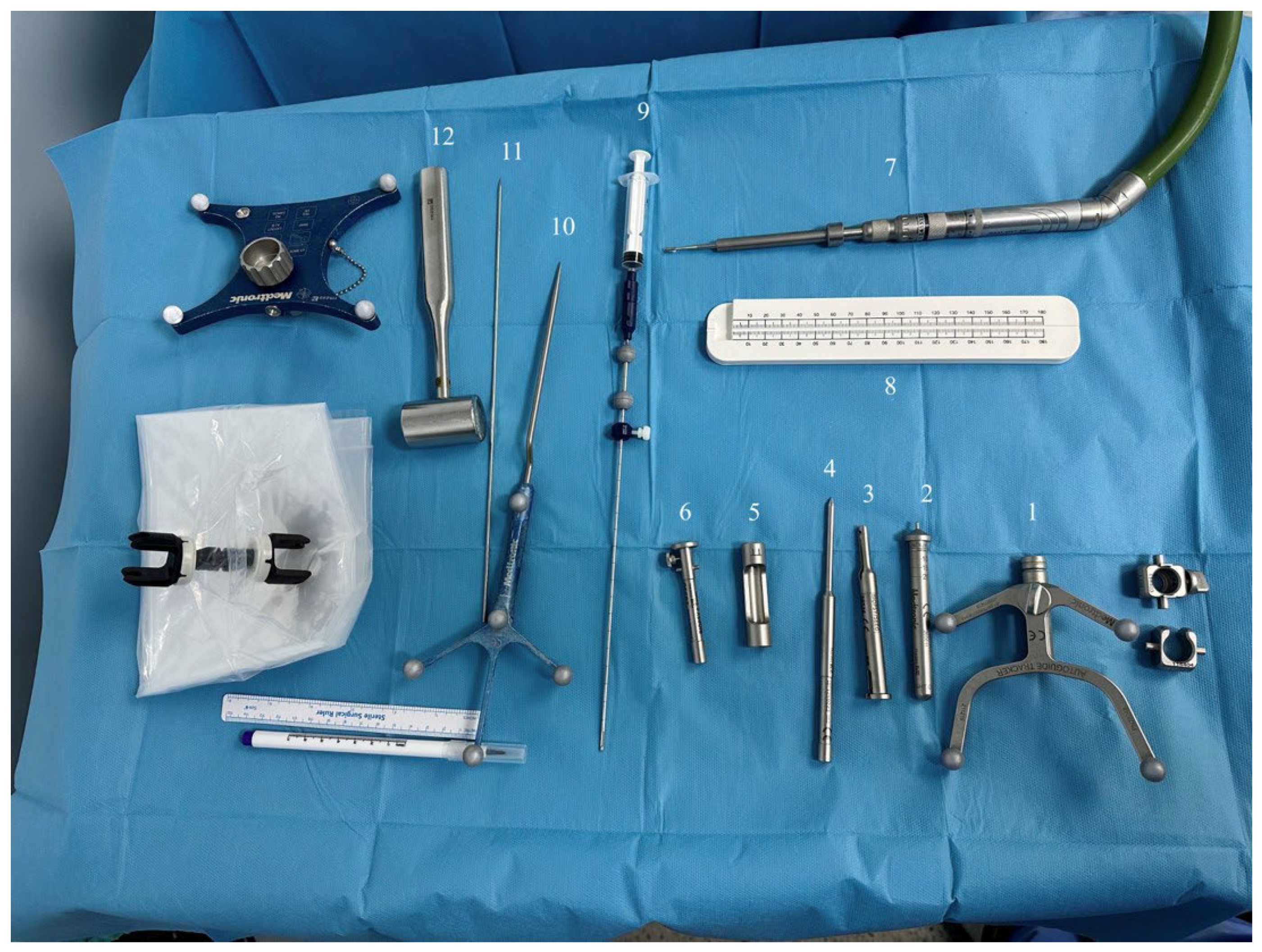
View of Surgical Materials for Autoguide Surgical Platform. 1- Stealth Autoguide Tracker 2- Visualase Height Guide 3- Autoguide Stab Incision Drill Guide 4- Autoguide Stab Incision Obturator 5- Autoguide tapping tube 6-Autoguide Reducing Tube 7- Midas Rex Drill 8- Measurement Tool 9- Autoguide Biopsy Needle With Syringe Attached 10- Navigation Probe 11-Kirschner Wire 12- Surgical Hammer.
Figure 1.
View of Surgical Materials for Autoguide Surgical Platform. 1- Stealth Autoguide Tracker 2- Visualase Height Guide 3- Autoguide Stab Incision Drill Guide 4- Autoguide Stab Incision Obturator 5- Autoguide tapping tube 6-Autoguide Reducing Tube 7- Midas Rex Drill 8- Measurement Tool 9- Autoguide Biopsy Needle With Syringe Attached 10- Navigation Probe 11-Kirschner Wire 12- Surgical Hammer.
The patient, Autoguide targeting unit and also Autoguide postitioning arm were draped in sterile fashion. The robotic system is manually repositioned by the surgeon to the planned entry point on the surface of the skin within a few centimeters and locked in place. Autoguide Stealth Tracker was verified at the navigation reference frame. Autoguide Height guide was flipped and reinserted into the Autoguide Stealth Tracker with the round tip facing down and locked. Autoguide Postitioning Arm was unlocked and patient’s scalp was touched with the round tip of the height guide.
Figure 2.
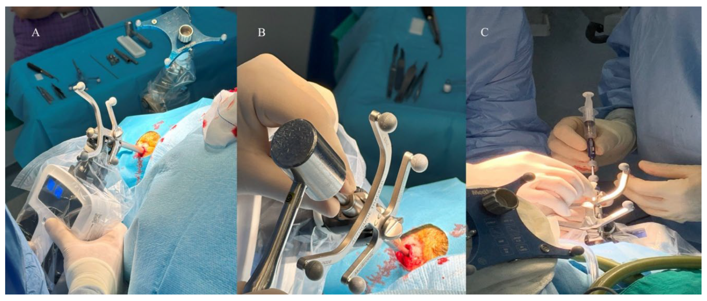
A-) Intraoperative view of Autoguide Robotic Platform. Visualase Height guide was reinserted into the Autoguide Stealth Tracker with the round tip facing down and locked. Autoguide Positioning Arm was unlocked and patient’s scalp was touched with the round tip of the height guide. B-) Anchoring the Stab incision Obturator guide at the skull C-) Moving the biopsy needle to the preoperatively defined target and obtaining tissue samples by using syringe.
Figure 2.
A-) Intraoperative view of Autoguide Robotic Platform. Visualase Height guide was reinserted into the Autoguide Stealth Tracker with the round tip facing down and locked. Autoguide Positioning Arm was unlocked and patient’s scalp was touched with the round tip of the height guide. B-) Anchoring the Stab incision Obturator guide at the skull C-) Moving the biopsy needle to the preoperatively defined target and obtaining tissue samples by using syringe.
When the symbols on the Autoguide targeting unit and Autoguide control unit indicate that the active surgical plan is within reach (dotted sphere touch the circle), positioning arm was locked and height guide was removed from the tracker. By using Autoguide control unit Autoguide Targeting Device was positioned automatically according to previously structured plan on navigation system.
The autoguide control and targeting unit were adjusted so that the dotted sphere was positioned inside the circle, as shown in Figure 3C. After aligning to the active surgical plan, deviation from the target point was detected on the navigation screen. 1 mm or less deviations were accepted in all cases.
Figure 3.
Intraoperative view of the Autoguide positioning arm and control unit A-) Autoguide positioning arm was adjusted to home position and locked by using ratchet locking mechanism on the Autoguide positioning arm. B-) Autoguide control unit indicate that the active surgical plan is within reach (dotted sphere touch the circle) C-) Autoguide control unit was checked and verified that dotted sphere was inside the circle.
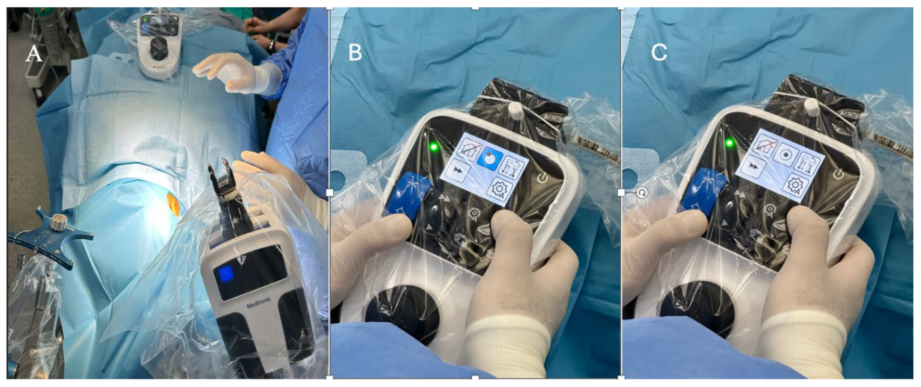
Figure 4.
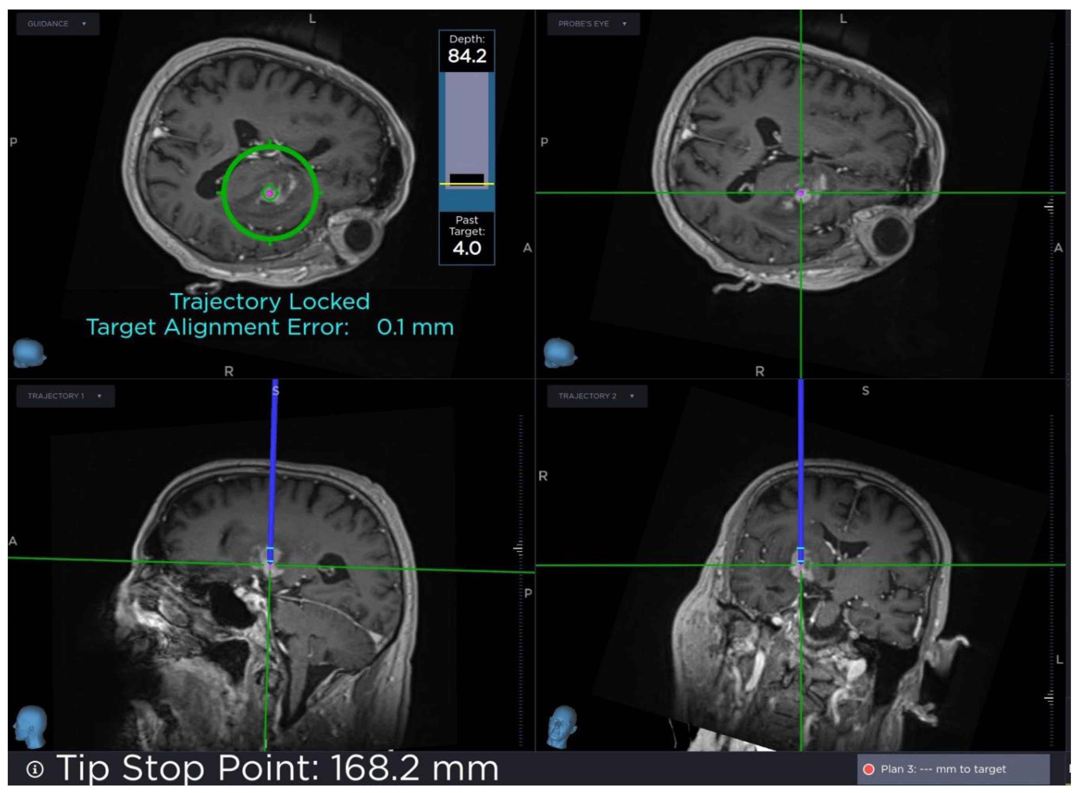
Heads-up display on the StealthStation monitor of real-time navigation information. Tip stop point and target alignment error was seen and customizable views of imaging sequences.
Figure 4.
Heads-up display on the StealthStation monitor of real-time navigation information. Tip stop point and target alignment error was seen and customizable views of imaging sequences.
Skin incision was made and Autoguide Stab Incision Drill guide was inserted into the Stealth Autoguide Tracker and tightened by using thumb screw on Stealth Autoguide Tracker. Autoguide Stab Incision Obturator was inserted into the Autoguide Stab Incision Drill Guide until it made contact with the skull. By using surgical hammer Stab Incision Obturator guide was anchored at the skull. Autoguide Stab Incision Drill Guide was unlocked and lowered until it made contact with the skull. Autoguide tapping tube was slided over the Autoguide then Stab Incision Obturator and the drill guide was hammered at the skull. Target Alignment Error was checked on the Stealth Station screen to determine whether the error is still within an acceptable range after hammering to anchor the drill guide. Midas Rex drill was inserted in the drill guide and stopper of the drill was adjusted according to tickness of the skull.
Figure 5.
Intraoperative view of Midas Rex drill which was inserted in the drill guide and stopper of the drill was adjusted according to tickness of the skull.
Figure 5.
Intraoperative view of Midas Rex drill which was inserted in the drill guide and stopper of the drill was adjusted according to tickness of the skull.
Preoperative CT scans were used to determine the tickness of the bone. Skull was drilled and dura was breached by using a kirschner wire. Autoguide reducing tube was inserted and biopsy needle was inserted into the reducing tube. Tip point and target alignment error were checked by using Stealth Navigation Software. Samples were examined under the microscope's yellow 560 filter after administering sodium fluorescein approximately 15 minutes before the procedure and it was confirmed that the biopsy was performed from the correct target. Furthermore, intraoperative pathologic examination confirmed that the target was accurate.
3.8. Frame-Based Stereotactic Biopsy Technique
Following induction of general anesthesia, patients were positioned supine and a Leksell stereotactic frame (Integra, CRW, New Jersey, USA) was applied to the skull using four fixation pins. After frame application, fiducial markers were attached to the frame and the patient underwent CT imaging. The CT images were used to establish a three-dimensional coordinate system relative to the frame. Preoperative MRI images were fused with CT images using navigation software (Brainlab AG, Munich, Germany) to optimize target localization. The CT images obtained after stereotactic frame application provided the coordinate reference system, while the MRI images offered superior lesion visualization and anatomical detail.
Target coordinates were calculated using the stereotactic planning software based on the lesion's location on CT imaging. (Figure 6)
The target was selected within contrast-enhancing regions when present, or based on T2/FLAIR hyperintensity for non-enhancing lesions. Entry point coordinates were calculated to provide the safest trajectory avoiding eloquent cortex, sulci, and major vascular structures.
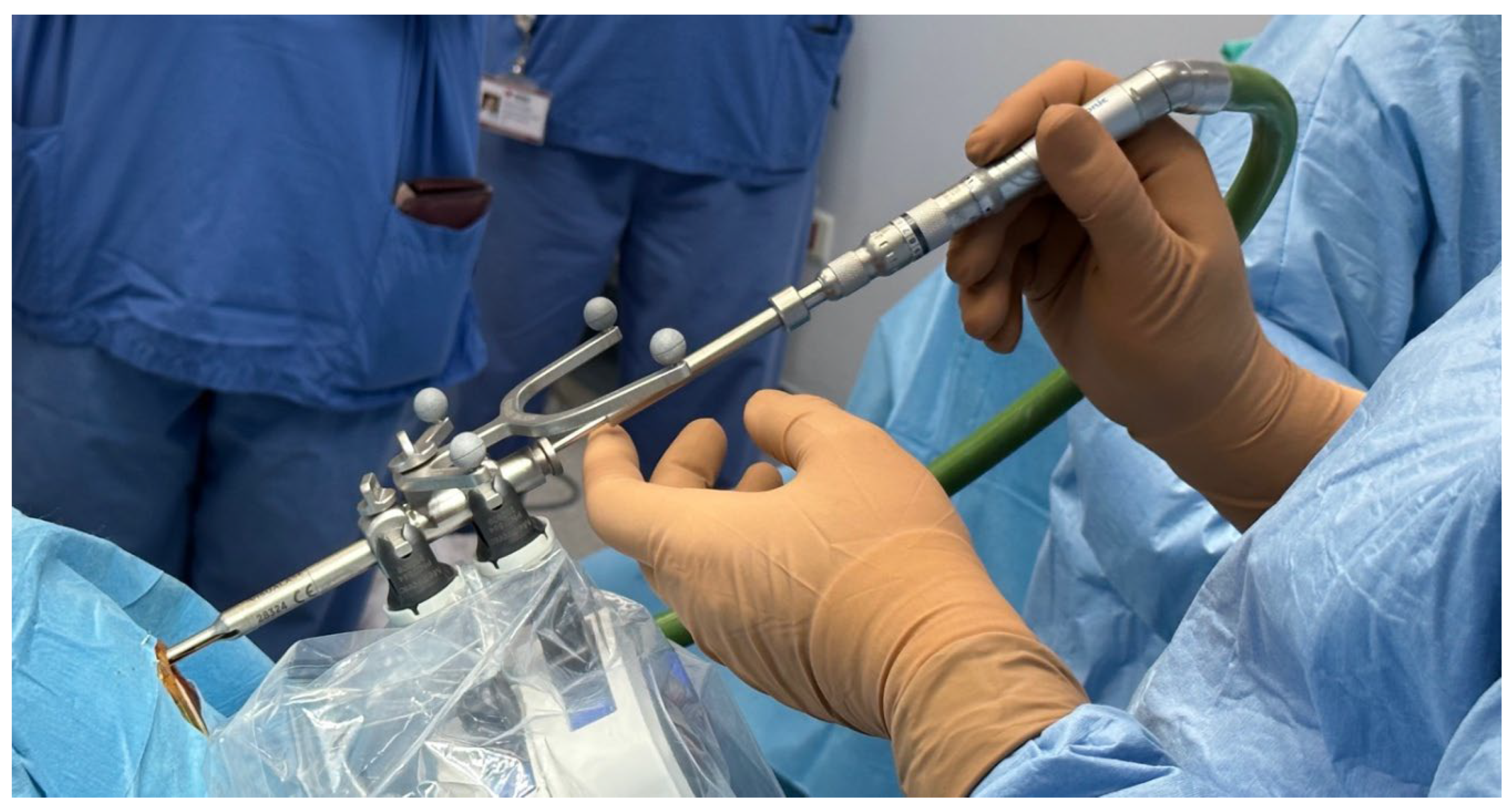
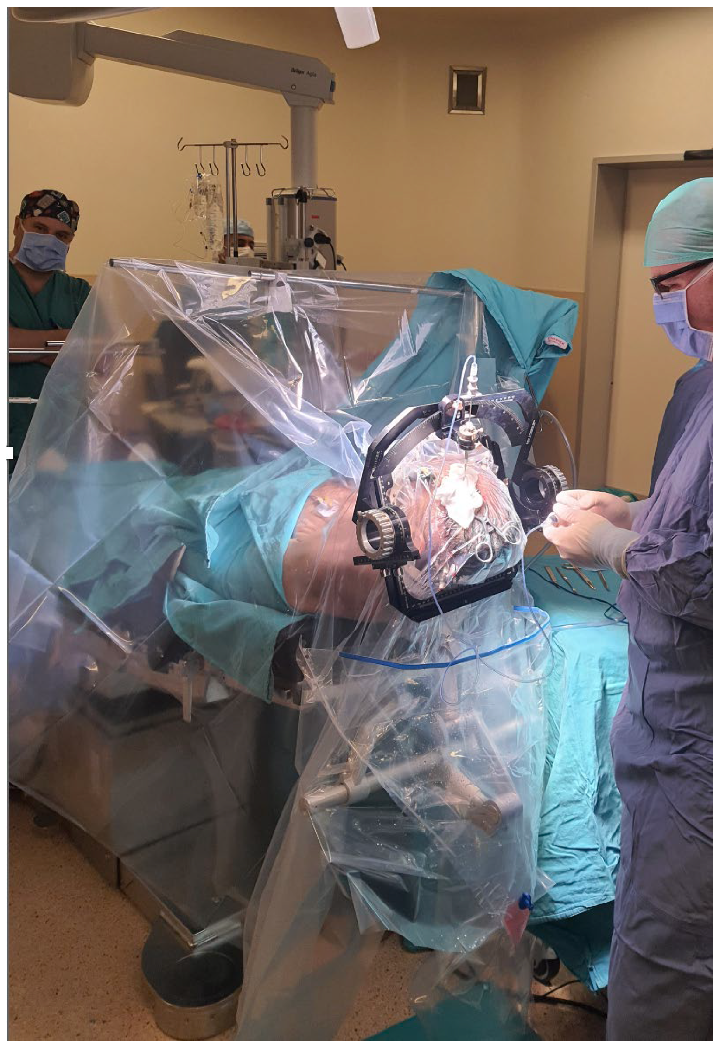
The patient was positioned on the operating table with the stereotactic frame secured to the table adapter. (Figure 7).
Using the calculated X, Y, and Z coordinates, the trajectory guide arc was mounted onto the stereotactic frame and adjusted to the predetermined coordinates. The entry site on the scalp was identified using the trajectory guide, and local anesthetic with epinephrine was infiltrated.
A small skin incision (approximately 3 cm) was made at the entry point, and a high speed drill (Medtronic, Minneapolis, MN, USA) was used to create a burr hole. The dura was coagulated and opened with a sharp needle. A side-cutting biopsy needle was introduced through the trajectory guide cannula and advanced to the calculated target.
Multiple tissue samples were obtained at target level (at least 4 samples were obtained if needed the needle was placed 1 mm above or below the target according to the target lesion for 4 more additional samples) and sent for frozen section examination and permanent histopathology. In all cases, immediate postoperative CT imaging was performed with the stereotactic frame still in place to measure actual needle trajectory and calculate targeting error by comparing achieved coordinates with planned coordinates. 1 milimeter or less accuracy was accepted during performing biopsy. The stereotactic frame was then removed, and the pin sites were dressed.
Figure 8.
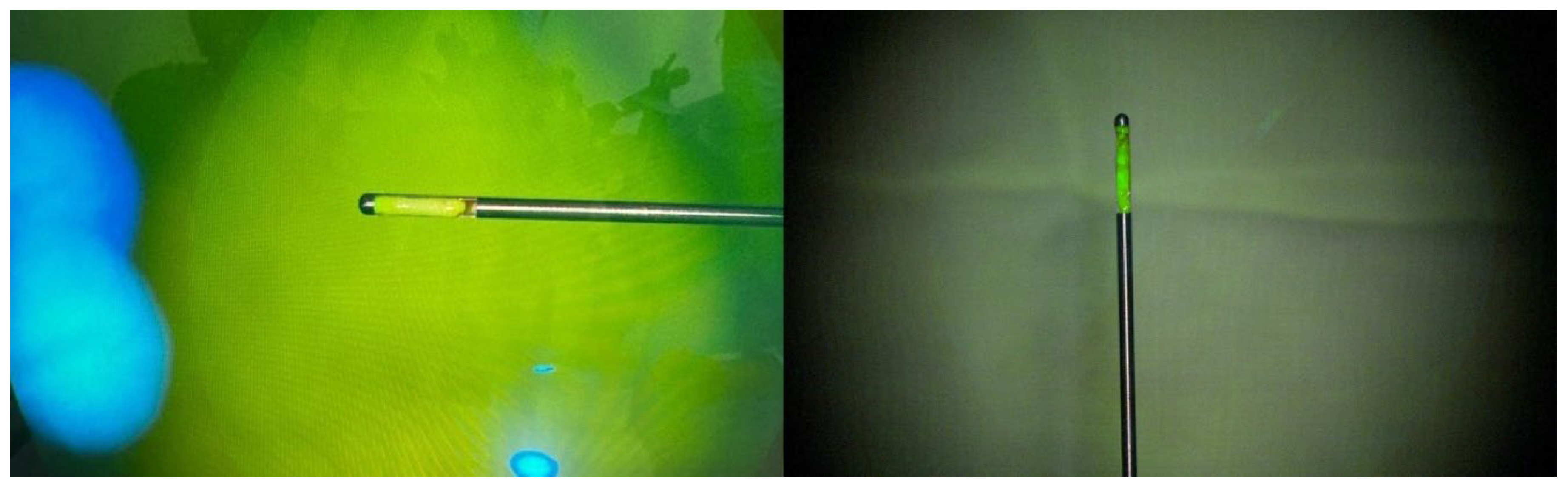
Intraoperative microscopic view of the specimens that were stained with sodium florescein.
Figure 8.
Intraoperative microscopic view of the specimens that were stained with sodium florescein.
4. Discussion
Many studies have been published in the literature regarding the reliability and usefulness of brain biopsy with stereotactic techniques [4,8,9]. Instead of the freehand biopsy technique, frame-based and frameless stereotactic and robot-assisted biopsy techniques have become more common today due to their increased safety and accuracy. With the advancement of robotic technology, frameless biopsy techniques have become more prominent for stereotactic biopsy compared to frame-based techniques [1,10,11]. Although frame-based techniques are considered the gold standard in stereotactic biopsy, frameless biopsy techniques have started to be preferred due to reasons such as the need for imaging after placing the frame, negatively impacting patient comfort, and prolonged surgical time [1]. Some of the most important advantages of robotic surgery compared to frame-based stereotactic procedures include automatic positioning of the robotic platform to the target, which helps prevent errors that may arise due to the manually controlling the tracking device and trajectory guide [12]. The Autoguide Robotic Platform is a suitable option for intracranial biopsy due to its small robotic arm, which occupies much less space in the operating room compared to similar robots, and its high accuracy and precision. The Autoguide Robotic Platform is a system with high maneuverability that can quickly respond to changes in the navigation plan and has a short learning curve. Unlike other robotic devices which have larger robotic arms, it does not require adjusting the robot to a reference point before making changes on the plan [2].
The most significant finding of our comparative analysis is the substantial difference in procedural efficiency between the two techniques. The robotic-biopsy group demonstrated significantly shorter mean surgical time (40.26±6.13 vs 52.47±8.92 minutes, p=0.002), representing approximately 23% reduction in operative time. Consistent with our findings, Dhawan et al.'s meta-analysis of 15 studies involving 2,400 patients found no significant difference in diagnostic yields between frame-based and frameless biopsy groups, but significantly shorter surgical time in the frameless group [5]. These time differences reflect fundamental procedural distinctions between the two techniques. Frame-based stereotactic biopsy requires several sequential steps that contribute to prolonged operative time [11]. Following induction of general anesthesia, the stereotactic frame must be rigidly fixed to the skull and after frame application, the patient must undergo CT imaging with stereotactic frame to establish the three-dimensional coordinate system. Subsequently, target coordinates must be manually calculated, entry points determined, and the trajectory arc manually adjusted to align with the calculated X, Y, and Z coordinates [13]. Each of these steps is sequential and time-dependent, with no opportunity for parallel workflow. In contrast, the Autoguide Robotic Platform features automatic positioning to the target, eliminating errors that may arise from manually controlling trajectory guides in frame-based procedures [12]. The small robotic arm occupies minimal operating room space compared to larger robotic systems, and its ergonomic design facilitates rapid setup and workflow. Unlike other robotic devices with larger arms, the Autoguide system does not require adjusting to a reference point before making trajectory changes, contributing to its efficiency and short learning curve [2]. Mean surgical time for intracranial biopsy varies between 30 to 127 minutes in the literature [12,14,15]. In our study, mean surgical time was 40.26+- 5.92 (range:38-55 minutes). The use of a small incision in surgery, the robot's ability to quickly and automatically position itself according to the navigation plan, and the real-time tracking of the biopsy needle's correct position in navigation can be cited as factors that contribute to shortening the surgery.
Postoperative hemorrhage is one of the significant complications of stereotactic biopsy, and it has been reported in the literature to occur in 2% to 5% of cases. It can be asymptomatic in 20% to 25% of patients [16,17]. In robotic biopsy group, one patient with a thalamic lesion (6.7%; 95% CI: 1.2%-29.8%) experienced an intraventricular hemorrhage detected on postoperative imaging and in frame-based biyopsy group one patient (6.7%; 95% CI: 1.2%-29.8%) experienced asymptomatic intralesional hemorrhage detected on postoperative imaging. This complication was asymptomatic and required no additional treatment or intervention.
In the robotic-biopsy group, we did not postoperatively determine accuracy through a secondary method because our diagnostic biopsy rate was 93.3%, indicating that the robot is accurate in comparison to preoperative plans based on neuroradiological imaging. As we considered the lesion to be an abscess based solely on preoperative imaging and clinical findings, the sample obtained from one patient was sent for microbiological examination instead of pathological analysis; therefore, this patient was not included in our calculation of the diagnostic biopsy rate. Following microbiological examination, the diagnosis of abscess was confirmed, and after appropriate antibiotic treatment, follow-up imaging showed near-complete regression of the lesion and improvement in the patient's symptoms.
In the study published by Kresuoulas et al., which represents one of the largest series in the literature involving biopsies performed using the Autoguide robotic platform, a diagnostic biopsy rate of 100% was reported. The authors emphasized that postoperative imaging is not required to confirm diagnostic accuracy and is not considered cost-effective.[2] It should be noted that it is not possible to determine true accuracy in real time without performing an intraoperative MRI or CT scan with the biopsy cannula in place. However, in clinical practice with Autoguide Robotic Platform, we believe obtaining intraoperative imaging is not required and is significantly more costly for the patients. In the robotic-biopsy group, we tracked the real-time position of the biopsy needle using the navigation software during the surgery and based our calculations on the needle tip error value computed by the navigation system. In the robotic biopsy group, intraoperative imaging was used for one patient. A biopsy was taken from a patient with a left temporal lesion in the prone position, by using the O-arm.
In the literature, the failure rate of stereotactic intracranial biopsies ranges from 2% to 10%, and it is an important issue to address as it necessitates repeat biopsies [7,18,19]. Fluorescent dyes, such as 5-aminolevulanic acid, indocyanine green, and sodium fluorescein, are substances used in microsurgery and stereotactic biopsies [20,21,22]. In our study, for lesions that were partially or entirely contrast-enhancing, the contrast-enhancing part of the lesion was targeted, and a biopsy was taken using Autoguide Robotic Platform. The biopsy specimen was then examined under the microscope with the yellow 560 filter, and the staining with sodium fluorescein confirmed that the biopsy was taken from the appropriate location. In their study, Thien and his colleagues reported that the use of sodium fluorescein had a positive predictive value of 100% and a negative predictive value of 25% in confirming the biopsy specimen by using stereotactic frame [23]. Mallereau reported in the literature that the inconclusive biopsy range is %2,6-11 in frameless biopsies and %0.7-15.7 in frame-based interventions. Atai published a retrospective review of 75 intracranial biopsy cases by using Autoguide Robotic Platform and diagnostic tissue was obtained in %100 of cases [24]. In 14 out of 15 patients intraoperative frozen section pathological examination revealed that all samples were taken from the preoperatively desired target. The lesion which was defined as an abcess didn’t send to pathology but the samples were cultured for defining the microorganism and deciding the suitable medical therapy. Our diagnostic yield was 93,33%. In our study 13 out of 15 specimens were stained with sodium fluorescein intraoperatively. The pathology of one of the lesion that did not stain with sodium fluorescein was determined to be cerebritis and also we couldn’t stain the lesion which was defined as abscess with sodium fluoresce because of its liquid nature. We would like to emphasize the importance of this study as it is the first in the literature to use sodium fluorescein for confirmation in intracranial biopsies taken with the Autoguide robot and to demonstrate that it improves biopsy accuracy. We believe that using sodium fluorescein staining is essential after robotic biopsy in order to increase the diagnostic yield and to confirm that biopsy was taken from the correct target.
Our study demonstrates that although frame-based stereotactic biopsy has been accepted as the gold standard in the literature for years, robotic-assisted biopsy, augmented with intraoperative sodium fluorescein, represents a safe and reliable alternative method with comparable diagnostic yield, targeting accuracy, and complication rates. Its significantly shorter surgical time and preoperative preparation time, while maintaining similar efficacy, constitute important advantages.
This study has several limitations that warrant acknowledgment. First, the retrospective design from two centers may introduce patient selection bias and institutional variability in protocols. Although procedures were performed by two experienced surgeons, inter-surgeon and inter-institutional variability in technique and decision-making may have influenced outcomes. Second, the sample size limits statistical power for detecting small differences in diagnostic yield or complication rates. Third, intraoperative sodium fluorescein was used only in the Robotic-Biopsy Group, precluding assessment of its independent contribution to diagnostic success separate from the robotic platform itself. As this is a preliminary comparative study, prospective randomized trials with larger cohorts, standardized protocols across institutions, and comprehensive cost-effectiveness analyses are required to confirm these findings and establish evidence-based guidelines for technique selection.
5. Conclusions
Robotic-assisted and frame-based stereotactic biopsy techniques achieve comparable targeting accuracy, diagnostic accuracy, and safety profiles with similar complication rates. However, the robotic biopsy group demonstrated significantly shorter surgical time compared to the frame-based biopsy group. The automated positioning capabilities of the robotic platform contribute to improved procedural efficiency and reduced anesthesia exposure. The use of intraoperative sodium fluorescein is a valuable adjunct technique for confirming that biopsy specimens are obtained from the intended target lesion. Both techniques represent safe and effective options for sampling deep-seated intracranial lesions, with technique selection guided by institutional resources and surgeon experience.
Author Contributions
Conceptualization, Utku Özgen, Mehmet Zeki Yıldız, Mehmet Osman Akçakaya; methodology, Talat Kırış.; software, -.; validation, Utku Özgen, Mehmet Zeki Yıldız, Mehmet Osman Akçakaya formal investigation Talat Kırış, Mehmet Osman Akçakaya ;resources, Utku Özgen, Mehmet Zeki Yıldız.; data curation, Utku Özgen; writing—original draft preparation, Utku Özgen writing—review and editing, Talat Kırış, Mehmet Osman Akçakaya; visualization, Talat Kırış.; supervision, Talat Kırış,Mehmet Osman Akçakaya; project administration, -.; funding acquisition, - All authors have read and agreed to the published version of the manuscript.
Funding
This research received no external funding.
Institutional Review Board Statement
According to the relevant regulations and laws of our local ethics committee, the retrospective non-experimental study of ‘Clinical Experience in Frameless Stereotactic Biopsy by using Stealth Autoguide Robotic Platform and Intraoperative Sodium Fluorescein’ was conducted based on data obtained without any additional processing, and therefore no ethics committee approval was required. Formal approval by the institutional ethics committee was not required because the study was based on secondary analyses of appropriately anonymized datasets of administrative data.
Informed Consent Statement
Written informed consent was obtained from all patients involved in the study to publish this paper.
Data Availability Statement
Study data is unavailable due to privacy and ethical restrictions of our hospital.
Acknowledgments
None.
Conflicts of Interest
The authors declare no conflicts of interest.
Abbreviations
CT: Computerised Tomography, MRI: Magnetic- Resonance Imaging, LITT: Laser interstitial Thermal Therapy.
References
- Sciortino, T.; Fernandes, B.; Conti Nibali, M.; Gay, L.G.; Rossi, M.; Lopci, E.; Colombo, A.E.; Elefante, M.G.; Pessina, F.; Bello, L.; et al. Frameless stereotactic biopsy for precision neurosurgery: diagnostic value, safety, and accuracy. Acta Neurochir (Wien) 2019, 161, 967-974. [CrossRef]
- Kreatsoulas, D.C.; Vignolles-Jeong, J.; Ambreen, Y.; Damante, M.; Akhter, A.; Lonser, R.R.; Elder, J.B. Surgical Characteristics of Intracranial Biopsy Using a Frameless Stereotactic Robotic Platform: A Single-Center Experience. Oper Neurosurg (Hagerstown) 2023. [CrossRef]
- Amin, D.V.; Lozanne, K.; Parry, P.V.; Engh, J.A.; Seelman, K.; Mintz, A. Image-guided frameless stereotactic needle biopsy in awake patients without the use of rigid head fixation. J Neurosurg 2011, 114, 1414-1420. [CrossRef]
- Dammers, R.; Haitsma, I.K.; Schouten, J.W.; Kros, J.M.; Avezaat, C.J.; Vincent, A.J. Safety and efficacy of frameless and frame-based intracranial biopsy techniques. Acta Neurochir (Wien) 2008, 150, 23-29. [CrossRef]
- Dhawan, S.; He, Y.; Bartek, J., Jr.; Alattar, A.A.; Chen, C.C. Comparison of Frame-Based Versus Frameless Intracranial Stereotactic Biopsy: Systematic Review and Meta-Analysis. World Neurosurg 2019, 127, 607-616.e604. [CrossRef]
- Lu, Y.; Yeung, C.; Radmanesh, A.; Wiemann, R.; Black, P.M.; Golby, A.J. Comparative effectiveness of frame-based, frameless, and intraoperative magnetic resonance imaging-guided brain biopsy techniques. World Neurosurg 2015, 83, 261-268. [CrossRef]
- Woodworth, G.F.; McGirt, M.J.; Samdani, A.; Garonzik, I.; Olivi, A.; Weingart, J.D. Frameless image-guided stereotactic brain biopsy procedure: diagnostic yield, surgical morbidity, and comparison with the frame-based technique. J Neurosurg 2006, 104, 233-237. [CrossRef]
- Lunsford, L.D.; Niranjan, A.; Khan, A.A.; Kondziolka, D. Establishing a benchmark for complications using frame-based stereotactic surgery. Stereotact Funct Neurosurg 2008, 86, 278-287. [CrossRef]
- McGirt, M.J.; Woodworth, G.F.; Coon, A.L.; Frazier, J.M.; Amundson, E.; Garonzik, I.; Olivi, A.; Weingart, J.D. Independent predictors of morbidity after image-guided stereotactic brain biopsy: a risk assessment of 270 cases. J Neurosurg 2005, 102, 897-901. [CrossRef]
- Hu, Y.; Cai, P.; Zhang, H.; Adilijiang, A.; Peng, J.; Li, Y.; Che, S.; Lan, F.; Liu, C. A Comparation Between Frame-Based and Robot-Assisted in Stereotactic Biopsy. Front Neurol 2022, 13, 928070. [CrossRef]
- Dorward, N.L.; Paleologos, T.S.; Alberti, O.; Thomas, D.G. The advantages of frameless stereotactic biopsy over frame-based biopsy. Br J Neurosurg 2002, 16, 110-118. [CrossRef]
- Bekelis, K.; Radwan, T.A.; Desai, A.; Roberts, D.W. Frameless robotically targeted stereotactic brain biopsy: feasibility, diagnostic yield, and safety. J Neurosurg 2012, 116, 1002-1006. [CrossRef]
- Vadera, S.; Chan, A.; Lo, T.; Gill, A.; Morenkova, A.; Phielipp, N.M.; Hermanowicz, N.; Hsu, F.P. Frameless Stereotactic Robot-Assisted Subthalamic Nucleus Deep Brain Stimulation: Case Report. World Neurosurg 2017, 97, 762.e711-762.e714. [CrossRef]
- Yasin, H.; Hoff, H.J.; Blümcke, I.; Simon, M. Experience with 102 Frameless Stereotactic Biopsies Using the neuromate Robotic Device. World Neurosurg 2019, 123, e450-e456. [CrossRef]
- Lefranc, M.; Capel, C.; Pruvot-Occean, A.S.; Fichten, A.; Desenclos, C.; Toussaint, P.; Le Gars, D.; Peltier, J. Frameless robotic stereotactic biopsies: a consecutive series of 100 cases. J Neurosurg 2015, 122, 342-352. [CrossRef]
- Mizobuchi, Y.; Nakajima, K.; Fujihara, T.; Matsuzaki, K.; Mure, H.; Nagahiro, S.; Takagi, Y. The risk of hemorrhage in stereotactic biopsy for brain tumors. J Med Invest 2019, 66, 314-318. [CrossRef]
- Riche, M.; Marijon, P.; Amelot, A.; Bielle, F.; Mokhtari, K.; Chambrun, M.P.; Joncour, A.L.; Idbaih, A.; Touat, M.; Do, C.H.; et al. Severity, timeline, and management of complications after stereotactic brain biopsy. J Neurosurg 2022, 136, 867-876. [CrossRef]
- Smith, J.S.; Quiñones-Hinojosa, A.; Barbaro, N.M.; McDermott, M.W. Frame-based stereotactic biopsy remains an important diagnostic tool with distinct advantages over frameless stereotactic biopsy. J Neurooncol 2005, 73, 173-179. [CrossRef]
- Ulm, A.J.; Bova, F.J.; Friedman, W.A. Stereotactic biopsy aided by a computer graphics workstation: experience with 200 consecutive cases. Surg Neurol 2001, 56, 366-371; discussion 371-362. [CrossRef]
- Hefti, M.; von Campe, G.; Moschopulos, M.; Siegner, A.; Looser, H.; Landolt, H. 5-aminolevulinic acid induced protoporphyrin IX fluorescence in high-grade glioma surgery: a one-year experience at a single institutuion. Swiss Med Wkly 2008, 138, 180-185. [CrossRef]
- Li, C.; Sullivan, P.Z.; Cho, S.; Nasrallah, M.P.; Buch, L.; Isaac Chen, H.C.; Lee, J.Y.K. Intraoperative Molecular Imaging with Second Window Indocyanine Green Facilitates Confirmation of Contrast-Enhancing Tissue During Intracranial Stereotactic Needle Biopsy: A Case Series. World Neurosurg 2019, 126, e1211-e1218. [CrossRef]
- Catapano, G.; Sgulò, F.G.; Seneca, V.; Iorio, G.; de Notaris, M.; di Nuzzo, G. Fluorescein-assisted stereotactic needle biopsy of brain tumors: a single-center experience and systematic review. Neurosurg Rev 2019, 42, 309-318. [CrossRef]
- Thien, A.; Han, J.X.; Kumar, K.; Ng, Y.P.; Rao, J.P.; Ng, W.H.; King, N.K.K. Investigation of the usefulness of fluorescein sodium fluorescence in stereotactic brain biopsy. Acta Neurochir (Wien) 2018, 160, 317-324. [CrossRef]
- Atai, N.A.; Mehta, V.A. Initial United States experience with Medtronic Stealth Autoguide cranial robotic guidance platform. J Neurosurg 2024, 1-7. [CrossRef]
Figure 6.
Stereotactic trajectory planning using BrainLab navigation software. The target lesion is outlined in orange with a planned trajectory (yellow line) shown in all three orthogonal planes. The right panel displays the trajectory coordinates including anterior-posterior (A-P), lateral, and vertical measurements, along with ring and arc angle settings for frame-based stereotactic guidance. The software allows precise three-dimensional localization and optimal trajectory selection to minimize neurovascular structures along the approach.
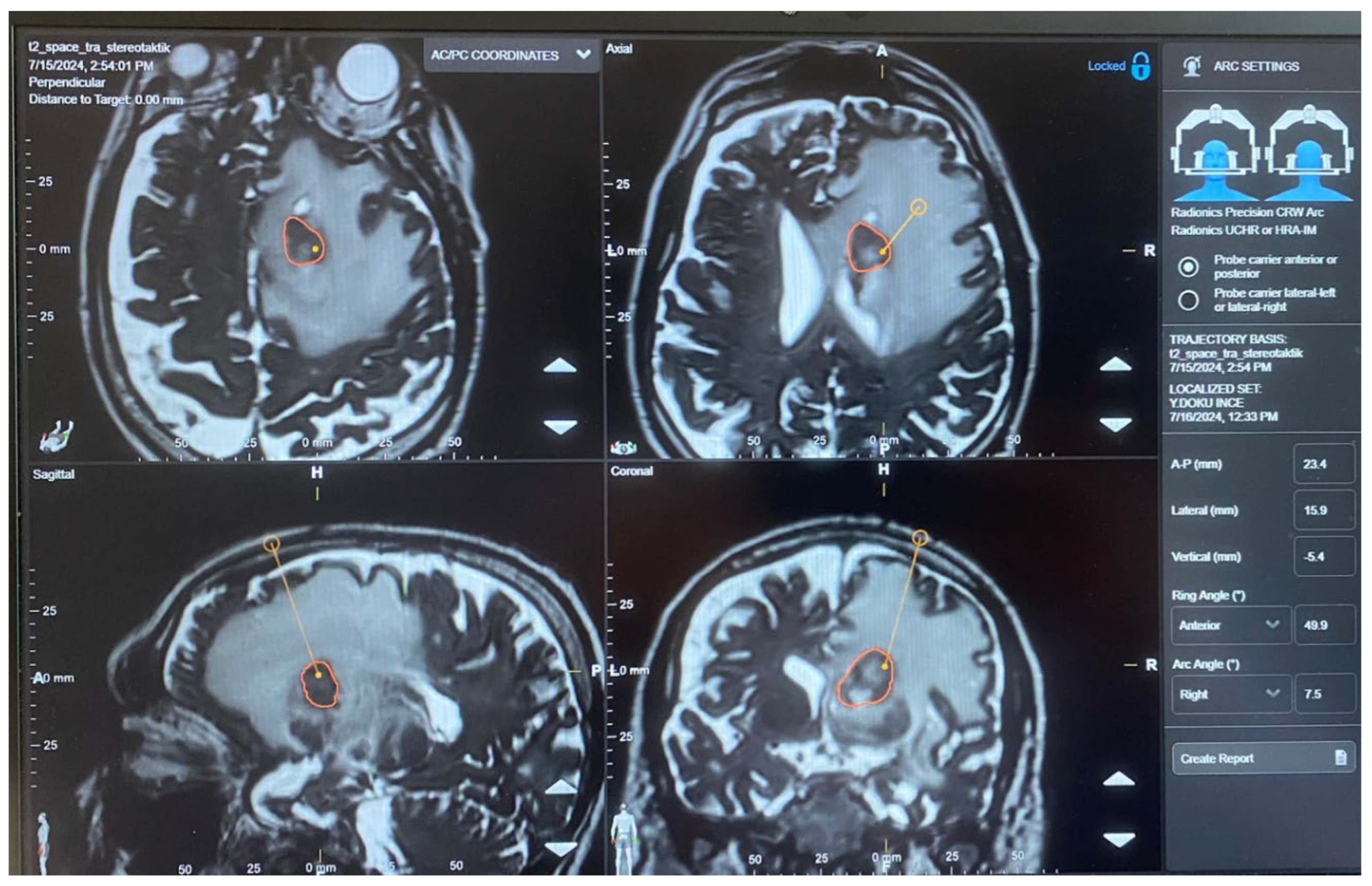
Figure 7.
Intraoperative view of frame-based stereotactic brain biopsy procedure. Demonstrates the operative setup during a frame-based stereotactic brain biopsy. The stereotactic frame is secured to the patient's head, providing a rigid reference system for precise target localization. The surgical team operates under standard neurosurgical lighting with stereotactic guidance for accurate needle trajectory planning and execution.
Disclaimer/Publisher’s Note: The statements, opinions and data contained in all publications are solely those of the individual author(s) and contributor(s) and not of MDPI and/or the editor(s). MDPI and/or the editor(s) disclaim responsibility for any injury to people or property resulting from any ideas, methods, instructions or products referred to in the content. |
© 2026 by the authors. Licensee MDPI, Basel, Switzerland. This article is an open access article distributed under the terms and conditions of the Creative Commons Attribution (CC BY) license (http://creativecommons.org/licenses/by/4.0/).
Copyright: This open access article is published under a Creative Commons CC BY 4.0 license, which permit the free download, distribution, and reuse, provided that the author and preprint are cited in any reuse.


