
Submitted:
16 January 2026
Posted:
19 January 2026
You are already at the latest version
Abstract
Microplastics (MPs) have become a widespread environmental contaminant, raising concern due to their persistence, capacity to transport pollutants, and potential risks to ecosystems and human health. Their increasing global production, prolonged degra-dation, and ubiquity in aquatic environments underscore the need for improved strategies for monitoring and mitigation. This review examines the definition, sources, environmental transport mechanisms, associated risks, and current detection methods for MPs in natural and engineered water systems. The methods discussed encompass a broad range of analytical and sensing technologies used to identify, characterize, and quantify MPs across diverse environmental matrices. The review highlights that no single technique is sufficient for comprehensive MP analysis; instead, the combination of multiple methods enhances sensitivity, specificity, and reliability. Current findings indicate widespread MP contamination, including within the human body, emphasizing significant ecological and health concerns. Progress in automated sample preparation, standardized protocols, and advanced sensing platforms is key to improving detection efficiency and comparability across different studies. Overall, the evidence presented supports the need for strengthened monitoring, continued technological innovation, and coordinated mitigation policies. Reducing MP pollution will require interdisciplinary collaboration, regulatory action, and increased public awareness to protect environ-mental integrity and human health.
Keywords:
microplastics
; environmental pollutants
; drinking water
; detection methods
; human exposure
1. Introduction
Microplastics (MPs) have garnered increasing attention in recent years due to their pervasive nature in ecosystems and potential threat to environmental and human health. Although plastics are essential in modern society [1], the growing problem of plastic pollution is challenging to address and solve [2]. In fact, plastic production and use are increasing annualy and reached an annual quantity of 368 million metric tons in 2019 [3] with a global polymer market valued at $533.6 billion and estimated to hit $838.5 billion by 2030 [4]. Plastic usage and waste grow year by year, while degradation processes are prolonged due to the poor water solubility and non-biodegradability of plastic polymers. Plastics can remain in the environment for centuries before being completely decomposed by photodegradation and thus re-entering normal biogeochemical cycles, particularly in the marine environment [5]. In addition, when plastics break down to a small enough form factor, they can be consumed by various marine life [6]. These plastic fragments can also combine with other pollutants, such as organic contaminants, antibiotics, heavy metals, and bacterial pathogens, thereby influencing and possibly enhancing the transport behaviour of environmental contamination. For these reasons, the problem of pollution caused by plastics has been recognized as the cause of deaths of many marine birds, mammals, and fish by entrapment or ingestion [7], as well as the origine of the creation of real plastic islands. It was reported by the UK Government in 2018 that 100,000 marine mammals and turtles and 1 million sea birds are killed by marine plastic pollution every year [8]. Indeed, due to the ubiquity of plastic waste and various transport processes, this form of contamination impacts all types of aquatic ecosystems and water bodies, including resources of drinking water [9]. This global high-level problem needs comprehensive and coordinated strategies that integrate prevention, monitoring, and remediation approaches across multiple scales. In particular, the early detection and quantification of microplastics in natural and engineered water systems have emerged as critical steps in understanding their distribution, assessing associated risks, and informing policy decisions. However, current analytical methods are often labor-intensive, time-consuming, and require specialized laboratory facilities, which limits large-scale and real-time monitoring efforts. Consequently, there is a growing interest in the development of advanced sensing technologies capable of enabling rapid, sensitive, and reliable detection of microplastics in complex environmental matrices. These innovations are essential not only for improving the assessment of microplastic pollution but also for supporting effective mitigation strategies and guiding future regulations aimed at reducing the ecological and human health impacts associated with plastic contamination.
In the following sections, we will describe the definition of MPs, their sources, the mechanisms through which they are transported across different environments down to water, the associated risks and the detection methods.
2. Definition of Microplastics
The term "microplastics" was introduced in 2004 by Professor Richard Thompson, a marine biologist at the University of Plymouth in the United Kingdom [10]. Then the National Oceanic and Atmospheric Administration, NOAA, defined in 2009 an upper size limit: “Pieces of plastic particles smaller than 5 mm” [11]. In 2015 the Joint Group of Experts on the Scientific Aspects of Marine Environmental Protection (GESAMP), added a lower limit, including for the first time, nanoplastics (down to 1 nm): Microplastics are particles in the size range from 1 nm to 5 mm [12]. The European Chemical Agency (ECHA) in their Annex XV Restriction Report on Intentionally Added Microplastics of August 2019 provides an additional constraint for fibers: a length of 3 nm ≤ x ≤ 15 mm and length-to-diameter ratio of >3 [13]. However, whether to include nanoscale in the definition of MP is always under debate. The most recent, all-inclusive and more descriptive definition of MP is provided by Joao and Roisin in 2019 “Microplastics are any synthetic solid particle or polymeric matrix, with regular or irregular shape and with size ranging from 1 μm to 5 mm, of either primary or secondary manufacturing origin, which are insoluble in water” [14]. Figure 1 gives an overview of plastic debris based on size in scientific studies and reports from various organizations [15]. Table 1 reports some examples of primary and secondary MPs.
3. Sources and Transport
3.1. Sources
As a focus of today's environmental problems, the prevalence of MPs in daily life has attracted widespread attention. Research has been conducted to collect and analyze MPs from air [16,17], freshwater [18,19], seawater [20,21], soil [22,23,24], wastewater [25,26,27], sediment [19,28,29] and even Antarctic snow [30]. MPs come from a variety of sources that are ubiquitous in our living environment, from household items to industrial products, almost everywhere. The major sources of MPs are shown in Figure 2. According to the reference [31], MPs release pathways include (a) human (e.g., synthetic textiles, personal care products), (b) transportation (e.g. erosion of synthetic rubber tires), and (c) industrial (e.g. plastic pellets) sources as shown in Figure 3.
Within this framework, MPs can be divided into two major categories: primary MPs, which are originally manufactured at small sizes, and secondary MPs, which result from breakdown, degradation and weathering of larger plastic debris. The most common compositions of MPs released in the environment are polyethylene (PE),polyethylene terephthalate (PET), polypropylene (PP), polyester (PES), polypropylene (PP), polystyrene (PS), polyvinylchloride (PVC), polyamide (PA) and polymethyl methacrylate (PMMA)
Primary MPs are intentionally manufactured for industrial or domestic use and are directly released into the environment as small plastic particles or microbeads. They are commonly found in personal care and cosmetic products (PCCPs) such as facial cleansers, shower gels, toothpaste, and cosmetics to provide a cleaning or polishing effect [32,33,34,35,36]. They are also used in industrial processes such as sandblasting, where plastic particles (PE and PP) are employed as an abrasive medium [37,38]. Despite efforts to regulate the use of microbeads in several countries, their presence in marine environments remains significant due to the lack of universal regulation.
Secondary MPs arise from the fragmentation of larger plastic items due to physical, chemical, or biological processes. often induced by weathering, such as photodegradation from ultraviolet (UV) light, heat, mechanical abrasion, and biodegradation.In all these processes, a significant role is played by the semicrystalline nature of 70% of commercial polymers, which are made of alternating crystalline and amorphous layers [39]. This leads to the presence of MPs in different ecosystems and thus even in food and water supplies. Common sources of secondary MPs include plastic bottles, fishing nets, fishing bait boxes, bags, and other items that deteriorate over time in natural environments [40,41,42]. Plastic packaging is one of the important sources of MPs, especially food and beverage packaging. Clothing made of synthetic fabrics like polyester, nylon, and spandex is also a major source of MPs. These fabrics shed microfibers during washing, wearing, and drying, releasing significant amounts of secondary MPs into water bodies and the environment [43,44,45]. A single load of laundry can release thousands of microfibers, which contribute significantly to MP pollution in aquatic environments [46].
MPs vary in density, color, chemical properties, shape and size, all of which influence their behavior in the environment. As regards chemical composition, 90% of MPs are composed of common polymers such as polyethylene (PE), polypropylene (PP), polystyrene (PS), and polyvinyl chloride (PVC) [47,48,49]. Table 2 provides information on products from daily life made of these polymers, along with their recycling details. The morphology of MPs was usually divided into fibers, fragments, films, pellets, foams, and others. Fibers and fragments account for more than 80% of the total MPs [50] and have extensive sources. The fibers are usually generated from washing garments and other textiles or the shedding of textile materials such as synthetic textiles, fishing tools, and woven bags. The fragments mainly come from the fragmentation and degradation of larger plastics in the environment [18]. Plastic mulch films have been identified as a primary source of film-shaped MPs. In fact, the environmental degradation of plastic mulch films makes them a major source of secondary MPs [51]. MP pellets are frequently used in cosmetic scrubbers or plastic production, such as facial cleansers and toothpaste [52]. Additionally, foam plastics primarily originate from fragmented lightweight foam materials for packaging and shipping floats [53]. Due to their higher specific surface area and lower density, foams are considered easily suspended within aquatic systems.
These plastic particles enter the environment through various pathways, including stormwater and wastewater, with wastewater treatment processes contributing to their partial removal. The small size of MPs allows them to be ingested by a wide range of organisms, from plankton to fish and birds, thus introducing them into the food chain. The ubiquity of MPs raises concerns due to their potential risks to humans and the environment, necessitating further research to establish regulations and management strategies.
3.2. Transport
MPs originate from a wide range of sources, both land-based and marine. These sources contribute to MP pollution through various pathways, including direct emissions and indirect inputs via rivers, runoff, and atmospheric deposition. Once MPs are released into the environment, their transport and distribution are influenced by a variety of physical, chemical, and biological processes. These include riverine transport, ocean currents, wind, and even the ingestion and excretion by organisms. Most MPs will accumulate in the ocean through river transport and direct discharge.
3.2.1. Riverine Transport
The majority of MP pollution is thought to originate from terrestrial sources, including urban, industrial, and agricultural activities. Land-based sources are responsible for approximately 80% of MPs that enter marine environments [54]. Rivers act as major conduits for transporting MPs from land-based sources to marine environments. They collect plastic debris from urban runoff, wastewater effluents, and stormwater systems, carrying them downstream to coastal areas. The concentration of MPs in rivers varies depending on factors such as population density, land use, and proximity to plastic manufacturing facilities. Large rivers, like the Yangtze [55] and Ganges [56], have been identified as major pathways in Asia for MP pollution to the ocean. The presence of MPs was investigated in marine water along the eastern coast of Geoje Island (South Korea), near the Nakdonggang River estuary, during the dry season (May) and rainy season (July) [57]. In May, 0.57 pieces/m3 of microplastics in the size range above 330 µm and 260–11410 pieces/m3 above 50 µm were found. In July, the number increased to 0.64–860 pieces/m3 and 210–15560 pieces/m3, respectively, most probably for the higher river flow.
3.2.2. Ocean Currents
About 71% of the earth's surface is occupied by oceans, which hold 97% of the earth’s water [58]. The remaining 3% is present as water in ponds, streams, glaciers, ice caps, and as water vapor in the atmosphere [6]. Marine sources of MPs include activities that occur at sea, such as commercial fishing, shipping, and offshore oil and gas operations. Lost or discarded fishing gear, such as nets and lines, can degrade into MPs over time, contributing to the accumulation of plastic particles in the ocean. Oceanic currents play a crucial role in the global transport of MPs [59]. These currents disperse MP particles across vast distances [60], leading to their accumulation in remote regions such as the Arctic and Antarctic [30,61,62], with a recent study suggesting that nanoplastics are the dominant fraction of marine plastic pollution [63]. The distribution of MPs is often linked to ocean gyres, where large amounts of plastic debris converge and form floating "garbage patches," such as the Great Pacific Garbage Patch [64,65]. The difference in density between plastics and seawater causes MPs to show up at various depths in the water, column, from the surface to the deep ocean, therefore they are ingested by different marine species [66,67,68,69]. Plastics with lower density (PE, PP, PS) are ingested more easily from the organisms that inhabit the upper part of the water column while plastics with higher density (PVC, PET) affect more easily the benthic organisms [70,71,72]. In addition, as these particles travel, they can accumulate organic contaminants from the water, potentially increasing their toxicity.
3.2.3. Atmospheric Transport
MPs have been detected in atmospheric deposition, indicating that wind plays a role in transporting plastic particles over long distances [73]. Once airborne, MPs can travel thousands of kilometers before settling back onto the Earth's surface. Studies have found MP contamination in remote locations such as the French Pyrenees [74] and the Arctic [75], far from any direct sources of plastic pollution. In addition to being a significant source of direct contamination in plants, thus in food [76], atmospheric transport is likely to be a consistent pathway for MP contamination in both terrestrial and aquatic environments, although research in this area is still in its early stages. In fact, atmospheric–marine exchange has been estimated to 1.2 tons of airborne MPs that are transported from air to the marine environment annually [77]. More research is required to gain a deeper understanding of the mechanisms that control the transport of MPs through the air.
3.2.4. Biological Transport
MPs can also be transported by living organisms. Marine organisms such as fish, seabirds, and marine mammals may ingest MPs, either accidentally or mistaking them for food [78]. These particles can then be transferred through the food web or excreted into the environment in different locations. MPs have been detected in land-based organisms, such as earthworms [79], which ingest tiny plastic particles present in the soil during feeding activities [80,81]. This suggests that MPs can also be mobilized within terrestrial ecosystems, although the full extent of this pathway remains unclear.
The pervasive presence of MPs in the environment, from the deepest oceans to the highest mountains, presents a significant ecological challenge. Their sources are diverse, ranging from urban areas and industrial processes to marine activities and atmospheric deposition. Once released, MPs can be transported over vast distances by rivers, ocean currents, the wind and evenliving organisms, ultimately affecting ecosystems around the globe. Further research is needed to fully understand the impacts of MPs on environmental and human health, as well as to develop effective strategies to mitigate their release into the environment.
4. Occurrence in Water Bodies
MPs have emerged as a pervasive pollutant in aquatic ecosystems globally. While early research concentrated on marine environments, growing evidence points to the presence of MPs in freshwater systems, including rivers, lakes, and drinking water sources. Figure 4 shows possible sources and transport mechanisms of MPs reaching fresh or drinking water. The distribution and ecological impacts of MPs in freshwater systems are increasingly concerning due to their potential to harm aquatic life and their implications for human health. This section provides an overview of the current understanding of MP pollution in freshwater systems, with a particular emphasis on its occurrence in drinking water sources.
4.1. Freshwater
Freshwater ecosystems, comprising rivers, streams, lakes, wetlands, reservoirs, glaciers, and icecaps are of utmost significance being the chief sources of potable water. They account for just 3% of the water on earth yet support up to 10% of its species, making them biodiversity hotspots [82]. MPs can enter freshwater systems through various pathways, including surface runoff, atmospheric deposition, and the application of sewage sludge in agriculture [83]. A recent study revealed a significant 1450% increase of MPs in treated soil after just four years of sludge application [84]. The transport of MPs is influenced by hydrodynamic processes, which can facilitate their movement from urban areas to remote aquatic environments [56,85]. In addition, the characteristics of MPs, such as their size and density, play a crucial role in their distribution and accumulation in water bodies [86]. The concentration of MPs varies significantly by location, ranging from over 1 million particles per cubic meter to fewer than 1 particle in 100 cubic meters [87]. A case study on surface waters in Flanders (Belgium) reported from 0 to 4.8 MPs per liter [88]. The abundance of MPs present depends on numerous factors, such as the surface basin, its depth, as well as the wind and tidal conditions that may arise. For instance, smaller MPs are more likely to remain suspended in the water column, while larger particles may settle in sediments, affecting their bioavailability and potential impact on aquatic organisms [89].
In freshwater systems, MPs enter via several pathways. Urban runoff, wastewater effluent, industrial discharges, and atmospheric deposition are the primary routes of entry. Wastewater treatment plants (WWTPs) have been recognized as major pathways for the release of MPs into the environment [90]. Studies have shown that WWTPs can contribute significantly to MP pollution, with treated effluent often containing residual MPs that are not effectively removed during the treatment process [91,92]. Similarly, stormwater runoff from urban areas can transport MPs from roads, tire wear, and other plastic products into rivers and lakes. The increasing use of synthetic fibers in textiles further exacerbates the problem since MP fibers released from domestic washing machines can enter wastewater systems and subsequently be discharged into receiving waters, contributing to the overall MP load in aquatic environments [45]. Conversely, it seems that the total mass of MPs and NPs released from plastic articles during mechanical dishwashing is low and minor compared to other known sources of plastic pollution [93].
MPs have been detected in various freshwater environments around the world. Studies have shown widespread contamination in rivers, lakes, and reservoirs, with concentrations varying based on proximity to urban centers, industrial activities, and the efficiency of local waste management systems. The spatial distribution of MPs in freshwater systems can be influenced by hydrological dynamics, such as flow rates, turbulence, and sedimentation processes. In Europe, the Rhine River was shown to contain high levels of MPs, with concentrations of up to 4,960 particles/m3 in 2015 [94]. A study by Stratmann et al. [95] reported the presence of MPs in the Seine River in France, with concentrations reaching up to 600 particles/m3 and 100 mg/m3. Scherer et al. [96] discovered MP pollution in the Elbe River in Germany, reporting an average concentration of 5.57 particles/m3 in the water phase. More recently, MP contents were measured in fine sediment material samples in the Elbe River from harbor basins (5.0 to 44.6 mg/kg) and in coarse material samples from shore areas (0.52 to 1.30 mg/kg) [97]. The abundance of MPs in Antuã River (Portugal) water ranged from 58-193 particles/m3 in March and from 71-1265 particles/m3 in October [98]. Sbarberi et al. [99] reported that 0.9 – 62.9 particles/m3 were found in Po River tributaries in Northern Italy.
4.2. Drinking Water
Studies suggest that drinking water may be the main source of human intake of MPs. It is estimated that Americans can ingest from 39,000 to 52,000 MP particles each year from food, with bottled water potentially contributing an additional 90,000 MPs compared to about 4,000 from tap water sources [100]. The implications of MP contamination in drinking water are significant, as they pose direct potential health risks to humans. The ingestion of MPs through drinking water may lead to adverse health effects, although research in this area is still emerging [101,102,103]. The presence of MPs in drinking water highlights the need for improved water treatment technologies and regulatory measures to mitigate their entry into water supplies. The occurrence of MPs in drinking water has been documented in various studies, revealing their presence in both raw (surface water and groundwater) and treated water sources (tap water and bottled water).
4.2.1. Raw Water
Surface water bodies that serve as sources of drinking water are often contaminated by MPs. Similarly, groundwater, which is traditionally considered to be less susceptible to pollution due to natural filtration processes, has also been found to contain MPs [104,105]. For example, a study conducted by Mintenig et al. [106] found low levels of MP particles in raw water samples from drinking water reservoirs in Germany, with concentrations ranging from 0 to 7 particles/L. Similarly, a pilot study conducted by Brancaleone et al. [101] found that MPs were detectable in Italian groundwater and fountains with a concentration ranging from 2 particles/L to a maximum of 5 particles/L. However, a significantly higher value of 38 ± 8 particles/L was detected in an unconfined groundwater aquifer in Victoria, Australia [107] indicating that even seemingly pristine water supplies can be contaminated. A study in Illinois, USA, detected MPs in well water samples at concentrations of up to 15.2 particles/L, raising concerns about the infiltration of MPs into groundwater aquifers [108]. Similarly, the study by Pivokonský et al. [109] further corroborated these findings, demonstrating the presence of MPs in both raw and treated drinking water, which raises concerns about the efficacy of current water treatment methods in removing these contaminants.
4.2.2. Treated Drinking Water
Since 2018, an increasing number of scientists investigated tap and bottled water originating from various locations around the globe. Kirstein et al. [110] summarized ten studies on MP pollution in bottled water and found that reported concentration of MPs ranged from 1.4 to 5.42×10⁷ particles per liter, with the latter value obtained by using non-validated methods for quantification. A study by Kosuth et al. [111] analyzed 159 tap water samples from 14 countries and found that 81% of the samples contained MPs, ranging from 0 to 61 particles per liter, with an overall average of 5.45 particles per liter. A global survey of bottled water by Mason et al. [112] revealed MP contamination in 93% of the samples, with concentrations ranging from 0 to 10,000 particles per liter. Altunisik et al. [113] analyzed 150 bottled natural water and mineral water samples from Turkey using Fourier transform infrared (FTIR) stereoscopic imaging and stereoscopic microscopy and found that MPs were present in 86% of the samples. Zhang et al. [114] summarized the MP pollution of tap water and bottled water from 22 countries and estimated that, based on the content of MPs in drinking water, each person ingested 0-4.7 ×10³ MPs through drinking water every year. Pratesi et al. [115] tested a total of 32 tap water samples collected from 2 main residential and commercial areas in Brasilia (Brazil) and found MP particles in 100% of the samples. The average MP concentration in samples from the north wing region was 438 particles per liter, exceeding the particles per liter found in the south wing, with the MP diameters ranging from 6 to 50 μm. Li et al. [116] examined 10 popular domestic and international brands of bottled drinking water available in Chinese supermarkets and discovered that the average MP abundance was 72.32±44.64 particles per liter. The number of MPs detected in PET bottled water (65.62±44.65 particles/L) and glass bottled water (87.94±46.38 particle/L) was higher than that in tap water (49.67±21.43 particle/L). Kirstein et al. [110] summarized ten studies on MP pollution in bottled water and found that reported MPs in bottled water ranged from 1.4 to 5.42×10⁷ particles/L. Schymanski et al. [117] obtained water samples from 12 recyclable plastic bottles, 10 single-use plastic bottles, 3 beverage cartons, and 9 glass bottles from German grocery stores, and found MP fragments less than 500 μm in diameter in each type of water. Almost 80% of MPs were between 5 and 20 μm in diameter. Oßmann et al. [118] studied the MPs in 21 different brands of mineral water. MPs were detected in almost all bottled water samples, among which disposable PET bottled water had the lowest MPs content, and most of the detected MPs were ≤ 5 μm in diameter. Danopoulos et al. [119] reported the maximum MP contamination was 628 particles/L for tap water and 4889 particles/L for bottled water in European samples after summarizing the results of 12 studies, with varied sizes from 1 to 100 μm.
A more recent review work by the Joint Research Centre, the European Commission’s science and knowledge service, summarized the results of 20 studies performed in DW samples of different parts of the world using different detection techniques [120]. The paper showed that the amount of microplastic particles found in DW vary between 0.0001 and 440 particles per liter, which encompasses a range of about 6 orders of magnitude. The reported particle size varied from 1 μm to 5 mm, even though most of the studies used filters with cut-off values of 10 μm or more, ruling out information about the presence of smaller MPs. In addition, some studies did not explicitly report the upper limit of size. PE, PET, PEST and PP were the prevalent polymers. Interestingly, the numbers of MPs seem to be lower in Europe (0.000-0.6 particles/L) than in America (12-316 particles/L) and Asia (0.7-440 particles/L). Part of this variability could be due to the use of different methods for MPs collection (“container-sampling” or “at-source filtering”), strategies for particle counting and analyzed water volumes. Similarly, a very recent study analyzed ten different brands of PET bottled water and one tap water sample from France using automated Raman microspectroscopy [121]. The MP concentrations ranged from 19 to 1154 (particles/L) across the different brands, with 413 particles/L in municipal tap water from the Toulouse Metropole. Significantly, 98 and 94% of MPs were found to be less than 20 and 10 μm in diameter, respectively, and are smaller than the 20-μm limit indicated by the EU Directive 2020/2184 methodology for consumable water quality. Appreciably lower number concentrations were found in ten brands of bottled water in Kerala (India) with values ranging from 9 ± 1.00 particles/L to 3 ± 1.73 particles/L across all brands [122]. Differently, an average of 92 particles/L, with a size detection limit of 1 µm, were found in six water brands available on the Croatian market [123]. A study performed on 11 bottled water brands in Nepal found that the lowest MPs concentration was 107 ± 23 particles/L, while the highest level was 365 ± 15 particles/L with the particle size ranging from 6.7 μm to 5 mm [124]. An investigation in 23 popular Iranian brands of bottled water found from 199.8 to 6626.7 particles/L) with 91.3% of detected particles in the size range between 1 and 10 μm [125]. However, another recent study [126] suggests that all these numbers could increase by several orders of magnitude if smaller, submicron particles (nanoplastics) are considered. In fact, as many as 2.4 ± 1.3 × 10⁵ particles/L were found in bottled water, about 90% of which were nanoplastics. In particular, the size distribution of polystyrene particles centered around 100 to 200 nm.
Clearly, as underlined by a remarkably recent systematic review of 140 articles, the large variability of MP pollution indicates the lack of standardized definitions, methods of quantitative identification, and comprehensive regulations for micro- and nanoplastics in bottled water [127]. In addition, the presence of MPs in treated drinking water suggests that current water treatment technologies may not be fully effective at removing these particles. Conventional water treatment processes, such as coagulation, flocculation, sedimentation, and filtration, can remove larger MPs but may be less effective for smaller particles, especially those smaller than 100 µm [109,123]. Advanced treatment processes, such as ultrafiltration, nanofiltration, and reverse osmosis, have shown higher efficacy in removing MPs, but these technologies are not yet widely implemented in drinking water treatment plants [128].
In conclusion, MPs are a widespread contaminant in freshwater systems and drinking water, originating from various sources, including wastewater treatment processes, stormwater runoff, and atmospheric deposition. Their occurrence in drinking water raises critical concerns regarding water quality and human health, necessitating further research to understand their sources, transport mechanisms, and potential impacts on public health, and efficient strategies for MP removal.
5. Environmental and Human Health Impact
MPs in the environment can contain contaminants of mainly two origins: they can be already present in plastic particles such as unreacted raw materials or auxiliary materials due to incomplete polymerization reactions during the manufacturing process [129], or there may be toxic contaminants that are adsorbed on the plastic surface after production, such as organic compounds (Persistent Organic Pollutants (POPs), Polychlorinated Biphenyls (PCBs), Polycyclic Aromatic Hydrocarbons (PAHs), Phthalates, dichlorodiphenyltrichloroethane (DDT), etc.) [64,130] or heavy [131,132,133,134], bacteria [135] and viruses [136]. This can increase the danger of contaminants. For instance, it was found that biofilm Escherichia coli cells attached to MPs had elevated multidrug resistance [137]. Therefore, MPs can be associated with a complex mixture of contaminants that can create significant chemical and physical risks in the ecosystem. The potential for MP-borne chemicals to be transported to organisms and through the food chain is an active area of research, as are the potential detrimental effects of MPs on organisms and entire ecosystems.
5.1. Environmental Impact
Research indicates that MPs can interact with a variety of biotas, leading to detrimental effects on aquatic and terrestrial organisms. For instance, studies have shown that MP fibers are prevalent in marine organisms such as mussels and zooplankton, which are critical components of marine food [138,139,140]. The ingestion of MPs by these organisms can disrupt feeding behaviors and reproductive functions, as evidenced by research on copepods, which demonstrated that polystyrene MPs adversely affect their fecundity and overall health [141]. Additionally, the chronic exposure of terrestrial organisms, such as earthworms, to polystyrene MPs has been shown to have transgenerational effects, indicating that the impacts of MP pollution can extend beyond immediate exposure [79].
The mechanisms by which MPs exert their effects on biota are complex and multifaceted. MPs can serve as vectors for harmful pollutants, including persistent organic pollutants (POPs) and heavy metals, which can adsorb onto their surfaces due to their large specific surface area [142]. This phenomenon raises concerns about the bioaccumulation of these toxic substances within food webs, potentially leading to significant ecological and health consequences. Furthermore, the internalization of MPs into cells has been observed, suggesting that they may not be as inert as previously assumed [143,144]. This "Trojan horse" effect, where MPs facilitate the entry of harmful substances into biological systems, highlights the need for further investigation into their toxicological profiles [82].
In addition to their direct effects on organisms, MPs also pose significant challenges for environmental management and remediation. Their persistent nature and resistance to degradation mean that MPs are likely to accumulate in the environment over time, complicating efforts to mitigate their impacts [145]. Current wastewater treatment processes often fail to effectively remove MPs, allowing them to enter natural water bodies and exacerbate pollution levels [146]. This highlights the urgent need for improved waste management strategies and innovative technologies to address MP contamination in aquatic and terrestrial ecosystems.
The life cycle assessment of MPs reveals that their environmental hazards may surpass those associated with mismanaged polymer waste, particularly due to their potential for photodegradation and the subsequent release of volatile organic compounds (VOCs) into the atmosphere [147]. The ecological consequences of MP pollution are profound, affecting not only individual species but also entire ecosystems. For example, the ingestion of MPs by zooplankton can disrupt food web dynamics and nutrient cycling, with potential cascading effects on higher trophic levels trophic levels [148]. Additionally, MPs can alter soil health and ecosystem functions, as evidenced by their impacts on soil-dwelling organisms and nutrient availability [149]. The widespread distribution of MPs across various habitats necessitates a holistic understanding of their ecological roles and the interrelationships between MP pollution and ecosystem health. Comprehensive studies are necessary to evaluate the environmental burdens of MPs across multiple impact categories, including freshwater ecotoxicity, climate change, and air pollution.
5.2. Human Health Impact
MPs are widespread environmental pollutants, and due to their ability to migrate their accumulation in the human body has become inevitable [150,151]. Exposure to human beings may occur in three main ways, ingestion, inhalation, and dermal contact with MPs present in products, foodstuff, and air. Figure 5 shows the three main routes of plastic particles' entry into the human body. Information on the impacts of MPs on humans is limited due to ethical constraints, strict biosecurity measures to handle human samples, and limited detection techniques.
5.2.1. Exposure to MPs
Ingestion. Oral ingestion is one of the main ways MPs enter the human body. MPs are widely found in food, beverage, pharmaceutical packaging and everyday products. In addition, through transmission via the food chain, MPs in oceans and rivers may also be ingested by aquatic organisms such as fish, and then enter the human food chain. In fact, the widely cited study [152] that estimated human ingestion of MPs to a value as high as 5 grams (a credit card) weekly has been brought into question by subsequent research [153]. However, recent studies confirm a relevant intake, even though with large variations across different geographic regions, primarily through seafood and drinking water [154]. Dietary exposure to MPs is increasingly recognized, for example in salts [155], sugar [156], vinegar [157], milk and dairy products [158], bottled water [159], seafood [160], beer [111], honey [161], beverages [162,163], packaged meat [164], packaged food ice-cubes [165] and so on. Catarino et al. [166] predicted that the fiber in the dust fallout on plates during mealtime (13,731- 68,415 particles/year/capita) is more important than MPs already present in food. The European Food Safety Authority considers that MPs larger than 150 μm in diameter are unlikely to be absorbed by the human body, while MPs smaller than 1.5 μm can penetrate deep into organs [110,167]. When these foods or drinks containing MPs are ingested by the human body, MP particles may be absorbed through the intestinal epithelial cells and enter the bloodstream. Toxicological data shows that the ingestion of these plastic particles can cause damage to the intestinal, hinder the passage of food or cause consequent reduction of normal food intake, and influence the availability of energy necessary for fundamental life processes [154,168]. They can also affect the immunological system, and cause death, neurotoxicity, and other problems. It should be noticed that significant risks are linked to the fact that the particles ingested may contain toxic contaminants attached to the MPs surface as mentioned before.
Inhalation. Airborne MP particles are also a significant source of human exposure, with a recent study suggesting that we inhale as many as 68,000 microplastic particles daily [169]. These particles may come from industrial emissions, vehicle exhaust, tire wearing, waste incineration, etc., or they may be kicked up by natural factors such as wind and water currents. When the body breathes in air containing MPs, the particles may enter the bloodstream through tissues such as the alveoli [170,171]. In the human body, the inhaled MP fibers (polyester) are taken up by lung tissue and may be related to tumors [172,173]. Research has shown that the MPs inhalation while wearing face masks increased with time during the COVID pandemic [174].
Dermal contact. Skin contact was long considered not the primary way MPs enter the body since dermal exposure to MP > 1 µm is limited by the barrier of the stratum corneum [175,176] and exposure was thought to be restricted to the negligible presence of nanoparticles in cosmetics and personal care products [177]. However, new evidence [175,178,179] suggests that MPs and NPs can trigger cutaneous alterations, elicit inflammatory reactions, interfere with the homeostasis of the skin's physiological functions, contribute to premature aging, and reduce the skin's normal response to damage and lesions. These effects could eventually lead to the proliferation of tumor cells and can be enhanced in damaged skin or mucous membranes (e.g., tattoos, medical implants, etc.), which can be a gateway for MPs to enter [180].
5.2.2. Presence of MPs in Human Specimens
The number of cases of MPs detected in human samples has increased in recent years, and these findings reveal the potential threat of MPs to human health. Here are some examples of specific human samples where MPs have been detected.
Blood samples. In a study published in 2022, a research team at Vrije University Amsterdam in the Netherlands found MP particles in the blood of human volunteers for the first time [181]. They tested blood samples from 22 healthy volunteers and found that 17 of them (77%) had MP particles in their blood, averaging 1.6 micrograms per milliliter of blood. This finding suggests that MPs may have spread throughout all the organs of the human body along with the blood circulation system, posing a great health risk. In a more recent study [182], MPs of 24 polymer types were identified in the blood of 18 volunteers out of 20 (90 %). The most abundant polymers were PE (32 %), Ethylene Propylene Diene Monomer (14 %), and Ethylene Vinyl Acetate (12 %) with a mean particle length of 128 ± 293 µm and a mean particle width of 58 ± 89 µm.
Placenta sample. In a study published in the journal Environment International in 2020, Italian researchers examined the placentas of six healthy pregnant women and detected MP particles between 5 and 10 microns in four of the placentas [183]. In a study published in 2024, researchers at the University of New Mexico detected MPs in 62 placental samples, with a detection rate of 100% [184]. Very recently, MPs with a size below 10 µm (mean diameter of 4.48 µm) were detected in 14 out of 18 samples of follicular fluid, with an average concentration of 2,191 particles/mL [185]. These findings suggest that MPs can circulate through the maternal blood, enter the placenta, cross the placental barrier and enter the fetus, posing a potential threat to the health of the fetus.
Heart and surrounding tissue samples. Researchers from Capital Medical University in China have discovered the presence of MPs in the heart and surrounding tissues [186]. Microplastic particles ranging in diameter from 20 to 469 μm were detected in five different types of tissue collected from the heart, including pericardium, epicardial adipose tissue (EAT), pericardial adipose tissue (PAT), myocardium, and left atrial appendage (LAA). MPs were detected in all types of samples, with the most common type of MP being polyethylene terephthalate (PET), which accounted for about 77 percent of the total. This finding further confirms the widespread distribution of MPs in the human body and suggests that MPs may entering the bloodstream can deposit in the body's vital organs.
Stool sample. In 2019, Schwabl et al. [187] reported that all eight human stool samples tested positive for MPs, with an average of 20 MPs per 10 grams of human stool with diameters between 50 and 500 microns. A total of nine plastic types were detected, with PP and PET being the most prevalent. This finding shows that MPs reach the human stomach and may be excreted through the digestive system, but they can also linger in the gut and potentially affect the digestive system.
Lung sample. After Dutch scientists detected MPs in the blood [181], British scientists have also found MP particles deep in the lungs of living people for the first time [172]. 39 MPs were detected within 85% of lung tissue samples with an average of 1.42 ± 1.50 particles/g of tissue. This finding is alarming because it suggests that MPs can enter the body not only through the digestive system but also through the respiratory system and into organs such as the lungs. A more recent investigation [150] detected an average of 14.19 ± 14.57 particles/g of tissue in the range size of 20–100 μm, and with PVC being the dominant polymer.
Nasal flush and sputum samples. Studies have shown that the presence of MPs has also been detected in the nasal rinses and sputum of indoor and outdoor workers such as couriers and office workers [188]. In the nasal lavage fluid of couriers, the predominant MP components were polyamide (PA) and PE, whereas for office staff, the main plastic components were PVC and PA. This indicates that MPs may enter the human respiratory tract through air inhalation and have potential effects on the respiratory system.
Semen samples. Researchers from Shandong Women and Children's Hospital [189] published a study showing that the detection rate of MPs was 100% in the semen samples of 36 subjects, with an average of 2 particles per sample, ranging from 0.72 to 7.02 μm in size. Eight different polymers were identified, with PS (31%) being the most common. In semen samples exposed to PS and PVC MPs, the sperm's ability to pull the thread was significantly reduced, which means that MPs may be harmful to men's reproductive health.
5.2.3. Disease related to MPS
The impact of MPs on human health is multifaceted, mainly including the following aspects:
Cancer risk. MPs may act as carriers for the transmission of carcinogens. Many plastic products contain carcinogenic substances, such as phthalates and polyvinyl chloride [152,190]. When these plastic products are broken down into MPs, these carcinogens may be released and enter the body. Once these carcinogens enter the blood circulation system, they may cause damage to the body's cells and tissues, which in turn increases the risk of cancer [191,192].
Immune system disorder. MPs entering the human body may trigger an abnormal immune system response [193]. The immune system is an important line of defense against external pathogens, and its balance is the key to maintaining good health. However, after MPs enter the blood circulation system as a foreign body, they may trigger the autophagy mechanism of immune cells and aggravate the stress response of cells, resulting in over-activation or suppression of the immune system, thus triggering immune system disorders. This disorder not only leads to an increased inflammatory response but also weakens the body's defenses against pathogens and increases the risk of autoimmune diseases and cancer.
Reproductive system damage. MPs are believed to have endocrine-disrupting effects that may interfere with the normal function of sex hormones [194]. Sex hormones play a crucial role in regulating the development and function of the reproductive system. However, the chemicals in MPs may mimic or interfere with the action of sex hormones and interfere with the body's endocrine balance. This interference can lead to problems such as abnormal development of the reproductive system, gonadal dysfunction, and reduced fertility. In particular, children and adolescents are easy targets for the effects of MPs on reproductive system function [195]. They are more sensitive to environmental chemicals at important stages of reproductive system development.
Accelerated cell aging. In recent years, studies have shown that the introduction of MPs into the blood can accelerate the cell aging process and cause significant health effects [196]. Cell aging is one of the important mechanisms of biological aging and disease, and the invasion of MPs may accelerate the process through oxidative stress and other mechanisms, leading to the decline of body function and disease.
Figure 6 indicates the sources, pathways and possible health impacts of MPs. Studies have shown that MPs can cause damage to target organs and cells by reducing cell vitality [197], inducing cell apoptosis [198], generating oxidative stress [199] and inflammation [200,201], activating related signaling pathways [202], affecting cell metabolism [203]. Some MPs contain harmful chemicals that can disrupt the body's endocrine system, leading to hormonal imbalances [194]. The immune system is unable to clear the synthetic particles, and long-term exposure may lead to chronic inflammation and increased tumor risk [204]. Nonetheless, knowledge of MP toxicity is still limited and largely influenced by exposure concentration, particle properties, adsorbed contaminants, tissues involved, and individual susceptibility, requiring further research.
6. Detection Methods
Since MP pollution has now turned out to be one of the most critical environmental concerns, the detection of MPs has become a critical area of research due to their pervasive presence in various environmental matrices and their potential impacts on ecosystems and human health. The development of effective detection and identification methodologies is beyond necessary. The analyses help reveal MP pollution’s prevalence, types, and impacts [205].
The present detection technology of MPs covers a variety of microscopic, spectroscopic, thermal and modern analytical methods. Typically, MP analysis involves two phases: physical characterization of the observed fragments, and chemical characterization to confirm the particles' chemical composition [206]. Microscopical techniques are the most commonly used methods for this purpose. Among analytical techniques, spectroscopy and thermal analysis are important for chemical characterization. However, certain microscopic methods also possess analytical potential, allowing for the identification and assessment of both the chemical and physical properties of polymers. While each technique has its advantages and limitations, their applications can vary based on the specific context of MP analysis. The combination of many can enhance the precision and reliability of MP detection, thus contributing to an improved understanding of the environmental impact.
6.1. Microscopy
Microscopic methods are essential for detecting, quantifying, and characterizing MPs, particularly in terms of their size, shape, and surface morphology. These techniques often complement spectroscopic methods by providing visual identification of MP particles, and when combined with spectroscopic techniques, they offer a comprehensive analysis of MPs. However, microscopy-based approaches present some common drawbacks. For example, it may require specific sample preparation depending on the material to examine under the microscope for MPs. For instance, in drinking water, filtration of large volumes may be necessary using specific filters suitable for microscope observation. Below are the microscopic methods used for MP detection, including optical microscopy and electron microscopic methods.
Optical Microscopy. Optical Microscopy is one of the most used methods for initial assessments of MP presence. It allows for the visualization of MPs with information on size, shape, color, and surface features. It is suitable for observing MP particles and fibers with larger particle sizes (usually greater than 100 microns). The optical microscopic method is a low-cost, convenient, and simple operational approach with small chemical requirements [207]. Studies have shown that optical microscopy can effectively identify MPs in environmental samples, such as sediments and water, by providing a straightforward means of visual inspection [208,209]. Specifically, stereo/dissecting microscope uses reflected illumination to provide a three-dimensional view of specimens. Suspected plastic material from the Stockholm Archipelago, Baltic Sea, was isolated by visual sorting with a stereo dissecting microscope and further characterized with spectroscopy, such as Raman or Fourier-transform infrared (FTIR) spectroscopy [210]. Polarized light optical microscopy (PLOM), on the other hand, can be used to observe materials exhibiting double reflection, either through transmission or reflection [208]. Sierra et al. [211] reported that MPs with different shapes and chemical compositions were positively identified by PLOM in the 70–600 μm range and confirmed by confocal Raman microscopy in wastewater samples of Montevideo, Uruguay.
However, this method may lead to false positives or negatives, as it relies heavily on the operator's ability to distinguish MPs from other particles [212,213]. Although simple and fast, it is limited by the size of MPs and the interference of organic and inorganic particles. For these reasons, it is usually used for preliminary screening [164,165].
Fluorescence Microscopy. Fluorescence Microscopy is widely utilized for the detection of MPs, particularly when combined with specific dyes such as Nile Red, which preferentially stain plastic particles. This method allows for the visualization of MPs in complex environmental matrices, enhancing the contrast between MPs and other materials [214,215]. The Nile Red dye technique has been proven to be an effective approach to the detection and quantification of small-sized MPs within environmental matrices, which can range from 20 μm to 1 mm. This technique makes the identification and quantification of MP polymers such as PE, PP, and PS much quicker. Recent advancements in fluorescence microscopy have enhanced the sensitivity of detection, enabling the localization of labeled MPs in biological studies, which is crucial for understanding their ecological impacts [214]. Using a laser confocal fluorescence microscope, most Nile Red labeled MPs detected in three WWTPs were found to be 50–200 µm in size [216]. PE, PP, and polyamide were the main polymer types. The use of counterstaining techniques has been shown to improve the accuracy of fluorescence microscopy by reducing false positives associated with non-plastic materials [217]. Several other dyes have been investigated, such as iDye pink, iDye blue, Rit pink, and Rit blue [218], Coumarine 6 [219] and Rhodamine B [220] to increase the number of polymers effectively stained and reduce the number of false positives due to organic matter and other materials.
Fluorescence Lifetime Imaging Microscopy (FLIM). This method detects and identifies MPs by assessing the decay time of fluorescence emitted from excited molecules in the sample. This technique offers spatial mapping of MPs based on their distinct fluorescence lifetimes, allowing for the differentiation of materials. It has emerged as a promising technique, allowing for rapid detection of fluorescence dye-stained MPs, significantly reducing the time needed for analysis compared to traditional methods [221]. FLIM combined with phasor analysis can distinguish between various types of MPs based on their specific fluorescence lifetimes, offering a fast, label-free, and sensitive method for identification [222].
Confocal Laser Scanning Microscopy (CLSM). Confocal Laser Scanning Microscopy (CLSM) enhances the resolution of fluorescence microscopy by providing three-dimensional imaging of MPs within complex samples. CLSM allows for the visualization of MPs in situ, enabling researchers to study their interactions with biological organisms and other environmental components [223]. This technique is particularly beneficial for understanding the ecological implications of MP pollution, as it can reveal how MPs are distributed within biological tissues [223]. Li et al. [224] reported an approach using CLSM for the imaging of fluorescent dye-encapsulated PS microbeads in plant tissues.
Hyperspectral Imaging (HIs). Hyperspectral microscopy provides a powerful approach for the effective detection and identification of MPs and NPs based on their spectral signatures. This technique captures a wide range of wavelengths, enabling the differentiation of various plastic types through their unique spectral profiles [225]. Hyperspectral imaging (HIS) has been successfully applied to analyze MPs in environmental samples, allowing for the identification of polymer types without the need for extensive sample preparation [223]. The ability to collect both spatial and spectral information simultaneously makes hyperspectral microscopy a valuable tool for comprehensive MP analysis. Zhang et al. [226] utilized hyperspectral imaging to identify and classify MPs in the intestinal tracts of fish, significantly reducing the need for complex tissue digestion protocols. More recently, HIS has successfully been applied to water samples as a way to avoid most of the time-consuming sample preparation before analysis with robust techniques such as Raman spectroscopy and Fourier Transform Infrared Spectroscopy (FTIR) [227]. Dark-field hyperspectral microscopy offers a label-free, non-destructive method for environmental monitoring and nanotoxicology, thus facilitating ecological studies [228].
Scanning Electron Microscopy (SEM). This method detects MPs by using focused electron beams to produce high-resolution images and can observe the detailed surface morphology, texture, and microstructure of MPs by detecting scattered electrons from the sample's surface. SEM has been utilized to examine surface characteristics and the colonization of microorganisms on MPs, revealing how these interactions can influence environmental dynamics [229]. Furthermore, SEM can be coupled with energy dispersive X-ray spectroscopy (EDX) to provide elemental composition data, enhancing the characterization of MP types [230]. This combination of techniques is particularly useful for identifying the chemical nature of MPs, which is essential for assessing their potential toxicity and environmental behavior.
Transmission electron microscopy (TEM). This microscopy method detects MPs by transmitting a beam of electrons through ultra-thin sample sections, creating high-resolution images of internal structures, and allowing for the detailed examination of MP morphology and surface characteristics at the nanometer scale. TEM has been utilized to study the structural properties of MPs, providing insights into their degradation processes and interactions with environmental factors [223]. This technique is particularly useful for analyzing smaller MPs and NPs, which are often challenging to characterize using conventional optical methods [223]. Watteau et al. [231] used TEM to distinguish polymers from organic matter in soil samples amended with municipal solid waste composts. Although TEM is not commonly used for MP detection, it may be used when extremely high resolution is required to observe the internal structure of MPs.
Atomic Force Microscopy (AFM). AFM is a powerful tool for characterizing MPs. It detects MPs by scanning surfaces with a sharp probe that measures surface topography and mechanical properties at the nanoscale based on atomic forces. It provides high-resolution imaging and allows for the identification of particle size, shape, and surface texture through force interactions between the probe and sample. Karpenko et al. [232] studied changes in the topography and mechanical properties of degraded polymer surfaces with AFM. Nanoscale infrared, thermal and mechanical properties of TiO2-pigmented MPs before and after aging were revealed by using an AFM-IR technique [233]. However, AFM is often limited by its inability to analyze large sample sizes quickly, which is critical for environmental monitoring [225].
Microscopic techniques are essential for MP analysis, each offering unique advantages in terms of resolution, specificity, and applicability to different sample types, providing visual and morphological insights into the detection and analysis of MPs. For larger particles, optical microscopy is fast and effective, while high-resolution techniques like SEM, TEM, and AFM are used to examine smaller particles, such as NPs, offering detailed surface and structural information. A summary of microscopic methods is presented in Table 3, which compares these techniques from various points of view. Each method has its advantages and limitations, depending on the particle size and the study's specific requirements. However, these methods often require coupling with spectroscopic techniques, such as FTIR or Raman, to determine chemical composition
6.2. Spectroscopy
The detection of MPs in diverse environmental matrices (such as water, air, soil, and food) using spectroscopic methods is a rapidly growing field. Spectroscopy provides non-destructive, accurate, and efficient techniques to identify the chemical composition of MPs and differentiate them from other particles or organic matter. Below are the spectroscopic methods used for MP detection in the literature.
Fourier Transform Infrared Spectroscopy (FTIR). FTIR is widely used for the identification of MPs due to its ability to provide molecular fingerprints of materials. It can determine the molecular structure and chemical composition of plastic by measuring the sample's absorption or transmission of infrared light. It is one of the most used techniques in MP detection and can identify many types of plastics. For example, Kerubo et al. [234] utilized FTIR alongside other techniques to identify various polymer types in surface waters and sediments, confirming the method's effectiveness in environmental monitoring. Additionally, Mikulec et al. [235] highlighted the efficiency of FTIR in detecting MPs in marine environments, emphasizing its role in advancing analytical methodologies for environmental applications. A new method successfully detected small MPs (20-100 μm) in freshwater, municipal wastewater, and landfill leachates using FTIR, demonstrating a large bias in MP determination without considering small particles [236]. However, challenges remain, such as the variability in baseline spectra due to sample geometry, which can complicate data interpretation [237]. A novel method using Micro-FTIR (μ-FTIR) [238] effectively quantified and identified MP fibers released during household washing, aiding in environmental cleanup efforts. Attenuated total reflectance FTIR (ATR-FTIR) is more sensitive for detecting smaller particles (e.g., 6 µm) and provides dual information about chemical nature and size [239].
Raman Spectroscopy. Raman spectroscopy has become one of the most widely applied techniques in the detection of MP particles. It analyzes the molecular vibrational and rotational states thus providing information on the sample based on the frequency change of scattered light, which is suitable for rapid and non-destructive detection of MPs directly on filter samples. Each polymer produces a unique Raman spectrum, enabling precise chemical identification. This technique, therefore, differentiates the various types of plastics and biological materials, hence improving the accuracy of identification in MPs. Aloia et al. [240] reviewed the very recent advancements in Raman spectroscopy for MP identification, and proposed a strategy for standardization of methods, protocols, modality of data presentation and quantification, which seems to be the most important issue in current research. In fact, Raman spectroscopy is suitable for multiple application variants. For example, Jung et al. [241] recently introduced an innovative method for estimating microplastic concentrations in aqueous samples, which employs the area ratio between the characteristic Raman peaks of microplastics and that of water. Vijay et al. [242] characterized secondary MPs from tea bags using micro-Raman spectroscopy (μ-Raman), which combines Raman spectroscopy with optical microscopy (often optical confocal microscopy). This enabled the study of materials at a micrometer-scale spatial resolution, thus showcasing the capability of μ-Raman for high-throughput screening and chemical characterization. This technique also lends itself to application to the detection of MPs in flowing liquids, thus avoiding the laborious and time-consuming filtration and separation steps. Kissel et al. [243] demonstrated the ability to acquire Raman spectra of individual particles as small as about 4 µm in flowing water, enabling the identification of their polymer type. Standing acoustic waves were used to focus and confine the plastic microparticles in the center of the flow-through tube, thus allowing for the identification of single particles, similar to cell cytometry. Surface-enhanced Raman Spectroscopy (SERS) has demonstrated significant sensitivity improvements for detecting MPs and has gained prominence for its ability to provide chemical identification of MPs at very small sizes, down to the nanometer scale [225,244], which generally shows analytical difficulties with conventional methods [245,246]. For instance, Xu et al. [244] demonstrated the use of SERS to detect MPs smaller than 1 μm in environmental samples, highlighting its sensitivity and applicability in complex matrices like water. Zhu et al. [247] developed a biomimetic Ag/ZnO@PDMS hybrid nanorod array that enhances Raman signals, achieving detection limits as low as 10-10 M for MPs. This technique allows for the detection of specific polymer types based on their vibrational signatures, making it a powerful tool for characterizing MPs in complex environmental matrices [248].
Near Infrared Spectroscopy (NIR). NIR spectroscopy has emerged as a complementary technique to FTIR, particularly for field applications [249]. It identifies MPs by analyzing how they absorb and reflect light in the near-infrared range. Different polymer types exhibit unique spectral signatures, allowing for material identification based on their light interaction properties. Pakhomova et al. [250] demonstrated the use of a miniature NIR spectrometer (MicroNIR) for identifying marine plastic litter, achieving a high identification rate for both macro and MPs. Furthermore, Wander et al. [251] developed a low-cost NIR method for quantifying MPs in soils, showcasing its potential for widespread application in environmental studies. Rani et al. [252] also noted the increasing adoption of NIR technology in MP analysis, highlighting its advantages over traditional methods due to its portability and reduced operational costs.
Laser Direct Infrared Spectrometry (LDIR). LDIR detects MPs by employing a quantum cascade laser light source to analyze the absorption spectra of materials directly on surfaces. This method permits the rapid identification of polymers based on their specific infrared signatures without requiring extensive sample preparation. Ourgaud et al. [253] reported that LDIR technique effectively identifies and quantifies marine MPs, with a high predominance of PVC, PE and PP in Mediterranean marine samples. LDIR enabled detecting and quantifying MPs in much lower size ranges (20–5000 μm) when compared to conventional spectroscopic or microscopic methods [254]. Huang et al. [255] applied LDIR for identifying the abundance and size distribution of MPs in a subset of sputum.
Nuclear Magnetic Resonance Spectroscopy (NMR). NMR spectroscopy is a powerful tool for the characterization of MPs. It detects MPs by analyzing the magnetic properties of atomic nuclei in a magnetic field, providing detailed information about the molecular structure and dynamics of the sample. NMR is particularly useful for analyzing complex mixtures, as it can detect a wide range of chemical compounds without the need for extensive sample preparation [256]. This technique has been applied to study the chemical composition of MPs in various environmental contexts [257]. Narykova et al. [258] that NMR has been employed in various studies to evaluate the structural properties of materials, which can be extended to the study of MPs. The technique can be particularly useful for identifying chemical additives in plastics that may leach into the environment. Additionally, NMR has been employed to study the interactions of MPs with biological systems, providing a deeper understanding of their potential health impacts [259].
Optical Photothermal InfraRed (O-PTIR) Spectroscopy. This method detects MPs by combining optical microscopy with photothermal techniques to analyze the absorption of infrared light by materials. Compared with traditional detection methods, O-PTIR spectroscopy significantly improves lateral resolution to sub and super-micrometer levels, allowing for high spatial resolution and non-contact infrared spectra, which facilitates the identification of MPs based on their unique thermal responses in complex biological tissues and environmental samples [260]. Lin et al. [261] found a large quantity of MPs released from PET bakeware with O-PTIR spectroscopy. Xie’s [262] study developed an efficient and reliable detection framework for MPs and NPs, achieving high accuracy and potential for standardizing detection methods.
Fluorescence Spectroscopy. Fluorescence Spectroscopy detects MPs by measuring the emission of light from a sample after it has been excited by a specific wavelength. The method identifies MPs based on their fluorescent properties or through the use of fluorescent dyes that bind to them. Gratzl et al. [263] investigated the potential of Nile Red staining alongside photoluminescence (PL) spectroscopy to identify the type of polymer and differentiate plastics from non-plastics. Fluorescence spectroscopy was applied to monitor inline thermal degradation of poly(ester imide)s (PEI) [264]. Seven plastic samples stained with acetone-based Nile Red were examined using a fluorescent spectrometer to determine the optimal emission peaks across UV-Vis excitation wavelengths by Prasad et al. [215]. Recently, staining MPs with 4-dimethylamino-4′-nitrostilbene (DANS) demonstrated selectivity across different polymers (PP, HDPE, LDPE, PS, and PET) in that the dye varied its emission spectrum depending on the polarity of the plastic surface. This property makes DANS quite promising for plastic type identification and discrimination [265]. Fluorescence spectroscopy, like Raman spectroscopy, also lends itself to application for the detection of MPs in flowing liquids by exciting microparticles with a focused laser beam as they travel through a transparent tube. Several investigations [266,267]. have been carried out in this field by using commercial apparatus for flow cytometry, usually used for analyzing biological cells. Recently, attempts have been made to make this method less expensive and bulky by designing simple, compact, and automatic devices for fluorescence analysis in a water stream with a certain degree of portability for on-site measurements [268].
Each spectroscopic method for detecting MPs has its own strengths and limitations. A summary of spectroscopic methods is presented in Table 4, which compares these techniques from various points of view. FTIR and Raman spectroscopy are the most used due to their excellent polymer identification abilities. However, depending on factors like the sample matrix, particle size, and detection limits, alternative techniques such as LIBS, fluorescence spectroscopy, or hyperspectral imaging may offer advantages.
6.3. Thermal Analysis
Thermal analysis methods, particularly thermogravimetric analysis (TGA), differential scanning calorimetry (DSC), and pyrolysis-gas chromatography-mass spectrometry (Py-GC-MS), have gained attention for their ability to characterize and quantify MPs by analyzing the thermal properties and chemical composition of polymers. These techniques help identify MPs in complex environmental samples by examining their thermal degradation behavior, melting points, or chemical structure, including water, soil, and biological samples. These methods, however, may be destructive and cannot be applied universally for each type of environmental sample. For this reason, non-destructive approaches have to be involved, like FTIR and Raman spectroscopy, combined with chemical analysis to enhance the accuracy and reliability of MP analysis, contributing to a better understanding of their distribution and impact on ecosystems [246,269,270].
Thermogravimetric Analysis (TGA). It is a thermal analysis technique that measures the mass change of a sample as it is heated at a controlled rate, enabling the characterization of decomposition, volatilization, and thermal stability of MPs. This technique can help differentiate between various types of plastics based on their thermal degradation profiles, which is essential for identifying the sources and types of MPs in environmental samples [271]. TGA can be coupled with mass spectrometry to analyze the volatile products released during the thermal degradation of MPs, allowing for the identification of specific polymer types present in environmental samples [272,273]. The combination of TGA with FTIR enhances the detection capabilities, enabling the identification of MPs based on their thermal degradation profiles and spectral fingerprints [272,274]. Moreover, the application of pyrolysis in conjunction with TGA has shown promise in the analysis of mixed MP samples. This method has been successfully employed to quantify PE and PET MPs in various matrices, demonstrating its effectiveness in providing quantitative data on MP pollution [198]. The integration of TGA with pyrolysis techniques allows for a comprehensive understanding of MP composition and concentration, which is crucial for environmental monitoring and risk assessment.
Differential scanning calorimetry (DSC). DSC measures heat flow in or out of a sample relative to a reference as it undergoes temperature changes, helping to identify melting, crystallization, and phase transitions of MPs. It is a thermal analysis technique that provides insights into the thermal properties of MPs, which can be indicative of their polymer type [275]. Bĕhálek et al. [276] discussed the application of DSC in the quality testing of plastics, illustrating its utility in identifying thermal transitions that are characteristic of specific polymers. In another study, Kurzweg et al. [275] employed DSC to analyze MPs in river sediments, demonstrating its effectiveness in assessing the thermal behavior of MP samples. Additionally, Barnett et al. [277] highlighted the role of DSC in conjunction with thermogravimetric analysis (TGA) for identifying polymers based on their thermal degradation profiles. While promising, this technique is less common in routine MP analysis compared to spectroscopic methods.
Pyrolysis Gas Chromatography-Mass Spectrometry (Py-GC-MS). This is a particularly noteworthy technique for analyzing MPs. Pyrolysis allows for the thermal decomposition of plastic samples at high temperatures in an oxygen-free environment where macromolecules are broken down into small volatile fragments. The GC separates these volatile fragments according to their retention times, which helps distinguish between different types of compounds present in the MP sample. The MS detects the ionized fragments and measures their mass-to-charge ratios (m/z) to identify the specific chemical structure of the pyrolyzed fragments, enabling the identification of the original polymer type. The combination of these methods not only improves detection limits but also facilitates the analysis of complex mixtures, which is often a challenge in environmental samples [278]. This method has been successfully applied to analyze MPs in various environmental matrices, for example, in marine environments, sediments, and even biological samples, providing insights into the types and quantities of plastics present [279]. Felline et al. [280] demonstrated that Py-GC-MS could qualitatively and quantitatively analyze MPs in edible fish species, highlighting its utility in environmental monitoring. Similarly, Fan et al. [281] noted that Py-GC-MS has been deployed in recent marine studies to identify polymer types and associated additives in MP samples. This capability is crucial for understanding the composition and potential toxicity of MPs in different ecosystems. Zhang et al. [282] developed a portable pyrolysis-mass spectrometry method that allows for rapid identification and quantification of MPs, addressing the urgent need for efficient analytical techniques in environmental monitoring. The use of complementary techniques, including FTIR and Raman spectroscopy, can provide additional insights into the chemical composition of MPs, enabling a more comprehensive characterization [283,284]. These spectroscopic methods can be particularly useful for identifying smaller particles that may not be effectively analyzed by Py-GC-MS alone [244].
Table 5 summarizes the key aspects of thermal analytical methods used for MP detection, including TGA, DSC, and Py-GC-MS. Each method has unique strengths, such as TGA’s ability to assess thermal stability and Py-GC-MS’s detailed chemical analysis. However, they also have limitations, particularly concerning sample size, complexity, and the need for calibration or additional methods for comprehensive analysis.
6.4. Other Techniques
The detection of MPs has become a critical and rapidly developing area of research due to their pervasive presence in the environment and potential ecological impacts. Recent advancements in detection methods have focused on enhancing sensitivity, speed, and accuracy. This section discusses various innovative techniques for MP detection, highlighting their principles and applications.
Flow Cytometry Flow cytometry is a less conventional strategy for MP detection but offers unique advantages in terms of particle size analysis and quantification, particularly in aquatic environments. This method can complement traditional techniques by offering insights into the size distribution and concentration of MPs in various environmental matrices. Flow cytometry can be particularly effective for detecting smaller MPs and NPs, which are often challenging to analyze using traditional methods [271]. Liu et al. [285] highlighted the integration of flow cytometry with other methods to enhance the detection of MPs, particularly in complex samples like seawater. This method is advantageous for distinguishing between different types of MPs based on their physical and chemical properties. Li et al. [267] used enhanced flow cytometry detection protocols for Nile red-stained MPs to enable distinct MP and NP enumeration, improving quantitative toxicity assessment of plastic pollution.
Microfluidic Devices Microfluidic devices represent an innovative approach to MP detection, allowing for the manipulation and analysis of small volumes of samples. These devices enable pretreatment, enrichment and separation of samples using tiny channels and microfluidics, with the advantages of high throughput, low consumption and automation, enhancing the efficiency of analysis [271]. They can integrate multiple analytical techniques, such as fluorescence and mass spectrometry, to provide rapid and sensitive detection of MPs [271]. Moreover, advancements in microfluidic technologies are paving the way for portable and in situ detection devices. For instance, Aghel's [286] droplet-based microfluidic impedance flow cytometer is designed for the rapid detection of micropollutants, including MPs, in water. Such devices can provide real-time data on MP characteristics, enhancing monitoring capabilities in aquatic ecosystems. The miniaturized microfluidic triboelectric sensor (M-TES) reported by Motalebizadeh et al. [287] effectively detects and measures MPs in water samples by measuring electrical charges generated by MP flow within water droplets. The sensor shows a linear correlation with MP size and concentration, demonstrating its potential for environmental monitoring. Combining low-cost plastic staining technologies with microfluidics, such as using Nile Red staining [288], allows for continuous and effective identification of MPs. These methods offer comparable performance to conventional techniques while enabling long-term monitoring. The micro-optofluidic platform enables fast quantification and identification of water MPs, especially in the 1–100 μm size range, using optical spectroscopy [289].
Electrochemical Analysis Electrochemical Analysis detects MPs by measuring the electrical properties of a sample in response to an applied voltage or current, allowing for the identification of specific chemical species. This method can provide insights into the redox behavior and concentration of MPs based on their interactions with electrodes. A hydrophobic carbon electrosensor based on cerium oxide nanoparticles was used for the electrochemical detection of PE and PP [290]. Du et al. [291] evaluated the electrochemical responses of a graphene electrode using electrochemical impedance spectroscopy (EIS) for PS under various optimal conditions, demonstrating a rapid and efficient method for the quantitative and fast detection of MPs in aquatic environments. A liquid-solid triboelectric Nanogenerator (LS-TENG) [215] detected MPs by generating electrical charges through triboelectric effects as liquid and solid materials interact, allowing for the identification of MPs based on their ability to produce voltage signals when in contact with specific materials.
Light Scattering Analysis This method is a promising technique for detecting MPs in transparent matrices, particularly due to its ability to characterize small particles that traditional methods may overlook. Isotropic Rayleigh scattering and anisotropic Mie scattering are two very important light scattering behaviors. Rayleigh scattering comes from smaller particles, i.e., with dimensions lower than the light wavelength, while Mie scattering occurs from larger particles, and the scattering pattern is affected by the angle between the light source and the detector. These methods can differentiate MPs based on size, refractive index, and material properties. Dynamic light scattering has been identified as a viable method for MP analysis, as it allows for the characterization of particle size distributions in a non-invasive manner [225]. Genc et al. [292] performed a numerical analysis and experimental validation of Mie scattering for MPs ranging from 1 to 100 µm in water. They found that the angular distribution of scattered light from MPs contains distinct signatures related to particle size and refractive index, which can be leveraged to develop a portable, low-cost setup for detecting MPs in water. Furthermore, Choobbari et al. [293] highlighted the utility of static light scattering combined with principal component analysis (PCA) and linear discriminant analysis (LDA) for determining the size and concentration of MPs in water. Their findings reinforce the notion that light scattering techniques can yield valuable insights into the surface characteristics of MPs, which are essential for accurate detection and quantification.
Digital Holography (DH) DH detects MPs by recording holograms of particles in a sample and reconstructing the 3D images using digital processing. This method allows for high-resolution imaging and is effective for high-throughput, non-invasive detection of MPs, providing quantitative morphological information and numerical refocusing capabilities [294]. DH combined with artificial intelligence, effectively identifies, characterizes, and classifies MPs in water samples, distinguishing them from marine diatoms [295]. Zhu et al. [296] introduced a pioneering zero-shot learning method that combines holographic images with semantic attributes to identify MPs in heterogeneous samples, even those not included in the training dataset. As reported by Huang et al. [297], polarization-sensitive holographic imaging effectively identifies transparent MPs through scattering media, providing the angle-independent distinction between different types.
Artificial Intelligence (AI) And Machine Learning (ML) Recent advancements in AI and ML have shown promise in enhancing the detection and analysis of MPs, especially in complex matrices such as water, soil, and biological samples. They present exciting opportunities to enhance traditional methods and improve accuracy, efficiency, and scalability. As research progresses and more high-quality data becomes available, AI and ML are likely to play increasingly critical roles in addressing the challenges posed by MP pollution. The integration of AI with traditional detection methods such as spectral analysis and neural networks is being explored to improve the efficiency and accuracy of MP analysis. Wang's research [298] on artificial intelligence-assisted nanodigital in-line holographic microscopy (AI-assisted nano-DIHM) demonstrates how AI can facilitate rapid in situ detection and characterization of MPs and NPs in water, potentially leading to scalable monitoring systems. The Fingerprint Region based Data Augmentation (FRDA) method [299], using explainable AI, improves FTIR MP classification by enhancing imbalanced datasets and enhancing machine learning performance. Machine learning algorithms can analyze large datasets generated from spectroscopic techniques, enabling the identification of MPs based on their spectral signatures [300]. For example, ML models can be trained to recognize patterns in the data that correspond to specific types of MPs, thereby improving detection accuracy and speed [301]. Hyperspectral imaging combined with ML algorithms has demonstrated significant potential for identifying MPs in complex samples, such as soil and water [302]. Furthermore, ML can assist in predicting the behavior and distribution of MPs in various environments, which is crucial for understanding their ecological impacts [303]. This integration represents a significant step towards automating MP detection processes.
In summary, the detection of MPs involves a diverse group of methodologies, each contributing unique insights into the characterization and quantification. In practical applications, appropriate detection technology or multiple technologies are usually selected according to factors such as sample type, detection target, and laboratory conditions. The challenges associated with MP detection, such as their small size and diverse morphology, necessitate the use of multiple analytical techniques to ensure accurate identification and quantification. Notably, the possibility of overestimation has been highlighted by a recent study, emphasizing the need to await further validation regarding contamination and interference issues and the standardization of observational methodologies [304,305]. The summary of the detection limit of various techniques applied in MP detection can be found in Figure 7. The integration of multiple techniques often enhances the sensitivity, specificity, reliability, and comprehensiveness of MP analysis, addressing the challenges posed by the complexities associated with MP contamination in various environmental contexts, ultimately aiding in the mitigation of MP pollution. Furthermore, the development of novel MP detection methods is crucial for addressing the environmental challenges posed by these pollutants. As highlighted by Anuar et al. [306], standardized methods for collecting and analyzing MPs are essential for global comparisons of MP pollution and its environmental impacts. Advancements in automated analysis and sample-preparation techniques are expected to streamline the detection process, making it more efficient and less labor-intensive.
7. Conclusions and Future Perspectives
In conclusion, the present review clearly demonstrates that microplastics (MPs) are ubiquitously distributed in environmental compartments and are increasingly detected within the human body. These findings highlight that MP pollution is no longer solely an environmental issue but represents a growing concern for human health. Based on the current state of knowledge, several critical aspects emerge:
- Urgent need for methodological standardization. One of the major limitations in MP research is the lack of standardized sampling and analytical protocols. Harmonization is urgently required with respect to sampled water volumes, units of measurement, size classification, polymer identification, and reporting formats. Without standardized methodologies, comparison among studies remains difficult and hampers the development of reliable global assessments.
- Identification of the most effective detection and quantification methods. Although numerous techniques are currently employed to sample, identify, and quantify MPs, not all methods offer the same levels of sensitivity, accuracy, or reproducibility. Regarding identification, Raman and FTIR have been used in most of the literature about this field and could be regarded as the gold standard for polymer classification. However, other spectroscopy methods such as fluorescence, MS, and NMR have been proposed for characterizing MPs. Integrating these techniques with optical microscopy and artificial intelligence could be the key to achieving a complete and reliable analysis of MPs. Moreover, the demand for simple, automated, inexpensive, portable systems for on-site inspection should also be considered. Future efforts should aim to critically evaluate existing approaches and define a set of validated, cost-effective, and widely applicable methods for MP detection in environmental and biological matrices.
- Growing evidence of risks to human health. An increasing body of scientific evidence indicates that MPs can enter the human body through multiple exposure pathways and accumulate in tissues and organs. This raises serious concerns regarding their potential toxicological effects, including inflammation, oxidative stress, and the transport of chemical additives and pathogens. The presence of MPs in human organs underscores the urgency of assessing their short- and long-term health implications.
- Research priorities and interdisciplinary action. Future research should focus on elucidating the mechanisms of MP toxicity, evaluating chronic exposure effects, and understanding dose–response relationships in humans. Addressing MP pollution requires coordinated efforts across environmental science, toxicology, medicine, and policy-making to develop comprehensive risk assessment and mitigation strategies.
- Regulatory measures and societal responsibility. To effectively reduce MP-related health risks, proactive measures are needed, including stricter regulations, standardized limits for MPs in products, reduction of single-use plastics, improved waste classification and recycling systems, and increased public awareness. Promoting sustainable and environmentally friendly lifestyles is essential to mitigate MP pollution at its source.
Only through standardized scientific approaches, robust regulatory frameworks, and collective societal action can the risks posed by microplastics to both environmental and public health be effectively addressed.
Author Contributions
Conceptualization, E.N. and R.P.; investigation, Y.L.; resources, Y.L., L.B.; data curation, Y.L.; writing—original draft preparation, Y.L., E.N., R.P.; writing—review and editing, Y.L., E.N., R.P., L.B.; visualization, L.B.; supervision, E.N., R.P. All authors have read and agreed to the published version of the manuscript.
Funding
This research received no external funding.
Data Availability Statement
Data is contained within the article.
Conflicts of Interest
The authors declare no conflicts of interest.
References
- Andrady, A.L.; Neal, M.A. Applications and Societal Benefits of Plastics. Philos. Trans. R. Soc. B Biol. Sci. 2009, 364, 1977–1984. [Google Scholar] [CrossRef] [PubMed]
- Gouin, T.; Becker, R.A.; Collot, G.; Davis, J.W.; Howard, B.; Inawaka, K.; Lampi, M.; Ramon, B.S.; Shi, J.; Hopp, P.W. Toward the Development and Application of an Environmental Risk Assessment Framework for Microplastic. Environ. Toxicol. Chem. 2019, 38, 2087–2100. [Google Scholar] [CrossRef] [PubMed]
- Europe, P. EPRO Plastics - the Facts 2019. Https://Www.Plasticseurope.Org/En/Resources/Market-Data. Accés: 3-05-2020. 2019, 2, 365–373. [Google Scholar]
- Prescient & Strategic Intelligence Polymer Market Research Report: Polymer Market Size, Share, Trend, Revenue & Forecast 2030; 2021.
- Jambeck, J.R.; Geyer, R.; Wilcox, C.; Siegler, T.R.; Perryman, M.; Andrady, A.; Narayan, R.; Law, K.L. Plastic Waste Inputs from Land into the Ocean. Science (80-. ). 2015, 347, 768–771. [Google Scholar] [CrossRef]
- Marcharla, E.; Vinayagam, S.; Gnanasekaran, L.; Soto-moscoso, M.; Chen, W.; Thanigaivel, S.; Ganesan, S.; Investigaci, I.D.A. Microplastics in Marine Ecosystems: A Comprehensive Review of Biological and Ecological Implications and Its Mitigation Approach Using Nanotechnology for the Sustainable Environment. Environ. Res. 2024, 256, 119181. [Google Scholar] [CrossRef]
- Ugwu, K.; Herrera, A.; Gómez, M. Microplastics in Marine Biota: A Review. Mar. Pollut. Bull. 2021, 169. [Google Scholar] [CrossRef]
- GOV. UK Prime Minister Launches 25 Year Environment Plan. Available online: https://www.gov.uk/government/news/prime-minister-launches-25-year-environment-plan (accessed on 10 December 2024).
- Albaseer, S.S.; Al-hazmi, H.E.; Agustiono, T.; Xu, X.; Abdulrahman, S.A.M.; Ezzati, P.; Habibzadeh, S.; Hollert, H.; Rabiee, N.; Lima, E.C.; et al. Microplastics in Water Resources: Global Pollution Circle, Possible Technological Solutions, Legislations, and Future Horizon. Sci. Total Environ. 2024, 946, 173963. [Google Scholar] [CrossRef]
- Thompson, R.C.; Olsen, Y.; Mitchell, R.P.; Davis, A.; Rowland, S.J.; John, A.W.G.; McGonigle, D.; Russell, A.E. Lost at Sea: Where Is All the Plastic? Science (80-. ). 2004, 304, 838–838. [Google Scholar] [CrossRef]
- Arthur, C.; Baker, J.; Bamford, H. Proceedings of the International Research Workshop on the Occurrence, Effects, and Fate of Microplastic Marine Debris. In Proceedings of the NOAA Technical Memorandum NOS-OR&R-30; 2009; p. 530.
- GESAMP Sources, Fate and Effects of Microplastics in the Marine Environment: A Global Assessment; London, 2015.
- ECHA Background Document to Committee for Risk Assessment (RAC) and Committee for Socio-Economic Analysis (SEAC) Opinions on Intentionally Added Microplastics; 2020; Vol. 1.
- Frias, J.P.G.L.; Nash, R. Microplastics: Finding a Consensus on the Definition. Mar. Pollut. Bull. 2019, 138, 145–147. [Google Scholar] [CrossRef]
- Hartmann, N.B.; Hüffer, T.; Thompson, R.C.; Hassellöv, M.; Verschoor, A.; Daugaard, A.E.; Rist, S.; Karlsson, T.; Brennholt, N.; Cole, M.; et al. Are We Speaking the Same Language? Recommendations for a Definition and Categorization Framework for Plastic Debris. Environ. Sci. Technol. 2019, 53, 1039–1047. [Google Scholar] [CrossRef]
- Shao, L.; Li, Y.; Jones, T.; Santosh, M.; Liu, P.; Zhang, M.; Xu, L.; Li, W.; Lu, J.; Yang, C.-X.; et al. Airborne Microplastics: A Review of Current Perspectives and Environmental Implications. J. Clean. Prod. 2022, 347, 959–6526. [Google Scholar] [CrossRef]
- Zhao, X.; Zhou, Y.; Liang, C.; Song, J.; Yu, S.; Liao, G. Airborne Microplastics: Occurrence, Sources, Fate, Risks and Mitigation. Sci. Total Environ. 2023, 858, 159943. [Google Scholar] [CrossRef]
- Zhao, M.; Cao, Y.; Chen, T.; Li, H.; Tong, Y.; Fan, W.; Xie, Y.; Tao, Y.; Zhou, J. Characteristics and Source-Pathway of Microplastics in Freshwater System of China: A Review. Chemosphere 2022, 297, 134192. [Google Scholar] [CrossRef]
- Yang, L.; Zhang, Y.; Kang, S.; Wang, Z.; Wu, C. Microplastics in Freshwater Sediment: A Review on Methods, Occurrence, and Sources. Sci. Total Environ. 2021, 754, 141948. [Google Scholar] [CrossRef] [PubMed]
- Hidalgo-Ruz, V.; Gutow, L.; Thompson, R.C.; Thiel, M. Microplastics in the Marine Environment: A Review of the Methods Used for Identification and Quantification. Environ. Sci. Technol. 2012, 46, 3060–3075. [Google Scholar] [CrossRef] [PubMed]
- Khalid, N.; Aqeel, M.; Noman, A.; Hashem, M.; Mostafa, Y.S.; Alhaithloul, H.A.S.; Alghanem, S.M. Linking Effects of Microplastics to Ecological Impacts in Marine Environments. Chemosphere 2021, 264, 128541. [Google Scholar] [CrossRef] [PubMed]
- Li, Y.; Yao, J.; Nie, P.; Feng, X.; Liu, J. An Effective Method for the Rapid Detection of Microplastics in Soil. Chemosphere 2021, 276, 128696. [Google Scholar] [CrossRef]
- Grause, G.; Kuniyasu, Y.; Chien, M.F.; Inoue, C. Separation of Microplastic from Soil by Centrifugation and Its Application to Agricultural Soil. Chemosphere 2022, 288, 132654. [Google Scholar] [CrossRef]
- Kumar, A.; Mishra, S.; Pandey, R.; Guo, Z.; Kumar, M.; Shiong, K.; Kumar, T.; Loke, P. Microplastics in Terrestrial Ecosystems: Un-Ignorable Impacts on Soil Characterises, Nutrient Storage and Its Cycling. Trends Anal. Chem. 2023, 158, 116869. [Google Scholar] [CrossRef]
- S. Tagg, A.; Sapp, M.; P. Harrison, J.; J. Ojeda, J. Identification and Quantification of Microplastics in Wastewater Using Focal Plane Array-Based Reflectance Micro-FT-IR Imaging. Anal. Chem. 2015, 87, 6032–6040. [CrossRef]
- Bakaraki Turan, N.; Sari Erkan, H.; Onkal Engin, G. Microplastics in Wastewater Treatment Plants: Occurrence, Fate and Identification. Process Saf. Environ. Prot. 2021, 146, 77–84. [Google Scholar] [CrossRef]
- Liu, W.; Zhang, J.; Liu, H.; Guo, X.; Zhang, X.; Yao, X.; Cao, Z.; Zhang, T. A Review of the Removal of Microplastics in Global Wastewater Treatment Plants: Characteristics and Mechanisms. Environ. Int. 2021, 146, 106277. [Google Scholar] [CrossRef] [PubMed]
- Oanh, D.T.; Thuy, D.T.; Huong, N.T.N.; Quynh, H.T.; Hieu, P.D.; Vu, D.M.; Nguyet, V.T.; Quynh, L.T.P.; Van Cuong, B.; Thuong, B.H. Preliminary Investigation of Microplastics in Sediments from Industrial Manufacturing Waste Sources. VNU J. Sci. Nat. Sci. Technol. 2022, 38, 1–8. [Google Scholar] [CrossRef]
- Drummond, J.D.; Schneidewind, U.; Li, A.; Hoellein, T.J.; Krause, S.; Packman, A.I. Microplastic Accumulation in Riverbed Sediment via Hyporheic Exchange from Headwaters to Mainstems. Sci. Adv. 2022, 8, eabi9305. [Google Scholar] [CrossRef]
- Aves, A.R.; Revell, L.E.; Gaw, S.; Ruffell, H.; Schuddeboom, A.; Wotherspoon, E.; Larue, M.; Mcdonald, A.J. First Evidence of Microplastics in Antarctic Snow. Cryosph. 2022, 1–31. [Google Scholar] [CrossRef]
- Wang, C.; Zhao, J.; Xing, B. Environmental Source, Fate, and Toxicity of Microplastics. J. Hazard. Mater. 2021, 407, 124357. [Google Scholar] [CrossRef]
- Ustabasi, G.S.; Baysal, A. Occurrence and Risk Assessment of Microplastics from Various Toothpastes. Environ. Monit. Assess. 2019, 191. [Google Scholar] [CrossRef]
- Madhumitha, C.T.; Karmegam, N.; Biruntha, M.; Arun, A.; Al Kheraif, A.A.; Kim, W.; Kumar, P. Extraction, Identification, and Environmental Risk Assessment of Microplastics in Commercial Toothpaste. Chemosphere 2022, 296, 133976. [Google Scholar] [CrossRef]
- Bashir, S.M.; Kimiko, S.; Mak, C.W.; Fang, J.K.H.; Gonçalves, D. Personal Care and Cosmetic Products as a Potential Source of Environmental Contamination by Microplastics in a Densely Populated Asian City. Front. Mar. Sci. 2021, 8. [Google Scholar] [CrossRef]
- Kukkola, A.; Chetwynd, A.J.; Krause, S.; Lynch, I. Beyond Microbeads: Examining the Role of Cosmetics in Microplastic Pollution and Spotlighting Unanswered Questions. J. Hazard. Mater. 2024, 476, 135053. [Google Scholar] [CrossRef]
- Guerranti, C.; Martellini, T.; Perra, G.; Scopetani, C.; Cincinelli, A. Microplastics in Cosmetics: Environmental Issues and Needs for Global Bans. Environ. Toxicol. Pharmacol. 2019, 68, 75–79. [Google Scholar] [CrossRef] [PubMed]
- Boucher, J.; Friot, D. Primary Microplastics in the Oceans: A Global Evaluation of Sources; 2017; ISBN 9782831718279. [Google Scholar]
- Miranda-Peña, L.; Buitrago-Duque, L.; Rangel-Buitrago, N.; Gracia C, A.; Arana, V.A.; Trilleras, J. Geographical Heterogeneity and Dominant Polymer Types in Microplastic Contamination of Lentic Ecosystems: Implications for Methodological Standardization and Future Research. RSC Adv. 2023, 13, 27190–27202. [Google Scholar] [CrossRef] [PubMed]
- Mendez, N.F.; Sharma, V.; Valsecchi, M.; Pai, V.; Lee, J.K.; Schadler, L.S.; Müller, A.J.; Watson-sanders, S.; Dadmun, M.; Kumaraswamy, G.; et al. Mechanism of Quiescent Nanoplastic Formation from Semicrystalline Polymers. Nat. Commun. 2025. [Google Scholar] [CrossRef] [PubMed]
- Castelvetro, V.; Corti, A.; Biale, G.; Ceccarini, A.; Degano, I.; La Nasa, J.; Lomonaco, T.; Manariti, A.; Manco, E.; Modugno, F.; et al. New Methodologies for the Detection, Identification, and Quantification of Microplastics and Their Environmental Degradation by-Products. Environ. Sci. Pollut. Res. 2021, 28, 46764–46780. [Google Scholar] [CrossRef]
- Liu, P.; Zhan, X.; Wu, X.; Li, J.; Wang, H.; Gao, S. Effect of Weathering on Environmental Behavior of Microplastics: Properties, Sorption and Potential Risks. Chemosphere 2020, 242, 125193. [Google Scholar] [CrossRef]
- Andrady, A.L. Microplastics in the Marine Environment. Mar. Pollut. Bull. 2011, 62, 1596–1605. [Google Scholar] [CrossRef]
- Acharya, S.; Rumi, S.S.; Hu, Y.; Abidi, N. Microfibers from Synthetic Textiles as a Major Source of Microplastics in the Environment: A Review. Text. Res. J. 2021, 91, 2136–2156. [Google Scholar] [CrossRef]
- Vassilenko, E.; Watkins, M.; Chastain, S.; Mertens, J.; Posacka, A.M.; Patankar, S.; Ross, P.S. Domestic Laundry and Microfiber Pollution: Exploring Fiber Shedding from Consumer Apparel Textiles. PLoS One 2021, 16. [Google Scholar] [CrossRef]
- Akyildiz, S.H.; Fiore, S.; Bruno, M.; Sezgin, H.; Yalcin-enis, I.; Yalcin, B.; Bellopede, R. Release of Microplastic Fibers from Synthetic Textiles during Household Washing. Environ. Pollut. 2024, 357, 124455. [Google Scholar] [CrossRef]
- Samal, K.; Samal, S.R.; Mishra, S.; Nayak, J.K. Sources, Transport, and Accumulation of Synthetic Microfiber Wastes in Aquatic and Terrestrial Environments. Water (Switzerland) 2024, 16, 1–19. [Google Scholar] [CrossRef]
- Parton, K.J.; Godley, B.J.; Santillo, D.; Tausif, M.; Omeyer, L.C.M.; Galloway, T.S. Investigating the Presence of Microplastics in Demersal Sharks of the North-East Atlantic. Sci. Rep. 2020, 10, 1–11. [Google Scholar] [CrossRef] [PubMed]
- Zheng, H.; Wu, Z.; Li, Q.; Yang, Y.; Wang, M.; An, L.; Wang, F. Study on the Occurrence of Microplastics in Bottled Drinking Water. J. Environ. Eng. Technol. 2024, 14, 758–763. [Google Scholar] [CrossRef]
- Sarkar, S.; Diab, H.; Thompson, J. Microplastic Pollution: Chemical Characterization and Impact on Wildlife. Int. J. Environ. Res. Public Health 2023, 20, 1745. [Google Scholar] [CrossRef] [PubMed]
- Santini, S.; De Beni, E.; Martellini, T.; Sarti, C.; Randazzo, D.; Ciraolo, R.; Scopetani, C.; Cincinelli, A. Occurrence of Natural and Synthetic Micro-Fibers in the Mediterranean Sea: A Review. Toxics 2022, 10. [Google Scholar] [CrossRef]
- Bai, R.; Li, Z.; Liu, Q.; Liu, Q.; Cui, J.; He, W. The Reciprocity Principle in Mulch Film Deterioration and Microplastic Generation. Environ. Sci. Process. Impacts 2023, 26, 8–15. [Google Scholar] [CrossRef]
- Not, C.; Chan, K.; Wing, M.; So, K.; Lau, W.; Tsz, L.; Tang, W. State of Microbeads in Facial Scrubs: Persistence and the Need for Broader Regulation. Environ. Sci. Pollut. Res. 2025, 32, 11063–11071. [Google Scholar] [CrossRef]
- Jian, M.; Zhang, Y.; Yang, W.; Zhou, L.; Liu, S.; Xu, E.G. Occurrence and Distribution of Microplastics in China’s Largest Freshwater Lake System. Chemosphere 2020, 261, 128186. [Google Scholar] [CrossRef]
- Acarer Arat, S. An Overview of Microplastic in Marine Waters: Sources, Abundance, Characteristics and Negative Effects on Various Marine Organisms. Desalin. Water Treat. 2024, 317, 100138. [Google Scholar] [CrossRef]
- Han, N.; Ao, H.; Mai, Z.; Zhao, Q.; Wu, C. Characteristics of (Micro)Plastic Transport in the Upper Reaches of the Yangtze River. Sci. Total Environ. 2023, 855, 158887. [Google Scholar] [CrossRef]
- Napper, I.E.; Baroth, A.; Barrett, A.C.; Bhola, S.; Chowdhury, G.W.; Davies, B.F.R.; Duncan, E.M.; Kumar, S.; Nelms, S.E.; Hasan Niloy, M.N.; et al. The Abundance and Characteristics of Microplastics in Surface Water in the Transboundary Ganges River. Environ. Pollut. 2021, 274, 116348. [Google Scholar] [CrossRef]
- Lee, Y.; Cho, J.; Sohn, J.; Kim, C. Health Effects of Microplastic Exposures: Current Issues and Perspectives in South Korea. 2023, 64, 301–308. [Google Scholar] [CrossRef] [PubMed]
- Issac, M.N.; Kandasubramanian, B. Effect of Microplastics in Water and Aquatic Systems. Environ. Sci. Pollut. Res. 2021, 28, 19544–19562. [Google Scholar] [CrossRef] [PubMed]
- Bajon, R.; Huck, T.; Grima, N.; Maes, C.; Blanke, B.; Richon, C.; Couvelard, X. Influence of Waves on the Three-Dimensional Distribution of Plastic in the Ocean. 2023, 187. [Google Scholar] [CrossRef]
- Pu, S.; Bushnaq, H.; Munro, C.; Gibert, Y.; Radhey, S.; Mishra, V.; Dumée, L.F. Perspectives on Transport Pathways of Microplastics across the Middle East and North Africa ( MENA ) Region. npj Clean Water 2024, 1–23. [Google Scholar] [CrossRef]
- Lusher, A.L.; Tirelli, V.; O’Connor, I.; Officer, R. Microplastics in Arctic Polar Waters: The First Reported Values of Particles in Surface and Sub-Surface Samples. Sci. Rep. 2015, 5, 14947. [Google Scholar] [CrossRef]
- Rota, E.; Bergami, E.; Corsi, I.; Bargagli, R. Macro- and Microplastics in the Antarctic Environment: Ongoing Assessment and Perspectives. Environments 2022, 9, 93. [Google Scholar] [CrossRef]
- Hietbrink, S.; Materić, D.; Holzinger, R.; Groeskamp, S.; Niemann, H. Nanoplastic Concentrations across the North Atlantic. Nature 2025, 643. [Google Scholar] [CrossRef]
- Katare, Y. Microplastics in Aquatic Environments: Sources, Ecotoxicity, Detection & Remediation. Biointerface Res. Appl. Chem. 2021, 12, 3407–3428. [Google Scholar] [CrossRef]
- Rede, D.; Delerue-Matos, C.; Fernandes, V.C. The Microplastics Iceberg: Filling Gaps in Our Understanding. Polymers (Basel). 2023, 15, 1–28. [Google Scholar] [CrossRef]
- Cincinelli, A.; Martellini, T.; Guerranti, C.; Scopetani, C.; Chelazzi, D.; Giarrizzo, T. A Potpourri of Microplastics in the Sea Surface and Water Column of the Mediterranean Sea. TrAC Trends Anal. Chem. 2019, 110, 321–326. [Google Scholar] [CrossRef]
- Borges-Ramírez, M.M.; Mendoza-Franco, E.F.; Escalona-Segura, G.; Osten, J.R. von Plastic Density as a Key Factor in the Presence of Microplastic in the Gastrointestinal Tract of Commercial Fishes from Campeche Bay, Mexico. Environ. Pollut. 2020, 267, 115659. [Google Scholar] [CrossRef] [PubMed]
- Hunter, A.; Thorpe, S.E.; McCarthy, A.H.; Manno, C. Microplastic Hotspots Mapped across the Southern Ocean Reveal Areas of Potential Ecological Impact. Sci. Rep. 2024, 14, 1–12. [Google Scholar] [CrossRef] [PubMed]
- Gao, S.; Yan, K.; Liang, B.; Shu, R.; Wang, N.; Zhang, S. The Different Ways Microplastics from the Water Column and Sediment Accumulate in Fi Sh in Haizhou Bay. Sci. Total Environ. 2023, 854, 158575. [Google Scholar] [CrossRef]
- Parolini, M.; Ferrario, C.; De Felice, B.; Gazzotti, S.; Bonasoro, F.; Candia Carnevali, M.D.; Ortenzi, M.A.; Sugni, M. Interactive Effects between Sinking Polyethylene Terephthalate (PET) Microplastics Deriving from Water Bottles and a Benthic Grazer. J. Hazard. Mater. 2020, 398, 122848. [Google Scholar] [CrossRef] [PubMed]
- Zhao, S.; Kvale, K.F.; Zhu, L.; Zettler, E.R.; Egger, M.; Mincer, T.J.; Amaral-Zettler, L.A.; Lebreton, L.; Niemann, H.; Nakajima, R.; et al. The Distribution of Subsurface Microplastics in the Ocean. Nature 2025, 641. [Google Scholar] [CrossRef]
- Gonzalez-Pineda, M.; Avila, C.; Lacerot, G.; Lozoya, J.P.; Teixeira de Mello, F.; Faccio, R.; Pignanelli, F.; Salvadó, H. Experimental Ingestion of Microplastics in Three Common Antarctic Benthic Species. Mar. Environ. Res. 2025, 204. [Google Scholar] [CrossRef]
- Collins, S.F.; Norton, A. Prevailing Wind Patterns Influence the Distribution of Plastics in Small Urban Lakes. Sci. Rep. 2024, 14, 1–11. [Google Scholar] [CrossRef]
- Allen, S.; Allen, D.; Phoenix, V.R.; Le Roux, G.; Durántez Jiménez, P.; Simonneau, A.; Binet, S.; Galop, D. Atmospheric Transport and Deposition of Microplastics in a Remote Mountain Catchment. Nat. Geosci. 2019, 12, 339–344. [Google Scholar] [CrossRef]
- Bergmann, M.; Mützel, S.; Primpke, S.; Tekman, M.B.; Trachsel, J.; Gerdts, G. White and Wonderful? Microplastics Prevail in Snow from the Alps to the Arctic. Sci. Adv. 2019, 5, 1–11. [Google Scholar] [CrossRef]
- Li, Y.; Zhang, J.; Xu, L.; Li, R.; Zhang, R.; Li, M.; Ran, C.; Rao, Z.; Wei, X.; Chen, M.; et al. Leaf Absorption Contributes to Accumulation of Microplastics in Plants. Nature 2025, 641, 666–673. [Google Scholar] [CrossRef]
- Liu, K.; Wu, T.; Wang, X.; Song, Z.; Zong, C.; Wei, N.; Li, D. Consistent Transport of Terrestrial Microplastics to the Ocean through Atmosphere. Environ. Sci. Technol. 2019. [Google Scholar] [CrossRef]
- Bhowmik, A.; Saha, G.; Saha, S.C. Microplastics in Animals: The Silent Invasion. Pollutants 2024, 4, 490–497. [Google Scholar] [CrossRef]
- Sobhani, Z.; Panneerselvan, L.; Fang, C.; Naidu, R.; Megharaj, M. Chronic and Transgenerational Effects of Polyethylene Microplastics at Environmentally Relevant Concentrations in Earthworms. Environ. Technol. Innov. 2022, 25, 102226. [Google Scholar] [CrossRef]
- Chang, X.; Fang, Y.; Wang, Y.; Wang, F.; Shang, L.; Zhong, R. Microplastic Pollution in Soils, Plants, and Animals: A Review of Distributions, Effects and Potential Mechanisms. Sci. Total Environ. 2022, 850, 157857. [Google Scholar] [CrossRef] [PubMed]
- Hodson, M.E.; Duffus-Hodson, C.A.; Clark, A.; Prendergast-Miller, M.T.; Thorpe, K.L. Plastic Bag Derived-Microplastics as a Vector for Metal Exposure in Terrestrial Invertebrates. Environ. Sci. Technol. 2017, 51, 4714–4721. [Google Scholar] [CrossRef]
- Upadhyay, S.; Sharma, P.K.; Dogra, K.; Bhattacharya, P.; Kumar, M.; Tripathi, V.; Karmakar, R. Microplastics in Freshwater: Unveiling Sources, Fate, and Removal Strategies. Groundw. Sustain. Dev. 2024, 26, 101185. [Google Scholar] [CrossRef]
- Li, Y.; Lu, Z.; Zheng, H.; Wang, J.; Chen, C. Microplastics in Surface Water and Sediments of Chongming Island in the Yangtze Estuary, China. Environ. Sci. Eur. 2020, 32. [Google Scholar] [CrossRef]
- Ramage, S.J.F.F.; Coull, M.; Cooper, P.; Campbell, C.D.; Prabhu, R.; Yates, K.; Dawson, L.A.; Devalla, S. Microplastics in Agricultural Soils Following Sewage Sludge Applications: Evidence from a 25-Year Study. Chemosphere 2025, 376, 144277. [Google Scholar] [CrossRef]
- Guo, M.; Noori, R.; Abolfathi, S. Microplastics in Freshwater Systems: Dynamic Behaviour and Transport Processes. Resour. Conserv. Recycl. 2024, 205, 107578. [Google Scholar] [CrossRef]
- Sulaiman, R.N.R.; Bakar, A.A.; Ngadi, N.; Kahar, I.N.S.; Nordin, A.H.; Ikram, M.; Nabgan, W. Microplastics in Malaysia’s Aquatic Environment: Current Overview and Future Perspectives. Glob. Challenges 2023, 7, 1–20. [Google Scholar] [CrossRef]
- Li, J.; Liu, H.; Paul Chen, J. Microplastics in Freshwater Systems: A Review on Occurrence, Environmental Effects, and Methods for Microplastics Detection. Water Res. 2018, 137, 362–374. [Google Scholar] [CrossRef]
- Semmouri, I.; Vercauteren, M.; Van Acker, E.; Pequeur, E.; Asselman, J.; Janssen, C. Distribution of Microplastics in Freshwater Systems in an Urbanized Region: A Case Study in Flanders (Belgium). Sci. Total Environ. 2023, 872. [Google Scholar] [CrossRef] [PubMed]
- Li, Y.; Peng, L.; Fu, J.; Dai, X.; Wang, G. A Microscopic Survey on Microplastics in Beverages: The Case of Beer, Mineral Water and Tea. Analyst 2022, 147, 1099–1105. [Google Scholar] [CrossRef] [PubMed]
- Ormaniec, P. Occurrence and Analysis of Microplastics in Municipal Wastewater, Poland. Environ. Sci. Pollut. Res. 2024, 31, 49646–49655. [Google Scholar] [CrossRef] [PubMed]
- Murphy, F.; Ewins, C.; Carbonnier, F.; Quinn, B. Wastewater Treatment Works (WwTW) as a Source of Microplastics in the Aquatic Environment. Environ. Sci. Technol. 2016, 50, 5800–5808. [Google Scholar] [CrossRef]
- Bhusare, S.; Satkar, S.G.; Sahu, A.; Savaliya, B.; Karale, T.; Gautam, R. Microplastic (MP) Pollution in Aquatic Ecosystems and Environmental Impact on Aquatic Animals. Uttar Pradesh J. Zool. 2024, 45, 59–68. [Google Scholar] [CrossRef]
- Okoffo, E.D.; Tscharke, B.J.; Thomas, K. V Release of Micro- and Nanosized Particles from Plastic Articles during Mechanical Dishwashing. ACS ES T Water 2025, 5, 2870–2881. [Google Scholar] [CrossRef]
- Mani, T.; Hauk, A.; Walter, U.; Burkhardt-Holm, P. Microplastics Profile along the Rhine River. Sci. Rep. 2015, 5, 17988. [Google Scholar] [CrossRef]
- Stratmann, C.N.; Dris, R.; Gasperi, J.; Buschman, F.A.; Markus, A.A.; Guerin, S.; Vethaak, A.D.; Tassin, B. Monitoring Microplastics in the Seine River in the Greater Paris Area. Front. Earth Sci. 2024, 12, 0–8. [Google Scholar] [CrossRef]
- Scherer, C.; Weber, A.; Stock, F.; Vurusic, S.; Egerci, H.; Kochleus, C.; Arendt, N.; Foeldi, C.; Dierkes, G.; Wagner, M.; et al. Comparative Assessment of Microplastics in Water and Sediment of a Large European River. Sci. Total Environ. 2020, 738, 139866. [Google Scholar] [CrossRef]
- Kurzweg, L.; Hauffe, M.; Schirrmeister, S.; Adomat, Y.; Socher, M.; Grischek, T.; Fery, A.; Harre, K. Microplastic Analysis in Sediments of the Elbe River by Electrostatic Separation and Differential Scanning Calorimetry. Sci. Total Environ. 2024, 930, 172514. [Google Scholar] [CrossRef] [PubMed]
- Rodrigues, M.O.; Abrantes, N.; Gonçalves, F.J.M.; Nogueira, H.; Marques, J.C.; Gonçalves, A.M.M. Spatial and Temporal Distribution of Microplastics in Water and Sediments of a Freshwater System (Antuã River, Portugal). Sci. Total Environ. 2018, 633, 1549–1559. [Google Scholar] [CrossRef] [PubMed]
- Sbarberi, R.; Magni, S.; Boggero, A.; Della Torre, C.; Nigro, L.; Binelli, A. Comparison of Plastic Pollution between Waters and Sediments in Four Po River Tributaries (Northern Italy). Sci. Total Environ. 2024, 912, 168884. [Google Scholar] [CrossRef] [PubMed]
- Cox, K.D.; Covernton, G.A.; Davies, H.L.; Dower, J.F.; Juanes, F.; Dudas, S.E. Human Consumption of Microplastics. Environ. Sci. Technol. 2019, 53, 7068–7074. [Google Scholar] [CrossRef]
- Brancaleone, E.; Mattei, D.; Fuscoletti, V.; Lucentini, L.; Favero, G.; Cecchini, G.; Frugis, A.; Gioia, V.; Lazzazzara, M. Microplastic in Drinking Water: A Pilot Study. Microplastics 2024, 3, 31–45. [Google Scholar] [CrossRef]
- Chowdhury, S.R.; Razzak, S.A.; Hassan, I.; Hossain, S.M.Z.; Hossain, M.M. Microplastics in Freshwater and Drinking Water: Sources, Impacts, Detection, and Removal Strategies; Springer International Publishing, 2023; Vol. 234, ISBN 1127002306677. [Google Scholar]
- Yang, L.; Kang, S.; Luo, X.; Wang, Z. Microplastics in Drinking Water: A Review on Methods, Occurrence, Sources, and Potential Risks Assessment. Environ. Pollut. 2024, 348, 123857. [Google Scholar] [CrossRef]
- Calero, E.C.; Viršek, M.K.; Mali, N. Microplastics in Groundwater: Pathways, Occurrence, and Monitoring Challenges. Water 2024, 1–22. [Google Scholar]
- Sangkham, S.; Islam, A.; Adhikari, S.; Kumar, R.; Sharma, P.; Sakunkoo, P.; Bhattacharya, P.; Tiwari, A. Evidence of Microplastics in Groundwater: A Growing Risk for Human Health. Groundw. Sustain. Dev. 2023, 23, 100981. [Google Scholar] [CrossRef]
- Mintenig, S.M.; Löder, M.G.J.; Primpke, S.; Gerdts, G. Low Numbers of Microplastics Detected in Drinking Water from Ground Water Sources. Sci. Total Environ. 2019, 648, 631–635. [Google Scholar] [CrossRef]
- Samandra, S.; Johnston, J.M.; Jaeger, J.E.; Symons, B.; Xie, S.; Currell, M.; Ellis, A.V.; Clarke, B.O. Microplastic Contamination of an Unconfined Groundwater Aquifer in Victoria, Australia. Sci. Total Environ. 2022, 802, 149727. [Google Scholar] [CrossRef]
- Panno, S.V.; Kelly, W.R.; Scott, J.; Zheng, W.; McNeish, R.E.; Holm, N.; Hoellein, T.J.; Baranski, E.L. Microplastic Contamination in Karst Groundwater Systems. Groundwater 2019, 57, 189–196. [Google Scholar] [CrossRef]
- Pivokonsky, M.; Cermakova, L.; Novotna, K.; Peer, P.; Cajthaml, T.; Janda, V. Occurrence of Microplastics in Raw and Treated Drinking Water. Sci. Total Environ. 2018, 643, 1644–1651. [Google Scholar] [CrossRef] [PubMed]
- Kirstein, I.V.; Gomiero, A.; Vollertsen, J. Microplastic Pollution in Drinking Water. Curr. Opin. Toxicol. 2021, 28, 70–75. [Google Scholar] [CrossRef]
- Kosuth, M.; Mason, S.A.; Wattenberg, E.V. Anthropogenic Contamination of Tap Water, Beer, and Sea Salt. PLoS One 2018, 13, e0194970. [Google Scholar] [CrossRef] [PubMed]
- Mason, S.A.; Welch, V.G.; Neratko, J. Synthetic Polymer Contamination in Bottled Water. Front. Chem. 2018, 6, 407. [Google Scholar] [CrossRef]
- Altunışık, A. Microplastic Pollution and Human Risk Assessment in Turkish Bottled Natural and Mineral Waters. Environ. Sci. Pollut. Res. 2023, 30, 39815–39825. [Google Scholar] [CrossRef]
- Zhang, Q.; Xu, E.G.; Li, J.; Chen, Q.; Ma, L.; Zeng, E.Y.; Shi, H. A Review of Microplastics in Table Salt, Drinking Water, and Air: Direct Human Exposure. Environ. Sci. Technol. 2020, 54, 3740–3751. [Google Scholar] [CrossRef]
- Pratesi, C.B.; Almeida, M.A.A.L.S.; Paz, G.S.C.; Teotonio, M.H.R.; Gandolfi, L.; Pratesi, R.; Hecht, M.; Zandonadi, R.P. Presence and Quantification of Microplastic in Urban Tap Water: A Pre-Screening in Brasilia, Brazil. Sustain. 2021, 13, 1–10. [Google Scholar] [CrossRef]
- LI, H.; ZHU, L.; MA, M.; WU, H.; AN, L.; YANG, Z. Occurrence of Microplastics in Commercially Sold Bottled Water. Sci. Total Environ. 2023, 867, 161553. [Google Scholar] [CrossRef]
- Schymanski, D.; Goldbeck, C.; Humpf, H.U.; Fürst, P. Analysis of Microplastics in Water by Micro-Raman Spectroscopy: Release of Plastic Particles from Different Packaging into Mineral Water. Water Res. 2018, 129, 154–162. [Google Scholar] [CrossRef]
- Oßmann, B.E.; Sarau, G.; Holtmannspötter, H.; Pischetsrieder, M.; Christiansen, S.H.; Dicke, W. Small-Sized Microplastics and Pigmented Particles in Bottled Mineral Water. Water Res. 2018, 141, 307–316. [Google Scholar] [CrossRef]
- Danopoulos, E.; Twiddy, M.; Rotchell, J.M. Microplastic Contamination of Drinking Water: A Systematic Review. PLoS One 2020, 15, 1–23. [Google Scholar] [CrossRef]
- JRC Analytical Methods to Measure Microplastics in Drinking Water (JRC Technical Report); Luxembourg, 2023.
- Hagelskjær, O.; Hagelskjær, F.; Margenat, H.; Yakovenko, N.; Sonke, J.E.; Le Roux, G. Majority of Potable Water Microplastics Are Smaller than the 20 Μm EU Methodology Limit for Consumable Water Quality. PLOS Water 2025, 4, 1–14. [Google Scholar] [CrossRef] [PubMed]
- Sarlin, P.J.; Morris, S.; Savitha, G.; Gopan, A.; Radhakrishnan, E.K. Occurrence and Characterization of Microplastics in Bottled Drinking Water. Discov. Environ. 2024, 2, 102. [Google Scholar] [CrossRef]
- Medić, A.; Fiedler, H.; Selvam, T.; Mohaček, V.; Sandy, G.; Lara, L.; Ivanda, M. Occurrence of Microplastics in Bottled Water from Croatia: A Raman Spectroscopy Approach. Environ. Sci. Pollut. Res. 2025, 32, 13918–13931. [Google Scholar] [CrossRef] [PubMed]
- Pandit, R.; Gautam, K.; Dahal, Y. An Investigative Study on the Prevalence of Microplastics in Commercial Bottled and Jar Water: A Nepalese Perspective. Water, Air, Soil Pollut. 2025, 236, 1–17. [Google Scholar] [CrossRef]
- Taheri, S.; Shoshtari-Yeganeh, B.; Pourzamani, H.; Ebrahimpour, K. Investigating the Pollution of Bottled Water by the Microplastics (MPs): The Effects of Mechanical Stress, Sunlight Exposure, and Freezing on MPs Release. Environ. Monit. Assess. 2023, 195. [Google Scholar] [CrossRef]
- Qian, N.; Gao, X.; Lang, X.; Deng, H.; Bratu, T.M.; Chen, Q.; Stapleton, P.; Yan, B.; Min, W. Rapid Single-Particle Chemical Imaging of Nanoplastics by SRS Microscopy. Proc. Natl. Acad. Sci. 2024, 121. [Google Scholar] [CrossRef]
- Sajedi, S.; An, C.; Chen, Z. Unveiling the Hidden Chronic Health Risks of Nano- and Microplastics in Single-Use Plastic Water Bottles: A Review. J. Hazard. Mater. 2025, 495, 138948. [Google Scholar] [CrossRef]
- Gao, W.; Zhang, Y.; Mo, A.; Jiang, J.; Liang, Y.; Cao, X.; He, D. Removal of Microplastics in Water: Technology Progress and Green Strategies. Green Anal. Chem. 2022, 3, 100042. [Google Scholar] [CrossRef]
- Stock, V.; Fahrenson, C.; Thuenemann, A.; Dönmez, M.H.; Voss, L.; Böhmert, L.; Braeuning, A.; Lampen, A.; Sieg, H. Impact of Artificial Digestion on the Sizes and Shapes of Microplastic Particles. Food Chem. Toxicol. 2020, 135, 111010. [Google Scholar] [CrossRef] [PubMed]
- Baruah, A.; Sharma, A.; Sharma, S.; Nagraik, R. An Insight into Different Microplastic Detection Methods. Int. J. Environ. Sci. Technol. 2022, 19, 5721–5730. [Google Scholar] [CrossRef]
- Zhang, T.; Jiang, B.; Xing, Y.; Ya, H.; Lv, M.; Wang, X. Current Status of Microplastics Pollution in the Aquatic Environment, Interaction with Other Pollutants, and Effects on Aquatic Organisms. Environ. Sci. Pollut. Res. 2022, 1, 3. [Google Scholar] [CrossRef] [PubMed]
- Jinhui, S.; Sudong, X.; Yan, N.; Xia, P.; Jiahao, Q.; Yongjian, X. Effects of Microplastics and Attached Heavy Metals on Growth, Immunity, and Heavy Metal Accumulation in the Yellow Seahorse, Hippocampus Kuda Bleeker. Mar. Pollut. Bull. 2019, 149, 110510. [Google Scholar] [CrossRef]
- Li, Y.; Zhang, Y.; Su, F.; Wang, Y.; Peng, L.; Liu, D. Adsorption Behaviour of Microplastics on the Heavy Metal Cr(VI) before and after Ageing. Chemosphere 2022, 302, 134865. [Google Scholar] [CrossRef] [PubMed]
- Pokhrel, A.; Islam, M.S.; Mitra, S. Generation of Eroded Nanoplastics from Real World Wastes and Their Capacity for Heavy Metal Adsorption. ACS ES T Water 2025, 5, 2291–2299. [Google Scholar] [CrossRef]
- He, L.; Wu, D.; Rong, H.; Li, M.; Tong, M.; Kim, H. Influence of Nano- and Microplastic Particles on the Transport and Deposition Behaviors of Bacteria in Quartz Sand. Environ. Sci. & Technol. 2018, 52, 11555–11563. [Google Scholar] [CrossRef]
- Moresco, V.; Oliver, D.M.; Weidmann, M.; Matallana-Surget, S.; Quilliam, R.S. Survival of Human Enteric and Respiratory Viruses on Plastics in Soil, Freshwater, and Marine Environments. Environ. Res. 2021, 199, 111367. [Google Scholar] [CrossRef]
- Gross, N.; Muhvich, J.; Ching, C.; Gomez, B.; Horvath, E.; Nahum, Y.; Zaman, M.H.; Gross, N.; Muhvich, J.; Ching, C.; et al. Effects of Microplastic Concentration, Composition, and Size on Escherichia Coli Biofilm-Associated Antimicrobial Resistance. Appl. Environ. Microbiol. 2025, 91, 1–17. [Google Scholar] [CrossRef]
- Santonicola, S.; Volgare, M.; Di Pace, E.; Cocca, M.; Mercogliano, R.; Colavita, G. Occurrence of Potential Plastic Microfibers in Mussels and Anchovies Sold for Human Consumption: Preliminary Results. Ital. J. Food Saf. 2021, 10. [Google Scholar] [CrossRef]
- Rodrigues, S.M.; Elliott, M.; Almeida, C.M.R.; Ramos, S. Microplastics and Plankton: Knowledge from Laboratory and Field Studies to Distinguish Contamination from Pollution. J. Hazard. Mater. 2021, 417, 126057. [Google Scholar] [CrossRef] [PubMed]
- Botterell, Z.L.R.; Beaumont, N.; Dorrington, T.; Steinke, M.; Thompson, R.C.; Lindeque, P.K. Bioavailability and Effects of Microplastics on Marine Zooplankton: A Review. Environ. Pollut. 2019, 245, 98–110. [Google Scholar] [CrossRef] [PubMed]
- Dong, Z.; Wang, W. Modeling the Vertical Transport of Copepod Fecal Particles under Nano/Microplastic Exposure. Environ. Sci. Technol. 2025. [Google Scholar] [CrossRef] [PubMed]
- Bryant, M.T.; Ma, X. Machine Learning Prediction of Adsorption Behavior of Xenobiotics on Microplastics under Different Environmental Conditions. ACS ES T Water 2024, 4, 991–999. [Google Scholar] [CrossRef]
- Ramsperger, A.F.R.M.; Narayana, V.K.B.; Gross, W.; Mohanraj, J.; Thelakkat, M.; Greiner, A.; Schmalz, H.; Kress, H.; Laforsch, C. Environmental Exposure Enhances the Internalization of Microplastic Particles into Cells. Sci. Adv. 2020, 6, 1–9. [Google Scholar] [CrossRef]
- Song, K.; Gao, S.H.; Pan, Y.; Gao, R.; Li, T.; Xiao, F.; Zhang, W.; Fan, L.; Guo, J.; Wang, A. Ecological and Health Risk Mediated by Micro(Nano)Plastics Aging Process: Perspectives and Challenges. Environ. Sci. Technol. 2025, 59, 5878–5896. [Google Scholar] [CrossRef]
- Sutkar, P.R.; Gadewar, R.D.; Dhulap, V.P. Recent Trends in Degradation of Microplastics in the Environment: A State-of-the-Art Review. J. Hazard. Mater. Adv. 2023, 11, 100343. [Google Scholar] [CrossRef]
- Carnevale, M.; Galafassi, S.; Zullo, R.; Torretta, V.; Rada, C. Microplastics Removal in Wastewater Treatment Plants: A Review of the Different Approaches to Limit Their Release in the Environment. Sci. Total Environ. 2024, 930, 172675. [Google Scholar] [CrossRef]
- Zhao, X.; You, F. Life Cycle Assessment of Microplastics Reveals Their Greater Environmental Hazards than Mismanaged Polymer Waste Losses. Environ. Sci. Technol. 2022, 56, 11780–11797. [Google Scholar] [CrossRef]
- Marmara, D.; Katsanevakis, S.; Brundo, M.V.; Tiralongo, F.; Ignoto, S.; Krasakopoulou, E. Microplastics Ingestion by Marine Fauna with a Particular Focus on Commercial Species: A Systematic Review. Front. Mar. Sci. 2023, 10, 1–16. [Google Scholar] [CrossRef]
- Paudel, P.; Kumar, R.; Pandey, M.K.; Paudel, P.; Subedi, M. Exploring the Impact of Micro-Plastics on Soil Health and Ecosystem Dynamics: A Comprehensive Review. J. Exp. Biol. Agric. Sci. 2024, 12, 163–174. [Google Scholar] [CrossRef]
- Zhu, L.; Kang, Y.; Ma, M.; Wu, Z.; Zhang, L.; Hu, R.; Xu, Q. Tissue Accumulation of Microplastics and Potential Health Risks in Human. Sci. Total Environ. 2024, 915, 170004. [Google Scholar] [CrossRef] [PubMed]
- Dzierżyński, E.; Gawlik, P.J.; Puźniak, D.; Flieger, W.; Jóźwik, K.; Teresiński, G.; Forma, A.; Wdowiak, P.; Baj, J.; Flieger, J. Microplastics in the Human Body: Exposure, Detection, and Risk of Carcinogenesis: A State-of-the-Art Review. Cancers (Basel). 2024, 16, 1–55. [Google Scholar] [CrossRef] [PubMed]
- Gruber, E.S.; Stadlbauer, V.; Pichler, V.; Resch-Fauster, K.; Todorovic, A.; Meisel, T.C.; Trawoeger, S.; Hollóczki, O.; Turner, S.D.; Wadsak, W.; et al. To Waste or Not to Waste: Questioning Potential Health Risks of Micro- and Nanoplastics with a Focus on Their Ingestion and Potential Carcinogenicity. Expo. Heal. 2023, 15, 33–51. [Google Scholar] [CrossRef]
- Pletz, M. Ingested Microplastics: Do Humans Eat One Credit Card per Week ? J. Hazard. Mater. Lett. 2022, 3, 100071. [Google Scholar] [CrossRef]
- Sofield, C.E.; Anderton, R.S.; Gorecki, A.M. Mind over Microplastics: Exploring Microplastic-Induced Gut Disruption and Gut-Brain-Axis Consequences. Curr. Issues Mol. Biol. 2024, 46, 4186–4202. [Google Scholar] [CrossRef]
- Makhdoumi, P.; Pirsaheb, M.; Amin, A.A.; Kianpour, S.; Hossini, H. Microplastic Pollution in Table Salt and Sugar: Occurrence, Qualification and Quantification and Risk Assessment. J. Food Compos. Anal. 2023, 119, 105261. [Google Scholar] [CrossRef]
- Afrin, S.; Rahman, M.M.; Hossain, M.N.; Uddin, M.K.; Malafaia, G. Are There Plastic Particles in My Sugar? A Pioneering Study on the Characterization of Microplastics in Commercial Sugars and Risk Assessment. Sci. Total Environ. 2022, 837, 155849. [Google Scholar] [CrossRef]
- Makhdoumi, P.; Naghshbandi, M.; Ghaderzadeh, K.; Mirzabeigi, M.; Yazdanbakhsh, A.; Hossini, H. Micro-Plastic Occurrence in Bottled Vinegar: Qualification, Quantification and Human Risk Exposure. Process Saf. Environ. Prot. 2021, 152, 404–413. [Google Scholar] [CrossRef]
- Visentin, E.; Niero, G.; Benetti, F.; O’Donnell, C.; De Marchi, M. Assessing Microplastic Contamination in Milk and Dairy Products. npj Sci. Food 2025, 9, 1–10. [Google Scholar] [CrossRef]
- Al-mansoori, M.; Stephenson, M.; Harrad, S.; Abdallah, M.A. Synthetic Microplastics in UK Tap and Bottled Water; Implications for Human Exposure. Emerg. Contam. 2025, 11, 100417. [Google Scholar] [CrossRef]
- Traylor, S.D.; Granek, E.F.; Duncan, M.; Brander, S.M. From the Ocean to Our Kitchen Table: Anthropogenic Particles in the Edible Tissue of U.S. West Coast Seafood Species. Front. Toxicol. 2024, 6, 1–13. [Google Scholar] [CrossRef]
- França, H.; Lins, D.C.; De Souza, L.; Cestaro, C. More than Just Sweet: Current Insights into Microplastics in Honey Products and a Case Study of Melipona Quadrifasciata Honey. Environ. Sci. Process. Impacts 2024. [Google Scholar] [CrossRef]
- Shruti, V.C.; Pérez-Guevara, F.; Elizalde-Martínez, I.; Kutralam-Muniasamy, G. First Study of Its Kind on the Microplastic Contamination of Soft Drinks, Cold Tea and Energy Drinks - Future Research and Environmental Considerations. Sci. Total Environ. 2020, 726, 138580. [Google Scholar] [CrossRef] [PubMed]
- Altunışık, A. Prevalence of Microplastics in Commercially Sold Soft Drinks and Human Risk Assessment. J. Environ. Manage. 2023, 336, 117720. [Google Scholar] [CrossRef]
- Kedzierski, M.; Lechat, B.; Sire, O.; Le Maguer, G.; Le Tilly, V.; Bruzaud, S. Microplastic Contamination of Packaged Meat: Occurrence and Associated Risks. Food Packag. Shelf Life 2020, 24, 100489. [Google Scholar] [CrossRef]
- Shruti, V.C.; Kutralam-Muniasamy, G.; Pérez-Guevara, F.; Roy, P.D.; Elizalde-Martínez, I. First Evidence of Microplastic Contamination in Ready-to-Use Packaged Food Ice Cubes. Environ. Pollut. 2023, 318, 120905. [Google Scholar] [CrossRef] [PubMed]
- Catarino, A.I.; Macchia, V.; Sanderson, W.G.; Thompson, R.C.; Henry, T.B. Low Levels of Microplastics (MP) in Wild Mussels Indicate That MP Ingestion by Humans Is Minimal Compared to Exposure via Household Fibres Fallout during a Meal. Environ. Pollut. 2018, 237, 675–684. [Google Scholar] [CrossRef]
- Panel, E.; Chain, F. Presence of Microplastics and Nanoplastics in Food, with Particular Focus on Seafood. EFSA J. 2016, 14. [Google Scholar] [CrossRef]
- Ng, P.L.; Kinn-Gurzo, S.S.; Chan, K.Y.K. Microplastics Impede Larval Urchin Selective Feeding. Sci. Total Environ. 2022, 155770. [Google Scholar] [CrossRef]
- Yakovenko, N.; Pérez-Serrano, L.; Segur, T.; Hagelskjaer, O.; Margenat, H.; Le Roux, G.; Sonke, J.E. Human Exposure to PM10 Microplastics in Indoor Air. PLoS One 2025, 20, 1–15. [Google Scholar] [CrossRef]
- Wieland, S.; Balmes, A.; Bender, J.; Kitzinger, J.; Meyer, F.; Ramsperger, A.F.; Roeder, F.; Tengelmann, C.; Wimmer, B.H.; Laforsch, C.; et al. From Properties to Toxicity: Comparing Microplastics to Other Airborne Microparticles. J. Hazard. Mater. 2022, 428, 128151. [Google Scholar] [CrossRef] [PubMed]
- Maurizi, L.; Simon-Sánchez, L.; Vianello, A.; Nielsen, A.H.; Vollertsen, J. Every Breath You Take: High Concentration of Breathable Microplastics in Indoor Environments. Chemosphere 2024, 361, 142553. [Google Scholar] [CrossRef] [PubMed]
- Jenner, L.C.; Rotchell, J.M.; Bennett, R.T.; Cowen, M.; Tentzeris, V.; Sadofsky, L.R. Detection of Microplastics in Human Lung Tissue Using ΜFTIR Spectroscopy. Sci. Total Environ. 2022, 831, 154907. [Google Scholar] [CrossRef] [PubMed]
- Chartres, N.; Cooper, C.B.; Bland, G.; Pelch, K.E.; Gandhi, S.A.; BakenRa, A.; Woodruff, T.J. Effects of Microplastic Exposure on Human Digestive, Reproductive, and Respiratory Health: A Rapid Systematic Review. Environ. Sci. Technol. 2024, 58, 22843–22864. [Google Scholar] [CrossRef]
- Ma, J.; Chen, F.; Xu, H.; Jiang, H.; Liu, J.; Li, P.; Chen, C.C.; Pan, K. Face Masks as a Source of Nanoplastics and Microplastics in the Environment: Quantification, Characterization, and Potential for Bioaccumulation. Environ. Pollut. 2021, 288, 117748. [Google Scholar] [CrossRef]
- Menichetti, A.; Mordini, D.; Montalti, M. Penetration of Microplastics and Nanoparticles Through Skin: Effects of Size, Shape, and Surface Chemistry. J. Xenobiotics 2025, 15. [Google Scholar] [CrossRef]
- Abafe, O.A.; Harrad, S.; Abdallah, M.A.E. Novel Insights into the Dermal Bioaccessibility and Human Exposure to Brominated Flame Retardant Additives in Microplastics. Environ. Sci. Technol. 2023, 57, 10554–10562. [Google Scholar] [CrossRef]
- Han, J.H.; Kim, H.S. Microplastics in Cosmetics: Emerging Risks for Skin Health and the Environment. Cosmetics 2025, 12, 1–17. [Google Scholar] [CrossRef]
- Amado Enrique, N.-F.; Marelis, P.-L.; Paula Montserrat, C.-B. Human Skin and Micro- and Nanoplastics: A Mini-Review. MOJ Ecol. Environ. Sci. 2024, 9, 122–125. [Google Scholar] [CrossRef]
- Aristizabal, M.; Jiménez-Orrego, K.V.; Caicedo-León, M.D.; Páez-Cárdenas, L.S.; Castellanos-García, I.; Villalba-Moreno, D.L.; Ramírez-Zuluaga, L.V.; Hsu, J.T.S.; Jaller, J.; Gold, M. Microplastics in Dermatology: Potential Effects on Skin Homeostasis. J. Cosmet. Dermatol. 2024, 23, 766–772. [Google Scholar] [CrossRef]
- Schneider, M.; Stracke, F.; Hansen, S.; Schaefer, U.F. Nanoparticles and Their Interactions with the Dermal Barrier. Dermatoendocrinol. 2009, 1, 197–206. [Google Scholar] [CrossRef]
- Leslie, H.A.; van Velzen, M.J.M.; Brandsma, S.H.; Vethaak, A.D.; Garcia-Vallejo, J.J.; Lamoree, M.H. Discovery and Quantification of Plastic Particle Pollution in Human Blood. Environ. Int. 2022, 163, 107199. [Google Scholar] [CrossRef]
- Leonard, S.V.L.; Liddle, C.R.; Atherall, C.A.; Chapman, E.; Watkins, M.; Calaminus, S.D.J.; Rotchell, J.M. Microplastics in Human Blood: Polymer Types, Concentrations and Characterisation Using μ FTIR. Environ. Int. 2024, 188, 108751. [Google Scholar] [CrossRef]
- Ragusa, A.; Svelato, A.; Santacroce, C.; Catalano, P.; Notarstefano, V.; Carnevali, O.; Papa, F.; Rongioletti, M.C.A.; Baiocco, F.; Draghi, S.; et al. Plasticenta: First Evidence of Microplastics in Human Placenta. Environ. Int. 2021, 146, 106274. [Google Scholar] [CrossRef] [PubMed]
- Garcia, M.A.; Liu, R.; Nihart, A.; El Hayek, E.; Castillo, E.; Barrozo, E.R.; Suter, M.A.; Bleske, B.; Scott, J.; Forsythe, K.; et al. Quantitation and Identification of Microplastics Accumulation in Human Placental Specimens Using Pyrolysis Gas Chromatography Mass Spectrometry. Toxicol. Sci. 2024, 199, 81–88. [Google Scholar] [CrossRef] [PubMed]
- Montano, L.; Raimondo, S.; Piscopo, M.; Ricciardi, M.; Guglielmino, A.; Chamayou, S.; Gentile, R.; Gentile, M. First Evidence of Microplastics in Human Ovarian Follicular Fluid: An Emerging Threat to Female Fertility. Ecotoxicol. Environ. Saf. 2025, 291, 117868. [Google Scholar] [CrossRef] [PubMed]
- Yang, Y.; Xie, E.; Du, Z.; Peng, Z.; Han, Z.; Li, L.; Zhao, R.; Qin, Y.; Xue, M.; Li, F.; et al. Detection of Various Microplastics in Patients Undergoing Cardiac Surgery. Environ. Sci. Technol. 2023, 57, 10911–10918. [Google Scholar] [CrossRef]
- Schwabl, P.; Koppel, S.; Konigshofer, P.; Bucsics, T.; Trauner, M.; Reiberger, T.; Liebmann, B. Detection of Various Microplastics in Human Stool: A Prospective Case Series. Ann. Intern. Med. 2019, 171, 453–457. [Google Scholar] [CrossRef]
- Jiang, Y.; Han, J.; Na, J.; Fang, J.; Qi, C.; Lu, J.; Liu, X.; Zhou, C.; Feng, J.; Zhu, W.; et al. Exposure to Microplastics in the Upper Respiratory Tract of Indoor and Outdoor Workers. Chemosphere 2022, 307, 136067. [Google Scholar] [CrossRef]
- Li, N.; Yang, H.; Dong, Y.; Wei, B.; Liang, L.; Yun, X.; Tian, J.; Zheng, Y.; Duan, S.; Zhang, L. Prevalence and Implications of Microplastic Contaminants in General Human Seminal Fluid: A Raman Spectroscopic Study. Sci. Total Environ. 2024, 937, 173522. [Google Scholar] [CrossRef]
- Sharma, M.D.; Elanjickal, A.I.; Mankar, J.S.; Krupadam, R.J. Assessment of Cancer Risk of Microplastics Enriched with Polycyclic Aromatic Hydrocarbons. J. Hazard. Mater. 2020, 398, 122994. [Google Scholar] [CrossRef] [PubMed]
- Goswami, S.; Adhikary, S.; Bhattacharya, S.; Agarwal, R. The Alarming Link between Environmental Microplastics and Health Hazards with Special Emphasis on Cancer. Life Sci. 2024, 355, 122937. [Google Scholar] [CrossRef] [PubMed]
- Pan, W.; Hao, J.; Zhang, M.; Liu, H.; Tian, F.; Zhang, X. Identification and Analysis of Microplastics in Peritumoral and Tumor Tissues of Colorectal Cancer. Sci. Rep. 2025, 1–13. [Google Scholar] [CrossRef] [PubMed]
- Vanetti, C.; Broggiato, M.; Pezzana, S.; Clerici, M.; Fenizia, C. Effects of Microplastics on the Immune System: How Much Should We Worry ? Immunol. Lett. 2025, 272, 106976. [Google Scholar] [CrossRef]
- Ullah, S.; Ahmad, S.; Guo, X.; Ullah, S.; Ullah, S.; Nabi, G.; Wanghe, K. A Review of the Endocrine Disrupting Effects of Micro and Nano Plastic and Their Associated Chemicals in Mammals. Front. Endocrinol. (Lausanne). 2023, 13, 1–17. [Google Scholar] [CrossRef]
- Wang, M.; Wu, Y.; Li, G.; Xiong, Y.; Zhang, Y.; Zhang, M. The Hidden Threat: Unraveling the Impact of Microplastics on Reproductive Health. Sci. Total Environ. 2024, 935, 173177. [Google Scholar] [CrossRef]
- Wu, D.; Zhang, M.; Bao, T.T.; Lan, H. Long-Term Exposure to Polystyrene Microplastics Triggers Premature Testicular Aging. Part. Fibre Toxicol. 2023, 20, 1–16. [Google Scholar] [CrossRef]
- Geppner, L.; Hellner, J.; Henjakovic, M. Effects of Micro- and Nanoplastics on Blood Cells in Vitro and Cardiovascular Parameters in Vivo, Considering Their Presence in the Human Bloodstream and Potential Impact on Blood Pressure. Environ. Res. 2025, 273, 121254. [Google Scholar] [CrossRef]
- Wang, L.; Pei, W.; Li, J.; Feng, Y.; Gao, X.; Jiang, P. Ecotoxicology and Environmental Safety Microplastics Induced Apoptosis in Macrophages by Promoting ROS Generation and Altering Metabolic Profiles. Ecotoxicol. Environ. Saf. 2024, 271, 115970. [Google Scholar] [CrossRef]
- Wang, S.; Han, Q.; Wei, Z.; Wang, Y.; Xie, J.; Chen, M. Polystyrene Microplastics Affect Learning and Memory in Mice by Inducing Oxidative Stress and Decreasing the Level of Acetylcholine. Food Chem. Toxicol. 2022, 162, 278–6915. [Google Scholar] [CrossRef] [PubMed]
- Deng, Y.; Zhang, Y.; Lemos, B.; Ren, H. Tissue Accumulation of Microplastics in Mice and Biomarker Responses Suggest Widespread Health Risks of Exposure. Sci. Rep. 2017, 7, 1–10. [Google Scholar] [CrossRef] [PubMed]
- da Silva Brito, W.A.; Singer, D.; Miebach, L.; Saadati, F.; Wende, K.; Schmidt, A.; Bekeschus, S. Comprehensive in Vitro Polymer Type, Concentration, and Size Correlation Analysis to Microplastic Toxicity and Inflammation. Sci. Total Environ. 2023, 854, 158731. [Google Scholar] [CrossRef] [PubMed]
- Hou, Z.; Meng, R.; Chen, G.; Lai, T.; Qing, R.; Hao, S.; Deng, J.; Wang, B. Distinct Accumulation of Nanoplastics in Human Intestinal Organoids. Sci. Total Environ. 2022, 838, 155811. [Google Scholar] [CrossRef]
- Mahmud, F.; Sarker, D.B.; Jocelyn, J.A.; Sang, Q.-X.A. Molecular and Cellular Effects of Microplastics and Nanoplastics: Focus on Inflammation and Senescence. Cells 2024, 13, 1788. [Google Scholar] [CrossRef]
- Cheng, Y.; Yang, Y.; Bai, L.; Cui, J. Microplastics: An Often-Overlooked Issue in the Transition from Chronic Inflammation to Cancer. J. Transl. Med. 2024, 22, 959. [Google Scholar] [CrossRef]
- Özgenç, E. Advanced Analytical Techniques for Assessing and Detecting Microplastic Pollution in Water and Wastewater Systems. Environ. Qual. Manag. 2024, 34, 1–14. [Google Scholar] [CrossRef]
- Santos, F.A.; Andre, R.S.; Alvarenga, A.D.; Alves, A.L.M.M.; Correa, D.S. Micro- and Nanoplastics in the Environment: A Comprehensive Review on Detection Techniques. Environ. Sci. Nano 2025, 3442–3467. [Google Scholar] [CrossRef]
- Singh, B.; Kumar, A. Advances in Microplastics Detection: A Comprehensive Review of Methodologies and Their Effectiveness. TrAC Trends Anal. Chem. 2024, 170, 117440. [Google Scholar] [CrossRef]
- Stefánsson, H.; Peternell, M.; Konrad-Schmolke, M.; Hannesdóttir, H.; Ásbjörnsson, E.J.; Sturkell, E. Microplastics in Glaciers: First Results from the Vatnajökull Ice Cap. Sustain. 2021, 13. [Google Scholar] [CrossRef]
- Mphaga, T.; Mhlongo, T.N.; Zikalala, S.; Topkin, J. Identification and Quantification of Microplastics in Wastewater Treatment Plants by Spectroscopic and Microscopic Techniques in Johannesburg East, South Africa. 2023, 18, 3124–3140. [Google Scholar] [CrossRef]
- Gewert, B.; Ogonowski, M.; Barth, A.; MacLeod, M. Abundance and Composition of near Surface Microplastics and Plastic Debris in the Stockholm Archipelago, Baltic Sea. Mar. Pollut. Bull. 2017, 120, 292–302. [Google Scholar] [CrossRef] [PubMed]
- Sierra, I.; Chialanza, M.R.; Faccio, R.; Carrizo, D.; Fornaro, L.; Pérez-Parada, A. Identification of Microplastics in Wastewater Samples by Means of Polarized Light Optical Microscopy. Environ. Sci. Pollut. Res. 2020, 27, 7409–7419. [Google Scholar] [CrossRef] [PubMed]
- Wagner, J.; Wang, Z.M.; Ghosal, S.; Murphy, M.; Wall, S.; Cook, A.M.; Robberson, W.; Allen, H. Nonestructive Extraction an Ientification of Microplastics from Freshwater Sport Fish Stomachs. Environ. Sci. Technol. 2019, 53, 14496–14506. [Google Scholar] [CrossRef] [PubMed]
- Zada, L.; Leslie, H.A.; Vethaak, A.D.; Tinnevelt, G.H.; Jansen, J.J.; de Boer, J.F.; Ariese, F. Fast Microplastics Identification with Stimulated Raman Scattering Microscopy. J. Raman Spectrosc. 2018, 49, 1136–1144. [Google Scholar] [CrossRef]
- Nguyen, B.; Tufenkji, N. Single-Particle Resolution Fluorescence Microscopy of Nanoplastics. Environ. Sci. Technol. 2022, 56, 6426–6435. [Google Scholar] [CrossRef]
- Prasad, S.; Bennett, A.; Triantafyllou, M. Characterization of Nile Red-Stained Microplastics through Fluorescence Spectroscopy. J. Mar. Sci. Eng. 2024, 12. [Google Scholar] [CrossRef]
- Pan, J.; Liu, H.; Xia, F.; Zhang, J.; Wang, D. Occurrence and Fate of Microplastics from Wastewater Treatment Plants Assessed by a Fluorescence-Based Protocol. Environ. Sci. Pollut. Res. 2022, 28690–28703. [Google Scholar] [CrossRef]
- Ribeiro, F.; Duarte, A.C.; da Costa, J.P. Staining Methodologies for Microplastics Screening. TrAC Trends Anal. Chem. 2024, 172, 117555. [Google Scholar] [CrossRef]
- Gao, Z.; Wontor, K.; Cizdziel, J. V Labeling Microplastics with Fluorescent Dyes for Detection, Recovery, and Degradation Experiments. Molecules 2022, 27, 7415. [Google Scholar] [CrossRef]
- Cheng, Y.; Zhang, J.; Nakano, H.; Ueyama, N.; Arakawa, H. Coumarin 6 Staining Method to Detect Microplastics. Mar. Pollut. Bull. 2023, 193, 115167. [Google Scholar] [CrossRef] [PubMed]
- Tong, H.; Jiang, Q.; Zhong, X.; Hu, X. Rhodamine B Dye Staining for Visualizing Microplastics in Laboratory-Based Studies. Environ. Sci. Pollut. Res. 2021, 28, 4209–4215. [Google Scholar] [CrossRef] [PubMed]
- Zhou, F.; Wang, X.; Wang, G.; Zuo, Y. A Rapid Method for Detecting Microplastics Based on Fluorescence Lifetime Imaging Technology (FLIM). Toxics 2022, 10, 118. [Google Scholar] [CrossRef] [PubMed]
- Monteleone, A.; Brandau, L.; Schary, W.; Wenzel, F. Using Autofluorescence for Microplastic Detection-Heat Treatment Increases the Autofluorescence of Microplastics. Clin. Hemorheol. Microcirc. 2020, 76, 473–493. [Google Scholar] [CrossRef]
- Jin, M.; Liu, J.; Yu, J.; Zhou, Q.; Wu, W.; Fu, L.; Yin, C.; Fernandez, C.; Karimi-Maleh, H. Current Development and Future Challenges in Microplastic Detection Techniques: A Bibliometrics-Based Analysis and Review. Sci. Prog. 2022, 105, 1–22. [Google Scholar] [CrossRef]
- Li, L.; Luo, Y.; Peijnenburg, W.J.G.M.; Li, R.; Yang, J.; Zhou, Q. Confocal Measurement of Microplastics Uptake by Plants. MethodsX 2020, 7, 100750. [Google Scholar] [CrossRef]
- Huang, Z.; Hu, B.; Wang, H. Analytical Methods for Microplastics in the Environment: A Review. Environ. Chem. Lett. 2023, 21, 383–401. [Google Scholar] [CrossRef]
- Zhang, Y.; Wang, X.; Shan, J.; Zhao, J.; Zhang, W.; Liu, L.; Wu, F. Hyperspectral Imaging Based Method for Rapid Detection of Microplastics in the Intestinal Tracts of Fish. Environ. Sci. Technol. 2019, 53, 5151–5158. [Google Scholar] [CrossRef]
- Gebejes, A.; Hrovat, B.; Semenov, D.; Kanyathare, B.; Itkonen, T.; Keinänen, M.; Koistinen, A.; Peiponen, K.E.; Roussey, M. Hyperspectral Imaging for Identification of Irregular-Shaped Microplastics in Water. Sci. Total Environ. 2024, 944. [Google Scholar] [CrossRef]
- Fakhrullin, R.; Nigamatzyanova, L.; Fakhrullina, G. Dark-Field/Hyperspectral Microscopy for Detecting Nanoscale Particles in Environmental Nanotoxicology Research. Sci. Total Environ. 2021, 772, 145478. [Google Scholar] [CrossRef]
- Wei, X.F.; Rindzevicius, T.; Wu, K.; Bohlen, M.; Hedenqvist, M.; Boisen, A.; Hakonen, A. Visualizing Undyed Microplastic Particles and Fibers with Plasmon-Enhanced Fluorescence. Chem. Eng. J. 2022, 442, 136117. [Google Scholar] [CrossRef]
- Kaile, N.; Lindivat, M.; Elio, J.; Thuestad, G.; Crowley, Q.G.; Hoell, I.A. Preliminary Results From Detection of Microplastics in Liquid Samples Using Flow Cytometry. Front. Mar. Sci. 2020, 7, 1–12. [Google Scholar] [CrossRef]
- Watteau, F.; Dignac, M.-F.; Bouchard, A.; Revallier, A.; Houot, S. Microplastic Detection in Soil Amended With Municipal Solid Waste Composts as Revealed by Transmission Electronic Microscopy and Pyrolysis/GC/MS. Front. Sustain. Food Syst. 2018, 2. [Google Scholar] [CrossRef]
- Karpenko, A.A.; Odintsov, V.S. Assessment of Microplastic Degradation in Bottom Sediments Using Raman Microspectroscopy and Atomic Force Microscopy. Russ. J. Mar. Biol. 2023, 49, 251–258. [Google Scholar] [CrossRef]
- Luo, H.; Xiang, Y.; Zhao, Y.; Li, Y.; Pan, X. Nanoscale Infrared, Thermal and Mechanical Properties of Aged Microplastics Revealed by an Atomic Force Microscopy Coupled with Infrared Spectroscopy (AFM-IR) Technique. Sci. Total Environ. 2020, 744, 140944. [Google Scholar] [CrossRef]
- Kerubo, J.O.; Onyari, J.M.; Muthumbi, A.W.N.; Andersson, D.R.; Kimani, E.N. Microplastic Polymers in Surface Waters and Sediments in the Creeks Along the Kenya Coast, Western Indian Ocean (WIO). Eur. J. Sustain. Dev. Res. 2021, 6, em0177. [Google Scholar] [CrossRef]
- Mikulec, V.; Adamović, P.; Cvetković, Ž.; Ivešić, M.; Gajdoš Kljusurić, J. Green Techniques for Detecting Microplastics in Marine with Emphasis on FTIR and NIR Spectroscopy—Short Review. Processes 2023, 11. [Google Scholar] [CrossRef]
- Xu, Z.; Sui, Q.; Li, A.; Sun, M.; Zhang, L.; Lyu, S.; Zhao, W. How to Detect Small Microplastics (20–100 Μm) in Freshwater, Municipal Wastewaters and Landfill Leachates? A Trial from Sampling to Identification. Sci. Total Environ. 2020, 733, 139218. [Google Scholar] [CrossRef]
- Willans, M.; Szczecinski, E.; Roocke, C.; Williams, S.; Timalsina, S.; Vongsvivut, J.; McIlwain, J.; Naderi, G.; Linge, K.L.; Hackett, M.J. Development of a Rapid Detection Protocol for Microplastics Using Reflectance-FTIR Spectroscopic Imaging and Multivariate Classification. Environ. Sci. Adv. 2023, 2, 663–674. [Google Scholar] [CrossRef]
- Yang, J.; Monnot, M.; Sun, Y.; Asia, L.; Wong-Wah-Chung, P.; Doumenq, P.; Moulin, P. Microplastics in Different Water Samples (Seawater, Freshwater, and Wastewater): Methodology Approach for Characterization Using Micro-FTIR Spectroscopy. Water Res. 2023, 232, 119711. [Google Scholar] [CrossRef]
- Liu, Y.; Lüttjohann, S.; Vianello, A.; Lorenz, C.; Liu, F. Detecting Small Microplastics down to 1. 3 μ m Using Large Area ATR-FTIR. Mar. Pollut. Bull. 2024, 198, 115795. [Google Scholar] [CrossRef]
- Aloia, R.; Annunziata, L.; Di Giacinto, F.; De Simone, S.; Profico, C.; Fanelli, I.; Di Francesco, G.; Mussi, V.; Profico, C.; Fanelli, I.; et al. Application of Raman Spectroscopy for the Analysis of Microplastics in Food and Beverages: A Comprehensive Review. Appl. Spectrosc. Rev. 2025, 0, 1–35. [Google Scholar] [CrossRef]
- Jung, E.S.; Choe, J.H.; Kim, J.S.; Ahn, D.W.; Yoo, J.; Choi, T.M.; Pyo, S.G. Quantitative Raman Analysis of Microplastics in Water Using Peak Area Ratios for Concentration Determination. npj Clean Water 2024, 1–6. [Google Scholar] [CrossRef]
- Vijay, A.; Mohandas, J.L.; Dutta-Gupta, S.; John, R. Label-Free Detection and Characterization of Secondary Microplastics from Tea Bags. Opt. Eng. 2024, 63. [Google Scholar] [CrossRef]
- Kissel, A.; Nogowski, A.; Kienle, A.; Foschum, F. Flow Raman Spectroscopy for the Detection and Identification of Small Microplastics. Sensors 2025, 25, 1–12. [Google Scholar] [CrossRef]
- Xu, G.; Cheng, H.; Jones, R.; Feng, Y.; Gong, K.; Li, K.; Fang, X.; Tahir, M.A.; Valev, V.K.; Zhang, L. Surface-Enhanced Raman Spectroscopy Facilitates the Detection of Microplastics . Environ. Sci. Technol. 2020, 54, 15594–15603. [Google Scholar] [CrossRef]
- Kniggendorf, A.K.; Wetzel, C.; Roth, B. Microplastics Detection in Streaming Tap Water with Raman Spectroscopy. Sensors (Switzerland) 2019, 19, 12–14. [Google Scholar] [CrossRef]
- Jin, N.; Song, Y.; Ma, R.; Li, J.; Li, G.; Zhang, D. Characterization and Identification of Microplastics Using Raman Spectroscopy Coupled with Multivariate Analysis. Anal. Chim. Acta 2022, 1197, 339519. [Google Scholar] [CrossRef]
- Zhu, Z.; Han, K.; Feng, Y.; Li, Z.; Zhang, A.; Wang, T.; Zhang, M.; Zhang, W. Biomimetic Ag/ZnO@PDMS Hybrid Nanorod Array-Mediated Photo-Induced Enhanced Raman Spectroscopy Sensor for Quantitative and Visualized Analysis of Microplastics. ACS Appl. Mater. Interfaces 2023, 15, 36988–36998. [Google Scholar] [CrossRef]
- Mikac, L.; Rigó, I.; Himics, L.; Tolić, A.; Ivanda, M.; Veres, M. Surface-Enhanced Raman Spectroscopy for the Detection of Microplastics. Appl. Surf. Sci. 2023, 608. [Google Scholar] [CrossRef]
- Bec, K.B.; Grabska, J.; Pfeifer, F.; Siesler, H.W.; Huck, C.W. Rapid On-Site Analysis of Soil Microplastics Using Miniaturized NIR Spectrometers: Key Aspect of Instrumental Variation. J. Hazard. Mater. 2024, 480. [Google Scholar] [CrossRef]
- Pakhomova, S.; Zhdanov, I.; van Bavel, B. Polymer Type Identification of Marine Plastic Litter Using a Miniature Near-Infrared Spectrometer (Micronir). Appl. Sci. 2020, 10, 1–14. [Google Scholar] [CrossRef]
- Wander, L.; Lommel, L.; Meyer, K.; Braun, U.; Paul, A. Development of a Low-Cost Method for Quantifying Microplastics in Soils and Compost Using near-Infrared Spectroscopy. Meas. Sci. Technol. 2022, 33. [Google Scholar] [CrossRef]
- Rani, M.; Ducoli, S.; Federici, S.; Depero, L.E. Influx of Near-Infrared Technology in Microplastic Community: A Bibliometric Analysis. Microplastics 2023, 2, 107–121. [Google Scholar] [CrossRef]
- Ourgaud, M.; Phuong, N.N.; Papillon, L.; Panagiotopoulos, C.; Galgani, F.; Schmidt, N.; Fauvelle, V.; Brach-Papa, C.; Sempéré, R. Identification and Quantification of Microplastics in the Marine Environment Using the Laser Direct Infrared (LDIR) Technique. Environ. Sci. Technol. 2022, 56, 9999–10009. [Google Scholar] [CrossRef] [PubMed]
- Ghanadi, M.; Joshi, I.; Dharmasiri, N.; Jaeger, J.E.; Burke, M.; Bebelman, C.; Symons, B.; Padhye, L.P. Quantification and Characterization of Microplastics in Coastal Environments: Insights from Laser Direct Infrared Imaging. Sci. Total Environ. 2024, 912, 168835. [Google Scholar] [CrossRef] [PubMed]
- Huang, S.; Huang, X.; Bi, R.; Guo, Q.; Yu, X.; Zeng, Q.; Huang, Z.; Liu, T.; Wu, H.; Chen, Y.; et al. Detection and Analysis of Microplastics in Human Sputum. Environ. Sci. Technol. 2022, 56, 2476–2486. [Google Scholar] [CrossRef]
- Günther, M.; Meier, J.; Imhof, W. Chemosphere Microplastic Quantification in Environmental Samples with Complex Organic Matrices by Diffusion NMR. Chemosphere 2025, 386, 144629. [Google Scholar] [CrossRef]
- Schäfer, T.; Buntkowsky, G.; Gutmann, T. Solid-State Nuclear Magnetic Resonance as a Versatile Tool to Identify the Main Chemical Components of Epoxy-Based Thermosets. ACS Omega 2020, 5, 5412–5420. [Google Scholar] [CrossRef]
- Sultana, M.; Arshad, U.; Khalid, M.; Akgül, A.; Albalawi, W.; Zahran, H.Y. A New Iterative Predictor-Corrector Algorithm for Solving a System of Nuclear Magnetic Resonance Flow Equations of Fractional Order. Fractal Fract. 2022, 6. [Google Scholar] [CrossRef]
- Dey, S.; Sen, K.; Chandra, N.; Saha, S. Green Analytical Chemistry Analytical Approaches for Quantifying and Characterizing Microplastics: Environmental Impacts and Bioaccumulation in Aquatic Systems. Green Anal. Chem. 2025, 12, 100191. [Google Scholar] [CrossRef]
- Yan, F.; Wang, X.; Sun, H.; Zhu, Z.; Sun, W.; Shi, X.; Zhang, J.; Zhang, L.; Wang, X.; Liu, M.; et al. Development of a Binary Digestion System for Extraction Microplastics in Fish and Detection Method by Optical Photothermal Infrared. Front. Mar. Sci. 2022, 9, 1–12. [Google Scholar] [CrossRef]
- Lin, X.; Gowen, A.A.; Chen, S.; Xu, J.-L. Baking Releases Microplastics from Polyethylene Terephthalate Bakeware as Detected by Optical Photothermal Infrared and Quantum Cascade Laser Infrared. Sci. Total Environ. 2024, 924, 171408. [Google Scholar] [CrossRef] [PubMed]
- Xie, J.; Yang, C.; Gowen, A.; Xu, J. Machine Learning Powered Framework for Detection of Micro- and Nanoplastics Using Optical Photothermal Infrared Spectroscopy. EPJ Web Conf. 2023, 287, 13016. [Google Scholar] [CrossRef]
- Gratzl, J.; Seifried, T.M.; Koyun, A.; Grothe, H. Characterization of Microplastics Using Fluorescence Spectroscopy and Online Single Particle Fluorescence Measurements. Environ. Res. 2022, 5, 2020–2021. [Google Scholar] [CrossRef]
- Funtan, A.; Michael, P.; Rost, S.; Omeis, J.; Lienert, K.; Binder, W.H. Self-Diagnostic Polymers—Inline Detection of Thermal Degradation of Unsaturated Poly(Ester Imide)S. Adv. Mater. 2021, 33, 1–9. [Google Scholar] [CrossRef]
- Sancataldo, G.; Ferrara, V.; Bonomo, F.P.; Chillura Martino, D.F.; Licciardi, M.; Pignataro, B.G.; Vetri, V. Identification of Microplastics Using 4-Dimethylamino-4′-Nitrostilbene Solvatochromic Fluorescence. Microsc. Res. Tech. 2021, 84, 2820–2831. [Google Scholar] [CrossRef]
- Ainé, L.; Jacquin, J.; Breysse, C.; Colin, C.; Andanson, J.M.; Delor-Jestin, F. Microplastics and Nanoplastics Detection Using Flow Cytometry: Challenges and Methodological Advances with Fluorescent Dye Application. MethodsX 2025, 14. [Google Scholar] [CrossRef]
- Li, J.; Huang, F.; Zhang, G.; Zhang, Z.; Zhang, X. Separation and Flow Cytometry Analysis of Microplastics and Nanoplastics. Front. Chem. 2023, 11, 1–15. [Google Scholar] [CrossRef]
- Pizzoferrato, R.; Li, Y.; Nicolai, E. Quantitative Detection of Microplastics in Water through Fluorescence Signal Analysis. Photonics 2023, 10, 508. [Google Scholar] [CrossRef]
- Elkhatib, D.; Oyanedel-Craver, V. A Critical Review of Extraction and Identification Methods of Microplastics in Wastewater and Drinking Water. Environ. Sci. Technol. 2020, 54, 7037–7049. [Google Scholar] [CrossRef]
- Ouyang, X.; Hu, Y.; Li, G. Integrated Sample-Pretreatment Strategy for Separation and Enrichment of Microplastics and Primary Aromatic Amines in the Migration of Teabag. J. Sep. Sci. 2022, 45, 929–937. [Google Scholar] [CrossRef]
- Xia, L. Research Progress on Separation and Detection Methods of Microplastics in Soil Environment. Acad. J. Sci. Technol. 2022, 3, 144–147. [Google Scholar] [CrossRef]
- Cho, M.H.; Song, Y.J.; Rhu, C.J.; Go, B.R. Pyrolysis Process of Mixed Microplastics Using TG-FTIR and TED-GC-MS. Polymers (Basel). 2023, 15. [Google Scholar] [CrossRef]
- Martín de la Fuente, A.; Marhuenda-Egea, F.C.; Ros, M.; Pascual, J.A.; Saez-Tovar, J.A.; Martinez-Sabater, E.; Peñalver, R. Thermogravimetry Coupled with Mass Spectrometry Successfully Used to Quantify Polyethylene and Polystyrene Microplastics in Organic Amendments. Environ. Res. 2022, 213. [Google Scholar] [CrossRef]
- Dang, T.T.; Sogut, E.; Uysal-unalan, I.; Corredig, M. Chemosphere Quantification of Polystyrene Microplastics in Water, Milk, and Coffee Using Thermogravimetry Coupled with Fourier Transform Infrared Spectroscopy. Chemosphere 2024, 368, 143777. [Google Scholar] [CrossRef]
- Kurzweg, L.; Schirrmeister, S.; Hauffe, M.; Adomat, Y.; Socher, M.; Harre, K. Application of Electrostatic Separation and Differential Scanning Calorimetry for Microplastic Analysis in River Sediments. Front. Environ. Sci. 2022, 10, 1–12. [Google Scholar] [CrossRef]
- Běhálek, L. Differential Scanning Calorimetry as a Tool for Quality Testing of Plastics. Key Eng. Mater. 2016, 669, 485–493. [Google Scholar] [CrossRef]
- Barnett, S.; Evans, R.; Quintana, B.; Miliou, A.; Pietroluongo, G. An Environmentally Friendly Method for the Identification of Microplastics Using Density Analysis. Environ. Toxicol. Chem. 2021, 40, 3299–3305. [Google Scholar] [CrossRef] [PubMed]
- Luo, P.; Bai, M.; He, Q.; Peng, Z.; Wang, L.; Dong, C.; Qi, Z.; Zhang, W.; Zhang, Y.; Cai, Z. A Novel Strategy to Directly Quantify Polyethylene Microplastics in PM2.5 Based on Pyrolysis-Gas Chromatography-Tandem Mass Spectrometry. Anal. Chem. 2023, 95, 3556–3562. [Google Scholar] [CrossRef]
- Primpke, S.; Fischer, M.; Lorenz, C.; Gerdts, G.; Scholz-Böttcher, B.M. Comparison of Pyrolysis Gas Chromatography/Mass Spectrometry and Hyperspectral FTIR Imaging Spectroscopy for the Analysis of Microplastics. Anal. Bioanal. Chem. 2020, 412, 8283–8298. [Google Scholar] [CrossRef]
- Felline, S.; Piccardo, M.; De Benedetto, G.E.; Malitesta, C.; Terlizzi, A. Microplastics’ Occurrence in Edible Fish Species (Mullus Barbatus and M. Surmuletus) from an Italian Marine Protected Area. Microplastics 2022, 1, 291–302. [Google Scholar] [CrossRef]
- Fan, W.; Salmond, J.A.; Dirks, K.N.; Cabedo Sanz, P.; Miskelly, G.M.; Rindelaub, J.D. Evidence and Mass Quantification of Atmospheric Microplastics in a Coastal New Zealand City. Environ. Sci. Technol. 2022, 56, 17556–17568. [Google Scholar] [CrossRef]
- Zhang, X.; Zhang, H.; Yu, K.; Li, N.; Liu, Y.; Liu, X.; Zhang, H.; Yang, B.; Wu, W.; Gao, J.; et al. Rapid Monitoring Approach for Microplastics Using Portable Pyrolysis-Mass Spectrometry. Anal. Chem. 2020, 92, 4656–4662. [Google Scholar] [CrossRef] [PubMed]
- Adhikari, S.; Kelkar, V.; Kumar, R.; Halden, R.U. Methods and Challenges in the Detection of Microplastics and Nanoplastics: A Mini-Review. Polym. Int. 2022, 71, 543–551. [Google Scholar] [CrossRef]
- Ahn, S.; Kim, N.; Choi, Y.; Kim, J.; Hwang, H.; Kim, C.; Lee, H.Y.; Kim, S.; Kim, J.S.; Lee, H.H.; et al. Peptide-Decorated Microneedles for the Detection of Microplastics. Biosensors 2024, 14. [Google Scholar] [CrossRef] [PubMed]
- Lv, L.; Yan, X.; Feng, L.; Jiang, S.; Lu, Z.; Xie, H.; Sun, S.; Chen, J.; Li, C. Challenge for the Detection of Microplastics in the Environment. Water Environ. Res. 2021, 93, 5–15. [Google Scholar] [CrossRef]
- Aghel, M.; Fardindoost, S.; Tasnim, N.; Hoorfar, M. A Droplet-Based Microfluidic Impedance Flow Cytometer for Detection of Micropollutants in Water. Environ. - MDPI 2024, 11. [Google Scholar] [CrossRef]
- Motalebizadeh, A.; Fardindoost, S.; Jungwirth, J.; Tasnim, N.; Hoorfar, M. Microplastic in Situ Detection Based on a Portable Triboelectric Microfluidic Sensor. Anal. Methods 2023, 15, 4718–4727. [Google Scholar] [CrossRef]
- Mesquita, P.; Gong, L.; Lin, Y. A Low-Cost Microfluidic Method for Microplastics Identification: Towards Continuous Recognition. Micromachines 2022, 13, 499. [Google Scholar] [CrossRef]
- Elsayed, A.A.; Erfan, M.; Sabry, Y.M.; Dris, R.; Gaspéri, J.; Barbier, J.S.; Marty, F.; Bouanis, F.; Luo, S.; Nguyen, B.T.T.; et al. A Microfluidic Chip Enables Fast Analysis of Water Microplastics by Optical Spectroscopy. Sci. Rep. 2021, 11. [Google Scholar] [CrossRef]
- Nguyen, H.H.T.; Kim, E.; Imran, M.; Choi, Y.H.; Kwak, D.H.; Ameen, S. Microplastic Contaminants Detection in Aquatic Environment by Hydrophobic Cerium Oxide Nanoparticles. Chemosphere 2024, 357, 141961. [Google Scholar] [CrossRef]
- Du, H.; Chen, G.; Wang, J. Highly Selective Electrochemical Impedance Spectroscopy-Based Graphene Electrode for Rapid Detection of Microplastics. Sci. Total Environ. 2023, 862, 160873. [Google Scholar] [CrossRef]
- Genc, S.; Icoz, K.; Erdem, T. Numerical Analysis and Experimental Verification of Optical Scattering from Microplastics. R. Soc. Open Sci. 2023, 10. [Google Scholar] [CrossRef]
- Choobbari, M.L.; Ciaccheri, L.; Chalyan, T.; Adinolfi, B.; Thienpont, H.; Meulebroeck, W.; Ottevaere, H. Batch Analysis of Microplastics in Water Using Multi-Angle Static Light Scattering and Chemometric Methods. Anal. Methods 2022, 14, 3840–3849. [Google Scholar] [CrossRef]
- Valentino, M.; Sirico, D.G.; Memmolo, P.; Miccio, L.; Bianco, V.; Ferraro, P. Digital Holographic Approaches to the Detection and Characterization of Microplastics in Water Environments. Appl. Opt. 2023, 62, D104. [Google Scholar] [CrossRef]
- Bianco, V.; Valentino, M.; Běhal, J.; Pirone, D.; Itri, S.; Mossotti, R.; Dalla Fontana, G.; Stella, E.; Miccio, L.; Memmolo, P.; et al. Digital Holography in Microplastic Identification. In Proceedings of the Unconventional Optical Imaging III; Georges, M.P., Popescu, G., Verrier, N., Eds.; SPIE, 20 May 2022; p. 75. [Google Scholar]
- Zhu, Y.; Lo, H.K.A.; Yeung, C.H.; Lam, E.Y. Microplastic Pollution Assessment with Digital Holography and Zero-Shot Learning. APL Photonics 2022, 7. [Google Scholar] [CrossRef]
- Huang, J.; Zhu, Y.; Li, Y.; Zhang, Y.; Lam, E.Y. Polarization-Sensitive Digital Holography for Microplastic Identification through Scattering Media. In Proceedings of the Optica Imaging Congress (3D, COSI, DH, FLatOptics, IS, pcAOP); Optica Publishing Group: Washington, D.C., 2023; p. HW3D.2.
- Wang, Z.; Pal, D.; Pilechi, A.; Ariya, P.A. Nanoplastics in Water: Artificial Intelligence-Assisted 4D Physicochemical Characterization and Rapid In Situ Detection. Environ. Sci. Technol. 2024, 58, 8919–8931. [Google Scholar] [CrossRef] [PubMed]
- Yan, X.; Cao, Z.; Murphy, A.; Ye, Y.; Wang, X.; Qiao, Y. FRDA: Fingerprint Region Based Data Augmentation Using Explainable AI for FTIR Based Microplastics Classification. Sci. Total Environ. 2023, 896, 165340. [Google Scholar] [CrossRef] [PubMed]
- Back, H. de M.; Vargas Junior, E.C.; Alarcon, O.E.; Pottmaier, D. Training and Evaluating Machine Learning Algorithms for Ocean Microplastics Classification through Vibrational Spectroscopy. Chemosphere 2022, 287, 131903. [Google Scholar] [CrossRef] [PubMed]
- Zhong, S.; Zhang, Y.; Zhang, H. Machine Learning-Assisted QSAR Models on Contaminant Reactivity Toward Four Oxidants: Combining Small Data Sets and Knowledge Transfer. Environ. Sci. Technol. 2022, 56, 681–692. [Google Scholar] [CrossRef]
- Zhang, L.; Wei, W.; Huang, G. Research on Identification and Classification Methods for Soil Microplastics in Hyperspectral Detection. Sci. J. Technol. 2024, 6, 1–7. [Google Scholar] [CrossRef]
- Halfar, J.; Brožová, K.; Čabanová, K.; Heviánková, S.; Kašpárková, A.; Olšovská, E. Disparities in Methods Used to Determine Microplastics in the Aquatic Environment: A Review of Legislation, Sampling Process and Instrumental Analysis. Int. J. Environ. Res. Public Health 2021, 18, 7608. [Google Scholar] [CrossRef]
- A. Monikh, F.; Materić, D.; Valsami-Jones, E.; Grossart, H.P.; Altmann, K.; Holzinger, R.; Lynch, I.; Stubenrauch, J.; Peijnenburg, W. Challenges in Studying Microplastics in Human Brain. Nat. Med. 2025, 31, 4034–4036. [Google Scholar] [CrossRef]
- Campen, M.J.; West, A.B.; Garcia, M.; Gullapalli, R.; El Hayek, E. Reply to: Challenges in Studying Microplastics in Human Brain. Nat. Med. 2025, 31, 4036–4038. [Google Scholar] [CrossRef]
- Anuar, S.T.; Altarawnah, R.S.; Mohd Ali, A.A.; Lee, B.Q.; Khalik, W.M.A.W.M.; Yusof, K.M.K.K.; Ibrahim, Y.S. Utilizing Pyrolysis–Gas Chromatography/Mass Spectrometry for Monitoring and Analytical Characterization of Microplastics in Polychaete Worms. Polymers (Basel). 2022, 14. [Google Scholar] [CrossRef] [PubMed]
Figure 1.
Examples of differences in the categorization of plastic debris according to size as applied (and/or defined) in scientific literature and institutional reports. Reported from Ref. [15].
Figure 1.
Examples of differences in the categorization of plastic debris according to size as applied (and/or defined) in scientific literature and institutional reports. Reported from Ref. [15].
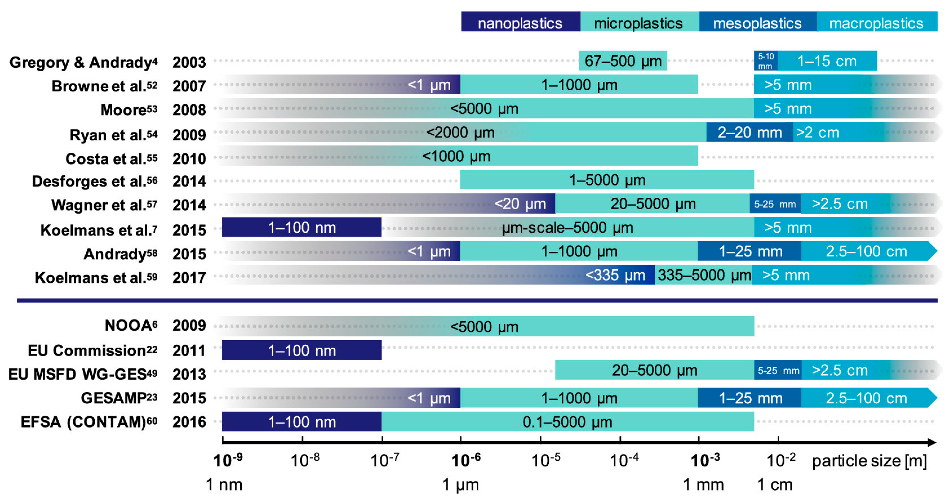
Figure 2.
Schematic representation of the major sources of MPs.
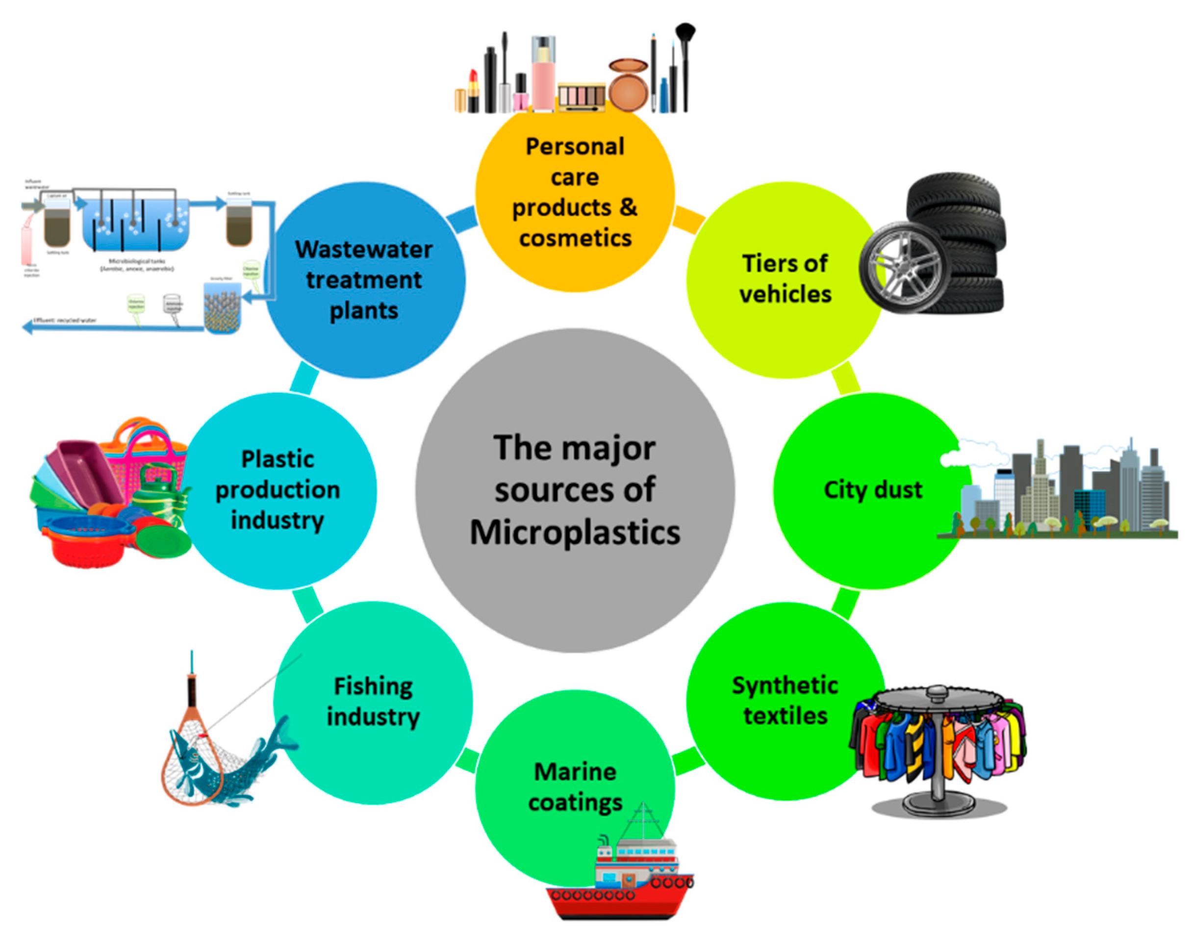
Figure 3.
Source, accumulation, fate, and human exposure pathways of MPs in the environment.
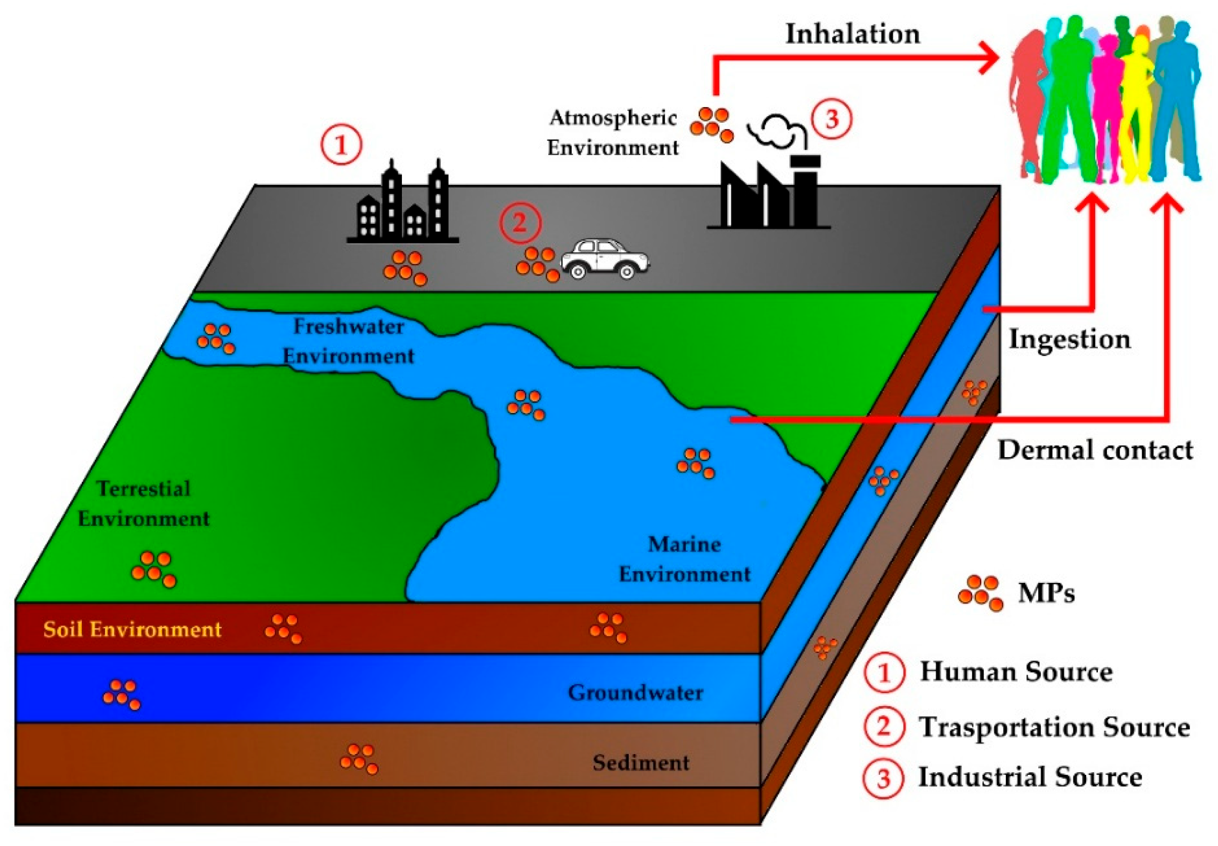
Figure 4.
Possible sources and transport mechanisms of MPs reaching fresh or drinking water.
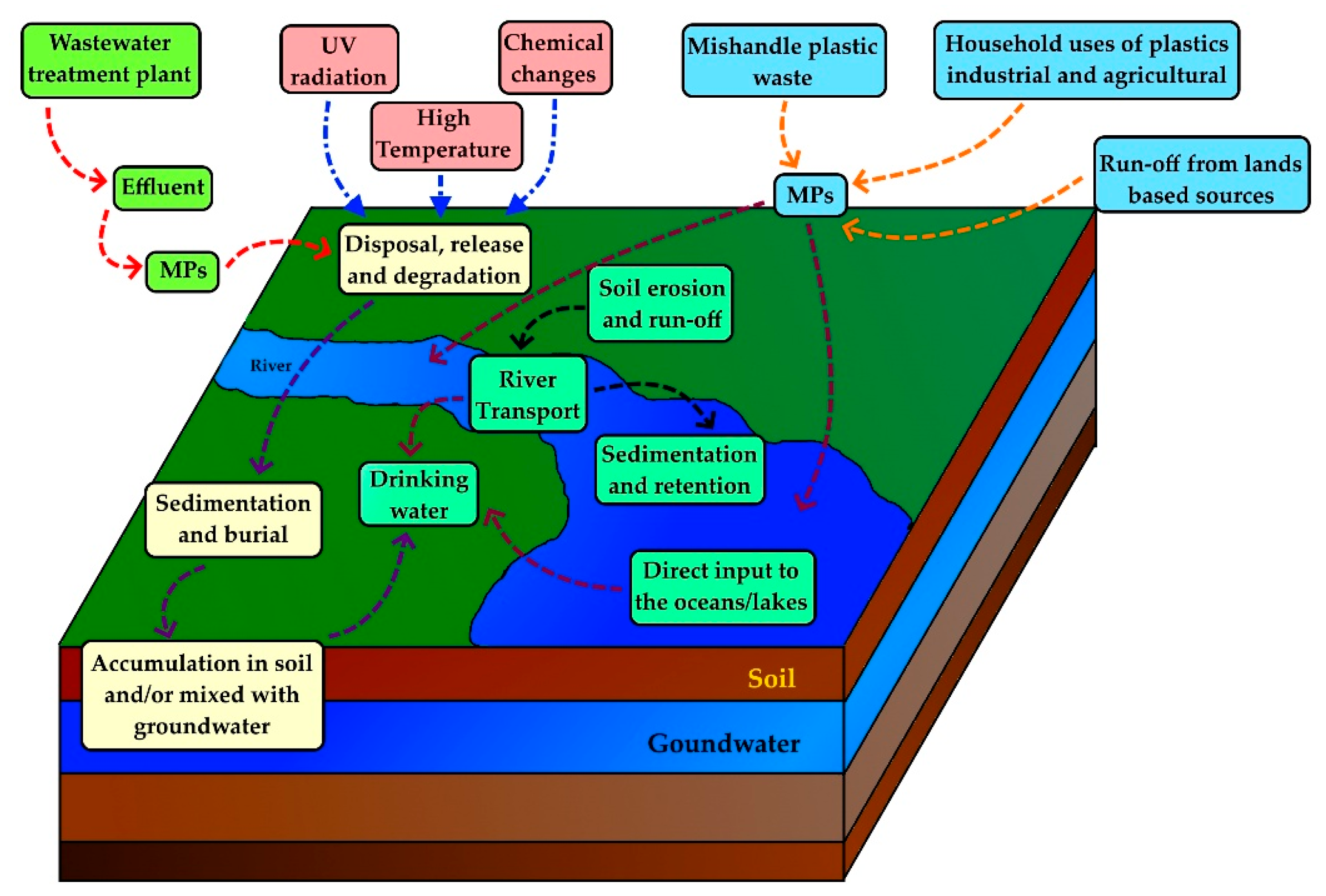
Figure 5.
Three key routes for micro- and nanoplastics entry into the human body.

Figure 6.
Environmental exposure of MPs to humans and possible health effects.
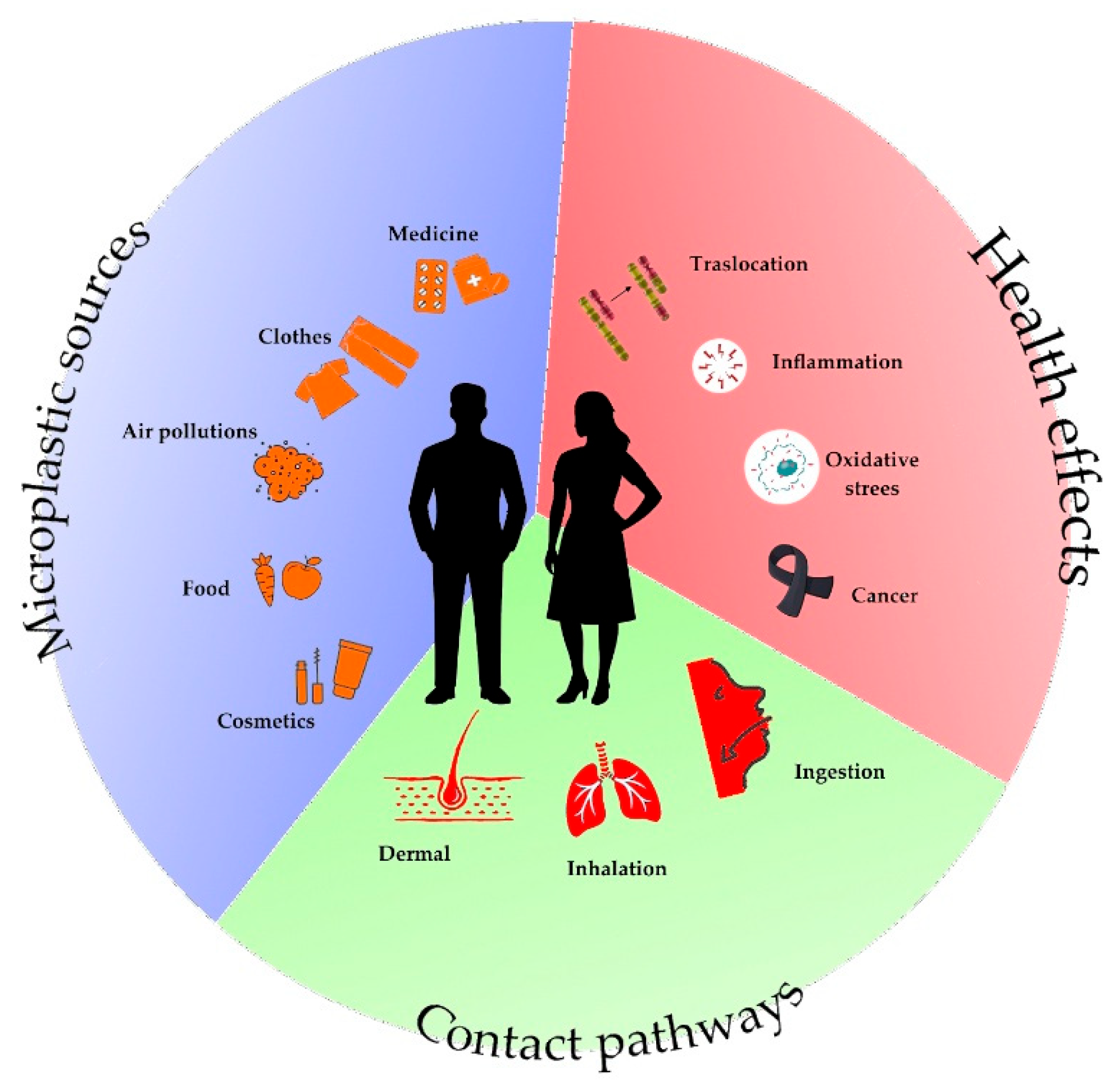
Figure 7.
Detectable MP sizes with different techniques.
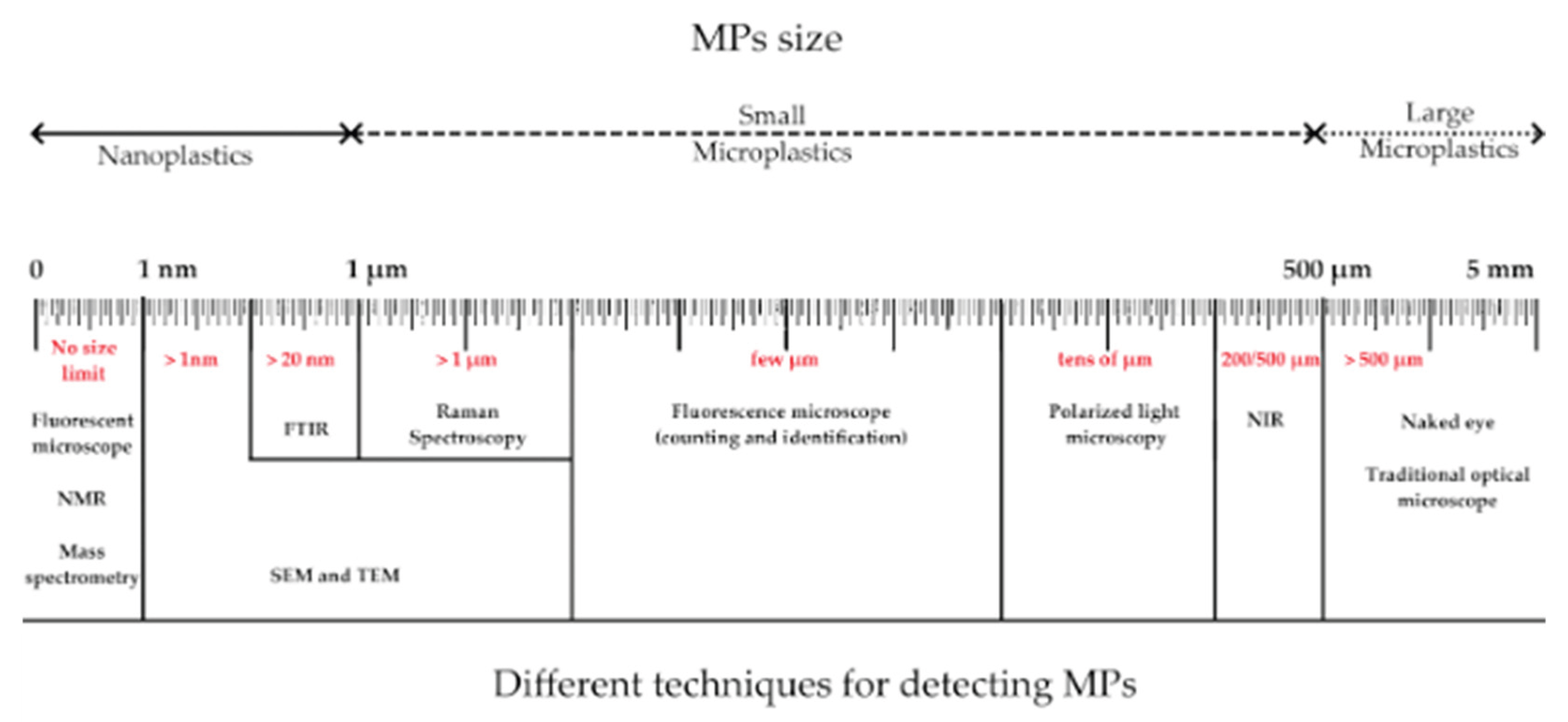

Table 1.
Examples of Primary and Secondary MPs.
| Category | Definition | Examples |
|---|---|---|
| Primary MPs | Microplastics are intentionally manufactured in small sizes for specific purposes | - Microbeads in cosmetics and personal care products (facial scrubs, exfoliants, toothpaste) - Pre-production plastic pellets (nurdles) - Abrasive media used in industrial blasting (air-blasting technology for cleaning surfaces) - Industrial plastic pellets (raw material used in plastic manufacturing) - Microcapsules in detergents, fertilizers, and pharmaceuticals |
| Secondary MPs | Microplastics are generated from the fragmentation, degradation, or weathering of larger plastic items. | - Fragments from bottles, bags, and packaging materials - Fibers from synthetic textiles (e.g., polyester, nylon) - Tire wear particles - Degraded fishing gear (nets, ropes) - Paint flakes (from road markings, ship hulls, household paints) - Degraded fishing nets and ropes (exposure to sun and saltwater breaks them into MPs) - Broken pieces from larger plastic objects (toys, containers, household items, etc.) - Cigarette filter debris (filters made of cellulose acetate degrade into MP) |
Table 2.
Plastic types with their densities, products, and recycling information.
| Polymer type | Density [g/cm3] | Products | Recycle sign | Recyclability |
|---|---|---|---|---|
| Polyethylene terephthalate (PET/PETE) | 1.30 - 1.40 | Soda bottles, water bottles, polyester film, containers for food, jars, fibers for clothing, and even carpets. | ♳ | Widely recycled |
| Polyethylene (HDPE) | 0.94 - 0.97 | Milk jugs, juice containers, grocery and trash bags, motor oil containers, shampoo and conditioner bottles, soap bottles, detergent containers, bleach containers, and toys. | ♴ | Widely recycled |
| Polyvinyl chloride (PVC) | 1.15 - 1.70 | Plumbing and sewage pipes, window frames, non-food packaging, cards, electrical cable insulation, flooring, and phonograph records. | ♵ | Not easily recyclable |
| Polyethylene (LDPE) | 0.917 - 0.94 | Plastic bags, computer components, trays, six-pack rings, milk and juice cartons, packaging for computer hardware, Ziploc frozen food bags. | ♶ | Recycle at specialist points |
| Polypropylene (PP) | 0.90 - 0.91 | Flip-top bottles, plastic diapers, Tupperware containers, margarine tubs, yogurt containers, prescription bottles, bottle caps, and even chairs. | ♷ | Widely recycled |
| Polystyrene (PS) | 1.04 - 1.05 | CD and DVD cases, packing peanuts, single-use disposable cutlery, trays, disposable razors, and smoke detector housings. | ♸ | Not easily recyclable |
| Polycarbonate (PC) | 1.15 - 1.20 | Plastic lenses in eyewear, medical devices, bulletproof glass, automotive components, protective gear, greenhouses, Digital Disks (CDs, DVDs, and Blu-ray), and exterior lighting fixtures. | ♹ | Recycle at specialist points |
| Polylactic acid (PLA) | 1.23 - 1.25 | Takeaway storage containers, takeaway cups and utensils, medical applications like implants, rods, and screws, home 3D printing | ♹ | Recycle at specialist points |
| Acrylonitrile butadiene styrene (ABS) | 1.02 - 1.21 | Lego bricks, computer keyboards, power tool housings, housing for home electrical appliances such as shavers, vacuum cleaners or food processors, automotive bumpers, golf club heads, toys, canoes, 3D printing. | ♹ | Recycle at specialist points |
| Polyamide (PA, nylon) | 1.01 - 1.60 | Toothbrushes, wear pads, wheels, gloves, guitar strings and pics, tennis racket strings, medical implants, electrical connectors, fishing line, tents, gears. | ♹ | Recycle at specialist points |
| Polyurethane (PU/PUT) | 1.23 - 1.35 | Flexible foam, rigid foam, coatings, adhesives, sealants and elastomers. | ♹ | Recycle at specialist points |
Table 3.
Microscopic methods for MP detection.
| Method | Size Detection Limit |
Chemical Information | Sample Preparation | Cost | Principle | Advantages | Limitation |
|---|---|---|---|---|---|---|---|
| Stereomicroscopy | 100 µm | No | Minimal | Low | Provides a 3D view of larger particles using optical paths. | Fast sorting of large particles by shape and color. | Low resolution, cannot detect small particles or provide chemical ID. |
| Polarized Light Microscopy (PLM) | 50 µm | Partial (crystalline) | Minimal | Low | Enhances contrast using polarized light to detect birefringence. | Useful for fiber identification. | Requires expertise, limited for small particles. |
| Fluorescence Microscopy | 1 µm (with fluorescence) | Partial (via fluorescent labeling) | Moderate (staining) | Moderate | Detects fluorescence from particles or dyes. | High contrast, detects very small particles. | Requires staining, can produce false positives. |
| Fluorescence Lifetime Imaging Microscopy (FLIM) |
100 nm | Yes (based on fluorescence lifetime) | Moderate (Requires staining or naturally fluorescent materials) | Medium to High | Measures the decay time of fluorescence emitted from excited molecules, providing information on molecular environment and interactions. | High spatial resolution, allows for detection of MPs and analysis of molecular interactions, can provide quantitative data on fluorophores. | Requires fluorescent dyes for non-fluorescent materials, can be complex to interpret, sensitive to environmental factors (pH, temperature). |
| Confocal Laser Scanning Microscopy (CLSM) | 500 nm | Partial (via fluorescent labeling) | Moderate (staining) | High | Uses laser scanning to create 3D images. | High-resolution, 3D imaging, useful in biological samples. | Expensive, requires fluorescence or labeling. |
| Hyperspectral Imaging (HIS) | 0.5 mm | Yes (spectral information for each pixel) | Minimal | Very High | Collects spatial and spectral data for each pixel in an image. | Simultaneous spatial and chemical identification. | Expensive, requires complex data processing, lower spatial resolution. |
| Scanning Electron Microscopy (SEM) |
1 µm | Partial (with EDX) | Complex (conductive coating) | High | Scans surface with electrons to create detailed images. | High-resolution surface morphology imaging. | Requires conductive coating, no chemical ID without EDX. |
| Transmission Electron Microscopy (TEM) | 1 nm | No | Complex (ultra-thin sections) | Very High | Transmits electrons through thin samples to visualize internal structures. | Ultra-high resolution, visualizes internal features. | Expensive, complex preparation, no polymer identification. |
| Atomic Force Microscopy (AFM) | 1 nm | No | Complex | High | Scans surfaces to create 3D topographical maps based on atomic forces. | Provides 3D surface detail and nanoplastic analysis. | Slow scanning, small sample area, no chemical ID. |
Table 4.
Spectroscopic methods for MP detection.
| Method | Size Detection Limit | Chemical Information | Sample Preparation | Cost | Principle | Advantages | Limitation |
|---|---|---|---|---|---|---|---|
| Fourier Transform Infrared Spectroscopy (FTIR) | 10 µm | Yes (IR absorption spectra) |
Minimal | Medium | Measures IR absorption to identify polymer composition via characteristic vibrations. | Non-destructive, identifies most polymers, can analyze mixtures. | Limited spatial resolution, unable to detect very small particles (<10 µm). |
| Raman Spectroscopy | 1 µm | Yes (Raman shift spectra) |
Minimal | Medium to High | Measures scattered light to provide molecular fingerprint based on vibrational modes. | High spatial resolution, detects small particles, identifies pigments. | Fluorescent samples may interfere, lower throughput compared to FTIR. |
| Near Infrared (NIR) Spectroscopy | 500 µm | Yes (broad molecular information) |
Minimal | Low to Medium | Analyzes molecular overtones and combinations of vibrations in the near-infrared region. | Fast and non-destructive, good for bulk analysis and polymer differentiation. | Limited sensitivity for small particles, broad spectral bands, not highly specific. |
| Laser Direct Infrared (LDIR) Spectrometry | 10 µm | Yes (IR absorption spectra) |
Minimal | High | Combines laser and IR spectroscopy for high-throughput polymer identification. | High throughput, automated mapping, good for polymer identification. | Expensive, resolution limited by laser spot size (~10 µm). |
| Nuclear Magnetic Resonance (NMR) Spectroscopy |
None (requires dissolved samples) |
Yes (chemical structure) |
Complex (requires sample dissolution) | Very High | Analyzes the magnetic properties of nuclei to provide detailed structural information. | Very detailed chemical information, differentiates complex polymers. | Requires dissolved or pure samples, not suitable for solid particles. |
| Optical Photothermal Infrared (O-PTIR) Spectroscopy |
500 nm | Yes (IR absorption spectra) |
Minimal | High | Measures infrared absorption indirectly by detecting photothermal effects. | High spatial resolution, can analyze very small particles (<1 µm). | Expensive, complex instrumentation, lower sensitivity for certain materials. |
Table 5.
Thermal analysis methods for MP detection.
| Method | Size Detection Limit | Chemical Information | Sample Preparation | Cost | Principle | Advantages | Limitation |
|---|---|---|---|---|---|---|---|
| Thermogravimetric Analysis (TGA) | 1 µm | Yes (mass loss profiles) | Minimal preparation | Medium to High | Measures weight changes in a sample as it is heated, providing information on thermal stability and composition. | Simple setup, can analyze small amounts of material, useful for assessing the thermal stability of plastics. | Limited to thermal decomposition, cannot provide specific chemical identities. |
| Differential Scanning Calorimetry (DSC) | 1 µm | Yes (thermal transitions) | Minimal preparation | Medium | Measures heat flow into or out of a sample as it is heated or cooled, providing insights into thermal transitions (melting, crystallization). | Provides specific thermal properties, useful for characterizing polymer behavior during heating. | Limited size range, cannot identify specific polymers directly. |
| Pyrolysis Gas Chromatography-Mass Spectrometry (Py-GC-MS) | 1 µm | Yes (chemical composition) | Moderate (requires pyrolysis) | High | Involves the thermal decomposition of polymers in an inert atmosphere followed by GC-MS analysis of the resulting gaseous products. | Highly sensitive and specific, provides detailed chemical identification of MPs. | Expensive, requires complex sample preparation, not suitable for quantitative analysis without calibration. |
Disclaimer/Publisher’s Note: The statements, opinions and data contained in all publications are solely those of the individual author(s) and contributor(s) and not of MDPI and/or the editor(s). MDPI and/or the editor(s) disclaim responsibility for any injury to people or property resulting from any ideas, methods, instructions or products referred to in the content. |
© 2026 by the authors. Licensee MDPI, Basel, Switzerland. This article is an open access article distributed under the terms and conditions of the Creative Commons Attribution (CC BY) license (http://creativecommons.org/licenses/by/4.0/).
Copyright: This open access article is published under a Creative Commons CC BY 4.0 license, which permit the free download, distribution, and reuse, provided that the author and preprint are cited in any reuse.


