Preprint
Review

This version is not peer-reviewed.

Rethinking Human Energy Metabolism

Submitted:

18 January 2026

Posted:

19 January 2026

You are already at the latest version

Abstract

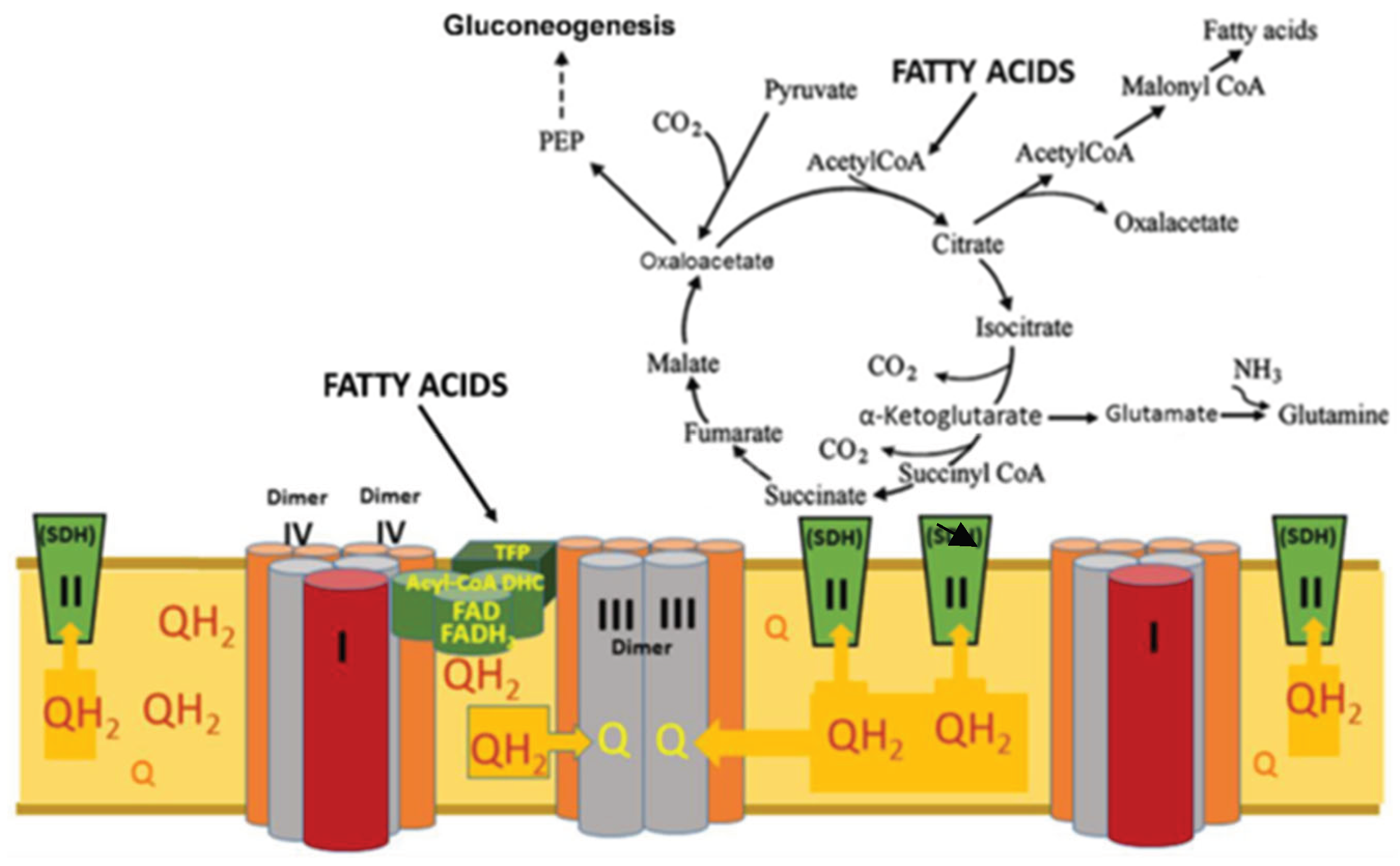
For a long time, glycolysis and mitochondrial oxidative phosphorylation were opposed to each other. Glycolysis work when there is a lack of oxygen, the mitochondria supply ATP in oxygen environment. In recent decades, it has been discovered that glycolysis in vivo works always and the final product is lactate. Lactate can accumulate and is the transport form for pyruvate. In this review, we look at how obligate lactate formation during glycolysis affects the tricarboxylic acid (TCA) cycle and mitochondrial respiration. We conclude that fatty acid β-oxidation is a prerequisite for obligate lactate formation during glycolysis, which in turn promotes and enhances the anaplerotic functions of the TCA cycle. In this way, a supply of two types of substrates for mitochondria is formed: fatty acids as the basic energy substrates, and lactate as an emergency substrate for the heart, skeletal muscles, and brain. High steady-state levels of lactate and ATP, supported by β-oxidation, stimulate gluconeogenesis and thus supporting the lactate cycle. It is concluded that mitochondrial fatty acids β-oxidation and glycolysis constitute a single interdependent system of energy metabolism of the human body.

Keywords: 
;  ;  ;  ;  ;  ;  

1. Introduction

Oxidative phosphorylation (OXPHOS) is the primary function of mitochondria. For decades, numerous mitochondrial functions have been studied in the in vitro system using isolated mitochondria. The respiratory chain of mitochondria in all mammals is basically the same, since it has been an effective energy producing device from the very beginning of evolution [1]. According to a long tradition, the sequence of protein complexes that are involved in electron transport and ATP synthesis are divided into five functional complexes, which are numbered in order: complexes I, II, III, IV, and V. Such numbering was associated with the logic of events in oxidative phosphorylation, that existed at that time (the middle of the 20th century). Complexes I, III and IV are involved in the transfer of electrons and generation of the transmembrane potential (Δp = ΔΨ -59 ΔpH). Complex V is ATP syntase consuming ΔpH + ATP/ADP antiporters (adenine nucleotide translocase – ANT) consuming ΔΨ. For a long time, the role of ANT was not taken into account in the overall energy balance of oxidative phosphorylation. Complexes II, aka succinate dehydrogenases (SDH), do not directly create a ∆pH or ΔΨ gradients, but they are the only enzymes in the tricarboxylic acid cycle (TCA) that are embedded into the inner membrane of mitochondria and reduce the membrane’s pool of ubiquinone (coenzyme Q) to ubiquinol (coenzyme QH2), which is oxidized by the respiratory complex III. The names of the complexes reflect their main function: complex I (CI; NADH:ubiquinone (UQ) oxidoreductase), complex II (CII; succinate:UQ oxidoreductase), complex III (CIII; cytochrome bc1) and complex IV (CIV; cytochrome c oxidase). Figure 1 reflects how our knowledge about the structure of the respiratory chain changed with time.
Unfortunately, the classical ideas about the structure and function of the respiratory chain turned out to be incorrect. In 2000, it was shown that the respiratory chain electron transporters are organized into superstructures called “respirasomes”. Each respirasome consist of three supercomplexes; two of them contain CI, one dimer of CIII, two dimers of CIV; the third, smaller supercolmplex, contains one dimer of CIII and two dimers of CIV [2,3]. Together, the three supercomplexes form the repirasome that also interacts with the polyenzymatic structures of the fatty acids β-oxidation of [4]. At first, the physiological significance of the megastructural construction of the respirasome seemed incomprehensible. However, taiking into account the known properties of the large supercomplexes containing complex I and the new data upon activation of the reverse electron transfer through SDH from CoQH2 to the TCA cycle, it was proposed that the main function of the respirasome is to carry out the β-oxidation of the long chain and middle chain fatty acids [5].
The third, smaller supercomplex of the respirasome, contains only a dimer of complex III and two dimers of complex IV (CIII2CIV4) [3]. This supercomplex directly oxidizes irreversibly and at a very high rate the inner membrane’s pool of CoQH2 (ubiquinol) . Due to the presence of the Q cycle in the complex III, the rate of oxidation of ubiquinol, although very high, is finite [6]. In addition, the rate of oxygen consumption by mitochondria is controlled by the rate of ATP synthases [5]. Brand noted that the highest steady-state levels of the coenzyme Q reduction are observed during fatty acid β-oxidation [7]. The high rate of ubiquinone reduction is achieved by the coordinated work of five mitochondrial acyl-CoA dehydrogenases: there are long-, medium-, and short-chain acyl-CoA dehydrogenases (LCAD, MCAD, and SCAD), which are most active with 14, 8, and 4 carbon substrates, respectively. These acyl-CoA dehydrogenases are homotetramers [8,9]. Very long-chain acetyl-CoA dehydrogenase (VLCAD has optimal chain length specificity for fatty acyl-CoAs with 16 carbons [10]. The fifths enzyme, human mitochondrial acyl-CoA dehydrogenase-9 plays a role in the mitochondrial β-oxidation of unsaturated fatty acids. [11]. In adition, acyl-CoA dehydrogenase-9 is required for the biogenesis of the respiratory complex I [12]. VLCAD and ACAD-9 are dimers and associated with the inner membrane. Thus, alltogether, approximately 14-16 acyl-CoA dehydrogenases of a single β-oxidation set of enzymes may work simultaneously maintaining the high steady-state level of ubiquinol in spite of its very fast oxidation by the smaller supercomplex. We do not yet know how many β-oxidation complexes are associated with a single respirasome, but definitely more than one.
At the high level of ubiqunol, SDH initiates the rerevrse electron transport creating high levels of reduction of the cell’s NADH/NAD+ and NADPH/NADP+ systems, as well as ATP production [7]. As the result, in vivo, glycolysis always produces lactate instead of pyruvate. A large body of evidence prove that lactate and the lactate cycle play a crucial roles in the overall energy balance in the body [13,14,15]. To grasp and to understand the consequences of the obligatory production of lactate, due to activation of the fatty acids β-oxidation, is the goal of this review.

2. A Quick Critical Look at the Energy Metabolism from the Point of View of the New Paradigms

The driving force for the movement of electrons down the respiratory chain are the differences in the redox potentials between hydrogen, as the primary source of energy, and water, as the final product. The redox potential is a measure of the readiness of a compound to give or accept electrons. Compounds with lower redox potential are ready to give away electrons to compounds with higher redox potentials that have higher affinity for electrons.
Normally, electrons are transported through the respiratory chain one at a time. When tracing the path of one electron from the reduced pyridine nucleotide (NADH), the first reaction of NADH will be with the enzyme NADH dehydrogenase, which is a component of the respiratory complex I and contains flavinmononucleotide (FMN). This is the rate-limiting step of the whole pathway of an electron, if mitochondria oxidize NADH only. From the reduced FMN, electrons are transferred one by one along the chain of Fe-S clusters, that form an isolated tunnel for electrons, to the complex-I-bound coenzyme Q (CoQ), reducing it to CoQH2. The reduced coenzyme CoQH2 (ubiquinol) of the complex I reacts directly with the tightly bound CoQ of the complex III, reducing it, and then electrons are transferred through the channel for electrons to the terminal cytochrome c1. Reduced cytochrome c1 transfers electrons to cytochrome c. Reduced cytochrome c is oxidized by complex IV, where the final reaction of oxygen (O2) reduction to 2H2O takes place. In this final reaction of water formation a large amount of heat is released. In accordance with the old paradigm, this is the sequence of events in the respiratory chain of mitochondria oxidizing the so called complex I substrates. It was generally accepted that NADH, the actual substrate for the respiratory chain, was supplied by the trcarboxylic acid cycle, which is started by condensation of acetyl-CoA and oxaloacetate (OAA). Most reseachers considered pyruvate as the main source of acetyl-CoA for the TCA cycle, and pyruvate as final product of glycolysis, which is wrong.

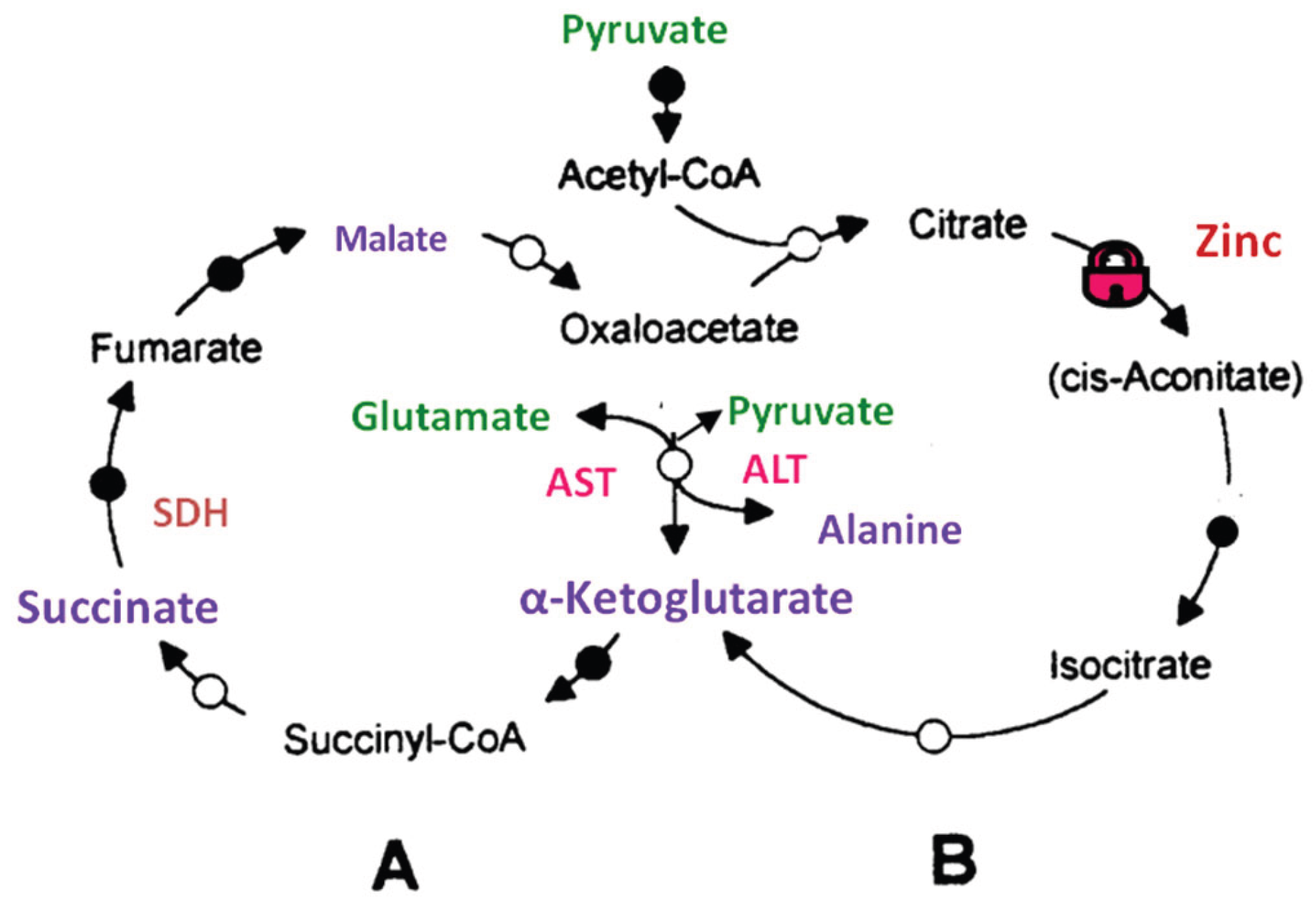
2.1. Glycolysis, Tricarboxylic Acid Cycle and the Mitochondrial Substrates

During the first decades of mitochondrial research, it was of most importance for reaseachers to a achive a stable and possibly high energization of the isolated mitochondria. To energize mitochondria in the experiments, researchers used different substrates. However, from the very beginning researchers encountered serious problems with the two key substrates, namely succinate and fatty acyl-carnitines. Isolated mitochondria from the brain and heart were unable oxidize succinate alone, but succinate + rotenone was a perfect substrate for mitochondria isolated from any organ. As regards acyl-carnitines, which in vivo are normal respiratory substrates for all types of mitochondria [16], in experiments in vitro the rates of β-oxidation were too slow to be physiologically relevant. For this reason, until recently, we knew very little about β-oxidation of long-chain and middle-chain fatty acids at the mitochondrial level. As the result, most of our knowledge about the energy-dependent functions of mitochondria were obtained on mitochondria energized either with the absolutely non-physiological substrate succinate + rotenone, or other physiologically irrelevant substrates, for example glutamate, which is a too valuable metabolite to be used as a sole source of energy in vivo, or pyruvate, which by itself, is not a substrate for the tricarboxylic acids (TCA)cycle.

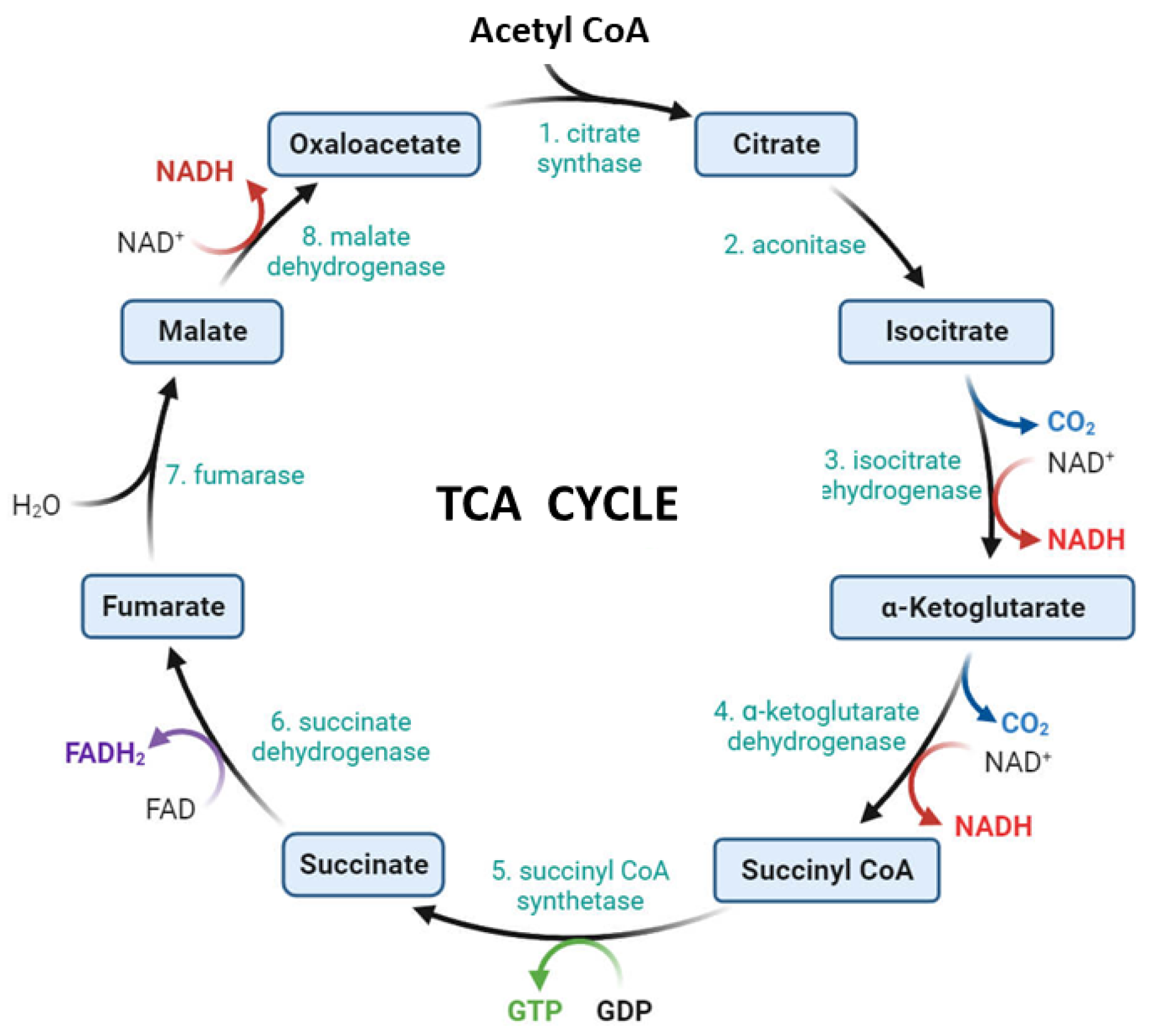
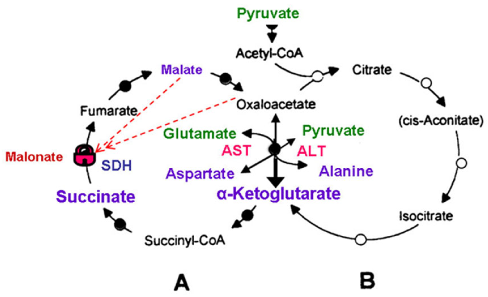
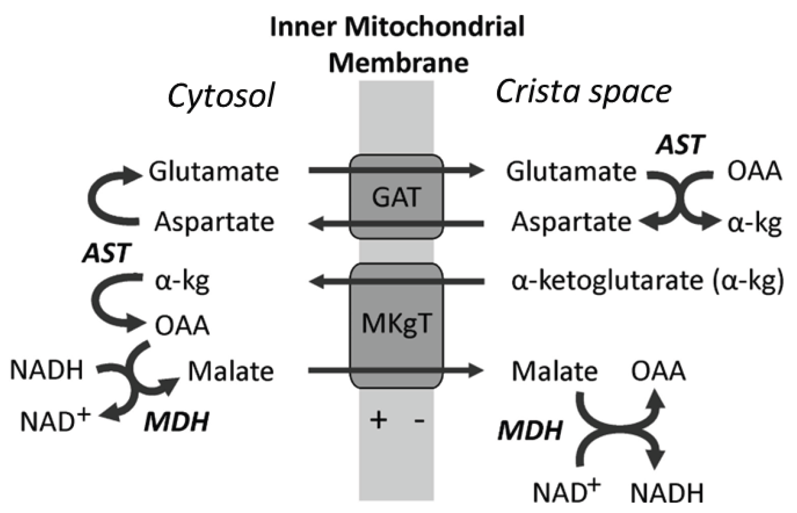
2.2. The Tricarboxylic Acid Cycle)

As stated above, in vivo, glycolysis does not produce pyruvate as a substrate for the mitochondria [17]. For this reason, in most cases the TCA cycle begins with the acyl-CoA, which originates from the fatty acids β-oxidation. Researchers, however, often used pyruvate + malate as substrates for the isolated mitochondria from most organs, except for the liver mitochondria. This is because in the fasted overnight animals, the liver pyruvate dehydrogenase complex is inhibited. Glutamate + malate was also a popular substrate for all types of mitochondria. Many researchers regarded pyruvate and glutamate as the substrates specific for the complex I, whereas succinate (usually with rotenone) served as a substrate for the complex II. However, in the mitochondria of most organs, pyruvate, and especially glutamate, are metabolized via transamination to form a-ketoglutarate and then succinate.
For a long time, it was believed that the enzymes of the TCA cycle reside in the matrix of mitochondria and the respiratory chain was presented as a sequence of the respiratory complexes, not as a respirasome. Already in the 90s of the last century it was known that quantitatively, the TCA cycle enzymes associated with the inner membrane, 200 times exceed those in the matrix [18]. Now we know that some of the experimental complications observed with the isolated mitochondria, such as endogenous inhibition of succinate dehydrogenase (SDH) and low rates of the long-chain and middle-chain acyl-carnitines β-oxidation, stem from the fact that practically all researchers utilized only one substrate or in combination with malate. By itself, malate is not oxidized by the mitochondria, but malate help to oxidize some other substrates because it is the source of oxaloacetate. In vivo, mitochondria always oxidize mixtures of substrates that are optimal for a given organ or tissue and the concrete metabolic situation.
For example, for the isolated synaptic brain mitochondria, which constutute more than 90% of the total brain mitochondria [19], the optimal substrate mixture is pyruvate + glutamate (neuromediator) [20]. Pyruvate originate from lactate, which in vivo serves as a main substrate for the synaptic mitochondria [21]. Lactate circulates in the blood at mM concentrations [22]. It was generally accepted that most of lactate originates from glucose. However, in the brain, liver and kidney fatty acids β-oxidation supplies energy and carbon atoms for the increased formation of OAA and thus gluconeogenesis [23,24,25]. Also, both glucose and lactate can be formed from other sugars, such as fructose, galactose, and mannose.
The tricarboxylic acids cycle is considered usually as a sequence of biochemical reactions aimed at oxidation of the main metabolites (see Figure 2). The rate of individual reactions may vary, but it was generally accepted that the TCA cycle almost always begins with the synthesis of citrate [26]. The first metabolite of the TCA cycle citrate is formed by condensation of oxaloacetate (OAA) with acetyl-CoA, which is produced either by decarboxylation of pyruvate, in the case of glycolysis, or by β-oxidatation of fatty acids. Then, the TCA cycle continues clockwise until the enzyme system returns to the formation of a new citrate molecule from the resulting OAA and a new molecule of acetyl-CoA. During the cycle, 2 molecules of CO2, 3 NADH and 1 FADH2 are released. However, in vivo, such a scenario could be one of several.
Back in the early 90s, it was shown that in the brain synaptic mitochondria the slowest step of the TCA cycle is between OAA and citrate [26]. In the isolated synaptosomes the activity of citrate synthase was found to be very high, whereas the activity of the pyruvate dehydrogenase complex (PDHC) was 10 times lower [27]. The flux between α-ketoglutarate (α-KG) and OAA was 3 – 5 times faster, depending on the presence or absence of glucose [26]. This suggests that in the brain, the TCA cycle may function as two conjugated cycles (Figure 3). The inhibition of PDHC in the formation of citrate becomes understandable in the light of a recent evidence that in vivo glycolysis always leads to the formation of lactate, not pyruvate [17,28,29]. Therefore, in vivo the glycolytic pyruvate will almost never be a source of citrate for the initiation of the TCA cycle. Most mitochondria possess the lactate dehydrogenase that provides pyruvate + H+ for oxidaton [13,30]. Therefore, lactate serves as a transport form of pyruvate.
As we see, the TCA cycle has a much more complex, though not yet fully studied, physiology. Yudkoff (1994) suggested that the malate-aspartate shunt (MAS) and related aminotransferases play a decisive role in the spliting of the TCA cycle (Figure 3 and Figure 4) [26]. Aspartate aminotransferase, which is the key enzyme of MAS, catalyzes the reversible reaction: glutamate + oxaloacetate α-ketoglutarate + aspartate. Alanine aminotransferase also catalyzes the reversible reaction: glutamate + pyruvate α-ketoglutarate + alanine. For the brain and spinal cord mitochondria, a mixture of glutamate + pyruvate + malate has been shown to be the optimal substrate mixture [20]. It must, however, be kept in mind that in vivo enzymes of the MAS work irreversibly in one way → or another ←, but in different compartments.
In the organs, where lactate becomes an important source of energy, MAS becomes indispensible. The importance of MAS has been proven in experiments with inhibitors of the transaminase and SDH [31]. We can suggest that in the presence of LDH and MAS, the TCA cycle in many organs works as two coupled cycles: left is running from a-ketoglutarate to OAA, and another extending from OAA to α-KG with the inclusion of the citrate condensation reaction. Under certain conditions, such a system could predominantly produce either OAA or isocitrate from the predecessor, without the need to involve the entire cycle, while maintaining energy production. In fact, such systems, predicted by Yudkoff, exist in the human body and are shown in Figure 3 and Figure 5. Increased production of isocitrate my be of a great importance for the functioning of the mitochondrial transhydrogenase [32].
Another very interesting example of the functional adaptation of the TCA cycle to the needs of an organ are mitochondria of the prostate. In the prostate of sexually mature men, mitochondria produce huge amounts of citrate. This has been found to be due to the inhibition of aconitase by zinc ions [33]. Therefore, the TCA cycle of the prostate mitochondria cannot work according to the classical scheme. Acetyl-CoA condenses with oxaloacetate to citrate, which is excreted entirely into a prostatic liquid, where the concentration of citrate can reach 10 mM. Under these conditions, only the small cycle A can fully function in the mitochondria of the prostate (Figure 5), where amino acids are used as a source of α-ketoglutarate. That is probably why men need to eat meat, which stimulates the prostate.
The physiological meaning of the high citrate concentration in the prostatic fluid is that Ca2+ ions are necessary for the active work of the spermatozoid’s tail. Therefore, when prostatic fluid mixes with spermatozoa, the citrate chelates Ca2+ and thus immobilize spermatozoids. At the right time and in the right place, calcium is released, the spermatozoids begin wag their tails, and if one of them is successful, the citrate will serve as a substrate for the zygote.

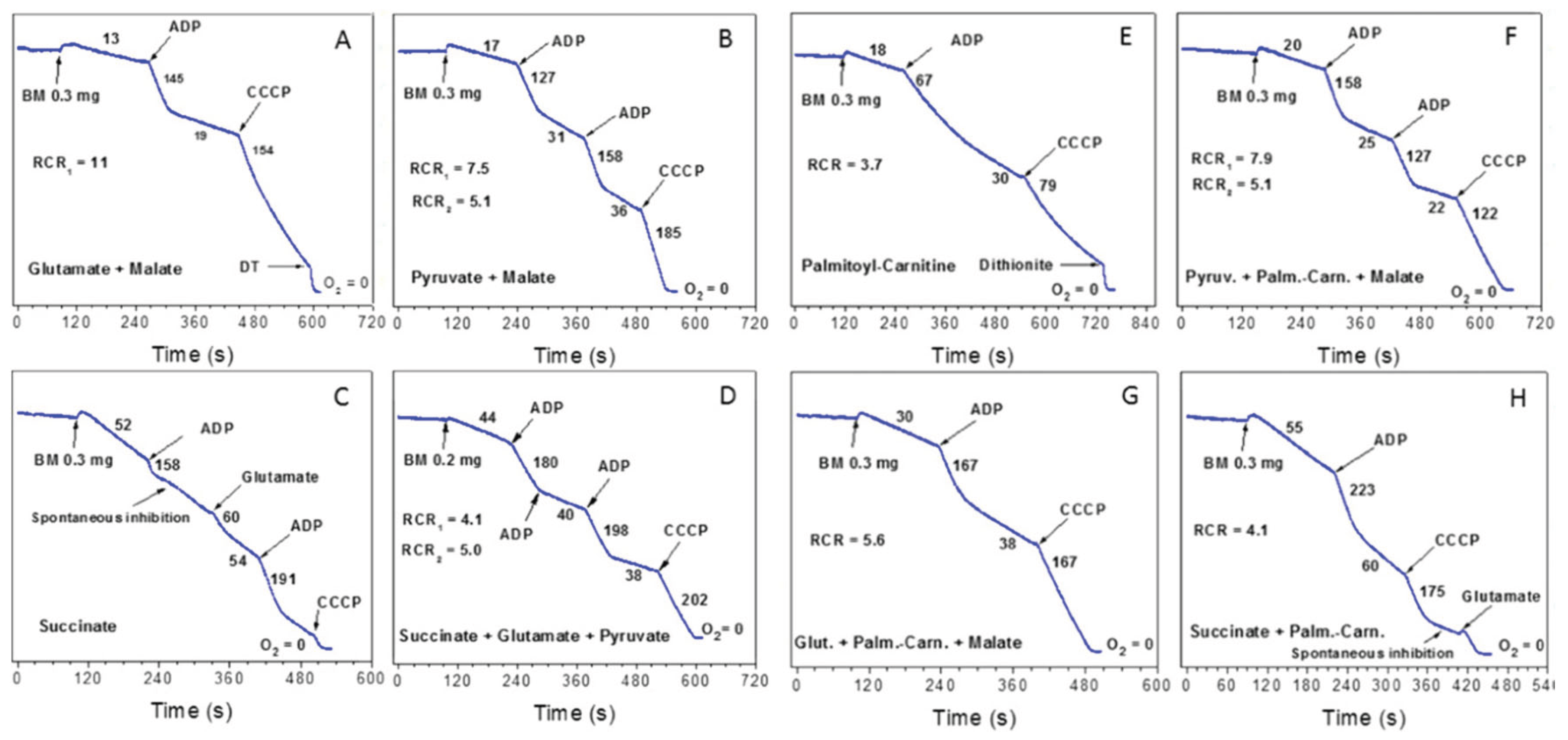
3. Respiratory Functions of the Isolated Mitochondria
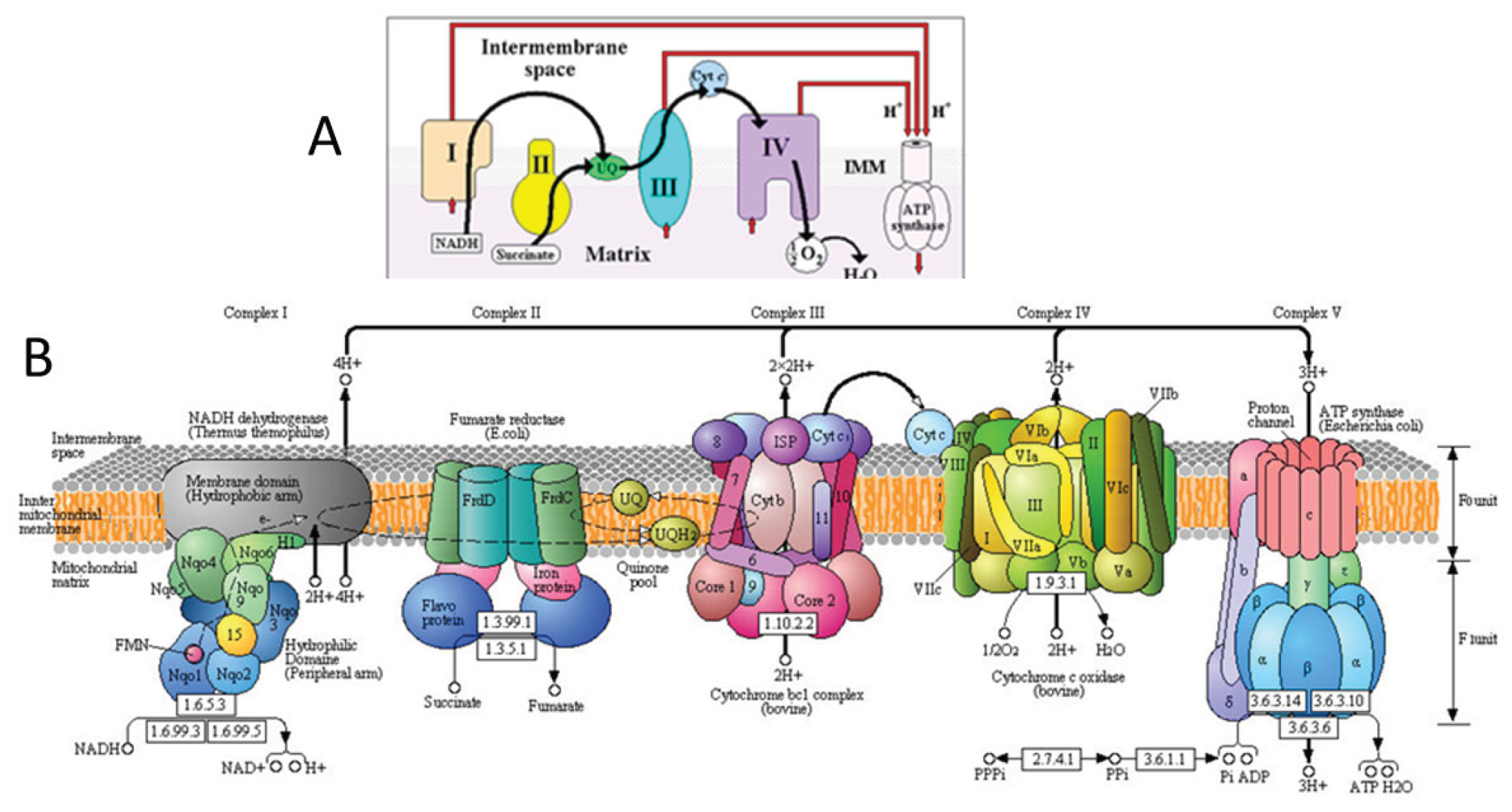
From the discussion of the metabolic properties of mitochondria we excluded liver mitochondria. This is because the liver and liver mitochondria have a number of exceptional functions and properties that other organs and their mitochondria do not have. For example, the liver and liver mitochondria are relatively resistant to prolonged hypoxia and are able to endure complete ischemia within 20-30 minutes. Their properties of the liver mitochondria vary greatly, depending on the composition of the food and the time that has passed after the last meal. Liver mitochondria have a large osmotically active volume and have a large storage of endogenous substrates, the liver is capable of active regeneration, etc. [35].].
As regards mitochondria from other than liver organs, the most difficult obstacles have been met during attempts to study respiratory activities with seemingly “simple” physiological substrates, namely succinate and fatty acyl carnitines. Figure 6 illustrates an experiment with the isolated synaptic mitochondria from the Sprague-Dawley rat. We mention the strain of animals because many functions, including respiratory activities, are species specific. This is important when comparing data from different species and strains of the same species. The composition of incubation medium, substrates concentrations and additions are described in the Figure 6 legend. For the readers not familiar with the polarographic assay we give some explanation. Changes in O2 concentration in the incubation medium were measured with the platinum Clark electrode [35]. The rates of O2 consumption are in nanomoles of O2 consumed per one minute per mg of mitochondrial protein. Substrates were added before mitochondria. Metabolic states: State 4, or resting respiration – O2 consumption before the addition of ADP; State 3, or active oxidative phosphorylation – the respiration rate after addition of ADP; uncoupled respiration – oxygen consumption after the addition of a protonophore (CCCP), which collapses (uncouples) the membrane potential. The respiratory control ratio (RCR) is the ratio of the respiratory rate with ADP to the respiratory rate in the absence of ADP. RCR is an indicator of the quality of the mitochondria: the value 1 – mitochondria are uncoupled, the higher the value, the better. Many mitochondrial functions are energy-dependent and thus decline upon uncoupling.
Figure 6A and 6B show respiratory rates with glutamate and pyruvate in the presence of malate. Many researchers add malate by default; however, we have found that the stimulatory effect of malate on oxidation of some substrates, acyl-carnitines in particular, is species-specific. As regards glutamate and pyruvate; in a separate experiment we have established that with the brain mitochondria about 60% of glutamate and 30% of pyruvate were oxidized via transamination. Figure 6C shows that these particular brain mitochondria were capable of oxidizing succinate in the State 4 and even begun to respond to the addition of ADP, but then, the State 3 respiration was quickly inhibited. It should be stressed that endogenous inhibitions of SDH in mitochondria is highly variable and species-specific. Some species display total inhibition of SDH [36]. Figure 6D shows that addition of glutamate and pyruvate totally release the inhibition of SDH.
Figure 6E shows oxidation of palmitoyl-carnitine (P-C) by synaptic mitochondria. This is a typical pattern also for the heart, and kidney mitochondria. The rates of O2 consumption during P-C oxidation strongly depend on the time passed after the isolation of mitochondria. This indicate that the endogenous substrates might have a stimulatory effect. Indeed, Figure 6F, 6G, and most impressively 6H show that in the presence of other metabolites palmitoyl-carnitine is oxidized at high rates in all metabolic states. Figure 6H is the most impressive because the two “bad substrates” separately, together showed the highest rates of oxidative phosphorylation. The increased State 4 respiratory rates with succinate and P-C were caused not by the uncoupling, but by the increased reverse electron transport associated with the increased content of the membrane’s ubiquinol. Both succinate and P-C reduce ubiquinone to ubiquinol [7].
Incubation medium:125 mM KCl, 10 mM NaCl, 10 mM MOPS, pH 7.2, 2 mM MgCl2, 2 mM KH2PO4, 1mM EGTA, 0.7 mM CaCl2. At Ca2+/EGTA = 0.7 the [Ca2+]Free was 1 µM. Chamber volume = 0.65 ml. Substrates: glutamate, 5 mM; malate, 2 mM; pyruvate, 2.5 mM; succinate, 5 mM; palmitoyl carnitine, 25 µM. Numbers at the traces are respiratory activities in nmol/min/mg mitochondrial protein. The respiratory activity ratio (RCR) is: V State 3/preceding V State 4. Additions: brain mitochondria 0.3mg, ADP 150 µM, CCCP 0.5 µM, glutamate 5mM. The figure is from [37].
High rates of oxidative phosphorylation with palmitoyl-carnitine + succinate we observed also with the kidney mitochondria [38] and the heart [37]. Tere were though, some organ-dependent differences. Because kidney mitochondria do not have endogenous inhibition of SDH, they oxidized succinate with a very high rate. Respiratory rate in state 3 with P-C + succinate was only 30% higher than with succinate alone. So, we did a control experiment with 0.5 mM succinate. There was no oxygen consumption by the kidney mitochondria with 0.5 mM succinate alone, however 0.5 mM succinate stimulated P-C oxidation 4-fold and 8-fold the oxidation of octanoyl-carnitine [38].
It was not possible for us to explain the above facts from the point of views of the- old paradigms. We tried to interpret the data of the Figure 6 from the standpoint of the respirasome as a functional unit for respiration.

4. Respirasome

4.1. History

In the 80s of the last century, it was shown that for the beef heart mitochondria the single set of respiratory complexes, involved in oxidative phosphorylation, namely complexes I:II:III:IV:V were related as 1:2:3:6-7:3-5 [39]. In 2000, Schägger and Pfeiffer (2000) presented evidence that the electron carriers are organized into three supercomplexes [1]. By interacting together, the three supercomplexes form a functional superstructure named “respirasome” [2]. Sometime after the discovery of respirasome, researchers started to designate the stoichiometry of complexes as I1III2IV4 [40] and found a signifucant variability in composition of the respirasomes among different species [41,42]. The respirasome presented by Schägger (2001) contained two large supercomplexes and one smaller supercomplex [2]. Each of the large supercomplexes comprised a monomer of complex I, a dimer of complex III and two dimers (four copies) of complex IV (Figure 7). The smaller supercomplex consisted of one dimer of complex III and two dimers of complex IV, or III2 IV4 [2]. The two active centers in the dimer of complexes III in the smaller supercomplex open into the lipid phase of the inner membrane and react with the membrane’s pool of ubiquinol. In the large supercomplexes the active centers of complexes III do not interact with the membrane’s pool of coenzyme-Q. Sousa et al. (2016) have shown that in the large supercomplex only one of the two Rieske iron-sulfur domains of the complex III dimer was active, indicating that the other monomer of complex III dimer was inactive [43].
The redox potential difference (ΔE°′) between ubiquinol (QH₂) (+0.045 V), which donates electrons to Complex III, and O₂/H₂O (+0.82 V), the terminal acceptor at Complex IV, is about 0.77 V. The energy released by the work of the small supercomplex (CIII2CIV2) is very high: ∆G = -nF∆E = −148 kJ per mol [44]. Therefore, the rate of ubiquinol oxidation is very fast, irreversible and releases a lot of heat
It is evident, that the membrane’s pool of the reduced Co-QН2, which swims inside the liquid lipid phase of the inner membrane at the temperature of +50oC [45], can be oxidized only by the smaller supercomplex of the respirasome. .

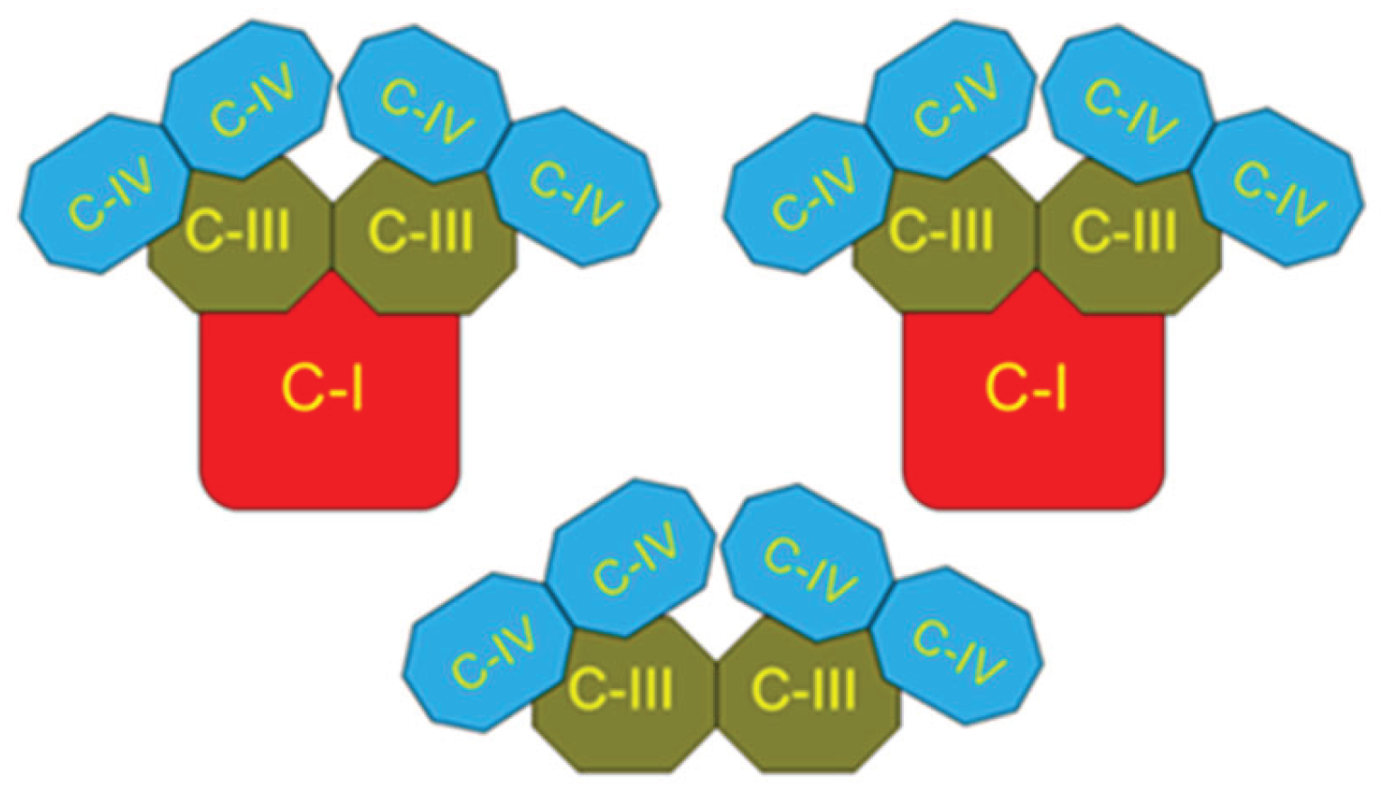
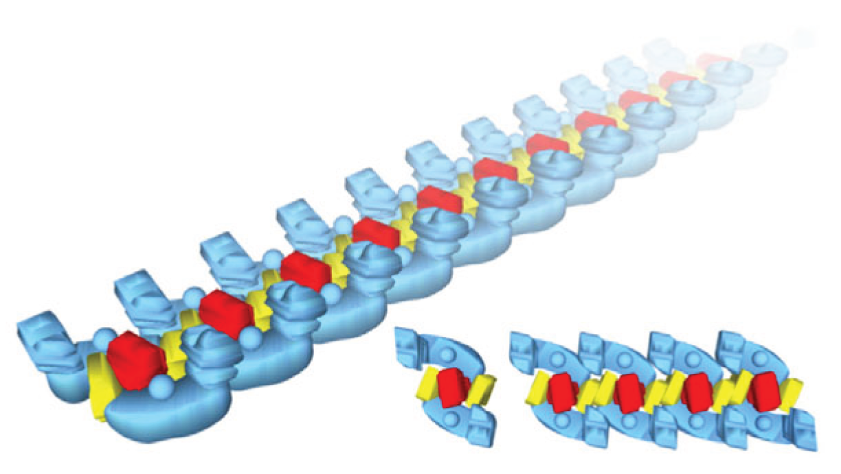
4.2. Respirasome Structure

After the discovery of the respirasome structure [1,2], there have been published a rather large number of papers describing various aspects of supercomplexes organization of which we mention a few [40,46,47,48,49,50].
Some authors observed that attenuation of the biogenesis of individual respiratory chain complexes was accompanied by increased formation of stable respiratory supercomplexes. This phenomenon was not accompanied by increased mitochondrial respiratory activity. Therefore, it was concluded that formation of the supersomplexes is necessary for the structural stabilization, but not for the enhancement of the respiratory chain catalysis [52]. Some authors believe that the term “respirasome” describes the phenomenon of formation in some organisms of the respiratory complexes’ clusters, but the complexes themselves can work independently [53].
It was shown that clusters of the respiratory complexes form the “respiratory string” [54,55], as shown in Figure 8. Association of basic units into a string is mediated by complex IV, which interacts with the neighboring complexes IV through a dimeric interface found in the X-ray structure [54]. The basic unit of the respiratory string (Figure 8 bottom center) has the structure I2 III2 IV2 thus indicating that the respirasome may be a structure of the higher order. The association of the base units into a chain is provided by complex IV, which forms dimers with the complexes IV of adjacent basic unit. Some authors presented different ratios and composition of supercomplexes in the respirasomes [43,56].

5. The Key Roles of Fatty Acids β-Oxidation and Lactate Accumulation and Oxiation in Human’s Metabolsim

5.1. Fatty Acids β-Oxidation

The highest rates of ubiquinone reduction to ubiquinol occur in the organs where β-oxidation of long-chain fatty acids is an important sourse of ATP and NADPH. Accordingly, the highest steady-state levels of CoQH2 are maintained in these organs, high enough to initiate the reversal of the electron flow from CoQH2 to the TCA cycle [7]. This increases the level of NADH in the mitochondria and the presence of the energy-dependent transhydrogenase translate a significant part of the mitochondrial redox potential NADH/NAD+ to the cytosolic NADPH/NADP+ [32,57]. Simultaneous increases in ATP and NADPH production stimulate anabolic and anaplerotic metabolic pathways as well as the main physiological functions of the organs (Figure 9).
The existing fallacy that mitochondrial respiration and energization depend on the external redox potential is correct only for the in vitro conditions when the isolated mitochondria lost metabolic communication with the cell and blood circulation. In the well-energized isolated mitochondria, the reverse electron transfer, which occurs during oxidation of the FAD-dependent substrates, restores the components of complex I, leading to increased production of superoxide radicals [7,58,59]. Under the in vitro experimental conditions, the excess of electrons cannot be directed to the cytoplasm because there are no conditions for activation of the mitochondrial transhydrogenase (MTH). It can be expected that in vivo there is no stimulation of the free radicals formation, at least not to the same extent as in vitro, since MTH and the reversal of the transfer of electrons through SDH (complex II) dump the excess of energy and electrons into the cytoplasm.
The high level of the redox potentials in the cytoplasm leads to the fact that glycolysis always results in the formation of lactate, rather than pyruvate [14,17,28]. Since pyruvate and other ketoacids are very unstable, we can consider lactate as the storage and transport form of pyruvate and the redox buffer. Most mitochondria have active lactate dehydrogenase that produce free pyruvate and a proton, which are oxidized by the mitochondria [60,61,62].

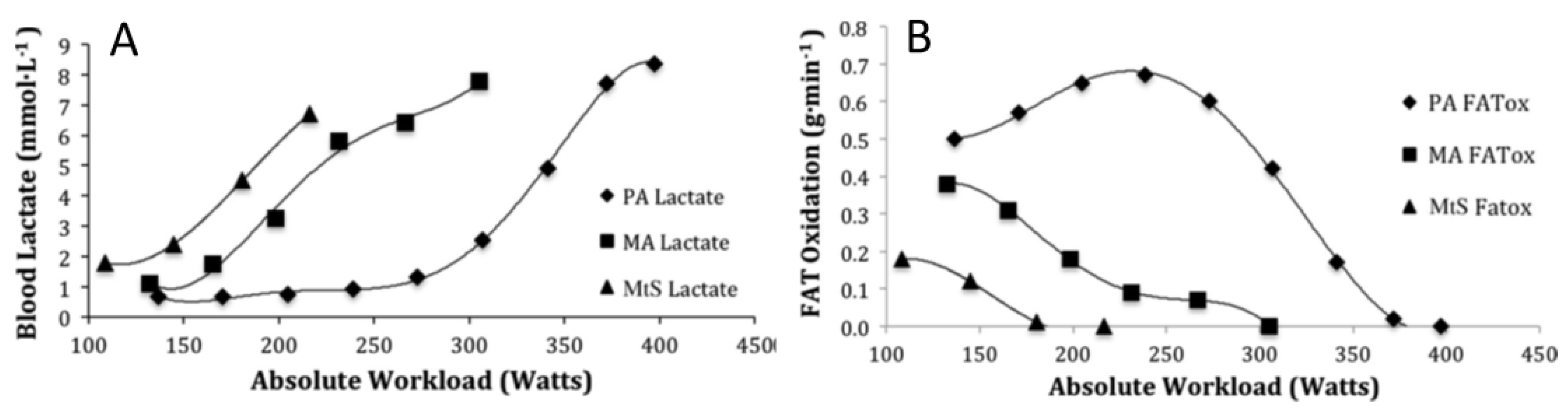
5.2. Oxidation of Lactate and Fatty Acids During Exercise

In this section, we discuss only the energy functions of lipids and carbohydrates, leaving aside numerous regulatory functions, such as acetylation, glycosylation, lactosylation, or the formation of prostaglandins, etc. The functional energy requirtments of different organs vary significantly. For example, the kidneys are constantly working and use only fatty acids as the energy source as glucose is used to reabsorb sodium. In addition, due to active gluconeogenesis, the kidneys, together with the liver, maintain blood glucose homeostasis and produce lactate. Other organs may change their functional load, sometimes many times. For example, skeletal muscle can increase oxygen consumption by 40-fold and the heart by 8-9 fold [63]. The rates of oxidative phosphorylation by the isolated mitochondria from any organs of mice and rats are approximately the same, and are limited by the rate of ATP synthase [5]. Even if the rate of ATP production by mitochondria in vivo would be several times higher than in vitro, mitochondria cannot provide enough ATP if, for example, the heart increases its energy demand by 9 times during intensive physical activity. Obviously, there must be an additional source of energy.
The discovery of the lactate cycle and the forced formation of lactate instead of pyruvate is an important event in the history of Bioenergetics [13,14,28,29]. It becomes evident that lactate plays an important role in the overall energy balance of the human body [5,14]. A good confirmation of this thesis are the data obtained during the studies of metabolic processes in athletes under various physical loads. The main advantage of these studies is that they are carried out on the whole body, although by indirect methods [22,64].
Figure 10 shows changes in the blood lactate (Figure 10A) and fatty acid intake (Figure 10B) depending on the intensity of physical activity [64]. Three groups of people were compared: people with metabolic syndrome (MtS), general people with moderate exercise (MAs), and high-end professional athletes (PAs).
Figure 10A and 10B show that changes in lactate and fatty acid (FA) consumption have different directions. In untrained people, the increase in physical activity leads to increased lactate content and a decrease in the consumption of fatty acids. In professional athletes, the lactate content is kept at a low level for a long time, which indicates a high rate of lactate consumption from the blood (Figure 10A). At the same time, the consumption of fatty acids begins to increase. In athletes, the content of blood lactate begins to increase only with heavy loads, when the consumption of fatty acids begins to decrease. Figure 10B indicates the following important properties of energy metabolism: 1) The rate of consumption of fatty acids is the inverse of the rate of consumption of lactate. This switching from one substrate to another is called “metabolic flexibility.” 2) High adaptability of energy metabolism. Lactate and FA intake can increase greatly with regular exercise. 3) With very intense physical activity, energy metabolism increasingly switches to lactate consumption. [22,64].
In spite of very interesting and inspiering data, we just dicussed, we will not rush to make and globalize the conclusions. Firstly, these results relate primarily to muscle tissue, secondly, these are the results of indirect methods of studying metabolism, and thirdly, we know very little about mitochondrial oxidation of fatty acids and its regulation. Finally, we know too little about the roles of SDH in β-oxidation, and the origins of lactate. We still know very little about the origin of the gender differences in energy metabolism. It is especially important to know gender differences in lipid metabolism [65]. Females of all mammals, including humans, live longer than males, produce fewer superoxide radicals, and age more slowly [66,67]. Women oxidize fatty acids differently during exercise, produce fewer superoxide radicals, and age more slowly [reviewed in [68].

6. Discussion

In mammals, there are two main sources of biological energy: carbohydrates and fatty acids. There are very large differences in the size of the storages and the rate of metabolism between these two energy sources. Fats have large storage, the metabolic pathways of catabolism and formation of triglycerides and phospholipides are very complex and require numerous cofactors (CoA, carnitine, CoQ, biotin). In comparison, the storages of glucose, glycogen, and lactate are limited. For these reasons, the two energy sources have different strategical roles in the body.
We suggest that fatty acids serve as a basic source of energy (ATP, NADPH, gradients), support anabolic and anaplerotic finctions. Catabolism of fatty acids via the minor supercomplex of the respirasome and complex II maintain in the body the high levels of reduction of the NAD and NADP systems, support the lactate cycle by obligatory reduction of pyruvate to lactate and suporting gluconeogenesis.
Carbohydrates, namely glucose and lactate have shorter and less complex metabolic steps. Therefore they have much faster metabolic turnover and more urgent functions in comparison with the fatty acids. Here we consider only energetic functions. Glucose via glycolysis supplies ATP to cell’s compartments, which lack mitochondria. For example in axsons of the CNS. Lactate accumulates in the blood and can quickly supply pyruvate for oxidation to the organs in the body, which increase their functional acivity.
Regetfully, metabolism of carbohydrate and lipids have been studied independently of each other and mostly in the in vitro system. The main difference between the in vitro system and the whole living organism is, that in vitro the connections between various metabolic processes and organs are lost. A striking example of the difference between the in vitro and in vivo metabolic pathways is the fact that in the whole organism, glycolysis always ends in the formation of lactate, not pyruvate [14,22]. As we show in this review, the obligatory formation and oxidation of lactate strongly affects the TCA cycle and mitochondrial oxidation of substrates.

7. Conclusions and Future Perspectives

The numerous discoveries, which have been made during the last several decades, force us to reconsider the energy metabolism of mammals and, most importantly, humans. We must discard old notions, such as aerobic-anaerobic glycolysis, the predominant role of mitochondrial oxidative phosphorylation in the body’s energy supply. Instead of pitting glycolysis and OXPHOS against each other, we must investigate their relationships.
We need to be more careful in transferring the conclusions, made in the in vitro experiments, to the whole organism, and investigate metabolic problems using different approaches. Unfortunately, we still have very little information about the mechanisms of β-oxidation of fatty acids with different aliphatic chain lengths, their physiological roles. We know very little about the key element in the β-oxidation and reverse electron flow: complex II (SDH). Gender differences in metabolism are one of the most pressing problems for biochemistry, physiology and medicine. Preliminary evidence suggests that these studies may have a huge impact on the understanding of the physiology and pathophysiology of metabolic processes in the human body.
And that is not all. In this review, we have touched only a small part of the new discoveries. There are still many analytical problems ahead that arise with the emergence of the new data. For example, how the discovery of the fact that mitochondrial cristae are independent cellular compartments will affect our understanding of the mechanisms of OXPHOS and energy metabolism in general?

Author Contributions

Conceptualization, AP, SD, AK, LYa; Experimental data: AP, VM, SD; Manuscript preparation: AP, AK, LYa, VM, SD; Figures preparation: AP, SD.

Funding

This research received no external funding.

Data Availability Statement

No new data were created or analyzed in this study.

Conflicts of Interest

The authors declare no conflict of interest.

References

  1. Swigonova, Z.; Mohsen, A.W.; Vockley, J. Acyl-CoA dehydrogenases: Dynamic history of protein family evolution. J. Mol. Evol. 2009, 69, 176–193. [Google Scholar] [CrossRef]
  2. Schagger, H.; Pfeiffer, K. Supercomplexes in the respiratory chains of yeast and mammalian mitochondria. EMBO J. 2000, 19, 1777–1783. [Google Scholar] [CrossRef] [PubMed]
  3. Schagger, H. Respiratory chain supercomplexes. IUBMB Life 2001, 52, 119–128. [Google Scholar] [CrossRef]
  4. Wang, Y.; Mohsen, A.W.; Mihalik, S.J.; Goetzman, E.S.; Vockley, J. Evidence for association of mitochondrial fatty acid oxidation and oxidative phosphorylation complexes. J. Biol. Chem. 2010, 285, 29834–29841. [Google Scholar] [CrossRef]
  5. Panov, A.V. The Structure of the Cardiac Mitochondria Respirasome Is Adapted for the Oxidation of Fatty Acids. Int. J. Mol. Sci. 2024, 25, 2410. [Google Scholar] [CrossRef]
  6. Crofts, A. R.; Holland, J.T.; Victoria, D.; Kolling, D.R.; Dikanov, S.A.; R. Gilbreth, R.; Lhee, SN.; Kuras, R.; Kuras, M.G. The Q-cycle reviewed: How well does a monomeric mechanism of the bc(1) complex account for the function of a dimeric complex? Biochim. Biophys. Acta 2008, 1777, 1001–1019. [Google Scholar] [CrossRef]
  7. Brand, M. D. Mitochondrial generation of superoxide and hydrogen peroxide as the source of mitochondrial redox signaling. Free Radic. Biol. Med. 2016, 100, 14–31. [Google Scholar] [CrossRef]
  8. Ikeda, Y.; Okamura-Ikeda, K.; Tanaka, K. Purification and characterization of short-chain, medium-chain, and long-chain acyl-CoA dehydrogenases from rat liver mitochondria. Isolation of the holo- and apoenzymes and conversion of the apoenzyme to the holoenzyme. J. Biol. Chem. 1985, 260, 1311–1325. [Google Scholar] [CrossRef] [PubMed]
  9. Izai, K.; Uchida, Y.; Orii, Y.; Yamamoto, T.; Hashimoto, S.T. Novel fatty acid beta-oxidation enzymes in rat liver mitochondria. I. Purification and properties of very-long-chain acyl-coenzyme A dehydrogenase. J. Biol. Chem. 1992, 267, 1027–1033. [Google Scholar] [CrossRef] [PubMed]
  10. McAndrew, R.P.; Wang, Y.; Mohsen, A.W.; He, M.; Vockley, J.; Kim, J.J. Structural basis for substrate fatty acyl chain specificity: crystal structure of human very-long-chain acyl-CoA dehydrogenase. J. Biol. Chem. 2008, 283, 9435–9443. [Google Scholar] [CrossRef]
  11. Ensenauer, R.; He, M.; Willard, J.M.; Goetzman, E.S.; Corydon, T.J.; et al. Human acyl-CoA dehydrogenase-9 plays a novel role in the mitochondrial beta-oxidation of unsaturated fatty acids. J. Biol. Chem. 2005, 280, 32309–32316. [Google Scholar] [CrossRef] [PubMed]
  12. Nouws, J.; Nijtmans, L.; S. M. Houten, S.M.; van den Brand, M.; Huynen, M.; et al. Acyl-CoA dehydrogenase 9 is required for the biogenesis of oxidative phosphorylation complex I. Cell Metab. 2010, 12, 283–294. [Google Scholar] [CrossRef] [PubMed]
  13. Gladden, L.B. Lactate metabolism: a new paradigm for the third millennium. J. Physiol. 2004, 558, 5–30. [Google Scholar] [CrossRef]
  14. Brooks, G.A.; Arevalo, J.A.; Osmond, J.A.; Leija, A.D.; Curl, R.G.; C.C.; Tovar, A.P. Lactate in contemporary biology: a phoenix risen. J. Physiol. 2022, 600, 1229–1251. [Google Scholar] [CrossRef]
  15. Panov, A.V.; Mayorov, V.I.; Dikalov, S.I. Role of Fatty Acids β-Oxidation in the Metabolic Interactions Between Organs. Int. J. Mol. Sci. 2024, 25, 12740. [Google Scholar] [CrossRef]
  16. Stanley, W.C.; Recchia, F.A.; Lopaschuk, G.D. Myocardial substrate metabolism in the normal and failing heart. Physiol. Rev. 2005, 85, 1093–1129. [Google Scholar] [CrossRef]
  17. Rogatzki, M.J.; Ferguson, B.S.; Goodwin, M.L.; Gladden, L.B. Lactate is always the end product of glycolysis. Front. Neurosci. 2015, 9, 22. [Google Scholar] [CrossRef]
  18. Stanley, J.C.; Perham, R.N. Purification of 2-oxo acid dehydrogenase multienzyme omplexes from ox heart by new method. Biochem. J. 1980, 191, 147–154. [Google Scholar] [CrossRef]
  19. Abeles, Moshe. Corticonics: Neural Circuits of the Cerebral Cortex; Cambridge Univ. Press, 1991; ISSN ISBN 0521374766. [Google Scholar]
  20. Panov, A.; Schonfeld, P.; Dikalov, S.; Hemendinger, R.; Bonkovsky, H.L.; Brooks, B.R. The Neuromediator Glutamate, through Specific Substrate Interactions, Enhances Mitochondrial ATP Production and Reactive Oxygen Species Generation in Nonsynaptic Brain Mitochondria. J. Biol. Chem. 2009, 284, 14448–14456. [Google Scholar] [CrossRef] [PubMed]
  21. Pellerin, L.; Magistretti, P.J. Neuroenergetics: calling upon astrocytes to satisfy hungry neurons. The Neuroscientist 2004, 10, 53–62. [Google Scholar] [CrossRef]
  22. Brooks, G.A. Muscle Fuel Utilization with Glycolysis Viewed Right Side Up. Adv. Exp. Med. Biol. 2025, 1478, 3–18. [Google Scholar]
  23. Adina-Zada, A.; Zeczycki, T.N.; Attwood, P.V. Regulation of the structure and activity of pyruvate carboxylase by acetyl CoA. Arch. Biochem. Biophys. 2012, 519, 118–130. [Google Scholar] [CrossRef] [PubMed]
  24. Valle, M. Pyruvate Carboxylase, Structure and Function. Subcell. Biochem. 2017, 83, 291–322. [Google Scholar] [PubMed]
  25. Panov, A.; Orynbayeva, Z.; Vavilin, V.; Lyakhovich, V. Fatty Acids in Energy Metabolism of the Central Nervous System. Review Article. BioMed. Res. Intern. 2014, 472459. [Google Scholar]
  26. Yudkoff, M.; Nelson, D.; Daikhin, Y.; Erecinska, M. Tricarboxylic acid cycle in rat brain synaptosomes. Fluxes and interactions with aspartate aminotransferase and malate/aspartate shuttle. J. Biol. Chem. 1994, 269, 27414–27420. [Google Scholar] [CrossRef]
  27. Ratnakumari, L.; Murthy, C.R. Activities of pyruvate dehydrogenase, enzymes of citric acid cycle, and aminotransferases in the subcellular fractions of cerebral cortex in normal and hyperammonemic rats. Neurochem. Res. 1989, 14, 221–228. [Google Scholar] [CrossRef]
  28. Brooks, G.A. Lactate as a fulcrum of metabolism. Redox Biol. 2020, 35, 101454. [Google Scholar] [CrossRef]
  29. Brooks, G. A. The Science and Translation of Lactate Shuttle Theory. Cell. Metab. 2018, 27, 757–785. [Google Scholar] [CrossRef]
  30. Kane, D. A. Lactate oxidation at the mitochondria: a lactate-malate-aspartate shuttle at work. Front. Neurosci. 2014, 8, 366. [Google Scholar] [CrossRef]
  31. Erecinska, M.; Nelson, D. Effects of 3-nitropropionic acid on synaptosomal energy and transmitter metabolism: relevance to neurodegenerative brain diseases. J. Neurochem. 1994, 63, 1033–1041. [Google Scholar] [CrossRef] [PubMed]
  32. Sazanov, L.A.; Jackson, J.B. Proton-translocating transhydrogenase and NAD- and NADP-linked isocitrate dehydrogenases operate in a substrate cycle which contributes to fine regulation of the tricarboxylic acid cycle activity in mitochondria. FEBS Letters 1994, 344, 109–l16. [Google Scholar] [CrossRef]
  33. Costello, L.C.; Franklin, R.B. Bioenergetic theory of prostate malignancy. The Prostate 1994, 25, 162–166. [Google Scholar] [CrossRef]
  34. Panov, A.; Orynbaeva, Z. Bioenergetic and antiapoptotic properties of mitochondria from cultured human prostate cancer cells PC-3, DU145 and LNCaP. PLoS ONE 2013, 8, e72078. [Google Scholar] [CrossRef] [PubMed]
  35. Panov, A. Practical Mitochondriology. Pitfalls and Problems in Studies of Mitochondria; Lexington, KY, 2013; ISBN 9781483963853. [Google Scholar]
  36. Panov, A.; Orynbayeva, Z. Determination of mitochondrial metabolic phenotype through investigation of the intrinsic inhibition of succinate dehydrogenase. Analyt. Biochem. 2018, 552, 30–37. [Google Scholar] [CrossRef]
  37. Panov, A.; Dikalov, S. Brain energy Metabolism. In Encyclopedia in Biochemstry, 3rd Edition ed; 2021; pp. Pages 1–16. [Google Scholar] [CrossRef]
  38. Panov, A.V.; Mayorov, V.I.; Dikalova, A.E.; Dikalov, S.I. Long-Chain and Medium-Chain Fatty Acids in Energy Metabolism of Murine Kidney Mitochondria. Int. J. Mol. Sci. 2023, 24, 379. [Google Scholar] [CrossRef]
  39. Hatefi, Y. The mitochondrial electron transport and oxidative phosphorylation system. Annu. Rev. Biochem. 1985, 54, 1015–1069. [Google Scholar] [CrossRef]
  40. Althoff, T.; Mills, D.J.; Popot, J.L.; Kuhlbrandt, W. Arrangement of electron transport chain components in bovine mitochondrial supercomplex I1III2IV1. EMBO J. 2011, 30, 4652–4664. [Google Scholar] [CrossRef]
  41. Muhleip, A.; Flygaard, R.K.; Baradaran, R.; Haapanen, O.; Gruhl, T.; et al. Structural basis of mitochondrial membrane bending by the I-II-III(2)-IV(2) supercomplex. Nature 2023, 615, 934–938. [Google Scholar] [CrossRef] [PubMed]
  42. Zhou, L.; Maldonado, M.; Padavannil, A.; Guo, F.; Letts, J.A. Structures of Tetrahymena’s respiratory chain reveal the diversity of eukaryotic core metabolism. Science 2022, 376, 831–839. [Google Scholar] [CrossRef] [PubMed]
  43. Sousa, J.S.; Mills, D.J.; Vonck, J.; Kuhlbrandt, W. Functional asymmetry and electron flow in the bovine respirasome. Elife 2016, 5, e21290. [Google Scholar] [CrossRef]
  44. CRC Handbook of Chemistry and Physics. 56th Edition; Weast, Robert C., Ed.; CRC Press; pp. 1975–1976.
  45. Chretien, D.; Benit, P.; Ha, H.H.; Keipert, S.; El-Khoury, R.; et al. Mitochondria are physiologically maintained at close to 50 degrees C. PLoS Biol. 2018, 16, e2003992. [Google Scholar] [CrossRef] [PubMed]
  46. Javadov, S.; Jang, S.; Chapa-Dubocq, X.R.; Khuchua, Z.; Camara, A.K. Mitochondrial respiratory supercomplexes in mammalian cells: structural versus functional role. J. Mol. Med. 2021, 99, 57–73. [Google Scholar] [CrossRef] [PubMed]
  47. Milenkovskaya, E.; Dowhan, W. Cardiolipin-dependent formation of mitochondrial respiratory supercomplexes. Chem. Phys. Lipids 2014, 179, 42–48. [Google Scholar] [CrossRef]
  48. Nesci, S.; Trombetti, F.; Pagliarani, A.; Ventrella, V.; Algieri, C.; et al. Molecular and Supramolecular Structure of the Mitochondrial Oxidative Phosphorylation System: Implications for Pathology. Life (Basel) 2021, 11, 242. [Google Scholar] [CrossRef]
  49. Vercellino, I.; Sazanov, L.A. SCAF1 drives the compositional diversity of mammalian respirasomes. Nat. Struct. Mol. Biol. 2024, 31, 1061–1071. [Google Scholar] [CrossRef]
  50. Wu, M.; Gu, J.; Guo, R.; Huang, Y.; Yang, M. Structure of Mammalian Respiratory Supercomplex I(1)III(2)IV(1). Cell. 2016, 167, 1598–1609.e1510. [Google Scholar] [CrossRef]
  51. Panov, A.; Mayorov, V.; Dikalov, S. Metabolic Syndrome and beta-Oxidation of Long-Chain Fatty Acids in the Brain, Heart, and Kidney Mitochondria. Int. J. Mol. Sci. 2022, 23. [Google Scholar] [CrossRef]
  52. Brischigliaro, M.; Cabrera-Orefice, A.; Arnold, A.; Viscomi, C.; Zeviani, M.; Fernandez-Vizarra, E. Structural rather than catalytic role for mitochondrial respiratory chain supercomplexes. Elife 2023, 12, RP88084. [Google Scholar] [CrossRef]
  53. Brzezinski, P.; Moe, A.; Adelroth, A.P. Structure and Mechanism of Respiratory III-IV Supercomplexes in Bioenergetic Membranes. Chem. Rev. 2021, 121, 9644–9673. [Google Scholar] [CrossRef]
  54. Bultema, J.B.; Braun, H.P.; Boekema, E.J.; Kouril, R. Megacomplex organization of the oxidative phosphorylation system by structural analysis of respiratory supercomplexes from potato. Biochim. Biophys. Acta 2009, 1787, 60–67. [Google Scholar] [CrossRef] [PubMed]
  55. Dudkina, N.V.; Kouril, R.; Peters, K.; BraunBoekema, H.P. Structure and function of mitochondrial supercomplexes. Biochim. Biophys. Acta 2021, 1797, 664–670. [Google Scholar] [CrossRef] [PubMed]
  56. Guo, R.; Zong, S.; Wu, M.; Gu, J.; Yang, M. Architecture of Human Mitochondrial Respiratory Megacomplex I(2)III(2)IV(2). Cell. 2017, 170, 1247–1257 e1212. [Google Scholar] [CrossRef]
  57. Hoek, J.B.; Rydstrom, J. Physiological roles of nicotinamide nucleotide transhydrogenase. Biochem. J. 1988, 254, 1–10. [Google Scholar] [CrossRef]
  58. Quinlan, C.L.; Perevoshchikova, I.V.; Hey-Mogensen, M.; Orr, A.L.; Brand, M.D. Sites of reactive oxygen species generation by mitochondria oxidizing different substrates. Redox. Biol. 2013, 1, 304–312. [Google Scholar] [CrossRef]
  59. Perevoshchikova, I.V.; Quinlan, C.L.; Orr, A.O.; Gerencser, A.A.; Brand, M.D. Sites of superoxide and hydrogen peroxide production during fatty acid oxidation in rat skeletal muscle mitochondria. Free Radic. Biol. Med. 2013, 61C, 298–309. [Google Scholar] [CrossRef] [PubMed]
  60. Elustondo, P.A.; White, A.E.; Hughes, M.E.; Brebner, K.; Pavlov, E.; Kane, D.A. Physical and functional association of lactate dehydrogenase (LDH) with skeletal muscle mitochondria. J. Biol. Chem. 2013, 288, 25309–25317. [Google Scholar] [CrossRef]
  61. Young, A.; Oldford, C.; Mailloux, R.J. Lactate dehydrogenase supports lactate oxidation in mitochondria isolated from different mouse tissues. Redox Biol. 2020, 28, 101339. [Google Scholar] [CrossRef]
  62. Altinok, O.; Poggio, J.L.; Stein, D.E.; Bowne, W.B.; Shieh, A.C.; et al. Malate-aspartate shuttle promotes l-lactate oxidation in mitochondria. J. Cell. Physiol. 2020, 235, 2569–2581. [Google Scholar] [CrossRef] [PubMed]
  63. Gonzalez-Freire, M.; de Cabo, R.; Bernier, M.; Sollott, S.J.; Fabbri, E.; et al. Reconsidering the Role of Mitochondria in Aging. J. Gerontol. A. Biol. Sci. Med. Sci. 2015, 70, 1334–1342. [Google Scholar] [CrossRef]
  64. San-Millan, I.; Brooks, G.A. Assessment of Metabolic Flexibility by Means of Measuring Blood Lactate, Fat, and Carbohydrate Oxidation Responses to Exercise in Professional Endurance Athletes and Less-Fit Individuals. Sports Med. 2018, 48, 467–479. [Google Scholar] [CrossRef]
  65. Wang, Z.; Ying, Z.; Bosy-Westphal, A.; Zhang, J.; Heller, M.; et al. Evaluation of specific metabolic rates of major organs and tissues: comparison between men and women. Am. J. Hum. Biol. 2011, 23, 333–338. [Google Scholar] [CrossRef] [PubMed]
  66. Vina, J.; Borras, C.; Gambini, J.; Sastre, J.; Pallardo, F.V. Why females live longer than males? Importance of the upregulation of longevity-associated genes by oestrogenic compounds. FEBS Lett. 2005, 579, 2541–2545. [Google Scholar] [CrossRef] [PubMed]
  67. Ballard, J.W.; Melvin, R.G.; Miller, J.T.; Katewa, S.D. Sex differences in survival and mitochondrial bioenergetics during aging in Drosophila. Aging Cell. 2007, 6, 699–708. [Google Scholar] [CrossRef] [PubMed]
  68. Panov, A.V.; Darenskaya, M.A.; Dikalov, S.I.; Kolesnikov, S.I. Metabolic syndrome as the first stage of eldership; the beginning of real aging. In Update in Geriatrics; Published; Somarnyotin, Somchai, Ed.; 14th April 2021; pp. Pp. 37–67. ISBN 978-1-83962-309-7. [Google Scholar] [CrossRef]
Figure 1. Structure and function of the mitochondrial respiratory chain according to the old paradigm. A. An early scheme of the respiratory chain; B. A more sophisticated presentation of the respiratory chain (this figure was adopted from Internet: Libretext. Biology. H. Yakubowski; P. Flatt).
Preprints 194877 g001
Figure 2. Classical presentation of the tricarboxylic acid cycle.
Preprints 194877 g002
Figure 3. Schematic presentation of the the TCA cycle work in the respiring mitochondria in the presence of malate-aspartate shuttle. Abbreviations: AST – aspartate aminotransferase, ALT – alanine aminotransferase, SDH succinate dehydrogenase. The red lock indicates the place of inhibition of succinate dehydrogenase activity by malonate and OAA. The dotted red lines from malate and oxaloacetate mean that they are also succinate dehydrogenase inhibitors. The figure was adapted from [20].
Preprints 194877 g003
Figure 4. Malate-Aspartate Shunt (MAS). The inner mitochondrial membrane is impermeable to NADH. In order to effectively use lactic acid as a substrate for respiration, lactate must first be converted to pyruvate in the reaction: Lactate + NAD+ → Pyruvate + NADH + H+. In energized mitochondria, MAC causes lactate dehydrogenase to work irreversibly towards proton oxidation. The driving force for this is the electrogenic exchange of aspartate for glutamate. Abbreviations: AST – aspartat aminotransferasa; α-kg – α-ketoglutarat; GAT-glutamat/aspartat transporter; MKgT - malat/ketoglutarate transporter; MDH – malate dehydrogenase; OAA- oxaloacetate. The Figure was adapted from [25].
Preprints 194877 g004
Figure 5. Functioning of the TCA cycle in the prostate mitochondria. Inhibition of aconitase by zinc ions leads to the accumulation of citrate and inhibition of the minor B cycle. The figure was adapted from [34].
Preprints 194877 g005
Figure 6. Oxygen consumption by rat brain mitochondria, isolated without BSA, oxidizing various substrates and their mixtures in different metabolic states.
Preprints 194877 g006
Figure 7. Schematic structure of the respirasome as suggested by Schägger [2].. The figure depicts two large and one small supercomplexes of the respiratory chain carriers. They are integral structures, which span the inner membrane and the figure shows view from the matrix side.The figure was adapted from [51].
Preprints 194877 g007
Figure 8. A schematic model on scale of the organization of respiratory chain complexes into respiratory string. The basic unit consists of two copies of complex I (blue), one copy of complex III2 (red), and two copies of complex IV (yellow). [54].
Preprints 194877 g008
Figure 9. Functioning of the respirasome and the tricarboxylic acid cycle during active β-oxidation of long-chain fatty acids. Abbreviations: Acyl-CoA DHC—acyl-CoA dehydrogenase complex, which includes three enzymes: acyl-CoA dehydrogenase, electron transfer flavoprotein (ETF), electron-transferring-flavoprotein dehydrogenase (ETFDH); PEP—phosphoenolpyruvate; TFP—trifunctional protein of the β-oxidation of fatty acids system; SDH—succinate dehydrogenase; Q—ubiquinone, oxidized form of coenzyme Q; QH2—ubiquinol, reduced form of coenzyme Q. The figure adapted from [38].
Preprints 194877 g009
Figure 10. A, Relationship between mean blood lactate level and exercise power output; 10B, Relationship between mean FATox (fatty acid oxidation) and exercise power output: Pas are professional endurance athletes, MA are moderately active healthy people, and MtS are people with metabolic syndrome [64].
Preprints 194877 g010
Disclaimer/Publisher’s Note: The statements, opinions and data contained in all publications are solely those of the individual author(s) and contributor(s) and not of MDPI and/or the editor(s). MDPI and/or the editor(s) disclaim responsibility for any injury to people or property resulting from any ideas, methods, instructions or products referred to in the content.
Copyright: This open access article is published under a Creative Commons CC BY 4.0 license, which permit the free download, distribution, and reuse, provided that the author and preprint are cited in any reuse.
Prerpints.org logo

Preprints.org is a free preprint server supported by MDPI in Basel, Switzerland.

Subscribe

Disclaimer

Terms of Use

Privacy Policy

Privacy Settings

© 2026 MDPI (Basel, Switzerland) unless otherwise stated