
Submitted:
16 January 2026
Posted:
19 January 2026
You are already at the latest version
Abstract
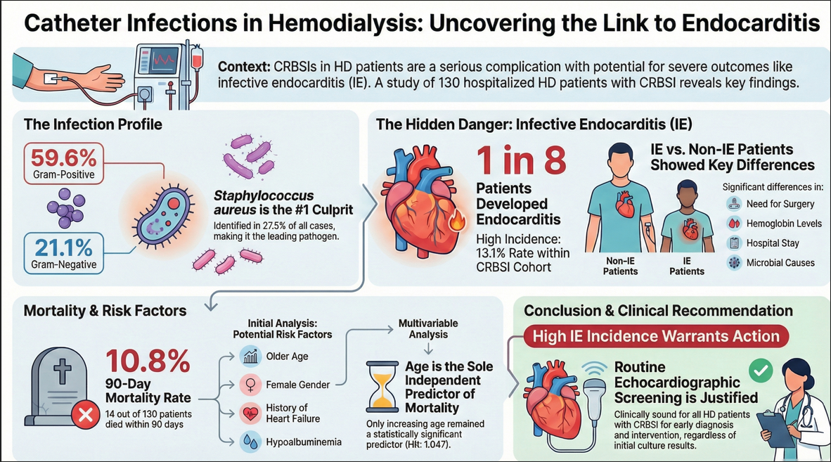
Background and Objectives: Catheter-related bloodstream infections (CRBSI) and infective endocarditis (IE) lead to substantial morbidity, prolonged hospitalizations, and increased mortality. This study aimed to determine the incidence of IE among hospitalized HD patients with CRBSI and identify risk factors associated with 90-day all-cause mortality. Materials and Methods: We conducted a retrospective analysis of patients diagnosed with CRBSI. Clinical, microbiological, and echocardiographic data were evaluated. Risk factors for 90-day mortality were analyzed using univariate analysis and multivariable Cox proportional hazards regression models. Results: A total of 130 patients were included. Gram-positive organisms were the predominant pathogens (59.6%), with Staphylococcus aureus identified in 27.5% (n=30) of cases. Gram-negative bacteria accounted for 21.1% of infections. IE was diagnosed in 17 patients, representing an incidence of 13.1% within the CRBSI cohort. Significant differences were observed between the IE and non-IE groups regarding the need for surgery, hemoglobin levels, length of hospital stay, and microbial etiology (p < 0.05). The 90-day all-cause mortality rate was 10.8% (n=14). Univariate analysis identified older age, female gender, history of heart failure, and hypoalbuminemia as factors associated with increased mortality (p < 0.05). In the multivariable Cox regression, age remained the sole independent predictor of 90-day mortality (Hazard Ratio: 1.047; 95% CI: 1.0–1.096; p=0.048). Conclusions: Staphylococcus aureus is the leading pathogen in HD patients with CRBSI and IE. Given the 13.1% IE incidence, routine echocardiographic screening represents a clinically sound and justified strategy for patients with CRBSI, regardless of initial culture results, to ensure early diagnosis and intervention.
Keywords:
hemodialysis
; catheter-related bloodstream infection
; infective endocarditis
; Staphylococcus aureus
; mortality
; echocardiography
1. Introduction
Catheter-related bloodstream infections (CRBSI) remain a formidable challenge in maintenance hemodialysis (HD), significantly escalating morbidity and healthcare cost [1]. Patients undergoing maintenance HD often necessitate prolonged central venous catheter (CVC) dependence, which is inherently associated with an elevated risk of CRBSI [2]. CRBSI typically arises via extra or intraluminal colonization, which is often dictated by the duration of catheter placement. Catheters inserted for short periods (e. g, less than 7 days) are frequently colonized via the intraluminal route, typically originating from hub contamination [3].
CRBSI in the chronic HD population is a critical public health concern, consistently linked to substantial increases in morbidity, prolonged hospitalization, and premature death [4]. A number of conditions and patient factors predispose individuals to CRBSI, including prolonged CVC duration, diabetes mellitus, advanced age, anemia, a history of previous catheter-related bacteremia, hypoalbuminemia, poor personal hygiene, moisture at the exit-site, Staphylococcus aureus nasal colonization, and specific catheter insertion techniques and sites [5,6,7]. Recent epidemiological evidence demonstrates a substantial difference in CRBSI rates based on device type: temporary CVCs exhibit an incidence of 3.25–10.8 events per 1,000 days of use, a frequency considerably higher than the 0.55–4.4 events recorded for permanent CVC [5]. A landmark meta-analysis confirms that catheter-reliant patients face a 1.5-fold increase in all-cause mortality and a more than twofold increase in fatal infection risk compared to those with fistulas [8]. The predominant causative agents for CRBSIs are Gram-positive bacteria, which constitute up to 80% of cases and primarily include Staphylococcus aureus and Coagulase−negative staphylococci [5,9,10].
The primary cause of IE is the pathogenic infiltration of the endocardium by microorganisms, including bacterial and fungal species [11]. The clinical consequence of this invasion is characterized by rapid destruction of the heart valve or the delicate lining of the ventricular wall. The incidence of IE in HD patients is reported to be between 2% and 5%, with recent studies indicating an alarmingly high mortality rate of 41.3% to 56% [12,13]. Staphylococcus aureus is also the predominant causative organism in this setting, responsible for 37% to 65% of IE cases [14].
This study aimed to determine the incidence of IE among hospitalized HD patients due to CRBSI and to identify the risk factors associated with 90-day all-cause mortality in this high-risk population.
2. Materials and Methods
2.1. Patient Selection Criteria, Study Design
This retrospective study utilized data from hospitalized HD patients at the nephrology department of Akdeniz University Hospital, a tertiary academic medical center serving a population of 2.7 million inhabitants in Antalya, Turkey. HD patients with suspected CRBSI from various surrounding centers and our in-center HD patients were selected. Comprehensive clinical data were extracted from electronic medical records using a standardized data collection form. The present study followed the principles of the Declaration of Helsinki. The study protocol received official approval from the local ethics committee (Antalya Akdeniz University Hospital Local Ethics Committee; 2023-336, 26 April 2023)
Patients were identified by searching the database for all admissions from January 2016 to December 2021 with a final diagnosis of definite, probable or possible CRSBI and IE per the modified Duke criteria [1,15]. Patients referred from external centers were admitted either through the emergency department or the nephrology outpatient clinic. Patients admitted to the nephrology inpatient service between the specified dates were individually examined, and those evaluated by the infectious disease specialists and diagnosed with CRBSI were included in the study. This study defines a suspected case of CRBSI as a patient with an indwelling dialysis catheter who presents with signs and symptoms of fever (>38 °C) or chills with no other obvious source of infection. The National Kidney Foundation Kidney Disease Outcomes Quality Initiative [1] definition of CRBSI was used. IE diagnosis has been based on the modified DUKE criteria [15]. Definite, probable or possible CRSBI and IE per the modified DUKE criteria are explained Appendix A.
2.2. Data Collection
Demographic information, duration and type of indwelling catheter, comorbidities (diabetes mellitus, hypertension, immunosuppressant use, congestive heart failure), predisposing cardiac conditions, causative organisms, laboratory results, echocardiographic findings, complications, length of hospitalization, and 90-day all-cause mortality were recorded. For all CRBSI patients, transthoracic echocardiography was performed to evaluate vegetation, cardiac function indices, and pulmonary artery pressure values. Echocardiographic evaluation could not be completed for 24 patients (18.5%). Given that this is a retrospective study, it is plausible that the lack of echocardiographic data for some patients was due to logistical difficulties in scheduling appointments during their hospitalization period. Transesophageal echocardiography was utilized when deemed necessary. In addition, extracardiac complications such as emboli to vital organs were documented. Septic pulmonary emboli were diagnosed using computed tomography (CT) of the lungs.
The inclusion criteria were patients aged 18 years and above, diagnosed with end-stage kidney disease and receiving maintenance HD, and having a CVC in situ for at least 7 days. Patients who were considered to be a source of infection other than catheter infection (such as pneumonia and urinary tract infection) were not included in the study.
2.3. Microbiological Methods
Simultaneous blood cultures from the catheter hub and a peripheral vein were obtained whenever possible, followed by the immediate administration of empirical antibiotics. At least two sets of blood cultures (for aerobic and anaerobic incubation) were obtained at the disease onset. Organism identification and antimicrobial susceptibility patterns were determined using standardized automated methods.
2.4. Statistical Analyses
All the collected data was entered into the Statistical Package for the Social Sciences version 22 software for further data cleaning and statistical analysis. Descriptive statistics were used to summarize the demographic and clinical characteristics. Continuous variables are reported as mean ± standard deviation for normally distributed data or median (interquartile range) for skewed data. Categorical variables are reported as numbers (percentages). Comparisons between the groups were made using the chi-square test for categorical data. For continuous data, normality was assessed using the Shapiro-Wilk test. If the data were normally distributed, an independent sample t-test was used between groups. For non-normally distributed continuous variables, the Mann–Whitney U test was applied. We also used a multiple logistic regression to look for the risk factors and confounders associated with the outcome (mortality).
3. Results
3.1. Demographic Data and Baseline Characteristics
The study cohort comprised 130 patients. The median age was 55.5 years (Interquartile Range: 42–66). The majority were men (57%), and common comorbidities included hypertension (60.8%) and diabetes mellitus (42.3%). Diabetic kidney disease was the main etiology of end stage kidney disease. 25 patients (19.2%) were on immunosuppressants and had received a renal transplantation. Six patients (4.6%) had malignancies. Three patients (2.3%) had a peritoneal dialysis history. Table 1 provides a comprehensive summary of the patients' characteristics, laboratory results and echocardiographic results. 24 patients did not have echocardiographic results.
77% of patients had a tunneled catheter, with the internal jugular vein being the primary insertion site (56%); the right side was chosen in 58 (45%) patients. The current locations of the catheters were as follows: right jugular 58 (44,6%), left jugular 16 (12,3%), right femoral 15 (11,5%), left femoral 14 (10.8%), right subclavian 18(13.8%), left subclavian 7 (5.4%), transhepatic 1(0.8%), missing 1(0.8%).
CRBSI was classified as definite in 93 patients, probable in 30, and possible in 17. Notably, infective endocarditis (IE) was diagnosed in 13.1% (n=17) of the CRBSI cohort (13 definite IE, 4 possible IE). The most frequent pathogen in the IE group was Staphylococcus aureus (8 patients, 56%). 16 vegetations were detected. Four patients underwent valve surgery, and four patients (3.1%) experienced extra-cardiac embolic events, with septic pulmonary embolism being the most common. 14 of 130 patients (10.8%) died within 90 days of admission (Table 1).
3.2. Microbiology Profiles
Only one episode of catheter infection was recorded per patient. Table 2 shows the etiology of catheter-related bacteremia from catheter culture and blood culture. Gram-positive organisms were responsible for the majority of bacteremia cases: 59.6% from catheter cultures (30 cases of Staphylococcus aureus) and 52.4% from blood cultures (31 cases of Staphylococcus aureus). Gram-negative bacteria accounted for 21.1% of catheter cultures and 21.8% of blood cultures. In particular, all Gram-positive bacterial isolates in the IE group were Staphylococcus aureus. Polymicrobial growth was detected in seven patients, with six cases involving different Gram-negative organisms and one case involving different Gram-positive organisms. Bacteremia caused by Candida glabrata only affected 0.8% of patients.
3.3. Comparison Between Non-IE and IE Patients
A clear statistical difference was found between the IE and non-IE groups with respect to vegetation, embolism, need for surgery, hemoglobin levels, length of hospitalization, and segmental wall motion abnormalities (p<0.05) (Table 1). Segmental wall motion deficits were significantly more prevalent in the IE group, even when accounting for a pre-existing heart failure diagnosis (p<0.05). There were no differences between groups regarding echocardiographic results except for segmental movement deficits. All Gram-positive bacterial isolates of IE were Staphylococcus aureus.
3.4. Mortality
14 out of 130 patients (10.8%) died within 90 days of admission. In-hospital mortality was 4.4% (5/113) for the non-IE group and 5.9% (1/17) for the IE group. 90-day all-cause mortality was similar in both groups. Univariate logistic regression for 90-day all-cause mortality identified age, female gender, heart failure, need for surgery, and albumin as statistically significant dependent variables(p<0.05) (Table 5).
Multivariable stepwise Cox proportional hazards regression analysis confirmed that age (Hazard Ratio: 1.047; 95% CI: 1.0–1.096; p=0.048) was the only independent significant predictor of 90-day all-cause mortality in the total CRBSI cohort (Table 6).
4. Discussion
To the best of our knowledge, this is the first study to have examined only the IE incidence rate and 90-day all-cause mortality for inpatient hemodialysis population with CRBSI. 90-day all-cause mortality was 10.8% (14/130). Although univariate analysis identified older age, female gender, history of heart failure, and lower albumin levels as factors associated with increased mortality, in the multivariable stepwise Cox proportional hazards regression analysis, age remained the only independent factor significantly associated with 90-day all-cause mortality.
The high prevalence of tunneled catheters (77%) in our cohort aligns with trends reported in other regions, including United States Renal Data System (USRDS) and the REIN register in France, and in Turkey, respectively 72.2%, 56.7% and 68.7 % of patients initiate HD, where CVCs unfortunately remain the predominant mode of vascular access for initiating renal replacement therapy due to the insufficient arteriovenous fistulas formation before renal replacement therapy [16,17,18]. This high catheter usage undoubtedly contributes to the elevated risk and subsequent cost associated with CRBSI and its complications.
Various studies have demonstrated the incidence of bacteremia (possible or confirmed) as 2.27 and 16.6 per 1,000 catheter-days for non-tunneled catheters and 0.37 to 10.8 per 1,000 catheter-days for tunneled catheters. Epidemiological findings indicate that the risk of bacteremia (encompassing both confirmed and suspected cases) is significantly differentiated by catheter type. Non-tunneled devices exhibit an incidence of 2.27 to 16.6 episodes per 1,000 days of use, whereas tunneled catheters present a lower, yet still broad, range of 0.37 to 10.8 episodes per 1,000 catheter-days [19,20].
Gram-positive organisms predominated in our cohort, accounting for 59.6% of catheter tip isolates and 52.4% of positive blood cultures. Within this group, Staphylococci emerged as the most frequent pathogens. Specifically, Staphylococcus aureus was identified in 27.5% (n=30) of catheter tips and 25% (n=31) of blood cultures, representing the leading cause of infection in both sites. Gram-negative bacteria were isolated in 21.1% of CRBSI cases, and 18.3% of cultures showed no bacterial growth from catheter tips. The microbial distribution in our study population aligns with previously published data. Gram-positive aerobes remain the predominant pathogens in CRBSIs, with Staphylococcus aureus and Coagulase-negative staphylococci accounting for 60%–80% of cases involving tunneled catheters [21,22]. Conversely, Gram-negative bacilli contribute to roughly 20% of CRBSIs [23].
CRBSI in HD patients, particularly those caused by Staphylococcus aureus, have been associated with a higher risk for hematogenous complications [23,24]. In our study, all cases of endocarditis with Gram-positive bacterial isolates were caused by Staphylococcus aureus and were associated with a higher rate of hematogenous complications (3 out of 4 patients). The sole Gram-negative case presenting with septic pulmonary emboli was caused by Stenotrophomonas maltophilia. In one study, Farrington et al. found polymicrobial infections in 40 (14%) of 289 patients with CRBSI [4]. In our study population, dual microorganism growth was detected in seven (5.4%) patients. Among these, six showed a combination of Gram-negative growths, while one presented with Gram-positive growth. We observed an 18.3% culture negativity rate. This relatively high rate may be attributed to the limited use of anaerobic cultures in our study, potentially resulting in an under-reporting of positive cultures due to missed diagnoses of fastidious bacteria.
Data regarding vascular access sites confirm that catheters placed in the internal jugular vein carry a reduced probability of infection. This pattern aligns with existing studies which have demonstrated that the femoral site is associated with a greater likelihood of bacteremia than either the subclavian or internal jugular insertion points [21,22,23,25]. Although in our study the internal jugular vein was the main of placement in 74 (56%) patients, we still encountered CRBSI.
Patients undergoing HD experience a significantly magnified risk of IE, with an incidence rate of 1.7–2.0 cases per 1,000 patients. This figure highlights a disparity of over 50-fold compared to the risk level found in those without end-stage renal disease [12]. A meta-analysis including 45,799 patients with IE on HD showed that approximately 2.7%–3.1% of the HD group suffer from endocarditis [12]. The higher incidence of IE observed in our study compared to previous reports can be attributed to our specific inclusion criteria, which focused exclusively on patients hospitalized with CRBSIs. In contrast to other studies where catheter use ranged from 26.4% to 79.4% [14,26,27,28], our cohort consisted entirely of (100%) patients undergoing HD via catheters—a population inherently at the highest risk for developing IE. Data from a 2024 retrospective study show that among a cohort of 254 CRBSI patients, the HD-specific subgroup (n=12) exhibited an infective endocarditis rate of 8.3% [23].
A meta-analysis found an overall in-hospital death rate of 29.5% (95% CI: 26.7%–46.6%) IE in the context of HD [12]. Specifically, the one-year survival prognosis for HD patients affected by IE in the United States is reported to be 46% [29]. In our study, one of the 17 IE patients died while hospitalization, and two of the remaining 16 patients died within 90 days. Our study did not yield a statistically significant difference in survival outcomes between the IE and non-IE groups; this lack of significance may be attributed to the limited sample size.
Some investigators found that females predominated in IE cases among HD patients, whereas other investigators noted a majority of males [26,27,29,30]. This disparity is potentially attributable to geographical variations among the investigated cohorts. In our study, there was no difference between sexes for IE, but female patients had a higher mortality rate in CRBSI cases.
The optimal management strategy for IE in HD patients remains a subject of debate. A recent meta-analysis by Ting et al. confirmed that survival outcomes do not significantly differ between surgical and medical interventions in this high-risk population [31]. Furthermore, the prognosis remains particularly grim for those requiring surgery; Elderia et al. recently reported a 1-year mortality rate as high as 75.6% [11]. Our findings align with these observations, as 50% (n=2/4) of our surgically managed patients died within 90 days.
In some studies it was reported negative blood cultures in nearly half of their study population [13,28,30]. Wang et all. reported positive culture rates exceeding 70% in their investigations [2]. In light of the high prevalence of Staphylococcus aureus reported both in our cohort and by Stahl et al. (58%) [32], clinicians should maintain a high index of suspicion for IE particularly when this pathogen is isolated [33]. Given that IE can occur even in the presence of negative, Gram-negative, or fungal cultures [14,32], and is associated with high mortality rates regardless of surgical intervention, echocardiography should be considered a standard diagnostic procedure for all hemodialysis patients presenting with suspected CRBSI. Both IE can occur even with negative, Gram-negative or fungal cultures [14,32] and IE is a condition with a high mortality rate (even though surgery is done) echocardiography should remain a standard diagnostic tool for all HD patients with suspected CRBSI.
Several methodological limitations should be considered when interpreting these results. Foremost, the reliance on a retrospective, observational design utilizing registry data inherently precludes the inference of definitive causal relationships. While the current study may be underpowered to detect subtle survival differences, the observed trends suggest that albumin, heart failure history warrants further investigation in larger cohorts. As this study included all hospitalized patients from both our center and the surrounding region, we were unable to determine the specific incidence rates of CRBSI and IE exclusive to our own HD unit.
5. Conclusions
This 7-year study highlights that CRBSI remains a major infectious complication in HD patients, with Staphylococcus aureus as the predominant pathogen in both CRBSI and IE. Age was found to be the only independent predictor of 90-day all-cause mortality within our specific cohort, given the limitations of the study (small number of participants). Our study identifies a notable 13.1% incidence of IE among HD patients hospitalized with CRBSI. In light of these findings, implementing routine echocardiographic screening for this high-risk population without awaiting culture results appears to be a clinically sound and justified strategy. Preventive efforts should focus on modifiable risk factors—particularly stringent infection control practices, reducing Gram-positive growth, standardized catheter care, and early transition to arteriovenous fistulas. Future large-scale multicenter studies are warranted to further elucidate survival predictors for hospitalized hemodialysis patients complicated by CRBSI and IE.
Author Contributions
Conceptualization, F.B., and F.S.; methodology, F.B., O.E.Y and U.C.; validation, O.E.Y., and U.C.; formal analysis, F.B., and U.C. investigation, F.B., U.C., and O.E.Y.; data curation, O.E.Y., and U.C.; writing-original draft preparation F.B. and O.E.Y.; writing-review and editing, F.B, U.C., and F.S.; supervision, F.B., and F.S.; all authors have read and agreed to the published version of the manuscript.
Funding
This research received no external funding.
Institutional Review Board Statement
The present study followed the principles of the Declaration of Helsinki. The study protocol received official approval from the local ethics committee (Akdeniz University Hospital Local Ethics Committee; 2023-336, 26 April 2023).
Informed Consent Statement
Informed consent was waived due to the retrospective nature of this study.
Data Availability Statement
data is unavailable due to privacy or ethical restrictions; a statement is still required.
Conflicts of Interest
The authors declare no conflict of interest.
Abbreviations
The following abbreviations are used in this manuscript:
| CI | Confidence interval |
| CKD | Chronic kidney disease |
| CRBSI | Catheter-related bloodstream infections |
| CVC | Central venous catheter |
| HD | Hemodialysis |
| IE | Infective endocarditis |
| ADPKD | Autosomal Dominant Polycystic Kidney Disease |
Appendix A
Definition of catheter-related bloodstream infections (CRBSI) and Infective Endocarditis (IE)
This study defines a suspected case of CRBSI as a patient with an indwelling dialysis catheter who presents with signs and symptoms of fever (>38 °C) or chills with no other obvious source of infection.
The National Kidney Foundation Kidney Disease Outcomes Quality Initiative [1] definition of CRBSI was used:
Definite: Same organism from a semiquantitative culture of the catheter tip (>15 CFU/catheter segment) and from a blood culture in a symptomatic patient with no other apparent source of infection.
- Probable: Symptoms subside with antibiotic therapy with or without catheter removal, and organisms grow only in blood culture with no growth from the catheter tip or not done.
- Possible: Defervescence of symptoms after antibiotic treatment or catheter removal in the absence of laboratory confirmation of bloodstream infection in a symptomatic patient with no other apparent source of infection.
IE diagnosis has been based on the modified DUKE criteria [15].
Here are the criteria:
Major criteria:
- •
- Blood culture positive for IE:
- ○
- Typical microorganisms consistent with IE from 2 separate blood cultures: Viridans streptococci, Streptococcus bovis, HACEK group, Staphylococcus aureus; or Community-acquired enterococci, in the absence of a primary focus
- ○
- Microorganisms consistent with IE from persistently positive blood cultures, defined as follows: At least 2 positive cultures of blood samples drawn >12 h apart; or
- ○
- All of 3 or a majority of >4 separate cultures of blood (with first and last sample drawn at least 1 h apart)
- ○
- Single positive blood culture for Coxiella burnetii or antiphase I IgG antibody titer >1:800
- •
- Evidence of endocardial involvement,
- •
- Echocardiogram positive for IE(transesophageal echocardiography recommended in patients with prosthetic valves, rated at least “possible IE” by clinical criteria, or complicated IE [paravalvular abscess]; transthoracic echocardiography as first test in other patients), defined as follows:
- ○
- Oscillating intracardiac mass on valve or supporting structures, in the path of regurgitant jets, or on implanted material in the absence of an alternative anatomic explanation; or
- ○
- Abscess; or
- ○
- New partial dehiscence of prosthetic valve
- •
- New valvular regurgitation (worsening or changing of pre-existing murmur not sufficient)
Minor criteria:
- Predisposition, predisposing heart condition or injection drug use,
- Fever, temperature >380C
- Vascular phenomena, major arterial emboli, septic pulmonary infarcts, mycotic aneurysm, intracranial hemorrhage, conjunctival hemorrhages, and Janeway’s lesions
- Immunologic phenomena: glomerulonephritis, Osler’s nodes, Roth’s spots, and rheumatoid factor
- Microbiological evidence: positive blood culture but does not meet a major criterion as noted abovea or serological evidence of active infection with organism consistent with IE
- Echocardiographic minor criteria eliminated
Definite infective endocarditis
Pathologic criteria:
- Microorganisms demonstrated by culture or histologic examination of a vegetation, a vegetation that has embolized, or an intracardiac abscess specimen; or
- Pathologic lesions; vegetation or intracardiac abscess confirmed by histologic examination showing active endocarditis
Clinical criteria
- 2 major criteria; or
- 1 major criterion and 3 minor criteria; or
- 5 minor criteria
Possible infective endocarditis
- 1 major criterion and 1 minor criterion; or
- 3 minor criteria
Rejected
- Firm alternate diagnosis explaining evidence of infective endocarditis; or
- Resolution of infective endocarditis syndrome with antibiotic therapy for <4 days; or
- No pathologic evidence of infective endocarditis at surgery or autopsy, with antibiotic therapy for <4 days; or
- Does not meet criteria for possible infective endocarditis, as above
References
- Lok, C.E.; Huber, T.S.; Lee, T.; Shenoy, S.; Yevzlin, A.S.; Abreo, K.; Allon, M.; Asif, A.; Astor, B.C.; Glickman, M.H.; et al. KDOQI Clinical practice guideline for vascular access: 2019 Update. Am. J. Kidney Dis. 2020, 75, S1–S164. [Google Scholar] [CrossRef] [PubMed]
- Wang, C.Y.; Wang, Y.C.; Yang, Y.S.; Chang, C.Y.; Chen, K.Y.; Lai, J.J.; Lin, J.C.; Chang, F.Y. Microbiological features, clinical characteristics and outcomes of ınfective endocarditis in adults with and without hemodialysis: a 10-year retrospective study in Northern Taiwan. J. Microbiol. Immunol. Infect. 2020, 53, 336–343. [Google Scholar] [CrossRef]
- Naveen Kumar, V.; van der Linden, M.; Menon, T.; Nitsche-Schmitz, D.P. Viridans and bovis group Streptococci that cause infective endocarditis in two regions with contrasting epidemiology. Int. J. Med. Microbiol. 2014, 304, 262–268. [Google Scholar] [CrossRef]
- Farrington, C.A.; Allon, M. Complications of hemodialysis catheter bloodstream infections: impact of infecting organism. Am. J. Nephrol. 2019, 50, 126–132. [Google Scholar] [CrossRef]
- Fysaraki, M.; Samonis, G.; Valachis, A.; Daphnis, E.; Karageorgopoulos, D.E.; Falagas, M.E.; Stylianou, K.; Kofteridis, D.P. Incidence, clinical, microbiological features and outcome of bloodstream infections in patients undergoing hemodialysis. Int. J. Med. Sci. 2013, 10, 1632–1638. [Google Scholar] [CrossRef]
- Fram, D.; Okuno, M.F.P.; Taminato, M.; Ponzio, V.; Manfredi, S.R.; Grothe, C.; Belasco, A.; Sesso, R.; Barbosa, D. Risk factors for bloodstream infection in patients at a Brazilian hemodialysis center: a case-control study. BMC Infect. Dis. 2015, 15. [Google Scholar] [CrossRef]
- Miller, L.M.; Clark, E.; Dipchand, C.; Hiremath, S.; Kappel, J.; Kiaii, M.; Lok, C.; Luscombe, R.; Moist, L.; Oliver, M.; et al. Hemodialysis tunneled catheter-related infections. Can. J. Kidney Heal. Dis. 2016, 3. [Google Scholar] [CrossRef]
- Ravani, P.; Palmer, S.C.; Oliver, M.J.; Quinn, R.R.; MacRae, J.M.; Tai, D.J.; Pannu, N.I.; Thomas, C.; Hemmelgarn, B.R.; Craig, J.C.; et al. Associations between hemodialysis access type and clinical outcomes: a systematic review. J. Am. Soc. Nephrol. 2013, 24, 465–473. [Google Scholar] [CrossRef] [PubMed]
- Patel, P.R.; Kallen, A.J.; Arduino, M.J. Epidemiology, surveillance, and prevention of bloodstream infections in hemodialysis patients. Am. J. Kidney Dis. 2010, 56, 566–577. [Google Scholar] [CrossRef] [PubMed]
- Dalgaard, L.S.; Nørgaard, M.; Jespersen, B.; Jensen-Fangel, S.; Østergaard, L.J.; Schønheyder, H.C.; Søgaard, O.S. Risk and prognosis of bloodstream infections among patients on chronic hemodialysis:a population-based cohort study. PLoS One 2015, 10. [Google Scholar] [CrossRef]
- Elderia, A.; Kiehn, E.; Djordjevic, I.; Gerfer, S.; Eghbalzadeh, K.; Gaisendrees, C.; Deppe, A.C.; Kuhn, E.; Wahlers, T.; Weber, C. Impact of chronic kidney disease and dialysis on outcome after surgery for infective endocarditis. J. Clin. Med. 2023, 12. [Google Scholar] [CrossRef]
- Sadeghi, M.; Behdad, S.; Shahsanaei, F. Infective endocarditis and its short and long-term prognosis in hemodialysis patients: a systematic review and meta-analysis. Curr. Probl. Cardiol. 2021, 46, 100680. [Google Scholar] [CrossRef]
- Guo, H.; Zhang, L.; He, H.; Wang, L. Risk factors for catheter-associated bloodstream infection in hemodialysis patients: a meta-analysis. PLoS One 2024, 19. [Google Scholar] [CrossRef]
- Zolfaghari, F.; Peighambari, M.M.; Kohansal, E.; Sadeghpour, A.; Moradnejad, P.; Shafii, Z. Comparative analysis of infective endocarditis in hemodialysis versus non-hemodialysis patients in Iran: implications for clinical practice and future research. BMC Cardiovasc. Disord. 2024, 24. [Google Scholar] [CrossRef]
- Li, J.S.; Sexton, D.J.; Mick, N.; Nettles, R.; Fowler, V.G.; Ryan, T.; Bashore, T.; Corey, G.R. Proposed modifications to the Duke criteria for the diagnosis of infective endocarditis. Clin Infect Dis 2000, 30, 633–638. [Google Scholar] [CrossRef] [PubMed]
- Johansen, K.L.; Chertow, G.M.; Gilbertson, D.T.; Ishani, A.; Israni, A.; Ku, E.; Li, S.; Li, S.; Liu, J.; Obrador, G.T.; et al. US Renal Data System 2022 Annual data report: Epidemiology of kidney disease in the United States. Am. J. Kidney Dis. 2023, 81, A8–A11. [Google Scholar] [CrossRef] [PubMed]
- Kazes, I.; Solignac, J.; Lassalle, M.; Mercadal, L.; Couchoud, C. Twenty years of the French renal epidemiology and information network. Clin. Kidney J. 2024, 17, 1–12. [Google Scholar] [CrossRef] [PubMed]
- Seyahi, N.; Koçyiğit, İ.; Ateş, K.; Süleymanlar, G. Current status of renal replacement therapy in Turkey: a summary of 2020 Turkish Society of nephrology registry report. Turkish J. Nephrol. 2022, 31, 103–109. [Google Scholar] [CrossRef]
- De Lima, C.S.; Vaz, F.B.; Campos, R.P. Bacteremia and mortality among patients with nontunneled and tunneled catheters for hemodialysis. Int. J. Nephrol. 2024. [Google Scholar] [CrossRef]
- Sahli, F.; Feidjel, R.; Laalaoui, R. Hemodialysis catheter-related infection: rates, risk factors and pathogens. J. Infect. Public Health 2017, 10, 403–408. [Google Scholar] [CrossRef]
- Almenara-Tejederas, M.; Rodríguez-Pérez, M.A.; Moyano-Franco, M.J.; de Cueto-López, M.; Rodríguez-Baño, J.; Salgueira-Lazo, M. Tunneled catheter-related bacteremia in hemodialysis patients: incidence, risk factors and outcomes. A 14-year observational study. J. Nephrol. 2023, 36, 203–212. [Google Scholar] [CrossRef]
- Farrington, C.A.; Allon, M. Management of the hemodialysis patient with catheter-related bloodstream infection. Clin. J. Am. Soc. Nephrol. 2019, 14, 611–613. [Google Scholar] [CrossRef] [PubMed]
- Ngo Bell, E.C.; Chapon, V.; Bessede, E.; Meriglier, E.; Issa, N.; Domblides, C.; Bonnet, F.; Vandenhende, M.A. Central venous catheter-related bloodstream infections: epidemiology and risk factors for hematogenous complications. Infect. Dis. Now 2024, 54, 104859. [Google Scholar] [CrossRef] [PubMed]
- Fowler, V.G.; Justice, A.; Moore, C.; Benjamin, D.K.; Woods, C.W.; Campbell, S.; Reller, L.B.; Corey, G.R.; Day, N.P.J.; Peacock, S.J. Risk factors for hematogenous complications of intravascular catheter-associated Staphylococcus aureus bacteremia. Clin. Infect. Dis. 2005, 40, 695–703. [Google Scholar] [CrossRef]
- Vanegas, J.M.; Salazar-Ospina, L.; Roncancio, G.E.; Jiménez, J.N. Staphylococcus aureus colonization increases the risk of bacteremia in hemodialysis patients: a molecular epidemiology approach with time-dependent analysis. Am. J. Infect. Control 2021, 49, 215–223. [Google Scholar] [CrossRef] [PubMed]
- Kwon, S.S.; Park, S.Y.; Bang, D.W.; Lee, M.H.; Hyon, M.S.; Lee, S.S.; Yun, S.; Song, D.; Park, B.W. Clinical characteristics and outcomes of infective endocarditis: impact of haemodialysis status, especially vascular access infection on short-term mortality. Infect. Dis. (Auckl). 2021, 53, 669–677. [Google Scholar] [CrossRef]
- Pericàs, J.M.; Llopis, J.; Jiménez-Exposito, M.J.; Kourany, W.M.; Almirante, B.; Carosi, G.; Durante-Mangoni, E.; Fortes, C.Q.; Giannitsioti, E.; Lerakis, S.; et al. Infective endocarditis in patients on chronic hemodialysis. J. Am. Coll. Cardiol. 2021, 77, 1629–1640. [Google Scholar] [CrossRef]
- Bentata, Y.; Haloui, I.; Haddiya, I.; Benzirar, A.; El Mahi, O.; Ismailli, N.; Elouafi, N. Infective endocarditis in hemodialysis patients: a 10-year observational single center study. J. Vasc. Access 2022, 23, 149–153. [Google Scholar] [CrossRef]
- Bhatia, N.; Agrawal, S.; Garg, A.; Mohananey, D.; Sharma, A.; Agarwal, M.; Garg, L.; Agrawal, N.; Singh, A.; Nanda, S.; et al. Trends and outcomes of infective endocarditis in patients on dialysis. Clin. Cardiol. 2017, 40, 423–429. [Google Scholar] [CrossRef]
- Zhang, W.; Ju, P.; Liu, X.; Zhou, H.; Xue, F. Comparison of clinical characteristics and outcomes of infective endocarditis between haemodialysis and non-haemodialysis patients in China. J. Int. Med. Res. 2020, 48. [Google Scholar] [CrossRef]
- Ting, S.W.; Chen, J.J.; Lee, T.H.; Kuo, G. Surgical versus medical treatment for infective endocarditis in patients on dialysis: a systematic review and meta-analysis. Ren. Fail. 2022, 44, 706–713. [Google Scholar] [CrossRef] [PubMed]
- Stahl, A.; Havers-Borgersen, E.; Østergaard, L.; Petersen, J.K.; Bruun, N.E.; Weeke, P.E.; Kristensen, S.L.; Voldstedlund, M.; Køber, L.; Fosbøl, E.L. Hemodialysis and its impact on patient characteristics, microbiology, cardiac surgery, and mortality in infective endocarditis. Am. Heart J. 2023, 264, 106–113. [Google Scholar] [CrossRef] [PubMed]
- Mermel, L.A.; Allon, M.; Bouza, E.; Craven, D.E.; Flynn, P.; O'Grady, N.P.; Raad, I.I.; Rijnders, B.J.; Sherertz, R.J.; Warren, D.K. Clinical practice guidelines for the diagnosis and management of intravascular catheter-related infection: 2009 Update by the Infectious Diseases Society of America. Clin Infect Dis. 2009, 49, 1–45. [Google Scholar] [CrossRef] [PubMed]

Table 1.
Comparison of infective endocarditis and non-infective endocarditis patients.
|
Total N:130 |
Non Infective Endocarditis n=113 |
Infective endocarditis N:17 |
p | |
| Age in years | 55.5±16.69 | 54.6±17.1 | 52.7±13.9 | 0.664 |
| Sex Female/Male, (Female%) | 55/77 | 46/67 (40.7%) | 9/8 (52.9%) | 0.341 |
| CKD etiology | ||||
| Diabetes mellitus | 39 (30%) | 34 (30.1%) | 5 (29.4%) | 0.436 |
| Hypertension | 33 (25.4%) | 31 (27.4%) | 2 (11.8%) | |
| Chronic glomerulonephritis |
11 (8.5%) |
9 (8%) |
1 (7.7%) |
|
| ADPKD | 4 (3.1%) | 4 (3.5%) | 0 | |
| Obstructive reasons | 9 (6.9%) | 7 (6.2%) | 2 (11.8%) | |
| Others | 14 (10.8%) | 13 (11.5%) | 1 (5.9%) | |
| Unknown | 20 (15.4%) | 15 (13.3%) | 5 (29.4%) | |
|
Diabetes mellitus yes/no, (yes%) |
55 (42.3%) | 48/65 (42.5%) | 7/10 (41.2%) | 0.919 |
| Hypertension yes/no, (yes%) | 79 (60.8%) | 69/44 (61.1%) | 10/7 (58.8%) | 0.860 |
| Heart failure, yes/no, (yes%) | 13 (10%) | 10/103 (8.8%) | 3/14 (17.6%) | 0.377 |
| Tunelled catheter, yes/no, (yes%) | 100 (77%) | 85/27 (75.9%) | 14/3 (82.4%) | 0.761 |
| Renal transplantation, yes/no, (yes%) | 25 (19.2%) | 23/90 (20.4%) | 2/15 (11.8%) | 0.524 |
| Catheter time, days | 45 (15-225) | 30 (7-4015) | 75 (25-2740) | 0.520 |
| Vegetation, yes/no, (yes%) | 16 (10.8%) | 2/87 (1.8%) | 14/3 (82.4%) | <0.001 |
| Malignancy, yes/no, (yes%) | 6 (4.6%) | 5/108 (4.4%) | 1/126(5.9%) | 0.576 |
| Embolism yes/no, (yes%) | 4 (3.1%) | 0/113 | 4/13 (23.5%) | <0.001 |
| Surgery yes/no, (yes%) | 4 (3.1%) | 1/112 (0.9%) | 3/14 (17.6%) | 0.007 |
| Mortality in 90 days, yes/no, (yes%) | 14 (10.8%) | 11/102 (9.7%) | 3/14 (17.6%) | 0.394 |
| Mortality in hospital, yes/no, (yes%) | 6 (4.6%) | 5/108 (4.4%) | 1/16 (5.9%) | 0.576 |
| Culture of catheter | 0.123 | |||
| Negative cultures | 21 | 15 (16.1%) | 6 (37.5%) | |
| G-positive | 65 | 57 (53.8%) | 8 (56%) | |
| G-negatif | 23 | 21 (30.1%) | 2 (12.5%) | |
| White blood cells, X103/mL | 9.745 (6.3-13.2) | 9.73 (0.64-44.7) | 10.4 (2.9-20.2) | 0.782 |
| Hemoglobin, g/dL | 9.2 (8.28-10.8) | 9.4 (6-17) | 8.4 (6.4-11) | 0.036 |
| Lymphocyte, X103/mL | 1.035 (595-1715) | 1.05 (0.06-13) | 1.01 (0.2-2.56) | 0.890 |
| Platelet, X103/mL | 178.5 (134.5-257.8) | 183 (29-496) | 165 (71-431) | 0.557 |
| Neutrophil, X103/mL | 6.88 (44.2-10.7) | 6.8 (0.41-7.3) | 7.4 (1.7-18.8) | 0.866 |
| CRP, mg/L | 109 (55.8-167.3) | 111 (14.7-230) | 107 (44-309) | 0.532 |
| Albumin, g/dL | 3.4 (3.02-3.6) | 3.4 (1.4-4.8) | 3.2 (2.6-4.2) | 0.120 |
| Hospitalization days | 17.5 (14-29) | 16 (3-165) | 35 (11-105) | <0.001 |
CKD: Chronic Kidney Disease, ADPKD: Autosomal Dominant Polycystic Kidney Disease.
Table 2.
Distribution of microbial isolates.
| Distribution of microbial isolates from catheter tip culture | Distribution of microbial isolates from blood culture | ||||
| Number | % | Number | % | ||
| Negative cultures | 21 | 18.3 | Negative cultures | 31 | 25 |
| Stap Aureus | 30 | 27.5 | Stap Aureus | 31 | 25 |
| G-positive | 35 | 32.1 | G-positive | 34 | 27.4 |
| G-negatif | 23 | 21.1 | G-negatif | 27 | 21.8 |
| Fungus | 1 | 0.8 | |||
| Total | 109 | Total | 124 | ||
Table 3.
Distribution of microbial isolates of catheter-related bacteremia based on catheter tip culture.
Table 3.
Distribution of microbial isolates of catheter-related bacteremia based on catheter tip culture.
| Distribution of microbial isolates from catheter tip culture | ||
| Number | ||
| Negative cultures | 21 | |
| Gram-positive | Staphylococcus aureus | 30 |
| Coagulase-negative staphylococci | 15 | |
| Staphylococcus hominis | 2 | |
| Staphylococcus epidermidis | 4 | |
| Streptococus mitis | 1 | |
| Kocuria rhizophila | 1 | |
| Diphtheroid basil | 1 | |
| Micrococcus luteus | 1 | |
| Streptecocus oralis | 1 | |
| Enterococcus faecium | 1 | |
| Enterococcus hirae | 1 | |
| Enterococcus faecalis | 7 | |
| Gram-negative | Enterobacteriaceae spp | 1 |
| Stenotrophomonas maltophilia | 1 | |
| Escherichia coli | 1 | |
| Klebsiella oxytoca | 2 | |
| Enterobacter cloacae | 5 | |
| Enterobacter asburiae | 2 | |
| Rhizobium radiobacter | 1 | |
| Klebsiella pneumoniae | 2 | |
| Moraxella osloensis | 1 | |
| Pseudomonas stutzeri | 1 | |
| Pseudomonas aeruginosa | 1 | |
| Pantoea agglomerans | 1 | |
Table 4.
Distribution of microbial isolates of catheter-related bacteremia based on blood culture.
| Distribution of microbial isolates from blood culture | ||
| Number | ||
| Negative cultures | 31 | |
| Gram-positive | Staphylococcus aureus | 31 |
| Coagulase-negative staphylococci | 16 | |
| Staphylococcus hominis | 2 | |
| Staphylococcus epidermidis | 6 | |
| Streptococus mitis | 1 | |
| Streptococcus anginosus | 1 | |
| Diphtheroid basil | 1 | |
| Enterococcus faecalis | 7 | |
| Gram-negative | ||
| Stenotrophomonas maltophilia | 3 | |
| Escherichia coli | 1 | |
| Klebsiella spp | 1 | |
| Ochrobactrum anthropi | 1 | |
| Klebsiella oxytoca | 1 | |
| Enterobacteriaceae spp | 1 | |
| Enterobacter cloacae | 4 | |
| Enterobacter kobei | 2 | |
| Enterobacter asburiae | 1 | |
| Rhizobium radiobacter | 1 | |
| Klebsiella pneumoniae | 5 | |
| Moraxella osloensis | 1 | |
| Pseudomonas stutzeri | 1 | |
| Acinetobacter baumannii | 1 | |
| Pantoea agglomerans | 1 | |
| Gemella morbillorum | 1 | |
| Enterobacter aerogenes | 1 | |
| Fungi | Candida glabrata | 1 |
Table 5.
Comparison of patients who survived and died within 90 days.
|
Survivors N=116 |
Died N=14 |
P | |
| Age in years | 53.1±16.7 | 64.9±13.1 | 0.012 |
| Sex female/male, (female%) | 45/71 (38.8%) | 10/4 (71.4%) | 0.020 |
| CKD etiology | 0.342 | ||
| Diabetes mellitus | 36 (31%) | 3 (21.4%) | |
| Hypertension | 29 (25%) | 4 (28.6%) | |
| Chronic glomerulonephritis | 11 (9.5%) | 0 | |
| ADPKD | 4 (3.4%) | 0 | |
| Obstructive reasons | 9 (7.8%) | 0 | |
| Others | 11 (9.5%) | 3 (21.4%) | |
| Unknown | 16 (13.8%) | 4 (28.6%) | |
| Diabetes Mellitus yes/no, (yes%) | 51/65 (44%) | 4/10 (28.6%) | 0.271 |
| Hypertension yes/no, (yes%) | 73/43 (62.9%) | 6/8 (42.9%) | 0.146 |
| Heart failure, yes/no, (yes%) | 9/107 (7.8%) | 4/10 (28.6%) | 0.035 |
| Tunelled catheter, yes/no, (yes%) | 89/26 (77.4%) | 10/4 (71.4%) | 0.738 |
| Renal transplantation, yes/no, (yes%) | 23/93 (19.8%) | 2/12 (14.3%) | 0.999 |
| Catheter time, days | 30 (7-4015) | 195 (8-2740) | 0.089 |
| Vegetation, yes/no, (yes%) | 13/84 (11.2%) | 3/6 (21.4%) | 0.076 |
| Malignancy, yes/no, (yes%) | 5/111 (4.3%) | 1/13 (7.1%) | 0.502 |
| Embolism yes/no, (yes%) | 4/112 (3.4%) | 0/14 | 0.998 |
| Surgery yes/no, (yes%) | 2/114 (1.7%) | 2/12 (14.3%) | 0.057 |
| Culture of catheter |
0.879 |
||
| Negative cultures | 19 (20%) | 2 (14.3%) | |
| G-positive | 56 (58.9%) | 9 (64.3%) | |
| G-negatif | 20 (21.1%) | 3 (21.4%) | |
| White blood cells, X103/mL | 9.3 (0.64-44.7) | 10.7 (1.29-22.9) | 0.311 |
| Hemoglobin, g/dL | 9.1 (6-17) | 9.6 (7.5-11.2) | 0.350 |
| Lymphocyte, X103/mL | 1.09 (0.06-13.1) | 1.01 (0.35-2.39) | 0.273 |
| Platelet, X103/mL | 179 (29-496) | 195 (64-415) | 0.176 |
| Neutrophil, X103/mL | 6.5 (0.41-7.3) | 9.5 (1.1-20.3) | 0.937 |
| CRP, mg/L | 106 (14.7-309) | 157 (44-226) | 0.059 |
| Albumin, g/dL | 3.4 (1.4-4.8) | 3.2 (2.1-3.7) | 0.045 |
| Hospitalization days | 17.5 (3-165) | 19 (6-69) | 0.383 |
| Echocardiografic Results | |||
| Ejection fraction % | 65 (27-69) | 60 (30-65) | 0.324 |
| Aortic root (cm) | 2.9±0.3 | 2.9±0.3 | 0.632 |
| Left atrium (cm) | 3.9 (2.6-5.3) | 4.5 (3.4-8.5) | 0.164 |
| Left ventricular end-diastolic diameter (cm) | 4.7±0.6 | 4.8±0.95 | 0.582 |
| Left ventricular end-sistolic diameter (cm) | 3 (1.8-5.5) | 2.9 (2.7-5.1) | 0.789 |
| Interventricular septum thickness (cm) | 1.2 (0.8-2.1) | 1.1 (1.1-1.3) | 0.314 |
| Left ventricular posterior wall thickness (cm) | 1.2 (0.8-1.7) | 1.1 (0.9-1.3) | 0.977 |
| Mitrale E wave (m/s) | 0.8 (0.4-2.1) | 0.9 (0.8-1.4) | 0.194 |
| Mitrale A wave (m/s) | 0.9 (0.5-1.7) | 0.65 (0.4-1.2) | 0.141 |
| Aort velosility (m/s) | 1.5 (1.1-4.5) | 1.2 (1.1-1.7) | 0.130 |
| Aortic apex gradient (mmHg) | 9 (4.8-81) | 8 (4.8-11.6) | 0.501 |
| Tricuspid regurgitation maximum velocity (m/s) | 2.5 (1.8-5.7) | 2.9 (2.2-3.3) | 0.232 |
| Pulmonary Velocity (m/s) | 1 (0.7-1.8) | 1.1 (0.8-1.2) | 0.995 |
| Segmental movement defect | 20/76 (17.2%) | 3/6 (21.4%) | 0.187 |
| Pericardial effusion (yes/no, yes%) | 21/76 (21.6%) | 1/8 (11.1%) | 0.681 |
| Pulmonary arterial pressure (mmHg) | 37 (28-104) | 45.5 (31-55.6) | 0.225 |
CKD: Chronic Kidney Disease, ADPKD: Autosomal Dominant Polycystic Kidney Disease.
Table 6.
Analysis of risk factors for 90-day all-cause mortality.
| Univariable analysis | Multivariable analysis | |||||
| Variable | Hazard ratio (95% CI) | p | Hazard ratio (95% CI) | p | ||
| Age | 1.010-1.094 | 1.051 | 0.015 | 1.0-1.096 | 1.047 | 0.048 |
| Sex, reference category male | 1.167-13.336 | 3.944 | 0.027 | |||
| Heart failure | 1.240-18.241 | 4.756 | 0.023 | |||
| Catheter duration time | 1.000-1.002 | 1.001 | 0.094 | |||
| Vegetation | 0.718-14.537 | 3.231 | 0.126 | |||
| Need for surgery | 1.225-73.665 | 9.500 | 0.031 | |||
| CRP | 0.999-1.017 | 1.008 | 0.072 | |||
| Albumin | 0.121-0.914 | 0.332 | 0.033 | |||
CI: Confidence interval.
Disclaimer/Publisher’s Note: The statements, opinions and data contained in all publications are solely those of the individual author(s) and contributor(s) and not of MDPI and/or the editor(s). MDPI and/or the editor(s) disclaim responsibility for any injury to people or property resulting from any ideas, methods, instructions or products referred to in the content. |
© 2026 by the authors. Licensee MDPI, Basel, Switzerland. This article is an open access article distributed under the terms and conditions of the Creative Commons Attribution (CC BY) license (http://creativecommons.org/licenses/by/4.0/).
Copyright: This open access article is published under a Creative Commons CC BY 4.0 license, which permit the free download, distribution, and reuse, provided that the author and preprint are cited in any reuse.


