
Submitted:
13 January 2026
Posted:
13 January 2026
You are already at the latest version
Abstract
Extracellular vesicles released by Leishmania spp. (LEVs) are increasingly recognized as key mediators of parasite communication and host immune modulation. Lipids, although central to LEV biogenesis and function, remain understudied in the context of Leishmania pathogenicity. Here, we investigated the presence and distribution of lipid-rich structures in Leishmania (L.) amazonensis promastigotes and intracellular amastigotes using transmission electron microscopy (TEM), scanning electron microscopy (SEM), and fluorescence microscopy with Bodipy staining. Promastigotes at the stationary phase exhibited abundant vesicle accumulation in the flagellar pocket, Golgi-associated autophagic structures, and lipid body–like inclusions. Infected BALB/c peritoneal macrophages contained amastigotes within parasitophorous vacuoles, also displaying lipid-rich compartments. Bodipy staining confirmed the presence of neutral lipid bodies in promastigotes, supporting their involvement in LEV formation. These findings suggest that lipid-enriched LEVs contribute to membrane remodeling, intracellular survival, and host cell modulation. Our study provides experimental evidence supporting lipid-centered mechanisms in Leishmania LEV biology and highlights potential targets for therapeutic intervention.
Keywords:
Leishmania amazonensis
; extracellular vesicles
; lipidomics
; lipid bodies
; flagellar pocket
; autophagy
; macrophage infection
; parasitophorous vacuole
; transmission electron microscopy
; bodipy staining
1. Introduction
Leishmania spp. are intracellular protozoan parasites responsible for leishmaniases, a group of neglected tropical diseases with substantial global impact [1,2]. Their ability to persist within macrophages reflects a long evolutionary history of adaptation, shaped by selective pressures that refined mechanisms of immune modulation, metabolic flexibility, and vesicle-mediated communication. Extracellular vesicles released by Leishmania (LEVs) have emerged as central mediators of parasite–host interaction, transporting proteins, lipids, and nucleic acids capable of reprogramming macrophage signalling and inflammatory responses [1,3,7].
Although the protein and RNA cargo of LEVs has been extensively characterized, their lipid composition remains comparatively underexplored [4,8,10]. Lipids are key regulators of membrane curvature, vesicle budding, cargo selection, and receptor engagement at the host–parasite interface [3,9]. Species-specific lipidomic signatures among Leishmania spp. suggest that lipid diversity contributes to differences in virulence, tissue tropism, drug resistance, and immune evasion [4,9,10,11]. These molecular features reflect deeper evolutionary processes that shaped parasite survival strategies across ecological contexts.
Leishmania (L.) amazonensis, associated with diffuse cutaneous leishmaniasis, exhibits remarkable adaptations, including the formation of large parasitophorous vacuoles and the attenuation of macrophage microbicidal responses [5,12]. Understanding the lipid-based mechanisms underlying LEV biogenesis and function in this species is therefore essential for elucidating how molecular evolution contributes to pathogenicity.
In this study, we analysed promastigotes and intracellular amastigotes of L. (L.) amazonensis using TEM, SEM, and fluorescence microscopy. By integrating ultrastructural observations with lipid-focused literature, we propose a lipid-centered model of LEV biogenesis and host-cell modulation [1,2,3,4,5,6,7,8,9,10,11,12,13,14].
The evolutionary dynamics of lipid-mediated parasite–host communication can also be contextualized within a One Health framework, which recognizes that parasitic diseases emerge and persist through interconnected human, animal, and environmental systems [5]. This conceptual relationship is illustrated in Figure 1.
2. Materials and Methods
2.1. Parasite Culture
Promastigotes of Leishmania (L.) amazonensis (MHOM/BR/1973/M2269) were cultured in Schneider’s medium supplemented with 10% fetal bovine serum (FBS), penicillin (100 U/mL), and streptomycin (100 µg/mL) at 26 °C, according to the protocol adopted by the laboratory. Stationary-phase promastigotes were harvested on day 7.
Promastigotes of L. (L.) amazonensis (MHOM/BR/26361) obtained from NNN medium cultures provided by the Leishmaniasis Program of the Instituto Evandro Chagas (Belém, Pará, Brazil) were maintained in RPMI 1640 medium supplemented with 10% FBS and incubated at 27 °C in a Biological Oxygen Demand (B.O.D.) chamber, following the standard protocol routinely used in our laboratory. Exponential-phase (day 4) and stationary-phase (day 7) promastigotes were collected according to the experimental design. Cultures were centrifuged at 2,500 rpm for 10 min at 27 °C, and the resulting pellet was resuspended in 1 mL of RPMI. Parasite density was determined using a Neubauer counting chamber and adjusted for each experimental condition.
2.2. Animals and Peritoneal Macrophage Isolation and Culture
Peritoneal macrophages were obtained from the abdominal cavity of male BALB/c mice (Mus musculus), 6–8 weeks of age. Animals were anesthetized and euthanized in a CO₂ chamber (Insight®) in accordance with the guidelines of the National Council for the Control of Animal Experimentation (CONCEA). After removal of the abdominal skin and exposure of the peritoneal cavity, 5 mL of sterile DMEM was injected, and the peritoneal lavage fluid was aspirated and centrifuged at 1,500 rpm for 10 min. The supernatant was discarded, and the cell pellet was resuspended in 1 mL of DMEM without fetal bovine serum (FBS), according to the protocol adopted by the laboratory. Cell counts were performed using a Neubauer chamber, and the concentration was adjusted according to the experimental design.
Macrophages were seeded onto sterile glass coverslips and incubated at 37 °C in a 5% CO₂ atmosphere for 2 h to allow cell adhesion. After this period, the culture medium was removed, and non-adherent cells were washed with sterile phosphate-buffered saline (PBS), pH 7.2. Fresh DMEM supplemented with 10% FBS was then added, and the cells were maintained at 37 °C in a 5% CO₂ atmosphere for 24 h, following the standard protocol routinely used in our laboratory, after which the experimental assays were initiated.
2.3. Infection of Macrophages
Macrophages were infected with stationary-phase promastigotes at a 10:1 parasite-to-cell ratio for 4 h. Non-internalized parasites were removed by washing, and cultures were incubated for 24–48 h.
2.4. Light Microscopy and Giemsa Staining
2.4.1. Giemsa Staining – Promastigotes
Promastigotes of Leishmania (L.) amazonensis in the exponential growth phase were used at a concentration of 1 × 10⁶ cells/mL. After the cultivation period, cells were fixed with 3% paraformaldehyde and allowed to adhere to poly-L-lysine–coated coverslips for 30 min. Samples were then stained with 10% Giemsa solution (diluted in phosphate buffer, pH 8.0) for 30 min. Slides were examined under an Olympus BX41 bright-field optical microscope, and images were acquired using an Axio Scope.A1 (Zeiss) microscope equipped with a Zeiss digital camera.
2.4.2. Giemsa Staining – Intracellular Amastigotes
Host-cell infection was performed using stationary-phase promastigotes of Leishmania (L.) amazonensis (day 6 of culture) at a ratio of 1:10 (one macrophage to ten parasites). After 72 h of incubation, cells were washed with sterile phosphate-buffered saline (PBS) to remove non-internalized parasites and fixed either with 3% paraformaldehyde diluted in 0.1 M PHEM buffer for 30 min or with cold methanol for 10 min, according to the protocol adopted by the laboratory. Samples were subsequently stained with Giemsa® (Sigma) for 30 min at room temperature. After staining, slides were gently rinsed with distilled water, air-dried, and mounted for analysis. Intracellular amastigotes were visualized under brightfield microscopy using an Olympus BX41 microscope, and representative images were acquired with an Axio Scope.A1 (Zeiss) microscope equipped with a digital camera.
2.5. Scanning Electron Microscopy (SEM)
Mouse peritoneal macrophages (10⁶ cells/mL) were cultured and subsequently infected with Leishmania (L.) amazonensis promastigotes as previously described. After 72 h of infection, cells were washed with sterile phosphate-buffered saline (PBS) and fixed with 2.5% glutaraldehyde. Samples were dehydrated in a graded ethanol series, critical-point dried, and fractured by gently applying a small piece of adhesive tape to the dried surface to remove the upper portion of the macrophages, thereby exposing the amastigotes within the parasitophorous vacuole. The samples were then sputter-coated with gold and examined using a MIRA 3 TESCAN scanning electron microscope.
2.6. Transmission Electron Microscopy (TEM)
Promastigotes of Leishmania (L.) amazonensis (2 × 10⁶ cells/mL) were cultured in tissue-culture flasks. After 72 h of growth, cells were fixed for 1 h in a solution containing 2.5% glutaraldehyde type II (70%), 4% paraformaldehyde, and 2.5% sucrose in 0.1 M sodium cacodylate buffer (pH 7.2). Following primary fixation, samples were post-fixed for 1% osmium tetroxide and 0.8% potassium ferrocyanide for 1 h. Cells were then dehydrated in a graded acetone series (50%, 70%, 90%, and three changes of 100%, 10 min each), gradually infiltrated with Epon® resin at increasing ratios (2:1, 1:1, and 1:2 acetone:Epon®), and finally placed in pure Epon® for 6 h before polymerization at 60 °C for 48 h. Resin blocks were sectioned using an ultramicrotome (Leica EM UC6), and ultrathin sections were stained with 5% uranyl acetate followed by lead citrate. Promastigotes and infected macrophages were examined using a LEO 906 E transmission electron microscope.
2.7. Bodipy Lipid Staining
Promastigotes of Leishmania (L.) amazonensis were fixed with 3% paraformaldehyde (PFA) and allowed to adhere to poly-L-lysine–coated coverslips for 30 min. As a preparatory step for staining, cells were permeabilized with 1% Triton X-100 and subsequently incubated with 50 mM ammonium chloride (NH₄Cl) for blocking. Nuclear staining was performed using ProLong™ Gold Antifade Mountant with DAPI (ThermoFisher). For lipid staining, promastigotes were incubated with BODIPY® 493/503 (1 µg/mL) for 15 min in the absence of light, washed with PBS, and mounted using a DAPI-containing antifade medium. Fluorescence images were acquired using FITC and DAPI filter sets on an Axio Scope.A1 (Zeiss) fluorescence microscope.
3. Results
3.1. Promastigote Morphology
Stationary-phase promastigotes exhibited elongated bodies and externalized flagella. TEM revealed vesicle accumulation in the flagellar pocket and lipid-body–like structures (Figure 2).
3.2. Intracellular Amastigotes
3.3. Lipid-Body–Like Structures
3.4. Bodipy Confirmation
Fluorescence microscopy confirmed neutral lipid bodies in promastigotes (Figure 6).
4. Discussion
Our findings provide compelling experimental evidence that Leishmania (L.) amazonensis produces lipid-rich structures intimately associated with LEV biogenesis, reinforcing the growing recognition of lipids as central regulators of EV formation, cargo selection, and host–parasite communication in trypanosomatids [1,3,7,10,11]. The pronounced vesicle accumulation observed in the flagellar pocket is consistent with its established role as the primary exocytic and endocytic hub in Leishmania spp. and other trypanosomatids [1,2,6,25], supporting the notion that this compartment orchestrates membrane turnover, secretion dynamics, and the release of extracellular vesicles enriched in bioactive lipids.
The identification of lipid body–like inclusions in both promastigotes and intracellular amastigotes indicates active lipid metabolism and storage, processes increasingly linked to membrane remodelling, stress adaptation, and the packaging of lipid-derived effectors into LEVs [3,4,6,9,11,13,25]. These structures resemble the neutral lipid bodies previously described in Leishmania spp., which function as reservoirs for triacylglycerols, sterol esters, and phospholipid precursors essential for vesicle formation and parasite survival under nutrient-limited or oxidative stress conditions [6,9,11,25]. Bodipy staining further corroborates the presence of neutral lipid pools, strengthening the hypothesis that lipid bodies contribute directly to LEV biogenesis and cargo loading, in line with current lipidomic and metabolomic approaches [4,10,21].
The presence of Golgi-associated autophagic structures in stationary-phase promastigotes adds an additional layer of mechanistic insight. Autophagy has been implicated in vesicular trafficking and organelle recycling in trypanosomatids, and our observations support the idea that autophagy-related pathways may facilitate the generation of lipid-rich vesicles or regulate the turnover of membranes destined for secretion [8,12,25]. This aligns with recent models proposing that autophagy intersects with EV biogenesis to modulate parasite adaptation, virulence, and metabolic plasticity, and is conceptually compatible with updated EV guidelines such as MISEV2023, which emphasize the importance of defining vesicle origin, cargo, and biogenesis pathways [2,22,23].
Within infected macrophages, the detection of lipid-rich compartments surrounding intracellular amastigotes suggests that parasite-derived lipids and LEVs may contribute to parasitophorous vacuole expansion, nutrient acquisition, and immune modulation. Previous studies have demonstrated that Leishmania lipids can suppress inflammatory signalling, inhibit macrophage activation, and promote intracellular survival [5,8,9,14]. Lipid-enriched LEVs are particularly well positioned to deliver such immunomodulatory molecules, including phosphatidylserine, sphingolipids, and eicosanoid-like mediators, which can alter host cell signalling, dampen pro-inflammatory responses, and reprogram macrophage metabolism [1,3,7,8,9,10,14]. This view is further supported by translational and veterinary studies in canine leishmaniasis, where lipid-bound vesicles have been proposed as diagnostic and pathogenic markers [24], and by methodological frameworks such as Leishmania 360°, which advocate standardized approaches for exosomal and EV research in this genus [23].
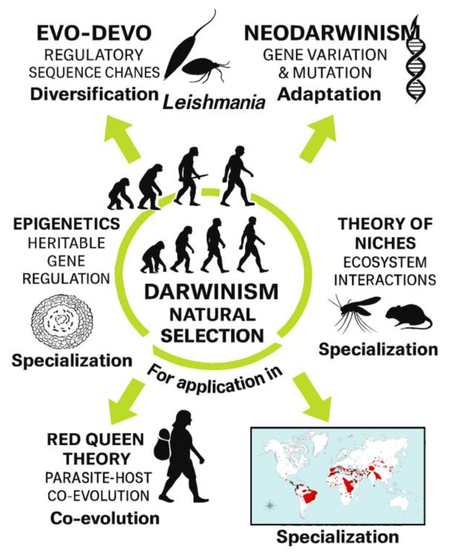
Importantly, the conceptual framework illustrated in Figure 7 situates these cellular and molecular findings within broader evolutionary and ecological principles. Evo-Devo perspectives highlight how conserved developmental modules and membrane-based communication systems may have been co-opted by trypanosomatids to enhance adaptability and host exploitation [16]. Comparative analyses with related parasites such as Trypanosoma cruzi, which also relies on lipid remodeling and vesicle-mediated metabolic plasticity [25], reinforce the idea that lipid-centered strategies represent an ancestral and evolutionarily conserved toolkit within the Trypanosomatidae. Niche theory provides a lens through which to interpret lipid-mediated interactions as mechanisms enabling parasites to occupy, modify, and stabilize intracellular environments [17]. Epigenetic regulation, including lipid-dependent chromatin remodeling and metabolic signaling, may further influence parasite differentiation and stress responses [18]. Finally, Red Queen co-evolutionary dynamics underscore how continuous host–parasite arms races drive innovation in communication strategies, including the evolution of lipid-rich EVs as tools for immune evasion and ecological specialization [19,20].
This evolutionary perspective is particularly relevant for zoonotic Leishmania species, whose ecological distribution, vector associations, and host range diversification remain major global health challenges [15,20,24]. By integrating ultrastructural, biochemical, methodological, and evolutionary insights, our results support a comprehensive lipid-centered model of LEV function in Leishmania, influencing vesicle biogenesis, host modulation, intracellular survival, and long-term adaptive trajectories [1,2,3,4,5,6,7,8,9,10,11,12,13,14,16,17,18,19,20,21,22,23,24,25]. Together, these findings highlight lipid-rich LEVs as key mediators of parasite fitness and pathogenicity, and as promising targets for therapeutic intervention, biomarker discovery, and standardized EV-based research pipelines [21,22,23,24].
5. Conclusions
Our results demonstrate that Leishmania (L.) amazonensis displays abundant lipid-rich structures closely associated with LEV biogenesis across both promastigote and amastigote stages. These lipid reservoirs and membrane-derived compartments likely support multiple facets of parasite physiology, including vesicle formation, cargo organization, host-cell modulation, and intracellular persistence. Collectively, the evidence reinforces the central role of lipid-mediated pathways in shaping Leishmania biology and pathogenicity. Adopting a lipid-centered perspective on LEV function not only advances our understanding of parasite–host communication but also highlights promising avenues for the identification of therapeutic targets and clinically relevant biomarkers. This framework provides a foundation for future translational, mechanistic, and evolutionary studies aimed at unraveling how lipid-rich vesicular systems contribute to disease progression and adaptive strategies in Leishmania infections.
Supplementary Materials
The following supporting information can be downloaded at: https://www.mdpi.com/article/doi/s1, Figure S1: title; Table S1: title.
Author Contributions
The authors contributed equally to the work. Roles: resources, conceptualization, investigation, formal analysis, writing, review and editing, Á.M.G.; investigation, writing, review and editing, A.G.-P.; resources, investigation, formal analysis, writing, review and editing, W.L.A.P.; investigation, formal analysis, writing, review and editing, K.W.P.; resources, formal analysis, supervision, writing, review and editing E.O.d.S. All authors have read and agreed to the published version of the manuscript.
Funding
This research was supported by the Coordination for the Improvement of Higher Education Personnel (CAPES) and by the Portuguese Foundation for Science and Technology (FCT), through the research project GHTM–UID/Multi/04413/2013 and Portugal-Brazil research project PTDC/SAU-PAR/28459/2017 EXOTRYPANO IHMT-NOVA/FMV-ULisboa/UFRN.
Institutional Review Board Statement
Not applicable.
Informed Consent Statement
Not applicable.
Data Availability Statement
The data presented in this study are available on request from the corresponding author (Á.M.G.). The data is not publicly available due to confidentiality.
Acknowledgments
All the authors of the manuscript acknowledge their respective laboratories/institutes/universities and would like to thank the anonymous reviewers for their thoughtful comments and efforts towards improving the manuscript. Á.M.G. gratefully acknowledge the Laboratory of Gabriela Santos-Gomes IHMT-UNL-Portugal for her permanent support, scientific guidance and thesis supervision.
Conflicts of Interest
The authors declare no conflict of interest.
Abbreviations
The following abbreviations are used in this manuscript:
| MDPI | Multidisciplinary Digital Publishing Institute |
| DOAJ | Directory of open access journals |
| TLA | Three letter acronym |
| LD | Linear dichroism |
| AG | Fatty acids |
| BODIPY | Boron-dipyrromethene |
| CRISPR | Clustered regularly interspaced short palindromic repeats |
| DNA | Desoxyribonucleic acid |
| EM | Electron microscopy |
| EV | Extracellular vesicle |
| FA | Fatty acid |
| GPI | Glycosylphosphatidylinositol |
| LD | Lipid droplet |
| LEV | Leishmania extracellular vesicle |
| LC-MS/MS | Liquid chromatography–tandem mass spectrometry |
| MS | Mass spectrometry |
| PBS | Phosphate-buffered saline |
| RNA | Ribonucleic acid |
| TEM | Transmission electron microscopy |
| Evo-Devo | Evolutionary developmental biology |
| RQT | Red Queen Theory |
| PHCE | Parasite–Host Co-Evolution |
| PHCED | Parasite–Host Co-Evolutionary Dynamics |
Supplementary Materials
Appendix A. Terminology and Conceptual Foundations
Appendix A.1 Terminology Used to Describe Lipid-Related Processes
A variety of lipid-related terms are used across biochemistry, pharmacology, and clinical sciences, often referring to distinct concepts. To ensure terminological consistency throughout this manuscript, we adopted standardized definitions based on peer-reviewed literature. Lipid-centered refers to biochemical mechanisms in which lipids act as primary substrates or reactive intermediates. Lipid-based is used for formulations, delivery systems, or structural assemblies that rely on lipids as functional components. Pharmacological interventions aimed at reducing circulating lipid levels are described as lipid-lowering, whereas tissues or samples with high lipid content are referred to as lipid-rich. Processes in which lipids act as mediators or signaling molecules are classified as lipid-mediated, while conditions or structures linked to lipid accumulation are described as lipid-associated. When a biological process requires lipids for activation or stability, the term lipid-dependent is used. Finally, agents or interventions that alter lipid metabolism are categorized as lipid-modifying. These definitions are summarized in Table 1 and visually represented in Figure 8 to facilitate conceptual clarity.
Appendix B. Integrated Conceptual and Methodological Frameworks
Appendix B.1 Integrated Frameworks, Resources, and Methodological Approaches for Lipid and Extracellular Vesicle Research in Leishmania
This appendix consolidates the principal resources, methodological standards, and conceptual models that support lipid-centered and extracellular vesicle (EV/LEV) research in Leishmania. Because lipid biology intersects with membrane dynamics, vesicle biogenesis, parasite physiology, and host–pathogen interactions, a unified analytical framework is essential for interpreting lipid-rich vesicles across experimental and translational contexts. Table 2 provides a comparative overview of the lipid-related resources, standards, and research frameworks most relevant to Leishmania and EV studies, while Figure 9 illustrates how these complementary sources converge to support a coherent, integrated approach.
Appendix C — Comparative Landscape of Lipid-Centered EV Research
Appendix C.1 Overview of Lipid-Centered Studies and Extracellular Vesicle (EV/LEV) Research in Trypanosomatids
Lipid-centered research in trypanosomatids has expanded considerably in recent years, driven by increasing recognition of the central role that lipids play in parasite physiology, vesicle biogenesis, host–pathogen communication, and disease progression. Extracellular vesicles (EVs/LEVs) released by these protozoa are now understood as lipid-rich structures that mediate immune modulation, nutrient acquisition, and intercellular signaling across diverse host environments. Despite these advances, available studies remain dispersed across species, methodological approaches, and biological contexts, making comparative synthesis essential for identifying shared mechanisms and species-specific adaptations.
To support this integrative perspective, Table 3 provides an overview of lipid-centered studies in trypanosomatids, including investigations of lipid-rich EVs/LEVs and the methodologies applied to characterize lipid-dependent and lipid-mediated processes. Together, these comparative data highlight conceptual advances, experimental strategies, and emerging themes that contextualize the findings presented in this manuscript.
References
- Atayde, V.D.; Hassani, K.; da Silva Lira Filho, A.; et al. Extracellular Vesicles in Leishmania Infection: Modulators of Host–Parasite Interaction. PLoS Pathog. 2023, 19, e1012636. [Google Scholar] [CrossRef]
- Silverman, J.M.; Clos, J.; de’Oliveira, C.C.; et al. Leishmania Exosomes Modulate Innate Immunity and Promote Infection. Proc. Natl. Acad. Sci. USA 2010, 107, 21635–21640. [Google Scholar] [CrossRef]
- Parreira de Aquino, G.; et al. Lipid Metabolism in Leishmania: Implications for Pathogenesis and Drug Resistance. Microb. Cell 2021, 8, 88–104. [Google Scholar] [CrossRef]
- Santos, L.C.; et al. Metabolomic Profiling of Leishmania Species Reveals Lipid Remodeling During Infection. Metabolites 2024, 14, 658. [Google Scholar] [CrossRef]
- Pal, P.; Das, S.; Chatterjee, N.; Bose, D.; Saha, K.D. Studies on the Anti-Inflammatory Effect of Leishmanial Lipid In Vitro and In Vivo. Prajnan O Sadhona 2015, 2, 1–XX. [Google Scholar]
- Sacks, D.; et al. Lipid Bodies in Leishmania: Structure, Function, and Role in Host Interaction. Mol. Biochem. Parasitol. 2009, 165, 1–9. [Google Scholar] [CrossRef]
- Gonçalves, R.; et al. Extracellular Vesicles from Protozoan Parasites: Biogenesis, Composition, and Function. Front. Microbiol. 2016, 7, 427. [Google Scholar] [CrossRef]
- Kumar, A.; et al. Lipid-Mediated Modulation of Host Immunity by Leishmania spp. Exp. Parasitol. 2021, 223, 107999. [Google Scholar] [CrossRef]
- Chowdhury, S.; et al. Host–Parasite Lipid Interactions in Leishmaniasis: Mechanisms and Therapeutic Perspectives. Int. J. Mol. Sci. 2022, 23, 2414. [Google Scholar] [CrossRef]
- Macedo, A.M.; et al. Lipidomic Signatures of Leishmania Extracellular Vesicles Reveal Species-Specific Patterns. Int. J. Mol. Sci. 2023, 24, 10637. [Google Scholar] [CrossRef]
- Rodrigues, J.C.F.; et al. Metabolic Adaptations of Leishmania Parasites: Lipid Remodeling and Survival Strategies. Microorganisms 2023, 13, 531. [Google Scholar] [CrossRef]
- Serrano, A.; et al. Lipid Droplets and Vesicular Trafficking in Leishmania: Ultrastructural Insights. Micron 2021, 142, 103089. [Google Scholar] [CrossRef]
- Parreira de Aquino, G.; et al. Lipid Remodeling in Leishmania: A Microbial Cell Perspective. Microb. Cell 2021, 8, 1–12. [Google Scholar] [CrossRef]
- Costa, D.L.; et al. Lipid-Rich Extracellular Vesicles in Leishmania Infection: Implications for Pathogenesis. Int. J. Parasitol. 2024, 54, 1–18. [Google Scholar] [CrossRef]
- World Health Organization (WHO). Leishmaniasis; WHO Fact Sheet, April 2017. Available online: https://www.who.int/mediacentre/factsheets/fs375/en/ (accessed on 24 July 2017).
- Carroll, S.B. Evo-Devo and the Evolution of Animal Diversity. Cell 2005, 120, 201–207. [Google Scholar] [CrossRef]
- Hutchinson, G.E. Concluding Remarks. Cold Spring Harb. Symp. Quant. Biol. 1957, 22, 415–427. [Google Scholar] [CrossRef]
- Bird, A. Perceptions of Epigenetics. Nature 2007, 447, 396–398. [Google Scholar] [CrossRef]
- Van Valen, L. A New Evolutionary Law. Evolutionary Theory 1973, 1, 1–30. [Google Scholar]
- Schmid-Hempel, P. Evolutionary Parasitology; Oxford University Press: Oxford, UK, 2011; ISBN 978-0199229482. [Google Scholar]
- Fisher Scientific. Lipid-related reagents and analytical products. Available online: https://www.fishersci.pt/pt/en/catalog/search/products?keyword=lipid (accessed on 7 January 2026).
- International Society for Extracellular Vesicles (ISEV). MISEV2023: Minimal Information for Studies of Extracellular Vesicles. J. Extracell. Vesicles 2023. Available online: https://isevjournals.onlinelibrary.wiley.com/doi/10.1002/jev2.12404 (accessed on 7 January 2026).
- Gabriel, Á.M.; Galué-Parra, A.; Pereira, W.L.A.; Pedersen, K.W.; da Silva, E.O. Leishmania 360°: Guidelines for Exosomal Research. Microorganisms 2021, 9, 2081. [Google Scholar] [CrossRef]
- Gabriel, Á.M.; Galvão, G.R.; Galué-Parra, A.; Casseb, L.M.N.; Pereira, W.L.A.; Pedersen, K.W.; Aguiar, D.C.F.; Gonçalves, E.C.; da Silva, E.O. Pathogenesis of canine leishmaniasis: diagnostic accuracy and experimental models targeting Leishmania lipid-bound vesicles. Acad. Biol. 2025, 3(1), 1–XX. [Google Scholar] [CrossRef]
- Booth, L.-A.; Smith, T.K. Lipid Metabolism in Trypanosoma cruzi: A Review. Mol. Biochem. Parasitol. 2020, 240, 111324. [Google Scholar] [CrossRef] [PubMed]
Figure 1.
Conceptual infographic illustrating the worldwide integration of human health, animal health, and environmental factors through the One Health approach. The circular triad emphasizes the interdependence of these domains, with silhouettes representing human evolutionary progression. The corrected label PARASITE LIPID–HOST CELL INTERACTION highlights the molecular interface between Leishmania and host cells, mediated by lipid-enriched extracellular vesicles (LEVs). This interaction is linked to Darwin’s Theory and an Evolutionary Perspective, underscoring the adaptive strategies of the parasite shaped by co-evolutionary pressures. The world map, adapted from WHO [15], displays the global distribution of cutaneous and visceral leishmaniasis (indicated by black dots and visceral leishmaniasis shown in pinkish-shaded areas), reinforcing the ecological and epidemiological relevance of lipid-centered host–parasite communication.
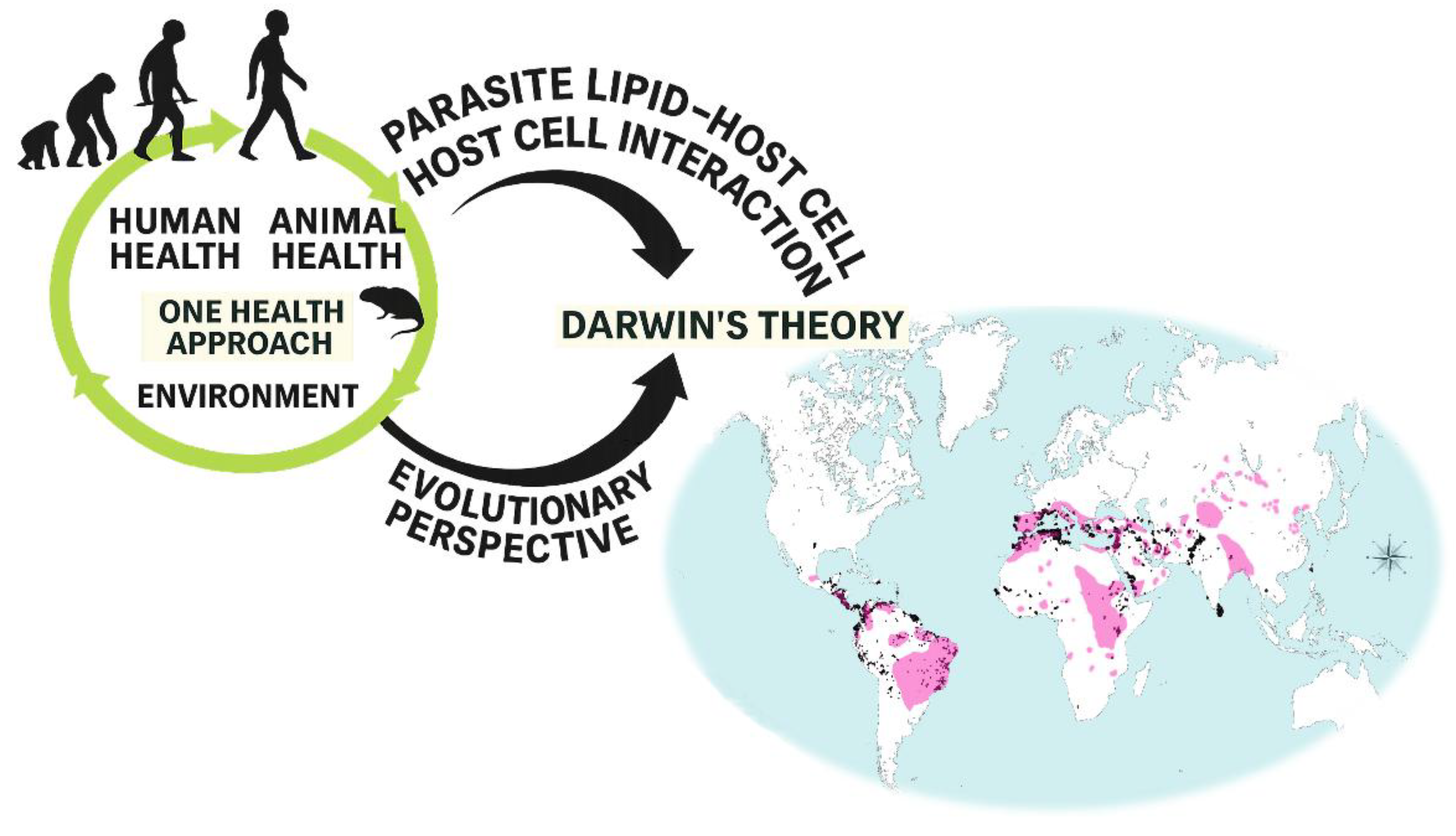
Figure 2.
A.Leishmania (L.) amazonensis promastigote on day seven of culture (stationary phase). Note the presence of metacyclic promastigote forms with elongated bodies and externalized flagella (arrows). B. Mouse peritoneal macrophages infected with L. (L.) amazonensis. Observe the presence of intracellular amastigotes within the parasitophorous vacuole (arrows). Samples were stained with Giemsa and examined by brightfield light microscopy.
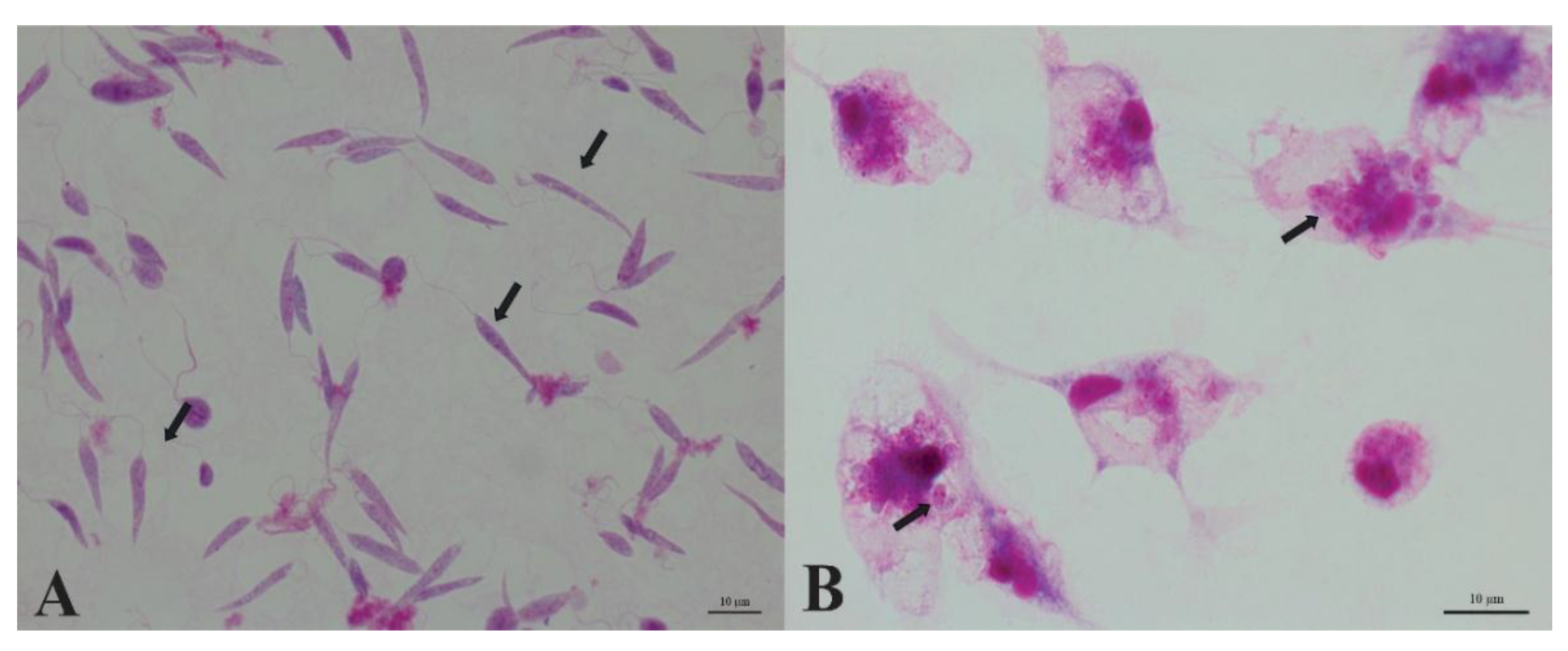
Figure 3.
Macrophage infected with Leishmania (L.) amazonensis, showing intracellular amastigote forms (arrows) visualized by scanning electron microscopy (SEM).
Figure 3.
Macrophage infected with Leishmania (L.) amazonensis, showing intracellular amastigote forms (arrows) visualized by scanning electron microscopy (SEM).
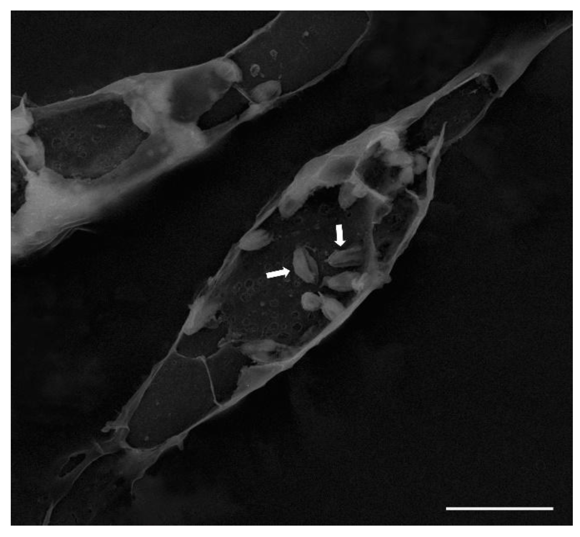
Figure 4.
Lipid-like structures present in promastigotes and amastigotes of Leishmania (L.) amazonensis. A) Promastigote; B) Amastigote. Note the presence of lipid bodies (*). FP: Flagellar pocket.
Figure 4.
Lipid-like structures present in promastigotes and amastigotes of Leishmania (L.) amazonensis. A) Promastigote; B) Amastigote. Note the presence of lipid bodies (*). FP: Flagellar pocket.
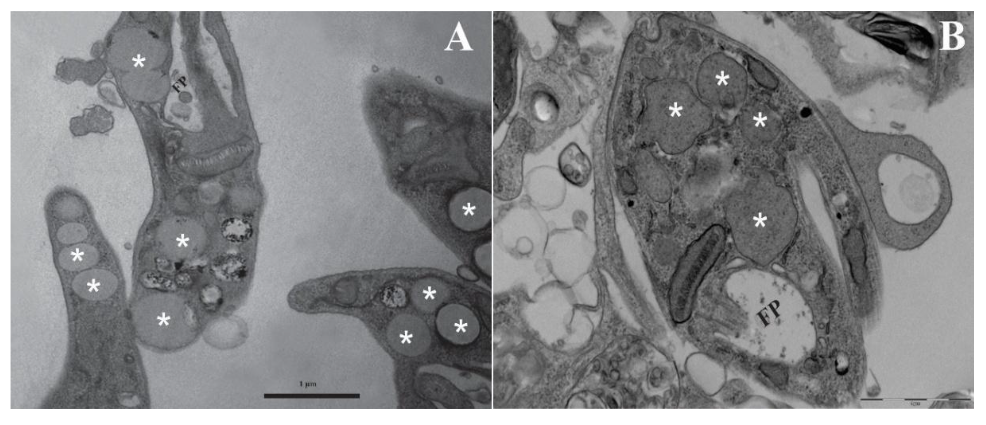
Figure 5.
Leishmania (L.) amazonensis promastigote showing Golgi cisternal structures with features consistent with an early autophagic process.
Figure 5.
Leishmania (L.) amazonensis promastigote showing Golgi cisternal structures with features consistent with an early autophagic process.
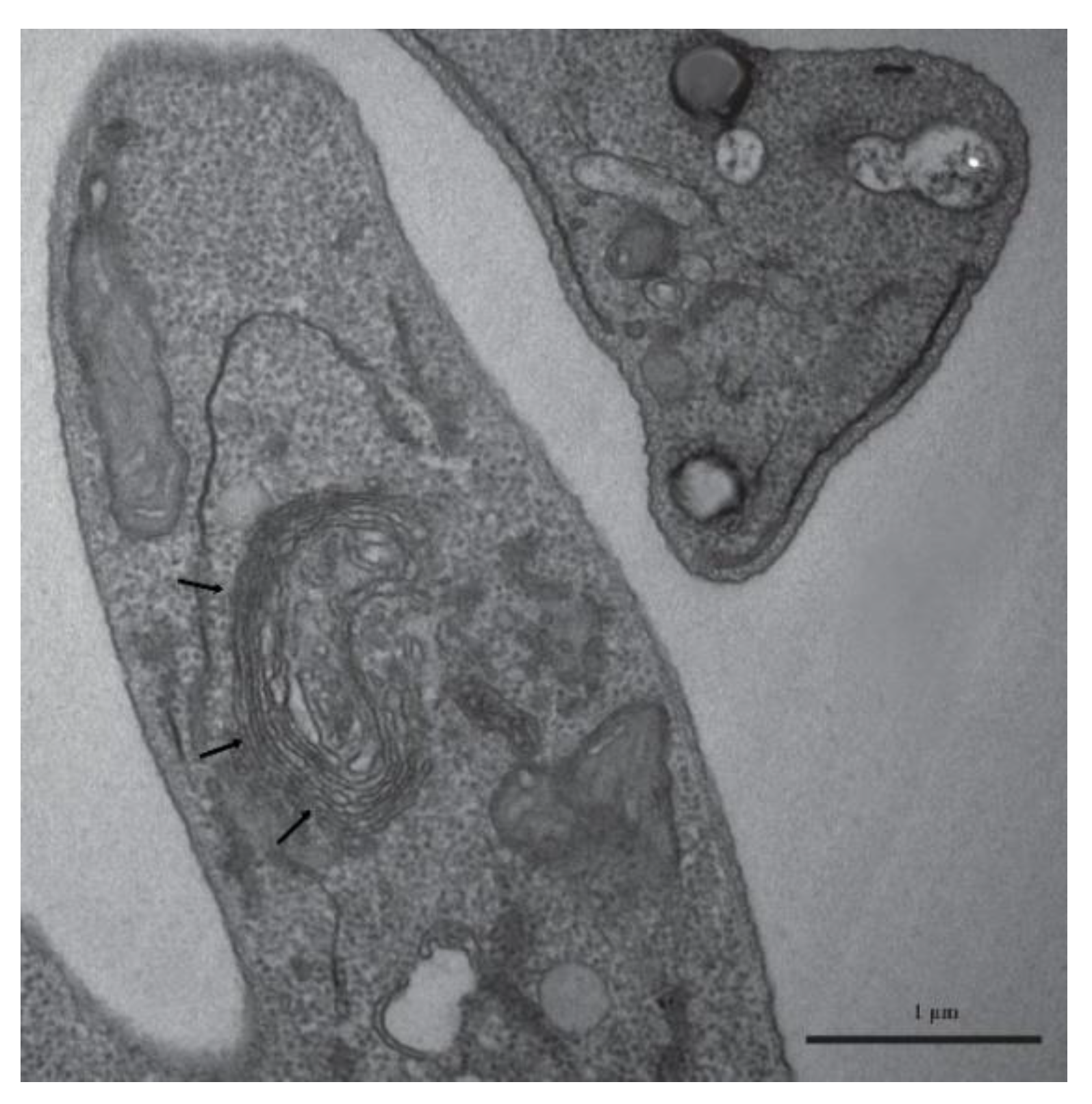
Figure 6.
Lipid body staining in Leishmania (L.) amazonensis promastigotes using BODIPY .A) Phase-contrast image; B) promastigotes labeled with BODIPY; C) promastigotes labeled with DAPI; D) merged image showing the superimposition of DAPI and BODIPY staining.
Figure 6.
Lipid body staining in Leishmania (L.) amazonensis promastigotes using BODIPY .A) Phase-contrast image; B) promastigotes labeled with BODIPY; C) promastigotes labeled with DAPI; D) merged image showing the superimposition of DAPI and BODIPY staining.
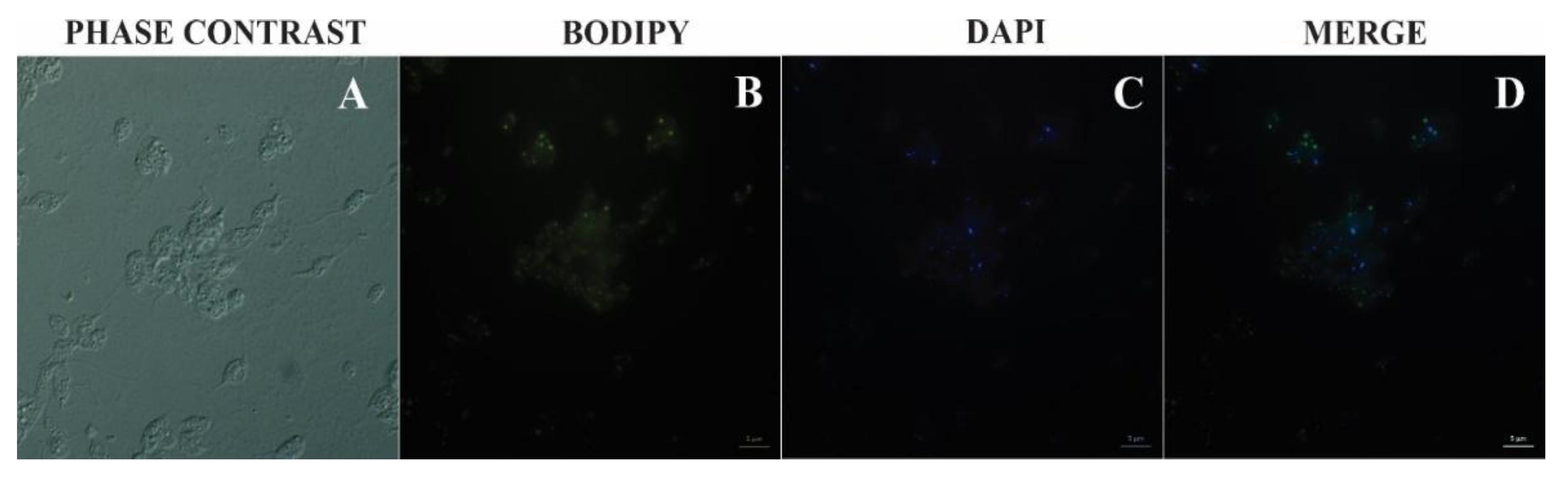
Figure 7.
Conceptual diagram illustrating Darwinian foundations and evolutionary extensions relevant to Leishmania research. The central theme of Darwinism and natural selection is represented by silhouettes of hominid evolution, symbolizing evolutionary pressure driving biological diversification. Arrows extend to five major theoretical domains: (1) Evo-Devo: regulatory sequence changes linked to morphological diversification, including parasite structures such as Leishmania flagella and vesicular membranes; (2) Neodarwinism: gene variation and mutation driving adaptation, illustrated by a DNA helix and relevant to parasite genomic plasticity; (3) Theory of Niches: ecosystem interactions and host specialization, represented by vectors and reservoirs (mosquito and rodent), emphasizing ecological constraints on transmission; (4) Epigenetics: heritable gene regulation contributing to phenotypic specialization, relevant to parasite stage transitions and host immune modulation; (5) Red Queen Theory: parasite–host co-evolution, depicted by a human figure under evolutionary pressure, highlighting the dynamic arms race between host defenses and parasite evasion. A world map at the base of the figure illustrates the global distribution and specialization of zoonotic Leishmania species. These conceptual associations align with lipid-centered analyses of trypanosomatids and their extracellular vesicles (see Supplementary Table S1), reinforcing the role of lipid-rich structures in vesicle biogenesis, host modulation, and parasite survival.
Disclaimer/Publisher’s Note: The statements, opinions and data contained in all publications are solely those of the individual author(s) and contributor(s) and not of MDPI and/or the editor(s). MDPI and/or the editor(s) disclaim responsibility for any injury to people or property resulting from any ideas, methods, instructions or products referred to in the content. |
© 2026 by the authors. Licensee MDPI, Basel, Switzerland. This article is an open access article distributed under the terms and conditions of the Creative Commons Attribution (CC BY) license (http://creativecommons.org/licenses/by/4.0/).
Copyright: This open access article is published under a Creative Commons CC BY 4.0 license, which permit the free download, distribution, and reuse, provided that the author and preprint are cited in any reuse.


