Submitted:
31 December 2025
Posted:
01 January 2026
You are already at the latest version
Abstract
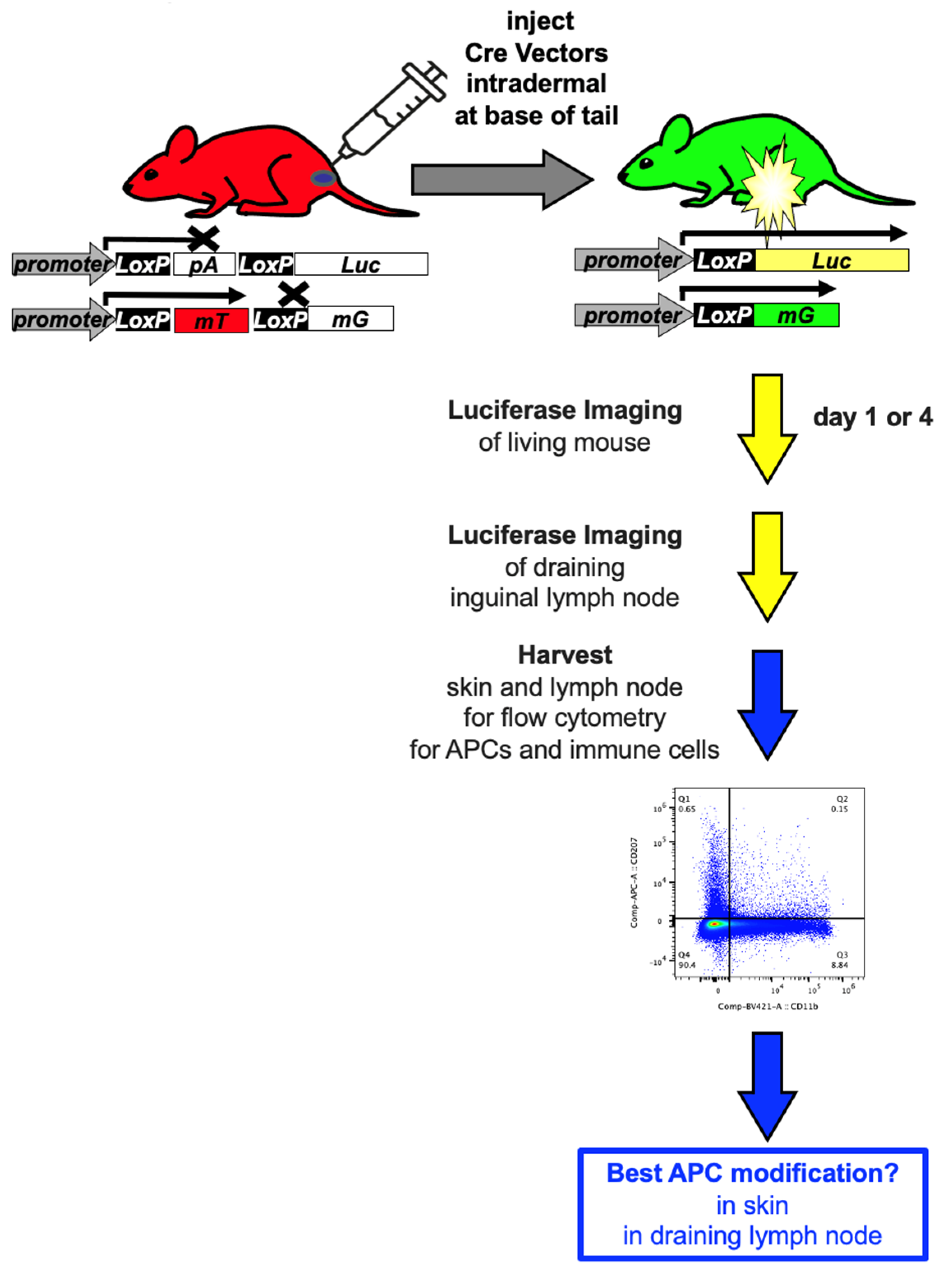
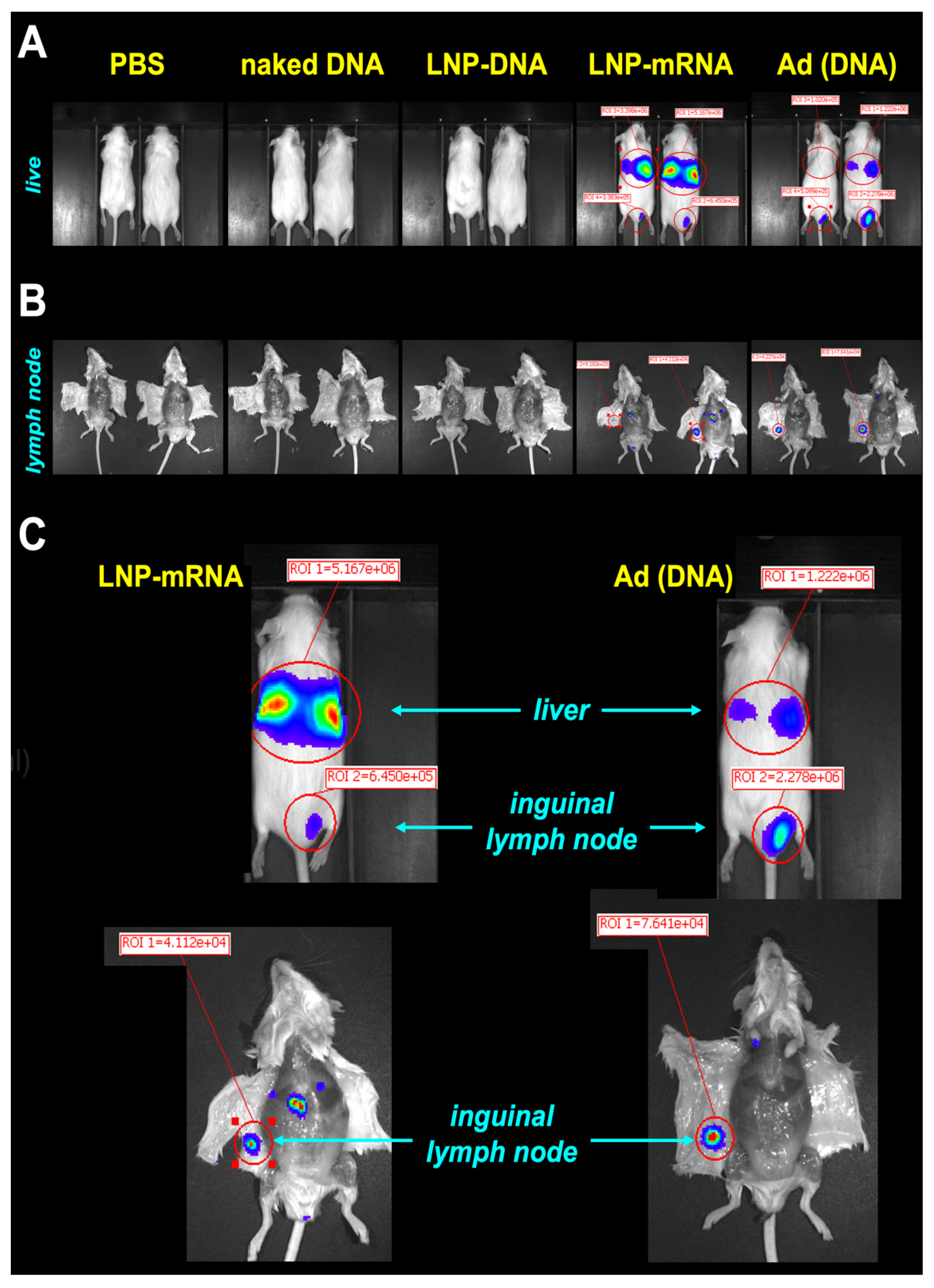
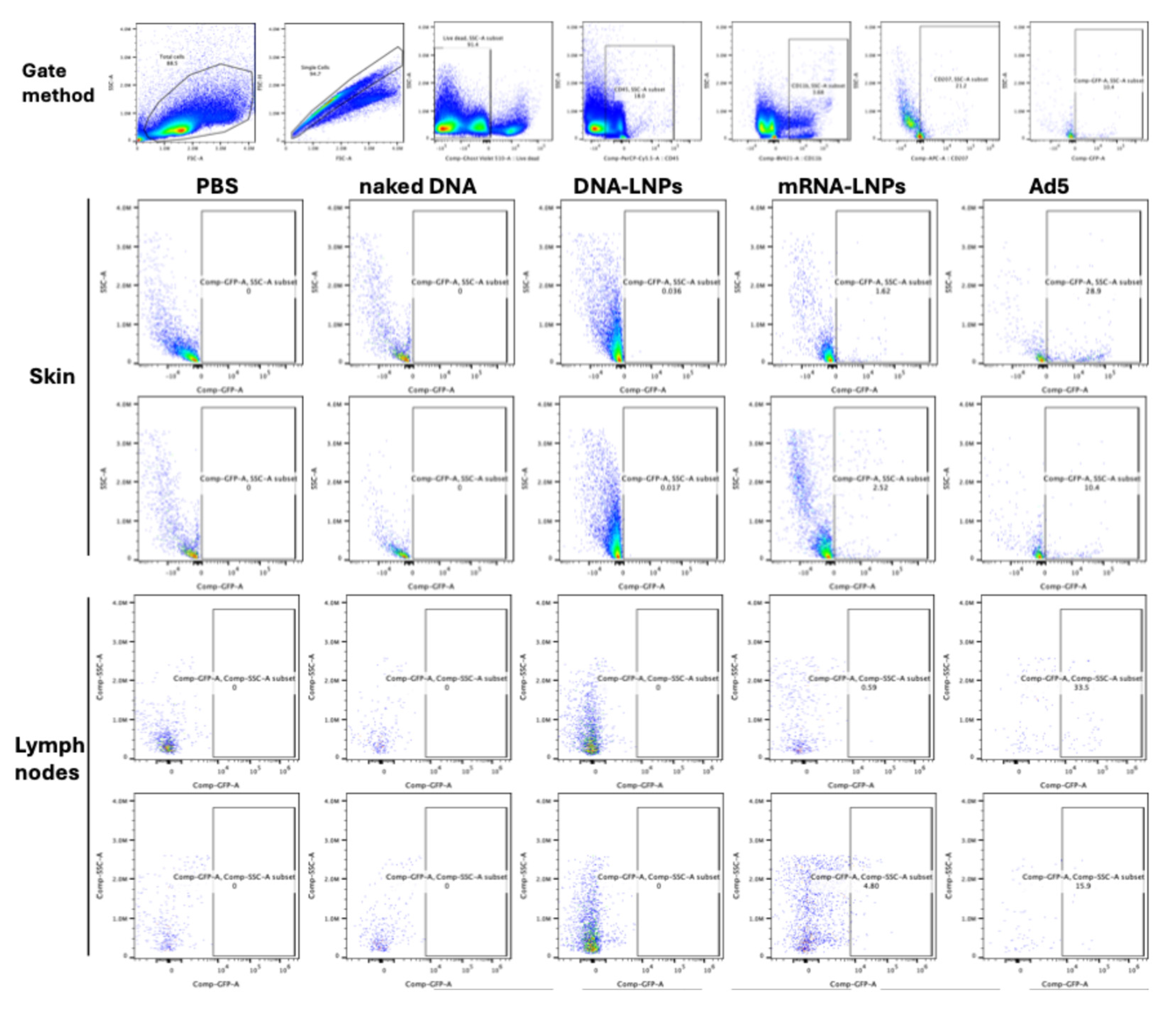
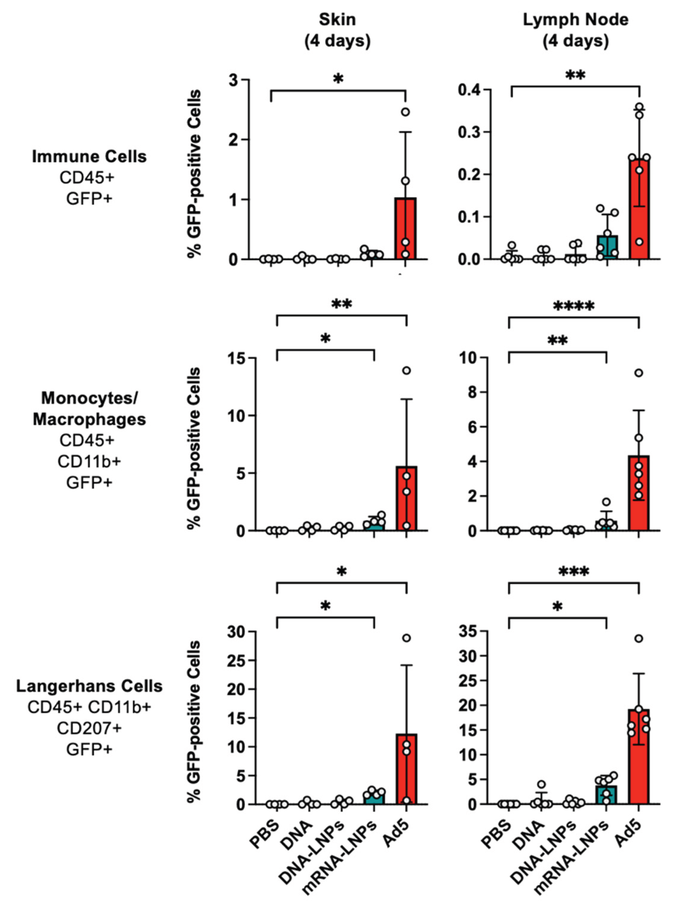
Background/Objectives: Antigen presenting cells (APCs) and immune cells have unique properties to drive or suppress immune responses. They are therefore key targets for the expression of vaccine antigens or transgene proteins. To better determine the utility of different molecular therapies to modify these cells, mRNA and DNA-based molecular therapy vectors were compared for their ability to genetically modify immune cells after intradermal injections in mice. DNA-based vectors included naked plasmid DNA, plasmid packaged in lipid nanoparticles (LNPs), and replication-defective adenovirus (Ad) vectors. mRNA delivery was mediated by packaging into LNPs like those used in COVID-19 vaccines. Methods: Each vector was used to deliver Cre recombinase into Cre reporter mice whose cells are activated to express green fluorescent protein (GFP) and firefly luciferase after Cre recombination. Mice were injected intradermally (ID) near the base of their tail at a site that drains into the inguinal lymph node. Luciferase activity was imaged in the living mice 1 or 4 days after vector injection. The animals were then euthanized and luciferase activity was imaged in the draining inguinal lymph node. Cells were prepared from the intradermal injection site and from the draining lymph node to determine which immune cells were genetically modified by phenotyping CD45, CD3, and CD11b GFP-positive cells by flow cytometry. Given that the skin uniquely contains Langerhans dendritic cells, these CD207+ cells were also phenotyped in skin samples and in the draining lymph node. Results: In both the skin and in the draining lymph node, the rank order of luciferase and GFP activation by the vectors were: 1) Ad; 2) mRNA-LNP; 3) DNA-LNP; and 4) naked DNA. Only mRNA-LNP and Ad vectors mediated obvious luciferase activity in the living animals and in the draining lymph nodes by imaging. Notably, both vectors appeared to leak from the ID injection site and not only modify the draining lymph node but also strongly modify the livers of the mice. Naked DNA and DNA-LNP mediated detectable GFP activation in the skin and draining lymph node in some mice, but this activity was low and did not reach statistical significance when compared to PBS-treated animals. mRNA-LNPs and Ad both mediated significant Cre delivery in CD45+, CD3+, CD11b+, and CD207+ immune cells in the skin and in the lymph node with adenovirus mediating consistently higher levels of expression in all of the tested cells. Conclusions: These data indicate that mRNA-LNP and Ad vectors mediate stronger modification of skin and lymph node immune cells after intradermal injections. Naked DNA and DNA-LNPs were markedly less potent at this activity than the other vectors. These data are consistent with the higher vaccine potency of mRNA-LNP and Ad vectors and suggest that approaches that increase targeting of immune cell subsets may have utility to increase efficacy while also reducing off target modification of tissues like the liver.
Keywords:
1. Introduction
2. Materials and Methods
2.1. Vectors
2.2. In Vitro Transcription (IVT) of mRNA
2.3. Lipid Nanoparticle Assembly
2.4. In Vivo Experiments
2.5. mT/mG: LSL-Lux Cre Reporter Mice
2.6. Vector Injections and Tissue Harvest
2.7. Flow Cytometry
2.8. Statistical Analysis
3. Results
3.1. Cre Reporter Mice
3.2. In Vivo Luciferase Imaging
3.3. Cre Delivery to Immune Cells in the Skin and in the Draining Lymph Node
4. Discussion
Author Contributions
Funding
Institutional Review Board Statement
Data Availability Statement
Acknowledgments
Conflicts of Interest
Abbreviations
| Ad | adenovirus |
| APC | antigen presenting cell |
| ID | intradermal |
| GFP | green fluorescent protein |
| Luc | luciferase |
| mG | membrane targeted GFP |
| RFP | red fluorescent protein |
| mT | membrane targeted mTomato RFP |
| VAERS | vaccine adverse event reporting system |
| VITT | vaccine-induced thrombotic thrombocytopenia |
References
- Prubeta, B.M. Current State of the First COVID-19 Vaccines. Vaccines 2021, 9. [Google Scholar] [CrossRef] [PubMed]
- Greinacher, A.; Langer, F.; Makris, M.; Pai, M.; Pavord, S.; Tran, H.; Warkentin, T.E. Vaccine-induced immune thrombotic thrombocytopenia (VITT): Update on diagnosis and management considering different resources. J Thromb Haemost 2021. [Google Scholar] [CrossRef] [PubMed]
- Rodriguez, E.V.C.; Bouazza, F.Z.; Dauby, N.; Mullier, F.; d’Otreppe, S.; Jissendi Tchofo, P.; Bartiaux, M.; Sirjacques, C.; Roman, A.; Hermans, C.; et al. Fatal vaccine-induced immune thrombotic thrombocytopenia (VITT) post Ad26.COV2.S: first documented case outside US. Infection 2021. [Google Scholar] [CrossRef] [PubMed]
- Heidecker, B.; Dagan, N.; Balicer, R.; Eriksson, U.; Rosano, G.; Coats, A.; Tschope, C.; Kelle, S.; Poland, G.A.; Frustaci, A.; et al. Myocarditis following COVID-19 vaccine: incidence, presentation, diagnosis, pathophysiology, therapy, and outcomes put into perspective. A clinical consensus document supported by the Heart Failure Association of the European Society of Cardiology (ESC) and the ESC Working Group on Myocardial and Pericardial Diseases. Eur J Heart Fail 2022, 24, 2000–2018. [Google Scholar] [CrossRef] [PubMed]
- Petito, E.; Gresele, P. VITT Pathophysiology: An Update. Vaccines 2025, 13. [Google Scholar] [CrossRef] [PubMed]
- Prather, A.A.; Dutcher, E.G.; Robinson, J.; Lin, J.; Blackburn, E.; Hecht, F.M.; Mason, A.E.; Fromer, E.; Merino, B.; Frazier, R.; et al. Predictors of long-term neutralizing antibody titers following COVID-19 vaccination by three vaccine types: the BOOST study. Sci Rep 2023, 13, 6505. [Google Scholar] [CrossRef] [PubMed]
- Menni, C.; May, A.; Polidori, L.; Louca, P.; Wolf, J.; Capdevila, J.; Hu, C.; Ourselin, S.; Steves, C.J.; Valdes, A.M.; et al. COVID-19 vaccine waning and effectiveness and side-effects of boosters: a prospective community study from the ZOE COVID Study. Lancet Infect Dis 2022, 22, 1002–1010. [Google Scholar] [CrossRef] [PubMed]
- Akyol, R.; Dalod, M. Identity, Functions, and the Spatiotemporal Maturation of Type 1 Conventional Dendritic Cells. Immunol Rev 2025, 336, e70079. [Google Scholar] [CrossRef] [PubMed]
- Vine, E.E.; Austin, P.J.; O’Neil, T.R.; Nasr, N.; Bertram, K.M.; Cunningham, A.L.; Harman, A.N. Epithelial dendritic cells vs. Langerhans cells: Implications for mucosal vaccines. Cell reports 2024, 43, 113977. [Google Scholar] [CrossRef] [PubMed]
- Banchereau, J.; Briere, F.; Caux, C.; Davoust, J.; Lebecque, S.; Liu, Y.J.; Pulendran, B.; Palucka, K. Immunobiology of dendritic cells. Annu Rev Immunol 2000, 18, 767–811. [Google Scholar] [CrossRef] [PubMed]
- Melero, I.; Vile, R.G.; Colombo, M.P. Feeding dendritic cells with tumor antigens: self-service buffet or a la carte? Gene Ther 2000, 7, 1167–1170. [Google Scholar] [CrossRef] [PubMed]
- Timares, L.; Takashima, A.; Johnston, S.A. Quantitative analysis of the immunopotency of genetically transfected dendritic cells. Proc Natl Acad Sci U S A 1998, 95, 13147–13152. [Google Scholar] [CrossRef] [PubMed]
- Hillestad, M.L.; Guenzel, A.J.; Nath, K.A.; Barry, M.A. A vector-host system to fingerprint virus tropism. Hum Gene Ther 2012, 23, 1116–1126. [Google Scholar] [CrossRef] [PubMed]
- Rubin, J.D.; Nguyen, T.V.; Allen, K.L.; Ayasoufi, K.; Barry, M.A. Comparison of Gene Delivery to the Kidney by Adenovirus, Adeno-Associated Virus, and Lentiviral Vectors After Intravenous and Direct Kidney Injections. Hum Gene Ther 2019, 30, 1559–1571. [Google Scholar] [CrossRef] [PubMed]
- Crosby, C.M.; Barry, M.A. IIIa deleted adenovirus as a single-cycle genome replicating vector. Virology 2014, 462-463, 158–165. [Google Scholar] [CrossRef] [PubMed]
- Parrett, B.J.; Yamaoka, S.; Barry, M.A. Reducing off-target expression of mRNA therapeutics and vaccines in the liver with microRNA binding sites. Mol Ther Methods Clin Dev 2025, 33, 101402. [Google Scholar] [CrossRef] [PubMed]
- Betageri, K.R.; Meridew, J.A.; Parrett, B.J.; Gilbert, R.M.; Link, P.A.; Schussler, N.A.; Mercado-Perez, A.; Caporarello, N.; Barry, M.A.; Tschumperlin, D.J. Lung-targeted Lipid Nanoparticle Delivery of a Matricellular mRNA Promotes Fibrotic Lung Repair. Am J Respir Cell Mol Biol 2025. [Google Scholar] [CrossRef] [PubMed]
- Weaver, E.A.; Nehete, P.N.; Buchl, S.S.; Senac, J.S.; Palmer, D.; Ng, P.; Sastry, K.J.; Barry, M.A. Comparison of replication-competent, first generation, and helper-dependent adenoviral vaccines. PloS one 2009, 4, e5059. [Google Scholar] [CrossRef] [PubMed]
Disclaimer/Publisher’s Note: The statements, opinions and data contained in all publications are solely those of the individual author(s) and contributor(s) and not of MDPI and/or the editor(s). MDPI and/or the editor(s) disclaim responsibility for any injury to people or property resulting from any ideas, methods, instructions or products referred to in the content. |
© 2026 by the authors. Licensee MDPI, Basel, Switzerland. This article is an open access article distributed under the terms and conditions of the Creative Commons Attribution (CC BY) license (http://creativecommons.org/licenses/by/4.0/).


