
Submitted:
30 December 2025
Posted:
31 December 2025
You are already at the latest version
Abstract
Cancer remains a leading cause of morbidity and mortality worldwide, and effective strategies for cancer prevention are urgently needed to complement therapeutic ad-vances. While dietary factors are known to influence cancer risk, the molecular mecha-nisms that mediate inter-individual responses to nutritional exposures remain poorly defined. Emerging evidence identifies long non-coding RNAs (lncRNAs) as pivotal regulators of gene expression, chromatin organization, metabolic homeostasis, immune signaling, and cellular stress responses, core processes that drive cancer initiation and progression and are highly sensitive to nutritional status. In parallel, advances in pre-cision nutrition have highlighted how variability in genetics, metabolism, microbiome composition, and epigenetic landscapes shapes dietary influences on cancer susceptibility. This review integrates these rapidly evolving fields by positioning lncRNAs as molecular conduits that translate dietary exposures into transcriptional and epigenetic programs governing cancer development, progression, and therapeutic vulnerability. We provide mechanistic evidence demonstrating how dietary bioactive compounds and micronu-trients, including polyphenols (curcumin, resveratrol, EGCG), flavonoids, alkaloids such as berberine, omega-3 fatty acids, folate, vitamin D, probiotic metabolites (such as bu-tyrate and propionate), and trace elements (such as selenium, and zinc), modulate on-cogenic and tumor-suppressive lncRNAs. These nutrient-lncRNA interactions influence cancer-relevant pathways controlling proliferation, epithelial-mesenchymal transition, inflammation, oxidative stress, and metabolic rewiring. We further discuss emerging lncRNA signatures that reflect nutritional and metabolic states, their potential utility as biomarkers for individualized dietary interventions, and their integration into liquid biopsy platforms. Leveraging multi-omics datasets and systems biology, we outline AI-driven frameworks to map nutrient-lncRNA regulatory networks and identify tar-getable nodes for cancer chemoprevention. Finally, we address translational challenges, including compound bioavailability, inter-individual variability, and limited clinical validation, and propose future directions for incorporating lncRNA profiling into preci-sion nutrition-guided cancer prevention trials. Together, these insights position lncRNAs at the nexus of diet and cancer biology and establish a foundation for mechanistically informed precision nutrition strategies in cancer chemoprevention.
Keywords:
long non‐coding RNAs (lncRNAs)
; precision nutrition
; cancer
; phytochemicals
; nutritional epigenomics
; micronutrients
; omega‐3 fatty acids
; metabolic rewiring
; chemoprevention
1. Introduction
Cancer development and progression are driven by coordinated disruptions in gene regulation, chromatin architecture, cellular metabolism, immune signaling, and stress-adaptive responses [1,2,3]. While genetic alterations initiate malignant transformation, sustained tumor growth and progression are shaped by dynamic regulatory networks that integrate intrinsic molecular programs with environmental cues [1]. Among the regulatory layers that orchestrate these processes, long non-coding RNAs (lncRNAs) have emerged as central determinants of oncogenic and tumor-suppressive states. By functioning as molecular scaffolds, guides, decoys, and regulatory effectors, lncRNAs influence transcriptional control, epigenetic remodeling, RNA stability, and signaling pathway integration across virtually all hallmarks of cancer [1,4,5,6].
Dysregulated lncRNA expression contributes to cancer initiation, epithelial–mesenchymal transition, metabolic rewiring, immune evasion, and therapeutic resistance [5,6,7]. Numerous lncRNAs act as context-dependent regulators that amplify oncogenic signaling pathways or reinforce tumor-suppressive checkpoints, thereby shaping cancer susceptibility and disease trajectory [8]. Importantly, lncRNAs exhibit cell type-specific, tissue-restricted, and environmentally responsive expression patterns, positioning them as sensitive molecular integrators of external stimuli [9,10]. These properties distinguish lncRNAs from protein-coding genes and underscore their relevance as mechanistic drivers, biomarkers, and potential therapeutic targets in cancer biology [11].
An emerging body of evidence indicates that lncRNA regulatory networks are highly responsive to nutritional and metabolic inputs [12]. Natural dietary compounds, including polyphenols, alkaloids, fatty acids, vitamins, probiotics, postbiotics, and trace elements, have been shown to modulate the expression and activity of oncogenic and tumor-suppressive lncRNAs across multiple cancer types [13,14]. Such nutrient-dependent modulation influences pathways central to carcinogenesis, including proliferation, inflammation, oxidative stress responses, epithelial–mesenchymal transition, stemness, and metabolic adaptation [15]. These findings suggest that diet-derived bioactive molecules do not act solely through classical antioxidant or anti-inflammatory mechanisms but can directly reprogram cancer-relevant transcriptional and epigenetic circuits via lncRNA-mediated regulation [16].
In parallel, advances in precision nutrition have highlighted the importance of inter-individual variability in shaping biological responses to dietary exposures [17]. Genetic background, metabolic phenotype, microbiome composition, and epigenetic state collectively determine how nutritional signals are interpreted at the molecular level [18]. Within this framework, lncRNAs emerge as plausible mechanistic interfaces through which dietary factors influence cancer susceptibility and progression [19,20,21]. The convergence of cancer biology, lncRNA regulation, and precision nutrition highlights the potential of nutrient-responsive lncRNAs as molecular targets and biomarkers for cancer chemoprevention.
Technological advances in multi-omics profiling, single-cell analysis, and systems biology are beginning to resolve the complexity of lncRNA-centered regulatory networks [22]. These approaches reveal how lncRNAs operate within interconnected molecular circuits that integrate environmental, metabolic, and inflammatory cues [19]. Moreover, computational modeling and artificial intelligence now provide powerful tools to map nutrient–lncRNA–pathway relationships, identify regulatory hubs, and predict context-specific responses that are difficult to capture through reductionist experimental strategies alone [23].
In this review, we synthesize current knowledge on the role of lncRNAs as molecular mediators linking nutrition to cancer biology. We focus on mechanistic insights into how dietary bioactive compounds and micronutrients modulate lncRNA-driven regulatory pathways underlying cancer genesis and cancer prevention. By integrating experimental evidence with systems-level and computational perspectives, we provide a conceptual foundation for lncRNA-guided precision nutrition strategies in cancer chemoprevention [21,24,25,26]. Understanding how modifiable dietary factors influence lncRNA regulatory circuits may ultimately lead to individualized approaches to reduce cancer risk and to establish prevention-focused translational strategies [27,28].
2. lncRNAs as Orchestrators of Cancer Initiation and Progression
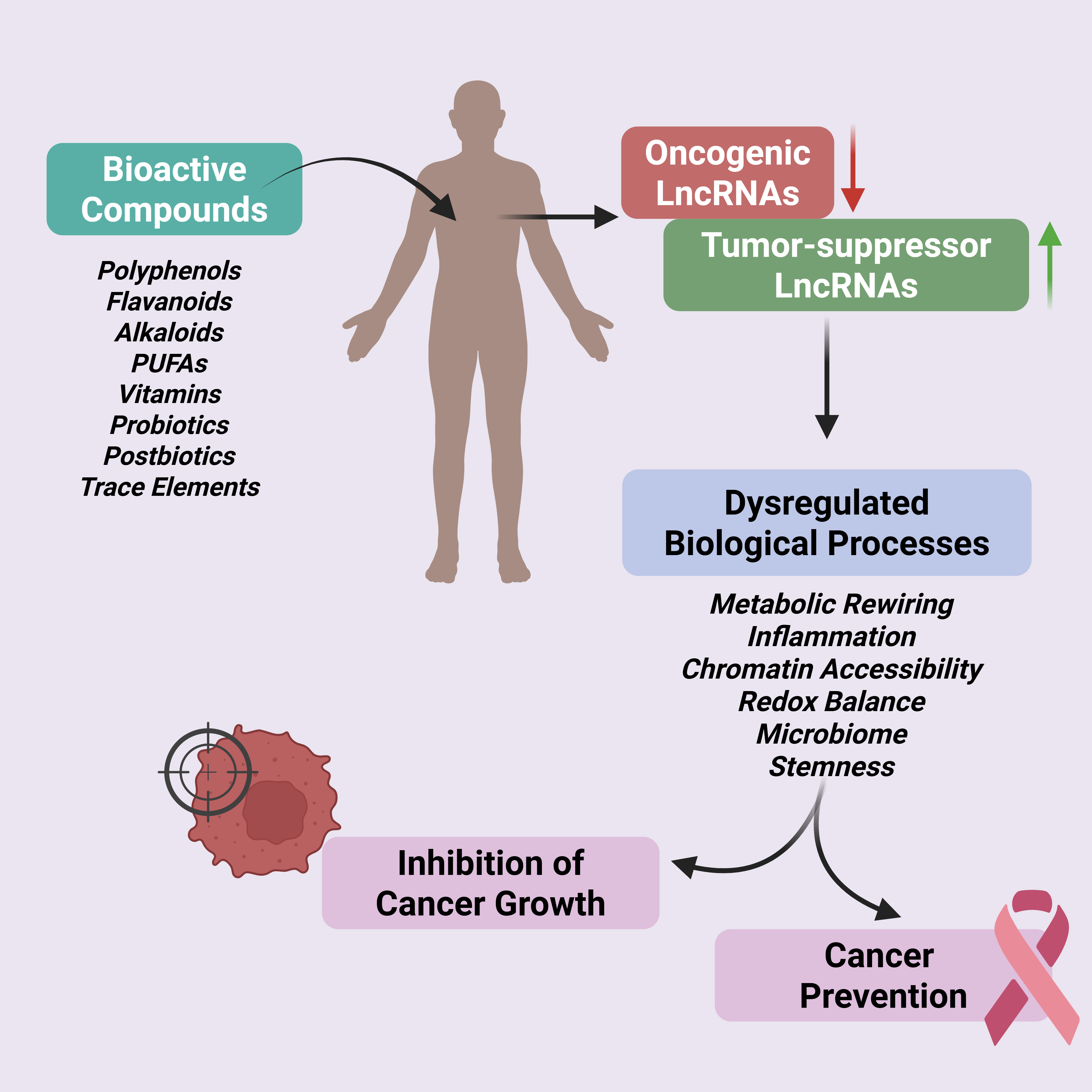
LncRNAs operate at the intersection of transcriptional control, epigenetic regulation, and signal integration, enabling them to coordinate complex cellular responses to both intrinsic and environmental cues [6]. In cancer, dysregulation of lncRNA expression and function contributes to virtually all hallmarks of malignancy, positioning these molecules as central regulators of oncogenic and tumor-suppressive states [7,29]. This section outlines the biological features of lncRNAs, their mechanisms of action, and their context-dependent roles in cancer-relevant regulatory networks, thus providing a mechanistic foundation for understanding how nutritional inputs can reshape lncRNA-driven programs. An overview of how dietary bioactive compounds interface with lncRNA-mediated regulatory networks to influence cancer susceptibility and prevention is illustrated in Figure 1
2.1. Biogenesis, Classification, and Regulatory Modalities of lncRNAs
LncRNAs are RNA transcripts longer than 200 nucleotides that lack protein-coding capacity yet exert diverse regulatory functions across the genome [12]. They arise from heterogeneous genomic contexts, including intergenic regions, introns, antisense strands, and overlapping loci with protein-coding genes [30,31]. Although many lncRNAs share canonical features with mRNAs, such as RNA polymerase II-mediated transcription, splicing, and polyadenylation, their processing efficiency, subcellular localization, and stability are highly variable, contributing to context-dependent regulatory activity [6,32]. This intrinsic plasticity allows lncRNAs to serve as dynamic regulators of gene expression programs [12,33].
Functionally, lncRNAs can be classified based on their molecular modes of action as signals, decoys, guides, and scaffolds [30]. Signaling lncRNAs reflect tightly regulated transcriptional states and carry temporal or spatial information in response to cellular cues. Decoy lncRNAs sequester transcription factors [34], RNA-binding proteins, or microRNAs, thereby buffering or redirecting signaling outputs [35]. Guide lncRNAs recruit chromatin-modifying complexes to specific genomic loci, directing epigenetic remodeling and transcriptional control [36]. Scaffold lncRNAs assemble multi-protein complexes, facilitating coordinated regulation of chromatin structure, transcription, or post-transcriptional processes [37,38]. These fundamental mechanisms often coexist within a single lncRNA, underscoring the modular and multifunctional nature of lncRNA-mediated regulation [12,30,38].
2.2. lncRNAs in Cancer Hallmarks
Aberrant lncRNA expression is now recognized as a pervasive feature of cancer, contributing to nearly all established hallmarks of malignancy [1,11]. Numerous oncogenic lncRNAs promote sustained proliferative signaling by reinforcing pathways such as PI3K–AKT, MAPK, Wnt/β-catenin, and STAT3, while tumor-suppressive lncRNAs restrain cell cycle progression and maintain genomic integrity [39,40]. Through interactions with transcriptional regulators, chromatin modifiers, and RNA-binding proteins, lncRNAs fine-tune the amplitude and duration of growth-promoting signals [39,41].
LncRNAs also play central roles in epithelial–mesenchymal transition (EMT), a critical process underlying invasion and metastasis [42]. By regulating EMT-associated transcription factors, cytoskeletal remodeling, and cell adhesion programs, lncRNAs facilitate phenotypic plasticity that enables cancer cells to disseminate and colonize distant sites [43]. In parallel, lncRNAs modulate inflammatory and stress-responsive pathways, shaping tumor-promoting microenvironments characterized by chronic cytokine signaling, oxidative stress, and immune suppression [44,45].
Metabolic reprogramming represents another major axis of lncRNA-mediated regulation in cancer. LncRNAs influence glycolysis, lipid metabolism, mitochondrial function, and redox balance, thereby supporting anabolic growth and adaptation to metabolic stress [46,47,48,49]. In immune contexts, lncRNAs regulate antigen presentation, immune checkpoint expression, and immune cell differentiation, contributing to tumor immune evasion [50,51]. Collectively, these functions position lncRNAs as master regulators that integrate signaling, metabolism, and microenvironmental interactions to sustain malignant phenotypes [52].
2.3. Oncogenic and Tumor-Suppressive lncRNAs: Context Dependency and Regulatory Balance
A defining feature of lncRNA biology in cancer is its pronounced context dependency. Some lncRNAs function predominantly as oncogenic drivers, promoting proliferation, invasion, and therapy resistance, whereas others act as tumor suppressors that constrain malignant progression [31,53]. This functional polarity is dictated by cellular context, tissue type, subcellular localization, and interaction partners rather than intrinsic sequence features alone.
Oncogenic lncRNAs often amplify nutrient- and growth factor–responsive signaling pathways, reinforcing transcriptional programs associated with proliferation, EMT, and metabolic adaptation [7,54]. In contrast, tumor-suppressive lncRNAs frequently stabilize genome integrity, repress oncogenic transcriptional networks, or enhance stress-induced checkpoints [55,56]. Importantly, the balance between oncogenic and tumor-suppressive lncRNA activity is highly sensitive to environmental and metabolic perturbations, making lncRNA networks particularly vulnerable to dysregulation during early tumorigenesis [57,58].
This dynamic equilibrium highlights why lncRNAs cannot be interpreted as universally beneficial or detrimental [59]. Instead, they function within finely tuned regulatory circuits whose outputs depend on cellular state and external inputs. Disruption of this balance contributes to malignant transformation and disease progression [60].
2.4. Early lncRNA Dysregulation in Carcinogenesis: Implications for Prevention
Accumulating evidence indicates that lncRNA dysregulation occurs early during carcinogenesis, often preceding overt histological transformation [61,62,63]. Changes in chromatin accessibility, metabolic stress, and inflammatory signaling can induce shifts in lncRNA expression that prime cells for malignant conversion [12,64]. These early alterations suggest that lncRNAs function not only as markers of established cancer but also as active participants in the initiation phase of tumor development [65].
Because lncRNAs respond rapidly to cellular stress and environmental cues, they represent attractive targets for early intervention. Transitional lncRNA expression patterns have been identified that mark critical inflection points in cancer evolution, such as the transition from localized growth to invasive or metastatic disease [11,42]. Targeting lncRNA-mediated regulatory circuits at these early stages may offer opportunities to intercept tumor progression before irreversible malignant programs are established.
Together, these observations establish lncRNAs as central orchestrators of cancer initiation and progression. Their regulatory versatility, context sensitivity, and early involvement in tumorigenesis provide the biological rationale for exploring upstream modulators, such as dietary and metabolic factors, that influence lncRNA networks. This framework sets the stage for examining how nutrition-derived bioactive compounds intersect with lncRNA-driven cancer pathways, as discussed in the following sections.
3. Nutritional Regulation of lncRNAs: A Precision Modulation Framework
Nutrition represents a uniquely actionable upstream regulator capable of influencing metabolic state, inflammatory tone, and chromatin accessibility, processes that converge on lncRNA-mediated regulatory networks [66,67]. Within this context, dietary bioactive compounds can be viewed as precision inputs that modulate lncRNA-driven transcriptional and epigenetic programs relevant to cancer susceptibility and prevention [16,68].
3.1. Nutrition as a Modulator of lncRNA-Driven Oncogenic Pathways
Although genetic and epigenetic alterations initiate and sustain malignant transformation, cancer progression is profoundly influenced by modifiable environmental factors [66,69,70]. Among these, nutrition represents a uniquely actionable factor capable of shaping cellular metabolism, redox balance, inflammatory tone, and epigenetic regulation [71,72,73] . However, the molecular mechanisms by which dietary exposures influence cancer susceptibility and progression remain incompletely defined. Emerging evidence suggests that lncRNAs, owing to their context-dependent, multi-functional role, serve as key molecular intermediaries linking nutrition to cancer-relevant transcriptional programs [14,28,74,75].
Unlike protein-coding mRNAs, lncRNAs exhibit highly dynamic, tissue-specific, and stimulus-responsive expression patterns[12]. This plasticity allows lncRNA networks to adapt to environmental cues and reconfigure gene regulatory states. Nutrient availability, dietary bioactive compounds, and metabolite fluxes can influence chromatin accessibility, transcription factor activity, RNA stability, and subsequent activation of specific signaling pathways via distinct sets of lncRNAs [76,77,78]. These properties position lncRNAs as susceptible targets of nutritional modulation, thus identifying them as promising targets for precision-based cancer prevention strategies.
3.2. Precision Nutrition as a Context for lncRNA-Mediated Cancer Modulation
Precision nutrition extends beyond generalized dietary recommendations by recognizing that genetic variation, metabolic phenotype, microbiome composition, and epigenetic states collectively shape biological responses to dietary inputs [79,80,81]. Within this framework, uniform nutritional interventions are likely to generate heterogeneous outcomes, including variable effects on cancer risk. In this context, LncRNAs offer a mechanistic vantage point from where this variability can be understood, as their expression and function are tightly coupled to cellular context and environmental signals.
From a cancer biology perspective, precision nutrition is not simply a lifestyle paradigm but a strategy to selectively influence molecular networks that govern oncogenic and tumor-suppressive pathways [17]. Nutrient-responsive lncRNAs may act as molecular sensors that translate dietary exposures into epigenetic and transcriptional outputs relevant to cancer genesis and progression. Thus, incorporating lncRNA biology into precision nutrition frameworks provides a mechanistic basis for stratifying individuals according to molecular responsiveness rather than dietary intake alone.
3.3. Dietary Bioactive Compounds as Regulators of lncRNAs
A growing body of experimental evidence demonstrates that specific dietary bioactive compounds can modulate the expression and activity of lncRNAs implicated in cancer initiation, promotion, and progression. These compounds include polyphenols, flavonoids, alkaloids, polyunsaturated fatty acids, vitamins, probiotics, postbiotics, and essential trace elements [13,21,82,83]. Rather than acting through single linear pathways, these nutrients influence lncRNA-centered regulatory circuits that intersect with proliferation, epithelial-to-mesenchymal transition (EMT), inflammation, oxidative stress responses, metabolic rewiring, and stemness [5,66,84,85,86,87,88,89].
Importantly, dietary compounds have been shown to differentially regulate oncogenic versus tumor-suppressive lncRNAs, thereby shifting the balance of transcriptional programs toward growth restraint, stress resolution, or differentiation. In many cases, these effects are mediated through epigenetic mechanisms, including chromatin remodeling, DNA methylation, histone modification, and modulation of transcription factor accessibility [66]. Such findings support the concept that nutrition-derived molecules can act as non-toxic epigenetic modulators that can reshape and restrain cancer-related lncRNA networks.
3.4. Moving Beyond Antioxidant Paradigms Toward lncRNA-Centric Mechanisms
Traditional explanations for the cancer-preventive effects of dietary compounds have focused primarily on antioxidant activity or generalized anti-inflammatory properties. While these mechanisms remain relevant, they do not fully explain the specificity and temporal kinetics of transcriptional changes observed in response to dietary interventions. Increasing evidence indicates that lncRNA-mediated regulation represents a higher-order mechanism through which dietary compounds exert context-dependent, spatio-temporal effects on the pathobiology of cancer [13,88,90,91].
By targeting lncRNA-driven regulatory hubs, dietary bioactive compounds can simultaneously influence multiple signaling pathways and cellular phenotypes. This network-level modulation differs from single-target pharmacological approaches and aligns with the complex, multifactorial nature of cancer development [83,92]. A lncRNA-centric perspective, therefore, redefines nutrition as a programmable epigenetic input capable of altering cancer trajectories through coordinated regulation of multiple gene expression networks.
4. Nutritional Modulation of lncRNAs: Molecular and Pathway-Level Insights
Experimental studies across diverse cancer models have shown that nutritional bioactive molecules influence lncRNA-centered regulatory circuits that control transcriptional programs, epigenetic remodeling, metabolic adaptation, and inflammatory signaling [67]. Rather than acting through isolated molecular targets, these compounds modify interconnected signaling networks by shifting the balance between oncogenic and tumor-suppressive lncRNAs [16,93,94], leading to changes in cancer hallmarks, including cell proliferation, EMT, stress responses, and immune modulation [85,93].
4.1. Polyphenols and Flavonoids as lncRNA-Modulating Agents in Cancer
Polyphenols and flavonoids are among the most thoroughly studied groups of dietary bioactive compounds with anticancer effects. Beyond their well-known antioxidant and anti-inflammatory roles, growing evidence shows that these compounds also directly influence lncRNA expression and function (Figure 2), ultimately modifying transcriptional and epigenetic pathways that are key to carcinogenesis [15,94,95]. Many of these effects target lncRNAs that control proliferation, epithelial–mesenchymal transition (EMT), inflammation, oxidative stress responses, metabolic changes, and stemness [15,20,91,96]. Mechanistically, polyphenols modulate lncRNA networks through multiple pathways, including chromatin remodeling, DNA methylation, transcription factor accessibility, RNA stability, and disruption of lncRNA-protein complexes. Additionally, it has been shown that polyphenols such as resveratrol, pterostilbene, and genistein, may elicit anticancer activity through the ncRNA-mediated stimulation of protective autophagy in lung cancer [95]. Importantly, these regulatory effects often manifest as coordinated network-level reprogramming rather than isolated pathway inhibition, reinforcing the concept that lncRNAs act as integrative hubs for diet-responsive cancer signaling [96,97].
4.1.1. Curcumin: Repression of Oncogenic lncRNAs and EMT-Associated Programs
Curcumin is one of the best-characterized polyphenols regarding lncRNA modulation in cancer. Several studies have shown that curcumin suppresses the oncogenic lncRNA HOTAIR in renal cell carcinoma, a key epigenetic regulator involved in chromatin remodeling, EMT, metastasis, and poor prognosis across various tumor types [97,98]. Downregulation of HOTAIR by curcumin is associated with decreased cancer cell migration and invasion, reduced expression of EMT markers and the restoration of epithelial traits in glioblastoma [99]. Curcumin also modulates the lncRNA PANDAR, which functions as a regulator of p53-dependent cell fate decisions [98]. In colorectal cancer, curcumin-induced upregulation of PANDAR promotes senescence rather than apoptosis, an effect that can be reversed upon PANDAR silencing, indicating a context-dependent role in growth arrest and stress adaptation [99]. In addition, curcumin suppresses other oncogenic lncRNAs, including H19, UCA1, and ROR, thereby disrupting Wnt/β-catenin–driven transcriptional programs linked to proliferation and stemness [13].
At the pathway level, curcumin-mediated lncRNA modulation intersects with TGF-β–dependent and independent EMT signaling, PI3K-AKT-mTOR activity, and NF-κB–driven inflammatory cascades. Through these coordinated effects, curcumin reprograms lncRNA-centered regulatory circuits that collectively restrain tumor progression [100,101,102].
4.1.2. Resveratrol: Modulation lncRNA-Driven Proliferative and Stress-Response Pathways
Resveratrol exerts anticancer activity in part through repression of the oncogenic lncRNA MALAT1, a regulator of alternative splicing, transcriptional elongation, and metastatic competence [103]. Suppression of MALAT1 by resveratrol has been documented in gastric, colorectal, ovarian, and hematological malignancies, leading to reduced proliferation, impaired migration, and altered cell cycle progression [104].
In addition to MALAT1, resveratrol modulates NEAT1, a nuclear retained lncRNA involved in paraspeckle formation, stress adaptation, and therapy resistance. Downregulation of NEAT1 by resveratrol has been linked to disruption of the unfolded protein response signaling and sensitization to cellular stress, particularly in multiple myeloma and solid tumor models[82,105,106]. These effects position resveratrol as a modulator of lncRNA-mediated stress resilience rather than a direct cytotoxic agent.
Transcriptomic analysis of lncRNAs in ovarian cancer cells exposed to a clinically relevant dose of resveratrol for 24 hours revealed significant modulation of numerous lncRNAs. Notably, there was a downregulation of oncogenic lncRNAs and an upregulation of tumor-suppressive lncRNAs [107]. Among the most upregulated lncRNAs were GAS5, NBR2, and HOTAIR, which are recognized for counteracting malignancy by promoting apoptosis, inhibiting cell proliferation and migration, and inducing autophagy. In contrast, the most downregulated lncRNAs included PVT1, UCA1, HULC, and MEG3, all of which are highly expressed in ovarian cancer and contribute to cell proliferation, migration, metastasis, glycolysis, and multidrug resistance. Additionally, Resveratrol downregulated the expression of several other oncogenic lncRNAs such as XIST, LINC00092, H19, MALAT1, and HOTAIR [107].
4.1.3. EGCG, Quercetin, and Berberine: lncRNA Regulation Across Diverse Cancer Contexts
Epigallocatechin gallate (EGCG), the primary catechin in green tea, influences multiple lncRNAs involved in apoptosis, drug response, and inflammatory signaling. EGCG has been shown to regulate NEAT1, facilitating increased cisplatin uptake by modulating drug transporter expression, thus enhancing chemotherapeutic sensitivity in non-small cell lung cancer models [110]. EGCG also influences lncRNA-linked DNA methylation patterns, contributing to sustained transcriptional reprogramming [111].
Quercetin exerts antiproliferative effects through lncRNA-dependent mechanisms involving UCA1 and INXS. In breast cancer models, quercetin-mediated downregulation of UCA1 and induction of the pro-apoptotic lncRNA INXS promote G2/M cell cycle arrest and apoptosis [112]. These effects highlight quercetin’s ability to shift the balance between survival and death pathways through the targeted modulation of lncRNAs.
Berberine suppresses lncRNAs that promote EMT, invasion, and metabolic plasticity. Its anticancer effects are associated with the repression of mesenchymal transcriptional programs and reduced expression of EMT-associated genes, including Snail, BMP7, and NODAL, in part through lncRNA-mediated regulatory circuits [113]. Berberine also triggers mitochondrial stress and apoptotic signaling in malignant pleural mesothelioma cells, reinforcing its multifaceted impact on lncRNA-driven cancer phenotypes [114]. In addition, berberine-mediated modulation of the lncRNA, CCDC18-AS1, appears to be involved in its inhibitory effects on esophageal cancer progression and in improved patient prognosis [115].
4.1.4. Convergent Signaling Axes Regulated by Polyphenol-Sensitive lncRNAs
Despite structural diversity, polyphenols converge on a limited set of oncogenic signaling pathways through lncRNA-mediated regulation. These include NF-κB, PI3K-AKT-mTOR, and Wnt/β-catenin signaling axes. LncRNAs such as HOTAIR, MALAT1, NEAT1, and H19, serve as regulatory nodes within these pathways, affecting transcriptional output, signal amplification, and pathway crosstalk [20,21,116].
Polyphenol-induced repression of NF-κB-associated lncRNAs attenuates chronic inflammatory signaling within the tumor microenvironment. Modulation of PI3K-AKT-linked lncRNAs alters metabolic adaptation and survival signaling, while regulation of Wnt/β-catenin-associated lncRNAs limits EMT and stemness programs [117,118,119]. Importantly, these pathways are interconnected through lncRNA-mediated feedback loops, allowing dietary compounds to exert systems-level regulatory effects rather than isolated molecular inhibition [120].
Together, these findings establish polyphenols and flavonoids as modulators of lncRNA-driven cancer networks and provide a mechanistic rationale for their inclusion in lncRNA-guided chemoprevention strategies.
4.2. Omega-3 and Omega-6 Fatty Acids: lncRNA-Mediated Control of Inflammation, Metabolism, and Tumor Progression
Polyunsaturated fatty acids (PUFAs), specifically omega-3 and omega-6 fatty acids, play critical roles in regulating inflammation, membrane dynamics, and metabolic signaling, processes that are closely associated with cancer development and progression. Beyond their established effects on lipid metabolism and eicosanoid synthesis, increasing evidence indicates that PUFAs modulate DNA-methylation and lncRNA expression, thereby influencing cancer-relevant transcriptional and epigenetic programs [121,122]. These findings position fatty acids as potent metabolic regulators of lncRNA-driven cancer pathways.
Omega-6 PUFAs, through their pro-inflammatory derivatives, are generally associated with tumor-promoting microenvironments, whereas omega-3 PUFAs, specifically eicosapentaenoic acid (EPA) and docosahexaenoic acid (DHA), exert anti-inflammatory and anti-tumorigenic effects. A key factor is that the ratio between omega-6 and omega-3—the omega-6/omega-3 ratio—is key, as a lower ratio is strongly linked to reduced breast cancer risk and fewer chronic diseases [123]. Importantly, these opposing biological outcomes are increasingly understood to be mediated, at least in part, through differential regulation of lncRNA networks that control inflammation, metabolic adaptation, and cellular plasticity.
Recent transcriptomic analyses of human visceral adipocytes provide direct support for this idea, showing that individual PUFAs trigger different and context-dependent lncRNA responses [121]. Exposure to the ω-6 PUFA arachidonic acid (AA) consistently changes the expression of several lncRNAs with known roles in inflammatory and cancer-related signaling. These include SNHG17, MIR100HG, NR2F1-AS1, MAGI2-AS3, POLR2J4, and NOP14-AS1, many of which are linked to colorectal cancer development, adipose tissue problems, or growth factor–driven pathways such as MAPK, EGFR, and NOTCH signaling. Network-based analyses also indicate that AA-responsive lncRNAs, especially MIR100HG and SNHG17, serve as regulatory hubs, connecting RNA-binding proteins with larger transcriptional programs that promote inflammation and support tumor growth within fat tissue.
In contrast, the ω-3 PUFA docosahexaenoic acid (DHA) selectively modulates lncRNAs linked to anti-inflammatory signaling and metabolic balance. DHA treatment decreases LUCAT1, a well-known oncogenic lncRNA previously shown to be elevated in obesity and colorectal cancer, and suppresses PSMG3-AS1, which has been associated with gastrointestinal cancers. At the same time, DHA increases LIPE-AS1, a lncRNA involved in adipocyte differentiation and lipid metabolism. Notably, this ω-3–induced lncRNA response is significantly diminished in adipocytes from obese individuals and colorectal cancer patients, while AA-driven lncRNA regulation remains mostly unaffected.
Taken together, these findings highlight lncRNAs as important molecular intermediates through which ω-3 and ω-6 PUFAs differentially shape adipose tissue inflammation, metabolic reprogramming, and tumor-associated signaling (Figure 3). This lncRNA-centered nutrient responsiveness offers a plausible mechanistic link between dietary fatty acid composition, adipose tissue dysfunction, and cancer progression (Figure 3).
4.2.1. EPA and DHA: Repression of Pro-Tumorigenic lncRNAs and Epigenetic Remodeling
EPA and DHA have been shown to suppress oncogenic lncRNAs implicated in cancer progression, metastasis, and therapy resistance. Among these, the imprinted lncRNA H19 and the chromatin-associated lncRNA HOTAIR emerge as key targets of regulation by omega-3 PUFA. Downregulation of H19 by EPA and DHA has been associated with reduced proliferative signaling, decreased invasive capacity, and altered DNA methylation patterns in cancer cells [124,125].
H19 is known to interact with epigenetic regulators such as DNMT1, linking fatty acid availability to locus-specific DNA methylation and transcriptional repression. Omega-3 PUFA-induced modulation of H19 may therefore influence broader epigenetic landscapes that govern inflammatory signaling and metabolic gene expression [126]. Similarly, suppression of HOTAIR by EPA and DHA interferes with Polycomb repressive complex recruitment and EMT-associated transcriptional programs, thereby contributing to reduced metastatic potential [127].
At the signaling level, EPA and DHA modulate lncRNA-regulated pathways involving PI3K-AKT, NF-κB, and TGF-β signaling. These coordinated effects reprogram cancer cells toward less aggressive phenotypes while simultaneously dampening pro-inflammatory transcriptional outputs [122].
4.2.2. lncRNAs in Fatty Acid–Regulated Inflammation and Macrophage Polarization
Chronic inflammation is a key driver of tumor initiation and progression, and macrophages play a central role in shaping inflammatory tumor microenvironments. LncRNAs have emerged as critical regulators of macrophage polarization, influencing the balance between pro-inflammatory (M1-like) and immunosuppressive (M2-like) states. Several inflammation-associated lncRNAs respond to PUFA availability, linking lipid metabolism to immune regulation [128].
The lncRNAs LINC00299 and AC002091.2 have been implicated in macrophage-mediated inflammatory responses and cytokine production. Altered expression of these lncRNAs influences NF-κB-dependent transcription and the expression of pro-inflammatory mediators such as TNF-α, IL-1β, and IL-6 [129]. Omega-3 fatty acids have been associated with epigenetic and transcriptional changes that modulate these lncRNAs, thereby attenuating inflammatory signaling and promoting immune resolution [122].
Through lncRNA-mediated regulation of macrophage polarization and inflammatory signaling, EPA and DHA contribute to tumor-suppressive microenvironments that limit cancer-promoting immune crosstalk.
4.2.3. lncRNA Control of Lipid-Sensitive Transcriptional Regulators: PPARγ and AMPK
Beyond inflammation, omega-3 fatty acids influence cancer metabolism through lncRNA-dependent modulation of lipid-sensitive transcriptional regulators [130]. The nuclear receptor PPARγ and the energy sensor AMP-activated protein kinase (AMPK) represent key nodes in metabolic adaptation, lipid handling, and tumor growth control [131,132].
The lncRNA lnc-HC negatively regulates PPARγ expression by modulating miRNA availability, thereby influencing lipid droplet formation and hepatic lipid accumulation. Changes in fatty acid composition, including omega-3 PUFA enrichment, alter lnc-HC expression and downstream PPARγ activity, linking dietary lipids to transcriptional control of metabolism [133].
AMPK signaling is also tightly regulated by lncRNAs. The lncRNA NBR2 directly interacts with the AMPKα subunit, enhancing AMPK activation under metabolic stress and suppressing tumorigenesis [134]. Conversely, lncRNAs such as H19, LINC00857, and LCAL1 modulate AMPK phosphorylation status and downstream mTOR signaling, influencing autophagy, proliferation, and metabolic rewiring [135]. Omega-3 fatty acids, by altering cellular energy balance and lipid flux, indirectly shape these lncRNA-AMPK circuits, reinforcing growth-restrictive metabolic states [136].
Collectively, omega-3 fatty acids exert anticancer effects through coordinated modulation of lncRNA networks that regulate inflammation, metabolism, and cellular plasticity. By repressing oncogenic lncRNAs such as H19 and HOTAIR, influencing macrophage-associated lncRNAs, and engaging lncRNA-dependent PPARγ and AMPK signaling, EPA and DHA reshape transcriptional and epigenetic programs central to tumor progression.These findings highlight fatty acids as metabolic regulators capable of exerting systems-level control over cancer-relevant lncRNA networks. Importantly, the lncRNA-mediated effects of omega-3 fatty acids complement those observed for polyphenols and flavonoids, reinforcing the concept that diverse dietary bioactive compounds converge on shared lncRNA-centered regulatory architectures. This convergence provides a strong mechanistic rationale for incorporating fatty acid–lncRNA interactions into precision nutrition–guided cancer chemoprevention strategies.
4.3. Niacin, NAD⁺ Metabolism, and Sirtuin–lncRNA Axes in Cancer Regulation
Cellular NAD⁺ metabolism occupies a central position at the interface of energy homeostasis, redox balance, chromatin regulation, and stress adaptation, processes that are frequently dysregulated in cancer [137]. Niacin (vitamin B3), as a dietary precursor of NAD⁺, influences intracellular NAD⁺ pools and thereby regulates the activity of NAD⁺-dependent enzymes, including sirtuins and poly (ADP-ribose) polymerases (PARPs) [138]. Emerging evidence indicates that lncRNAs actively participate in these NAD⁺-sensitive regulatory circuits, positioning them as key intermediaries linking niacin availability to cancer-relevant transcriptional and epigenetic programs [139,140].
Unlike static metabolic cofactors, NAD⁺ functions as a dynamic signaling molecule that integrates nutrient availability with chromatin remodeling and transcriptional control [141]. LncRNAs respond to fluctuations in NAD⁺ levels and, in turn, regulate the expression, localization, and activity of NAD⁺-dependent enzymes. This bidirectional relationship places lncRNAs at the center of nutrient-sensitive epigenetic regulation in cancer cells [142].
4.3.1. Sirtuin-lncRNA Interactions in Chromatin Remodeling and Tumor Suppression
Sirtuins (SIRT1–SIRT7) are NAD⁺-dependent deacetylases that regulate histone modification, transcription factor activity, mitochondrial function, and genome stability[143]. Several lncRNAs directly or indirectly modulate sirtuin expression and function, thereby influencing cancer cell fate.
The lncRNA H19 negatively regulates SIRT1 by acting as a competing endogenous RNA for miR-675 and related microRNAs, linking nutrient availability to chromatin acetylation states and proliferative signaling[144]. Increased NAD⁺ levels, driven by niacin supplementation or enhanced salvage pathway activity (see below), can counterbalance H19-mediated repression by promoting SIRT1 activity, leading to deacetylation of p53, FOXO transcription factors, and NF-κB subunits[145]. This shift favors cell cycle arrest, stress resistance, and suppression of inflammatory signaling.
Conversely, tumor-suppressive lncRNAs such as MEG3 enhance p53 stability and transcriptional activity, an effect that is reinforced by SIRT1-mediated chromatin remodeling under conditions of adequate NAD⁺ availability[146]. These findings highlight how niacin-sensitive sirtuin activity intersects with lncRNA networks to regulate tumor suppressor pathways.
4.3.2. NAD⁺ Salvage Pathway, NAMPT, and lncRNA Control of Metabolic Plasticity
The NAD⁺ salvage pathway, primarily driven by nicotinamide phosphoribosyl transferase (NAMPT), is often upregulated in cancer cells to support high metabolic and biosynthetic needs [147]. LncRNAs have become key regulators of NAMPT expression and activity, thus affecting NAD⁺ availability and downstream signaling.
The oncogenic lncRNA UCA1 promotes glycolytic flux and metabolic reprogramming in part by enhancing NAMPT expression and stabilizing NAD⁺ pools, supporting sustained proliferation and therapy resistance [148,149]. Such lncRNA-driven circuits reinforce the metabolic adaptability of cancer cells under nutrient stress.
In contrast, lncRNAs such as NBR2, which activate AMPK signaling during stress, indirectly suppress excessive NAD⁺ consumption by limiting anabolic growth and mTOR signaling [134]. Through this mechanism, NBR2 acts as a metabolic checkpoint that counterbalances NAD⁺-driven oncogenic programs. Niacin availability, affected by NAD⁺ synthesis, modulates the balance between these competing lncRNA-regulated metabolic states [150].
4.3.3. PARP Activity, DNA Damage Responses, and lncRNA Regulation
NAD⁺ also serves as an essential substrate for PARPs, which regulate DNA damage responses and genome stability. Excessive PARP activation can deplete NAD⁺ pools, leading to metabolic collapse, whereas controlled PARP activity supports DNA repair and cell survival [151]. The LncRNA EGFR-AS1 has been shown to promote DNA-repair through the modulation of PARP-dependent pathways [152].
Other lncRNAs, including LINP1, scaffold DNA repair complexes and influence non-homologous end joining efficiency, intersecting with PARP-dependent repair mechanisms [153]. Niacin-mediated modulation of NAD⁺ levels therefore impacts not only metabolic regulation but also lncRNA-governed DNA damage responses relevant to cancer susceptibility and progression [154].
Collectively, niacin and NAD⁺ metabolism influence cancer biology through tightly coupled lncRNA-dependent regulatory networks that integrate chromatin remodeling, metabolic adaptation, inflammatory signaling, and genome stability [52]. By modulating sirtuin activity, NAD⁺ salvage pathways, and PARP-dependent DNA repair, niacin-sensitive lncRNA circuits shape transcriptional and epigenetic landscapes that determine cancer cell fate.
4.4. Folate, One-Carbon Metabolism, and lncRNA-Driven Epigenetic Regulation
Folate metabolism plays a crucial role in one-carbon transfer reactions that support nucleotide biosynthesis, DNA methylation, and chromatin remodeling. Also, vitamins B2, B6, and B12 act as cofactors in one-carbon metabolism, facilitating folate cycling and methionine regeneration. This process produces S-adenosylmethionine (SAM), the primary methyl donor for epigenetic and metabolic methylation reactions. Homocysteine is either recycled to methionine or converted to cysteine, with dietary betaine serving as an additional methyl donor [155,156]. Disruption of folate homeostasis alters epigenetic landscapes and genome stability, processes that are intimately linked to carcinogenesis. Increasing evidence indicates that lncRNAs function as sensitive regulators as well as targets of folate-dependent epigenetic mechanisms, connecting dietary folate intake to lncRNA-driven cancer pathways [5].
Through its role in generating S-adenosylmethionine (SAM), folate influences DNA and histone methylation patterns that govern lncRNA transcription. Conversely, lncRNAs modulate the expression and activity of enzymes involved in one-carbon metabolism, creating bidirectional regulatory loops that shape epigenetic states relevant to cancer development [157].
4.4.1. Vitamin B-Dependent DNA Methylation and lncRNA Expression
Altered folate levels influence the methylation status of lncRNA promoters, leading to changes in lncRNA expression that affect cancer susceptibility. Hypomethylation-driven overexpression of oncogenic lncRNAs such as HOTAIR, H19, and UCA1 has been associated with enhanced proliferation, EMT, and metastatic potential [5]. Folate deficiency exacerbates these effects by reducing methyl donor availability, thereby promoting permissive chromatin states that favor oncogenic lncRNA activation.
Folate deficiency is associated with DNA strand breaks, impaired DNA repair, and chromosomal instability, all of which contribute to carcinogenesis. LncRNAs such as NORAD and LINP1, which are involved in genome maintenance and DNA repair pathway coordination, are sensitive to epigenetic and metabolic perturbations linked to folate status [153,158]. Altered expression of these lncRNAs under folate-deficient conditions compromises genome integrity and promotes accumulation of oncogenic mutations.
Importantly, excessive folate supplementation in established tumors may also have unintended consequences by supporting nucleotide biosynthesis and proliferation. This duality underscores the context-dependent nature of folate–lncRNA interactions and reinforces the need for lncRNA-guided stratification when considering folate-based interventions for cancer prevention [150].
In addition, bioinformatic evidence suggests that the riboflavin transporter SLC52A2 is embedded within lncRNA-centered regulatory networks in hepatocellular carcinoma, notably involving the THUMPD3-AS1/hsa-miR-139-5p/SLC52A2 axis, highlighting a potential connection between riboflavin metabolism, lncRNA regulation, and cancer-related epigenetic processes [159]. Moreover, vitamin B6 metabolism, via the enzyme PNPO, is linked to breast invasive ductal carcinoma (IDC) development. PNPO is overexpressed in ovarian and breast cancer, promoting tumor progression, and positively correlates with the lncRNA MALAT1, which inversely correlates with miR-216b-5p. This suggests a ceRNA regulatory axis-MALAT1/miR-216b-5p/PNPO-that plays a key role in IDC and could be a therapeutic target [160]. Vitamin B12 also affects lncRNAs, particularly MALAT1, in mouse N2A neuroblastoma cells [150].
4.4.2. lncRNAs as Regulators of One-Carbon Metabolic Enzymes
Beyond being epigenetic targets, lncRNAs actively regulate enzymes involved in folate and one-carbon metabolism. The lncRNA HULC has been shown to influence methionine cycle enzymes and SAM availability, indirectly affecting global methylation capacity and transcriptional regulation [161]. Similarly, MALAT1 and NEAT1 modulate chromatin-associated metabolic enzymes, linking lncRNA expression to metabolic control of epigenetic states [162].
These regulatory interactions establish feedback loops in which folate availability shapes lncRNA expression, and lncRNAs, in turn, influence the efficiency and fidelity of one-carbon metabolic processes. Disruption of these loops can promote epigenetic instability and facilitate malignant transformation. Together, folate-dependent one-carbon metabolism and lncRNA-mediated epigenetic regulation form an interconnected network that influences cancer initiation and progression. By modulating DNA methylation, chromatin structure, and genome stability through lncRNA-dependent mechanisms, folate availability shapes cancer-relevant transcriptional landscapes.
These findings position folate not merely as a nutritional requirement but as an epigenetic regulator whose effects on cancer risk are mediated through lncRNA-centered networks. Incorporating folate-lncRNA interactions into precision nutrition frameworks may help distinguish protective versus permissive epigenetic states and guide more informed chemopreventive strategies. Figure 4 summarizes the emerging roles of niacin (vitamin B3) and folate in reshaping cancer-associated lncRNA networks through metabolic and epigenetic regulatory pathways.
Beyond direct effects on cancer cells, several B vitamins exert measurable immunomodulatory effects that may indirectly influence lncRNA-regulated inflammatory and tumor-permissive environments. In human immune cells, vitamins E, K, B5, B6, and B9 have been shown to differentially modulate immune cell subsets and activation states, as assessed by flow cytometry, highlighting the capacity of micronutrients to reshape immune contexts relevant to cancer initiation and progression [163].
4.5. Vitamin D-lncRNA Networks in Cancer Regulation
Vitamin D, through its active metabolite 1,25-dihydroxyvitamin D₃, exerts pleiotropic effects on cellular differentiation, immune regulation, and growth control[164]. These effects are primarily mediated by the vitamin D receptor (VDR), a ligand-activated nuclear receptor that functions as a transcriptional regulator. Beyond classical protein-coding targets, accumulating evidence indicates that VDR signaling intersects extensively with lncRNA networks, positioning lncRNAs as critical downstream effectors of vitamin D-dependent cancer regulation [150].
VDR binding to vitamin D response elements within promoter or enhancer regions influences the transcription of multiple lncRNAs involved in cell cycle control, differentiation, and immune modulation. In the cancer context, vitamin D signaling has been linked to the repression of oncogenic lncRNAs and induction of tumor-suppressive lncRNAs, thereby reshaping transcriptional programs linked to cell proliferation and invasion [165]. In particular, Vitamin D3 has been shown to inhibit glycolysis and lactate production in colorectal cancer by inducing the expression of MEG3 [166]. These effects underscore a direct epigenetic interface between vitamin D availability and lncRNA-driven cancer pathways.
4.5.1. VDR-Regulated lncRNAs in Proliferation and Differentiation
Several lncRNAs are transcriptionally regulated by VDR signaling and contribute to vitamin D-mediated growth restraint. The lncRNA MEG3, a well-established tumor suppressor that enhances p53 activity and represses oncogenic transcriptional programs, is positively regulated by vitamin D in multiple epithelial cancer models [167]. Upregulation of MEG3 in response to vitamin D correlates with reduced proliferation and increased differentiation, reinforcing its role as a mediator of VDR-dependent tumor suppression.
Conversely, vitamin D signaling represses oncogenic lncRNAs such as H19 and MALAT1, which are implicated in EMT, metastasis, and therapy resistance. Suppression of these lncRNAs attenuates proliferative signaling and disrupts transcriptional programs associated with cellular plasticity [163]. These findings suggest that vitamin D-VDR activity modulates a balance between oncogenic and tumor-suppressive lncRNA networks that influence cancer progression.
4.5.2. Vitamin D, Immune Regulation, and lncRNA-Mediated Tumor Microenvironment Control
Vitamin D is a potent immunomodulator that shapes both innate and adaptive immune responses. LncRNAs have emerged as key regulators of immune cell differentiation, cytokine signaling, and immune checkpoint expression, providing a mechanistic link between vitamin D signaling and tumor immune surveillance.
The lncRNA NEAT1 plays a central role in inflammatory signaling and immune cell activation. Vitamin D-mediated suppression of NEAT1 has been associated with reduced NF-κB activation and attenuation of pro-tumorigenic inflammatory signaling within the tumor microenvironment [165]. Additional immune-related lncRNAs, including LINC00511 and GAS5, respond to vitamin D signaling and modulate T-cell function, macrophage polarization, and cytokine production in breast cancer [168]. Through these mechanisms, vitamin D-regulated lncRNA networks contribute to immune contexts that are less permissive to tumor growth.
Collectively, vitamin D signaling engages lncRNA-dependent regulatory circuits that constrain proliferation, promote differentiation, and modulate tumor-associated inflammation. By acting through VDR-regulated lncRNAs, vitamin D influences cancer biology at the transcriptional and epigenetic levels rather than through isolated signaling pathways. These findings provide a mechanistic rationale for considering vitamin D status within lncRNA-guided frameworks for cancer risk modulation and chemoprevention.
4.6. Probiotics and Postbiotics in lncRNA-Mediated Prevention of Colorectal Carcinogenesis
Dietary elements, including amino acids, carbohydrates, lipids, and fibers (acting as prebiotics) along with polyphenols, influence the growth and metabolism of the microbial flora that inhabit the gut, which in turn influence with their metabolites (postbiotics) the integrity and homeostasis of intestinal tissue [73,169]. Alterations in the microbiota composition (dysbiosis) may lead to intestinal inflammatory diseases (such as inflammatory bowel disease and Chron’s disease) and favor colorectal carcinogenesis [170,171]. Interestingly, CARINH, a lncRNA transcribed from a locus associated with increased risk of inflammatory bowel disease, has been shown to protect the intestinal mucosa by maintaining gut eubiosis [172]. Certain bacteria have been shown to promote colorectal carcinogenesis and progression by modulating the expression of lncRNAs in intestinal cells [173]. For instance, Fusobacterium Nucleatum has been shown to promote the metastatization of colorectal cancer through inducing the expression of two onco-lncRNAs EVADR and KRT7-AS [174,175]
On the contrary, certain lactobacillus strains, with their metabolites (particularly the short-chain fatty acids butyrate, propionate and acetate), have been shown to dampen inflammation, revert the macrophage M2 into M1 phenotype, stimulate the immune response and restore intestinal epithelial barrier, thus providing preventive and therapeutic benefits in colorectal cancer [171,176,177,178]. Among the variety of mechanisms involved in such anticancer activity by probiotics and postbiotics is their ability to modulate the expression of tumor suppressor lncRNAs [173,179,180]. A recent placebo-controlled clinical trial demonstrated that lncRNAs may play a key role in mediating the anticancer effects of beneficial probiotics. In colorectal cancer patients undergoing chemo-radiotherapy, consumption of probiotic Lactobacillus acidophilus for 13 weeks led to substantial downregulation of the expression of eleven onco-lncRNAs (including PVT1, HOTAIR, MALAT1, UCA1, CCAT1, CRNDE, XLOC_006844, LOC152578, XLOC-000303, and BCAR4) and the upregulation of the expression of the tumor suppressor lncRNA, LincRNA-P21 [181]. These interconnected relationships between diet, gut microbial composition, microbial metabolites, and lncRNA-mediated regulation of intestinal homeostasis and colorectal carcinogenesis are schematically summarized in Figure 5.
4.7. Essential Trace Elements-lncRNA Axis in Cancer
Essential trace elements such as selenium and zinc play indispensable roles in maintaining cellular redox balance, genome integrity, and adaptive stress responses, biological processes that are frequently disrupted during carcinogenesis [182,183,184,185]. Beyond their classical functions as enzymatic cofactors, emerging evidence indicates that selenium and zinc modulate the expression of lncRNAs, directly and/or indirectly, thereby linking micronutrient availability to cancer-relevant transcriptional and epigenetic programs [186,187].
Selenium is incorporated into a specialized class of selenoproteins [188], including glutathione peroxidases and thioredoxin reductases, which are central to antioxidant defense and redox signaling. Through these systems, selenium availability influences cellular responses to oxidative stress, inflammation, and DNA damage, conditions known to regulate the expression profiles of lncRNAs in cancer [189].
Primarily, these trace elements modulate lncRNA expression by altering redox-sensitive transcription factors and epigenetic regulators, rather than by direct interaction with lncRNA loci. Although the underlying mechanism is not known, it has been shown that sodium selenite inhibits the malignant progression of gastric cancer through the downregulation of lncRNA, HOXB-AS1 [190].
Zinc serves as a structural and catalytic cofactor for a wide range of transcription factors and chromatin-associated proteins, including p53, NF-κB, and zinc-finger regulators [191]. Through these roles, zinc availability indirectly shapes lncRNA expression programs involved in DNA damage responses, inflammatory signaling, and cell survival.
Collectively, these findings position redox-sensitive, selenium- and zinc-responsive lncRNAs as molecular intermediaries linking micronutrient availability to redox control and cancer susceptibility. Incorporating trace element-lncRNA interactions into precision nutrition frameworks may therefore provide additional leverage points for redox-oriented cancer prevention strategies [refs], complementing broader dietary and metabolic interventions discussed elsewhere in this review.
5. Integrative and Systems-Level Approaches to Decode Nutrient–lncRNA-Cancer Networks
5.1. Rationale for Systems-Level Analysis of Nutrient-Responsive lncRNA Regulation
The regulatory role of lncRNAs in cancer biology is inherently network-based rather than linear. Individual lncRNAs interact with chromatin modifiers, transcription factors, RNA-binding proteins, microRNAs, and signaling pathways in highly context-dependent ways. When combined with nutritional inputs, which simultaneously influence metabolism, redox balance, inflammatory signaling, and epigenetic regulation, the resulting regulatory landscape becomes multidimensional and non-linear. Therefore, traditional reductionist approaches are inadequate to fully understand how nutrient-responsive lncRNA networks influence cancer susceptibility and progression. Figure 6 outlines a systems-level paradigm that unifies multi-omics profiling, computational network inference, and AI/ML-driven prediction to enable mechanistic interpretation and translational insight into nutrient-lncRNA-cancer networks.
Systems biology offers a conceptual and computational framework to integrate complex interactions across molecular layers. By modeling lncRNAs as dynamic nodes within regulatory networks, systems-level approaches help identify emergent properties, regulatory bottlenecks, and context-specific vulnerabilities that are not visible from single-gene or single-pathway analyses [192,193]. This perspective is particularly useful for studying nutrition-lncRNA interactions, where small disturbances can propagate through interconnected networks and lead to biologically meaningful results.
5.2. Multi-Omics Integration to Map lncRNA-Centered Cancer Regulatory Circuits
Advances in high-throughput technologies have enabled comprehensive profiling of genomic, epigenomic, transcriptomic, proteomic, and metabolomic states in cancer. Integrating these multi-omics datasets is essential for resolving how lncRNAs coordinate responses to nutritional and metabolic cues.
Transcriptomic analyses capture nutrient-induced changes in lncRNA expression, while epigenomic profiling reveals associated alterations in chromatin accessibility, DNA methylation, and histone modifications. Proteomic and interactomic datasets identify lncRNA-binding partners and downstream signaling effects, whereas metabolomic profiling links lncRNA regulation to shifts in metabolic flux and redox balance. When analyzed collectively, these datasets enable reconstruction of lncRNA-centered regulatory circuits that connect dietary inputs to cancer-relevant phenotypes [194,195].
Single-cell multi-omics technologies could further refine this framework by resolving cell-type-specific lncRNA expression patterns and metabolic states within heterogeneous tumor ecosystems [196]. These approaches could reveal how nutrient-responsive lncRNA networks differ across cancer cells, immune populations, and stromal compartments, providing insights into spatial and functional heterogeneity that influence cancer progression and therapeutic response.
5.3. Network Inference and Pathway Modeling of Nutrient–lncRNA Interactions
Computational network inference methods allow systematic identification of regulatory relationships between lncRNAs, signaling pathways, and phenotypic outputs. Correlation-based networks, Bayesian inference models, and causal network reconstruction approaches have been applied to large-scale cancer datasets to identify lncRNAs that function as regulatory hubs or bottlenecks [197,198,199].
In the context of nutrition, these models can incorporate dietary variables, metabolite levels, or nutrient-responsive transcriptional signatures as external inputs. This would enables the identification of lncRNAs that mediate the effects of specific nutrients on oncogenic pathways such as NF-κB, PI3K-AKT-mTOR, Wnt/β-catenin, and TGF-β signaling. Importantly, network modeling will identify how multiple dietary components may converge on shared lncRNA-regulated circuits, reinforcing the concept of coordinated, systems-level modulation rather than isolated molecular effects.
Dynamic modeling approaches could further enable simulation of temporal responses to nutritional perturbations, capturing feedback loops and adaptive responses that influence long-term cancer trajectories. These models would be particularly relevant for chemoprevention, where sustained, low-intensity interventions must be evaluated over extended timescales.
5.4. Artificial Intelligence and Machine Learning for Predictive lncRNA-Guided Nutrition Strategies
Artificial intelligence (AI) and machine learning (ML) approaches offer powerful tools for extracting predictive insights from complex, high-dimensional datasets [200]. Supervised and unsupervised learning algorithms can be applied to multi-omics and clinical datasets to identify lncRNA signatures associated with dietary exposure, metabolic state, and cancer risk [201].
Feature selection and dimensionality reduction methods enable prioritization of lncRNAs that are most informative for distinguishing protective versus permissive regulatory states. Graph-based learning models and neural networks can capture non-linear interactions between lncRNAs, metabolites, and signaling pathways, facilitating prediction of context-specific responses to nutritional interventions.
Importantly, AI-driven frameworks can be used to stratify individuals based on lncRNA expression profiles, metabolic features, and genetic background, thereby supporting precision nutrition strategies tailored to cancer risk modulation. Such models also enable in silico testing of hypothetical interventions, accelerating hypothesis generation and reducing reliance on trial-and-error experimental designs [202,203].
5.5. Translational Applications: Biomarkers, Liquid Biopsies, and Prevention-Focused Trials
The integration of systems biology and AI approaches positions lncRNAs as promising biomarkers for nutrition-informed risk assessment for specific diseases including cancer [204]. Nutrient-responsive lncRNA signatures can potentially be detected in minimally invasive liquid biopsy platforms, including circulating tumor cells, extracellular vesicles, and cell-free RNA [205]. These signatures may reflect both cancer-associated regulatory states and modifiable metabolic or nutritional exposures.
In translational settings, lncRNA-guided stratification could inform prevention-focused clinical studies by identifying individuals most likely to benefit from targeted dietary interventions. Such approaches may enable rational trial design, improved compliance monitoring, and more precise interpretation of outcomes. Importantly, systems-level models can also identify unintended activation of oncogenic pathways, helping to mitigate risks associated with indiscriminate supplementation.
5.6. Challenges and Future Directions
Despite significant advances, several challenges must be addressed to fully realize the potential of systems-level lncRNA-guided precision nutrition. These include limited availability of longitudinal, nutrition-annotated multi-omics datasets, variability in lncRNA annotation across platforms, and the need for standardized analytical pipelines. Moreover, causal relationships inferred from computational models require experimental validation in relevant biological contexts.
Future efforts should prioritize integrative study designs that combine controlled nutritional interventions with comprehensive lncRNA profiling and systems-level analysis. Such approaches will be essential for translating mechanistic insights into actionable strategies for cancer chemoprevention.
6. Therapeutic and Preventive Potential of Nutrition-lncRNA Axis
The convergence of nutritional biology and lncRNA regulation offers tangible opportunities for cancer prevention and early intervention when grounded in specific molecular mechanisms. Rather than serving as broad lifestyle modifiers, dietary bioactive compounds can be viewed as precision inputs capable of reshaping lncRNA-centered regulatory networks that govern oncogenic signaling, metabolic adaptation, inflammation, and cellular plasticity.
6.1. Targeting Oncogenic lncRNAs Through Dietary Bioactive Compounds
Several dietary compounds have demonstrated the capacity to repress oncogenic lncRNAs that function as central drivers of cancer progression. For example, polyphenols such as curcumin and resveratrol consistently suppress HOTAIR, a chromatin-associated lncRNA that promotes epithelial–mesenchymal transition, metastasis, and epigenetic silencing of tumor suppressor genes. In preclinical cancer models, dietary suppression of HOTAIR is accompanied by reduced invasion, altered chromatin accessibility, and attenuation of EMT-associated transcriptional programs[13,206,207]. These findings suggest that individuals exhibiting elevated HOTAIR expression may represent candidates for lncRNA-guided dietary modulation aimed at limiting early metastatic potential. In this context, resveratrol, by inducing tumor-suppressive lncRNAs such as GAS5, NBR2, and HOTAIR, may serve as a promising preventive and adjuvant therapeutic agent [107]. Similarly, repression of MALAT1 and NEAT1 by resveratrol, EGCG, and vitamin D intersects with pathways controlling cell cycle progression, stress adaptation, and therapy resistance. Because MALAT1 and NEAT1 are frequently upregulated across multiple tumor types and detectable in circulating RNA pools, their nutrient responsiveness supports both biomarker-driven risk stratification and intervention monitoring in prevention-focused settings [20,83,208,209].
6.2. Modulating Inflammatory and Immune-Permissive lncRNA Networks
Chronic inflammation represents a permissive environment for tumor initiation and progression, and several lncRNAs act as key regulators of inflammatory signaling. Omega-3 fatty acids provide a clear example of how dietary inputs influence inflammatory lncRNA circuits. Supplementation with eicosapentaenoic acid (EPA) and docosahexaenoic acid (DHA) has been associated with suppression of H19 and inflammation-associated lncRNAs that reinforce NF-κB–dependent transcriptional programs [77,210,211,212].
In macrophages and stromal cells, omega-3–responsive lncRNAs modulate polarization states, favoring tumor-suppressive immune contexts over pro-tumorigenic inflammatory signaling. These effects suggest that dietary fatty acid composition may indirectly regulate tumor immune microenvironments through lncRNA-mediated control of cytokine production and immune cell differentiation.
Probiotics and postbiotics have been shown to effectively shift cancer-associated macrophages from the M2 to the anticancer M1 phenotype [213]and to suppress the expression of oncogenic lncRNAs such as PVT1, HOTAIR, MALAT1, and UCA1 (among others) in peripheral blood of rectal cancer patients [181].
6.3. Metabolic Checkpoint lncRNAs as Leverage Points for Chemoprevention
Metabolic reprogramming is a hallmark of early tumorigenesis, and lncRNAs play critical roles in coordinating energy sensing and anabolic growth. The lncRNA NBR2, which activates AMPK signaling under metabolic stress, functions as a tumor suppressor by restraining mTOR-driven proliferation. Nutrient states that enhance AMPK activity—such as omega-3 fatty acid enrichment or modulation of NAD⁺ metabolism—indirectly reinforce NBR2-dependent growth checkpoints [140,219].
Conversely, oncogenic lncRNAs such as UCA1 promote glycolysis and metabolic flexibility through enhancement of NAD⁺ salvage pathways and PI3K–AKT signaling. Dietary modulation of niacin availability and NAD⁺ flux may therefore influence the balance between tumor-suppressive and tumor-promoting metabolic lncRNA circuits. These examples illustrate how lncRNA-guided metabolic stratification could inform personalized dietary interventions aimed at suppressing early oncogenic metabolic adaptation [35,220,221].
6.4. Epigenetic Vulnerability Windows Defined by Folate- and Vitamin D–Responsive lncRNAs
Epigenetic instability represents an early and reversible stage in carcinogenesis, making it particularly amenable to preventive intervention. Folate-dependent one-carbon metabolism influences DNA methylation patterns that regulate lncRNA expression, including oncogenic lncRNAs such as HOTAIR, H19, and UCA1, as well as tumor-suppressive lncRNAs such as MEG3. Balanced folate availability supports methylation-dependent repression of oncogenic lncRNA promoters, whereas deficiency or excess can destabilize these regulatory states [11,222,223,224].
Vitamin D provides an additional example of epigenetic modulation through nuclear receptor–lncRNA networks. VDR-regulated induction of MEG3 and repression of MALAT1 and NEAT1 link vitamin D status to p53 activity, immune regulation, and differentiation programs. These effects suggest that lncRNA-responsive epigenetic windows may exist during which dietary modulation exerts disproportionate influence on cancer risk trajectories [225,226,227].
6.5. Toward lncRNA-Guided Precision Prevention Strategies
Together, these examples illustrate how the nutrition–lncRNA axis can be operationalized into mechanistically informed prevention strategies. Rather than universal dietary prescriptions, this framework supports stratifying individuals based on lncRNA expression patterns, metabolic state, and inflammatory tone. Nutritional interventions can then be selected to reinforce tumor-suppressive lncRNA networks or dampen oncogenic circuits.
Importantly, these approaches complement existing prevention and therapeutic strategies. Dietary modulation of lncRNA networks may enhance immune surveillance, reduce inflammatory permissiveness, and sensitize cells to pharmacologic interventions, positioning nutrition as a biological co-modulator rather than an alternative therapy. As multi-omics profiling and systems-level modeling mature, these lncRNA-guided strategies may evolve into actionable tools for reducing cancer risk in defined populations.
7. Conclusions
Cancer prevention has historically been constrained by a limited molecular understanding of how modifiable environmental factors interface with the regulatory circuitry that governs tumor initiation and early progression. While epidemiological studies have long implicated diet as a determinant of cancer risk, the absence of a mechanistic framework has hindered translation into actionable, precision-guided strategies. The emergence of lncRNAs as central regulators of transcriptional, epigenetic, metabolic, and immune programs fundamentally reframes this challenge. LncRNAs do not simply accompany oncogenic transformation; they actively integrate genetic context with environmental and metabolic inputs to shape cancer susceptibility.
This review synthesizes growing evidence that nutritional signals—delivered through dietary bioactive compounds, fatty acids, vitamins, probiotics, and micronutrients—consistently converge on lncRNA-centered regulatory networks. Through these networks, nutrition influences core hallmarks of cancer, including epithelial–mesenchymal transition, inflammatory permissiveness, metabolic rewiring, stress adaptation, genome stability, and immune surveillance. Importantly, these effects occur at the level of regulatory architecture rather than isolated signaling pathways, providing a mechanistic explanation for how sustained, low-intensity nutritional inputs can exert durable and biologically meaningful effects on cancer-relevant states.
By positioning lncRNAs at the nexus of nutrition and cancer biology, a new paradigm for cancer chemoprevention emerges—one that is mechanistically grounded, context dependent, and inherently aligned with precision medicine. Within this paradigm, nutrition is no longer viewed as a generalized lifestyle modifier but as a programmable biological input capable of reshaping transcriptional and epigenetic landscapes. Nutrient-responsive lncRNAs serve as both effectors and sensors within this system, enabling identification of vulnerable regulatory states that may be intercepted before malignant programs become fixed.
The integration of systems biology, multi-omics profiling, and artificial intelligence further accelerates the translational potential of this framework. Network-based modeling enables identification of lncRNA regulatory hubs, prediction of pathway-level responses to nutritional modulation, and stratification of individuals according to molecular risk and responsiveness. Importantly, these approaches support prevention-oriented strategies that prioritize early intervention, adaptability, and low toxicity, complementing existing therapeutic modalities rather than competing with them.
Looking forward, translating the nutrition–lncRNA axis into clinical impact will require longitudinal, nutrition-annotated molecular datasets, functional validation in relevant preclinical models, and development of minimally invasive lncRNA-based biomarkers suitable for population-level application. As these efforts mature, lncRNAs may function not only as mechanistic links between diet and cancer biology but also as practical guides for individualized preventive interventions. Importantly, such strategies will require careful context-dependent validation to avoid unintended metabolic or epigenetic consequences.
In summary, the convergence of nutritional science and lncRNA biology provides a coherent and actionable roadmap for precision cancer prevention. Leveraging dietary modulation of lncRNA regulatory networks offers a scalable, biologically rational approach to intercept early oncogenic trajectories and shift the emphasis of cancer control from treatment toward prevention. Embracing this framework has the potential to redefine how nutritional strategies are conceptualized, evaluated, and deployed in the effort to reduce cancer burden. By placing lncRNAs at the intersection of nutrition and cancer biology, this review defines a new paradigm in which lncRNA-guided nutritional modulation becomes a rational and scalable strategy for precision cancer prevention.
Author Contributions
Conceptualization, C.M., C.I., and D.N.D.; methodology, C.M. and D.N.D.; software, R.N. and S.B.; validation, C.M., R.N., S.B., and D.N.D.; formal analysis, C.M.; investigation, C.M., R.N., S.B., J.H.H., S.T., E.P., and L.A.; resources, C.M., and D.D.; data curation, C.M., R.N., S.B., J.H.H., S.T., E.P., and L.A.; writing—original draft preparation, S.I., E.P., L.A., and C.M.; writing—review and editing, C.M., D.D., R.N., S.B., A.F., and C.I.; supervision, C.M. and D.D.; project administration, D.N.D.; funding acquisition, D.N.D. All authors have read and agreed to the published version of the manuscript.
Funding
Please add: The work was supported by the Department of Defense Ovarian Cancer Research Program Award (grant no. W81XWH-22-1-0415). Computational and bioinformatics services were provided by the research cores supported by the National Institute of General Medical Sciences (grant no. P30GM154635) and the National Cancer Institute of the National Institutes of Health (grant no. P30CA225520). Figures were created by the authors using BioRender.com under an academic license.
Data Availability Statement
Data supporting the narrative are cited within the paper.
Acknowledgments
In this section, you can acknowledge any support given which is not covered by the author contribution or funding sections. This may include administrative and technical support, or donations in kind (e.g., materials used for experiments). Where GenAI has been used for purposes such as generating text, data, or graphics, or for study design, data collection, analysis, or interpretation of data, please add “During the preparation of this manuscript/study, the author(s) used [tool name, version information] for the purposes of [description of use]. The authors have reviewed and edited the output and take full responsibility for the content of this publication.”.
Conflicts of Interest
The authors declare no conflicts of interest.:
Abbreviations
The following abbreviations are used in this manuscript:
| AI | Artificial intelligence |
| AMPK | AMP-activated protein kinase |
| APC | Adenomatous polyposis coli |
| AKT | Protein kinase B |
| ATP | Adenosine triphosphate |
| β-catenin | Beta-catenin |
| ccRNA | Competing endogenous RNA |
| DHA | Docosahexaenoic acid |
| DNMT | DNA methyltransferase |
| EGCG | Epigallocatechin gallate |
| EMT | Epithelial–mesenchymal transition |
| EPA | Eicosapentaenoic acid |
| FOXO | Forkhead box O |
| HOTAIR | HOX transcript antisense RNA |
| IL | Interleukin |
| lncRNA | Long non-coding RNA |
| MALAT1 | Metastasis-associated lung adenocarcinoma transcript 1 |
| MAPK | Mitogen-activated protein kinase |
| MEG3 | Maternally expressed gene 3 |
| miRNA | MicroRNA |
| ML | Machine learning |
| mTOR | Mechanistic target of rapamycin |
| NAD⁺ | Nicotinamide adenine dinucleotide |
| NAMPT | Nicotinamide phosphoribosyltransferase |
| NBR2 | Neighbor of BRCA1 gene 2 |
| NEAT1 | Nuclear paraspeckle assembly transcript 1 |
| NF-κB | Nuclear factor kappa-light-chain-enhancer of activated B cells |
| PARP | Poly(ADP-ribose) polymerase |
| PI3K | Phosphoinositide 3-kinase |
| PPARγ | Peroxisome proliferator-activated receptor gamma |
| PUFA | Polyunsaturated fatty acid |
| SAM | S-adenosylmethionine |
| SIRT | Sirtuin |
| STAT | Signal transducer and activator of transcription |
| TGF-β | Transforming growth factor beta |
| TNF-α | Tumor necrosis factor alpha |
| UCA1 | Urothelial carcinoma-associated 1 |
| VDR | Vitamin D receptor |
| Wnt | Wingless-related integration site |
References
- Hanahan, D. Hallmarks of Cancer: New Dimensions. Cancer Discov 2022, 12(1), 31–46. [Google Scholar] [CrossRef]
- Zhao, H.; et al. Inflammation and tumor progression: signaling pathways and targeted intervention. Signal transduction and targeted therapy 2021, 6(1), 263. [Google Scholar] [CrossRef] [PubMed]
- You, M.; et al. Signaling pathways in cancer metabolism: mechanisms and therapeutic targets. Signal Transduction and Targeted Therapy 2023, 8(1). [Google Scholar] [CrossRef] [PubMed]
- Yuan, S.; Almagro, J.; Fuchs, E. Beyond genetics: driving cancer with the tumour microenvironment behind the wheel. Nature Reviews Cancer 2024, 24(4), 274–286. [Google Scholar] [CrossRef] [PubMed]
- Nadhan, R.; et al. LncRNAs and the cancer epigenome: Mechanisms and therapeutic potential. Cancer Letters 2024, 605, 217297. [Google Scholar] [CrossRef]
- Nadhan, R.; et al. Signaling by LncRNAs: Structure, Cellular Homeostasis, and Disease Pathology. Cells 2022, 11(16), 2517. [Google Scholar] [CrossRef]
- Nadhan, R.; Dhanasekaran, D.N. Decoding the Oncogenic Signals from the Long Non-Coding RNAs. Onco 2021, 1(2), 176–206. [Google Scholar] [CrossRef]
- Nadhan, R.; Dhanasekaran, D.N. Regulation of Tumor Metabolome by Long Non-Coding RNAs. Journal of Molecular Signaling 2022, 1. [Google Scholar] [CrossRef]
- Nadhan, R.; et al. Long non-coding RNAs in cancer, in Handbook of Oncobiology: From Basic to Clinical Sciences; Springer, 2024; pp. 773–817. [Google Scholar]
- Wei, C.; et al. Role of long non-coding RNAs in cancer: From subcellular localization to nanoparticle-mediated targeted regulation. Molecular Therapy - Nucleic Acids 2023, 33, 774–793. [Google Scholar] [CrossRef]
- Bhan, A.; Soleimani, M.; Mandal, S.S. Long Noncoding RNA and Cancer: A New Paradigm. Cancer Research 2017, 77(15), 3965–3981. [Google Scholar] [CrossRef]
- Statello, L.; et al. Gene regulation by long non-coding RNAs and its biological functions. Nature Reviews Molecular Cell Biology 2021, 22(2), 96–118. [Google Scholar] [CrossRef]
- Hayakawa, S.; et al. Contribution of Non-Coding RNAs to Anticancer Effects of Dietary Polyphenols: Chlorogenic Acid, Curcumin, Epigallocatechin-3-Gallate, Genistein, Quercetin and Resveratrol. Antioxidants 2022, 11(12), 2352. [Google Scholar] [CrossRef]
- Lovell, C.D.; Anguera, M.C. Long Noncoding RNAs That Function in Nutrition: Lnc-ing Nutritional Cues to Metabolic Pathways. Annual Review of Nutrition 2022, 42(1), 251–274. [Google Scholar] [CrossRef] [PubMed]
- Nishida, A.; Andoh, A. The role of inflammation in cancer: mechanisms of tumor initiation, progression, and metastasis. Cells 2025, 14(7), 488. [Google Scholar] [CrossRef] [PubMed]
- Kalhori, M.R.; et al. Regulation of Long Non-Coding RNAs by Plant Secondary Metabolites: A Novel Anticancer Therapeutic Approach. Cancers 2021, 13(6), 1274. [Google Scholar] [CrossRef] [PubMed]
- Singh, V.K.; et al. Precision nutrition-based strategy for management of human diseases and healthy aging: Current progress and challenges forward. Frontiers in Nutrition 2024, 11, 1427608. [Google Scholar] [CrossRef]
- Chaudhary, D.; et al. Nutrigenomics and Personalized Diets-Tailoring Nutrition for Optimal Health. In Applied Food Research; 2025; p. 100980. [Google Scholar]
- Salido-Guadarrama, I.; Romero-Cordoba, S.L.; Rueda-Zarazua, B. Multi-omics mining of lncRNAs with biological and clinical relevance in cancer. International Journal of Molecular Sciences 2023, 24(23), 16600. [Google Scholar] [CrossRef]
- Biersack, B. Non-coding RNA/microRNA-modulatory dietary factors and natural products for improved cancer therapy and prevention: Alkaloids, organosulfur compounds, aliphatic carboxylic acids and water-soluble vitamins. Non-coding RNA Research 2016, 1(1), 51–63. [Google Scholar] [CrossRef]
- Dey, S.; et al. Dietary flavonoids as modulators of non-coding RNAs in hormone-associated cancer. Food Chemistry Advances 2023, 2, 100321. [Google Scholar] [CrossRef]
- Chiu, H.-S. Single-cell multi-omics as a window into the non-coding transcriptome. Hereditas 2025, 162(1), 179. [Google Scholar] [CrossRef]
- Li, J.; Zhang, X.; Liu, C. The computational approaches of lncRNA identification based on coding potential: status quo and challenges. Computational and Structural Biotechnology Journal 2020, 18, 3666–3677. [Google Scholar] [CrossRef]
- Xiao, Y.-L.; et al. Effects of dietary intervention on human diseases: molecular mechanisms and therapeutic potential. Signal Transduction and Targeted Therapy 2024, 9(1). [Google Scholar] [CrossRef]
- Singh, V.K.; et al. Precision nutrition-based strategy for management of human diseases and healthy aging: current progress and challenges forward. Frontiers in Nutrition 2024, 11. [Google Scholar] [CrossRef] [PubMed]
- Rajabi, S.; et al. Long Non-Coding RNAs as Novel Targets for Phytochemicals to Cease Cancer Metastasis. Molecules 2023, 28(3), 987. [Google Scholar] [CrossRef] [PubMed]
- Homayoonfal, M.; Asemi, Z.; Yousefi, B. Targeting long non coding RNA by natural products: Implications for cancer therapy. Critical Reviews in Food Science and Nutrition 2023, 63(20), 4389–4417. [Google Scholar] [CrossRef]
- Montgomery, M.; Srinivasan, A. Epigenetic Gene Regulation by Dietary Compounds in Cancer Prevention. Advances in Nutrition 2019, 10(6), 1012–1028. [Google Scholar] [CrossRef]
- Schmitt, A.M.; Chang, H.Y. Long Noncoding RNAs in Cancer Pathways. Cancer Cell 2016, 29(4), 452–463. [Google Scholar] [CrossRef]
- Mattick, J.S.; et al. Long non-coding RNAs: definitions, functions, challenges and recommendations. Nature Reviews Molecular Cell Biology 2023, 24(6), 430–447. [Google Scholar] [CrossRef] [PubMed]
- Basu, S.; Nadhan, R.; Dhanasekaran, D.N. Long Non-Coding RNAs in Ovarian Cancer: Mechanistic Insights and Clinical Applications. Cancers 2025, 17(3), 472. [Google Scholar] [CrossRef]
- Bridges, M.C.; Daulagala, A.C.; Kourtidis, A. LNCcation: lncRNA localization and function. Journal of Cell Biology 2021, 220(2). [Google Scholar] [CrossRef]
- Gao, N.; et al. Long Non-Coding RNAs: The Regulatory Mechanisms, Research Strategies, and Future Directions in Cancers. Frontiers in Oncology 2020, 10. [Google Scholar] [CrossRef]
- Eun, J.W.; et al. A New Understanding of Long Non-Coding RNA in Hepatocellular Carcinoma—From m6A Modification to Blood Biomarkers. Cells 2023, 12(18), 2272. [Google Scholar] [CrossRef]
- Ha, J.H.; et al. Deciphering a GPCR-lncrna-miRNA nexus: Identification of an aberrant therapeutic target in ovarian cancer. Cancer Letters 2024, 591, 216891. [Google Scholar] [CrossRef]
- Segal, D. J. Dostie, The Talented LncRNAs: Meshing into Transcriptional Regulatory Networks in Cancer. Cancers 2023, 15(13), 3433. [Google Scholar] [CrossRef]
- He, R.-Z.; Luo, D.-X.; Mo, Y.-Y. Emerging roles of lncRNAs in the post-transcriptional regulation in cancer. Genes & Diseases 2019, 6(1), 6–15. [Google Scholar]
- Herman, A.B.; Tsitsipatis, D.; Gorospe, M. Integrated lncRNA function upon genomic and epigenomic regulation. Molecular Cell 2022, 82(12), 2252–2266. [Google Scholar] [CrossRef] [PubMed]
- Peng, W.X.; Koirala, P.; Mo, Y.Y. LncRNA-mediated regulation of cell signaling in cancer. Oncogene 2017, 36(41), 5661–5667. [Google Scholar] [CrossRef] [PubMed]
- Li, Q.; et al. Signaling pathways involved in colorectal cancer: pathogenesis and targeted therapy. Signal Transduction and Targeted Therapy 2024, 9(1). [Google Scholar] [CrossRef] [PubMed]
- Zhang, X.-Z.; Liu, H.; Chen, S.-R. Mechanisms of Long Non-Coding RNAs in Cancers and Their Dynamic Regulations. Cancers 2020, 12(5), 1245. [Google Scholar] [CrossRef]
- Baba, S.K.; et al. Long non-coding RNAs modulate tumor microenvironment to promote metastasis: novel avenue for therapeutic intervention. Frontiers in Cell and Developmental Biology 2023, 11. [Google Scholar] [CrossRef]
- Ahmad, M.; et al. Involvement of lncRNAs in cancer cells migration, invasion and metastasis: cytoskeleton and ECM crosstalk. Journal of Experimental & Clinical Cancer Research 2023, 42(1). [Google Scholar] [CrossRef]
- Jiao, J.; et al. LncRNAs in tumor metabolic reprogramming and tumor microenvironment remodeling. Frontiers in Immunology 2024, 15. [Google Scholar] [CrossRef] [PubMed]
- Mathy, N.W.; Chen, X.-M. Long non-coding RNAs (lncRNAs) and their transcriptional control of inflammatory responses. Journal of Biological Chemistry 2017, 292(30), 12375–12382. [Google Scholar] [CrossRef]
- Gluba-Sagr, A.; et al. The Role of Selected lncRNAs in Lipid Metabolism and Cardiovascular Disease Risk. International Journal of Molecular Sciences 2024, 25(17), 9244. [Google Scholar] [CrossRef]
- Peng, X.; et al. Regulatory function of glycolysis-related lncRNAs in tumor progression: Mechanism, facts, and perspectives. Biochemical Pharmacology 2024, 229, 116511. [Google Scholar] [CrossRef]
- Guo, Y.; et al. Role of LncRNAs in regulating cancer amino acid metabolism. Cancer Cell International 2021, 21(1). [Google Scholar] [CrossRef]
- Kacso, T.P.; et al. Reactive Oxygen Species and Long Non-Coding RNAs, an Unexpected Crossroad in Cancer Cells. International Journal of Molecular Sciences 2022, 23(17), 10133. [Google Scholar] [CrossRef]
- Guo, Y.; Xie, Y.; Luo, Y. The Role of Long Non-Coding RNAs in the Tumor Immune Microenvironment. Frontiers in Immunology 2022, 13. [Google Scholar] [CrossRef]
- Zhang, Z.; et al. Pan-cancer characterization of lncRNA modifiers of immune microenvironment reveals clinically distinct de novo tumor subtypes. npj Genomic Medicine 2021, 6(1). [Google Scholar] [CrossRef] [PubMed]
- Lin, Y.-H. Crosstalk of lncRNA and Cellular Metabolism and Their Regulatory Mechanism in Cancer. International Journal of Molecular Sciences 2020, 21(8), 2947. [Google Scholar] [CrossRef] [PubMed]
- Huang, Y.; et al. Oncogene or tumor suppressor? Long noncoding RNAs role in patient's prognosis varies depending on disease type. Translational Research 2021, 230, 98–110. [Google Scholar] [CrossRef]
- Taniue, K.; Akimitsu, N. The Functions and Unique Features of LncRNAs in Cancer Development and Tumorigenesis. International Journal of Molecular Sciences 2021, 22(2), 632. [Google Scholar] [CrossRef] [PubMed]
- Han, Y.; et al. Tumor-suppressive function of long noncoding RNA MALAT1 in glioma cells by downregulation of MMP2 and inactivation of ERK/MAPK signaling. Cell Death & Disease 2016, 7(3), e2123–e2123. [Google Scholar]
- Pickard, M.; Williams, G. Molecular and Cellular Mechanisms of Action of Tumour Suppressor GAS5 LncRNA. Genes 2015, 6(3), 484–499. [Google Scholar] [CrossRef] [PubMed]
- Liu, H.; et al. Long non-coding RNAs involved in cancer metabolic reprogramming. Cellular and Molecular Life Sciences 2019, 76(3), 495–504. [Google Scholar] [CrossRef]
- Ren, Y.; et al. Long non-coding RNAs in cancer glycolysis and metabolism: mechanisms and translational opportunities. Cell Death & Disease 2025. [Google Scholar] [CrossRef]
- Arunkumar, G. LncRNAs: the good, the bad, and the unknown. Biochemistry and Cell Biology 2024, 102(1), 9–27. [Google Scholar] [CrossRef]
- Zhao, S.; et al. Long noncoding RNAs: fine-tuners hidden in the cancer signaling network. Cell Death Discovery 2021, 7(1). [Google Scholar] [CrossRef]
- Badowski, C.; He, B.; Garmire, L.X. Blood-derived lncRNAs as biomarkers for cancer diagnosis: the Good, the Bad and the Beauty. npj Precision Oncology 2022, 6(1). [Google Scholar] [CrossRef]
- Lotfi, E.; et al. Circulating miRNAs and lncRNAs serve as biomarkers for early colorectal cancer diagnosis. Pathology - Research and Practice 2024, 255, 155187. [Google Scholar] [CrossRef]
- Lin, Y.; et al. A Plasma Long Noncoding RNA Signature for Early Detection of Lung Cancer. Translational Oncology 2018, 11(5), 1225–1231. [Google Scholar] [CrossRef]
- Connerty, P.; Lock, R.B.; De Bock, C.E. Long Non-coding RNAs: Major Regulators of Cell Stress in Cancer. Frontiers in Oncology 2020, 10. [Google Scholar] [CrossRef]
- Perez-Medina, M.; et al. A Comprehensive Review of Long Non-Coding RNAs in the Cancer–Immunity Cycle: Mechanisms and Therapeutic Implications. International Journal of Molecular Sciences 2025, 26(10), 4821. [Google Scholar] [CrossRef]
- Marei, H.E. Epigenetic regulators in cancer therapy and progression. NPJ Precision Oncology 2025, 9(1), 1–18. [Google Scholar] [CrossRef]
- Lovell, C.D.; Anguera, M.C. Long noncoding rnas that function in nutrition: lnc-ing nutritional cues to metabolic pathways. Annual Review of Nutrition 2022, 42, 251–274. [Google Scholar] [CrossRef]
- Kondo, Y.; Shinjo, K.; Katsushima, K. Long non-coding RNA s as an epigenetic regulator in human cancers. Cancer science 2017, 108(10), 1927–1933. [Google Scholar] [CrossRef] [PubMed]
- Mbemi, A.; et al. Impact of Gene–Environment Interactions on Cancer Development. International Journal of Environmental Research and Public Health 2020, 17(21), 8089. [Google Scholar] [CrossRef] [PubMed]
- Gu, M.; et al. Epigenetic regulation in cancer. MedComm 2024, 5(2), p. e495. [Google Scholar] [CrossRef] [PubMed]
- Souza, A.P.d.; Marinho, V.; Marques, M.R. The fundamental role of nutrients for metabolic balance and epigenome integrity maintenance. Epigenomes 2025, 9(3), 23. [Google Scholar] [CrossRef]
- Munteanu, C.; Schwartz, B. The relationship between nutrition and the immune system. Frontiers in Nutrition 2022. [Google Scholar] [CrossRef]
- Munteanu, C.; Schwartz, B. Interactions between Dietary Antioxidants, Dietary Fiber and the Gut Microbiome: Their Putative Role in Inflammation and Cancer. International Journal of Molecular Sciences 2024, 25(15), 8250. [Google Scholar] [CrossRef]
- Han, Y.; et al. Novel insights into lncRNAs as key regulators of post-translational modifications in cancer: mechanisms and therapeutic potential. Cellular Oncology 2025, 1–26. [Google Scholar] [CrossRef] [PubMed]
- Ekine-Afolabi, B.A.; et al. The Impact of Diet on the Involvement of Non-Coding RNAs, Extracellular Vesicles, and Gut Microbiome-Virome in Colorectal Cancer Initiation and Progression. Frontiers in Oncology 2020, 10. [Google Scholar] [CrossRef]
- Beck, Z.T.; Xing, Z.; Tran, E.J. LncRNAs: Bridging environmental sensing and gene expression. RNA biology 2016, 13(12), 1189–1196. [Google Scholar] [CrossRef]
- Mierziak, J.; et al. Influence of the Bioactive Diet Components on the Gene Expression Regulation. Nutrients 2021, 13(11), 3673. [Google Scholar] [CrossRef]
- Lam, F.; Leisegang, M.S.; Brandes, R.P. LncRNAs Are Key Regulators of Transcription Factor-Mediated Endothelial Stress Responses. International Journal of Molecular Sciences 2024, 25(17), 9726. [Google Scholar] [CrossRef]
- Petre, M.L.; et al. Precision nutrition: Is tailor-made dietary intervention a reality yet? (Review). Biomed Rep 2025, 22(5), 86. [Google Scholar] [CrossRef]
- De Toro-Martín, J.; et al. Precision Nutrition: A Review of Personalized Nutritional Approaches for the Prevention and Management of Metabolic Syndrome. Nutrients 2017, 9(8), 913. [Google Scholar] [CrossRef] [PubMed]
- Heianza, Y.; Qi, L. Gene-Diet Interaction and Precision Nutrition in Obesity. International Journal of Molecular Sciences 2017, 18(4), 787. [Google Scholar] [CrossRef]
- Zhang, L.; et al. Regulation of main ncRNAs by polyphenols: A novel anticancer therapeutic approach. Phytomedicine 2023, 120, 155072. [Google Scholar] [CrossRef] [PubMed]
- Mishra, S.; et al. Long non-coding RNAs are emerging targets of phytochemicals for cancer and other chronic diseases. Cellular and Molecular Life Sciences 2019, 76(10), 1947–1966. [Google Scholar] [CrossRef]
- You, Z.; et al. Therapeutic target genes and regulatory networks of gallic acid in cervical cancer. Frontiers in Genetics 2025, 15, 1508869. [Google Scholar] [CrossRef]
- Sellitto, A.; et al. Regulation of Metabolic Reprogramming by Long Non-Coding RNAs in Cancer. Cancers 2021, 13(14), 3485. [Google Scholar] [CrossRef]
- Zhou, L.; et al. Revisiting cancer hallmarks: insights from the interplay between oxidative stress and non-coding RNAs. Molecular Biomedicine 2020, 1(1). [Google Scholar] [CrossRef]
- Munteanu, C.; Mârza, S.M.; Papuc, I. The immunomodulatory effects of vitamins in cancer. Frontiers in Immunology 2024, 15. [Google Scholar] [CrossRef] [PubMed]
- Bakrim, S.; et al. Epi-nutrients for cancer prevention: Molecular mechanisms and emerging insights. Cell Biology and Toxicology 2025, 41(1). [Google Scholar] [CrossRef] [PubMed]
- Zhu, S.-R.; et al. Dietary modulation of epigenetics: Implications for cancer prevention and progression. Journal of Nutritional Oncology 2025, 10(1), 8–15. [Google Scholar]
- Tang, P.C.-T.; et al. LncRNA-dependent mechanisms of transforming growth factor-β: from tissue fibrosis to cancer progression. Non-coding RNA 2022, 8(3), 36. [Google Scholar] [CrossRef]
- Pradas-Juni, M.; et al. A MAFG-lncRNA axis links systemic nutrient abundance to hepatic glucose metabolism. Nature Communications 2020, 11(1). [Google Scholar] [CrossRef] [PubMed]
- Sung, W.J.; Hong, J. Targeting lncRNAs of colorectal cancers with natural products. Frontiers in Pharmacology 2023, 13, 1050032. [Google Scholar] [CrossRef]
- Liu, Q.-P.; et al. LncRNAs in tumor microenvironment: The potential target for cancer treatment with natural compounds and chemical drugs. Biochemical Pharmacology 2021, 193, 114802. [Google Scholar] [CrossRef] [PubMed]
- Lv, H.; et al. Modulation of long noncoding RNAs by polyphenols as a novel potential therapeutic approach in lung cancer: A comprehensive review. Phytotherapy Research 2024, 38(6), 3240–3267. [Google Scholar] [CrossRef]
- Vidoni, C.; et al. Epigenetic targeting of autophagy for cancer prevention and treatment by natural compounds. Seminars in Cancer Biology 2020, 66, 34–44. [Google Scholar] [CrossRef]
- Lv, H.; et al. Modulation of long noncoding RNAs by polyphenols as a novel potential therapeutic approach in lung cancer: A comprehensive review. Phytotherapy Research 2024, 38(6), 3240–3267. [Google Scholar] [CrossRef]
- Pang, H.; Badehnoosh, B. Synergistic strength: unleashing exercise and polyphenols against breast cancer. Cancer Cell International 2025, 25(1). [Google Scholar] [CrossRef]
- Nocito, M.C.; et al. Antitumoral Activities of Curcumin and Recent Advances to ImProve Its Oral Bioavailability. Biomedicines 2021, 9(10), 1476. [Google Scholar] [CrossRef]
- Chen, T.; et al. Silence of long noncoding RNA PANDAR switches low-dose curcumin-induced senescence to apoptosis in colorectal cancer cells. OncoTargets and therapy 2017, 483–491. [Google Scholar] [CrossRef]
- Wang, Z.; et al. Curcumin suppresses glioblastoma cell proliferation by p-AKT/mTOR pathway and increases the PTEN expression. Archives of Biochemistry and Biophysics 2020, 689, 108412. [Google Scholar] [CrossRef]
- Cai, J.; et al. Curcumin attenuates lncRNA H19-induced epithelial-mesenchymal transition in tamoxifen-resistant breast cancer cells. Molecular Medicine Reports 2020, 23(1), 1–1. [Google Scholar] [CrossRef] [PubMed]
- Wang, B.-S.; et al. Curcumin inhibits the growth and invasion of gastric cancer by regulating long noncoding RNA AC022424.2. World Journal of Gastrointestinal Oncology 2024, 16(4), 1437–1452. [Google Scholar] [CrossRef] [PubMed]
- Hussein, M.A.; et al. MALAT-1 Is a Key Regulator of Epithelial–Mesenchymal Transition in Cancer: A Potential Therapeutic Target for Metastasis. Cancers 2024, 16(1), 234. [Google Scholar] [CrossRef]
- Yang, Z.; Xia, L. Resveratrol inhibits the proliferation, invasion, and migration, and induces the apoptosis of human gastric cancer cells through the MALAT1/miR-383-5p/DDIT4 signaling pathway. Journal of gastrointestinal oncology 2022, 13(3), p. 985. [Google Scholar] [CrossRef]
- Smith, N.E.; et al. The Long and the Short of It: NEAT1 and Cancer Cell Metabolism. Cancers 2022, 14(18), 4388. [Google Scholar] [CrossRef] [PubMed]
- Geng, W.; et al. Resveratrol inhibits proliferation, migration and invasion of multiple myeloma cells via NEAT1-mediated Wnt/β-catenin signaling pathway. Biomedicine & Pharmacotherapy 2018, 107, 484–494. [Google Scholar]
- Vallino, L.; et al. Modulation of non-coding RNAs by resveratrol in ovarian cancer cells: In silico analysis and literature review of the anti-cancer pathways involved. Journal of Traditional and Complementary Medicine 2020, 10(3), 217–229. [Google Scholar] [CrossRef]
- Asemi, R.; et al. Modulation of long non-coding RNAs by resveratrol as a potential therapeutic approach in cancer: A comprehensive review. Pathology-Research and Practice 2023, 246, 154507. [Google Scholar] [CrossRef] [PubMed]
- Yang, Z.; et al. Resveratrol suppresses the invasion and migration of human gastric cancer cells via inhibition of MALAT1-mediated epithelial-to-mesenchymal transition. Experimental and Therapeutic Medicine 2019, 17(3), 1569–1578. [Google Scholar] [CrossRef]
- Jiang, P.; et al. NEAT1 upregulates EGCG-induced CTR1 to enhance cisplatin sensitivity in lung cancer cells. Oncotarget 2016, 7(28), p. 43337. [Google Scholar] [CrossRef] [PubMed]
- Bakrim, S.; et al. Epi-nutrients for cancer prevention: Molecular mechanisms and emerging insights. Cell Biology and Toxicology 2025, 41(1), 116. [Google Scholar] [CrossRef]
- Rezaie, F.; Mokhtari, M.J.; Kalani, M. Quercetin arrests in G2 phase, upregulates INXS LncRNA and downregulates UCA1 LncRNA in MCF-7 cells. International Journal of Molecular and Cellular Medicine 2022, 10(3), 208. [Google Scholar]
- Xiong, R.-G.; et al. Anticancer effects and mechanisms of berberine from medicinal herbs: an update review. Molecules 2022, 27(14), 4523. [Google Scholar] [CrossRef]
- Yao, Z.; et al. Berberine induces mitochondrial-mediated apoptosis and protective autophagy in human malignant pleural mesothelioma NCI-H2452 cells. Oncology reports 2018, 40(6), 3603–3610. [Google Scholar] [CrossRef] [PubMed]
- Wu, C.-Y.; et al. Berberine alleviates the proliferation and metastasis of ESCA by promoting CCDC18-AS1 expression based on bioinformatics and in vitro experimental verification. Scientific Reports 2025, 15(1). [Google Scholar] [CrossRef]
- Veeraraghavan, V.P.; et al. Effects of polyphenols on ncRNAs in cancer—An update. Clinical and Experimental Pharmacology and Physiology 2022, 49(6), 613–623. [Google Scholar] [CrossRef] [PubMed]
- Wenzel, U.; et al. Dietary flavone is a potent apoptosis inducer in human colon carcinoma cells. Cancer research 2000, 60(14), 3823–3831. [Google Scholar] [PubMed]
- Han, S.; et al. Targeting lncRNA/Wnt axis by flavonoids: A promising therapeutic approach for colorectal cancer. Phytotherapy Research 2022, 36(11), 4024–4040. [Google Scholar] [CrossRef]
- Jantan, I.; et al. Dietary polyphenols suppress chronic inflammation by modulation of multiple inflammation-associated cell signaling pathways. The Journal of Nutritional Biochemistry 2021, 93, 108634. [Google Scholar] [CrossRef]
- Sánchez-Marín, D.; et al. LncRNAs driving feedback loops to boost drug resistance: sinuous pathways in cancer. Cancer Letters 2022, 543, 215763. [Google Scholar] [CrossRef]
- Frankhouser, D.E.; et al. Dietary omega-3 fatty acid intake impacts peripheral blood DNA methylation-anti-inflammatory effects and individual variability in a pilot study. The Journal of nutritional biochemistry 2022, 99, 108839. [Google Scholar] [CrossRef]
- Tait, S.; et al. Gene and lncRNA Profiling of ω3/ω6 Polyunsaturated Fatty Acid-Exposed Human Visceral Adipocytes Uncovers Different Responses in Healthy Lean, Obese and Colorectal Cancer-Affected Individuals. International Journal of Molecular Sciences 2024, 25(6), 3357. [Google Scholar] [CrossRef]
- Simopoulos, A.P. The importance of the ratio of omega-6/omega-3 essential fatty acids. Biomed Pharmacother 2002, 56(8), 365–79. [Google Scholar] [CrossRef]
- Kiyasu, Y.; et al. EPA, DHA, and resolvin effects on cancer risk: The underexplored mechanisms. Prostaglandins & Other Lipid Mediators 2024, 174, 106854. [Google Scholar] [CrossRef]
- Zhou, J.; et al. H19 lncRNA alters DNA methylation genome wide by regulating S-adenosylhomocysteine hydrolase. Nature communications 2015, 6(1), 10221. [Google Scholar] [CrossRef]
- Lee, H.-S.; et al. Dietary supplementation with polyunsaturated fatty acid during pregnancy modulates DNA methylation at IGF2/H19 imprinted genes and growth of infants. Physiological genomics 2014, 46(23), 851–857. [Google Scholar] [CrossRef] [PubMed]
- Raju, G.S.R.; et al. HOTAIR: a potential metastatic, drug-resistant and prognostic regulator of breast cancer. Molecular cancer 2023, 22(1), 65. [Google Scholar] [CrossRef]
- Jiang, P.; Li, X. Regulatory mechanism of lncRNAs in M1/M2 macrophages polarization in the diseases of different etiology. Frontiers in Immunology 2022, 13, 835932. [Google Scholar] [CrossRef] [PubMed]
- Valverde, A.; Naqvi, R.A.; Naqvi, A.R. Non-coding RNA LINC01010 regulates macrophage polarization and innate immune functions by modulating NFκB signaling pathway. Journal of cellular physiology 2024, 239(5), p. e31225. [Google Scholar] [CrossRef]
- Zong, Y.; et al. Decoding the regulatory roles of non-coding RNAs in cellular metabolism and disease. Molecular Therapy 2023, 31(6), 1562–1576. [Google Scholar] [CrossRef]
- Hernandez-Quiles, M.; Broekema, M.F.; Kalkhoven, E. PPARgamma in Metabolism, Immunity, and Cancer: Unified and Diverse Mechanisms of Action. Frontiers in Endocrinology 2021, 12. [Google Scholar] [CrossRef] [PubMed]
- Keerthana, C.K.; et al. The role of AMPK in cancer metabolism and its impact on the immunomodulation of the tumor microenvironment. Frontiers in Immunology 2023, 14. [Google Scholar] [CrossRef]
- Lan, X.; et al. Long noncoding RNA lnc-HC regulates PPARγ-mediated hepatic lipid metabolism through miR-130b-3p. Molecular therapy Nucleic acids 2019, 18, 954–965. [Google Scholar] [CrossRef]
- Liu, X.; et al. LncRNA NBR2 engages a metabolic checkpoint by regulating AMPK under energy stress. Nature cell biology 2016, 18(4), 431–442. [Google Scholar] [CrossRef]
- Li, J.; et al. LncRNAs are involved in regulating ageing and age-related disease through the adenosine monophosphate-activated protein kinase signalling pathway. Genes & Diseases 2024, 11(5), p. 101042. [Google Scholar]
- Liu, S.-H.; et al. Omega-3 fatty acids-enriched fish oil activates AMPK/PGC-1α signaling and prevents obesity-related skeletal muscle wasting. Marine drugs 2019, 17(6), 380. [Google Scholar] [CrossRef]
- Navas, L.E.; Carnero, A. NAD+ metabolism, stemness, the immune response, and cancer. Signal Transduction and Targeted Therapy 2021, 6(1). [Google Scholar] [CrossRef]
- Xie, N.; et al. NAD+ metabolism: pathophysiologic mechanisms and therapeutic potential. Signal Transduction and Targeted Therapy 2020, 5(1). [Google Scholar] [CrossRef] [PubMed]
- Covarrubias, A.J.; et al. NAD+ metabolism and its roles in cellular processes during ageing. Nature reviews Molecular cell biology 2021, 22(2), 119–141. [Google Scholar] [CrossRef]
- Nadhan, R.; Dhanasekaran, D.N. Regulation of Tumor Metabolome by Long Non-Coding RNAs. Journal of Molecular Signaling 2022, 16. [Google Scholar] [CrossRef]
- Podyacheva, E.; Toropova, Y. The Role of NAD+, SIRTs Interactions in Stimulating and Counteracting Carcinogenesis. International Journal of Molecular Sciences 2023, 24(9), 7925. [Google Scholar] [CrossRef] [PubMed]
- Izzo, L.T.; Affronti, H.C.; Wellen, K.E. The bidirectional relationship between cancer epigenetics and metabolism. Annual review of cancer biology 2021, 5(1), 235–257. [Google Scholar] [CrossRef]
- Wu, Q.-J.; et al. The sirtuin family in health and disease. Signal Transduction and Targeted Therapy 2022, 7(1). [Google Scholar] [CrossRef]
- Su, Z.; et al. Lnc RNA H19 functions as a competing endogenous RNA to regulate AQP 3 expression by sponging miR-874 in the intestinal barrier. FEBS letters 2016, 590(9), 1354–1364. [Google Scholar] [CrossRef]
- Zhang, T.; et al. Enzymes in the NAD+ salvage pathway regulate SIRT1 activity at target gene promoters. Journal of Biological Chemistry 2009, 284(30), 20408–20417. [Google Scholar] [CrossRef]
- Xu, J.; et al. A review of current evidence about lncRNA MEG3: a tumor suppressor in multiple cancers. Frontiers in cell and developmental biology 2022, 10, 997633. [Google Scholar] [CrossRef] [PubMed]
- Yaku, K.; et al. NAD Metabolism in Cancer Therapeutics. Frontiers in Oncology 2018, 8. [Google Scholar] [CrossRef]
- Wang, C.-J.; et al. The lncRNA UCA1 promotes proliferation, migration, immune escape and inhibits apoptosis in gastric cancer by sponging anti-tumor miRNAs. Molecular cancer 2019, 18(1), 115. [Google Scholar] [CrossRef] [PubMed]
- Wang, H.; et al. LncRNA UCA1 in anti-cancer drug resistance. Oncotarget 2017, 8(38), 64638. [Google Scholar] [CrossRef]
- Yazarlou, F.; et al. Tracing vitamins on the long non-coding lane of the transcriptome: vitamin regulation of LncRNAs. Genes & nutrition 2024, 19(1), 5. [Google Scholar]
- Laspata, N.; Muoio, D.; Fouquerel, E. Multifaceted Role of PARP1 in Maintaining Genome Stability Through Its Binding to Alternative DNA Structures. Journal of Molecular Biology 2024, 436(1), 168207. [Google Scholar] [CrossRef]
- Gao, K.; et al. lncRNA EGFR-AS1 promotes DNA damage repair by enhancing PARP1-mediated PARylation. Journal of Cell Biology 2026, 225(1). [Google Scholar] [CrossRef] [PubMed]
- Wang, X.; et al. LINP1 facilitates DNA damage repair through non-homologous end joining (NHEJ) pathway and subsequently decreases the sensitivity of cervical cancer cells to ionizing radiation. Cell Cycle 2018, 17(4), 439–447. [Google Scholar] [CrossRef]
- Surjana, D.; Halliday, G.M.; Damian, D.L. Role of nicotinamide in DNA damage, mutagenesis, and DNA repair. Journal of nucleic acids 2010, 2010(1), 157591. [Google Scholar] [CrossRef]
- Lyon, P.; et al. B Vitamins and One-Carbon Metabolism: Implications in Human Health and Disease. Nutrients 2020, 12(9). [Google Scholar] [CrossRef]
- Munteanu, C.; Schwartz, B.; Vitamins, B. Vitamins, Glucoronolactone and the Immune System: Bioavailability, Doses and Efficiency. Nutrients 2023, 16(1). [Google Scholar] [CrossRef]
- Zhang, X.; et al. Overexpression of cytosolic long noncoding RNA cytb protects against pressure-overload-induced heart failure via sponging microRNA-103-3p. Molecular Therapy Nucleic Acids 2022, 27, 1127–1145. [Google Scholar] [CrossRef] [PubMed]
- Zhang, X.; et al. Long non-coding RNA NORAD serves as a promoter of oncogenesis and inhibits ferroptosis via miR-144-3p-mTOR-ferritinophagy axis in cancer. European Journal of Medical Research 2025, 30(1), 704. [Google Scholar] [CrossRef]
- Zhang, L.; et al. Systematic analysis of the role of SLC52A2 in multiple human cancers. Cancer Cell Int 2022, 22(1), p. 8. [Google Scholar] [CrossRef] [PubMed]
- Ren, W.; et al. Pyridoxine 5'-phosphate oxidase is correlated with human breast invasive ductal carcinoma development. Aging (Albany NY) 2019, 11(7), 2151–2176. [Google Scholar] [CrossRef]
- Wang, C.; et al. Interactome analysis reveals that lncRNA HULC promotes aerobic glycolysis through LDHA and PKM2. Nature communications 2020, 11(1), 3162. [Google Scholar] [CrossRef] [PubMed]
- Bitaraf, A.; et al. MALAT1 as a molecular driver of tumor progression, immune evasion, and resistance to therapy. Molecular Cancer 2025, 24(1), 245. [Google Scholar] [CrossRef]
- Munteanu, C.; et al. E, K, B5, B6, and B9 vitamins and their specific immunological effects evaluated by flow cytometry. Front Med (Lausanne) 2022, 9, 1089476. [Google Scholar] [CrossRef]
- Daniel, Vitamin D Metabolism, Mechanism of Action, and Clinical Applications. Chemistry & Biology 2014, 21(3), 319–329.
- Bikle, D.D. Vitamin D regulation of and by long non coding RNAs. Molecular and Cellular Endocrinology 2021, 532, 111317. [Google Scholar] [CrossRef]
- Zuo, S.; et al. Long Non-coding RNA MEG3 Activated by Vitamin D Suppresses Glycolysis in Colorectal Cancer via Promoting c-Myc Degradation. Frontiers in Oncology 2020. [Google Scholar] [CrossRef] [PubMed]
- Zhu, Y.; et al. MEG3 activated by vitamin D inhibits colorectal cancer cells proliferation and migration via regulating clusterin. EBioMedicine 2018, 30, 148–157. [Google Scholar] [CrossRef]
- Zhao, L.; et al. The emerging roles of LINC00511 in breast cancer development and therapy. Frontiers in Oncology 2024, 14, 1429262. [Google Scholar] [CrossRef]
- Zhang, P. Influence of Foods and Nutrition on the Gut Microbiome and Implications for Intestinal Health. International Journal of Molecular Sciences 2022, 23(17), 9588. [Google Scholar] [CrossRef] [PubMed]
- Garvey, M. Intestinal Dysbiosis: Microbial Imbalance Impacts on Colorectal Cancer Initiation, Progression and Disease Mitigation. Biomedicines 2024, 12(4), 740. [Google Scholar] [CrossRef] [PubMed]
- Garavaglia, B.; et al. The Anti-Inflammatory, Immunomodulatory, and Pro-Autophagy Activities of Probiotics for Colorectal Cancer Prevention and Treatment: A Narrative Review. Biomedicines 2025, 13(7), 1554. [Google Scholar] [CrossRef]
- Ma, H.; et al. A lncRNA from an inflammatory bowel disease risk locus maintains intestinal host-commensal homeostasis. Cell Research 2023, 33(5), 372–388. [Google Scholar] [CrossRef]
- Fan, S.; et al. Effects of Long Non-Coding RNAs Induced by the Gut Microbiome on Regulating the Development of Colorectal Cancer. Cancers 2022, 14(23), 5813. [Google Scholar] [CrossRef] [PubMed]
- Lu, X.; et al. Long non-coding RNA EVADR induced by Fusobacterium nucleatum infection promotes colorectal cancer metastasis. Cell Reports 2022, 40(3), p. 111127. [Google Scholar] [CrossRef] [PubMed]
- Chen, S.; et al. Fusobacterium nucleatum promotes colorectal cancer metastasis by modulating KRT7-AS/KRT7. Gut Microbes 2020, 11(3), 511–525. [Google Scholar] [CrossRef]
- Ghorbani, E.; et al. Role of lactobacillus strains in the management of colorectal cancer: An overview of recent advances. Nutrition 2022, 103-104, 111828. [Google Scholar] [CrossRef]
- Vallino, L.; et al. Cell-free Lactiplantibacillus plantarum OC01 supernatant suppresses IL-6-induced proliferation and invasion of human colorectal cancer cells: Effect on β-Catenin degradation and induction of autophagy. Journal of Traditional and Complementary Medicine 2023, 13(2), 193–206. [Google Scholar] [CrossRef]
- Garavaglia, B.; et al. Butyrate Inhibits Colorectal Cancer Cell Proliferation through Autophagy Degradation of β-Catenin Regardless of APC and β-Catenin Mutational Status. Biomedicines 2022, 10(5), 1131. [Google Scholar] [CrossRef]
- Gao, Y.; et al. LncRNA lncLy6C induced by microbiota metabolite butyrate promotes differentiation of Ly6Chigh to Ly6Cint/neg macrophages through lncLy6C/C/EBPβ/Nr4A1 axis. Cell Discovery 2020, 6(1). [Google Scholar] [CrossRef]
- Yang, X.; Wei, L. Analysis of long Non-Coding RNA and mRNA expression in Clostridium butyricum-Induced apoptosis in SW480 colon cancer cells. Gene 2025, 940, 149208. [Google Scholar] [CrossRef]
- Khodaii, Z.; et al. Novel targets in rectal cancer by considering lncRNA–miRNA–mRNA network in response to Lactobacillus acidophilus consumption: a randomized clinical trial. Scientific Reports 2022, 12(1). [Google Scholar] [CrossRef] [PubMed]
- Liu, R.; et al. A critical review on the relevance, essentiality, and analytical techniques of trace elements in human cancer. Metallomics 2025, 17(2). [Google Scholar] [CrossRef]
- Rayman, M.P. Selenium and human health. Lancet 2012, 379(9822), 1256–68. [Google Scholar] [CrossRef]
- Bendellaa, M.; et al. Roles of zinc in cancers: From altered metabolism to therapeutic applications. Int J Cancer 2024, 154(1), 7–20. [Google Scholar] [CrossRef]
- Yildiz, A.; Kaya, Y.; Tanriverdi, O. Effect of the Interaction Between Selenium and Zinc on DNA Repair in Association With Cancer Prevention. J Cancer Prev 2019, 24(3), 146–154. [Google Scholar] [CrossRef]
- Lammi, M.J.; Qu, C. Selenium-Related Transcriptional Regulation of Gene Expression. Int J Mol Sci 2018, 19(9). [Google Scholar] [CrossRef]
- Ho, E. Zinc deficiency, DNA damage and cancer risk. J Nutr Biochem 2004, 15(10), 572–578. [Google Scholar] [CrossRef] [PubMed]
- Angelone, T.; et al. Expanding the frontiers of guardian antioxidant selenoproteins in cardiovascular pathophysiology. Antioxidants & Redox Signaling 2024, 40(7-9), 369–432. [Google Scholar]
- Stolwijk, J.M.; et al. Understanding the Redox Biology of Selenium in the Search of Targeted Cancer Therapies. Antioxidants (Basel) 2020, 9(5). [Google Scholar] [CrossRef] [PubMed]
- Jiang, H.; et al. Sodium Selenite Regulates the Proliferation and Apoptosis of Gastric Cancer Cells by Suppressing the Expression of LncRNA HOXB-AS1. Evid Based Complement Alternat Med 2022, 2022, 6356583. [Google Scholar] [CrossRef]
- Kamaliyan, Z.; Clarke, T.L. Zinc finger proteins: guardians of genome stability. Front Cell Dev Biol 2024, 12, 1448789. [Google Scholar] [CrossRef]
- Ma, C.; et al. Systems biology analysis of long non-coding RNAs and targets to identify key modules and biomarkers in breast cancer. Discov Oncol 2025. [Google Scholar] [CrossRef]
- Mitra, R.; Adams, C.M.; Eischen, C.M. Systematic lncRNA mapping to genome-wide co-essential modules uncovers cancer dependency on uncharacterized lncRNAs. Elife 2022. [Google Scholar]
- Salido-Guadarrama, I.; Romero-Cordoba, S.L.; Rueda-Zarazua, B. Multi-Omics Mining of lncRNAs with Biological and Clinical Relevance in Cancer. Int J Mol Sci 2023, 24(23). [Google Scholar] [CrossRef]
- Liu, Y.; et al. Machine learning combined with multi-omics to identify immune-related LncRNA signature as biomarkers for predicting breast cancer prognosis. Sci Rep 2025, 15(1), 23863. [Google Scholar] [CrossRef] [PubMed]
- Cao, G.; Chen, D. Unveiling Long Non-coding RNA Networks from Single-Cell Omics Data Through Artificial Intelligence. Methods Mol Biol 2025, 2883, 257–279. [Google Scholar]
- Mercatelli, D.; et al. Gene regulatory network inference resources: A practical overview. Biochim Biophys Acta Gene Regul Mech 2020, 1863(6), 194430. [Google Scholar] [CrossRef] [PubMed]
- Le, K.; et al. Gene and lncRNA co-expression network analysis reveals novel ceRNA network for triple-negative breast cancer. Sci Rep 2019, 9(1), 15122. [Google Scholar] [CrossRef] [PubMed]
- Qu, C.; et al. The landscape of immune checkpoint-related long non-coding RNAs core regulatory circuitry reveals implications for immunoregulation and immunotherapy responses. Commun Biol 2024, 7(1), 327. [Google Scholar] [CrossRef]
- Nadhan, R.; et al. Decoding lysophosphatidic acid signaling in physiology and disease: mapping the multimodal and multinodal signaling networks. Signal Transduct Target Ther 2025, 10(1), p. 337. [Google Scholar] [CrossRef]
- Benso, A.; Politano, G. Artificial intelligence and machine learning heuristics for discovery of ncRNAs. Prog Mol Biol Transl Sci 2025, 214, 145–162. [Google Scholar]
- Kim, Y.; Lee, M. Deep Learning Approaches for lncRNA-Mediated Mechanisms: A Comprehensive Review of Recent Developments. Int J Mol Sci 2023, 24(12). [Google Scholar] [CrossRef]
- de Gonzalo-Calvo, D.; et al. Machine learning for catalysing the integration of noncoding RNA in research and clinical practice. EBioMedicine 2024, 106, 105247. [Google Scholar] [CrossRef]
- Shi, Y.; et al. Identification of Accessible Hepatic Gene Signatures for Interindividual Variations in Nutrigenomic Response to Dietary Supplementation of Omega-3 Fatty Acids. Cells 2021, 10(2). [Google Scholar] [CrossRef] [PubMed]
- Ma, L.; et al. Liquid biopsy in cancer current: status, challenges and future prospects. Signal Transduct Target Ther 2024, 9(1), 336. [Google Scholar] [CrossRef] [PubMed]
- Maleki Dana, P.; et al. The role of polyphenols in overcoming cancer drug resistance: a comprehensive review. Cellular & Molecular Biology Letters 2022, 27(1). [Google Scholar] [CrossRef]
- Truong, T.T.; et al. Plant-Derived Antioxidants as Modulators of Redox Signaling and Epigenetic Reprogramming in Cancer. Cells 2025, 14(24), 1948. [Google Scholar] [CrossRef]
- Saghafi, T.; et al. Phytochemicals as Modulators of Long Non-Coding RNAs and Inhibitors of Cancer-Related Carbonic Anhydrases. International Journal of Molecular Sciences 2019, 20(12), 2939. [Google Scholar] [CrossRef]
- Mazurek, M.; et al. Plasma Circulating lncRNAs: MALAT1 and NEAT1 as Biomarkers of Radiation-Induced Adverse Effects in Laryngeal Cancer Patients. Diagnostics 2025, 15(6), 676. [Google Scholar] [CrossRef] [PubMed]
- Allam-Ndoul, B.; et al. Effect of n-3 fatty acids on the expression of inflammatory genes in THP-1 macrophages. Lipids in Health and Disease 2016, 15(1). [Google Scholar] [CrossRef]
- Wang, B.; et al. The Roles of H19 in Regulating Inflammation and Aging. Frontiers in Immunology 2020, 11. [Google Scholar] [CrossRef]
- Li, T.; et al. Modulation of lncRNA H19 enhances resveratrol-inhibited cancer cell proliferation and migration by regulating endoplasmic reticulum stress. Journal of Cellular and Molecular Medicine 2022, 26(8), 2205–2217. [Google Scholar] [CrossRef]
- Garavaglia, B.; et al. Probiotic-Derived Metabolites from Lactiplantibacillus plantarum OC01 Reprogram Tumor-Associated Macrophages to an Inflammatory Anti-Tumoral Phenotype: Impact on Colorectal Cancer Cell Proliferation and Migration. Biomedicines 2025, 13(2), 339. [Google Scholar] [CrossRef]
- Shan, K.; et al. Resolvin D1 and D2 inhibit tumour growth and inflammation via modulating macrophage polarization. Journal of Cellular and Molecular Medicine 2020, 24(14), 8045–8056. [Google Scholar] [CrossRef] [PubMed]
- Liang, P.; et al. Effect of Dietary Omega-3 Fatty Acids on Tumor-Associated Macrophages and Prostate Cancer Progression. The Prostate 2016, 76(14), 1293–1302. [Google Scholar] [CrossRef]
- Westheim, A.J.F.; et al. Fatty Acids as a Tool to Boost Cancer Immunotherapy Efficacy. Frontiers in Nutrition 2022. [Google Scholar] [CrossRef]
- Hu, J.; et al. Long Noncoding RNA GAS5 Regulates Macrophage Polarization and Diabetic Wound Healing. Journal of Investigative Dermatology 2020, 140(8), 1629–1638. [Google Scholar] [CrossRef]
- Chini, A.; et al. Novel long non-coding RNAs associated with inflammation and macrophage activation in human. Scientific Reports 2023, 13(1). [Google Scholar] [CrossRef]
- Liu, X.; et al. LncRNA NBR2 engages a metabolic checkpoint by regulating AMPK under energy stress. Nature Cell Biology 2016, 18(4), 431–442. [Google Scholar] [CrossRef]
- Li, Z.; et al. Long non-coding RNA UCA1 promotes glycolysis by upregulating hexokinase 2 through the mTOR–STAT3/microRNA143 pathway. Cancer Science 2014, 105(8), 951–955. [Google Scholar] [CrossRef]
- Myong, S.; Nguyen, A.Q.; Challa, S. Biological Functions and Therapeutic Potential of NAD+ Metabolism in Gynecological Cancers. Cancers 2024, 16(17), 3085. [Google Scholar] [CrossRef]
- Sobral, A.F.; et al. Unveiling the Therapeutic Potential of Folate-Dependent One-Carbon Metabolism in Cancer and Neurodegeneration. International Journal of Molecular Sciences 2024, 25(17), 9339. [Google Scholar] [CrossRef]
- Zhou, J.; et al. H19 lncRNA alters DNA methylation genome wide by regulating S-adenosylhomocysteine hydrolase. Nature Communications 2015, 6(1), 10221. [Google Scholar] [CrossRef]
- Ranjbar, M.; et al. Mutual interaction of lncRNAs and epigenetics: focusing on cancer. Egyptian Journal of Medical Human Genetics 2023, 24(1). [Google Scholar] [CrossRef]
- Yao, K.; et al. Identification of MYC and STAT3 for early diagnosis based on the long noncoding RNA-mRNA network and bioinformatics in colorectal cancer. Frontiers in Immunology 2025, 15. [Google Scholar] [CrossRef]
- Castillo, J.; Stueve, T.R.; Marconett, C.N. Intersecting transcriptomic profiling technologies and long non-coding RNA function in lung adenocarcinoma: discovery, mechanisms, and therapeutic applications. Oncotarget 2017, 8(46), 81538–81557. [Google Scholar] [CrossRef]
- Trujano-Camacho, S.; et al. Vitamin D as an Epigenetic Regulator: A Hypothetical Mechanism for Cancer Prevention via Inhibition of Oncogenic lncRNA HOTAIR. International Journal of Molecular Sciences 2025, 26(16), 7997. [Google Scholar] [CrossRef]
Figure 1.
lncRNAs integrating nutrition with cancer susceptibility and prevention. Nutritional inputs, including polyphenols, flavonoids, alkaloids, polyunsaturated fatty acids, vitamins probiotics, postbiotics, and trace elements, modulate lncRNA expression and function within genetic and epigenetic contexts. As regulatory hubs, lncRNAs integrate these signals to shape key biological processes, including metabolic state, inflammation, chromatin accessibility, redox balance, microbiome interactions, and cellular stemness. These upstream effects converge on cell signaling pathways, shifting the balance between oncogenic and tumor-suppressive programs and ultimately influencing cancer susceptibility or prevention.
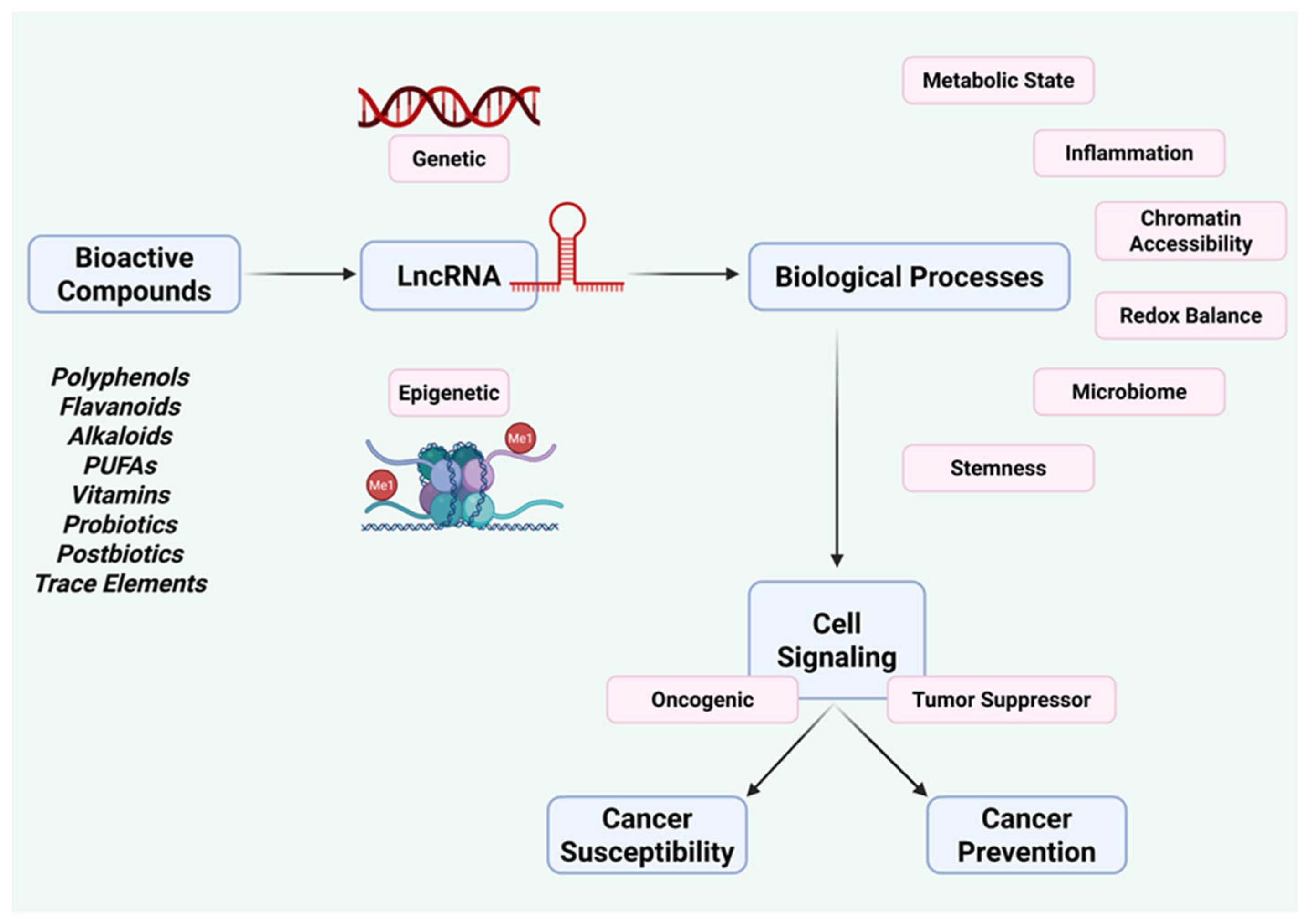
Figure 2.
Modulation of lncRNAs by polyphenols and flavonoids. Polyphenols and flavonoids, including curcumin, resveratrol, epigallocatechin gallate, quercetin, and berberine, selectively suppress oncogenic lncRNAs (e.g., HOTAIR, H19, MALAT1, NEAT1, UCA1, PVT1) while inducing tumor-suppressive lncRNAs (e.g., GAS5, MEG3, NBR2, CASC2, INXS), converging on Wnt/β-catenin, TGF-β, PI3K–AKT–mTOR, and NF-κB signaling pathways.
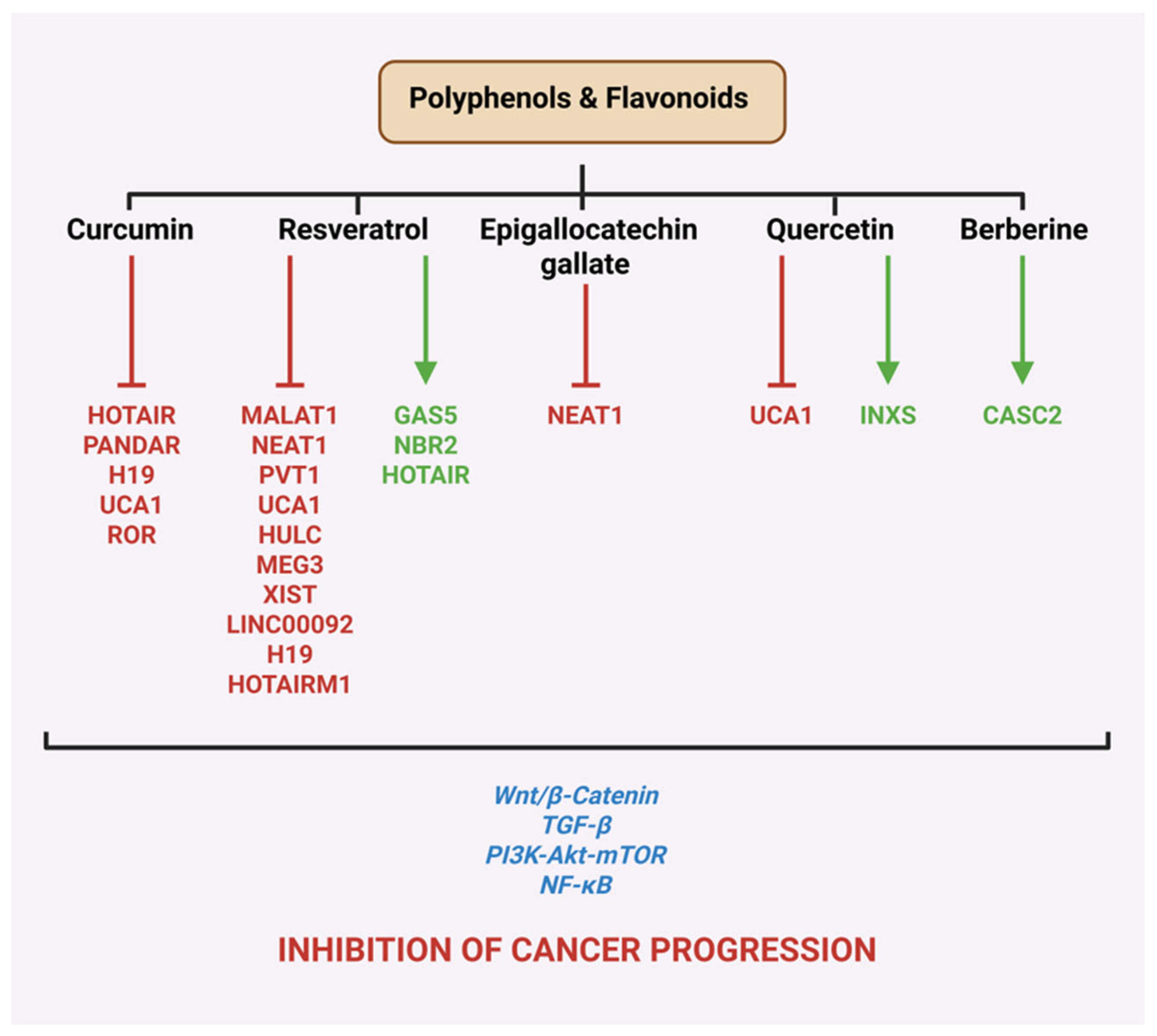
Figure 3.
Differential regulation of lncRNA networks by omega-3 and omega-6 fatty acids. This schematic illustrates how distinct classes of dietary omega fatty acids modulate cancer-relevant lncRNA programs and signaling pathways. Omega-3 fatty acids, including eicosapentaenoic acid (EPA) and docosahexaenoic acid (DHA), suppress multiple oncogenic lncRNAs (e.g., H19, HOTAIR, SNHG3, LINC00857, LCAL1, LUCAT1, and PSMG3-AS1) while inducing tumor-suppressive lncRNAs such as LINC00299, AC002091.2, NBR2, and LIPE-AS1. These changes converge on key pathways governing inflammation, metabolism, and growth control, including TGF-β, PI3K–AKT–mTOR, NF-κB, PPARγ, and AMPK signaling. In contrast, omega-6 fatty acids, represented by arachidonic acid, are associated with induction of distinct lncRNAs (e.g., SNHG17, MIR100HG, NR2F1-AS1, MAGI2-AS3, POLR2J4, and NOP14-AS1) that preferentially engage MAPK, EGFR, and NOTCH signaling cascades. Together, the figure highlights how the balance between omega-3 and omega-6 fatty acids shapes lncRNA-centered regulatory networks with opposing consequences for cancer progression.
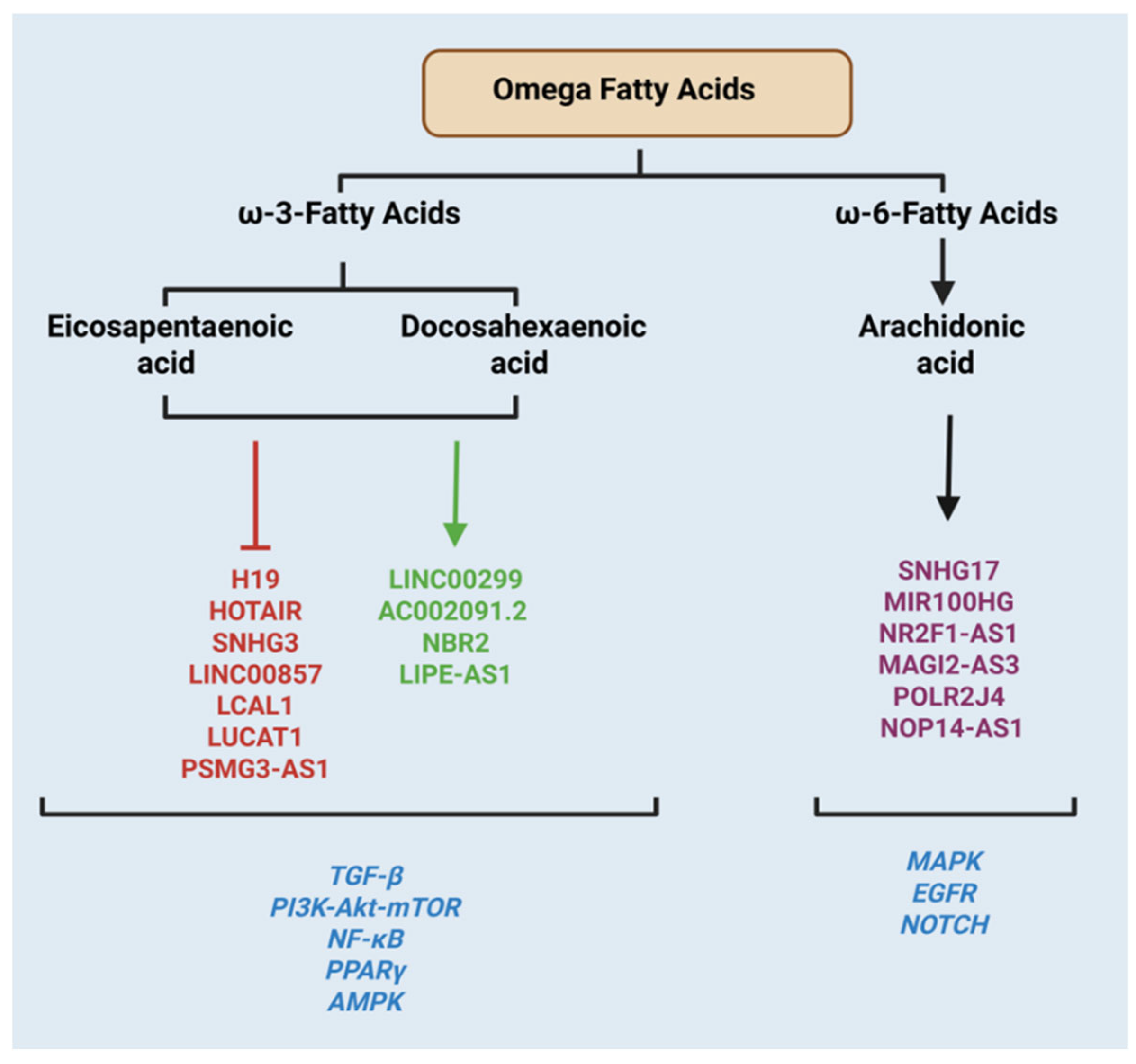
Figure 4.
Regulation of cancer-associated lncRNAs by niacin (vitamin B3) and folate. This figure summarizes how niacin (vitamin B3) and folate shape cancer-relevant lncRNA programs through complementary metabolic and epigenetic routes. Niacin acts primarily through NAD⁺-dependent processes, including sirtuin (SIRT1) signaling, NAMPT-driven NAD⁺ salvage, and PARP-mediated DNA repair. Through these pathways, niacin is linked to reduced expression of oncogenic lncRNAs such as H19, UCA1, and HOTAIR, alongside induction of tumor-suppressive lncRNAs including MEG3, NBR2, NORAD, and LINP1, consistent with enhanced genome maintenance and stress resilience. Folate influences lncRNA expression through its role in one-carbon metabolism and DNA methylation. Adequate folate availability is associated with repression of oncogenic lncRNAs (HOTAIR, H19, UCA1) and increased expression of tumor-suppressive lncRNAs such as MEG3 and LINC00261. Together, these vitamin-responsive lncRNA networks converge on pathways that favor genomic stability, metabolic balance, and restraint of cancer progression.
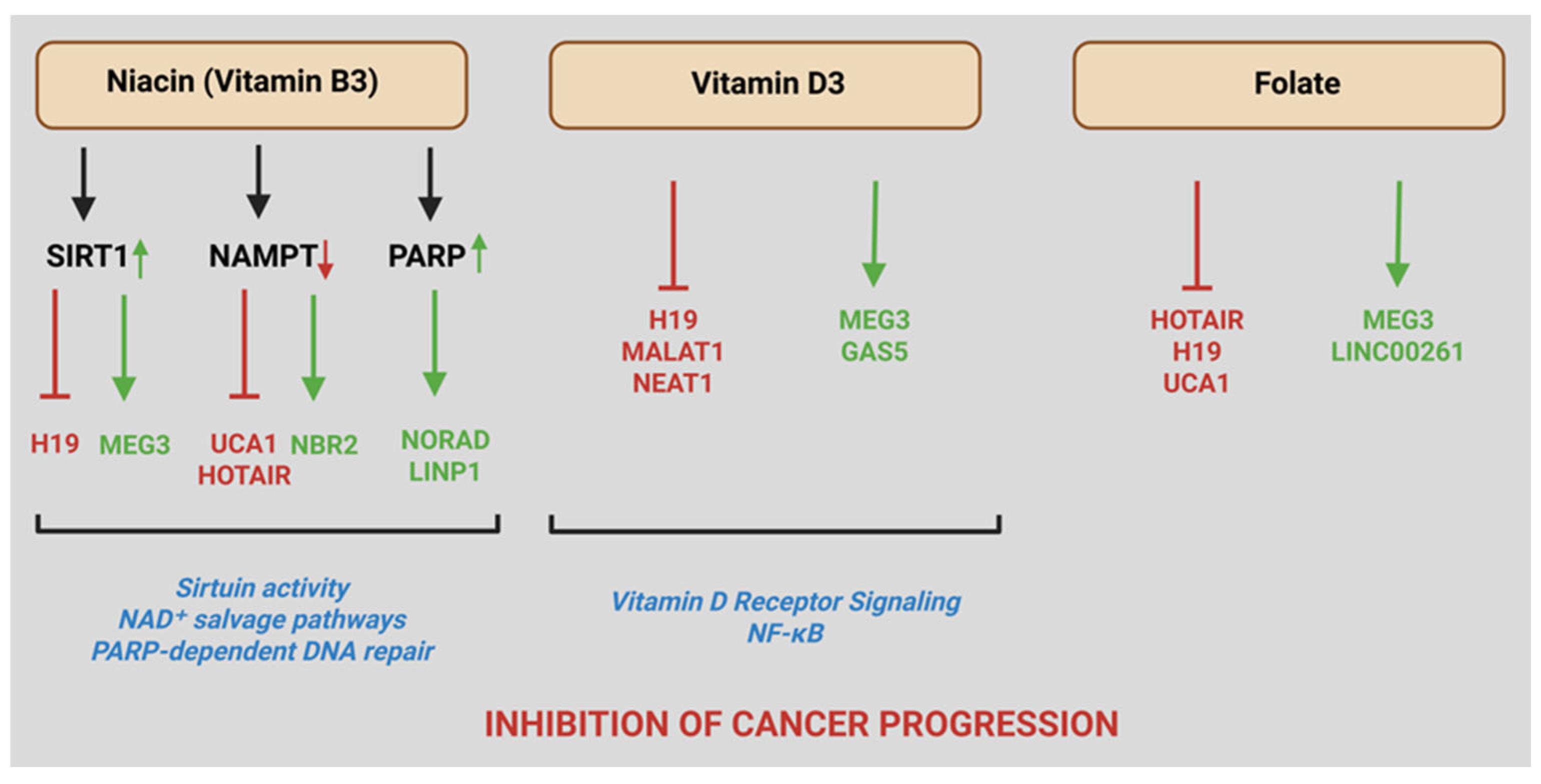
Figure 5.
lncRNA-mediated integration of diet, gut microbiota, and colorectal cancer progression. This schematic illustrates how dietary patterns shape gut microbial composition and downstream lncRNA-regulated pathways that influence intestinal inflammation and colorectal cancer progression. A fat- and sugar-rich diet promotes intestinal dysbiosis, characterized by enrichment of pro-inflammatory bacteria such as Fusobacterium nucleatum, leading to chronic inflammation, inflammatory bowel disease (IBD), and a tumor-promoting microenvironment. Dysbiosis-associated bacteria induce oncogenic lncRNAs, including EVADR and KRT-AS, which facilitate tumor growth, immune evasion, and metastatic progression. In contrast, a diet rich in fruits, fiber, and fish supports gut eubiosis, characterized by short-chain fatty acid (SCFA)–producing bacteria such as Lactobacillus acidophilus. SCFAs, particularly butyrate, suppress oncogenic lncRNAs including MALAT1, UCA1, PVT1, and HOTAIR, thereby restraining malignant signaling. The intestinal epithelial lncRNA CARINH contributes to maintenance of eubiosis and mucosal homeostasis, limiting inflammation and colorectal tumorigenesis. Collectively, this axis highlights lncRNAs as molecular mediators through which diet–microbiota interactions modulate intestinal inflammation, tumor growth, and metastatic potential.
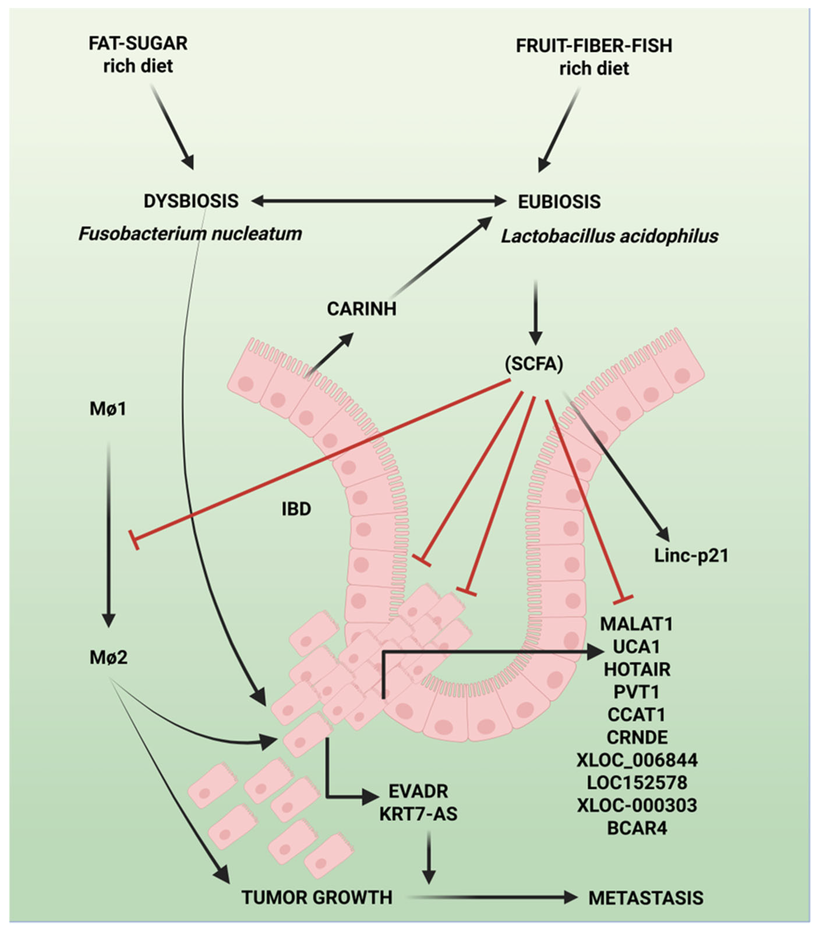
Figure 6.
AI and ML paradigm for decoding nutrient–lncRNA–cancer regulatory networks. This schematic illustrates an emerging framework for systematically resolving how nutritional inputs shape cancer biology through lncRNA-centered regulatory networks. Multi-omics and single-cell profiling provide foundational molecular layers to capture nutrient-responsive lncRNA dynamics across genetic, epigenetic, transcriptional, proteomic, and metabolic dimensions. These data are integrated using computational network inference strategies—including correlation-based modeling, Bayesian and causal network reconstruction, dynamic modeling, and systems biology approaches—to identify regulatory hierarchies, context-specific control nodes, and emergent network behavior. Artificial intelligence and machine-learning strategies further extend this paradigm by enabling data integration at scale, pattern discovery, and predictive modeling of nutrient–lncRNA–pathway interactions. Together, these approaches define a forward-looking systems framework to guide mechanistic discovery, biomarker development, and precision nutrition–based cancer prevention strategies.
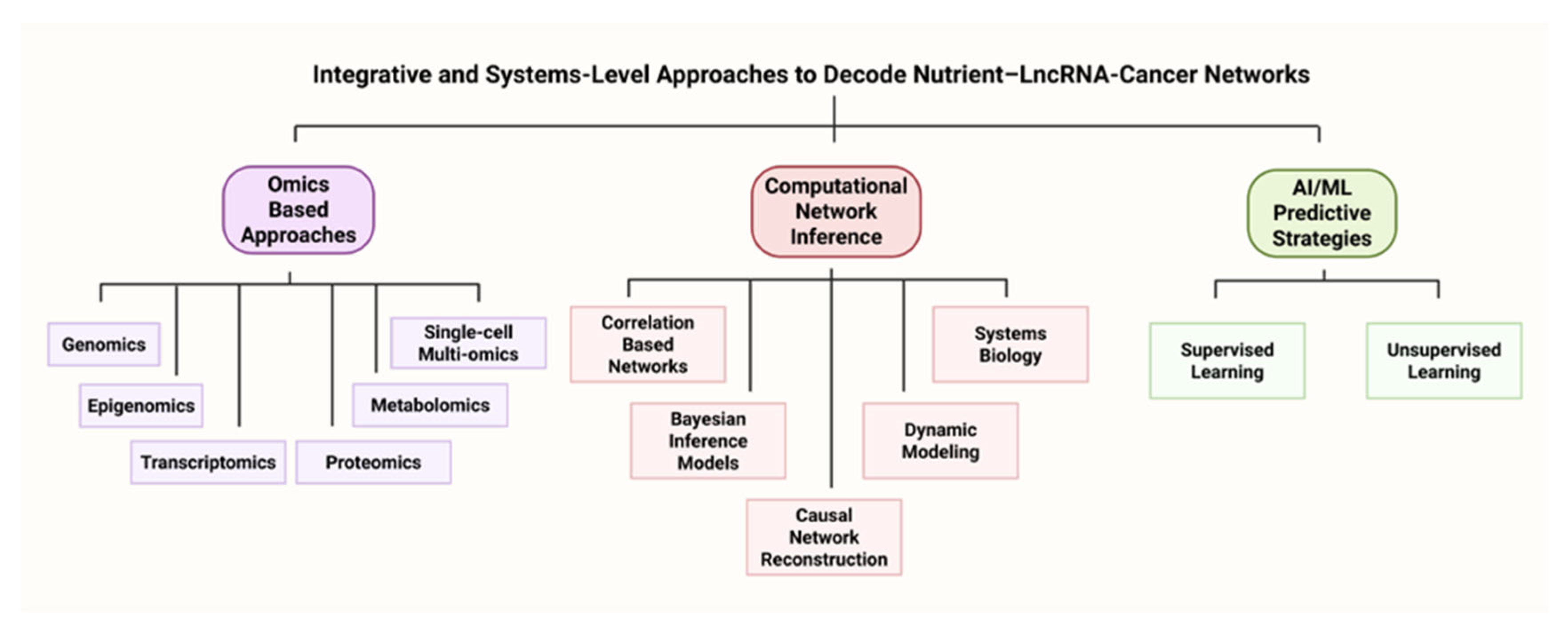
Disclaimer/Publisher’s Note: The statements, opinions and data contained in all publications are solely those of the individual author(s) and contributor(s) and not of MDPI and/or the editor(s). MDPI and/or the editor(s) disclaim responsibility for any injury to people or property resulting from any ideas, methods, instructions or products referred to in the content. |
© 2025 by the authors. Licensee MDPI, Basel, Switzerland. This article is an open access article distributed under the terms and conditions of the Creative Commons Attribution (CC BY) license (http://creativecommons.org/licenses/by/4.0/).
Copyright: This open access article is published under a Creative Commons CC BY 4.0 license, which permit the free download, distribution, and reuse, provided that the author and preprint are cited in any reuse.


