Preprint
Article

This version is not peer-reviewed.

Cancer-Associated Thrombosis in Breast Cancer: Risk Factors and Personalized Management

A peer-reviewed article of this preprint also exists.

Submitted:

30 December 2025

Posted:

31 December 2025

You are already at the latest version

Abstract

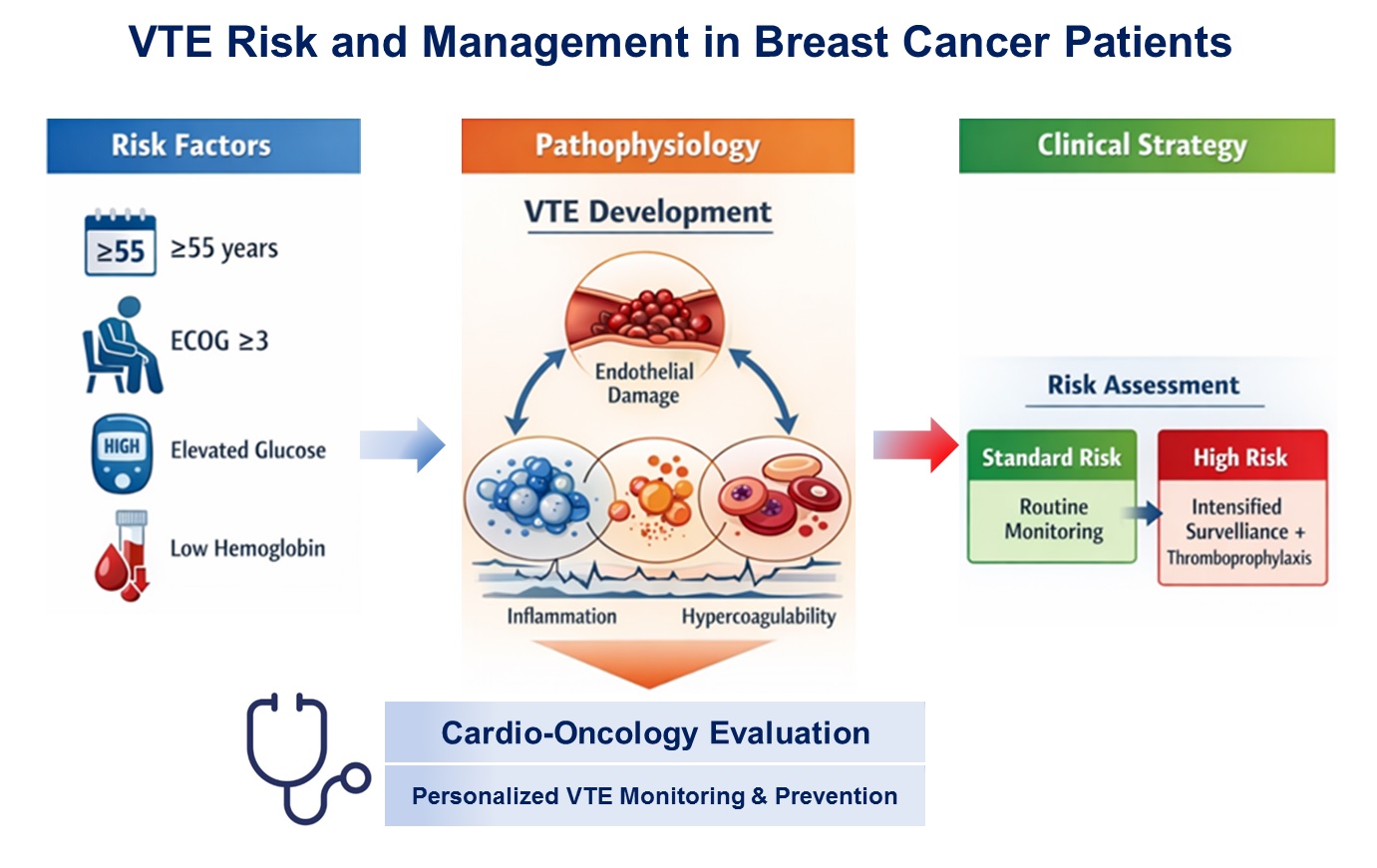
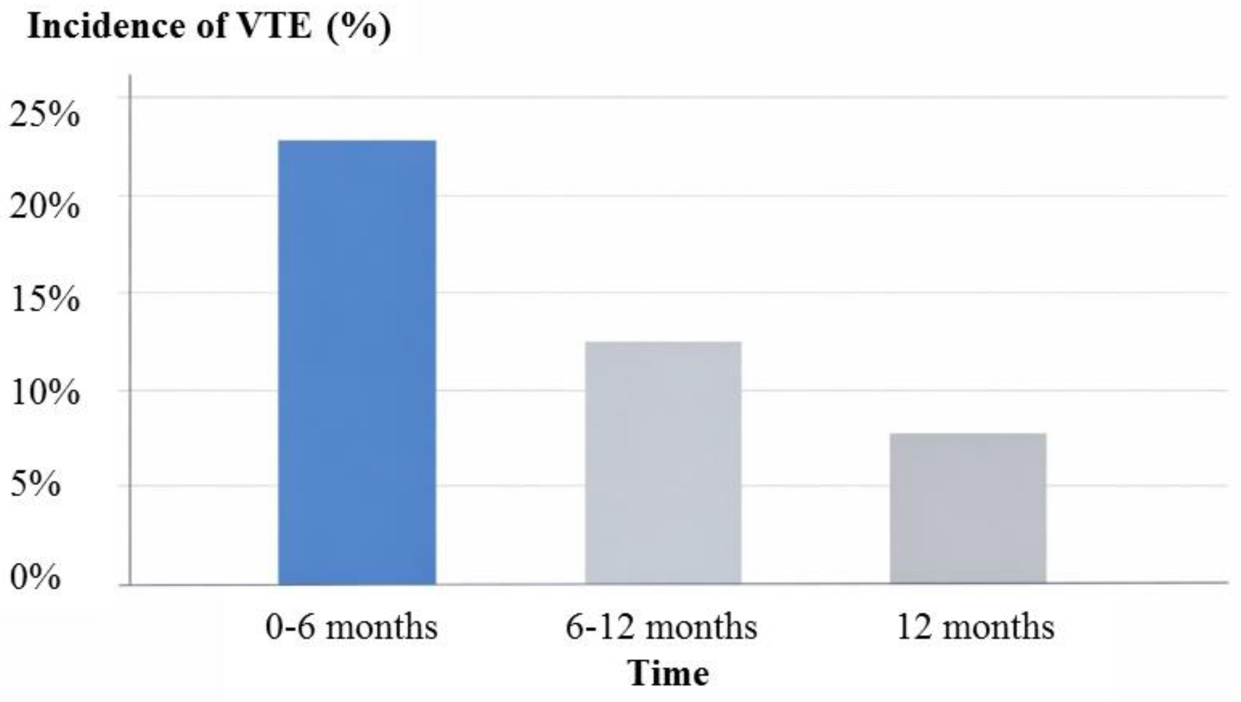
Background & Objectives: Venous thromboembolism (VTE) is a major cardiovascular complication in cancer patients and leading cause of morbidity and mortality. The aim of the study was to evaluate the incidence, timing, clinical predictors, and management of VTE in patients with breast cancer (BC), undergoing oncological therapy, and to propose a risk-adapted strategy for thrombosis monitoring and prevention. Methods: In this retrospective single-center study, 116 women with histologically confirmed BC (stages I–IV) treated between 2021 and 2024 were included. Patients were divided according to the occurrence of objectively confirmed VTE. Clinical characteristics, comorbidities, laboratory parameters, cancer-related factors, and treatment modalities were analyzed. Univariate and multivariate logistic regression analyses were performed to identify independent predictors of VTE. Results: VTE occurred in 25 patients (21.6%), predominantly within the first 12 months after cancer diagnosis. Patients who developed VTE were significantly older and more frequently had hypertension, dyslipidemia, hyperglycemia, anemia, and leukocytosis. Multivariate analysis identified age≥55 years, poor performance status (ECOG ≥3), and elevated glucose level as independent predictors of VTE. Deep vein thrombosis of the lower and upper extremities was the most common manifestation (52%), while pulmonary embolism was present in 24% of cases, either alone or in combination (20%). Direct oral anticoagulants were the most frequently used long-term anticoagulant therapy. Conclusions: VTE is a clinically relevant and relatively frequent complication in patients with BC, particularly during the early period of anticancer treatment. Patient-related and metabolic factors play a key role in thrombosis risk, underscoring the need for individualized, risk-adapted approaches to VTE prevention and monitoring in these populations.

Keywords: 
;  ;  ;  ;  

1. Introduction

Venous thromboembolism (VTE), encompassing deep vein thrombosis (DVT) and pulmonary embolism (PE), represents one of the most common and potentially life-threatening complications in patients with cancer. Cancer-associated thrombosis (CAT) is responsible for significant morbidity, increased healthcare utilization, interruption of oncological therapy, and remains a leading cause of non-cancer-related mortality in cancer patients. The prothrombotic state in cancer is multifactorial and results from complex interactions between tumor biology, host-related factors, and anticancer treatments [1].
Breast cancer is the most frequently diagnosed malignancy among women worldwide and in Ukraine as well, and is associated with continuously improving survival due to advances in early detection and systemic therapy [2]. Historically, breast cancer has been categorized as a malignancy with a relatively low risk of VTE, particularly when compared with gastrointestinal, pancreatic, or lung cancers. Consequently, routine pharmacological thromboprophylaxis is not generally recommended for most patients with BC in current clinical guidelines. However, emerging evidence from population-based registries and real-world cohorts suggests that the risk of VTE in BC patients may be higher than previously assumed, especially during period of active treatment [3,4].
Several factors may contribute to the development of VTE in BC patients, including advanced age, comorbid cardiovascular and metabolic conditions, reduced functional status, and cancer stage progression. In addition, modern treatment strategies, such as chemotherapy (CT), endocrine therapy, radiation therapy (RT), and surgical intervention, may further increase thrombotic risk through endothelial injury, systemic inflammation, and alterations in coagulation pathway [5]. Importantly, a conventional risk stratification tools, such as the Khorana Risk Score, has demonstrated limited predictive value in BC population, highlighting the need for alternative or refined risk assessment approaches [6,7,8,9].
The early identification of patients at risk of VTE is essential to guide individualized prevention strategies and to balance the benefits of thromboprophylaxis against the risk of bleeding. From a cardio-oncology perspective, comprehensive cardiovascular assessment and close monitoring are particularly relevant, as thrombotic complications often coexist with other cardiovascular comorbidities and may adversely affect overall prognosis [10].
Therefore, the present study aimed to assess the incidence, timing, clinical characteristics, and predictors of VTE in patients with BC undergoing cancer treatment in a real-world setting. Additionally, based on the identified risk factors, we proposed a risk-adapted approach for thrombosis monitoring and prevention to support personalized management in this patient population.

2. Materials and Methods

2.1. Study Design and Participants

This retrospective, single-center observation study included 116 consecutive patients with histologically confirmed BC (stages I–IV) who underwent cancer treatment between 2021 and 2024. All patients received therapy in accordance with current guidelines and protocols at the National Cancer Institute of the Ministry of Health of Ukraine. Patients were referred for cardiovascular evaluation and monitoring to the Cardio-Oncology Center at the National Scientific Center “The M.D. Strazhesko Institute of Cardiology, Clinical and Regenerative Medicine of the National Academy of Medical Sciences of Ukraine”.
This study was conducted in compliance with the Declaration of Helsinki, and the study protocol was approved by the local institutional ethics committee, and all data were analyzed anonymously.

2.2. Study Endpoint and Group Stratification

The primary endpoint of the study was the occurrence of objectively confirmed VTE during the follow-up period. Based on the presence or absence of VTE, patients were divided into two groups: Group 1: patients who developed VTE during follow-up (n = 25), Group 2: patients without VTE events (n = 91).

2.3. Definition and Diagnosis of VTE

VTE included DVT of the upper or lower extremities, PE, and combined PE and DVT. Upper extremity DVT included thrombosis of the axillary, subclavian, and internal jugular veins. Lower extremity DVT involved thrombosis of the iliac, femoral, popliteal, and tibial veins. Peripheral DVT was confirmed using compression ultrasonography of the affected limb veins. PE was diagnosed by contrast-enhanced computed tomography.

2.4. Clinical and Cardiovascular Assessment

Patients’ comprehensive clinical and cardiovascular evaluation, along with detailed medical history, cardiovascular risk factors, and comorbidities, cancer-related characteristics (stage, histology, treatment modalities, CT regimens, and cumulative doses, RT exposure, surgical interventions) were performed.
Assessment of functional status using Eastern Cooperative Oncology Group performance status (ECOG PS), physical examination, 12-lead electrocardiography, and transthoracic echocardiography. Left ventricular ejection fraction (LV EF) was measured using standard echocardiographic techniques.

2.5. Laboratory Assessment

Routine laboratory investigations included: complete blood count (white blood cells, red blood cells, hemoglobin, thrombocytes), biochemical parameters (serum creatinine, glucose, and cholesterol panel). All laboratory tests were performed in certified hospital laboratories using standardized methods.

2.6. Assessment of Cardiovascular Risk Factors and Comorbidities

Traditional cardiovascular risk factors were evaluated in all patients, including body mass index (BMI), smoking status, and dyslipidemia. Comorbid cardiovascular disease included coronary artery disease (CAD), arterial hypertension, and diabetes mellitus (DM).

2.7. Anticoagulant Therapy

The choice of anticoagulant therapy for the treatment of VTE was based on current clinical recommendations and individual patient characteristics. Long-term anticoagulation was performed using direct oral anticoagulants (DOACs) or low molecular weight heparin (LMWHs). Treatment selection considered bleeding risk, renal function, drug-drug interaction, and patient preference.

2.8. Statistical analysis

Data analysis was performed using SPSS version 26.0 (IBM Corp., USA). Continuous variables are presented as mean ± standard error of the mean (M ± m), while categorical variables are expressed as absolute numbers and percentages. Group comparisons were conducted using Student’s t-test for continuous variables and the chi-square test for categorical variables. Univariate logistic regression analysis was used to identify variables associated with VTE occurrence. Variable with significance level of p < 0.05 in univariate analysis were subsequently entered into multivariate stepwise logistic regression model to identify independent predictors of VTE. Odds ratios (OR) and 95% confidence intervals (CI) were calculated. A two-side p value < 0.05 was considered statistically significant.

3. Results

3.1. Baseline Characteristic of the Study Population

A total of 116 patients with histologically confirmed DC were included in the analysis. The baseline demographic, anthropometric, and clinical characteristics of the study population are summarized in Table 1.
The mean age of the cohort was 53.1 ± 1.2 years, and all patients were female. Sixteen patients (13.8%) were older than 65 years. The mean BMI was 28.1 kg/m2. Dyslipidemia was presented in 26.7% of patients, arterial hypertension in 38.8%, coronary artery disease in 10.3% and diabetes mellitus in 2.6%.
The majority of patients (93.9%) had stage II or III BC at diagnosis. Most patients (64%) received anthracycline-based CT regimens, including epirubicin or doxorubicin, in combination with cyclophospamide, taxanes, trastuzumab, or other agents.

3.2. Incidence of Venous Thromboembolism

During the follow-up period, VTE occurred in 25 patients (21.6%). Based on VTE occurrence, patients were divided into two groups:
Group 1: patients who developed VTE (n=25)
Group 2: patients without VTE (n=91).
A comparison of baseline clinical, oncological, laboratory, and hemodynamic characteristics between two groups is presented in Table 2.
Patients who developed VTE were significantly older than those without VTE (63.8 ± 1.9 vs. 50.2 ± 1.3 years, p<0.01). Arterial hypertension was more prevalent in Group 1 compared with Group 2 (56.0 vs. 34.2%, p<0.05), as was dyslipidemia (40.0 vs. 23.1, p<0.05).
There were no significant differences between groups with respect to BMI, smoking status, CAD, or DM. Patients with VTE tended to have a poorer functional status, with ECOG performance ≥ 3 observed more frequently in Group 1, although this difference did not reach statistical significance.
Systolic and diastolic BP values, as well as heart rate, were comparable between groups. However, LV EF was significantly lower in patients with VTE compared with those without VTE (58.1 ± 1.2% vs. 61.8 ± 0.5%, p<0.05), which may be explained by impaired LV contractility, particularly in patients with PE.
Patients who developed VTE has significantly higher levels of total cholesterol and fasting glucose compared with patients without VTE (p < 0.05 for both).
Additionally, Group 1 demonstrated significantly higher WBC counts, as well as lower RBC counts and hemoglobin level (p<0.05 for all comparisons). Elevated WBC count and anemia are recognized contributors to thrombotic risk in oncology patients [12,13].
Anthracycline-containing CT regimens were used more frequently in patients without VTE (68.1%) than in those with VTE (48.0%), however, the cumulative anthracycline dose did not differ significantly between groups. HER2/neu-positive status and trastuzumab use were also more common in Group 2. No significant differences were observed regarding RT or endocrine therapy exposure (Table 3).
Surgical treatment was performed less frequently in Group 1, however, in two patients VTE developed within 30 days after surgery. According to available evidence, postoperative VTE risk in BC patients is considered relatively low, but perioperative events remain clinically relevant [11].
The type, timing and treatment of VTE events are detailed in Table 4. DVT of the lower or upper extremities was the most frequent manifestation (48.0%), followed by PE alone (24.0%) and combined PE and DVT (20.0%). Catheter-related thrombosis and isolated upper extremity DVT were observed less frequently (8%).
VTE most commonly occurred within the first 12 months after BC diagnosis: 40.0% within the first 6 month and 44.0% between 6 and 12 months, while 16.0% occurred after one year (Figure 1). This temporal distribution reflects the period of highest thrombotic vulnerability during cancer treatment.
Varicose veins or a history of thrombophlebitis were presented in 40.0% of patients with VTE, suggesting an additional predisposing factor for thrombosis.
DOAC were used as long-term anticoagulant therapy in the majority of patients with VTE (76.0%), whereas LMWH were prescribed in 24.0% of cases.

3.3. Predictors of Venous Thromboembolism

In univariate analysis, age ≥ 55 years, ECOG performance status ≥ 3, elevated glucose level, and decreased hemoglobin level were significantly associated with VTE development (P < 0.05 for all). No significant association was observed between the Khorana Risk Score and VTE occurrence.
Multivariate logistic regression analysis identify three independent predictors of VTE: age ≥ 55 years (OR 1.74, 95% CI: 1.18 to 2.72), ECOG performance status ≥ 3 (OR 1.92, 95% CI: 1.29 to 2.12), and elevated glucose level (OR 2.21, 95% CI: 1.53 to 3.14).

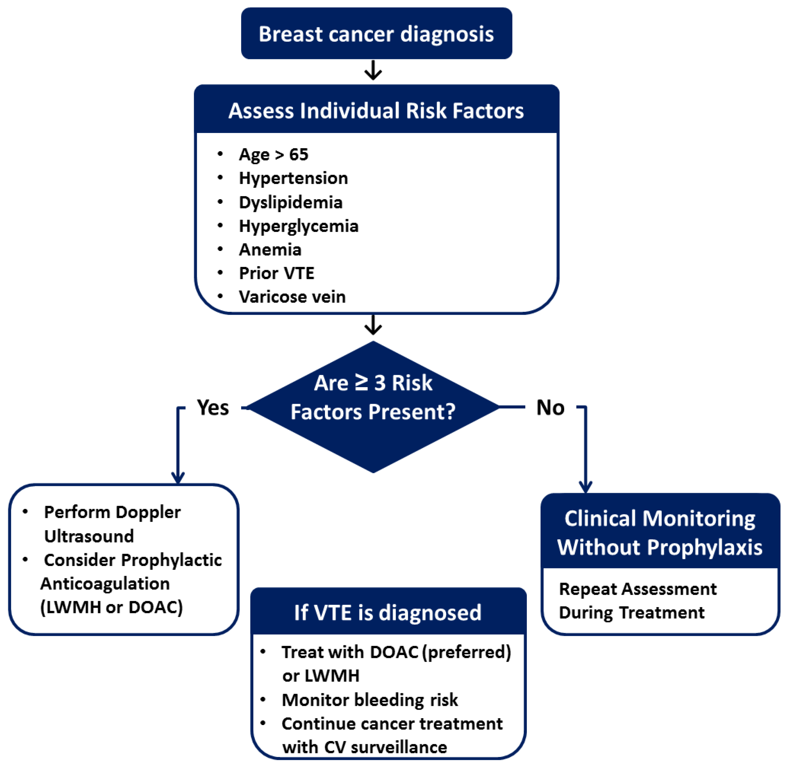
3.4. Risk-Adapted Monitoring and Prevention Strategy

Based on the identified predictors, a risk-adapted algorithm for VTE monitoring and prevention in BC patients was proposed (Figure 2). This strategy integrates clinical, laboratory, and treatment-related factors to personalize thrombosis risk and prophylaxis, aiming to improve patient safety while minimizing bleeding risk.
Study limitations: This study has several limitations, including its single center, retrospective design and relatively small sample size, which may limit generalizability. The absence of a standardized protocol of thromboprophylaxis and potential selection bias should also be acknowledged.

4. Discussion

CAT represents a major challenge in contemporary cardio-oncology, reflecting the complex interplay between malignancy, anticancer therapies, cardiovascular comorbidities, and systemic metabolic disturbances. Although BC has traditionally been classified as a tumor with a relatively low thrombotic risk, the present study demonstrates that VTE is a frequent and clinically relevant complication in patients undergoing modern multimodal BC treatment [14].
These findings call into question conventional risk stratification paradigms and underscore the need for an integrated cardio-oncological approach to thrombosis prevention and monitoring.
In our cohort, VTE occurred in 21.6% of patients, with the highest incidence observed within the first 12 months after BC diagnosis. This temporal pattern is consistent with large population-based studies indicating that the peri-diagnostic and early treatment phase represent the period of maximal thrombotic vulnerability in cancer patients [15,16,17]. From a cardio-oncology perspective, this interval coincides with cumulative exposure to surgical intervention, systemic CT, RT, and targeted therapies, all of which may promote endothelial injury, inflammatory activation, and dysregulations of the coagulation cascade [18,19].
Importantly, this early high risk window overlaps with the phase of intensified cardiovascular surveillance in cardio-oncology practice, highlighting the necessity of integrating thrombotic risk assessment into routine cardiovascular monitoring during initial cancer treatment.
A key finding of the present study is that patient-related factors, rather than cancer-specific or treatment-related variables, were the primary determinants of VTE development. Advanced age emerged as a strong independent predictor, in line with previous reports and established clinical risk models, including the Geneva score [20]. Aging is associated with endothelial dysfunction, impaired fibrinolysis, and a higher burden of cardiovascular comorbidities, all of which amplify thrombotic risk in oncology populations.
Poor functional status (ECOG performance status ≥3) was independently associated with VTE occurrence. This likely reflects the combined effects of reduced mobility, frailty, systemic inflammation, and heightened cardiovascular vulnerability. Similar observations have been reported in large BC registries, where impaired performance status consistently predicted adverse thrombotic outcomes [21,22].
Of particular relevance to cardio-oncology is the identification of hyperglycemia as an independent VTE predictor. Metabolic dysregulation is increasingly recognized as a key contributor to thrombogenesis through mechanisms involving endothelial dysfunction, oxidative stress, platelet hyperactivity, and activation of procoagulant pathways. In patients with BC, metabolic abnormalities frequently coexist with hypertension, dyslipidemia, and subclinical cardiovascular disease, forming a high-risk cardiometabolic phenotype that predisposes to CAT. These findings reinforce the concept that metabolic optimization should be considered an integral component of thrombosis prevention strategies in cardio-oncology practice.
Patients who developed VTE exhibited higher leukocyte counts and lower hemoglobin levels. Leukocytosis reflects systemic inflammation and neutrophil-driven prothrombotic mechanisms, including neutrophil extracellular trap, which has been increasingly implicated in the pathogenesis of CAT [11,12]. Anemia, particularly at moderate-to-severe levels, is a recognized thrombotic risk factor and may further exacerbate tissue hypoxia, endothelial dysfunction, and cardiovascular strain [13].
From a cardio-oncological perspective, these laboratory abnormalities should be interpreted not merely as oncological markers but as indicator of global cardiovascular and thrombotic risk, warranting closer surveillance and potentially earlier preventive interventions.
The Khorana Risk Score did not demonstrate predictive utility in this BC cohort, consistent with prior systematic reviews and meta-analyses [10]. Although widely applied in ambulatory oncology populations, the Khorana score appears insufficient for tumors traditionally classified as low risk, such as BC. This limitation is particularly relevant in cardio-oncology, where reliance on generalized risk models may result in under-recognition of patients with high-risk cardiovascular phenotypes.
Our findings support the growing consensus that tumor-agnostic risk scores should be complemented or replaced by individualized, cardio-oncology-oriented risk assessment frameworks incorporating functional status, metabolic parameters, inflammatory markers, and cardiovascular comorbidities.
In contrast, exposure to anthracyclines, trastuzumab, RT, or endocrine therapy was not independently associated with VTE in multivariate analysis. Although several anticancer therapies are known to affect endothelial function and coagulation pathways, current evidence does not support routine pharmacological thromboprophylaxis in unselected BC populations [8,22,23,24].
Nevertheless, treatment-related cardiovascular toxicity may indirectly influence thrombotic risk by impairing cardiac function or promoting vascular injury. The lower LV EF in patients with VTE, particularly those with PE, highlights the bidirectional relationship between cardiac dysfunction and thromboembolic events and reinforces the need for integrated cardiac and thrombotic monitoring.
DOACs were the predominant long-term anticoagulant therapy in this cohort, reflecting current guideline-supported practice. Accumulating evidence supports the efficacy and acceptable safety profile of DOACs in CAT, particularly in patients without gastrointestinal malignancies or high bleeding risk [25]. In BC patients, who generally exhibit a lower hemorrhagic risk profile, DOAC-based strategies appear especially feasible within a cardio-oncology framework.
Based on the identified predictors, we propose a risk-adapted algorithm for VTE monitoring and prevention that integrates age, functional status, cardiometabolic abnormalities, hematological parameters, and cardiovascular comorbidities. This approach aligns with the principles of modern cardio-oncology, emphasizing early risk stratification, personalized surveillance, and judicious use of preventive anticoagulation.

5. Conclusions

VTE represents a significant and often underestimated complication in patients with BC, particularly during early phases of treatment. Our findings emphasize that patient-related and cardiometabolic factors are central drivers of thrombotic risk and should be systematically addressed within cardio-oncology programs. Prospective, multicenter studies are warranted to validate these observations and to develop BC-specific, cardio-oncology-oriented risk models capable of guiding personalized thromboprophylaxis strategies.

Author Contributions

Conceptualization, S.K.; methodology, N.D., S.K.; formal analysis, N.D.; investigation, S.K., N.D., O.L., I.S, A.L., S.L.; writing-original draft preparation, N.D.; writing-review and editing, S.K., N.D., S.L. All authors have read and agreed to the published version of the manuscript.

Funding

no funding.

Institutional Review Board Statement

The study was conducted in accordance with the Declaration of Helsinki and approved by the local ethics committee (№ 1-04, date of approval 20.01.2021).

Informed Consent Statement

Informed consent was obtained from all subjects involved in the study.

Data Availability Statement

Data are available upon request.

Acknowledgments

We sincerely thank all participants for their time, effort, and invaluable contributions to this study.

Conflicts of Interest

no conflicts of interest.

Abbreviations

The following abbreviations are used in this manuscript:
BC Breast cancer
VTE Venous thromboembolism
DVT Deep vein thrombosis
PE Pulmonary embolism
PH Pulmonary hypertension
CAT Cancer associated thrombosis
CCT Comprehensive antitumor therapy
CT Chemotherapy
RT Radiation therapy
BMI Body mass index
CAD Coronary artery disease
DM Diabetes mellitus
DOAC Direct oral anticoagulants
LMWH Low molecular weight heparin

References

  1. Wan, T.; Song, J.; Zhu, D. Cancer-associated venous thromboembolism: a comprehensive review. Thrombosis J 2025, 23(1), 35. [Google Scholar]
  2. Cancer in Ukraine 2022-2023: Incidence, mortality, prevalence and other relevant statistics. Bulletin of the National Cancer Registry of Ukraine 2024, 25, 130 p.
  3. Razouki, ZA; Ali, NT; Nguyen, VQ; Escalante, CP. Risk factors associated with venous thromboembolism in breast cancer: a narrative review. Support Care Cancer 2022, 30(10), 8589–8597. [Google Scholar] [CrossRef]
  4. Kirwan, CC; Blower, EL. Contemporary breast cancer treatment-associated thrombosis. Thromb Res 2022, 213, S8–S15. [Google Scholar] [CrossRef]
  5. Rubovszky, G; Kocsis, J; Boér, K; Chilingirova, N; Dank, M; Kahán, Z; Kaidarova, D; Kövér, E; Krakovská, BV; Máhr, K; et al. Systemic Treatment of Breast Cancer. 1st Central-Eastern European Professional Consensus Statement on Breast Cancer. Pathol Oncol Res 2022, 28, 1610383. [Google Scholar] [CrossRef] [PubMed]
  6. Key, NS; Khorana, AA; Kuderer, NM; Bohlke, K; Lee, AYY; Arcelus, JI; Wong, SL; Balaban, EP; Flowers, CR; Francis, CW; et al. Venous Thromboembolism Prophylaxis and Treatment in Patients With Cancer: ASCO Guideline Update. J Clin Oncol 2023, 41(16), 3063–71. [Google Scholar] [CrossRef]
  7. Gradishar, WJ; Moran, MS; Abraham, J; Abramson, V; Aft, R; Agnese, D; Allison, KH; Anderson, B; Bailey, J; Burstein, HJ; et al. Breast Cancer, Version 3.2024, NCCN Clinical Practice Guidelines in Oncology. J Natl Compr Canc Netw 2024, 22(5), 331–57. [Google Scholar]
  8. Falanga, A; Ay, C; Di Nisio, M; Gerotziafas, G; Jara-Palomares, L; Langer, F; Lecumberri, R; Mandala, M; Maraveyas, A; Pabinger, I; et al. ESMO Guidelines Committee. Venous thromboembolism in cancer patients: ESMO Clinical Practice Guideline. Ann Oncol 2023, 34(5), 452–67. [Google Scholar] [CrossRef]
  9. Overvad, TF; Ording, AG; Nielsen, PB; Skjøth, F; Albertsen, IE; Noble, S; Vistisen, AK; Gade, IL; Severinsen, MT; Piazza, G; Larsen, TB. Validation of the Khorana score for predicting venous thromboembolism in 40 218 patients with cancer initiating chemotherapy. Blood Adv 2022, 6(10), 2967–2976. [Google Scholar]
  10. Mulder, FI; Candeloro, M; Kamphuisen, PW; Di Nisio, M; Bossuyt, PM; Guman, N; Smit, K; Büller, HR; van Es, N. CAT-prediction collaborators. The Khorana score for prediction of venous thromboembolism in cancer patients: a systematic review and meta-analysis. Haematologica 2019, 104(6), 1277–87. [Google Scholar] [CrossRef]
  11. Maesaka, JY; Reis, YN; Elias, LM; Akerman, D; Baracat, EC; Filassi, JR. Venous thromboembolism incidence in postoperative breast cancer patients. Clinics (Sao Paulo) 2023, 78, 100229. [Google Scholar] [CrossRef] [PubMed]
  12. Ikeda, S; Yamashita, Y; Morimoto, T; Chatani, R; Kaneda, K; Nishimoto, Y; Ikeda, N; Kobayashi, Y; Ikeda, S; Kim, K; et al. Association Between White Blood Cell Counts at Diagnosis and Clinical Outcomes in Venous Thromboembolism - From the COMMAND VTE Registry-2. Circ J 2025, 89(5), 592–601. [Google Scholar] [CrossRef]
  13. Douce, DR; Holmes, CE; Cushman, M; MacLean, CD; Ades, S; Zakai, NA. Risk factors for cancer-associated venous thromboembolism: The venous thromboembolism prevention in the ambulatory cancer clinic (VTE-PACC) study. J Thromb Haemost 2019, 17(12), 2152–59. [Google Scholar] [PubMed]
  14. Carmona-Bayonas, A; Gómez, D; Martínez de Castro, E; Pérez Segura, P; Muñoz Langa, J; Jimenez-Fonseca, P; Sánchez Cánovas, M; Ortega Moran, L; García Escobar, I; Rupérez Blanco, AB; et al. A snapshot of cancer-associated thromboembolic disease in 2018–2019: First data from the TESEO prospective registry. Eur J Intern Med 2020, 78, 41–49. [Google Scholar] [PubMed]
  15. Walker, AJ; West, J; Card, TR; Crooks, C; Kirwan, CC; Grainge, MJ. When are breast cancer patients at highest risk of venous thromboembolism? A cohort study using English health care data. Blood 2016, 127(7), 849–57. [Google Scholar] [CrossRef]
  16. Khan, UT; Walker, AJ; Baig, S; Card, TR; Kirwan, CC; Grainge, MJ. Venous thromboembolism and mortality in breast cancer: cohort study with systematic review and meta-analysis. BMC Cancer 2017, 17(1), 747. [Google Scholar] [CrossRef]
  17. Brand, JS; Hedayati, E; Bhoo-Pathy, N; Bergh, J; Hall, P; Humphreys, K; Ludvigsson, JF; Czene, K. Time-dependent risk and predictors of venous thromboembolism in breast cancer patients: A population-based cohort study. Cancer 2017, 123(3), 468–75. [Google Scholar]
  18. Girardi, L; Wang, TF; Ageno, W; Carrier, M. Updates in the Incidence, Pathogenesis, and Management of Cancer and Venous Thromboembolism. Arterioscler Thromb Vasc Biol 2023, 43(6), 824–31. [Google Scholar]
  19. Park, JH; Ahn, SE; Kwon, LM; Ko, HH; Kim, S; Suh, YJ; Kim, HY; Park, KH; Kim, D. The Risk of Venous Thromboembolism in Korean Patients with Breast Cancer: A Single-Center Experience. Cancers 2023, 15, 3124. [Google Scholar] [CrossRef]
  20. Harder, EM; Desai, O; Marshall, PS. Clinical Probability Tools for Deep Venous Thrombosis, Pulmonary Embolism, and Bleeding. Clin Chest Med 2018, 39(3), 473–482. [Google Scholar]
  21. Ioffe, D; Bhatia-Patel, SC; Gandhi, S; Hamad, EA; Dotan, E. Cardiovascular Concerns, Cancer Treatment, and Biological and Chronological Aging in Cancer: JACC Family Series. JACC CardioOncol 2024, 6(2), 143–158. [Google Scholar] [CrossRef]
  22. Drăgan, A; Drăgan, AŞ. Novel Insights in Venous Thromboembolism Risk Assessment Methods in Ambulatory Cancer Patients: From the Guidelines to Clinical Practice. Cancers 2024, 16, 458. [Google Scholar] [CrossRef]
  23. Sánchez Cánovas, M; López Robles, J; Adoamnei, E; Cacho Lavin, D; Diaz Pedroche, C; Coma Salvans, E; Quintanar Verduguez, T; García Verdejo, FJ; Cejuela Solís, M; García Adrián, S; et al. Thrombosis in breast cancer patients on cyclin-dependent kinase inhibitors: Survival impact and predictive factors. A study by the Cancer and Thrombosis Group of the Spanish Society of Medical Oncology (SEOM). Eur J Intern Med 2024, 130, 98–105. [Google Scholar] [CrossRef]
  24. Kozhukhov, S; Dovganych, N. Thromboembolism in Patients with Cancer: A Practical Guide to Recurrent Events. Rev Cardiovasc Med 2024, 25(11), 406. [Google Scholar] [CrossRef]
  25. Zakai, NA; Walker, RF; MacLehose, RF; Adam, TJ; Alonso, A; Lutsey, PL. Impact of anticoagulant choice on hospitalized bleeding risk when treating cancer-associated venous thromboembolism. J Thromb Haemost 2018, 16(12), 2403–12. [Google Scholar] [CrossRef]
Figure 1. Temporal distribution of VTE in BC patients during follow-up.
Preprints 192135 g001
Figure 2. Risk-adapted algorithm for VTE monitoring and prevention in BC patients.
Preprints 192135 g002
Table 1. Baseline demographic and clinical characteristics of patients with breast cancer.
Variable Value of the indicator
(n=116)
Baseline characteristics
Age, years 53.1±1.2
Patients >65 years old, n (%) 16 (13.8)
Smoking, n (%) 2 (1.7)
BMI, kg/m2 28.1±0.6
Dyslipidemia, n (%) 31 (26.7)
Comorbidity
Coronary artery disease, n (%) 12 (10.3)
Diabetes mellitus, n (%) 3 (2.6)
Hypertension, n (%) 45 (38.8)
Abbreviations: BMI - body mass index. Data are presented as mean ± standard error of the mean or absolute number (%).
Table 2. Clinical, laboratory, and hemodynamic characteristics of patients with and without VTE.
Variable VTE group (n=25) Non- VTE group (n=91) p-value
Age, years 63.8±1.9 50.2±1.3 р<0.01
BMI, kg/m2 29.3±0.9 27.8±0.8 NS
Hypertension, n (%) 14 (56.0) 31 (34.1) р<0.01
Coronary artery disease, n (%) 4 (16.0) 8 (8.8) NS
Smoking, n (%) 1 (4.0) 1 (1.1) NS
Diabetes mellitus, n (%) 2 (8.0) 1 (1.1) NS
Dyslipidemia, n (%) 10 (40.0) 21 (23.1) р<0.05
Cancer stage, n (%)
І
II
III
019 (76.0)
3 (12.0)

2 (2.2)
66 (72.5)
21 (23.1)
IV 3 (12.0) 2 (2.2)
ECOG performance status ≥ 3 6 (24.0) 14 (15.4) NS
Laboratory parameters
Creatinine, μmol/L 79.4±3.3 71.8±3.2 NS
Total cholesterol, mmol/L 6.1±0.1 5.3±0.3 р<0.05
Glucose, mmol/L 6.4±0.7 5.3±0.2 р<0.05
White blood cells, ×109/L 6.8±0.7 5.1±0.3 р<0.05
Hemoglobin, g/L 114.7±3.1 126.2±3.2 р<0.05
Red blood cells, ×1012/L 4.1±0.1 4.5±0.1 р<0.05
Hemodynamics
Systolic BP, mm Hg 123.6±3.2 128.1±2.0 NS
Diastolic BP, mm Hg 80.6±12.0 87.4±1.6 NS
Heart rate, beats/min 85.2±2.8 87.2±2.0 NS
Left ventricular ejection fraction, (%) 58.1±1.2 61.8±0.5 р<0.05
Abbreviations: VTE - venous thromboembolism; BMI - body mass index; ECOG - Eastern Cooperative Oncology Group; BP - blood pressure; NS - not significant.
Table 3. Cancer treatment of BC patients.
Variable VTE group (n=25) Non- VTE group (n=91) p-value
Anthracycline-based chemotherapy, n (%) 12 (48.0) 62 (68.1) р<0.05
Cumulative anthracycline dose mg/m2 224.5±11.2 227.5±16.8 NS
Trastuzumab, n (%) 5 (20.0) 31 (34.1) р<0.05
Radiation therapy, n (%) 8 (32.0) 30 (33.0) NS
Endocrine therapy, n (%) 4 (16.0) 18 (19.8) NS
Surgical treatment, n (%) 10 (40.0) 58 (63.7) р<0.05
Table 4. Characteristics of VTE and risk factors in patients with BC.
Variable Value
Type of VTE, n (%)
Pulmonary embolism (PE) 6 (24.0)
Deep vein thrombosis (DVT) 12 (48.0)
PE + DVT 5 (20.0)
Upper extremity DVT 1 (4.0)
Catheter-related DVT (port-associated) 1 (4.0)
Time of VTE occurrence after BC diagnosis, n (%)
0-6 months 10 (40.0)
6-12 months 11 (44.0)
> 12 months 4 (16.0)
D-dimer, ng/mL 2261.1± 492
Risk factors for VTE, n (%)
Surgical treatment within 30 days 2 (8.0)
Varicose veins / thrombophlebitis 10 (40.0%)
Anticoagulant therapy, n (%)
Low-molecular-weight heparin 6 (24.0)
Direct oral anticoagulants 19 (76.0)
Abbreviations: BC - breast cancer; VTE - venous thromboembolism; PE - pulmonary embolism; DVT - deep vein thrombosis.
Disclaimer/Publisher’s Note: The statements, opinions and data contained in all publications are solely those of the individual author(s) and contributor(s) and not of MDPI and/or the editor(s). MDPI and/or the editor(s) disclaim responsibility for any injury to people or property resulting from any ideas, methods, instructions or products referred to in the content.
Copyright: This open access article is published under a Creative Commons CC BY 4.0 license, which permit the free download, distribution, and reuse, provided that the author and preprint are cited in any reuse.
Prerpints.org logo

Preprints.org is a free preprint server supported by MDPI in Basel, Switzerland.

Subscribe

Disclaimer

Terms of Use

Privacy Policy

Privacy Settings

© 2026 MDPI (Basel, Switzerland) unless otherwise stated