Preprint
Article

This version is not peer-reviewed.

A Novel Anti-CDH5/VE‐Cadherin Monoclonal Antibody (Ca5Mab-8) for Flow Cytometry, Western Blotting, and Immunohistochemistry

Submitted:

25 December 2025

Posted:

26 December 2025

You are already at the latest version

Abstract

Cadherin-5 (CDH5), also known as vascular endothelial cadherin (VE-cadherin), plays essential roles in endothelial cell adhesion, vascular barrier function, and signaling. CDH5 coordinates endothelial cell–cell junction during vascular remodeling, which is indispensable for both vascular homeostasis and adaptive responses to pathological stimuli. Although anti-CDH5 monoclonal antibodies (mAbs) can be used for individual applications including flow cytometry, western blotting, and immunohistochemistry (IHC), highly sensitive and versatile anti-CDH5 mAbs for all applications remain limited. Here, novel anti-human CDH5 mAbs, designated Ca5Mabs, were developed using a flow cytometry-based high-throughput screening. Among them, a clone Ca5Mab-8 (IgG2a, κ) recognized CDH5-overexpressed Chinese hamster ovary-K1 (CHO/CDH5) cells in flow cytometry. Furthermore, Ca5Mab-8 also recognized endogenous CDH5-expressing human endothelial cell lines (HUVEC/TERT2 and HDMVEC/TERT164-B) and a cervical cancer cell line (Hela). These reactivities were superior to a commercially available anti-CDH5 mAb (clone BV9). The dissociation constant value of Ca5Mab-8 for CHO/CDH5 was determined as 6.1 × 10⁻9 M. Ca5Mab-8 can detect endogenous CDH5 in Western blotting. Moreover, Ca5Mab-8, but not BV9, is available for IHC to detect endothelial cells in formalin-fixed paraffin-embedded tissues. These results indicate that Ca5Mab-8 is versatile for research and are expected to contribute to clinical applications, such as tumor diagnosis and therapy.

Keywords: 
;  ;  ;  ;  ;  

1. Introduction

Cadherin-5 (CDH5), also known as vascular endothelial cadherin (VE-cadherin) or CD144, is an endothelial cell adhesion molecule that plays essential roles in the endothelial cell adhesion, vascular barrier function, and signaling [1]. CDH5 is a classical type II cadherin which is composed of five extracellular cadherin (EC1~5) repeats, a transmembrane domain, and a cytoplasmic tail [2]. The CDH5 extracellular domain mediates calcium-dependent homophilic binding at adherens junctions between endothelial cells [2]. The cytoplasmic domain interacts with α-catenin, β-catenin, p120-catenin, and plakoglobin, which link CDH5 to the actin cytoskeleton [3,4]. Therefore, CDH5 is essential for maintaining vascular integrity under both physiological and pathological conditions.
Major function of CDH5 is the regulation of vascular permeability [5]. By forming the adherens junctions, CDH5 restricts paracellular flux of materials and cells across the endothelium [6]. Disruption of CDH5–mediated adherens junctions through the internalization, or proteolytic cleavage leads to increased vascular permeability [7]. Inflammatory cytokines including tumor necrosis factor-α and interleukin-1β, or vascular endothelial growth factors (VEGFs) increase vascular permeability through modulation of CDH5 to facilitate immune cell extravasation and leakage of materials [8,9,10,11].
CDH5 also plays a pivotal role in vascular development and angiogenesis [12]. Loss of CDH5 in mice results in embryonic lethality due to severe vascular defects, indicating the essential role in blood vessel formation [13]. During angiogenesis, dynamic regulation of CDH5 allows endothelial sprouting through transient loss of cell–cell contacts, increased cell motility, and re-establish stable junctions mediate newly vessel formation [14]. Furthermore, CDH5 modulates signaling pathways to promote endothelial proliferation and survival [15]. CDH5 forms complexes with VEGFR2, and stimulates the downstream signaling, thereby coordinating endothelial cell responses [16,17].
In pathological condition, dysregulation of CDH5 is implicated in various diseases such as tumor, atherosclerosis, and inflammatory disorders [18,19]. In tumor angiogenesis, abnormal CDH5 distribution and function mediate leaky and disorganized vasculature [20]. Consequently, CDH5 has emerged as a potential therapeutic target for diseases through regulation of angiogenesis [21] For targeting CDH5, monoclonal antibody (mAb) is an important strategy to develop the various therapeutic modalities.
Anti-CDH5 mAbs (clones Cad 5, BV6, BV9) against the extracellular domain were developed to inhibit angiogenesis [22]. These mAbs could inhibit CDH5 reorganization, increase paracellular permeability, induce endothelial cell apoptosis, and prevent angiogenesis in vitro [22]. Epitope mapping analyses demonstrated that Cad 5, BV6, and BV9 interact with EC1, EC3, and EC3-EC4, respectively, which affect CDH5 adhesion/clustering, alter endothelial cell permeability and vascular tube formation [22]. In preclinical studies, an anti-mouse CDH5 mAb (clone BV13) was evaluated. BV13 inhibited CDH5-mediated adherens junction formation, endothelial capillary formation, and blocks angiogenesis in the mouse cornea in vivo [23,24]. Furthermore, administration of BV13 decreased the growth and metastasis of Lewis lung tumors [24]. However, anti-CDH5 mAb has not been developed in clinical trials.
Anti-CDH5 mAbs have been developed for various applications. However, few mAbs have been developed for both flow cytometry and immunohistochemistry (IHC). We previously established anti-CDH1/E-Cadherin [25] and anti-CDH15/M-Cadherin [26] mAbs for flow cytometry, Western blotting, and IHC using the Cell-Based Immunization and Screening (CBIS) method. The CBIS method includes immunization of antigen-overexpressed cells and a high-throughput flow cytometry–mediated screening. MAbs obtained by the CBIS method generally recognize conformational epitopes, which allows the use for flow cytometry. A part of the mAbs has been demonstrated to be suitable for Western blotting and IHC. In this study, we employed the CBIS method to develop highly versatile anti-CDH5 mAbs.

2. Materials and Methods

2.1. Cell Lines

Mouse myeloma P3X63Ag8U.1 (P3U1), Chinese hamster ovary (CHO)-K1, human glioblastoma LN229, an hTERT-immortalized endothelial HUVEC/TERT2, and human cervical cancer Hela cell lines were obtained from American Type Culture Collection (ATCC, Manassas, VA, USA). Human dermal microvascular endothelial cell line (lymphatic origin) HDMVEC/TERT164-B was obtained from EVERCYTE (Vienna, Austria). CHO-K1, P3U1, LN229, and Hela were maintained as described previously [25]. HUVEC/TERT2 was cultured in Vascular Cell Basal Medium and Vascular Endothelial Cell Growth Kit-VEGF (ATCC). HDMVEC/TERT164-B was cultured in an Endopan MV kit (PAN Biotech, Bayern, Germany) supplemented with G418. All the cells were cultured in a humidified incubator at 37°C with 5% CO2.

2.2. Plasmid Construction and Establishment of Stable Transfectants

Genes encoding CDH5 (NM_001795.5) were synthesized by Eurofins Genomics KK (Tokyo, Japan). The CDH5 cDNA without pro-peptide was subcloned into the pCAG-Ble vector with an N-terminal PA16 tag [27]. Additionally, the CDH5 cDNA with an N-terminal MAP16 tag [28] was constructed. These plasmids were transfected into CHO-K1 or LN229 cells and stable transfectants were sorted using anti-PA16 tag mAb (clone NZ-1) [27] or anti-MAP16 tag mAb (clone PMab-1) [28]. Finally, PA16-CDH5-overexpressed CHO-K1 (CHO/CDH5) and MAP16-CDH5-overexpressed LN229 (LN229/CDH5) were established.
We previously established the CDHs-overexpressed stable transfectants as described previously [29]. To confirm the expression of CDHs in these transfectants, 1 μg/mL of an anti-CDH1 mAb (clone Ca1Mab-3) [25], 1 μg/mL of an anti-CDH3 mAb (clone MM0508-9V11, Abcam, Cambridge, UK), 1 μg/mL of an anti-CDH15 mAb (clone Ca15Mab-1)[26], or 0.1 μg/mL of an anti-PA16 tag mAb, NZ-33 [30] were used.

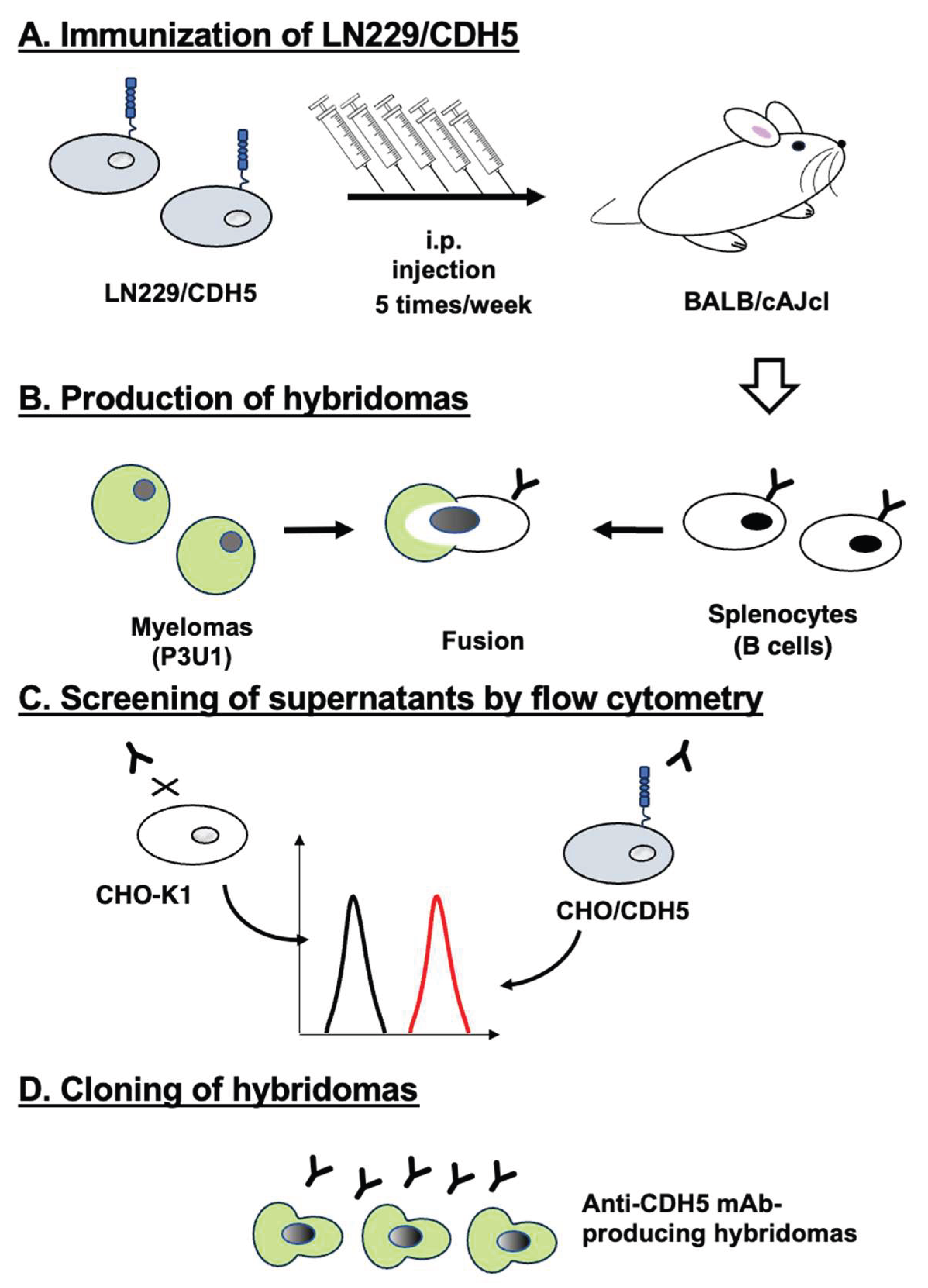
2.3. Production of Hybridomas

Female BALB/cAJcl mice (CLEA Japan, Tokyo, Japan) were intraperitoneally immunized with LN229/CDH5 cells (1 × 10⁸ cells/injection), and hybridomas were generated as previously described [26]. Supernatants positive for CHO/CDH5 and negative for CHO-K1 were screened using an SA3800 Cell Analyzer (Sony Corporation, Tokyo, Japan).

2.4. Flow Cytometry

Cells harvested with 1 mM ethylenediaminetetraacetic acid were incubated with mAbs for 30 min at 4°C. Then, the cells were treated with Alexa Fluor 488-conjugated anti-mouse IgG (2,000-fold dilution; Cell Signaling Technology, Danvers, MA, USA) for 30 min at 4°C. Flow cytometry were performed as described previously [25].

2.5. Determination of Dissociation Constant Values Using Flow Cytometry

CHO/CDH5 were treated with serially diluted Ca5Mab-8 and BV9. Subsequently, the cells were treated with anti-mouse IgG conjugated with Alexa Fluor 488 (200-fold dilution) for 30 minutes at 4°C. The dissociation constant (KD) values were determined as described previously [25].

2.6. Western Blotting

Western blotting was performed using 1 μg/mL of Ca5Mab-8, 1 μg/mL of BV9, or 1 μg/mL of an anti-isocitrate dehydrogenase 1 (IDH1) mAb (clone RcMab-1, rat IgG2a) as described previously [26].

2.7. IHC Using Cell Blocks

The formalin-fixed paraffin-embedded (FFPE) cell sections were prepared and stained with 0.5 μg/mL of Ca5Mab-8 or 0.5 μg/mL of BV9 using BenchMark ULTRA PLUS with the ultraView Universal DAB Detection Kit (Roche Diagnostics, Indianapolis, IN, USA).

2.8. IHC Using a Tissue Array

The FFPE of liver tumor with a liver tissue array including pathology grade, TNM and clinical stage (T032d) was purchased from TissueArray.Com LLC (Derwood, MD, USA). The sections were stained with 2 μg/mL of Ca5Mab-8 or 2 μg/mL of isotype control mouse IgG2a mAb (MpMab-23, http://www.med-tohoku-antibody.com/topics/001_paper_antibody_PDIS.htm) using BenchMark ULTRA PLUS with the ultraView Universal DAB Detection Kit.

3. Results

3.1. Development of anti-CDH5 mAbs

An antigen LN229/CDH5 was established as described in materials and methods. LN229/CDH5 (1 × 10⁸ cells/mouse) was immunized for five times in two BALB/cAJcl mice (Fig. 1A). Then, hybridomas were generated by fusing the splenocyte and P3U1 (Fig. 1B). The hybridoma supernatants were screened to identify supernatants that were positive for CHO/CDH5 and negative for CHO-K1 (Fig. 1C). As a result, 243 positive wells out of 958 wells (25.4%) were obtained. Subsequently, limiting dilution was performed and anti-CDH5 mAb-producing hybridomas were cloned (Fig. 1D). Finally, 14 clones were finally established (http://www.med-tohoku-antibody.com/topics/001_paper_antibody_PDIS.htm#CDH5+).
Figure 1. Schematic representation of anti-CDH5 mAbs production. (A) LN229/CDH5 was injected into BALB/cAJcl mice intraperitoneally. (B) After five immunizations, spleen cells were fused with P3U1. (C) The supernatants from hybridomas were screened by flow cytometry using CHO/CDH5 and CHO-K1 cells. (D) Anti-CDH5 mAb-producing hybridoma clones (Ca5Mabs) were established through limiting dilution.
Preprints 191487 g001

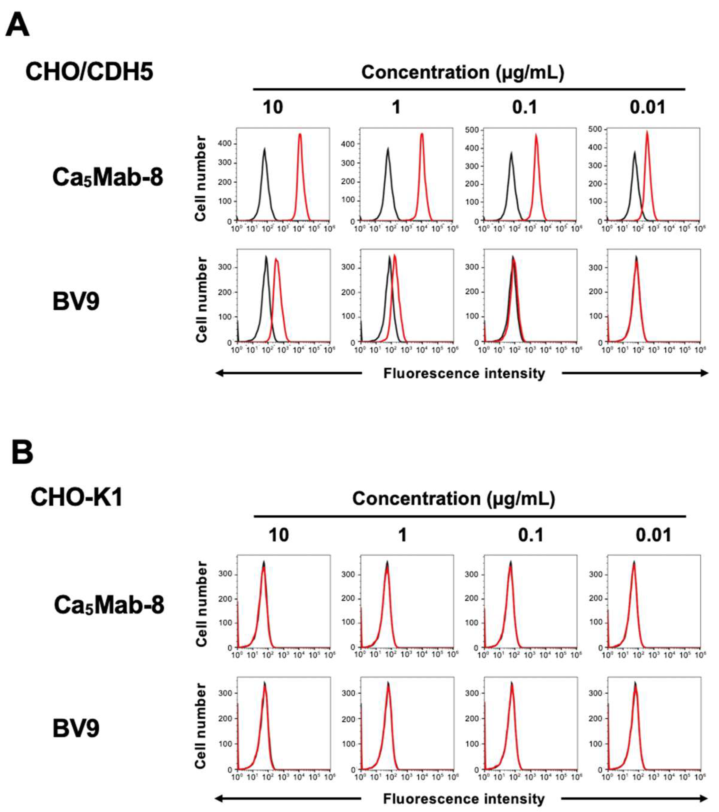
3.2. Flow Cytometry Using anti-CDH5 mAb Against CDH5-Overexpressed CHO-K1

We next conducted the screening of applications including flow cytometry, western blotting, and IHC using the supernatants of clones. As a result, Ca5Mab-8 (IgG2a, κ) can be applied to the three applications. We next prepared the purified mAbs from the supernatants and investigated the properties of these mAbs. Fig. 2 showed flow cytometric analysis using these mAbs and a commercially available anti-CDH5 mAb (clone BV9) against CHO/CDH5 and CHO-K1 cells. Ca5Mab-8 and BV9 reacted with CHO/CDH5 in a dose-dependent manner from 10 to 0.01 μg/mL (Fig. 2A). Compared to BV9, Ca5Mab-8 clearly showed a higher reactivity to CHO/CDH5. In contrast, Ca5Mab-8 and BV9 did not recognize CHO-K1 even at 10 μg/mL (Fig. 2B). The binding affinity of Ca5Mab-8 and BV9 was measured using flow cytometry. Next, the fitting binding isotherms of Ca5Mab-8 and BV9 to CHO/CDH5 were shown in Supplementary Fig. 1. The KD values of Ca5Mab-8 for CHO/CDH5 was 6.1 × 10⁻9 M. Although the KD values of BV9 for CHO/CDH5 was provisionally determined as 9.9 × 10⁻8 M, the fitting binding isotherms did not reach plateau. These results showed that Ca5Mab-8 possesses superior binding affinity to CHO/CDH5 compared to BV9.
Figure 2. Flow cytometric analysis of Ca5Mab-8 and BA9 against CHO/CDH5 and CHO-K1. CHO/CDH5 (A) and CHO-K1 (B) were treated with Ca5Mab-8 and BA9 at the indicated concentrations (red) or with blocking buffer (black, negative control). The mAbs-treated cells were incubated with Alexa Fluor 488-conjugated anti-mouse IgG.
Preprints 191487 g002

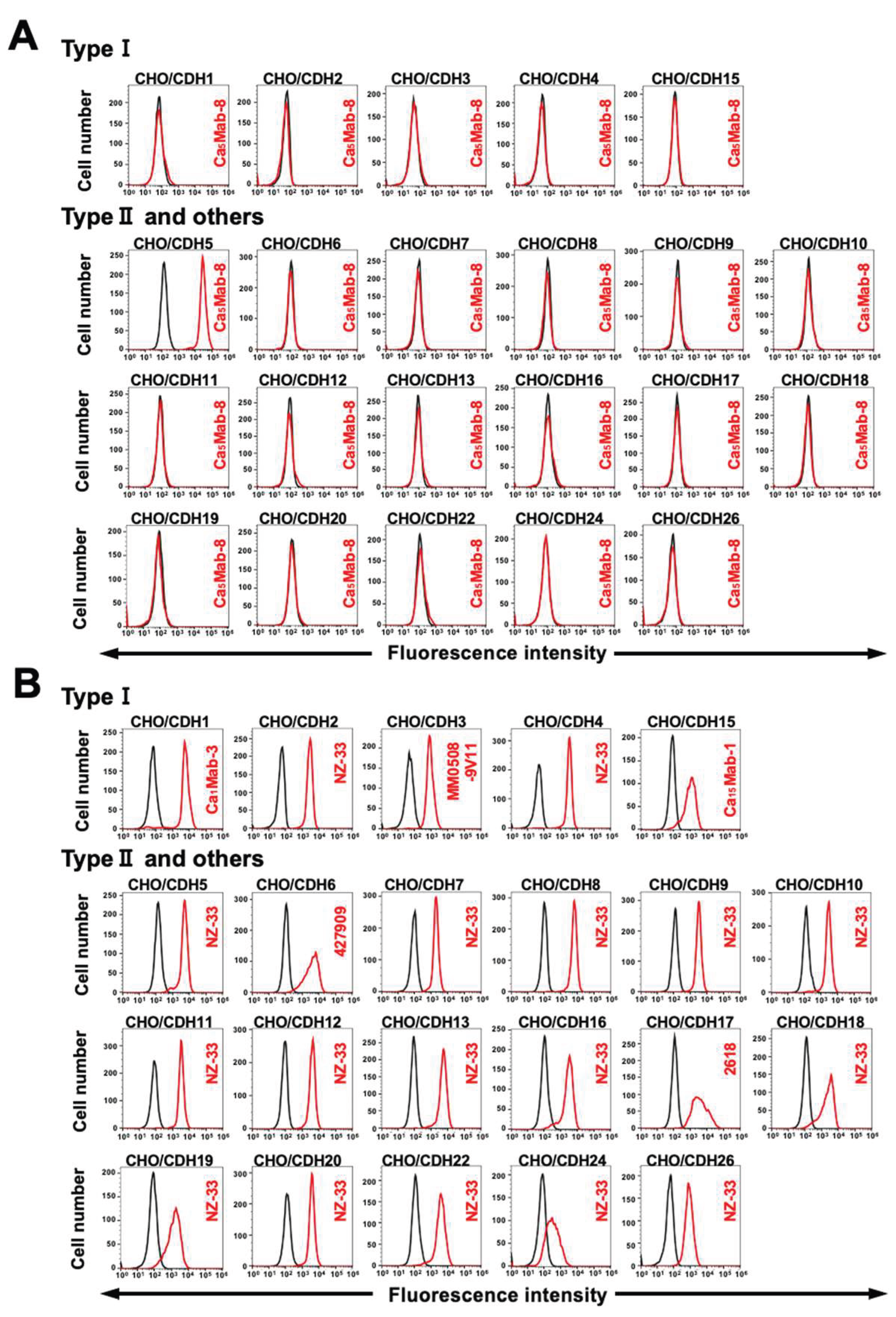
3.3. Specificity of Ca5Mab-8 to CDHs-Overexpressed CHO-K1

We previously established CHO-K1 overexpressed type I CDHs (CDH1, CDH2, CDH3, CDH4, and CDH15) [25,26], type II CDHs (CDH5, CDH6, CDH7, CDH8, CDH9, CDH10, CDH11, CDH12, CDH18, CDH19, CDH18, CDH20, and CDH22), 7D CDHs (CDH16 and CDH17), a truncated CDH (CDH13), and an atypical CDH (CDH26) [29]. Therefore, the specificity of Ca5Mabs to those CDHs was determined. As shown in Fig. 3A, Ca5Mab-8 reacted with CHO/CDH5 but did not react with other CDHs-overexpressed in CHO-K1. The cell surface expression of CDHs were confirmed in Fig. 3B. These results indicate that Ca5Mab-8 is a specific mAb to CDH5 among those CDHs.
Figure 3. Flow cytometry analysis of Ca5Mab-8 in CDHs-overexpressed CHO-K1. (A) The type I CDHs (CDH1, CDH2, CDH3, CDH4, and CDH15) type II CDHs (CDH5, CDH6, CDH7, CDH8, CDH9, CDH10, CDH11, CDH12, CDH18, CDH19, CDH18, CDH20, and CDH22), 7D CDHs (CDH16 and CDH17), a truncated CDH (CDH13), and an atypical CDH (CDH26)- overexpressed CHO-K1 were treated with 10 µg/mL of Ca5Mab-8 (red) or with control blocking buffer (black, negative control), followed by treatment with anti-mouse IgG conjugated with Alexa Fluor 488. (B) Each CDH expression was confirmed by 1 µg/mL of an anti-CDH1 mAb (clone Ca1Mab-3), 1 µg/mL of an anti-CDH3 mAb (clone MM0508-9V11), 1 µg/mL of an anti-CDH15 mAb (clone Ca15Mab-1), and 1 µg/mL of an anti-PA16-tag mAb (clone NZ-33) to detect other CDHs, followed by the treatment with Alexa Fluor 488-conjugated secondary mAbs.
Preprints 191487 g003

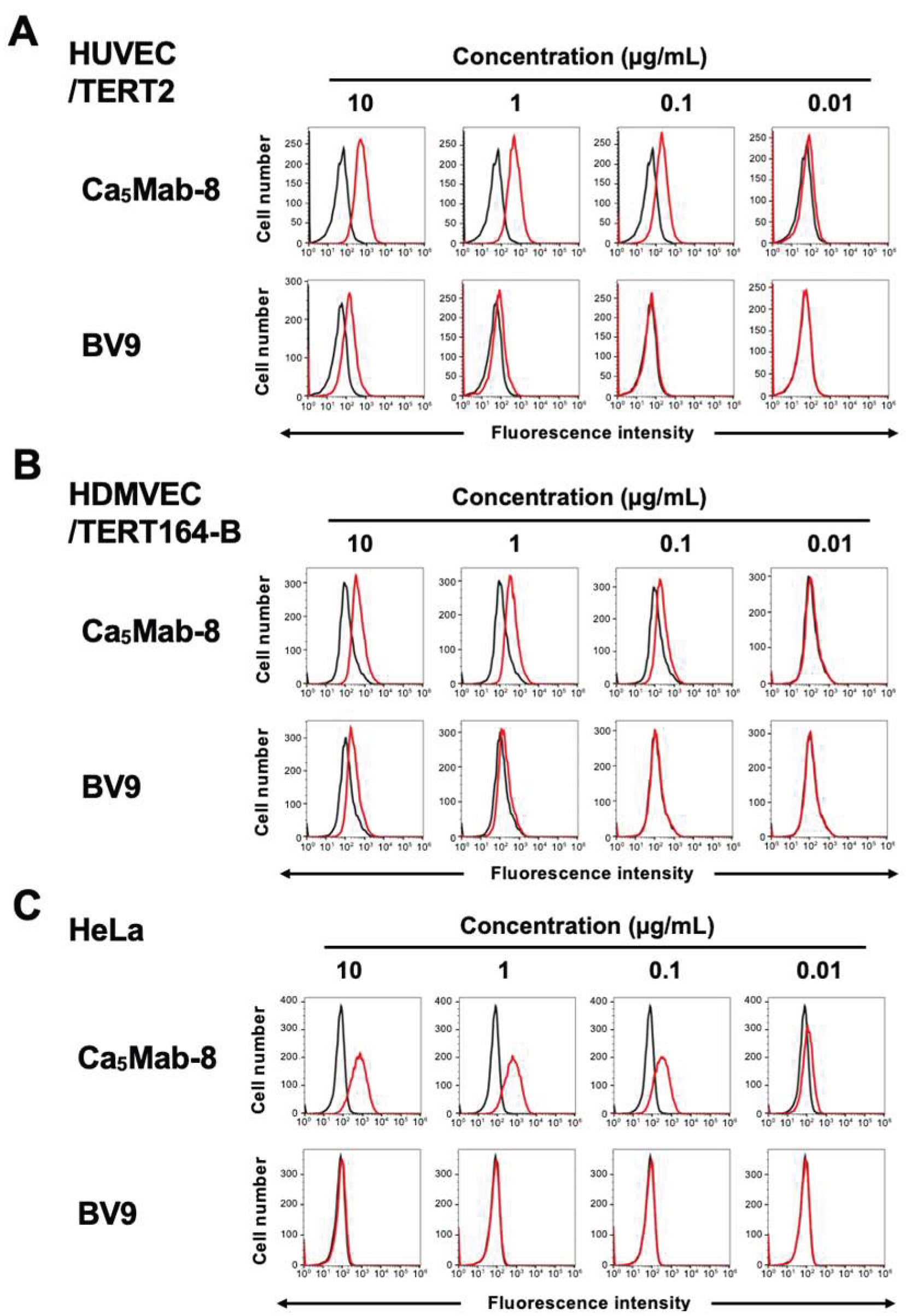
3.4. Flow Cytometry Using anti-CDH5 mAb Against Endogenous CDH5-Expressing Cells

CDH5 expression is known to be detected in endothelial cells. Ca5Mab-8 and BV9 recognized vascular endothelial HUVEC/TERT2 and lymphatic endothelial HDMVEC/TERT164-B cell lines in clow cytometry (Fig. 4A and B). Ca5Mab-8 also showed a higher reactivity compared to BV9. Since CDH5 was detected in several cancer cell lines [31,32], we screened the reactivity of Ca5Mab-8 to our cancer cell line panel. As a result, Ca5Mab-8 dose-dependently reacted with cervical cancer Hela cell line but BV9 did not (Fig. 4C). These results indicate that Ca5Mab-8 can be applied to flow cytometry to detect endogenous CDH5.
Figure 4. Flow cytometry analysis of Ca5Mab-8 and BA9 against HUVEC/TERT2, HDMVEC/TERT164-B, and Hela. HUVEC/TERT2 (A), HDMVEC/TERT164-B (B), and Hela (C) were treated with Ca5Mab-8 and BA9 at the indicated concentrations (red) or with blocking buffer (black, negative control). The mAbs-treated cells were incubated with Alexa Fluor 488-conjugated anti-mouse IgG.
Preprints 191487 g004

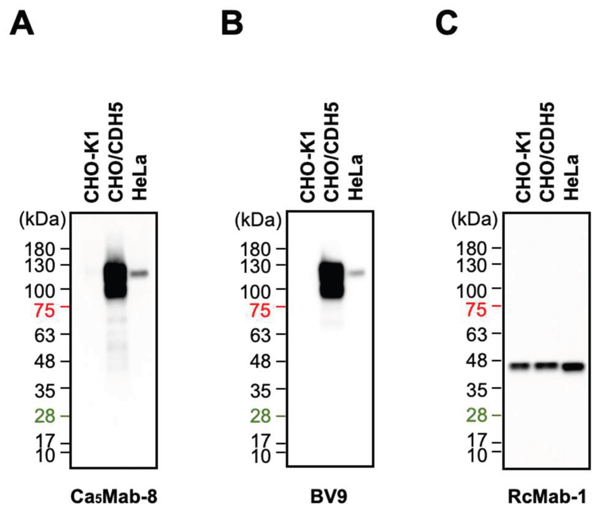
3.5. Western Blotting Using Ca5Mab-8 and BV9

We next examined whether Ca5Mab-8 is suitable for Western blotting. Whole-cell lysates of CHO-K1, CHO/CDH5, and Hela were analyzed. Ca5Mab-8 and BV9 detected clear bands from 100 to 130 kDa in CHO/CDH5 and ~120 kDa band in Hela, but not in CHO-K1 (Fig. 5A and B). An anti-IDH1 mAb (RcMab-1) served as an internal control (Fig. 5C). These results indicate that Ca5Mab-8 and BV9 can detect exogenous and endogenous CDH5 in Western blotting.
Figure 5. Western blotting using Ca5Mab-8 and BA9. The cell lysate (10 μg/lane) of CHO-K1, CHO/CDH5, and Hela were electrophoresed and transferred onto polyvinylidene difluoride membranes. The membranes were incubated with 1 μg/mL of Ca5Mab-8 (A), 1 μg/mL of BA9 (B), and 1 μg/mL of RcMab-1 (an anti-IDH1 mAb) (C), followed by the treatment with anti-mouse (Ca5Mab-8 and BA9) or anti-rat IgG (RcMab-1)-conjugated with horseradish peroxidase.
Preprints 191487 g005

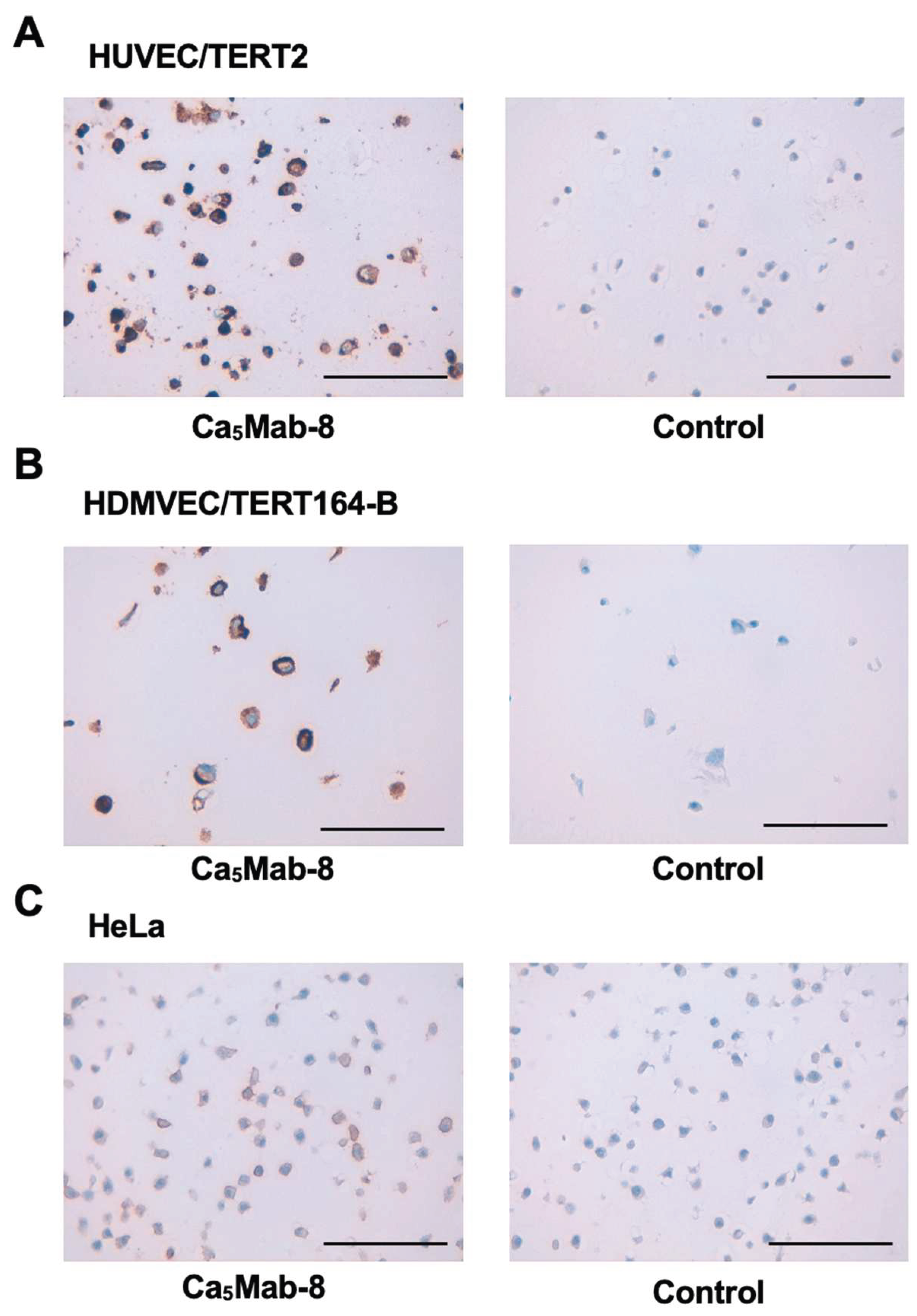
3.6. IHC Using Ca5Mab-8 in Formalin-Fixed Paraffin-Embedded Cell Blocks

We examined whether Ca5Mab-8 is suitable for the IHC of FFPE sections of CHO-K1, CHO/CDH5, HUVEC/TERT2, and HDMVEC/TERT164-B. Both intense cytoplasmic and membranous staining by Ca5Mab-8 were detected in CHO/CDH5 but not in CHO-K1 (supplementary Fig. 2). Furthermore, cytoplasmic and membranous staining by Ca5Mab-8 were also observed in HUVEC/TERT2, HDMVEC/TERT164-B, and Hela (Fig. 6). In contrast, BV9 cannot detect endogenous CDH5 in HUVEC/TERT2 and Hela in same experimental setting (supplementary Fig. 3). These results indicate that Ca5Mab-8 can detect exogenous and endogenous CDH5 in IHC of FFPE sections of cultured cells.
Figure 6. Immunohistochemistry using Ca5Mab-8 in formalin-fixed paraffin-embedded cell blocks. HUVEC/TERT2 (A), HDMVEC/TERT164-B (B), and Hela (C) sections were treated with 0.5 μg/mL of Ca5Mab-8 or control (without primary Ab). The staining was performed using BenchMark ULTRA PLUS with the ultraView Universal DAB Detection Kit, Scale bar = 100 μm.
Preprints 191487 g006

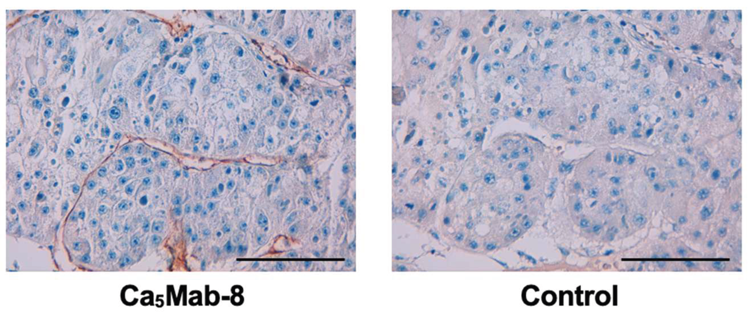
3.7. IHC Using Ca5Mab-8 in Formalin-Fixed Paraffin-Embedded Tissues

The FFPE liver cancer tissue array was stained with Ca5Mab-8. Ca5Mab-8 stained the vascular structure in tumor stroma but an isotype control IgG2a mAb did not (Fig. 7). In the tissue array, Ca5Mab-8 did not detect CDH5 in tumors. These results indicated that Ca5Mab-8 is suitable for detecting CDH5 in FFPE sections.
Figure 7. Immunohistochemistry using Ca5Mab-8 in formalin-fixed paraffin-embedded tissues. The sequential sections of a FFPE liver cancer tissue array were treated with 2 μg/mL of Ca5Mab-8 (A) and isotype control IgG2a (B). The staining was performed using BenchMark ULTRA PLUS with the ultraView Universal DAB Detection Kit, Scale bar = 100 μm.
Preprints 191487 g007

4. Discussion

This study demonstrated novel anti-CDH5 mAbs using the CBIS method (Fig. 1). Among them, an anti-CDH5 mAb, Ca5Mab-8 recognized both exogenous and endogenous CDH5 in flow cytometry with high reactivity compared to commercially available anti-CDH5 mAbs (BV9) (Fig. 2 and Fig. 4) and a superior affinity (supplementary Fig. 1). Ca5Mab-8 showed the specificity among CDHs (Fig. 3). Furthermore, Ca5Mab-8 are suitable for Western blotting (Fig. 5), IHC using cell block (Fig. 6), and detection of blood vessels in FFPE tissues (Fig. 7). Since IHC were performed using an automated slide staining system, it is possible to standardize the staining conditions. Therefore, Ca5Mab-8 is highly versatile for basic research in vascular biology and diagnosis.
Tumor cells evolve within a complex tumor microenvironment composed of diverse stromal and immune cell populations [33]. Among these components, endothelial cells play a central role in tumor angiogenesis, a key process driving tumor growth, progression, and metastasis [34]. Endothelial cells also contribute to tumor progression and metastasis by undergoing reprogramming into mesenchymal-like cells termed endothelial-to-mesenchymal transition (EndoMT) [35]. This endothelial plasticity is regulated by a broad spectrum of cytokines such as transforming growth factor-β [35]. EndoMT participates in multiple stages of tumor progression, including pathological angiogenesis, cancer cell intravasation and extravasation, generation of cancer-associated fibroblasts, and resistance to therapies [35]. During EndoMT, endothelial cells lose their barrier functions through downregulation of endothelial markers including CDH5, Claudin-5, and Tie2 [36]. Since Ca5Mab-8 is highly versatile, it would contribute to the molecular analyses in cultured endothelial cells and pathological analyses using tissue sections.
Several mAbs such as Cad 5, BV6, and BV9 possess the biological functions to inhibit CDH5 reorganization and formation of adherens junctions [22]. The epitopes of Cad 5, BV6, and BV9 were identified within EC1, EC3, and EC3-EC4, respectively [22]. Peptide scanning and the competition analyses revealed that Cad 5 recognizes KVFRVDAETGDVFAI on EC1 and BV6 recognizes TIDLRY located EC3. In contrast, detailed epitope of BV9 was not determined [22]. Therefore, validation of the epitopes and biological functions of Ca5Mab-8 are essential to apply the therapeutic uses.
An anti-mouse CDH5 mAb (BV13) showed in vitro biological effects and in vivo antitumor efficacy in preclinical models [24]. However, BV13 inhibited not only vascular tube formation during tumor angiogenesis but also disrupted adherens junctions of normal vasculature with an increased vascular permeability, which lead to pulmonary edema and death [37]. Another anti-mouse CDH5 mAb (clone E4G10) was developed to inhibit CDH5 function during angiogenesis without disrupting adherens junctions on normal vasculature [38]. E4G10 exhibited the comparable antitumor effects to BV13 with lower side effects in lung [38]. However, the strategy has not been applied in clinic.
CDH5 has been reported to be expressed in non-endothelial cells such as tumor cells. As shown in Fig. 4C, Ca5Mab-8, but not BV-9, recognized Hela in flow cytometry. Glioblastoma (GBM) is a highly angiogenic and the most aggressive of human cancers [39]. GBMs exhibit extensive network of abnormal vasculature [40]. A subset of the CD133⁺/CDH5⁺ stem-like progenitor from GBM was reported to have the capacity to differentiate into both tumor and tumor endothelium [41]. This result indicates that GBM-derived endothelial progenitors contribute the tumor angiogenesis [42]. In mouse models with human GBM stem cell-derived tumors, 70% of the CD31⁺ cells in the tumor core were GBM-derived, while almost all CD31⁺ cells in the periphery were host-derived [43]. During the initial phase of endothelial commitment, GBM stem cells upregulate CDH5, which is subsequently downregulated in a maturation phase [42]. Therefore, anti-CDH5 mAbs including Ca5Mabs have potential for the development of antitumor agent against GBM. Ca5Mab-8 recognized both tumor and endothelial CDH5 (Fig. 4), suggesting that Ca5Mab-8 can target tumor-derived and preexisting endothelium. Further investigations are essential to select CDH5-positive GBM cells and appropriate in vivo tumor models. Additionally, we have developed cancer-specific mAbs (CasMabs) against HER2 [44,45], one of which has been evaluated in a phase I clinical trial (NCT06241456) [46]. The anti-HER2 CasMabs recognized HER2-positive breast cancer cells, but not normal epithelial cells. We will generate more Ca5Mab clones from resting positive-wells and investigate the reactivity to tumor cells and normal endothelial cells.
We previously cloned the cDNA of mAbs and produced recombinant mouse IgG2a mAbs to confer antibody-dependent cellular cytotoxicity (ADCC). These mouse IgG2a mAbs have been evaluated the antitumor efficacies in human tumor xenograft models [47,48]. Since we have cloned the cDNA of Ca5Mab-8, recombinant Ca5Mab-8 will be produced and evaluated in vitro ADCC activity and antitumor efficacy in mouse GBM xenograft models. Additionally, we should investigate the antiangiogenic effect of Ca5Mab-8 in vitro and in vivo.

Author Contributions

Haruto Yamamoto: Investigation. Mika K. Kaneko: Conceptualization. Hiroyuki Suzuki: Writing – review and editing. Yukinari Kato: Conceptualization, Funding acquisition, Project administration, Writing – review and editing. All authors have read and agreed to the published version of the manuscript.

Funding

This research was supported in part by Japan Agency for Medical Research and Development (AMED) under Grant Numbers: JP25am0521010 (to Y.K.), JP25ama121008 (to Y.K.), JP25ama221153 (to Y.K.), JP25ama221339 (to Y.K.), and JP25bm1123027 (to Y.K.), and by the Japan Society for the Promotion of Science (JSPS) Grants-in-Aid for Scientific Research (KAKENHI) grant no. 25K10553 (to Y.K.).

Institutional Review Board Statement

The animal study protocol was approved by the Animal Care and Use Committee of Tohoku University (Permit number: 2022MdA-001) for studies involving animals.

Informed Consent Statement

Not applicable.

Data Availability Statement

All related data and methods are presented in this paper. Additional inquiries should be addressed to the corresponding authors.

Conflicts of Interest

The authors declare no conflict of interest involving this article.

References

  1. Nan, W.; He, Y.; Wang, S.; Zhang, Y. Molecular mechanism of VE-cadherin in regulating endothelial cell behaviour during angiogenesis. Front Physiol 2023, 14, 1234104. [CrossRef]
  2. van Roy, F. Beyond E-cadherin: roles of other cadherin superfamily members in cancer. Nat Rev Cancer 2014, 14, 121-134. [CrossRef]
  3. Lin, W.H.; Cooper, L.M.; Anastasiadis, P.Z. Cadherins and catenins in cancer: connecting cancer pathways and tumor microenvironment. Front Cell Dev Biol 2023, 11, 1137013. [CrossRef]
  4. Ratheesh, A.; Yap, A.S. A bigger picture: classical cadherins and the dynamic actin cytoskeleton. Nat Rev Mol Cell Biol 2012, 13, 673-679. [CrossRef]
  5. Dejana, E.; Orsenigo, F. Endothelial adherens junctions at a glance. J Cell Sci 2013, 126, 2545-2549. [CrossRef]
  6. Bazzoni, G.; Dejana, E. Endothelial cell-to-cell junctions: molecular organization and role in vascular homeostasis. Physiol Rev 2004, 84, 869-901. [CrossRef]
  7. Dejana, E.; Giampietro, C. Vascular endothelial-cadherin and vascular stability. Curr Opin Hematol 2012, 19, 218-223. [CrossRef]
  8. Rahimi, N. Defenders and Challengers of Endothelial Barrier Function. Front Immunol 2017, 8, 1847. [CrossRef]
  9. Marcos-Ramiro, B.; García-Weber, D.; Millán, J. TNF-induced endothelial barrier disruption: beyond actin and Rho. Thromb Haemost 2014, 112, 1088-1102. [CrossRef]
  10. Parnell, E.; Smith, B.O.; Palmer, T.M.; Terrin, A.; Zaccolo, M.; Yarwood, S.J. Regulation of the inflammatory response of vascular endothelial cells by EPAC1. Br J Pharmacol 2012, 166, 434-446. [CrossRef]
  11. Weis, S.; Cui, J.; Barnes, L.; Cheresh, D. Endothelial barrier disruption by VEGF-mediated Src activity potentiates tumor cell extravasation and metastasis. J Cell Biol 2004, 167, 223-229. [CrossRef]
  12. Delgado-Bellido, D.; Oliver, F.J.; Vargas Padilla, M.V.; Lobo-Selma, L.; Chacón-Barrado, A.; Díaz-Martin, J.; de Álava, E. VE-Cadherin in Cancer-Associated Angiogenesis: A Deceptive Strategy of Blood Vessel Formation. Int J Mol Sci 2023, 24. [CrossRef]
  13. Vittet, D.; Buchou, T.; Schweitzer, A.; Dejana, E.; Huber, P. Targeted null-mutation in the vascular endothelial-cadherin gene impairs the organization of vascular-like structures in embryoid bodies. Proc Natl Acad Sci U S A 1997, 94, 6273-6278. [CrossRef]
  14. Wen, L.; Yan, W.; Zhu, L.; Tang, C.; Wang, G. The role of blood flow in vessel remodeling and its regulatory mechanism during developmental angiogenesis. Cell Mol Life Sci 2023, 80, 162. [CrossRef]
  15. Wallez, Y.; Vilgrain, I.; Huber, P. Angiogenesis: the VE-cadherin switch. Trends Cardiovasc Med 2006, 16, 55-59. [CrossRef]
  16. Coon, B.G.; Baeyens, N.; Han, J.; Budatha, M.; Ross, T.D.; Fang, J.S.; Yun, S.; Thomas, J.L.; Schwartz, M.A. Intramembrane binding of VE-cadherin to VEGFR2 and VEGFR3 assembles the endothelial mechanosensory complex. J Cell Biol 2015, 208, 975-986. [CrossRef]
  17. Tzima, E.; Irani-Tehrani, M.; Kiosses, W.B.; Dejana, E.; Schultz, D.A.; Engelhardt, B.; Cao, G.; DeLisser, H.; Schwartz, M.A. A mechanosensory complex that mediates the endothelial cell response to fluid shear stress. Nature 2005, 437, 426-431. [CrossRef]
  18. Dabravolski, S.A.; Kashtalap, V.V.; Rozhkova, U.V.; Maksaeva, A.O.; Sukhorukov, V.N.; Orekhov, A.N. The role of cell junctions in atherosclerosis: implications for inflammation, endothelial dysfunction, and plaque stability. J Physiol Biochem 2025, 81, 573-587. [CrossRef]
  19. Brandon, K.D.; Frank, W.E.; Stroka, K.M. Junctions at the crossroads: the impact of mechanical cues on endothelial cell-cell junction conformations and vascular permeability. Am J Physiol Cell Physiol 2024, 327, C1073-c1086. [CrossRef]
  20. Berx, G.; van Roy, F. Involvement of members of the cadherin superfamily in cancer. Cold Spring Harb Perspect Biol 2009, 1, a003129. [CrossRef]
  21. Blaschuk, O.W.; Devemy, E. Cadherins as novel targets for anti-cancer therapy. Eur J Pharmacol 2009, 625, 195-198. [CrossRef]
  22. Corada, M.; Liao, F.; Lindgren, M.; Lampugnani, M.G.; Breviario, F.; Frank, R.; Muller, W.A.; Hicklin, D.J.; Bohlen, P.; Dejana, E. Monoclonal antibodies directed to different regions of vascular endothelial cadherin extracellular domain affect adhesion and clustering of the protein and modulate endothelial permeability. Blood 2001, 97, 1679-1684. [CrossRef]
  23. Corada, M.; Zanetta, L.; Orsenigo, F.; Breviario, F.; Lampugnani, M.G.; Bernasconi, S.; Liao, F.; Hicklin, D.J.; Bohlen, P.; Dejana, E. A monoclonal antibody to vascular endothelial-cadherin inhibits tumor angiogenesis without side effects on endothelial permeability. Blood 2002, 100, 905-911. [CrossRef]
  24. Liao, F.; Li, Y.; O'Connor, W.; Zanetta, L.; Bassi, R.; Santiago, A.; Overholser, J.; Hooper, A.; Mignatti, P.; Dejana, E.; et al. Monoclonal antibody to vascular endothelial-cadherin is a potent inhibitor of angiogenesis, tumor growth, and metastasis. Cancer Res 2000, 60, 6805-6810.
  25. Ubukata, R.; Suzuki, H.; Kaneko, M.K.; Kato, Y. Development of novel anti-CDH1/E-cadherin monoclonal antibodies for versatile applications. Biochemistry and Biophysics Reports 2026, 45, 102401. [CrossRef]
  26. Ubukata, R.; Suzuki, H.; Tanaka, T.; Kaneko, M.K.; Kato, Y. Development of an anti-CDH15/M-cadherin monoclonal antibody Ca(15)Mab-1 for flow cytometry, immunoblotting, and immunohistochemistry. Biochem Biophys Rep 2025, 43, 102138. [CrossRef]
  27. Fujii, Y.; Kaneko, M.; Neyazaki, M.; Nogi, T.; Kato, Y.; Takagi, J. PA tag: a versatile protein tagging system using a super high affinity antibody against a dodecapeptide derived from human podoplanin. Protein Expr Purif 2014, 95, 240-247. [CrossRef]
  28. Fujii, Y.; Kaneko, M.K.; Kato, Y. MAP Tag: A Novel Tagging System for Protein Purification and Detection. Monoclon Antib Immunodiagn Immunother 2016, 35, 293-299. [CrossRef]
  29. Satofuka, H.; Suzuki, H.; Kaneko, M.K.; Kato, Y. Development of Anti-Human Cadherin-26 Monoclonal Antibody, Ca26Mab-6, for Flow Cytometry. Preprints 2025. [CrossRef]
  30. Fujisawa, S.; Yamamoto, H.; Tanaka, T.; Kaneko, M.K.; Suzuki, H.; Kato, Y. Development and characterization of Ea7Mab-10: A novel monoclonal antibody targeting ephrin type-A receptor 7. MI 2025. [CrossRef]
  31. Zhou, L.; Yang, Y.; Ye, Y.; Qiao, Q.; Mi, Y.; Liu, H.; Zheng, Y.; Wang, Y.; Liu, M.; Zhou, Y. Notch1 signaling pathway promotes growth and metastasis of gastric cancer via modulating CDH5. Aging (Albany NY) 2024, 16, 11893-11903. [CrossRef]
  32. Hung, M.S.; Chen, I.C.; Lung, J.H.; Lin, P.Y.; Li, Y.C.; Tsai, Y.H. Epidermal Growth Factor Receptor Mutation Enhances Expression of Cadherin-5 in Lung Cancer Cells. PLoS One 2016, 11, e0158395. [CrossRef]
  33. de Visser, K.E.; Joyce, J.A. The evolving tumor microenvironment: From cancer initiation to metastatic outgrowth. Cancer Cell 2023, 41, 374-403. [CrossRef]
  34. Leone, P.; Malerba, E.; Susca, N.; Favoino, E.; Perosa, F.; Brunori, G.; Prete, M.; Racanelli, V. Endothelial cells in tumor microenvironment: insights and perspectives. Front Immunol 2024, 15, 1367875. [CrossRef]
  35. Watabe, T.; Takahashi, K.; Pietras, K.; Yoshimatsu, Y. Roles of TGF-β signals in tumor microenvironment via regulation of the formation and plasticity of vascular system. Semin Cancer Biol 2023, 92, 130-138. [CrossRef]
  36. Huang, M.; Liu, T.; Ma, P.; Mitteer, R.A., Jr.; Zhang, Z.; Kim, H.J.; Yeo, E.; Zhang, D.; Cai, P.; Li, C.; et al. c-Met-mediated endothelial plasticity drives aberrant vascularization and chemoresistance in glioblastoma. J Clin Invest 2016, 126, 1801-1814. [CrossRef]
  37. Corada, M.; Mariotti, M.; Thurston, G.; Smith, K.; Kunkel, R.; Brockhaus, M.; Lampugnani, M.G.; Martin-Padura, I.; Stoppacciaro, A.; Ruco, L.; et al. Vascular endothelial-cadherin is an important determinant of microvascular integrity in vivo. Proc Natl Acad Sci U S A 1999, 96, 9815-9820. [CrossRef]
  38. Liao, F.; Doody, J.F.; Overholser, J.; Finnerty, B.; Bassi, R.; Wu, Y.; Dejana, E.; Kussie, P.; Bohlen, P.; Hicklin, D.J. Selective targeting of angiogenic tumor vasculature by vascular endothelial-cadherin antibody inhibits tumor growth without affecting vascular permeability. Cancer Res 2002, 62, 2567-2575.
  39. Singh, S.; Dey, D.; Barik, D.; Mohapatra, I.; Kim, S.; Sharma, M.; Prasad, S.; Wang, P.; Singh, A.; Singh, G. Glioblastoma at the crossroads: current understanding and future therapeutic horizons. Signal Transduct Target Ther 2025, 10, 213. [CrossRef]
  40. Nowacka, A.; Śniegocki, M.; Smuczyński, W.; Bożiłow, D.; Ziółkowska, E. Angiogenesis in Glioblastoma-Treatment Approaches. Cells 2025, 14. [CrossRef]
  41. Wang, R.; Chadalavada, K.; Wilshire, J.; Kowalik, U.; Hovinga, K.E.; Geber, A.; Fligelman, B.; Leversha, M.; Brennan, C.; Tabar, V. Glioblastoma stem-like cells give rise to tumour endothelium. Nature 2010, 468, 829-833. [CrossRef]
  42. Buruiana, A.; Florian, S.I.; Florian, A.I.; Soritau, O.; Susman, S. Endothelial transdifferentiation of glioma stem cells: a literature review. Acta Neuropathol Commun 2025, 13, 181. [CrossRef]
  43. Ricci-Vitiani, L.; Pallini, R.; Biffoni, M.; Todaro, M.; Invernici, G.; Cenci, T.; Maira, G.; Parati, E.A.; Stassi, G.; Larocca, L.M.; et al. Tumour vascularization via endothelial differentiation of glioblastoma stem-like cells. Nature 2010, 468, 824-828. [CrossRef]
  44. Kaneko, M.K.; Suzuki, H.; Kato, Y. Establishment of a Novel Cancer-Specific Anti-HER2 Monoclonal Antibody H(2)Mab-250/H(2)CasMab-2 for Breast Cancers. Monoclon Antib Immunodiagn Immunother 2024, 43, 35-43. [CrossRef]
  45. Arimori, T.; Mihara, E.; Suzuki, H.; Ohishi, T.; Tanaka, T.; Kaneko, M.K.; Takagi, J.; Kato, Y. Locally misfolded HER2 expressed on cancer cells is a promising target for development of cancer-specific antibodies. Structure 2024, 32, 536-549.e535. [CrossRef]
  46. Hosking, M.P.; Shirinbak, S.; Omilusik, K.; Chandra, S.; Kaneko, M.K.; Gentile, A.; Yamamoto, S.; Shrestha, B.; Grant, J.; Boyett, M.; et al. Preferential tumor targeting of HER2 by iPSC-derived CAR T cells engineered to overcome multiple barriers to solid tumor efficacy. Cell Stem Cell 2025, 32, 1087-1101.e1084. [CrossRef]
  47. Ubukata, R.; Ohishi, T.; Kaneko, M.K.; Suzuki, H.; Kato, Y. EphB2-Targeting Monoclonal Antibodies Exerted Antitumor Activities in Triple-Negative Breast Cancer and Lung Mesothelioma Xenograft Models. Int J Mol Sci 2025, 26. [CrossRef]
  48. Kaneko, M.K.; Suzuki, H.; Ohishi, T.; Nakamura, T.; Tanaka, T.; Kato, Y. A Cancer-Specific Monoclonal Antibody against HER2 Exerts Antitumor Activities in Human Breast Cancer Xenograft Models. Int J Mol Sci 2024, 25. [CrossRef]
Disclaimer/Publisher’s Note: The statements, opinions and data contained in all publications are solely those of the individual author(s) and contributor(s) and not of MDPI and/or the editor(s). MDPI and/or the editor(s) disclaim responsibility for any injury to people or property resulting from any ideas, methods, instructions or products referred to in the content.
Copyright: This open access article is published under a Creative Commons CC BY 4.0 license, which permit the free download, distribution, and reuse, provided that the author and preprint are cited in any reuse.
Prerpints.org logo

Preprints.org is a free preprint server supported by MDPI in Basel, Switzerland.

Subscribe

Disclaimer

Terms of Use

Privacy Policy

Privacy Settings

© 2026 MDPI (Basel, Switzerland) unless otherwise stated