
Submitted:
16 December 2025
Posted:
26 December 2025
You are already at the latest version
Abstract
Background/Objectives: Photophobia is one of the most prevalent migraine symptoms, both during and outside of attacks, but its pathogenesis is unknown. The posterior thalamic nuclei may directly affect ambient light discomfort. This study examined the link between photophobia and the structure and morphometry of the thalamus and its subregions, including the lateral geniculate nuclei and pulvinar subnuclei. Methods: Twenty patients with episodic migraine without aura (MO) and 20 healthy controls (HCs) underwent high-resolution T1-weighted magnetic resonance imaging and comprehensive ophthalmological assessment. Patients were scanned interictally and none of them were under preventive therapy. Volumetric segmentation encompassed the whole thalamus and the lateral geniculate nuclei and pulvinar subregions. Interictal photophobia was evaluated using a visual analogue scale, ranging from 0 to 10. All thalamic and subregion volumes were used as independent variables and photophobia levels as dependent variables in general linear models. The model considered gender as a factor and total intracranial volume as a covariate. Results: No statistically significant differences were observed in the overall thalamic volume or in any of its subregions between MO patients and healthy controls (punc > 0.05). No relationships emerged between thalamic volumes and interictal subjective photophobia levels (p > 0.05). Conclusions: Our results suggest that photophobia is not linked to thalamic macroscopic volumes alterations during the interictal phase of MO patients. Further research is needed to determine whether these results could be extended to patients with migraine with aura or during other phases of the migraine cycle.
Keywords:
photophobia
; freesurfer
; thalamus
; segmentation
; migraine
; MRI
1. Introduction
Despite significant progress in migraine research, the anatomical and functional mechanisms behind canonical migraine-related symptoms, such as photophobia, remain largely unexplored [1]. Photophobia is typically characterised as an excessive sensitivity to ambient light stimuli [2] and constitutes one of the principal criteria for migraine diagnosis [3]. Although photophobia is more common during the migraine pain, patients with migraine may experience it also during the pain-free interval [4].
Evidence suggests a correlation between hyperexcitability of the occipital cortex and photophobia [5,6,7], alongside a CGRP-dependent mechanism of light sensitisation [8]. For some researchers, photophobia is facilitated by a subset of intrinsically photosensitive retinal ganglion cells that link the retina to the spinal trigeminal nucleus, as well as the lateral geniculate and pulvinar thalamic nuclei [9,10,11,12]. The triangulation of thalamic, brainstem, and retinal systems could enable the perception of the light signal as painful [13].
In this study, we aimed to examine the relationship between the volumes of thalamic nuclei implicated in visual processing, such as the lateral geniculate nucleus and the pulvinar, and subjective photophobia levels in patients with migraine without aura during the interictal phase.
The Freesurfer’s automated segmentation software – a probabilistic atlas based on histological examination of the thalamus that integrates magnetic resonance imaging and histological data through a 3D reconstruction informed by blockface images and augmented by in vivo magnetic resonance (MR) segmentations – was used to study thalamic nuclei and subnuclei volumes [14]. Additionally, to rule out a possible influence of alterations of the anterior segment of the eye on photophobia, a comprehensive series of ophthalmological test were conducted in all participants [2].
2. Materials and Methods
We performed a secondary analysis of the data collected from our previous study, using only data from patients who also were asked for the subjective interictal photophobia level on the day of the examination [15]. Twenty patients diagnosed with episodic migraine without aura (MO) in accordance with the International Classification of Headache Disorders (ICHD III) [3] were recruited from two Headache Centers (Rome and Latina) (Table 1).
All patients maintained a headache diary provided to them upon reserving a consultation appointment for at least one month prior to their initial visit. During the first consultation, we collected detailed information on the patient’s clinical characteristics, including the years from disease onset, the frequency and duration of migraine episodes each month, the intensity of headaches (assessed using a VAS scale from 0 to 10), and the days elapsed since the most recent migraine attack (Table 1). Additionally, all patients were requested to respond to the following question: In the absence of migraine pain, what is the average intensity of discomfort caused by exposure to bright light? (on a scale from 0 to 10, with 0 indicating no discomfort and 10 representing the highest level of discomfort). Exclusion criteria were the use of preventive therapy in the preceding three months, side-locked head pain, concomitant diagnosis of other primary or secondary headache types, and history of other relevant concomitant diseases, including neurological and neuro-ophthalmological diseases.
Twenty healthy controls (HCs) of similar age and gender were recruited among medical students and healthcare practitioners for comparative investigation. Exclusion criteria for HCs were medical or pertinent ophthalmological disorders, personal or familial history of primary headaches or epilepsy and did not engage in frequent use of drugs. Further exclusion criteria for both HCs and MOs were the existence of structural brain abnormalities on magnetic resonance imaging (MRI).
All subjects received a complete ophthalmological assessment, which included best-corrected visual acuity measurements (Snellen Equivalent), slit-lamp biomicroscopy for anterior segment evaluation, indirect ophthalmoscopy following pupil dilation (Tropicamide 1% drops) for posterior segment examination, and Goldmann applanation tonometry for intraocular pressure (IOP) measurement. All participants recruited were right-handed.
All participants were provided with a comprehensive overview of the study and granted written informed consent. The Ethical Committee of the Faculty of Medicine, Sapienza University of Rome, authorized the study protocol (Protocol No. 0295/2023).
MRI Protocol
Scanning sessions were scheduled in the afternoon (4:00–7:00 p.m.). Female participants were uniformly scanned throughout the mid-cycle phase. Participants were directed to refrain from sleep deprivation and alcohol consumption the day before the scanning procedure. Caffeinated beverages were banned on the day of the scan. All patients with MO were scanned during the interictal phase, i.e., minimum of three consecutive days before or after a migraine attack as verified through follow-up telephone interviews. Additionally, symptomatic medication was prohibited on the day of the scan.
All participants underwent MRI on a 3 Tesla Siemens Magnetom Vida scanner, using a 32-channel head coil. High-resolution T1-weighted sagittal magnetization-prepared rapid gradient echo (MPRAGE) sequences were acquired (TR: 2300 ms, TE: 2.25 ms, 208 sagittal slices, voxel dimensions 0.8 x 0.8 x 0.8 mm3, base resolution 320, field of view 256 mm, slice thickness 0.8 mm).
Imaging Data Processing: Thalamus Subunits Analysis
Thalamic segmentation was performed utilising a feature in Freesurfer 7.4.1 (https://surfer.nmr.mgh.harvard.edu/fswiki/SubregionSegmentation), a Python application that delineates the thalamus into 25 nuclei, employing a probabilistic atlas derived from histology data [14].
Two experienced investigators (F.C. and I.G.) examined each participant’s and thalamic segmentations, ultimately excluding those with artefacts.
We collected the volumes of the following thalamic nuclei: lateral geniculate, pulvinar anterior, pulvinar medial, pulvinar lateral, pulvinar inferior; their volumes were calculated and recorded for each participant in the group (Figure 1).
Statistical Analysis
Descriptive and inferential statistics were used, concentrating on volumetric differences between groups and the correlation between volumes and clinical factors in the MO group. SPSS for Windows (v.21 IBM Corp.) were used for the analysis.
The total intracranial volume (TIV) of each participant was computed using Freesurfer (version 7.4.1) [16,17,18,19,20,21,22,23,24].
To evaluate differences in the subnuclei volumes between groups (dependent variables), we applied a general linear model (GLM) that used age and TIV as covariates (independent variables) and gender and group as factors. Additionally, we applied a new GLM to assess the relationship between the volume of the MO group in each thalamic subunit (dependent variables) and values of photophobia (covariates of interest), controlling for age and TIV (covariates of no interest) and incorporating gender as a factor.
The Anderson-Darling and/or Kolmogorov-Smirnov tests were performed for each model’s covariates and responses to examine normality distributions. P-values were established at 0.025 (Bonferroni correction) for volume discrepancies between groups of the entire thalamus (left and right) and its subunits at p < 0.05, adjusted for multiple comparisons using the Benjamini-Hochberg method to address for the number of regions of interest.
3. Results
Twenty patients with MO (15 females, mean age 29.00 ± 5.06) and 20 HCs (15 females, mean age 34.35 ± 11.96) were recorded. Participants with MO had a mean of 6.3 ± 3.3 headache days per month. Mean interictal photophobia was 3.0 ± 1.8. Complete demographic and clinical characteristics of the participants are reported in Table 1.
None of the patients showed any relevant ophthalmological condition and all of them had IOP <20 mmHg and BCVA of 20/20 Snellen in both eyes.
Table 2 reports the volumes of the entire thalamus and its subregions in both hemispheres. No statistically significant differences were found between MO patients and HCs in either total thalamic volume or its subregions (puncorrected > 0.05, Table 2).

Table 2.
Mean volumes (mm3) ± standard deviations and interquartile ranges (25th–75th percentile) of thalamic subunits and total right and left thalamus in patients with episodic migraine without aura (MO) and healthy controls (HCs). Results from general linear models (GLMs) assessing between-group differences are reported.
| Left | Right | |||||
| HCs | MO | Inferential statistics | HCs | MO | Inferential statistics | |
| Lateral geniculate nucleus (LGN) | 304.81 ± 52.77 270.19 – 342.93 |
264.4 ± 44.9 229.8 – 310.1 |
F = 0.76; p = 0.39 |
304.77 ± 45.60 268.98 – 334.25 |
275.0 ± 50.6 244.3 – 310.4 |
F = 0.06; p = 0.81 |
| Pulvinar inferior (PuI) | 258.15 ± 50.21 213.93 – 295.78 |
236.45 ± 42.51 201.37 – 266.20 |
F = 2.96; p = 0.09 |
287.71 ± 56.17 241.56 – 328.45 |
259.43 ± 41.13 231.49 – 287.72 |
F = 0.57; p = 0.46 |
| Pulvinar medial (PuM) | 1181.50 ± 185.27 1016.70 – 1284.45 |
1105.4 ± 150.4 971.9 – 1235.7 |
F = 2.16; p = 0.15 |
1300.82 ± 201.51 1119.74 – 1461.13 |
1199.3 ± 147.8 1073.5 – 1316.5 |
F = 0.83; p = 0.37 |
| Pulvinar anterior (PuA) | 246.60 ± 40.66 223.51 – 271.38 |
229.24 ± 31.42 203.7 – 256.4 |
F = 1.85; p = 0.18 |
260.87 ± 38.70 226.02 – 287.91 |
244.28 ± 32.02 219.14 – 270.55 |
F = 1.37; p = 0.25 |
| Pulvinar lateral (PuL) | 205.99 ± 48.33 161.67 – 248.88 |
189.81 ± 38.30 153.21 – 223.64 |
F = 1.80; p = 1.88 |
217.01 ± 52.24 177.49 – 250.18 |
199.05 ± 34.69 172.83 – 209.91 |
F = 0.98; p = 0.33 |
| Whole thalamus | 7527 ± 1286 6527 – 8285 |
6821 ± 830 6327 - 7608 |
F = 0.21; p = 0.65 |
7775 ± 1184 6717 – 8508 |
7153 ± 903 6438 – 7763 |
F = 0.46; p = 0.50 |
Additionally, no significant relationships emerged between photophobia values and total volume of the entire thalamus or of its subregions (puncorrected > 0.05, Table 3). Multiple comparison corrections were not performed because all p-values were greater than 0.05.
Table 3.
General linear model (F; p) values between photophobia scale and left and right thalamic subregion volumes.
Table 3.
General linear model (F; p) values between photophobia scale and left and right thalamic subregion volumes.
| Left | Right | |
| Lateral geniculate nucleus (LGN) | 0.01; 0.914 | 0.00; 0.976 |
| Pulvinar inferior (PuI) | 0.44; 0.517 | 0.05; 0.831 |
| Pulvinar medial (PuM) | 0.82; 0.380 | 0.14; 0.717 |
| Pulvinar anterior (PuA) | 1.33; 0.267 | 1.60; 0.225 |
| Pulvinar lateral (PuL) | 0.66; 0.429 | 0.01; 0.936 |
| Whole thalamus | 3.55; 0.079 | 0.60; 0.451 |
4. Discussion
The most notable finding of this study is the absence of any relationship between the perceived level of photophobia during the interictal phase and thalamic volumes, both total and its subregions (lateral geniculate nuclei and pulvinar subnuclei). These results suggest that interictal light sensitivity is not associated with macrostructural volumetric abnormalities in the thalamus and in the lateral geniculate nuclei and pulvinar subnuclei.
Although various conditions may be associated with photophobia, the cerebral processes underlying its generation remain unidentified.
Several evidence support a link between the trigeminovascular system and the visual system in the development of photophobia [2]. Anatomically, second-order neurons from the spinal trigeminal nucleus project to the posterior thalamus, encompassing the pulvinar [9,11,25]. Indeed, outside migraine, photophobia is also a common symptom in both meningitis and subarachnoid haemorrhage [26], both conditions with involvement of the meninges. Additionally, it has to be noted that the integrity of the visual system is not strictly essential for the perception of light discomfort, as blind individuals can still report this sensation [26].
In line with this, in previous study, some authors found no differences in the retinal cone visual pathway (both M-L cones and S-cone systems) between healthy controls and subjects with migraine with and without aura recorded during the interictal phase. Additionally, no correlation emerged with subjective discomfort to environmental light exposure [27], suggesting that the retinal cone visual pathway does not play a direct role in interictal photophobia.
Some authors have hypothesized a direct link between the retina and the trigeminal system, as well as an indirect pathway via the dilatation of the choroidal arteries [2]. These alternative pathways include the non-image producing pathway, which involves the activation of retinal ganglion cells [28], whose axons transmit signals to the visual cortices through the optic nerve, the lateral geniculate nucleus, and the pulvinar in the posterior thalamus [29]. They proposed that the aggravation of migraine headache by light is mediated by photic signals relayed from the retina through the optic nerve to central neurones that process nociceptive signals from the meninges and to hypothalamic nuclei, including the suprachiasmatic nucleus, which play a role in regulating autonomic functions and emotions [30]. Case findings indicate that the pulvinar exhibited markedly enhanced bilateral activation during photophobia [10,11], and photophobia has been seen in individuals with demyelinating lesions affecting the posterior thalamus [31]. However, hypermetabolism of the posterior thalamus has been documented in individuals with essential blepharospasm, another condition associated with photophobia [32]. Notwithstanding this evidence, our study’s results indicate that the degree of photophobia experienced during the pain-free interval is unrelated to these macrostructural metrics of the visual thalamus.
Nonetheless, our results do not rule out the involvement of the thalamic nuclei in migraine photophobia from a metabolic and/or functional point of view. Suzuki et al. [33] found that the bilateral thalamus had hypermetabolism in patients with migraine who experience interictal photophobia compared to patients who do not. However, it is important to note that the authors did not grade the level of photophobia. Another study using functional MRI in response to visual stimuli found no significant relationship between the blood oxygen level dependent (BOLD) response of the lateral geniculate nuclei and the level of visual discomfort in patients with both migraine with and without aura [34]. During photophobia, a distinct functional activation pattern was observed in the ventroposteromedial thalamus alongside the trigeminal system [25].
In addition to the thalamus, substantial evidence indicates functional cortical involvement, encompassing both visual and sensorimotor aspects, in the pathophysiology of photophobia during the pericritical phase of migraine. Electrophysiological evidence indicates a negative correlation between subjective photophobia levels and the amplitude of the electroencephalographic response to intermittent photic stimulation [35], as well as with post-movement beta event-related synchronization, which reflects post-activation excitability in the sensorimotor cortices [36]. Consequently, we cannot rule out that other areas of the brain besides the direct visual pathway may play a role in the pathophysiology of photophobia.
Our study has some limitations. These include the small sample size and the use of a subjective photophobia rating scale, which may cause reporting biases. However, no objective and validated tests for photophobia exist to date. Additionally, the inclusion of patients without aura only during the interictal phase limits the generalizability of our results to other migraine phases, as well as to patients with migraine with aura.
5. Conclusions
Our study found no relationship between the subjective experience of photophobia and the neuronal density of the visual thalamic nuclei during the interictal phase of migraine, suggesting that interictal photophobia is not associated with macrostructural abnormalities in the thalamus and its subnuclei. These results support the idea that migraine is not characterized by macrostructural abnormalities, while metabolic and/or functional abnormalities cannot be ruled out based on our findings. Future research should investigate whether this phenomenon also occurs during the presence of aura and at other phases of the migraine cycle.
Author Contributions
Conceptualization, G.C. and F.C.; methodology, A.D.R.; software, A.D.R. and A.P.; formal analysis, A.D.R. and L.Z.; investigation, G.S., D.C., G.G., F.C., and C.A.; data curation, M.S.; writing—original draft preparation, G.C.; writing—review and editing, G.S., L.Z., and V.P.; supervision, M.A. All authors have read and agreed to the published version of the manuscript.
Funding
The authors did not receive funding for the design of the study and collection, analysis, and interpretation of data and in writing the manuscript.
Institutional Review Board Statement
All the participants provided written informed consent to participate in the study, which was approved by the ethical review board of the Faculty of Medicine at the University of Rome, Italy (N° 0295/2023).
Informed Consent Statement
Informed consent was obtained from all subjects involved in the study.
Data Availability Statement
The informed consent signed by all participants in this study did not include a provision stating that individual raw data can be made publicly accessible. Therefore, in agreement with the Italian data protection law, individual de-identified participant raw data cannot be shared publicly. Researchers meeting the criteria for access to confidential data may access the data upon request, involving the documentation of data access.
Acknowledgments
The contribution of the G.B. Bietti Foundation to this paper was supported by the Italian Ministry of Health and Fondazione Roma.
Conflicts of Interest
The authors declare no conflicts of interest.
Abbreviations
The following abbreviations are used in this manuscript:
| BOLD | blood oxygen level dependent |
| FC | functional connectivity |
| GLM | general linear model |
| MRI | Magnetic Resonance Imaging |
| HC | Healthy Control |
| MO | Migraine without aura |
| TIV | Total Intracranial Volume |
References
- Ashina, M. Migraine. New England Journal of Medicine 2020, 383, 1866–1876. [Google Scholar] [CrossRef] [PubMed]
- Digre, K.B.; Brennan, K.C. Shedding Light on Photophobia. Journal of neuro-ophthalmology: the official journal of the North American Neuro-Ophthalmology Society 2012, 32, 68–81. [Google Scholar] [CrossRef] [PubMed]
- Headache Classification Committee of the International Headache Society (IHS) The International Classification of Headache Disorders, 3rd Edition. Cephalalgia 2018, 38, 1–211. [CrossRef] [PubMed]
- Main, A.; Dowson, A.; Gross, M. Photophobia and Phonophobia in Migraineurs between Attacks. Headache 1997, 37, 492–495. [Google Scholar] [CrossRef]
- Cucchiara, B.; Datta, R.; Aguirre, G.K.; Idoko, K.E.; Detre, J. Measurement of Visual Sensitivity in Migraine: Validation of Two Scales and Correlation with Visual Cortex Activation. Cephalalgia: an international journal of headache 2015, 35, 585–592. [Google Scholar] [CrossRef]
- Boulloche, N.; Denuelle, M.; Payoux, P.; Fabre, N.; Trotter, Y.; Géraud, G. Photophobia in Migraine: An Interictal PET Study of Cortical Hyperexcitability and Its Modulation by Pain. Journal of neurology, neurosurgery, and psychiatry 2010, 81, 978–984. [Google Scholar] [CrossRef]
- Chong, C.D.; Starling, A.J.; Schwedt, T.J. Interictal Photosensitivity Associates with Altered Brain Structure in Patients with Episodic Migraine. Cephalalgia: an international journal of headache 2016, 36, 526–533. [Google Scholar] [CrossRef]
- Mason, B.N.; Kaiser, E.A.; Kuburas, A.; Loomis, M.-C.M.; Latham, J.A.; Garcia-Martinez, L.F.; Russo, A.F. Induction of Migraine-Like Photophobic Behavior in Mice by Both Peripheral and Central CGRP Mechanisms. The Journal of Neuroscience 2017, 37, 204–216. [Google Scholar] [CrossRef]
- Noseda, R.; Kainz, V.; Jakubowski, M.; Gooley, J.J.; Saper, C.B.; Digre, K.; Burstein, R. A Neural Mechanism for Exacerbation of Headache by Light. Nature neuroscience 2010, 13, 239–245. [Google Scholar] [CrossRef]
- Rosini, F.; Cerase, A.; Pretegiani, E.; Lucii, G.; Federighi, P.; Federico, A.; Rufa, A. Photophobia and Bilateral Pulvinar Involvement in Non-Alcoholic Wernicke’s Encephalopathy. Neurol Sci 2013, 34, 1867–1869. [Google Scholar] [CrossRef]
- Panorgias, A.; Lee, D.; Silva, K.E.; Borsook, D.; Moulton, E.A. Blue Light Activates Pulvinar Nuclei in Longstanding Idiopathic Photophobia: A Case Report. Neuroimage Clin 2019, 24, 102096. [Google Scholar] [CrossRef] [PubMed]
- Noseda, R.; Burstein, R. Advances in Understanding the Mechanisms of Migraine-Type Photophobia. Curr Opin Neurol 2011, 24, 197–202. [Google Scholar] [CrossRef] [PubMed]
- Okamoto, K.; Tashiro, A.; Chang, Z.; Bereiter, D.A. Bright Light Activates a Trigeminal Nociceptive Pathway. Pain 2010. [Google Scholar] [CrossRef] [PubMed]
- Iglesias, J.E.; Insausti, R.; Lerma-Usabiaga, G.; Bocchetta, M.; Van Leemput, K.; Greve, D.N.; van der Kouwe, A.; Fischl, B.; Caballero-Gaudes, C.; et al.; Alzheimer’s Disease Neuroimaging Initiative A Probabilistic Atlas of the Human Thalamic Nuclei Combining Ex Vivo MRI and Histology. Neuroimage 2018, 183, 314–326. [Google Scholar] [CrossRef]
- Giardina, I.; Di Renzo, A.; Chiffi, D.; Giuliani, G.; Sebastianelli, G.; Casillo, F.; Abagnale, C.; Ziccardi, L.; Pucci, A.; Parisi, V.; et al. 3T MRI Thalamic Segmentation Reveals No Macrostructural Changes in Interictal Episodic Migraine without Aura Compared to Healthy Controls. J Headache Pain 2025, 26, 243. [Google Scholar] [CrossRef]
- Chong, C.D.; Aguilar, M.; Schwedt, T.J. Altered Hypothalamic Region Covariance in Migraine and Cluster Headache: A Structural MRI Study. Headache 2020, 60, 553–563. [Google Scholar] [CrossRef]
- Reuter, M.; Rosas, H.D.; Fischl, B. Highly Accurate Inverse Consistent Registration: A Robust Approach. Neuroimage 2010, 53, 1181–1196. [Google Scholar] [CrossRef]
- Ségonne, F.; Dale, A.M.; Busa, E.; Glessner, M.; Salat, D.; Hahn, H.K.; Fischl, B. A Hybrid Approach to the Skull Stripping Problem in MRI. Neuroimage 2004, 22, 1060–1075. [Google Scholar] [CrossRef]
- Han, X.; Jovicich, J.; Salat, D.; van der Kouwe, A.; Quinn, B.; Czanner, S.; Busa, E.; Pacheco, J.; Albert, M.; Killiany, R.; et al. Reliability of MRI-Derived Measurements of Human Cerebral Cortical Thickness: The Effects of Field Strength, Scanner Upgrade and Manufacturer. NeuroImage 2006, 32, 180–194. [Google Scholar] [CrossRef]
- Fischl, B.; Salat, D.H.; van der Kouwe, A.J.W.; Makris, N.; Ségonne, F.; Quinn, B.T.; Dale, A.M. Sequence-Independent Segmentation of Magnetic Resonance Images. Neuroimage 2004, 23 Suppl 1, S69–84. [Google Scholar] [CrossRef]
- Fischl, B.; Sereno, M.I.; Dale, A.M. Cortical Surface-Based Analysis. II: Inflation, Flattening, and a Surface-Based Coordinate System. Neuroimage 1999, 9, 195–207. [Google Scholar] [CrossRef] [PubMed]
- Fischl, B.; Sereno, M.I.; Tootell, R.B.; Dale, A.M. High-Resolution Intersubject Averaging and a Coordinate System for the Cortical Surface. Hum Brain Mapp 1999, 8, 272–284. [Google Scholar] [CrossRef]
- Fischl, B.; Liu, A.; Dale, A.M. Automated Manifold Surgery: Constructing Geometrically Accurate and Topologically Correct Models of the Human Cerebral Cortex. IEEE Trans Med Imaging 2001, 20, 70–80. [Google Scholar] [CrossRef] [PubMed]
- Fischl, B.; Dale, A.M. Measuring the Thickness of the Human Cerebral Cortex from Magnetic Resonance Images. Proc Natl Acad Sci U S A 2000, 97, 11050–11055. [Google Scholar] [CrossRef]
- Moulton, E.; Becerra, L.; Borsook, D. An fMRI Case Report of Photophobia: Activation of the Trigeminal Nociceptive Pathway. Pain 2009, 145, 358–363. [Google Scholar] [CrossRef]
- Amini, A.; Digre, K.; Couldwell, W.T. Photophobia in a Blind Patient: An Alternate Visual Pathway. Case Report. J Neurosurg 2006, 105, 765–768. [Google Scholar] [CrossRef]
- Casillo, F.; Di Renzo, A.; Sebastianelli, G.; Abagnale, C.; Martelli, F.; Di Lorenzo, C.; Serrao, M.; Falsini, B.; Parisi, V.; Coppola, G. Lack of a Direct Link between Macular Cones Function and Photophobia in Interictal Migraine. Cephalalgia 2024, 44, 3331024241276501. [Google Scholar] [CrossRef]
- Matynia, A.; Parikh, S.; Chen, B.; Kim, P.; McNeill, D.S.; Nusinowitz, S.; Evans, C.; Gorin, M.B. Intrinsically Photosensitive Retinal Ganglion Cells Are the Primary but Not Exclusive Circuit for Light Aversion. Exp Eye Res 2012, 105, 60–69. [Google Scholar] [CrossRef]
- Maleki, N.; Becerra, L.; Upadhyay, J.; Burstein, R.; Borsook, D. Direct Optic Nerve Pulvinar Connections Defined by Diffusion MR Tractography in Humans: Implications for Photophobia. Human brain mapping 2012, 33, 75–88. [Google Scholar] [CrossRef]
- Noseda, R.; Copenhagen, D.; Burstein, R. Current Understanding of Photophobia, Visual Networks and Headaches. Cephalalgia 2019, 39, 1623–1634. [Google Scholar] [CrossRef]
- Kawasaki, A.; Borruat, F.-X. Photophobia Associated with a Demyelinating Lesion of the Retrochiasmal Visual Pathway. Am J Ophthalmol 2006, 142, 854–856. [Google Scholar] [CrossRef]
- Emoto, H.; Suzuki, Y.; Wakakura, M.; Horie, C.; Kiyosawa, M.; Mochizuki, M.; Kawasaki, K.; Oda, K.; Ishiwata, K.; Ishii, K. Photophobia in Essential Blepharospasm--a Positron Emission Tomographic Study. Mov Disord 2010, 25, 433–439. [Google Scholar] [CrossRef]
- Suzuki, Y.; Kiyosawa, M.; Wakakura, M.; Ishii, K. Hyperactivity of the Medial Thalamus in Patients with Photophobia-Associated Migraine. Headache 2024, 64, 1005–1014. [Google Scholar] [CrossRef]
- Datta, R.; Aguirre, G.K.; Hu, S.; Detre, G.A.; Cucchiara, B. Interictal Cortical Hyperresponsiveness in Migraine Is Directly Related to the Presence of Aura. Cephalalgia: an international journal of headache 2013, 33, 365–374. [Google Scholar] [CrossRef]
- Bjørk, M.; Hagen, K.; Stovner, L.; Sand, T. Photic EEG-Driving Responses Related to Ictal Phases and Trigger Sensitivity in Migraine: A Longitudinal, Controlled Study. Cephalalgia 2011, 31, 444–455. [Google Scholar] [CrossRef]
- Mykland, M.S.; Bjørk, M.H.; Stjern, M.; Sand, T. Alterations in Post-Movement Beta Event Related Synchronization throughout the Migraine Cycle: A Controlled, Longitudinal Study. Cephalalgia 2018, 38, 718–729. [Google Scholar] [CrossRef]
Figure 1.
Thalamic nuclei segmentation. FreeSurfer 3D rendering of thalamic nuclei in their lateral-medial (left) and caudal-rostral (right) aspects on T1-weighted multiecho MPRAGE data of a representative sample subject. Lateral geniculate nucleus (LGN), Pulvinar inferior (PuI), Pulvinar medial (PuM), Pulvinar anterior (PuA), Pulvinar lateral (PuL).
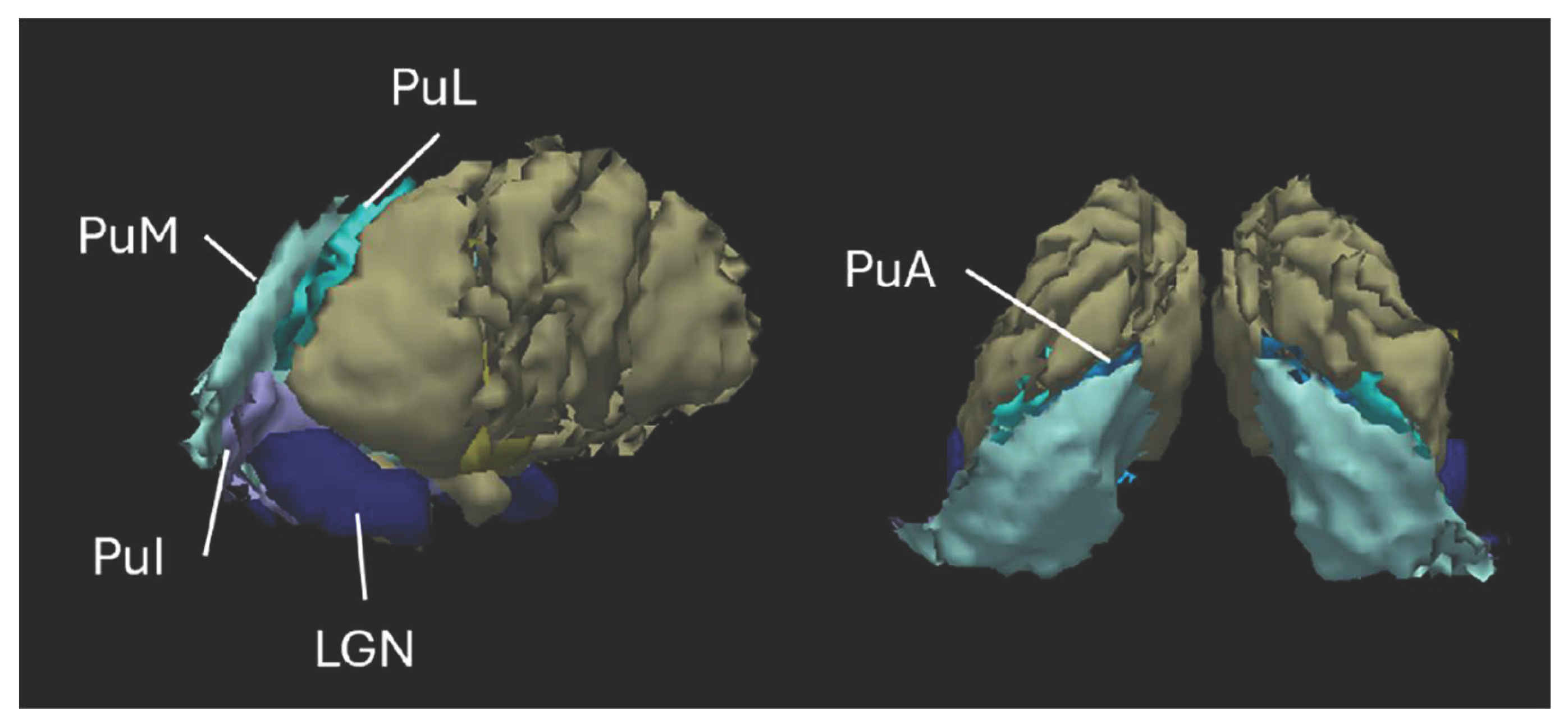
Table 1.
Demographic and clinical characteristics of healthy controls (HCs) and patients with migraine without aura scanned during migraine-free intervals (MO). Data are presented as mean ± standard deviation. Group differences were tested using Student’s t-test.
Table 1.
Demographic and clinical characteristics of healthy controls (HCs) and patients with migraine without aura scanned during migraine-free intervals (MO). Data are presented as mean ± standard deviation. Group differences were tested using Student’s t-test.
| HCs (n = 20) |
MO (n = 20) |
Statistics | |
| Female (n) | 15 | 15 | |
| Age (years) | 29.00 ± 5.06 | 34.35 ± 11.96 | t = -1.84, p = 0.08 |
| History of migraine (years) | 17.0 ± 12.9 | ||
| Attack frequency/month (n) | 6.3 ± 3.3 | ||
| Attack duration (hours) | 33.2 ± 19.9 | ||
| Severity of headache (0-10) | 8.3 ± 1.0 | ||
| Days from the last migraine attack (n) | 5.8 ± 3.9 | ||
| MIDAS | 21.0 ± 17.9 | ||
| HIT-6 | 61.9 ± 8.0 | ||
| ASC-12 | 4.1 ± 4.2 | ||
| Interictal photophobia (0-10) | 3.0 ± 1.8 | ||
Disclaimer/Publisher’s Note: The statements, opinions and data contained in all publications are solely those of the individual author(s) and contributor(s) and not of MDPI and/or the editor(s). MDPI and/or the editor(s) disclaim responsibility for any injury to people or property resulting from any ideas, methods, instructions or products referred to in the content. |
© 2025 by the authors. Licensee MDPI, Basel, Switzerland. This article is an open access article distributed under the terms and conditions of the Creative Commons Attribution (CC BY) license (http://creativecommons.org/licenses/by/4.0/).
Copyright: This open access article is published under a Creative Commons CC BY 4.0 license, which permit the free download, distribution, and reuse, provided that the author and preprint are cited in any reuse.


