Preprint
Review

This version is not peer-reviewed.

Hepatic Sinusoidal Obstruction Syndrome Induced by Pyrrolizidine Alkaloids from Gynura segetum: Mechanisms and Therapeutic Advances

A peer-reviewed article of this preprint also exists.

Submitted:

19 December 2025

Posted:

22 December 2025

You are already at the latest version

Abstract

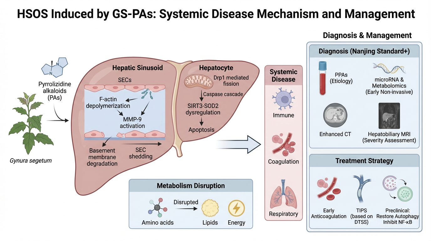
Gynura segetum, a traditional Chinese medicinal herb, has been increasingly recognized for its hepatotoxic potential due to its content of PAs. These compounds are among the most common causes of HILI in China and are strongly associated with HSOS. This review systematically explores the pathogenesis, diagnostic evolution, and therapeutic strategies of PAs-induced HSOS. We detail the molecular mechanisms underlying PA metabolism, including cytochrome P450-mediated bioactivation and the formation of pyrrole–protein adducts, which initiate sinusoidal endothelial cell injury and hepatocyte apoptosis. Advances in diagnostic criteria, such as the Nanjing Criteria and Drum Tower Severity Scoring System, are discussed alongside emerging biomarkers like circulating microRNAs and pyrrole–protein adducts. Imaging modalities, including contrast-enhanced CT and Gd-EOB-DTPA MRI, have transitioned from descriptive tools to quantitative and prognostic instruments. Therapeutic approaches have evolved from supportive care to precision interventions, including anticoagulation, TIPS, and autophagy-modulating agents. This review highlights the need for integrated diagnostic and therapeutic frameworks and calls for enhanced public awareness and regulatory oversight to mitigate PAs-related liver injury.

Keywords: 
;  ;  

1. Introduction

Gynura segetum (Lour.) Merr. (GS), belonging to the Asteraceae family, is a perennial herb widely known in China as “Tu-San-Qi” or “Ju-San-Qi”. In China, the medicinal use of GS can be traced back to the Ming Dynasty, with its therapeutic properties traditionally believed to include promoting blood circulation, exerting hemostatic effects, detoxifying, and alleviating edema [1,2]. However, hepatotoxicity associated with G. segetum has been repeatedly reported. The toxicity is primarily attributed to its content of pyrrolizidine alkaloids (PAs), which are among the most frequent causes of herb-induced liver injury(HILI) in China [3]. Even a single ingestion of as little as 10 grams of GS can lead to liver injury and rapidly progress to a severe hepatic vascular disorder known as hepatic sinusoidal obstruction syndrome (HSOS), also referred to as veno-occlusive disease (VOD) [4,5].
In Western countries, HSOS most commonly occurs as a complication of hematopoietic stem cell transplantation (HSCT), with an incidence of 5–20% and mortality exceeding 80% in severe cases. The principal pathogenic mechanism involves sinusoidal endothelial detachment and swelling induced by radiotherapy or chemotherapy, resulting in sinusoidal blockage [6]. In contrast, in China and several other Asian regions, ingestion of PAs-containing herbal remedies represents the predominant etiology. G. segetum and its closely related species Tu-San-Qi have been well-documented as major causative agents due to their high PAs content. The defining histopathological feature is selective injury to hepatic sinusoidal endothelial cells, leading to non-thrombotic occlusion of sinusoids and subsequent post-sinusoidal portal hypertension [4]. Clinically, the disease typically manifests as acute right upper quadrant pain, hepatomegaly, ascites, and rapid weight gain secondary to fluid retention. Currently, no specific antidote exists for PAs-induced HSOS. Once progression to hepatic failure occurs, the condition is associated with a markedly elevated mortality rate, highlighting the critical importance of early recognition, accurate diagnosis, and immediate cessation of toxin exposure [7].
Diagnosis of PAs-HSOS remains challenging. Many patients lack a definitive history of exposure to PAs-containing substances, and the clinical presentation of PAs-induced HSOS overlaps with Budd-Chiari syndrome and right heart failure, both conditions that manifest sinusoidal portal hypertension through distinct pathophysiological mechanisms. No effective pharmacological therapy has yet been established to reverse the hepatotoxicity of G. segetum or restore hepatic parenchymal integrity. The diagnosis of drug-induced liver injury (DILI) and herb-induced liver injury (HILI) continues to be hindered by the absence of reliable biomarkers for routine clinical application [8]. Alarmingly, recent reports include a case of a four-year-old child developing PAs-HSOS after accidental ingestion of GS, emphasizing the urgent need to raise public awareness of its potential toxicity [9].
In summary, GS represents a medicinal plant with a long therapeutic history but carries a significant risk of severe hepatotoxicity. HSOS induced by its PAs components constitutes a complex pathological process spanning botany, toxicology, and clinical medicine. This review aims to comprehensively summarize the molecular mechanisms underlying PAs-induced hepatotoxicity, current diagnostic strategies—including both conventional criteria and emerging biomarkers—and potential therapeutic interventions, thereby providing insights to improve understanding and management of this clinically challenging disease.

2. Food and Pharmaceutical Safety Recommendations Regarding PAs

The first reported case of PAs-induced HSOS dates back to 1920 and was linked to the consumption of PAs-contaminated wheat [10], Since then, PAs-related safety issues have been documented across numerous countries, including Afghanistan, the United Kingdom, China, Germany, Hong Kong, India, Jamaica, South Africa, Switzerland, and the United States, with approximately 8,160 poisoning cases recorded to date. This substantial public health burden has prompted many countries to establish regulations restricting or prohibiting the use of PAs-containing herbs [11,12]. The International Agency for Research on Cancer (IARC) classifies PAs as carcinogenic substances. To date, more than 660 PAs have been identified in over 6,000 plant species. These compounds are naturally occurring phytotoxins serving as chemical defenses against herbivores and are found in thousands of species from families such as Asteraceae, Boraginaceae, and Fabaceae. Many of these plants are used medicinally or appear as contaminants in honey, tea, and cereal products, posing a significant public health concern [13,14]. In nature, PAs coexist with their more water-soluble N-oxides (PAN-oxides), and the ratio between these forms fluctuates within a single plant species depending on environmental conditions and growth stage [15,16]. The European Food Safety Authority (EFSA) has implemented systematic monitoring and regulatory limits for PAs and PAN-oxides in contaminated foods to maintain minimal concentrations throughout the food chain. EFSA established a reference point of 237 µg/kg body weight per day to assess the carcinogenic risk associated with PAs exposure and concluded that PAs may pose health concerns for regular consumers of tea and herbal infusions, particularly among younger populations [17]. The UK Medicines and Healthcare Products Regulatory Agency (MHRA) proposed adding Senecio-containing products to the list of prohibited substances, making the sale, supply, or import of unlicensed oral preparations containing such plants illegal within the UK [18,19]. Nevertheless, not all PAs exhibit toxicity, as their harmful potential largely depends on specific structural features of the molecule. Therefore, a comprehensive international regulatory framework to prevent PAs exposure and its associated hepatotoxicity has yet to be established. The major regulations of each period and each country are presented in Table 1.

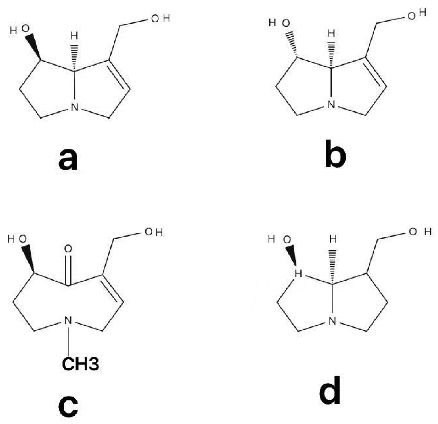
3. Metabolism of PAs

PAs are esters composed of a necine base—an amino alcohol containing two fused five-membered rings with a shared nitrogen atom—and one or more necic acid moieties. PAs are toxic to humans and other mammals. Based on the N-oxidation state and the degree of saturation of the pyrrolizidine ring, PAs generally exist in four structural forms: three tertiary amines (saturated, unsaturated, and otonecine types) and one N-oxide form. in Figure 1 [20]. The PAs molecule typically contains two fused five-membered rings sharing a nitrogen atom at the C-4 position. Most naturally occurring PAs are derivatives of 1-methylpyrrolizidine, with some existing as 1-hydroxymethyl-1,2-dehydropyrrolizidine esters that exhibit hepatotoxic properties. The core structure responsible for PAs-induced hepatotoxicity consists of an unsaturated necine base with a 1,2-double bond, one or two hydroxyl groups, and esterified side chains derived from branched necic acids. Only PAs containing the 1,2-unsaturated double bond display marked hepatotoxicity and genotoxicity, while their saturated analogues are largely non-toxic [21]. This distinction arises from differences in metabolic activation: 1,2-unsaturated PAs are bioactivated by hepatic cytochrome P450 enzymes-primarily CYP3A4 and CYP2B6 to generate highly reactive dehydropyrrolizidines (dehydro-PAs). These intermediates covalently bind to cellular DNA and proteins, leading to hepatocyte necrosis, apoptosis, and sinusoidal endothelial injury, culminating in HSOS [4].
Both the roots and aerial parts of GS contain hepatotoxic PAs in concentrations sufficient to cause liver injury, and therefore the entire plant should not be consumed as either medicine or food [22]. The major hepatotoxic PAs identified in G. segetum include senecionine, seneciphylline, and integerrimine [23]. Their strong binding affinity to hepatic tissue (39.22 ± 1.90 nmol/g liver in rats) provides direct evidence linking PAs-induced hepatotoxicity to the formation of pyrrole–protein adducts.
After intestinal absorption, PAs are transported to the liver, where they undergo metabolic activation by cytochrome P450 isoenzymes (mainly CYP2B and CYP3A) to form dehydropyrrolizidine alkaloids (DHPAs). DHPAs are further hydrolyzed to dehydroretronecine (DHR), both of which are highly reactive. These metabolites can conjugate with intracellular glutathione (GSH) to form GSH conjugates, thereby detoxifying reactive intermediates. However, when DHPAs and DHR form stable pyrrole-protein and pyrrole-DNA adducts, they trigger the key molecular event underlying PAs-induced hepatotoxicity. These adducts disrupt normal cellular function and initiate oxidative stress, mitochondrial injury, and apoptosis [13,24,25,26]. The rate of pyrrole–protein adduct formation serves as a crucial indicator of PA bioactivation. Structural diversity among PAs results in variable toxicity, with diester-type PAs (e.g., lasiocarpine) producing the greatest quantity of adducts and exhibiting the highest hepatotoxic potential [27,28,29]. PAs exposure induces widespread metabolic disturbances that exhibit time– and dose–dependent patterns, primarily affecting amino acid, lipid, and energy metabolism pathways. Amino acid dysregulation impairs hepatic nitrogen handling; lipid metabolic disruption leads to alterations in arachidonic and linoleic acid pathways and elevations in glycerol and triglycerides, contributing to lipid peroxidation and hepatic injury; while energy metabolism dysfunction, reflected by abnormal levels of citric acid and lactate, indicates mitochondrial impairment [30]. Liver sinusoidal endothelial cells (LSECs), which express high levels of CYP450 enzymes but possess limited detoxification capacity, are recognized as the primary targets of PAs toxicity. Their injury compromises the sinusoidal barrier and causes luminal obstruction, producing the characteristic pathology of HSOS [31,32,33]. Chen et al. demonstrated that following repeated administration of G. segetum extract, the clearance of PAs from liver tissue was markedly slower than from serum, with residues detectable up to eight weeks after the final dose. This persistence explains the cumulative toxicity of PAs and the continued progression of disease even after discontinuation, underscoring the importance of long-term clinical follow-up in affected patients [34].

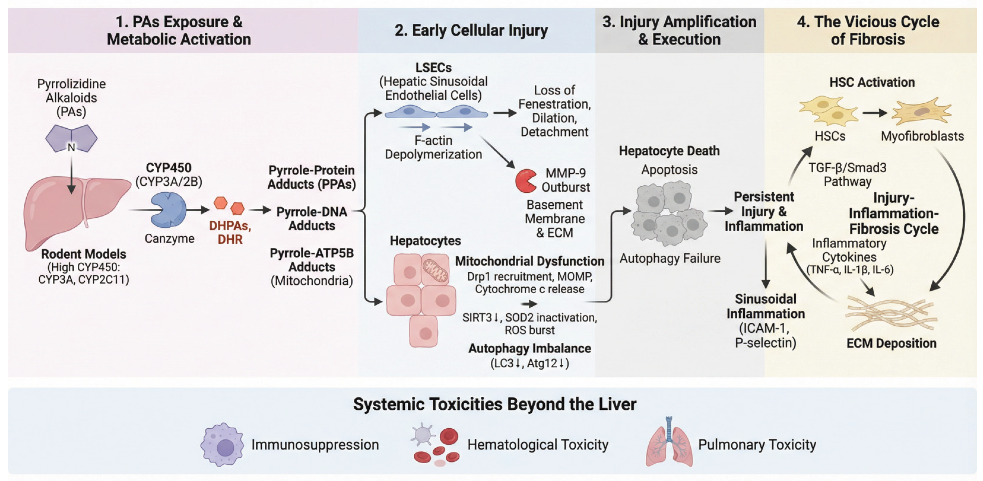
4. Pathogenesis: From Molecular Events to Pathological Outcomes

3.1. Establishment of Experimental Models

In vitro studies using hepatic S9 fractions have demonstrated that the biological half-life of PAs is shorter in rats than in humans. In human liver microsomes, PAs are primarily detoxified via UGT1A4-catalyzed N-glucuronidation reactions. By contrast, murine and rat microsomal systems lack this detoxification pathway. Consequently, CYP450-mediated bioactivation of PAs occurs at significantly higher rates in rodents compared to humans, amplifying their susceptibility to PAs-induced hepatotoxicity [35,36]. Rodent models of PAs-induced HSOS exhibit pathological features that closely resemble those observed in human HSOS. Owing to their higher metabolic activation rate and comparable histopathological manifestations, mice have become the preferred species for model establishment. Male rodents are particularly suited for PAs-induced HSOS models because sex hormones drive high hepatic expression of CYP3A and CYP2C11 enzymes. The synergistic activity of these enzymes markedly enhances the metabolic activation of PAs, enabling the generation of sufficient reactive metabolites even at relatively low exposure doses. This process reproducibly induces characteristic HSOS pathology. The establishment of this model provides a robust experimental platform for elucidating the molecular mechanisms underlying HSOS and for identifying early diagnostic biomarkers, such as circulating microRNAs (miRNAs) [37,38]. The pathogenesis of each system is illustrated in Figure 2.

3.2. Initiation of Toxic Metabolism and Core Molecular Events

The N-oxide metabolites of PAs can be enzymatically reduced back to their parent compounds in both the intestine and liver. Following intestinal absorption, PAs are transported to the liver, where they undergo metabolic activation catalyzed predominantly by cytochrome P450 enzymes (mainly CYP3A and CYP2B), generating reactive intermediates known as DHPAs. DHPAs are subsequently hydrolyzed to DHR [15]. These electrophilic metabolites form stablePPAs and pyrrole–DNA adducts with cellular macromolecules—an event recognized as the key molecular initiation step of PA-induced hepatotoxicity [3]. Lu et al. further demonstrated that PAs metabolites form pyrrole–ATP5B adducts with the β-subunit of mitochondrial ATP synthase, directly impairing ATP production and precipitating cellular energy failure [13].

3.3. Selective Injury to Hepatic Sinusoidal Endothelial Cells

LSECs represent the primary cellular targets of PA toxicity. PAs disrupt the cytoskeletal architecture of LSECs by inducing depolymerization of F-actin, resulting in dilation, fusion, and eventual loss of fenestrations. These structural alterations compromise sinusoidal hemodynamics and microcirculatory integrity [39]. The damage is highly cell-specific, attributable to the high expression of CYP450 enzymes and relatively weak detoxification capacity in LSECs [40].

3.4. Amplification and Execution of Injury: MMP-9 Outburst and Hepatocyte Death

Initial LSEC injury triggers excessive expression and activation of matrix metalloproteinase-9 (MMP-9), which degrades the endothelial basement membrane and extracellular matrix, thereby exacerbating endothelial detachment and structural disintegration of hepatic sinusoids. Administration of MMP inhibitors (MMPi) markedly attenuates sinusoidal damage and intrahepatic hemorrhage in rat HSOS models, confirming the pivotal role of MMP-9 in disease progression and highlighting its potential as a therapeutic target [40].

3.5. Hepatocyte Death: Mitochondrial Apoptosis and Autophagy Imbalance

Hepatocellular death under PAs exposure involves both mitochondrial apoptosis and disrupted autophagic regulation. Exposure to PAs promotes the recruitment of dynamin-related protein 1 (Drp1) to mitochondria, resulting in excessive mitochondrial fission and fragmentation. This process induces mitochondrial outer membrane permeabilization (MOMP), leading to cytochrome c release, caspase cascade activation, and execution of apoptosis [41,42]. Concurrently, PAs downregulate the deacetylase SIRT3, causing acetylation-dependent inactivation of the antioxidant enzyme SOD2. The ensuing burst of mitochondrial reactive oxygen species (ROS) further amplifies apoptotic signaling [43]. Autophagy, which normally exerts cytoprotective effects, is markedly suppressed under PAs exposure, as evidenced by reduced expression of its key markers LC3 and Atg12. Impaired autophagic flux aggravates mitochondrial dysfunction and accelerates apoptotic progression [44].

3.6. The Vicious Cycle of Fibrosis

Persistent hepatic injury initiates pathological repair responses characterized by activation of hepatic stellate cells (HSCs) and excessive deposition of extracellular matrix (ECM) components [45]. The canonical TGF-β/Smad3 signaling pathway is activated, driving transcription of profibrotic genes and acting synergistically with inflammatory cytokines such as TNF-α, IL-1β, and IL-6. Suppression of autophagy induced by PAs coincides with sinusoidal endothelial inflammation, marked by upregulated expression of ICAM-1 and P-selectin. Downregulation of the RNA-binding protein CPEB4 may represent a novel mechanistic link between PAs exposure and autophagy inhibition, reinforcing a self-perpetuating “injury-inflammation-fibrosis” cycle [32].

3.7. Systemic Toxicities Beyond the Liver in PAs-HSOS

3.7.1. Immunosuppression

GS exerts marked suppressive effects on both innate and adaptive immunity. It specifically inhibits macrophage phagocytosis, lymphocyte proliferation, cytokine release, and nitric oxide production—key components of immune defense. Such broad immunosuppressive activity may predispose affected individuals to heightened susceptibility to infections [46].

3.7.2. Hematological Toxicity

GS exposure leads to a generalized elevation in leukocytes, including neutrophils, lymphocytes, monocytes, and eosinophils, while markedly reducing platelet count and platelet hematocrit. Coagulation assays reveal prolonged prothrombin time (PT), activated partial thromboplastin time (APTT), and thrombin time (TT), alongside decreased fibrinogen (FIB) concentration and platelet aggregation rate. Concomitantly, serum levels of endothelin and nitric oxide are significantly elevated. Histological analysis of the spleen demonstrates architectural abnormalities, including reduction of splenic lobules and loss of germinal centers [47].

3.7.3. Pulmonary Toxicity

Beyond hepatic injury, PAs have also been implicated in pulmonary toxicity. Mechanistic studies indicate that although metabolic activation of PAs is minimal within lung tissue, reactive dehydro-PAs metabolites generated in the liver may translocate to the lungs, where they form pyrrole–protein adducts that mediate tissue injury. Experimental evidence shows that multiple PAs, including those present in GS, induce pulmonary lesions in rats at hepatotoxic doses, suggesting that lung injury may represent a shared toxicological feature of PAs [48].

4. Evolution of the Diagnostic Framework: From Clinical Criteria to Precision Biomarkers

4.1. Establishment and Optimization of Clinical Diagnostic Standards

The “Nanjing Criteria” proposed by Chinese experts, provide a pivotal foundation for diagnosing PAs-HSOS. These criteria emphasize four key elements: (1) a documented history of PA exposure; (2) characteristic clinical manifestations such as abdominal distension, right upper quadrant pain, hepatomegaly, and ascites; (3) abnormal liver function tests; and (4) typical radiologic findings. Compared with the revised Seattle and Baltimore criteria, the Nanjing criteria not only incorporate PA exposure history and radiological features but also eliminate requirements for weight gain and specific bilirubin thresholds, thereby enhancing alignment with the clinical manifestations of PAs-HSOS [49]. The Roussel Uclaf Causality Assessment Method (RUCAM) further offers a structured framework for evaluating the causal relationship in suspected cases of HILI [50]. A recent advance in diagnosis and treatment is the development of the Drum Tower Severity Score (DTSS). The DTSS system assigns points based on four parameters: aspartate aminotransferase, total bilirubin, fibrinogen, and portal vein peak velocity. Each parameter is scored according to its value, yielding a total score ranging from 4 to 16. Patients with mild severity (4–6 points) respond well to anticoagulation therapy, with a negative predictive value as high as 88%. They are recommended for outpatient anticoagulation and biweekly follow-up. Those with moderate severity (7–10 points) show no response to anticoagulation in approximately 45.6% of cases; hospitalization and close monitoring are advised, with TIPS intervention considered if ineffective. In severe cases (11–16 points), the probability of non-response to anticoagulation is 78.3%, and direct TIPS is recommended to prevent clinical deterioration. The DTSS system accurately predicts the non-response rate to the foundational regimen of supportive care plus anticoagulation. It enables early stratification of disease severity and provides an objective, quantitative tool for guiding clinical decisions—such as identifying high-risk patients who may require early escalation to TIPS—thereby facilitating more precise management of PAs-HSOS [51]. Other Diagnostic Criteria for HSOS is shown in Table 2.

4.2. Advances in Quantitative and Functional Imaging Diagnosis

Imaging plays an indispensable role in the diagnosis, severity assessment, and prognostic evaluation of PAs-HSOS. With the advancement of computed tomography (CT) and magnetic resonance imaging (MRI), particularly the integration of quantitative imaging analysis, diagnostic radiology has evolved from traditional morphological observation toward functional and precision-based quantification. This paradigm shift provides objective and reproducible diagnostic support that complements and extends beyond conventional clinical tools such as the RUCAM scale and the early “Nanjing Criteria”.
Contrast-enhanced CT has emerged as a pivotal modality for identifying and grading PAs-HSOS. A large-scale retrospective study involving 71 patients with PAs-HSOS and 222 controls systematically characterized its hallmark CT features: heterogeneous low attenuation of the liver parenchyma (100%), patchy enhancement (92.96%), and hepatic vein narrowing (87.32%) [64]. These objective imaging markers strengthen the imaging component of the Nanjing diagnostic framework, enhancing both consistency and clinical applicability. The study further demonstrated that contrast-enhanced CT outperforms the traditional Seattle criteria in overall diagnostic accuracy.
Quantitative CT analysis enables an objective assessment of disease burden and severity Wang et al. innovatively applied a threshold-based region-growing algorithm to segment PAs-HSOS lesions and calculate the lesion-to-liver volume ratio. This ratio showed a strong positive correlation with serum ALT, AST, total bilirubin levels, and clinical severity scores. As a quantitative imaging biomarker, it provides a reproducible and intuitive metric for evaluating disease burden, compensating for the limitations of RUCAM and the Nanjing Criteria in grading disease severity, and offering a more data-driven foundation for treatment planning and prognostic prediction [52].
Functional Role of MRI-Specific Contrast Agents in Diagnosis and Prognosis, MRI-specific hepatobiliary contrast agents provide unique functional insights that enhance both diagnostic precision and prognostic evaluation in PAs-HSOS. Guo et al. demonstrated the distinct value of gadolinium ethoxybenzyl diethylenetriamine pentaacetic acid (Gd-EOB-DTPA)-enhanced MRI in this context. All patients exhibited heterogeneous hypointensity of the hepatic parenchyma during the hepatobiliary phase (HBP), reflecting impaired hepatocellular function and offering functional imaging evidence for diagnosis. Notably, the severity of HBP hypointensity correlated positively with prothrombin time (PT) and the international normalized ratio (INR), and was identified as an independent predictor of mortality [53].
This finding represents a pivotal transition-positioning imaging not merely as a diagnostic modality, but as a quantitative and functional tool for prognostic prediction, extending beyond the capabilities of RUCAM and the exposure-based Nanjing Criteria.
Collectively, quantitative CT and functional MRI have ushered PAs-HSOS diagnosis into a new era of precision and quantification. Future investigations should focus on: integrating objective imaging parameters (e.g., lesion-to-liver volume ratio, HBP signal intensity) into diagnostic and grading systems to compensate for the non-quantitative limitations of RUCAM and existing standards, exploring the potential of radiomics to uncover latent pathological patterns and prognostic signatures; and developing multimodal diagnostic frameworks that combine imaging-based metrics with emerging biomarkers such as pyrrole-protein adducts (PPAs) and circulating microRNAs. Such integration may yield a more comprehensive and predictive diagnostic ecosystem for PA-HSOS.

4.3. Emerging Frontiers in Precision Biomarkers

4.3.1. Etiology-Specific Biomarker: PPAs

Lin et al. first detected PPAs in the serum of patients with PAs-HSOS [4]. Subsequent work by Ma et al. demonstrated a strong positive correlation between serum alanine aminotransferase (ALT) levels and hepatic PPAs content, establishing PPA as a shared pathogenic event and a specific biomarker of PAs-induced hepatotoxicity across different (PAs) structures [54]. Quantification of PPAs in serum using UPLC-MS has been recommended as the optimal diagnostic biomarker for PAs-HSOS [55]. The slow hepatic clearance and persistence of PPAs further explain the cumulative nature of its toxicity [34].

4.3.2. Early Diagnostic and Prognostic Biomarkers: microRNAs and Metabolomics

Recent studies identified significant upregulation of miR-148a-3p, miR-362-5p, and miR-194-5p in both PAs-HSOS patients and experimental rat models. Their expression levels correlated positively with the severity of liver injury, indicating strong potential as early, non-invasive diagnostic biomarkers [37]. Metabolomic profiling revealed that GS exposure induces broad disturbances in amino acid, lipid, and energy metabolism. Metabolites such as arginine, creatine, valine, and citric acid were highlighted as potential biomarker clusters [30,56]. Furthermore, metabolomics enables clear discrimination between non-toxic Panax notoginseng and hepatotoxic GS. UPLC-Q/TOF-MS analysis of urine and plasma samples showed distinct endogenous metabolite patterns: L-glutamic acid, L-methionine, cytosine, and L-tyrosine predominated in the Panax group, while phytosphingosine, creatine, and sphinganine were enriched in the Gynura group. In plasma, key discriminative metabolites included arachidonic acid, L-tyrosine, linoleic acid, α-linolenoyl ethanolamide, and lysophosphatidylcholine (15:0) for Panax, versus L-arginine, L-valine, arachidonic acid, and lysophosphatidylcholine (18:2 (9Z, 12Z)) for Gynura [30].

4.3.3. Systems Biology Perspective: The Gut-Liver Axis

A systems-level investigation revealed a critical link between GS-induced hepatotoxicity and gut microbiota dysbiosis. GS extracts markedly reduced the abundance of Lactobacillus species, known for their hepatoprotective roles. The perturbed gut microbial composition exhibited close metabolic interactions with host peripheral metabolites, particularly those involved in energy, lipid, and amino acid metabolism. These findings suggest that GS-induced hepatotoxicity represents a systemic pathological process mediated through the “gut microbiota-host metabolism axis” rather than a liver-confined toxic response [30].

5. Evolution of Therapeutic Strategies: From Supportive Care to Multimodal Precision Intervention

The Nanjing Clinical Guidelines emphasize that the cornerstone of PAs-HSOS management lies in early recognition, timely intervention, and stratified treatment. The primary principles are as follows: (1) Immediate discontinuation of any suspected causative agent to halt ongoing hepatotoxicity; (2) Comprehensive supportive therapy focusing on symptomatic relief and prevention of complications—stabilizing hepatic function, controlling ascites, improving coagulation, and preventing infection. For patients with rapidly progressive disease or poor response to standard management, the guidelines recommend targeted pharmacologic interventions (e.g., anticoagulants and hemodynamic modulators), and, when indicated, interventional or surgical approaches such as transjugular intrahepatic portosystemic shunt (TIPS) or liver transplantation [49].
This hierarchical and integrative therapeutic framework reflects a paradigm shift from empirical supportive care toward evidence-based, precision-oriented, multimodal intervention, marking a new stage in the systematic.

5.1. Management of PA-HSOS

In recent years, Basic Supportive Therapy and Targeted Pharmacological Interventions, as clinical experience with PA-HSOS has accumulated, research focus has progressively shifted from the establishment of diagnostic criteria toward the optimization of therapeutic strategies, refinement of prognostic assessment, and advancement in the management of special patient populations.
In the context of autophagy regulation, bicyclol exerts its hepatoprotective effects primarily through restoration of autophagic function. In PAs-exposed mice, pretreatment with bicyclol significantly reversed the decline in the LC3-II/LC3-I ratio and modulated the expression of the autophagy-suppressive protein Bcl-2, indicating alleviation of PA-induced autophagy inhibition. This protection is mechanism-specific, occurring mainly via the autophagic pathway rather than through apoptosis suppression. Moreover, bicyclol bidirectionally regulates cytochrome P450 isoenzymes, thereby mitigating the metabolic activation of PAs and indirectly protecting hepatocytes—including sinusoidal endothelial cells—from autophagic dysregulation [44].
Prednisone acts through multiple molecular targets to inhibit inflammation and fibrosis. It suppresses the activation of inflammatory mediators such as TNF-α and the transcription factor NF-κB p65, while simultaneously downregulating profibrotic factors including TGF-β1 and CTGF. Consequently, prednisone confers protection during both acute inflammatory and chronic fibrotic stages. As a pivotal transcriptional regulator of inflammation, cell adhesion, and survival, NF-κB plays a central role in sinusoidal endothelial injury and inflammatory amplification in PAs-HSOS [57].
Salvia miltiorrhiza (Danshen) markedly improves hepatic function and histopathological injury in GS–induced HSOS in a dose-dependent manner. Mechanistically, Danshen treatment significantly reduces hepatic protein levels of TNF-α, VCAM-1, and ICAM-1, and suppresses NF-κB p65 expression, indicating that its protective effects are mediated through inhibition of NF-κB signaling activation [58].
Ligustrazine exerts its therapeutic effects by suppressing the expression of early growth response factor-1 (Egr-1) and NF-κB p65, thereby downregulating their common downstream target, tissue factor (TF). This newly identified Egr-1/NF-κB/TF signaling axis suggests that ligustrazine alleviates the procoagulant state and microthrombus formation within hepatic sinusoids, offering a novel molecular target for the management of PAs-HSOS-related microcirculatory dysfunction [59]. The treatment guidelines in the Nanjing standard are shown in Table 3.

5.2. Precision Application of Interventional and Surgical Therapies and Advances in Prognostic Evaluation

5.2.1. Anticoagulation Therapy: From Exploratory Use to Established Role and Risk Management

Early management of PAs-HSOS primarily relied on symptomatic and supportive care; however, recent studies have clearly demonstrated the pivotal role of early anticoagulation therapy in promoting disease resolution. In a retrospective cohort of 49 patients, the remission rate was significantly higher in the anticoagulation group than in the standard care group (P=0.037) [57]. An independent study involving 75 patients validated this conclusion, demonstrating that the anticoagulation group exhibited a significantly higher cure rate (65.3%) compared to the non-anticoagulation group, thereby establishing anticoagulation as a first-line therapeutic strategy for this condition [58]. Recent advances emphasize the importance of precise patient selection and dynamic risk balancing between therapeutic benefit and hemorrhagic complications. Although anticoagulation significantly improves remission and cure rates, it is associated with a bleeding incidence of up to 12.2%. Consequently, current clinical practice has shifted away from indiscriminate use toward early, risk-stratified anticoagulation in patients without high bleeding risk, accompanied by vigilant monitoring and individualized dose adjustment [59,60].

5.2.2. Interventional Therapy: Earlier Application and Development of Prognostic Models

In patients with refractory ascites unresponsive to medical therapy, transjugular intrahepatic portosystemic shunt (TIPS) has evolved from a last-resort “salvage” intervention to an essential bridging therapy. Multiple studies have demonstrated its remarkable clinical efficacy—TIPS effectively controls ascites, reduces portal pressure, and significantly improves survival outcomes. One study reported that patients undergoing TIPS had a more than ninefold higher probability of six-month survival compared with those not receiving the procedure [61].
Recent investigations have focused on developing prognostic models to refine postoperative assessment. Baseline prolongation of prothrombin time (PT) and a serum total bilirubin level exceeding 9 mg/dL on day five after TIPS were identified as independent predictors of mortality [62]. These quantitative parameters assist in identifying optimal candidates for TIPS and inform postoperative surveillance strategies, advancing the precision management of interventional therapy.

5.2.3. Combined Procedures and the Expanding Role of Liver Transplantation

In complex cases, direct intrahepatic portocaval shunt (DIPS) combined with inferior vena cava stent placement has demonstrated favorable long-term outcomes, with one-, three-, and five-year survival rates of 98%, 89.59%, and 80%, respectively. This combined approach offers a viable therapeutic option for patients with outflow obstruction. Meanwhile, liver transplantation remains the definitive treatment for end-stage disease, with its efficacy and indications supported by multiple studies [63].
Notably, emerging research has highlighted patients with underlying liver diseases, such as alcoholic cirrhosis, as a particularly vulnerable subgroup. Case reports indicate that PAs-HSOS in these individuals is often overlooked or misdiagnosed due to overlapping clinical manifestations. Studies emphasize that in patients with preexisting hepatic disorders, unexplained acute or subacute liver injury or worsening ascites should prompt strong suspicion of PAs-HSOS. Targeted diagnostic evaluation, such as liver biopsy, facilitates early identification and intervention, thereby improving prognosis [64,65].

6. Summary and Perspectives

In summary, clinical research on PAs-HSOS has entered an advanced stage characterized by proactive treatment strategies, precise clinical decision-making, and individualized patient management. Future directions include validating and refining prognostic models such as DTSS, conducting prospective studies to determine the optimal timing for anticoagulation and TIPS intervention, and strengthening regulation and public awareness regarding PAs-containing foods and herbal products to prevent the onset of this severe hepatic injury at its source.

Author Contributions

The majority content of this manuscript was written by Zheng Zhou and Wenzhe Ma. Practicality correction and review of clinical practice literature, as well as verification of literature citations by Tong Chu. Format correction and adjustment by Dongfan Yang. Dayuan Zheng,Kuanyun Zhang,Shaokui Liang,Lu Yang,Yanchao Yang make revisions and conduct reviews on the content. Supervision,project administration,funding acquisition by Wenzhe Ma.All authors have given approval to the final version of the manuscript.

Funding

Science and Technology Development Fund, Macau SAR (File no. 0075/2024/RIB2, 0105/2022/A2 and 006/2023/SKL).

Data Availability Statement

Not applicable to this article as no new data were created or analyzed in this study.

Acknowledgments

Thanks to all the developers of the drawing software FigDraw.

Conflicts of Interest

The authors declare no competing financial interest.

Abbreviations

The following abbreviations are used in this manuscript.
HSOS Hepatic Sinusoidal Obstruction Syndrome
PAs Pyrrolizidine Alkaloids
PPAs pyrrole-protein adducts
GS Gynura segetum
HILI Herb-Induced Liver Injury
DILI Drug-Induced Liver Injury
LSECs Liver Sinusoidal Endothelial Cells
DTSS Drum Tower Severity Score
HSCT hematopoietic stem cell transplantation
VOD veno-occlusive disease
HSCT hematopoietic stem cell transplantation
IARC The International Agency for Research on Cancer
EFSA The European Food Safety Authority
MHRA Medicines and Healthcare Products Regulatory Agency
DHPAS dehydropyrrolizidine alkaloids
MMP-9 metalloproteinase-9
MMPi MMP inhibitors
DRP1 dynamin-related protein 1
MOMP mitochondrial outer membrane permeabilization
ECM excessive deposition of extracellular matrix
PT prothrombin time
APTT activated partial thromboplastin time
TT thrombin time
FIB alongside decreased fibrinogen
DIPS direct intrahepatic portocaval shunt
TIPS transjugular intrahepatic portosystemic shunt
Gd-EOBDTPA gadolinium ethoxybenzyl diethylenetriamine pentaacetic acid

References

  1. Seow, L. J.; Beh, H. K.; Umar, M. I.; Sadikun, A.; Asmawi, M. Z. Anti-inflammatory and antioxidant activities of the methanol extract of Gynura segetum leaf. Int. Immunopharmacol. 2014, 23(1), 186–191. [Google Scholar] [CrossRef]
  2. Rautou, P. E. Management of hepatic vascular diseases. J. Hepatol. 2012, 56 Suppl 1, S25–S38. [Google Scholar] [CrossRef]
  3. Wang, J. Y.; Gao, H. Tusanqi and hepatic sinusoidal obstruction syndrome. J. Dig. Dis. 2014, 15(3), 105–107. [Google Scholar] [CrossRef]
  4. Lin, G.; Wang, J. Y.; Li, N.; Li, M.; Gao, H.; Ji, Y.; Zhang, F.; Wang, H.; Zhou, Y.; Ye, Y. Hepatic sinusoidal obstruction syndrome associated with consumption of Gynura segetum. J. Hepatol. 2011, 54(4), 666–673. [Google Scholar] [CrossRef]
  5. DeLeve, L. D.; Valla, D.-C.; Garcia-Tsao, G. Vascular disorders of the liver#†. Hepatology 2009, 49(5), 1729–1764. [Google Scholar] [CrossRef] [PubMed]
  6. Kikuta, A. Diagnosis and treatment of sinusoidal obstruction syndrome (veno-occlusive disease)]. Rinsho Ketsueki 2021, 62(8), 1256–1264. [Google Scholar] [CrossRef]
  7. Zhang, Z.; Zou, H.; Dai, Z.; Shang, J.; Sure, S.; Lai, C.; Shi, Y.; Yang, Q.; Xiang, G.; Yao, Y.; Feng, T.; Zhong, D.; Huang, X. Gynura segetum-induced liver injury leading to acute liver failure: a case report and literature review. BMC Complement Med Ther 2022, 22(1), 61. [Google Scholar] [CrossRef] [PubMed]
  8. Zhu, L.; Zhang, C. Y.; Li, D. P.; Chen, H. B.; Lin, G. Tu-San-Qi (Gynura japonica): the culprit behind pyrrolizidine alkaloid-induced liver injury in China. Acta Pharmacol. Sin. 2020, 1212–1222. [Google Scholar] [CrossRef]
  9. Zheng, Q.; Zhang, H. Gynura segetum induces hepatic sinusoidal obstruction syndrome in a child: A case report. Medicine (Baltimore) 2024, 103(11), e37341. [Google Scholar] [CrossRef] [PubMed]
  10. Mackay, A. H. The Alkaloids of Senecio Jacobæa. Nature 1920, 106, 503. [Google Scholar] [CrossRef]
  11. Organization, W. H. Pyrrolizidine alkaloids: health and safety guide. In Geneva World Health Organization; 1989. [Google Scholar]
  12. Zhang, L.; Li, Q.; Makamure, J.; Zhao, D.; Liu, Z.; Zheng, C.; Liang, B. Transjugular intrahepatic portosystemic shunt for hepatic sinusoidal obstruction syndrome associated with consumption of Gynura segetum. BMC Gastroenterol. 2021, 21(1), 26. [Google Scholar] [CrossRef]
  13. Lu, Y.; Ma, J.; Song, Z.; Ye, Y.; Fu, P. P.; Lin, G. The role of formation of pyrrole–ATP synthase subunit beta adduct in pyrrolizidine alkaloid-induced hepatotoxicity. Arch. Toxicol. 2018, 92(11), 3403–3414. [Google Scholar] [CrossRef]
  14. Rute, M.; David, P.; Patrícia, V. o.; Paula, A. Pyrrolizidine Alkaloids: Chemistry, Pharmacology, Toxicology and Food Safety. Int. J. Mol. Sci. 2018, 19(6), 1668. [Google Scholar] [CrossRef] [PubMed]
  15. Mattocks, A.R. The Occurrence and Analysis of Pyrrolizidine Alkaloid N-Oxides. Xenobiotica 1971, 1(4-5), 451–453. [Google Scholar] [CrossRef]
  16. Seeff, L.; Stickel, F.; Navarro, V. J. Hepatotoxicity of Herbals and Dietary Supplements - ScienceDirect. Drug-Induced Liver Disease (Third Edition) 2013, 631–657. [Google Scholar] [CrossRef]
  17. Knutsen, H. K.; Alexander, J.; Barregard, L.; Binaglia, M. Risks for human health related to the presence of pyrrolizidine alkaloids in honey, tea, herbal infusions and food supplements; John Wiley & Sons Ltd, 2017; pp. 1831–4732. [Google Scholar] [CrossRef]
  18. Knutsen, H. K.; Alexander, J.; Barregård, L.; Bignami, M.; Brüschweiler, B.; Ceccatelli, S.; Cottrill, B.; Dinovi, M.; Edler, L.; Grasl-Kraupp, B.; Hogstrand, C.; Hoogenboom, L. R.; Nebbia, C. S.; Oswald, I. P.; Petersen, A.; Rose, M.; Roudot, A. C.; Schwerdtle, T.; Vleminckx, C.; Vollmer, G.; Wallace, H.; Gomez Ruiz, J. A.; Binaglia, M. Risks for human health related to the presence of pyrrolizidine alkaloids in honey, tea, herbal infusions and food supplements. Efsa j 2017, 15(7), e04908. [Google Scholar] [CrossRef]
  19. (MHRA), M. a. H. p. R. A., Public consultation: proposals to prohibit the sale, supply or importation of unlicensed medicinal products for internal use which contain Senecio species and proposals to amend three existing orders. Agency, M. a. H. p. R., Ed. London, UK, 2007.
  20. Schramm, S.; Köhler, N.; Rozhon, W. Pyrrolizidine Alkaloids: Biosynthesis, Biological Activities and Occurrence in Crop Plants. Molecules 2019, 24(3), 498. [Google Scholar] [CrossRef] [PubMed]
  21. Schrenk, D. Toxicology of pyrrolizidine alkaloids. Food Chem. Toxicol. 2020, 135, 110938. [Google Scholar] [CrossRef] [PubMed]
  22. Xiong, A.; Shao, Y.; Fang, L.; Yang, X.; Zhang, S.; Zheng, J.; Ding, W.; Yang, L.; Wang, Z. Comparative analysis of toxic components in different medicinal parts of Gynura japonica and its toxicity assessment on mice. Phytomedicine 2019, 54, 77–88. [Google Scholar] [CrossRef]
  23. Wang, W.; Yang, X.; Chen, Y.; Ye, X.; Wang, Z. Seneciphylline, a main pyrrolizidine alkaloid in Gynura japonica, induces hepatotoxicity in mice and primary hepatocytes via activating mitochondria﹎ediated apoptosis. J. Appl. Toxicol. 2020, 40(11), 1534–1544. [Google Scholar] [CrossRef]
  24. Ma, J.; Xia, Qingsu; Fu, Peter P.; Lin, Ge. Pyrrole-protein adducts – A biomarker of pyrrolizidine alkaloid-induced hepatotoxicity. Journal of Food and Drug Analysis 2018, 26(3), 965–972. [Google Scholar] [CrossRef]
  25. Neuman, M. G.; Lawrence, C.; Mihai, O.; Nanau, R. M.; Hyunjin, J. Hepatotoxicity of Pyrrolizidine Alkaloids. J. Pharm. Pharm. Sci. 2015, 18(4), 825–843. [Google Scholar] [CrossRef]
  26. Chojkier, M. Hepatic sinusoidal-obstruction syndrome: toxicity of pyrrolizidine alkaloids. J. Hepatol. 2003, 39(3), 437–446. [Google Scholar] [CrossRef] [PubMed]
  27. Ruan, J.; Yang, M.; Fu, P.; Ye, Y.; Lin, G. Metabolic activation of pyrrolizidine alkaloids: insights into the structural and enzymatic basis. Chem. Res. Toxicol. 2014, 27(6), 1030–1039. [Google Scholar] [CrossRef]
  28. Ruan, J.; Liao, C.; Ye, Y.; Lin, G. Lack of metabolic activation and predominant formation of an excreted metabolite of nontoxic platynecine-type pyrrolizidine alkaloids. Chem. Res. Toxicol. 2014, 27(1), 7–16. [Google Scholar] [CrossRef]
  29. He, Y.; Zhu, L.; Ma, J.; Lin, G. Metabolism-mediated cytotoxicity and genotoxicity of pyrrolizidine alkaloids. Arch. Toxicol. 2021, 95(3), 1917–1942. [Google Scholar] [CrossRef] [PubMed]
  30. Gu, X.; Li, S.; Lu, M.; Li, Y.; Wang, Q.; Chen, L.; Jia, Y.; Cao, S.; Zhang, T.; Zhou, M.; Gou, X. Investigation of Gynura segetum root extract (GSrE) induced hepatotoxicity based on metabolomic signatures and microbial community profiling in rats. Front. Microbiol. 2022, 13, 947757. [Google Scholar] [CrossRef] [PubMed]
  31. Yang; Ji-Rong; Huo; Hong-Yi; Zhu; Zhe; Chen; Xiang-Yang; Wang. The effect of Salvia miltiorrhiza in a mouse model of hepatic sinusoidal obstruction syndrome induced by Gynura segetum. Revista espanola de enfermedades digestivas: organo oficial de la Sociedad Espanola de Patologia Digestiva 2019, 111(11), 823–827. [Google Scholar] [CrossRef]
  32. Zhang, F.; Zhou, Y.; Yang, X.; Xiong, A. Z.; Wang, Z. T.; Yang, L. Gynura Rhizoma containing pyrrolizidine alkaloids induces the hepatic sinusoidal obstruction syndrome in mice via upregulating fibrosis-related factors. Acta Pharmacol. Sin. 2019, 40(6), 781–789. [Google Scholar] [CrossRef]
  33. Yang, M.; Ruan, J.; Fu, P. P.; Lin, G. Cytotoxicity of pyrrolizidine alkaloid in human hepatic parenchymal and sinusoidal endothelial cells: Firm evidence for the reactive metabolites mediated pyrrolizidine alkaloid-induced hepatotoxicity. Chem. Biol. Interact. 2016, 243, 119–126. [Google Scholar] [CrossRef]
  34. Zhu, L.; Xue, J.; Xia, Q.; Fu, P. P.; Lin, G. The long persistence of pyrrolizidine alkaloid-derived DNA adducts in vivo: kinetic study following single and multiple exposures in male ICR mice. Arch. Toxicol. 2017, 91(2), 1–17. [Google Scholar] [CrossRef]
  35. Kolrep, F.; Numata, J.; Kneuer, C.; Preiss-Weigert, A.; Lahrssen-Wiederholt, M.; Schrenk, D.; These, A. In vitro biotransformation of pyrrolizidine alkaloids in different species. Part I: Microsomal degradation. Arch. Toxicol. 2018, 92(3), 1089–1097. [Google Scholar] [CrossRef]
  36. He, Y. Q.; Yang, L.; Liu, H. X.; Zhang, J. W.; Liu, Y.; Fong, A.; Xiong, A. Z.; Lu, Y. L.; Yang, L.; Wang, C. H. Glucuronidation, a new metabolic pathway for pyrrolizidine alkaloids. Chem. Res. Toxicol. 2010, 23(3), 591–599. [Google Scholar] [CrossRef]
  37. Wang, X.; Zhang, W.; Yang, Y.; Chen, Y.; Wang, Z. Blood microRNA Signatures Serve as Potential Diagnostic Biomarkers for Hepatic Sinusoidal Obstruction Syndrome Caused by Gynura japonica Containing Pyrrolizidine Alkaloids. Front. Pharmacol. 12, 627126. [CrossRef] [PubMed]
  38. DeLeve, L. D.; McCuskey, R. S.; Wang, X.; Hu, L.; McCuskey, M. K.; Epstein, R. B.; Kanel, G. C. Characterization of a reproducible rat model of hepatic veno-occlusive disease. Hepatology 1999, 29(6), 1779–91. [Google Scholar] [CrossRef] [PubMed]
  39. DeLeve; Laurie, D.; Wang; Xiangdong; Tsai; Jeffrey; Kanel; Gary; Strasberg; Steven. Sinusoidal obstruction syndrome (veno-occlusive disease) in the rat is prevented by matrix metalloproteinase inhibition. Gastroenterology 2003, 125(3), 882–890. [Google Scholar] [CrossRef]
  40. Yu, X. Z.; Ji, T.; Bai, X. L.; Liang, L.; Wang, L. Y.; Chen, W.; Liang, T. B. Expression of MMP-9 in hepatic sinusoidal obstruction syndrome induced by Gynura segetum. J Zhejiang Univ Sci B 2013, 14(1), 68–75. [Google Scholar] [CrossRef]
  41. Wen, C.; Zhou, T.; Chang, Y.; Wei, Y.; Zhang, H.; Yang, Z. Exposure to Gynura japonica (Thunb.) Juel plants induces hepatoxicity in rats and Buffalo rat liver cells. J. Ethnopharmacol. 2024, 335, 118692. [Google Scholar] [CrossRef]
  42. Yang, X.; Wang, H.; Ni, H. M.; Xiong, A.; Wang, Z.; Sesaki, H.; Ding, W. X.; Yang, L. Inhibition of Drp1 protects against senecionine-induced mitochondria-mediated apoptosis in primary hepatocytes and in mice. Redox Biol 2017, 12, 264–273. [Google Scholar] [CrossRef] [PubMed]
  43. Wang, W.; Yang, X.; Chen, Y.; Ye, X.; Jiang, K.; Xiong, A.; Yang, L.; Wang, Z. Seneciphylline, a main pyrrolizidine alkaloid in Gynura japonica, induces hepatotoxicity in mice and primary hepatocytes via activating mitochondria-mediated apoptosis. J. Appl. Toxicol. 2020, 40(11), 1534–1544. [Google Scholar] [CrossRef]
  44. Yao, J.; Wu, J.; Jia, S.; Shao, J.; Zhang, X.; Xu, Z.; Zhang, H.; Li, H.; Yao, X. Effects of bicyclol on hepatic sinusoidal obstruction syndrome induced by Gynura segetum. J. Clin. Lab. Anal. 2022, 36(12), e24793. [Google Scholar] [CrossRef]
  45. Zhao, Y. Q.; Deng, X. W.; Xu, G. Q.; Lin, J.; Lu, H. Z.; Chen, J. Mechanical homeostasis imbalance in hepatic stellate cells activation and hepatic fibrosis. Front Mol Biosci 2023, 10, 1183808. [Google Scholar] [CrossRef] [PubMed]
  46. Yuandani; Jantan, I.; Husain, K. 4,5,4’-Trihydroxychalcone, 8,8’-(ethene-1,2-diyl)-dinaphtalene-1,4,5-triol and rutin from Gynura segetum inhibit phagocytosis, lymphocyte proliferation, cytokine release and nitric oxide production from phagocytic cells. BMC Complement. Altern. Med. 2017, 17(1), 211. [Google Scholar] [CrossRef] [PubMed]
  47. Fang, J.; Zhang, G.; Teng, X.; Zhang, Z.; Pan, J.; Shou, Q.; Chen, M. Hematologic toxicity of Gynura segetum and effects on vascular endothelium in a rat model of hepatic veno-occlusive disease. Zhonghua Gan Zang Bing Za Zhi 2015, 23(1), 59–63. [Google Scholar] [CrossRef] [PubMed]
  48. Song, Z.; He, Y.; Ma, J.; Fu, P. P.; Lin, G. Pulmonary toxicity is a common phenomenon of toxic pyrrolizidine alkaloids. J Environ Sci Health C Toxicol Carcinog 2020, 38(2), 124–140. [Google Scholar] [CrossRef]
  49. Gastroenterology, H. C. G. o. C. S. o., Expert consensus on diagnosis and treatment of pyrrolidine alkaloid-related hepatic sinusoidal obstruction syndrome (Nanjing, 2017). Chinese Journal of Digestion 2017, (8), 513–522. [CrossRef]
  50. Danan, G.; Teschke, R. RUCAM in Drug and Herb Induced Liver Injury: The Update. Int. J. Mol. Sci. 2015, 17(1), 14. [Google Scholar] [CrossRef]
  51. Wang, X.; Zhang, W.; Zhang, M.; Zhang, F.; Xiao, J.; Yin, Q.; Han, H.; Li, T.; Lin, G.; Zhuge, Y. Development of a Drum Tower Severity Scoring (DTSS) system for pyrrolizidine alkaloid-induced hepatic sinusoidal obstruction syndrome. Hepatol. Int. 2022, 16(3), 669–679. [Google Scholar] [CrossRef]
  52. Wang, C.; Wu, X.; Xie, W.; Ren, X.; Zhang, W.; Xu, J. Quantitative Analysis of CT Images in Patients with Pyrrolizidine Alkaloid-Induced Sinusoidal Obstruction Syndrome. Sci. Rep. 2019, (1). [Google Scholar] [CrossRef]
  53. Guo, T.; Li, X.; Yang, X.; Kong, X.; Liu, H.; Bai, T.; Xu, K.; Ye, J.; Song, Y. Gadoxetic Acid-Enhanced Hepatobiliary-Phase Magnetic Resonance Imaging for Pyrrolizidine Alkaloid-Induced Hepatic Sinusoidal Obstruction Syndrome and Association with Liver Function. Sci. Rep. 2019, 9(1), 1231. [Google Scholar] [CrossRef]
  54. Ma, J.; Xia, Q.; Fu, P. P.; Lin, G. Pyrrole-protein adducts - A biomarker of pyrrolizidine alkaloid-induced hepatotoxicity. J Food Drug Anal 2018, 26(3), 965–972. [Google Scholar] [CrossRef]
  55. Gao, H.; Li, N.; Wang, J. Y.; Zhang, S. C.; Lin, G. Definitive diagnosis of hepatic sinusoidal obstruction syndrome induced by pyrrolizidine alkaloids. J. Dig. Dis. 2015, 13(1), 33–39. [Google Scholar] [CrossRef] [PubMed]
  56. Zhang, Y.; Zhang, H.; Shi, J.; Qiu, S.; Fei, Q.; Zhu, F.; Wang, J.; Huang, Y.; Tang, D.; Chen, B. Metabolomics Based Comparison on the Biomarkers between Panax Notoginseng and its Counterfeit Gynura Segetum in Rats. Current Pharmaceutical Analysis 2020. [Google Scholar] [CrossRef]
  57. Tan, Y.; Zhou, X. Anticoagulant therapy likely increases risk of bleeding in Gynura segetum-induced hepatic sinus obstruction syndrome. Medicine (Baltimore) 2024, 103(6), e35914. [Google Scholar] [CrossRef]
  58. Wang, Y.; Qiao, D.; Li, Y.; Xu, F. Risk factors for hepatic veno-occlusive disease caused by Gynura segetum: a retrospective study. BMC Gastroenterol. 2018, 18(1), 156. [Google Scholar] [CrossRef] [PubMed]
  59. Tan, Y.; Zheng, S. Clinicopathological characteristics and diagnosis of hepatic sinusoidal obstruction syndrome caused by Tusanqi - Case report and literature review. Open Med (Wars) 2023, 18(1), 20230737. [Google Scholar] [CrossRef]
  60. Sun, Z.; Kang, J.; Zhang, Y. Hepatic veno-occlusive disease related to Gynura segetum: A case report. Medicine (Baltimore) 2018, 97(17), e0552. [Google Scholar] [CrossRef]
  61. Xiao, J.; Tu, J.; Zhang, H.; Zhang, F.; Zhang, W.; Xu, H.; Yin, Q.; Yang, J.; Han, H.; Wang, Y.; Zhang, B.; Peng, C.; Zou, X.; Zhang, M.; Zhuge, Y. Risk factors of poor prognosis in patients with pyrrolidine alkaloid-induced hepatic sinusoidal obstruction syndrome after transjugular intrahepatic portosystemic shunt. Hepatol. Int. 2021, 15(3), 720–729. [Google Scholar] [CrossRef]
  62. Xiao, J.; Tu, J.; Zhang, H.; Zhang, F.; Zhuge, Y. Risk factors of poor prognosis in patients with pyrrolidine alkaloid-induced hepatic sinusoidal obstruction syndrome after transjugular intrahepatic portosystemic shunt. Hepatol. Int. DOI. [CrossRef]
  63. Shihua, L.; Jianguo, C.; He, H.; Kechun, Y. Direct Intrahepatic Portocaval Shunt for Sinusoidal Obstruction Syndrome Associated with Hepatotoxicity of Pyrrolizidine Alkaloids. BioMed Res. Int. 2018, 2018, 1–8. [Google Scholar] [CrossRef]
  64. Gao, Y.; Zhang, X.; Zhou, X.; Wang, J.; Gong, L.; Chen, G. A rare case of Gynura-segetum-related hepatic sinus obstruction syndrome complicated with alcoholic liver disease. J. Pak. Med. Assoc. 2024, 74(7), 1355–1357. [Google Scholar] [CrossRef] [PubMed]
  65. Cen, P.; Ding, J.; Jin, J. Hepatic sinusoidal obstruction syndrome caused by the ingestion ofGynura segetumin a patient with alcoholic cirrhosis: a case report. J. Int. Med. Res. 2021, 49(4), 157–168. [Google Scholar] [CrossRef] [PubMed]
Figure 1. Structures of the most common necine bases.
Preprints 190584 g001
Figure 2. Pathogenesis of PAs-induced HSOS and liver fibrosis.
Preprints 190584 g002
Table 1. The laws and regulations of various countries regarding PAs.
Period Country/Region Key Regulatory Actions/Events Core Content and Impact
1920s–1950s Initial Recognition South Africa Report and Confirmation of “Seneciosis” in Livestock First scientific confirmation of a direct link between consumption of PAs-containing plants (Crotalaria) and animal hepatic veno-occlusive disease (VOD), laying the foundation for PAs toxicology research.
1950s–1970s Widespread Discovery & Local Response Jamaica, India, Afghanistan Documentation of Mass Human Poisonings, e.g., “Jamaican Vomiting Sickness” Outbreaks of VOD in multiple regions linked to PAs-contaminated grains confirmed severe human toxicity, triggering international concern.
1970s–1990s Systematic Regulatory Beginnings Germany Early National Restrictions on Herbal Products Based on toxicological research, Germany’s Federal Ministry of Health issued an ordinance in 1992 limiting specific plants for herbal teas and proposing a provisional tolerable daily intake (TDI), forming one of the earliest national PA regulatory frameworks.
1990s–Present Establishment of Global Standards European Union (EMA) EMA/HMPC/893108/2011 Guideline on Pyrrolizidine Alkaloids in Herbal Medicinal Products Adopted on 21 July 2016, this guideline sets unified limits (e.g., maximum daily intake of 1.0 μg for adults) for PAs in all herbal medicines marketed in the EU, mandating risk assessment and profoundly impacting the global industry.
1990s–Present Establishment of Global Standards United Kingdom (MHRA) 2007 Public Consultation: Proposal to Prohibit Unlicensed Oral Medicines Containing Senecio This consultation led to a ban on specific PAs-containing plants in unlicensed medicines, exemplifying a targeted national action with a precautionary principle.
1990s–Present Establishment of Global Standards United States (FDA) Enforcement via Warning Letters & Import Alerts (e.g., Import Alert 54–10) The FDA monitors PAs risks primarily through enforcement tools like warning letters and import alerts for non-compliant products (e.g., herbal teas, supplements), without setting a unified federal limit.
1990s–Present Establishment of Global Standards Australia (TGA) Limits Specified in the Therapeutic Goods (Standard for Medicinal Plants) Order (Schedule 14) The Order lists permit PAs-containing plants with strict conditions, doses, and mandatory warning labels, establishing clear national standards.
Recent Developments: Extension to the Food Sector European Union Setting Maximum Levels in Food: Commission Regulation (EU) 2020/2040 This regulation, amending Regulation (EC) No 1881/2006, sets specific PAs maximum levels for dried herbs, herbal infusions, and food supplements, marking a key expansion of PAs regulation into the food chain.
Recent Developments: Extension to the Food Sector European Union, China, etc. Enhanced Monitoring in Honey and Other Foods Recognizing PAs contamination in honey, salads, etc., regulators have intensified monitoring and research. The EU and China include honey in official control plans, though a uniform EU-wide maximum level for honey is not yet established.
Table 2. Other diagnostic criteria for HSOS.
No. Standard Name Year of Publication/Proposal Institution/Country Applicable Type Core Diagnostic Points
1 Seattle Criteria (Original) 1984 Seattle Bone Marrow Transplant Group (USA) Transplant/Chemotherapy-related HSOS Within 20 days post-transplant, presence of≥2 criteria: (1) Jaundice (elevated TBil); (2) Hepatomegaly or right upper quadrant pain; (3) Weight gain >2%.
2 Baltimore Criteria 1987 Johns Hopkins University (USA) Transplant/Chemotherapy-related HSOS (1) TBil ≥2 mg/dL; (2) Hepatomegaly/right upper quadrant pain; (3) Weight gain >5%; (4) Ascites–Diagnosis requires TBil plus at least one of the other criteria.
3 Modified Seattle Criteria 1993 Multicenter Revision (USA) Transplant/Chemotherapy-related HSOS Still requires ≥2 criteria within 20 days post-transplant, but now includes imaging and fluid balance assessments.
4 EBMT Diagnostic and Severity Criteria (Adult) 2016 EBMT (European Society for Blood and Marrow Transplantation) Transplant/Chemotherapy-related HSOS (1) Jaundice, hepatomegaly, weight gain, ascites; (2) Occurs within 21 days or may have late onset; (3) Incorporates ultrasound, hemodynamic, and organ function parameters; introduces severity grading.
Table 3. Nanjing standard for the treatment of PAs-HSOS.
Treatment category Specific measures Explanation/Action mechanism Precautions
Basic and Supportive Care Discontinuation of PAs-containing Plants and Products Complete cessation and avoidance of re-exposure to plants containing pyrrolizidine alkaloids and related products Control disease progression from the source
Salt Restriction and Diuresis Restrict sodium intake (<2 g/day); rational use of diuretics (e.g., spironolactone combined with furosemide) Basic measures for controlling ascites and alleviating symptoms
Liver-protective Therapy Administration of hepatoprotective drugs such as polyene phosphatidylcholine, silymarin compounds, and glycyrrhizin preparations Reduce hepatocyte damage
Albumin Supplementation Infusion of human albumin to increase plasma colloid osmotic pressure Applicable for patients with hypoalbuminemia; aids in ascites resolution
Nutritional Support Provide adequate calories and protein to maintain a positive nitrogen balance Supports overall patient recovery
Specific Drug Therapy Low Molecular Weight Heparin Anticoagulation Improves hepatic microcirculation and prevents microthrombus formation Suitable for early-stage patients without bleeding tendency
Prostaglandin E1 Vasodilation and inhibition of platelet aggregation May help improve hepatic blood flow
Corticosteroids Suppresses early inflammatory response Efficacy remains controversial, not recommended for routine use; requires careful benefit-risk assessment by experienced physicians
Interventional Therapy Transjugular Intrahepatic Portosystemic Shunt (TIPS) Establishes an intrahepatic portal vein-hepatic vein shunt to reduce portal pressure and promote ascites absorption Indicated for refractory ascites unresponsive to medical diuretic therapy; potential complications include hepatic encephalopathy and shunt stenosis
Surgical Treatment Liver Transplantation Replaces the diseased liver and restores liver function Applicable to end-stage liver disease patients refractory to all medical and interventional therapies; suitable for irreversible damage such as liver failure and severe cirrhosis
Disclaimer/Publisher’s Note: The statements, opinions and data contained in all publications are solely those of the individual author(s) and contributor(s) and not of MDPI and/or the editor(s). MDPI and/or the editor(s) disclaim responsibility for any injury to people or property resulting from any ideas, methods, instructions or products referred to in the content.
Copyright: This open access article is published under a Creative Commons CC BY 4.0 license, which permit the free download, distribution, and reuse, provided that the author and preprint are cited in any reuse.
Prerpints.org logo

Preprints.org is a free preprint server supported by MDPI in Basel, Switzerland.

Subscribe

Disclaimer

Terms of Use

Privacy Policy

Privacy Settings

© 2026 MDPI (Basel, Switzerland) unless otherwise stated