
Submitted:
15 December 2025
Posted:
16 December 2025
You are already at the latest version
Abstract
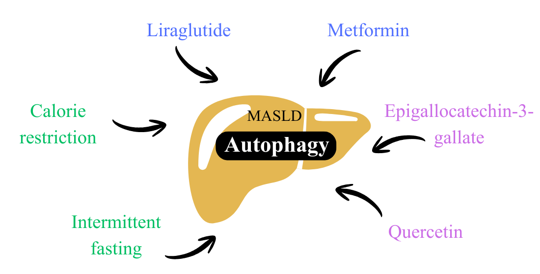
Metabolic dysfunction-associated steatotic liver disease (MASLD) is a global health problem driven primarily by obesity and insulin resistance. Growing evidence indicates that hepatic autophagy dysfunction contributes to lipid accumulation, inflammation, and disease progression, making this process a promising therapeutic target. This review discusses pharmacological, nutritional, and bioactive compound interventions that modulate autophagy. Among pharmacological therapies, in experimental models, liraglutide and metformin reportedly activate the AMP-activated protein kinase/ mammalian target of rapamycin pathway, increasing the expression of microtubule-associated proteins 1A/1B light chain 3 form II and beclin 1, decreasing sequestosome 1 expression, and augmenting the nuclear translocation of transcription factor EB, indicating effective autophagy induction. Moreover, nutritional strategies, such as calorie restriction and intermittent fasting, regulate energy-sensing pathways and have been associated with improvements in steatosis, oxidative stress, and metabolic parameters. Bioactive compounds also modulate autophagy signaling and reduce hepatic lipid accumulation. Taken together, the evidence reinforces autophagy as a central mechanism by which various therapeutic approaches can attenuate MASLD, supporting the development of more effective and mechanism-based interventions.
Keywords:
metabolic dysfunction-associated steatotic liver disease
; autophagy
; liraglutide
; metformin
; calorie restriction
; intermittent fasting
; bioactive compounds
1. Introduction
Metabolic dysfunction-associated steatotic liver disease (MASLD) represents a substantial and growing global health challenge, affecting approximately one quarter of the world’s adult population [1]. Its continued increase is intrinsically linked to obesity and sedentary lifestyle pandemics. The socioeconomic burden of MASLD is substantial, being one of the leading causes of hepatocellular carcinoma and the need for liver transplantation in the United States, with lifetime healthcare costs that reached approximately US$100 billion in 2016 [2].
MASLD is defined by the presence of hepatic steatosis (fat accumulation ≥ 5% in hepatocytes) in individuals with at least one of five metabolic disorders: high body mass index (BMI ≥ 25 kg/m2), elevated fasting glucose (≥ 100 mg/dL), high blood pressure (≥ 130/85 mmHg), high triglyceride levels (TG ≥ 150 mg/dL), or high-density lipoprotein cholesterol (HDL-C). This new nomenclature, adopted in 2023, emphasizes the central contribution of metabolic dysfunction, without requiring the exclusion of alcohol consumption [3,4,5].
Clinically, MASLD is a progressive disease. Its initial stage is simple hepatic steatosis, characterized by fat accumulation without significant inflammation. The most advanced stage is metabolic dysfunction-associated steatohepatitis (MASH), evidenced by hepatocellular inflammation, ballooning, and risk of developing fibrosis, cirrhosis, and hepatocellular carcinoma, with consequent increased mortality [5,6].
Autophagy is a crucial catabolic process that contributes to cellular homeostasis by degrading dysfunctional components and lipids. Lipophagy, a selective autophagic mechanism targeting lipid vesicles, is the selective mechanism by which lipid vesicles are sequestered by autophagosomes, fused to lysosomes, and degraded by lysosomal acid lipase [7]. Evidence indicates that MASLD is positively correlated with autophagy suppression in the liver [8,9]. It is suggested that the abundance of cellular energy, typical of obesity and insulin resistance (IR), inhibits the AMP-activated protein kinase (AMPK)/ mammalian target of rapamycin complex 1 (mTORC1) pathway, which regulates autophagy [10,11].
Inhibition of mTORC1 is essential to activate the autophagic machinery [e.g., Unc-51-like kinase 1 (ULK1), the beclin 1 complex, conversion of microtubule-associated protein 1 light chain 3 form I (LC3-I) to form II (LC3-II)]. Impaired hepatic autophagy leads to increased lipid accumulation, which, in turn, further suppresses autophagy, creating a vicious cycle. This accumulation not only perpetuates steatosis but also contributes to cellular stress and inflammation, driving MASLD progression [12,13].
Some evidence suggests the potential of pharmacological and non-pharmacological agents to induce autophagy, including antidiabetic drugs such as glucagon-like peptide-1 (GLP-1) agonists and metformin, dietary interventions such as calorie restriction and intermittent fasting, and bioactive compounds [14]. Given this scenario, the relevance of autophagy as a mechanism for attenuating the pathogenesis of MASLD stands out, as does the importance of a better understanding of potential therapeutic agents and of modulating this pathway.
Thus, this review aims to compile existing evidence on the effects of pharmacological, nutritional, and bioactive compound interventions on autophagy and to explore their potential role in the pathogenesis of MASLD.
1.1 Pharmacological interventions
1.1.1 GLP-1 analogs
GLP-1 analogs are a class of drugs used to treat type 2 diabetes mellitus (T2DM) and obesity. In general, these drugs exert their effects through the activation of membrane receptors coupled to G proteins, expressed mainly in the lung, kidney, stomach, heart, intestine, alpha and beta cells of the pancreatic islets, and in multiple regions of the central nervous system, promoting effects such as increased satiety, augmented glucose-dependent insulin secretion, decreased glucagon secretion, and attenuated gastric emptying [15,16].
Within this family, liraglutide has demonstrated consistent positive effects on MASLD in experimental and clinical studies. Some evidence suggests that autophagy activation may be a mechanism underlying the improvement in MASLD observed in some experimental models using liraglutide. For example, a study using in vivo and in vitro approaches showed that liraglutide alleviates hepatic steatosis induced by a high-fat diet (HFD) by activating the transcription factor EB (TFEB)-regulated autophagy–lysosomal pathway and promoting autophagy-dependent lipid degradation. This activation occurs via the GLP-1R/ TFEB pathway, highlighting the therapeutic potential of liraglutide in MASLD. In this sense, liraglutide promoted nuclear translocation of TFEB and restored autophagic activity by improving lysosomal function, leading to reduced hepatic expression of LC3-II and sequestosome 1 (p62) [17,18,19,20,21,22].
Interestingly, a study in an HFD-induced MASLD model with Sprague–Dawley rats and HepG2 cells treated with palmitic acid (PA) showed that liraglutide administration for 4 weeks increased beclin 1 and autophagy-related 7 (ATG7) protein expression, as well as the LC3-II/ LC3-I ratio, which were reduced in response to HFD consumption, indicating induction of autophagy. Additionally, an increase in the expression of phospho-AMP-activated protein kinase (p-AMPK) was observed, along with a decrease in the expression of phospho-mammalian target of rapamycin (p-mTOR), suggesting that liraglutide modulates the AMPK/ mammalian target of rapamycin (mTOR) pathway to promote autophagy (Table 1) [14].
Another study investigated the effect of liraglutide on hepatocellular steatosis in C57BL/6 mice and L-O2 cells treated with free fatty acids (FFAs). It was demonstrated that liraglutide administration for 12 weeks increased the expression of LC3-II and beclin 1 (reduced with HFD) and reduced that of p62 (increased with HFD), indicating induction of autophagy. An increase in p-AMPK expression and a decrease in p-mTOR expression were also reported, suggesting that liraglutide modulates the AMPK/ mTOR pathway to promote autophagy (Table 1) [23].
In a study investigating the effects of liraglutide on T2DM-associated hepatic steatosis in an animal model, liraglutide significantly reduced liver damage in mice with MASLD and T2DM, as evidenced by histological and biochemical analyses. The authors also found that liraglutide activated the AMPK/ mTOR signaling pathway, which was suppressed in the MASLD model (Table 1) [24].
Another study evaluated whether liraglutide could attenuate MASLD in mice fed an HFD, exploring the underlying molecular mechanisms, with emphasis on mitochondrial architecture and autophagy mediated by the SIRT1/ SIRT3-FOXO3a pathways. Treatment with liraglutide for 4 weeks resulted in a significant decrease in body mass, improved glucose tolerance, and reduced hepatic triglyceride content in mice with MASLD. Additionally, decreased p62 gene and protein expression, and increased beclin 1 expression and LC3-II/I ratio were observed, indicating that liraglutide promoted hepatic autophagy (Table 1) [25].
1.1.2 Metformin
Metformin is a synthetic biguanide with an antihyperglycemic effect, used in the treatment of T2DM. Its effects are mainly due to increased insulin sensitivity, reduced hepatic glucose synthesis (by inhibiting hepatic gluconeogenesis), and decreased intestinal glucose absorption, with the safe dose for the treatment of T2DM between 250–2550 mg/day [26].
Two studies showed that metformin significantly improves steatosis in patients with MASLD without T2DM, although it has no impact on fibrosis or necroinflammation in these individuals [27,28]. Additionally, patients with MASH presented metformin-improved plasma alanine aminotransferase (ALT) levels and liver histology in 30% of patients [29].
Regarding autophagy, a study reported that this pathway is involved in improving MASLD with metformin use. More specifically, in an HFD-induced MASLD mouse model, metformin treatment for 9 weeks increased LC3-II protein expression, the number of autophagic vesicles and TFEB in the nuclear fraction of the liver, and decreased p62 expression in the liver, simultaneously improving obesity, steatosis, and IR [30].
Furthermore, a study conducted on human carcinoma cells showed that metformin induced autophagy and increased autophagic flux by activating the AMPK/ mTOR pathway, as shown in Table 2. Although these findings do not derive directly from hepatic MASLD models, the identified mechanism is highly biologically relevant, as AMPK/ mTOR signaling dysfunction and reduced autophagic capacity are central to the disease’s pathophysiology [31].
1.2. Calorie restriction
Calorie restriction is a dietary intervention that consists of a significant reduction in calorie intake without compromising the nutritional content of the diet. In animal models and primates, this type of intervention is associated with increased longevity, through adaptations at the metabolic and molecular levels [32].
In metabolic terms, one study showed that a 30% reduction in calorie intake in primates reduced the presence of T2DM and cardiovascular diseases [33,34]. An association between negative energy balance and improved MASLD has also been suggested, possibly through reduced body mass, with improvements in steatosis, liver histology, and plasma ALT levels [35,36].
The activation of autophagy has been studied as a potential mechanism involved in the metabolic effects observed with calorie restriction. A study using in vivo and in vitro methods demonstrated that calorie restriction and its mimetics activate chaperone-mediated autophagy, promoting the selective degradation of damaged proteins and conferring cellular protection against stress and aging. Concerning MASLD, characterized by the accumulation of dysfunctional proteins and impairment of autophagic pathways, this mechanism offers a plausible explanation for the beneficial metabolic effects of calorie restriction and reinforces its potential as a therapeutic intervention (Table 3) [37].
A study aimed at understanding the effects of calorie restriction and resveratrol on hepatic steatosis showed that both reduced hepatic steatosis and hepatocyte ballooning induced by HFD. In this study, calorie restriction provided superior protection compared with resveratrol, associated with lower total calorie intake and reduced body mass. Moreover, animals fed an HFD had decreased SIRT1 levels and autophagy markers LC3-II and beclin 1, as well as p62 accumulation, indicating compromised autophagic flux. Treatment with calorie restriction increased the expression of SIRT1, LC3-II, and beclin 1, in addition to reducing p62 levels, suggesting a restoration of hepatic autophagic flux (Table 3) [38].
1.3. Intermittent fasting
Intermittent fasting is a dietary intervention characterized by alternating periods of fasting and eating. The most commonly used protocols include alternate-day fasting, in which full days of fasting or very reduced calorie intake alternate with days of ad libitum eating, and full-day fasting, which involves 1–3 days of total or partial fasting per week; and time-restricted feeding, limiting eating to a short daily window (usually 4–10 hours) and maintaining fasting during the remaining hours of the day. As a consequence of these cycles of food abstinence, there is greater mobilization of fatty acids and increased production of ketone bodies, a metabolic process associated with improved cardiometabolic parameters, including plasma lipids, insulin sensitivity, and blood pressure [39].
Some studies suggest that certain intermittent fasting protocols are associated with reduced body mass and inhibition of MASLD progression [40,41,42,43]. There is also evidence suggesting a relationship between autophagy, MASLD, and intermittent fasting.
One clinical trial investigated how intermittent fasting influences hepatic autophagy in patients with MASLD, evaluating the effects of the intervention on autophagy markers, including autophagy-related 5 (ATG5) and beclin 1. Intermittent fasting increased serum ATG5, reduced hepatic steatosis, hepatic fibrosis, body mass, BMI, abdominal circumference, ALT, and aspartate aminotransferase (AST). The study suggests that intermittent fasting may be an effective strategy to improve hepatic steatosis and activate autophagy in patients with MASLD. These effects may contribute to enhanced hepatic metabolism and reduced risk of progression to more severe forms of liver disease (Table 4) [44].
Another study demonstrated that a 24-hour intermittent fasting protocol over three non-consecutive days increased LC3 protein and gene expression in the livers of C57BL/6 mice fed both an HFD and a control diet. Increases in lysosomal-associated membrane protein 1 (LAMP1) protein expression and beclin 1 gene expression in the liver of mice fed a control diet were also observed (Table 4) [45].
1.4. Bioactive compounds
1.4.1 Epigallocatechin-3-gallate
Epigallocatechin-3-gallate (EGCG) is the catechin present in the greatest quantity in green tea (Camellia sinensis), contributing to its antioxidant, anti-inflammatory, and cardioprotective properties. Doses between 800 and 1600 mg/day are well tolerated and safe, and some studies have demonstrated hepatoprotective and antifibrotic effects with EGCG, with a reduction in hepatic steatosis [46,47,48,49,50].
A study using C57BL/6 mice showed that 2 weeks of EGCG treatment exerted anti-obesity effects, mainly by activating beclin 1-mediated autophagy in gonadal white adipose tissue, resulting in reduced visceral adiposity and improved glucose tolerance. These findings highlight the therapeutic potential of EGCG in the prevention and treatment of obesity and associated metabolic disorders, such as MASLD (Table 5) [51].
Another study demonstrated that EGCG stimulates autophagy in primary mouse hepatocytes. These effects are partially mediated by activation of the AMPK pathway, leading to increased protein expression. Thus, EGCG may have therapeutic potential for the prevention and treatment of hepatic steatosis and other hepatic conditions associated with autophagy dysfunction [52]. Additionally, it was demonstrated that EGCG increased the protein expression of LC3 and AMPK in differentiated adipocytes [53]. Table 5 presents the effects of EGCG on autophagy in both studies.
1.4.2 Quercetin
Quercetin (3,5,7,3′,4′-pentahydroxyflavone) is a polyphenol found mainly in fruits, vegetables, and derivatives, such as onions, apples, and wine, and is attributed with antioxidant, anti-inflammatory, and antitumor effects. In addition, quercetin exhibits cardioprotective effects, including a reduction in blood pressure [55]. Although beneficial effects are attributed to this bioactive compound, its low oral bioavailability makes quercetin poorly usable in the human body, with a maximum of 20 mg/day absorbed by the digestive tract [56,57].
A hepatoprotective effect is attributed to quercetin, although the mechanisms underlying this effect remain poorly elucidated. It has been proposed that this polyphenol attenuates inflammation by improving mitochondrial function, regulating inflammatory cytokine production, and modulating oxidative stress [58,59,60,61,62].
Supplementation of Goto–Kakizaki (GK) rats with quercetin was able to promote hepatic autophagy, increasing the expression of proteins involved in autophagy and related pathways, such as LC3, beclin 1, PTEN-induced putative kinase 1 protein (Pink-1) and Parkin RBR E3 ubiquitin protein ligase (Parkin), and reducing the expression of p62, phosphatidylinositol 3-kinase (PI3K), phospho-phosphatidylinositol 3-kinase (p-PI3K), protein kinase B (AKT), phospho-protein kinase B (p-AKT), mTOR, and p-mTOR proteins, reversing the protein expression scenario observed in GK animals fed an HFD (Table 6) [63].
Another study used ApoE-deficient mice fed an HFD as a model. Quercetin, by promoting autophagy, reduced the hepatic accumulation of oxidized low-density lipoprotein (ox-LDL). In this study, increased LC3-II protein expression and reduced p62 expression were observed, suggesting augmented autophagic degradation (Table 6) [64].
Additionally, in a mouse model of HFD-induced MASLD, 4 weeks of quercetin treatment reduced the NAFLD Activity Score (NAS), a marker of MALD severity, possibly by modulating autophagy-related proteins, as evidenced by increased LC3 protein expression in the liver. However, hepatic p62 expression was also increased by quercetin treatment (Table 6) [65].
2. Conclusions
Autophagy emerges as a central mechanism in maintaining hepatic homeostasis and protecting against MASLD progression. The evidence gathered in this review demonstrates that different pharmacological, nutritional, and bioactive compound interventions converge on activating pathways associated with increased autophagic flux—especially those mediated by AMPK/ mTOR signaling and the regulation of TFEB.
Among the drugs analyzed, GLP-1 agonists, particularly liraglutide, show consistent effects in attenuating steatosis, inflammation, and mitochondrial dysfunction, frequently accompanied by the induction of autophagy in experimental models. Metformin also restores autophagic flux and reduces steatosis, although the magnitude of these effects in humans remains limited. Nutritional interventions, such as calorie restriction, support the notion that energy-deficient states are potent physiological inducers of autophagy, thereby reversing structural and functional alterations associated with MASLD. Bioactive compounds with autophagoregulatory potential offer promising perspectives, but still lack robust evidence.
Collectively, the data indicate that modulating autophagy represents a plausible and strategic therapeutic target in MASLD. However, its clinical translation depends on studies evaluating dose, duration, safety, and the magnitude of long-term benefits. Advancing the understanding of the mechanisms regulating autophagic flux, as well as identifying sensitive and specific markers of its activity in vivo, will be essential for the development of effective and personalized interventions. Thus, integrating pharmacological and nutritional strategies that optimize autophagy may represent a promising approach to halt the progression of MASLD and improve metabolic and hepatic outcomes.
Author Contributions
Conceptualization, J.S.A; writing—original draft preparation, J.S.A.; writing—review and editing, J.S.A. and C.R.O.C.
Funding
This research was funded by the National Council for Scientific and Technological Development (CNPq) (grant number 140217/2022-3) and São Paulo Research Foundation (FAPESP) (grant numbers 2016/25129-4; 2017/18972-0 and 2021/10469-2).
Acknowledgments
We would like to thank the National Council for Scientific and Technological Development (CNPq) (grant number 140217/2022-3) and the São Paulo Research Foundation (FAPESP) (grant numbers 2016/25129-4; 2017/18972-0 and 2021/10469-2) for the financial support.
Conflicts of Interest
The authors declare no conflict of interest.
Abbreviations
The following abbreviations are used in this manuscript:
| AKT | Protein Kinase B |
| ALT | Alanine aminotransferase |
| AMPK | AMP-activated protein kinase |
| AST | Aspartate aminotransferase |
| ATG5 | Autophagy-related 5 |
| ATG7 | Autophagy-related 7 |
| ATG12 | Autophagy-related 12 |
| BMI | Body mass index |
| CMA | Chaperone-mediated autophagy |
| EGCG | Epigallocatechin-3-gallate |
| ELISA | Enzyme-linked immunosorbent assay |
| FFA | Free fatty acid |
| GLP-1 | Glucagon-like peptide-1 |
| HDL-C | High-density lipoprotein cholesterol |
| HFD | High-fat diet |
| IF | Intermittent fasting |
| IHC | Immunohistochemistry |
| IR | Insulin resistance |
| LAMP1 | Lysosomal-associated membrane protein 1 |
| LAMP2A | Lysosomal-associated membrane protein type 2A |
| LC3 | Microtubule-associated proteins 1A/1B light chain 3 |
| LC3-I | Form I of LC3 |
| LC3-II | Form II of LC3 |
| LC3-II/I | Ratio between forms II and I of LC3 |
| MASH | Metabolic dysfunction-associated steatohepatitis |
| MASLD | Metabolic dysfunction-associated steatotic liver disease |
| mTOR | Mammalian target of rapamycin |
| mTORC1 | Mammalian target of rapamycin complex 1 |
| NAS | NAFLD Activity Score |
| ox-LDL | Oxidized low-density lipoprotein |
| p62 | Sequestosome-1 |
| PA | Palmitic acid |
| p-AKT | Phospho-AKT |
| p-AMPK | Phospho-AMPK |
| Parkin | Parkin RBR E3 ubiquitin protein ligase |
| PI3K | Phosphatidylinositol 3-kinase |
| Pink-1 | PTEN-induced putative kinase 1 protein |
| p-mTOR | Phospho-mTOR |
| p-PI3K | Phospho-PI3K |
| qRT-PCR | Quantitative real-time polymerase chain reaction |
| RT-PCR | Real-time polymerase chain reaction |
| T2DM | Type 2 diabetes mellitus |
| TG | Triglycerides |
| TFEB | Transcription factor EB |
| ULK1 | Unc-51–like kinase 1 |
| WB | Western blot |
References
- Yanai, H; Yoshida, H. Metabolic-Dysfunction-Associated Steatotic Liver Disease—Its Pathophysiology, Association with Atherosclerosis and Cardiovascular Disease, and Treatments. Int J Mol Sci 2023, 24(20), 15493. [Google Scholar] [CrossRef]
- Miao, L; Wang, X; Chen, K. Current status and future trends of the global burden of MASLD. Trends Endocrinol Metab 2024, 35(2), 112–25. [Google Scholar] [CrossRef]
- Chan, W-K; Chuah, K-H; Rajaram, RB; Goh, K-L. Metabolic Dysfunction-Associated Steatotic Liver Disease (MASLD): a state-of-the-art review. J Obes Metab Syndr 2023, 32(3), 197–213. [Google Scholar] [CrossRef]
- Eslam, M; Sanyal, AJ; George, J; Sanyal, A; Neuschwander-Tetri, B; Tiribelli, C; Kleiner, DE; Brunt, E; Bugianesi, E; Yki-Järvinen, H; et al. A new definition for metabolic dysfunction-associated fatty liver disease: an international expert consensus statement. J Hepatol 2020, 73(1), 202–9. [Google Scholar] [CrossRef]
- Sangro, P; Cubero, FJ; Trautwein, C. Metabolic dysfunction–associated fatty liver disease (MAFLD): an update of the recent advances in pharmacological treatment. J Physiol Biochem 2023, 79(4), 869–85. [Google Scholar] [CrossRef] [PubMed]
- Moreira, RO; Salles, JE; Oliveira, CP; Cotrim, HP; Carvalho, M; Borges, JL; de Paiva, A; Lottenberg, A; Bertoluci, MC. Brazilian evidence-based guideline for screening, diagnosis, treatment, and follow-up of metabolic dysfunction-associated steatotic liver disease (MASLD) in adult individuals with overweight or obesity: a joint position statement. Arch Endocrinol Metab 2023, 67(6), e000646. [Google Scholar] [CrossRef] [PubMed]
- Zechner, R; Madeo, F; Kratky, D. Cytosolic lipolysis and lipophagy: two sides of the same coin. Nat Rev Mol Cell Biol 2017, 18(11), 671–84. [Google Scholar] [CrossRef] [PubMed]
- Gluchowski, NL; Becuwe, M; Walther, TC; Farese, RV. Lipid droplets and liver disease: from basic biology to clinical implications. Nat Rev Gastroenterol Hepatol 2017, 14(6), 343–55. [Google Scholar] [CrossRef]
- Jakubek, P; Rychter, AM; Ratajczak, AE; Szymczak-Tomczak, A; Zawada, A; Eder, P; Dobrowolska, A; Krela-Kaźmierczak, I. Autophagy alterations in obesity, type 2 diabetes, and metabolic dysfunction-associated steatotic liver disease: the evidence from human studies. Intern Emerg Med 2024, 19(5), 1207–20. [Google Scholar] [CrossRef]
- Dann, SG; Selvaraj, A; Thomas, G. MTOR Complex1–S6K1 signaling: at the crossroads of obesity, diabetes and cancer. Trends Mol Med 2007, 13(6), 252–9. [Google Scholar] [CrossRef]
- Um, SH; Frigerio, F; Watanabe, M; Picard, F; Joaquin, M; Sticker, M; Fumagalli, S; Allegrini, PR; Kozma, SC; Auwerx, J; et al. Absence of S6K1 protects against age- and diet-induced obesity while enhancing insulin sensitivity. Nature 2004, 431(7005), 200–5. [Google Scholar] [CrossRef]
- Saxton, RA; Sabatini, DM. mTOR Signaling in Growth, Metabolism, and Disease. Cell 2017, 168(6), 960–76. [Google Scholar] [CrossRef]
- Xiang, H; Zhang, J; Lin, C; Zhang, L; Liu, B; Ouyang, L. Targeting autophagy-related protein kinases for potential therapeutic purpose. Acta Pharm Sin B 2020, 10(4), 569–81. [Google Scholar] [CrossRef]
- He, Y; Ao, N; Yang, J; Wang, X; Jin, S; Du, J. The preventive effect of liraglutide on the lipotoxic liver injury via increasing autophagy. Ann Hepatol 2020, 19(1), 44–52. [Google Scholar] [CrossRef]
- Bifari, F; Manfrini, R; Dei Cas, M; Berra, C; Siano, M; Zuin, M; Paroni, R; Folli, F. Multiple target tissue effects of GLP-1 analogues on non-alcoholic fatty liver disease (NAFLD) and non-alcoholic steatohepatitis (NASH). Pharmacol Res 2018, 137, 219–29. [Google Scholar] [CrossRef]
- Jin, T; Weng, J. Hepatic functions of GLP-1 and its based drugs: current disputes and perspectives. Am J Physiol Endocrinol Metab 2016, 311(3), E620-7. [Google Scholar] [CrossRef] [PubMed]
- Ghosal, S; Datta, D; Sinha, B. A meta-analysis of the effects of glucagon-like-peptide 1 receptor agonist (GLP1-RA) in nonalcoholic fatty liver disease (NAFLD) with type 2 diabetes (T2D). Sci Rep 2021, 11(1), 22063. [Google Scholar] [CrossRef] [PubMed]
- Li, Z; Yuan, T; Hu, X; Shi, H; Mo, X; Chen, X. Liraglutide protects against inflammatory stress in non-alcoholic fatty liver by modulating Kupffer cells M2 polarization via cAMP-PKA-STAT3 signaling pathway. Biochem Biophys Res Commun 2019, 510(1), 20–6. [Google Scholar] [CrossRef]
- Luo, Y; Yang, P; Li, Z; Chen, Y; Zhang, L; Liu, T; Xie, Z; Zhou, S. Liraglutide Improves Non-Alcoholic Fatty Liver Disease In Diabetic Mice By Modulating Inflammatory Signaling Pathways. Drug Des Devel Ther 2019, 13, 4065–74. [Google Scholar] [CrossRef]
- Newsome, PN; Buchholtz, K; Cusi, K; Linder, M; Okanoue, T; Ratziu, V; Sanyal, AJ; Sejling, A-S; Harrison, SA. A Placebo-Controlled Trial of Subcutaneous Semaglutide in Nonalcoholic Steatohepatitis. N Engl J Med 2021, 384(12), 1113–24. [Google Scholar] [CrossRef] [PubMed]
- Rezaei, S; Tabrizi, R; Nowrouzi-Sohrabi, P; Jalali, M; Atkin, SL; Alipour, S; Jamialahmadi, T; Sahebkar, A. GLP-1 Receptor Agonist Effects on Lipid and Liver Profiles in Patients with Nonalcoholic Fatty Liver Disease: systematic review and meta-analysis. Can J Gastroenterol Hepatol 2021, 2021, 8936865. [Google Scholar] [CrossRef]
- Fang, Y; Ji, L; Zhu, C; Xiao, Y; Zhang, J; Lu, J; Yin, J; Wei, L. Liraglutide Alleviates Hepatic Steatosis by Activating the TFEB-Regulated Autophagy-Lysosomal Pathway. Front Cell Dev Biol 2020, 8, 602574. [Google Scholar] [CrossRef]
- He, Q; Sha, S; Sun, L; Zhang, J; Dong, M. GLP-1 analogue improves hepatic lipid accumulation by inducing autophagy via AMPK/mTOR pathway. Biochem Biophys Res Commun 2016, 476(4), 196–203. [Google Scholar] [CrossRef]
- Liao, Z; Huang, L; Chen, J; Chen, T; Kong, D; Wei, Q; Chen, Q; Deng, B; Li, Y; Zhong, S. Liraglutide Improves Nonalcoholic Fatty Liver Disease in Diabetic Mice by Activating Autophagy Through AMPK/mTOR Signaling Pathway. Diabetes Metab Syndr Obes 2024, 17, 575–84. [Google Scholar] [CrossRef]
- Tong, W; Ju, L; Qiu, M; Xie, Q; Chen, Y; Shen, W; Sun, W; Wang, W; Tian, J. Liraglutide ameliorates non-alcoholic fatty liver disease by enhancing mitochondrial architecture and promoting autophagy through the SIRT1/SIRT3–FOXO3a pathway. Hepatol Res 2016, 46(9), 933–43. [Google Scholar] [CrossRef]
- Dutta, S; Shah, RB; Singhal, S; Dutta, SB; Bansal, S; Sinha, S; Haque, M. Metformin: a review of potential mechanism and therapeutic utility beyond diabetes. Drug Des Devel Ther 2023, 17, 1907–32. [Google Scholar] [CrossRef]
- Isaacs, SD; Farrelly, FV; Brennan, PN. Role of anti-diabetic medications in the management of MASLD. Frontline Gastroenterol 2025, 16(3), 239–49. [Google Scholar] [CrossRef]
- Bugianesi, E; Gentilcore, E; Manini, R; Natale, S; Vanni, E; Villanova, N; David, E; Rizzetto, M; Marchesini, G. A Randomized Controlled Trial of Metformin versus Vitamin E or Prescriptive Diet in Nonalcoholic Fatty Liver Disease. Am J Gastroenterol 2005, 100(5), 1082–90. [Google Scholar] [CrossRef] [PubMed]
- Loomba, R; Lutchman, G; Kleiner, DE; Ricks, M; Feld, JJ; Borg, BB; Modi, A; Nagabhyru, P; Sumner, AE; Liang, TJ. Clinical trial: pilot study of metformin for the treatment of non-alcoholic steatohepatitis. Aliment Pharmacol Ther 2008, 29(2), 172–82. [Google Scholar] [CrossRef]
- Zhang, D; Ma, Y; Liu, J; Deng, Y; Zhou, B; Wen, Y; Li, M; Wen, D; Ying, Y; Luo, S. Metformin Alleviates Hepatic Steatosis and Insulin Resistance in a Mouse Model of High-Fat Diet-Induced Nonalcoholic Fatty Liver Disease by Promoting Transcription Factor EB-Dependent Autophagy. Front Pharmacol 2021, 12, 689111. [Google Scholar] [CrossRef] [PubMed]
- Gao, C; Fang, L; Zhang, H; Zhang, W-S; Li, X-O; Du, S-Y. Metformin Induces Autophagy via the AMPK-mTOR Signaling Pathway in Human Hepatocellular Carcinoma Cells. Cancer Manag Res 2020, 12, 5803–11. [Google Scholar] [CrossRef] [PubMed]
- Most, J; Tosti, V; Redman, LM; Fontana, L. Calorie restriction in humans: an update. Ageing Res Rev 2017, 39, 36–45. [Google Scholar] [CrossRef]
- Colman, RJ; Anderson, RM; Johnson, SC; Kastman, EK; Kosmatka, KJ; Beasley, TM; Allison, DB; Cruzen, C; Simmons, HA; Kemnitz, JW. Caloric Restriction Delays Disease Onset and Mortality in Rhesus Monkeys. Science 2009, 325(5937), 201–4. [Google Scholar] [CrossRef]
- Colman, RJ; Beasley, TM; Kemnitz, JW; Johnson, SC; Weindruch, R; Anderson, RM. Caloric restriction reduces age-related and all-cause mortality in rhesus monkeys. Nat Commun 2014, 5, 3557. [Google Scholar] [CrossRef]
- Simancas-Racines, D; Annunziata, G; Verde, L; Fascì-Spurio, F; Reytor-González, C; Muscogiuri, G; Frias-Toral, E; Barrea, L. Nutritional Strategies for Battling Obesity-Linked Liver Disease: the role of medical nutritional therapy in metabolic dysfunction-associated steatotic liver disease (masld) management. Curr Obes Rep 2025, 14(1), 1–17. [Google Scholar] [CrossRef]
- Koutoukidis, DA; Astbury, NM; Tudor, KE; Morris, E; Henry, JA; Noreik, M; Jebb, SA; Aveyard, P. Association of Weight Loss Interventions With Changes in Biomarkers of Nonalcoholic Fatty Liver Disease. JAMA Intern Med 2019, 179(9), 1262–71. [Google Scholar] [CrossRef]
- Jafari, M; Macho-González, A; Diaz, A; Lindenau, K; Santiago-Fernández, O; Zeng, M; Massey, AC; de Cabo, R; Kaushik, S; Cuervo, AM. Calorie restriction and calorie-restriction mimetics activate chaperone-mediated autophagy. Proc Natl Acad Sci U S A 2024, 121(26), e2317945121. [Google Scholar] [CrossRef] [PubMed]
- Ding, S; Jiang, J; Zhang, G; Bu, Y; Zhang, G; Zhao, X. Resveratrol and caloric restriction prevent hepatic steatosis by regulating SIRT1-autophagy pathway and alleviating endoplasmic reticulum stress in high-fat diet-fed rats. PLoS One 2017, 12(8), e0183541. [Google Scholar] [CrossRef] [PubMed]
- Vasim, I; Majeed, CN; DeBoer, MD. Intermittent Fasting and Metabolic Health. Nutrients 2022, 14(3), 631. [Google Scholar] [CrossRef]
- Zhang, Z; Kong, AP-S; Wong, VW-S; Hui, HX. Intermittent fasting and metabolic dysfunction-associated steatotic liver disease: the potential role of the gut-liver axis. Cell Biosci 2025, 15(1), 62. [Google Scholar] [CrossRef]
- Wilkinson, MJ; Manoogian, ENC; Zadourian, A; Lo, H; Fakhouri, S; Shoghi, A; Wang, X; Fleischer, JG; Navlakha, S; Panda, S. Ten-Hour Time-Restricted Eating Reduces Weight, Blood Pressure, and Atherogenic Lipids in Patients with Metabolic Syndrome. Cell Metab 2020, 31(1), 92–104.e5. [Google Scholar] [CrossRef] [PubMed]
- Xiao, Y; Liu, Y; Zhao, L; Zhou, Y. Effect of 5: 2 fasting diet on liver fat content in patients with type 2 diabetic with nonalcoholic fatty liver disease. Metab Syndr Relat Disord 2022, 20(8), 459–65. [Google Scholar] [CrossRef] [PubMed]
- Johari, MI; Yusoff, K; Haron, J; Nadarajan, C; Ibrahim, KN; Wong, MS; Hafidz, MIA; Chua, BE; Hamid, N; Arifin, WN. A Randomised Controlled Trial on the Effectiveness and Adherence of Modified Alternate-day Calorie Restriction in Improving Activity of Non-Alcoholic Fatty Liver Disease. Sci Rep 2019, 9(1), 11232. [Google Scholar] [CrossRef]
- Karahan, TO; Akyuz, EY; Karadag, DY; Yilmaz, Y; Eren, F. Effects of Intermittent Fasting on Liver Steatosis and Fibrosis, Serum FGF-21 and Autophagy Markers in Metabolic Dysfunction-Associated Fatty Liver Disease: a randomized controlled trial. Life 2025, 15(5), 696. [Google Scholar] [CrossRef] [PubMed]
- Chaudhary, R; Liu, B; Bensalem, J; Sargeant, TJ; Page, AJ; Wittert, GA; Hutchison, AT; Heilbronn, LK. Intermittent fasting activates markers of autophagy in mouse liver, but not muscle from mouse or humans. Nutrition 2022, 101, 111662. [Google Scholar] [CrossRef]
- Capasso, L; De Masi, L; Sirignano, C; Maresca, V; Basile, A; Nebbioso, A; Rigano, D; Bontempo, P. Epigallocatechin Gallate (EGCG): pharmacological properties, biological activities and therapeutic potential. Molecules 2025, 30(3), 654. [Google Scholar] [CrossRef]
- Janota, B; Janion, K; Bużek, A; Janczewska, E. Dietary Strategies in the Prevention of MASLD: a comprehensive review of dietary patterns against fatty liver. Metabolites 2025, 15(8), 528. [Google Scholar] [CrossRef]
- Hefer, M; Petrovic, A; Roguljic, LK; Kolaric, TO; Kizivat, T; Wu, CH; Tabll, AA; Smolic, R; Vcev, A; Smolic, M. Green Tea Polyphenol (-)-Epicatechin Pretreatment Mitigates Hepatic Steatosis in an In Vitro MASLD Model. Curr Issues Mol Biol 2024, 46(8), 8981–94. [Google Scholar] [CrossRef]
- Zhang, J; Wang, S; Zhang, T; Zi, M; Wang, S; Zhang, Q. Green tea epigallocatechin gallate attenuate metabolic dysfunction-associated steatotic liver disease by regulation of pyroptosis. Lipids Health Dis 2025, 24(1), 140. [Google Scholar] [CrossRef]
- Hidalgo, I; Ortiz-Flores, M; Villarreal, F; Fonseca-Coronado, S; Ceballos, G; Meaney, E; Nájera, N. Is it possible to treat nonalcoholic liver disease using a flavanol-based nutraceutical approach? Basic and clinical data. J Basic Clin Physiol Pharmacol 2022, 33(6), 703–14. [Google Scholar] [CrossRef]
- Choi, C; Song, H-D; Son, Y; Cho, YK; Ahn, S-Y; Jung, Y-S; Yoon, YC; Kwon, SW; Lee, Y-H. Epigallocatechin-3-Gallate Reduces Visceral Adiposity Partly through the Regulation of Beclin1-Dependent Autophagy in White Adipose Tissues. Nutrients 2020, 12(10), 3072. [Google Scholar] [CrossRef] [PubMed]
- Zhou, J; Farah, BL; Sinha, RA; Wu, Y; Singh, BK; Bay, B-H; Yang, CS; Yen, PM. Epigallocatechin-3-Gallate (EGCG), a Green Tea Polyphenol, Stimulates Hepatic Autophagy and Lipid Clearance. PLoS One 2014, 9(1), e87161. [Google Scholar] [CrossRef]
- Kim, S-N; Kwon, H-J; Akindehin, S; Jeong, H; Lee, Y-H. Effects of Epigallocatechin-3-Gallate on Autophagic Lipolysis in Adipocytes. Nutrients 2017, 9(7), 680. [Google Scholar] [CrossRef]
- Santamarina, AB; Oliveira, JL; Silva, FP; Carnier, J; Mennitti, LV; Santana, AA; de Souza, GHI; Ribeiro, EB; do Nascimento, CMO; Lira, FS. Green Tea Extract Rich in Epigallocatechin-3-Gallate Prevents Fatty Liver by AMPK Activation via LKB1 in Mice Fed a High-Fat Diet. PLoS One 2015, 10(11), e0141227. [Google Scholar] [CrossRef]
- Aghababaei, F; Hadidi, M. Recent Advances in Potential Health Benefits of Quercetin. Pharmaceuticals 2023, 16(7), 1020. [Google Scholar] [CrossRef] [PubMed]
- Wang, W; Sun, C; Mao, L; Ma, P; Liu, F; Yang, J; Gao, Y. The biological activities, chemical stability, metabolism and delivery systems of quercetin: a review. Trends Food Sci Technol 2016, 56, 21–38. [Google Scholar] [CrossRef]
- Fujimori, M; Kadota, K; Shimono, K; Shirakawa, Y; Sato, H; Tozuka, Y. Enhanced solubility of quercetin by forming composite particles with transglycosylated materials. J Food Eng 2015, 149, 248–54. [Google Scholar] [CrossRef]
- Katsaros, I; Sotiropoulou, M; Vailas, M; Kapetanakis, EI; Valsami, G; Tsaroucha, A; Schizas, D. Quercetin’s Potential in MASLD: investigating the role of autophagy and key molecular pathways in liver steatosis and inflammation. Nutrients 2024, 16(22), 3789. [Google Scholar] [CrossRef]
- Chen, L; Liu, J; Mei, G; Chen, H; Peng, S; Zhao, Y; Yao, P; Tang, Y. Quercetin and non-alcoholic fatty liver disease: a review based on experimental data and bioinformatic analysis. Food Chem Toxicol 2021, 154, 112314. [Google Scholar] [CrossRef]
- Liu, P; Lin, H; Xu, Y; Zhou, F; Wang, J; Liu, J; Zhu, X; Guo, X; Tang, Y; Yao, P. Frataxin-Mediated PINK1–Parkin-Dependent Mitophagy in Hepatic Steatosis: the protective effects of quercetin. Mol Nutr Food Res 2018, 62(16), e1800164. [Google Scholar] [CrossRef] [PubMed]
- Miltonprabu, S; Tomczyk, M; Skalicka-Woźniak, K; Rastrelli, L; Daglia, M; Nabavi, SF; Alavian, SM; Nabavi, SM. Hepatoprotective effect of quercetin: from chemistry to medicine. Food Chem Toxicol 2017, 108, 365–74. [Google Scholar] [CrossRef]
- Sotiropoulou, M; Katsaros, I; Vailas, M; Lidoriki, I; Papatheodoridis, GV; Kostomitsopoulos, NG; Valsami, G; Tsaroucha, A; Schizas, D. Nonalcoholic fatty liver disease. Saudi J Gastroenterol 2021, 27(6), 319–30. [Google Scholar] [CrossRef]
- Guo, Z; Zhang, J; Li, M; Xing, Z; Li, X; Qing, J; Zhang, Y; Zhu, L; Qi, M; Zou, X. Mechanism of action of quercetin in regulating cellular autophagy in multiple organs of Goto-Kakizaki rats through the PI3K/AKT/mTOR pathway. Front Med 2024, 11, 1442071. [Google Scholar] [CrossRef] [PubMed]
- Liu, L; Gao, C; Yao, P; Gong, Z. Quercetin Alleviates High-Fat Diet-Induced Oxidized Low-Density Lipoprotein Accumulation in the Liver: implication for autophagy regulation. Biomed Res Int 2015, 2015, 607531. [Google Scholar] [CrossRef] [PubMed]
- Katsaros, I; Sotiropoulou, M; Vailas, M; Papachristou, F; Papakyriakopoulou, P; Grigoriou, M; Kostomitsopoulos, N; Giatromanolaki, A; Valsami, G; Tsaroucha, A. The Effect of Quercetin on Non-Alcoholic Fatty Liver Disease (NAFLD) and the Role of Beclin1, P62, and LC3: an experimental study. Nutrients 2024, 16(24), 4282. [Google Scholar] [CrossRef]

Table 1.
Characteristics of studies evaluating the effects of liraglutide, focusing on the modulation of autophagy.
Table 1.
Characteristics of studies evaluating the effects of liraglutide, focusing on the modulation of autophagy.
| Reference | Model | Intervention | Effect on autophagy (In vivo) |
Effect on autophagy (In vitro) |
DOI |
|---|---|---|---|---|---|
| [22] | MASLD: C57BL/6J mice fed HFD for 18 weeks, plus HepG2 cells and primary hepatocytes treated with PA |
In vivo: 0.4 mg/kg, once daily for 4 weeks; In vitro: 0.0001–0.0005 mM for 24h |
↑ ATG7 protein expression (WB), ↑ beclin 1 protein expression (WB), ↓ LC3-II protein expression (WB), ↓ p62 protein expression (WB), ↑ TFEB expression in the nuclear fraction (WB) in the liver | ↓ LC3-II protein expression (WB) and ↓ p62 protein expression (WB) | 10.3389/fcell.2020.602574 |
| [14] | MASLD: Sprague-Dawley rats fed HFD for 12 weeks, plus HepG2 cells treated with PA | In vivo: 0.05–0.2 mg/kg, twice daily for 4 weeks; In vitro: 1×10⁻⁵, 5×10⁻⁵, 1×10⁻⁴ or 5×10⁻⁴ mM for 24h | ↑ LC3-II/I protein expression (WB), ↑ beclin 1 protein expression (WB), ↑ ATG7 protein expression (WB) in the liver | ↑ LC3-II/I protein expression (WB), ↑ beclin 1 protein expression (WB), ↑ ATG7 protein expression (WB), ↑ p-AMPK protein expression (WB), ↑ p-mTOR protein expression (WB) | 10.1016/j.aohep.2019.06.023 |
| [23] | MASLD: C57BL/6 mice fed HFD for 12 weeks, plus L-O2 cells treated with FFA | In vivo: 0.1 mg/kg, once daily for 12 weeks; In vitro: 0.0001 mM for 24h | ↑ LC3-II protein expression (WB), ↓ p62 protein expression (WB) in the liver | ↑ p-AMPK protein expression (WB), ↓ p-mTOR protein expression (WB), ↑ beclin 1 protein expression (WB) | 10.1016/j.bbrc.2016.05.086 |
| [24] | MASLD and T2DM: Sprague-Dawley rats fed HFD, plus 1% STZ | 3.6 mg/kg, twice daily for 4 weeks |
↑ LC3-II/I protein expression (WB), ↑ LC3 gene expression (RT-PCR), ↑ beclin 1 gene and protein expression (WB and RT-PCR), ↑ p-AMPK protein expression (WB), ↑ AMPK gene expression (RT-PCR), ↓ p-mTOR protein expression (WB), ↓ mTOR gene expression (RT-PCR) in the liver | - | 10.2147/DMSO.S447182 |
| [25] | MASLD: C57BL/6J mice fed HFD for 16 weeks | 150 mg/kg, once daily for 4 weeks | ↓ p62 gene and protein expression (WB and RT-PCR), ↑ beclin 1 gene and protein expression (WB and RT-PCR), ↑ LC3-II/I protein expression (WB); ↑ LC3 gene expression (RT-PCR) in the liver | - | 10.1111/hepr.12634 |
The arrows indicate the direction of the effect: ↑ – Increase; ↓ – Reduction. Abbreviations: MASLD – metabolic dysfunction–associated steatotic liver disease; HFD – High fat diet; ATG7 – autophagy-related 7; LC3 – microtubule-associated proteins 1A/1B light chain 3; LC3-II – form II of microtubule-associated protein 1 light chain 3; p62 – sequestosome 1; TFEB – Transcription factor EB; PA – Palmitic acid; FFA – Free fatty acids; p-AMPK – phospho-AMP-activated protein kinase; p-mTOR – phospho-mammalian target of rapamycin; T2DM – type 2 diabetes mellitus; STZ – Streptozotocin; AMPK – AMP-activated protein kinase; mTOR – mammalian target of rapamycin; LC3–II/I – ratio between forms II and I of microtubule-associated proteins 1A/1B light chain 3; WB – Western blot; RT-PCR – real-time polymerase chain reaction.
Table 2.
Characteristics of studies evaluating the effects of metformin, focusing on the modulation of autophagy.
Table 2.
Characteristics of studies evaluating the effects of metformin, focusing on the modulation of autophagy.
| Reference | Model | Intervention | Effect on autophagy | DOI |
|---|---|---|---|---|
| [30] | MASLD: C57BL/6J mice fed HFD for 14 weeks | 300 mg/kg, once daily for 9 weeks | ↑ LC3-II protein expression (WB), ↓ p62 protein expression (WB), ↑ TFEB protein expression in the nuclear fraction (WB) in the liver | 10.3389/fphar.2021.689111 |
| [31] | MHCC97H and HepG2 cells | 10 mM, for 12–72 h | ↑ LC3-II protein expression (WB), ↓ p62 protein expression (WB), ↑ p-AMPK protein expression (WB), and ↓ p-mTOR protein expression (WB) in both cell lines | 10.2147/CMAR.S257966 |
The arrows indicate the direction of the effect: ↑ – increase; ↓ – reduction. Abbreviations: MASLD – metabolic dysfunction-associated steatotic liver disease; HFD – high-fat diet; LC3-II – microtubule-associated protein 1 light chain 3 form II; p62 – sequestosome 1; TFEB – transcription factor EB; AMPK – AMP-activated protein kinase; p-AMPK – phospho-AMP-activated protein kinase; p-mTOR – phospho-mammalian target of rapamycin; WB – Western blot; IR – insulin resistance.
Table 3.
Characteristics of studies evaluating the effects of calorie restriction, focusing on the modulation of autophagy.
Table 3.
Characteristics of studies evaluating the effects of calorie restriction, focusing on the modulation of autophagy.
| Reference | Model | Intervention | Effect on autophagy | DOI |
|---|---|---|---|---|
| [37] | Fisher-344 rats aged 4, 12, and 22 months | 40% calorie restriction of energy intake | Robust CMA activation in tissues of aged rodents via LAMP2A stabilization | 10.1073/pnas.2317945121 |
| [38] | Wistar rats fed HFD for 18 weeks | 70% calorie restriction of energy intake | ↑ LC3-II gene and protein expression (RT-PCR and WB), ↑ beclin 1 gene expression (RT-PCR), ↑ SIRT1 gene and protein expression (RT-PCR and WB), ↓ p62 gene and protein expression (RT-PCR and WB) in the liver | 10.1371/journal.pone.0183541 |
The arrows indicate the direction of the effect: ↑ – increase; ↓ – reduction. Abbreviations: CMA – chaperone-mediated autophagy; LAMP2A – lysosomal-associated membrane protein type 2A; HFD – high-fat diet; LC3-II – microtubule-associated protein 1 light chain 3 form II; SIRT1 – sirtuin 1; p62 – sequestosome 1; WB – Western blot; RT-PCR – real-time polymerase chain reaction.
Table 4.
Characteristics of studies evaluating the effects of intermittent fasting, focusing on the modulation of autophagy.
Table 4.
Characteristics of studies evaluating the effects of intermittent fasting, focusing on the modulation of autophagy.
| Reference | Model | Intervention | Effect on autophagy | DOI |
|---|---|---|---|---|
| [44] | Obese humans with MASLD | Diet with 22–25 kcal/kg/day, plus IF 16:8 (16 h fasting and 8 h feeding) for 8 weeks | ↑ Serum ATG5 in the IF group (ELISA) | 10.3390/life15050696 |
| [45] | C57BL/6 mice fed HFD for 8 weeks | IF 24 h, on 3 non-consecutive days/week for 8 weeks | ↑ LC3 protein and gene expression (WB and qRT-PCR) in the liver of control and HFD-fed animals, ↑ LAMP1 protein expression in the liver of control animals (WB), and ↑ beclin 1 gene expression (qRT-PCR) in the liver of control animals. | 10.1016/j.nut.2022.111662 |
The arrows indicate the direction of the effect: ↑ – increase; ↓ – reduction. Abbreviations: MASLD – metabolic dysfunction–associated steatotic liver disease; IF – intermittent fasting; ATG5 – autophagy-related 5; ELISA – enzyme-linked immunosorbent assay; HFD – high-fat diet; LAMP1 – lysosomal-associated membrane protein 1; WB – Western blot; qRT-PCR – quantitative real-time polymerase chain reaction.
Table 5.
Characteristics of studies evaluating the effects of EGCG, focusing on the modulation of autophagy.
Table 5.
Characteristics of studies evaluating the effects of EGCG, focusing on the modulation of autophagy.
| Reference | Model | Intervention | Effect on autophagy | DOI |
|---|---|---|---|---|
| [51] | C57BL/6 mice fed HFD for 8 weeks | 20 mg/kg, once daily for 2 weeks | ↑ LC3-II/I protein expression (WB), ↑ beclin 1 protein expression (WB), ↑ ATG7 and ATG12 protein expression (WB) in gonadal white adipose tissue | 10.3390/nu12103072 |
| [52] | Primary mouse hepatocytes | 0.04 mM, for 24 h | ↑ LC3-II protein expression (WB), ↓ p62 protein expression (WB), ↑ AMPK protein expression (WB) | 10.1371/journal.pone.0087161 |
| [53] | Differentiated adipocytes (C3H10T1/2) | 0.01 mM, for 24 h | ↑ LC3 protein expression (WB) and ↑ AMPK protein expression (WB) | 10.3390/nu9070680 |
| [54] | Swiss rats fed HFD for 16 weeks | 50 mg/kg, once daily for 16 weeks | ↑ SIRT1 and p-AMPK protein expression (WB) in the liver | 10.1371/journal.pone.0141227 |
The arrows indicate the direction of the effect: ↑ – increase; ↓ – reduction. Abbreviations: EGCG – epigallocatechin-3-gallate; HFD – high-fat diet; LC3-II/I – ratio between forms II and I of microtubule-associated proteins 1A/1B light chain 3; ATG7 – autophagy-related 7; ATG12 – autophagy-related 12; LC3-II – form II microtubule-associated protein 1 light chain 3; LC3- microtubule-associated proteins 1A/1B light chain 3; p62 – sequestosome 1; AMPK – AMP-activated protein kinase; SIRT1 – sirtuin 1; p-AMPK – phospho-AMP-activated protein kinase; WB – Western Blot.
Table 6.
Characteristics of studies evaluating the effects of quercetin, focusing on the modulation of autophagy.
Table 6.
Characteristics of studies evaluating the effects of quercetin, focusing on the modulation of autophagy.
| Reference | Model | Intervention | Effect on autophagy | DOI |
|---|---|---|---|---|
| [63] | Goto-Kakizaki rats fed HFD for 10 weeks | 50 or 75 mg/kg, once daily for 8 weeks | ↑ LC3 protein expression (WB), ↑ beclin 1 protein expression (WB), and ↑ Pink1/Parkin protein expression (WB), ↓ p62 protein expression (WB), ↓ mTOR protein expression (WB), ↓ p-mTOR protein expression (WB), ↓ PI3K protein expression (WB), ↓ p-PI3K protein expression (WB), ↓ AKT protein expression (WB), and ↓ p-AKT protein expression (WB), in the liver | 10.3389/fmed.2024.1442071 |
| [64] | ApoE mice (C57BL/6J background) fed HFD for 24 weeks | 100 mg/kg, once daily for 24 weeks | ↑ Protein expression of LC3-II (WB), ↓ Protein expression of p62 (WB), ↓ Protein expression of mTOR (WB) in the liver | 10.1155/2015/607531 |
| [65] | C57BL/6J mice fed HFD for 12 weeks | 10 and 50 mg/kg, once daily for 4 weeks | ↑ Expression of LC3 and p62 in the liver (IHC), ↑ Protein expression of LC3 in the liver (WB) | 10.3390/nu16244282 |
The arrows indicate the direction of the effect: ↑ – increase; ↓ – reduction. Abbreviations: HFD – high-fat diet; LC3 – microtubule-associated protein 1 light chain 3; Pink1 - PTEN-induced putative kinase 1 protein; Parkin – Parkin RBR E3 ubiquitin protein ligase; p62 – sequestosome 1; mTOR – mammalian target of rapamycin; PI3K – phosphatidylinositol 3-kinase; AKT – protein kinase B; LC3-II – form II of microtubule-associated protein 1 light chain 3; IHC – immunohistochemistry; WB – Western blot.
Disclaimer/Publisher’s Note: The statements, opinions and data contained in all publications are solely those of the individual author(s) and contributor(s) and not of MDPI and/or the editor(s). MDPI and/or the editor(s) disclaim responsibility for any injury to people or property resulting from any ideas, methods, instructions or products referred to in the content. |
© 2025 by the authors. Licensee MDPI, Basel, Switzerland. This article is an open access article distributed under the terms and conditions of the Creative Commons Attribution (CC BY) license (http://creativecommons.org/licenses/by/4.0/).
Copyright: This open access article is published under a Creative Commons CC BY 4.0 license, which permit the free download, distribution, and reuse, provided that the author and preprint are cited in any reuse.


