
Submitted:
15 December 2025
Posted:
16 December 2025
You are already at the latest version
Abstract
Purpose: We investigated the long-term effects of cataract surgery by the eight-chop technique on intraocular pressure (IOP) in cataract patients.Methods: The patients were classified into three groups (Grade II, III, and IV) according to the lens hardness. The operative time, phaco time, aspiration time, cumulative dissipated energy, and volume of fluid used were measured intraoperatively. The best-corrected visual acuity and corneal endothelial cell density were measured. The IOP was monitored for 5 years. Based on the preoperative IOP, eyes were classified into two groups for analysis: IOP > 15 mmHg and < 15 mmHg.Results: The operative time in Grades II, III, and IV were 4.63 ± 0.88 min, 5.48 ± 1.52 min, and 7.77 ± 1.47 min, respectively. The rate of corneal endothelial cell density loss was 1.9 ± 8.3% at 19 weeks. Postoperatively, the IOPs at 1 year were 12.6 ± 2.4 mmHg, 13.2 ± 2.3 mmHg, and 11.7 ± 2.2 mmHg, and at 5 years were 13.1 ± 2.5 mmHg, 12.0 ± 2.0 mmHg, and 12.0 ± 0.6 mmHg, in Grades II, III, and IV, respectively. In patients with a preoperative IOP < 15 mmHg, the IOP remained significantly lower even after 5 years of surgery.Conclusions: The eight-chop technique can lower the IOP and this effect persists for 5 years. This procedure is short and is associated with a minimal reduction in corneal endothelial cell density. Thus, this technique is very effective in lowering IOP in patients with cataracts.
Keywords:
cataract surgery
; phacoemulsification
; corneal endothelial cell
; intraocular pressure
; eight-chop technique
Introduction
Phacoemulsification can reduce the intraocular pressure (IOP) in patients with primary open-angle glaucoma (POAG) [1,2], primary angle-closure glaucoma,[3,4] and pseudoexfoliation glaucoma [1,5]. Investigating changes in IOP following phacoemulsification in patients with glaucoma may be important when considering glaucoma treatment. However, it is difficult to assess how phacoemulsification and intraocular lens implantation affect IOP in glaucoma patients with uncontrolled IOP.
Previous studies showed that phacoemulsification changes IOP in patients with cataract without glaucoma [6,7], although there is conflicting evidence regarding the extent of IOP reduction, and it remains unclear to what extent phacoemulsification and lens implantation reduce IOP in normal eyes.
Previous studies reported changes in IOP following phacoemulsification performed using various techniques, such as the divide-and-conquer technique [2,8]; however, there are no reports on phacoemulsification performed using the eight-chop technique [9,10]. Furthermore, no previous studies have evaluated the intraoperative parameters, therefore, surgery details are unknown.
In the eight-chop technique [9,10], the nucleus under the ophthalmic viscosurgical device is manually divided prior to phacoemulsification. Compared to conventional grooving, divide-and-conquer, and phaco-chop techniques, the operative time is drastically reduced, and the aspiration time and the volume of the fluid used are significantly lower when the eight-chop technique is used [9,10].
In our previous study, we improved the prechop technique [11] and developed the eight-chop technique. In this technique, the lens nucleus is divided into eight segments instead of only four as in the prechop technique.
Investigating postoperative changes after phacoemulsification using the eight-chop technique will help in understanding how phacoemulsification and lens implantation affect IOP in settings of minimal surgical involvement.
When investigating the relationship between phacoemulsification and IOP, it is necessary to consider the effects of surgery on intraocular tissues. More accurate results can be obtained if changes in IOP are examined by measuring intraoperative parameters and considering the surgical involvement of the intraocular tissues. Furthermore, it is important to examine how IOP changes in the long term when considering the treatment for chronic glaucoma; however, there are very few studies on the long-term IOP-lowering effects of clear corneal phacoemulsification.
This study aimed to evaluate the long-term effects of phacoemulsification performed using the eight-chop technique on IOP in normal eyes over 5 years.
Methods
Patients with cataracts who underwent phacoemulsification and posterior chamber intraocular lens (IOL) implantation between January 2016 and December 2022 were enrolled. Patients who had visited the clinic with a diagnosis of cataracts were enrolled in the study. The exclusion criteria were the presence of corneal disease or opacity, uveitis, pupillary dilation problems, and a history of trauma or surgery.
The study protocol adhered to the guidelines of the Declaration of Helsinki and was approved by the Sato Eye Clinic review board (approval number, 160101). Informed consent was obtained from all patients before initiating the study. Preoperatively, all patients underwent slit-lamp and retinal examinations, and their best-corrected visual acuity (BCVA) and IOP were measured. Patients enrolled in this study had no abnormalities on Humphrey 30-2 visual field defects (3 contiguous points greater than -5 dB or 1point greater than -10 dB) during follow-up, or had no history of glaucoma, no ocular medications, and did not have glaucomatous optic nerve changes defined as a cup-to-disc ratio of more than 0.7, disc asymmetry, total or partial thinning of the neural rim, and peripapillary atrophy. Corneal endothelial cell density (CECD; cells/mm2) was measured using a noncontact specular microscope (EM-3000, Topcon, Hasunumacho, Tokyo, Japan). The firmness of the nucleus was graded using the Emery classification [12], based on which patients were classified into three groups (Grade II, Grade III, or Grade IV). Grade IV included Grades IV and V cataracts. Phacoemulsification was performed by the same surgeon, who was experienced in the eight-chop technique, using the Centurion® phacoemulsification unit (Alcon Labs Inc., Fort Worth, Texas, USA).
Three new surgical instruments were designed and developed for performing the eight-chop technique [9,10]. The research team designed these eight-choppers and requested a manufacturing company to produce them. The tip of the Eight-chopper I (SP-8193; ASICO, Westmont, Illinois, USA) was smaller than that of the conventional prechopper, with a length and width of 3.2 mm and 1.4 mm, respectively, as well as a sharp leading edge, and was used for Grade II patients. The Eight-chopper II (SP-8402; ASICO, Westmont, Illinois, USA) used for Grade III patients had a smaller tip (2.5 mm long and 0.8 mm wide) that was angled so that it could be inserted vertically into the lens nucleus. For Grade IV patients, the Lance-chopper (SP-9989; ASICO, Westmont, Illinois, USA) was used that had a 3.0 mm long and 1.3 mm wide tip with a sharper leading edge.
In all surgeries, a temporal, clear corneal incision was made using a 3.0-mm steel keratome. After injecting sodium hyaluronate into the anterior chamber, a 6.2–6.5 mm continuous curvilinear capsulorhexis was created using capsule forceps. The soft-shell technique [13] was used for Grades III and IV patients. Brilliant Blue G (0.025%) was used to improve capsule visualization in cases with dense cataracts or corneal opacity. Hydrodissection was performed using a 27-G cannula; however, hydrodelineation was not performed. The lens nucleus was cracked into eight segments using Eight-chopper I for Grade II patients, Eight-chopper II for Grade III patients, and Lance-chopper for Grade IV patients. A sideport incision was made using a 23-G micro-vitreoretinal knife at 90° from the main incision using the Lance-chop technique. The Lance-chop is an eight-chop technique that uses a Lance-chopper and nucleus sustainer for cases with a hard lens nucleus.[9] The eight segments were phacoemulsified and aspirated from the depth of the iris plane. The capsular bag was aspirated with an irrigation/aspiration tip to remove the cortical material. An ophthalmic viscosurgical device was injected and a foldable IOL (Alcon Labs Inc., Fort Worth, Texas, USA) with polymethyl methacrylate haptics was inserted into the capsular bag using an injector system. The ophthalmic viscosurgical device was then aspirated. The Centurion® phacoemulsification unit (Alcon Labs Inc, Fort Worth, Texas, USA) was used in all cases with a flow rate adjusted to 32 mL/min, a maximum ultrasound power of 80%, and a 1.1-mm tip. If necessary, the wound was sealed by stromal hydration. At the end of the surgery, the anterior chamber was replaced with a balanced salt solution containing moxifloxacin (0.5 mg/mL).
The intraoperative outcome measures included operative time (min), phaco time (s), aspiration time (s), cumulative dissipated energy (CDE), volume of fluid used (mL), and occurrence of intraoperative complications. Operative time was defined as the duration between the beginning of the corneal incision and the end of the ophthalmic viscosurgical device aspiration. Patients were followed up on postoperative days 1 and 2, at 1, 3, 7, and 19 weeks, and at 1, 2, 3, 4, and 5 years. The postoperative outcome measures were BCVA, IOP, and CECD (cells/mm2). For BCVA and CECD evaluations, data obtained on postoperative weeks 7 and 19 were used. For IOP measurements, data obtained on postoperative weeks 7 and 19, and years 1, 2, 3, 4, and 5 were used. The eyes were assigned to two groups for analysis: preoperative IOP > 15 mmHg and preoperative IOP < 15 mmHg, according to the method of Poley et al. [14].
Statistical analyses were performed using the one-way analysis of variance (ANOVA), Kruskal-Wallis test, or Kruskal-Wallis test followed by the Steel-Dwass post-hoc test using Excel Toukei® (version 7.0, Esumi Co. Ltd., Shimane, Japan). Paired t-test was used to compare preoperative and postoperative IOPs. A p-value < 0.05 was considered statistically significant.
Results
This study included 86 eyes of 59 patients with cataracts who underwent phacoemulsification and posterior chamber IOL implantation. Patient characteristics and intraoperative parameters are shown in Table 1. There were no significant differences in the mean age of all groups (p = 0.192, Kruskal-Wallis test). The operative time was significantly different among groups (p < 0.01, Kruskal-Wallis test). Furthermore, the operative time was significantly different between Grades II and III (p = 0.04, Steel-Dwass post hoc test) although no significant difference was observed between Grades III and IV (p = 0.064, Steel-Dwass post hoc test). The operative time in Grade II was significantly shorter than that in Grade IV (p < 0.01, Steel-Dwass post hoc test). The phaco time was significantly different among groups (p < 0.01, Kruskal-Wallis test). The phaco time was significantly different between Grades II and III (p = 0.018, Steel-Dwass post hoc test); however, no significant difference was observed between Grades III and IV (p = 0.268, Steel-Dwass post hoc test). The phaco time was significantly shorter in Grade II compared to that in Grade IV (p = 0.010, Steel-Dwass post hoc test). Although the aspiration time was significantly different among groups (p < 0.01, Kruskal-Wallis test), there were no significant differences between Grades II and III (p = 0.110, Steel-Dwass post hoc test) and between Grades III and IV (p = 0.091, Steel-Dwass post hoc test). However, the aspiration time was significantly shorter in Grade II compared to that in Grade IV (p = 0.011, Steel-Dwass post hoc test). Similarly, there were significant differences in CDE among groups (p < 0.01, Kruskal-Wallis test). CDE was significantly different between Grades II and III (p < 0.01, Steel-Dwass post hoc test) although no significant difference was observed between Grades III and IV (p = 0.109, Steel-Dwass post hoc test). CDE was significantly lower in Grade II compared to that in Grade IV (p < 0.01, Steel-Dwass post hoc test). The volume of fluid used during the surgery was significantly different among groups (p < 0.01, Kruskal-Wallis test), although no significant differences were observed between Grades II and III (p = 208, Steel-Dwass post hoc test) and between Grades III and IV (p = 0.091, Steel-Dwass post hoc test). The volume of fluid used was significantly lower in Grade II compared to that used in Grade IV (p = 0.010, Steel-Dwass post hoc test).
As shown in Table 2, there were significant differences between preoperative BCVA, 7 weeks postoperative BCVA, and 19 weeks postoperative BCVA (p < 0.01, Kruskal-Wallis test). Preoperative BCVA was significantly different from 7 weeks postoperative (p < 0.01, Steel-Dwass post hoc test) and 19 weeks postoperative BCVA (p < 0.01, Steel-Dwass post hoc test). However, there was no significant difference in BCVA at 7 and 19 weeks postoperatively (p = 0.963, Steel-Dwass post hoc test).
Compared to the preoperative value, CECD decreased by 0.6% at 7 weeks and 1.9% at 19 weeks postoperatively (Table 2) although the change was not statistically significant (p = 0.772, Kruskal-Wallis test).
As shown in Table 3, there were no significant differences in IOP among groups preoperatively (p = 0.972, one-way ANOVA) and postoperatively (p = 0.146–0.646, one-way ANOVA). In all groups, the IOP was significantly decreased (p < 0.01, paired t-test) at 7 weeks postoperatively and then increased until 1 year postoperatively, although the values remained significantly lower than the preoperative IOP. Over the next 4 years, the IOP continued to decrease significantly in all groups, and the decrease remained significant (p < 0.01, paired t-test) at 5 years postoperatively. Changes in the IOP of groups with preoperative IOP > 15 mmHg and < 15 mmHg are shown in Table 4. In the group with preoperative IOP > 15 mmHg, IOP significantly decreased (p < 0.01, paired t-test) at 7 weeks postoperatively and then increased until postoperative year 4, although the postoperative IOP remained significantly lower than the preoperative IOP. Furthermore, in the group with a preoperative IOP < 15 mmHg, a similar trend was observed.
Values represent mean ± SD. BCVA, best-corrected visual acuity; MAR, minimum angle of resolution; CECD, corneal endothelial cell density. P-values derived from the Steel-Dwass post hoc test are shown in the following order: preoperative vs. 7 weeks postoperatively, 7 weeks postoperatively vs. 19 weeks postoperatively, and preoperative vs. 19 weeks postoperatively. †Significant differences between groups (Kruskal-Wallis test followed by Steel-Dwass post hoc test). ⁎No significant differences between groups (Kruskal-Wallis test followed by Steel-Dwass post hoc test).
As shown in Figure 1, the rate of reduction in IOP was 16.8–30.0% at 7 weeks postoperatively and 9.1–19.8% at 1 year postoperatively. Over the next 4 years, the rate of reduction in IOP was maintained at 8.8–21.2% while at postoperative year 5, the rate of reduction in IOP was 10.6–17.4%.
No intraoperative complications were observed and no capsulorhexis tears occurred in any of the three groups.
Discussion
Herein, we showed that the eight-chop technique requires less operative time (4–6 min) for Grades II and III patients compared to other techniques that usually take 10–19 min [13,15,16]. Even in patients with hard lens nuclei, the eight-chop technique can be completed in less than 8 min, which is shorter than the operative time required when the phaco-chop technique is used (12 min) [17]. Furthermore, eight-chop requires less phaco time and has a lower CDE compared to other techniques. Moreover, the eight-chop technique requires approximately one-third to one-fourth volume of fluid used in other techniques [16,18]; thus, the ultrasound and irrigation/aspiration tips are inserted into the eye for a shorter period of time, which may prevent damage to the trabecular meshwork cells, Schlemm's canal cells, and corneal endothelial cells.
CECD represents the true summation of intraocular insults received during surgery, thus, it is important to assess CECD when comparing the efficacy of various techniques.[19] Previous studies have reported a 5–16% reduction in CECD after cataract surgery during the first few postoperative months [13,15,16,20]; however, in the present study, the decrease was 0.6% at 7 weeks postoperatively and 1.9% at 19 weeks postoperatively in all the groups. These results indicate that the eight-chop technique is advantageous in terms of the surgical involvement of intraocular tissues, including the trabecular meshwork and Schlemm's canal.
Several studies [6,7] have reported a reduction in IOP after phacoemulsification cataract extraction and intraocular lens implantation in patients without any preexisting disease. Irak-Dersu et al. observed a 4–8% reduction in IOP after cataract surgery.[8] Shingleton et al. reported a slightly higher (9–10%) reduction in IOP after clear corneal phacoemulsification in healthy patients [21]. Poley et al. reviewed the charts of 223 eyes of healthy patients who underwent phacoemulsification and reported that IOP reduction was 8.8% (1.4 mmHg) at 1 year postoperatively, although the mean reduction was 10.0% (1.6 mmHg) at 10 years postoperatively.[14] Furthermore, the authors reported that the change in postoperative IOP is proportional to that in preoperative IOP. In the group with a mean preoperative IOP of 12.7 mmHg, a mean increase of 0.1% (0.2 mmHg) was observed 4 years postoperatively [14]. In the present study, the rate of reduction in IOP was 9.1–19.8% at 1 year postoperatively and 10.6–17.4% at 5 years postoperatively, which is higher than that reported previously. Furthermore, in the group with preoperative IOP < 15 mmHg, the IOP significantly decreased (p < 0.01, paired t-test) at 7 weeks and remained significantly lower at 5 years postoperatively. The mean preoperative IOP of this group was 12.7 mmHg, which is similar to that of the lowest preoperative IOP group in the study by Poly et al [14]. However, compared to the preoperative IOP, we observed that the IOP remained significantly lower by 1.6 mmHg at 5 years postoperatively, while Poly et al. reported a 0.2 mmHg increase in the postoperative IOP [14]. This suggests that the eight-chop technique is superior to the divide-and-conquer and phaco-chop techniques in terms of surgical involvement of the intraocular tissues. Our results showed that phacoemulsification can lower the postoperative IOP, and the higher the preoperative IOP, the greater its IOP-lowering effects. It has been suggested that surgical techniques may influence the effects of phacoemulsification on IOP reduction. Poly et al. were unable to detect IOP reduction in the group with the lowest preoperative IOP, which suggests that the surgical technique used might have significantly counteracted the effects of phacoemulsification on IOP reduction. The eight-chop technique is less invasive and minimally influences the IOP-lowering effects of phacoemulsification.
In POAG, there is a loss of trabecular meshwork cells [22,23], which results in a reduced outflow facility [22]. We observed that the decrease in CDCE following cataract surgery performed using the eight-chop technique is much lower compared to that performed using the divide-and-conquer and phaco-chop techniques. We speculate that the loss of trabecular meshwork cells might be lower when the eight-chop technique is used, and it is possible that a normal trabecular meshwork function is maintained, resulting in IOP reduction which is sustained for a longer period.
Previous histological examinations have shown that during glaucoma development, fibronectin accumulation occurs in the trabecular meshwork and endothelial lining of the Schlemm’s canal [24,25]. Increased fibronectin accumulation in the drainage outflow system of the trabecular meshwork may impair the regulation of outflow mechanisms and contribute to abnormally high resistance in POAG. Furthermore, a decrease in the trabecular meshwork cell number may increase fibronectin accumulation. We previously reported that bovine trabecular meshwork cells grow slower and have significantly higher fibronectin protein levels when cultured in a high-glucose medium [26]. It might be possible that the use of divide-and-conquer and phaco-chop techniques may reduce the number of trabecular meshwork cells, leading to the accumulation of fibronectin, which results in lower IOP reduction after the surgery.
The major resistance sites within the trabecular structures likely reside in the cribriform portion of the meshwork [27,28]. However, some investigators consider that the main resistance lies slightly proximal to the cribriform meshwork tissue [29,30]. This suggests that the resistance site is located within the inner wall and the very flimsy basement membrane of the inner wall endothelial cells. The cribriform portion of the meshwork is composed of trabecular meshwork cells connected to each other and to the inner wall endothelium of the Schlemm's canal and immersed in the extracellular matrix. These trabecular meshwork cells are actively phagocytic and are involved in the turnover of the extracellular matrix and a dense network of elastic-like fibers connected to the inner wall of the Schlemm's canal by connecting fibrils [31]. Anterior ciliary muscle tendons are anchored within this cribriform plexus such that muscle contraction may influence the width of the cribriform layer and change the form of the intercellular spaces in this area, thereby influencing outflow resistance [32]. In primate eyes with well-developed accommodation, ciliary muscle contraction can expand the trabecular meshwork, thereby enhancing the filtration area of the inner wall of Schlemm's canal and reducing outflow resistance [32]. After phacoemulsification cataract surgery, traction to the ciliary body via the Zonule of Zinn is lost and the physical action of the ciliary muscle on the trabecular meshwork may change, resulting in decreased resistance to aqueous humor outflow.
In previous studies on changes in IOP following phacoemulsification, the divide-and-conquer [2,8], stop-and-chop [7,8,33], and other [14,34,35,36,37] techniques have been used, and there have been no reports on prechop, eight-chop, or phaco-chop techniques. It is important to measure the phaco time, aspiration time, CDE, volume of fluid used, and operative time when examining changes in IOP after phacoemulsification cataract surgery. However, to the best of our knowledge, no previous studies evaluating the effects of phacoemulsification cataract surgery on IOP have analyzed the intraoperative parameters. Since the phacoemulsification technique used may influence the extent of postoperative IOP reduction, as suggested by Hayashi et al. [35], it is necessary to employ a well-developed phacoemulsification technique that minimizes the surgical involvement of intraocular tissues when examining changes in IOP. Majstruk et al. reported that in patients with medically controlled mild or moderate POAG, the decrease in IOP following phacoemulsification is clinically insignificant with no change in the number of glaucoma medications at 1 year of follow-up. The authors concluded that in POAG patients, additional procedures should be considered since the effect of standalone cataract surgery is not very beneficial [2]. However, the study employed the divide-and-conquer phacoemulsification technique, and intraoperative parameters, including the operative time, have not been evaluated. In addition, the posterior capsule rupture rate was extremely high (5.71%).
Our study supports previous reports highlighting the benefits of phacoemulsification surgery for managing eyes with cataracts that require IOP reduction. In cases of non-glaucomatous eyes, the expected reduction in IOP by 1.6–2.6 mmHg or 10.6–17.4% following cataract surgery is not essential, although there is no indication of harmful effects of the lower IOP. The 10.6–17.4% decrease in IOP is comparable to the response that can be achieved using a single pharmacological antihypertensive agent. Moreover, the effects of phacoemulsification on IOP last for at least 5 years.
In the present study, we did not compare our results with those obtained in previous studies that employed prechop, phaco-chop, and divide-and-conquer techniques, which is a limitation of our study. Since several previous studies have used the phaco-chop and divide-and-conquer techniques, we believe that the efficiency of the eight-chop technique can be partially evaluated by comparing our results with the results of these previous studies.
Conclusions
The eight-chop technique requires less operative time, less phaco time, a lower CDE, and less volume of fluid used compared to other techniques. This technique is advantageous in terms of the surgical involvement of intraocular tissues, including the corneal endothelial cell, trabecular meshwork, and Schlemm's canal. Therefore, this technique may preserve a normal trabecular meshwork function, resulting in IOP reduction which is sustained for a longer period. Cataract surgery should be considered for glaucoma treatment and phacoemulsification techniques that minimize damage to the intraocular tissues should be used.
Abbreviation
IOP = intraocular pressure; PAOG = primary open-angle glaucoma; IOL = intraocular lens; BCVA = best-corrected visual acuity; CECD = corneal endothelial cell density; CDE = cumulative dissipated energy; AVOVA = analysis of variance; SD = standard deviation; MAR = minimum angle of resolutions
Author Contributions
Tsuyoshi Sato wrote the main manuscript text and prepared a figure, tables. Tsuyoshi Sato reviewed the manuscript.
Funding
There was no funding for this study.
Institutional Review Board Statement
All of the research and measurements followed the tenets of the Declaration of Helsinki and were reviewed and approved by the Sato Eye Clinic Approval Committee (approval number 160106). Written informed consent for participation was obtained from each patient after explaining the nature and possible consequences of the study.
Data Availability Statement
The data that support the findings of this study are available on request from the corresponding author. The data are not publicly available due to privacy or ethical restrictions.
Acknowledgments
We would like to thank Editage (www.editage.com) for English language editing.
Conflicts of Interest
The author has no commercial or proprietary interest in any of the companies, products, or methods described in this article.
References
- Elgin, U; et al. Early postoperative effects of cataract surgery on anterior segment parameters in primary open-angle glaucoma and pseudoexfoliation glaucoma. Turk J Ophthalmol 2016, 46.3, 95–98. [Google Scholar] [CrossRef] [PubMed]
- Majstruk, L; et al. Long term effect of phacoemulsification on intraocular pressure in patients with medically controlled primary open-angle glaucoma. BMC Ophthalmol 2019, 19.1, 149. [Google Scholar] [CrossRef] [PubMed]
- Liu, CJ; et al. Determinants of long-term intraocular pressure after phacoemulsification in primary angle-closure glaucoma. J Glaucoma 2011, 20.9, 566–570. [Google Scholar] [CrossRef] [PubMed]
- Tian, T; et al. The effect of phacoemulsification plus goniosynechialysis in acute and chronic angle closure patients with extensive goniosynechiae. BMC Ophthalmol 2019, 19.1, 65. [Google Scholar] [CrossRef]
- Damji, KF; et al. Intraocular pressure following phacoemulsification in patients with and without exfoliation syndrome: a 2 year prospective study. Br J Ophthalmol 2006, 90.8, 1014–1018. [Google Scholar] [CrossRef]
- Wirbelauer, C; et al. Intraocular pressure in nonglaucomatous eyes with pseudoexfoliation syndrome after cataract surgery. Ophthalmic Surg Lasers 1998, 29.6, 466–471. [Google Scholar] [CrossRef]
- Shingleton, BJ; et al. Long-term changes in intraocular pressure after clear corneal phacoemulsification: normal patients versus glaucoma suspect and glaucoma patients. J Cataract Refract Surg 1999, 25.7, 885–890. [Google Scholar] [CrossRef]
- Irak-Dersu, I; et al. Intraocular pressure change after temporal clear corneal phacoemulsification in normal eyes. Acta Ophthalmol 2010, 88.1, 131–134. [Google Scholar] [CrossRef]
- Sato, T. Efficacy and safety of the eight-chop technique in phacoemulsification for cataract patients. J Cataract Refract Surg 2023, 49.5, 479–484. [Google Scholar] [CrossRef]
- Sato, T. Effects of the eight-chop technique in phacoemulsification on intraocular pressure in patients with primary open-angle glaucoma and controls. EC Ophthalmology 2023, 14.8, 1–11. [Google Scholar]
- Akahoshi, T. Phaco prechop." Phaco Chop and Advanced Phaco Techniques; Chang, DF, Ed.; SLACK Incorporated: Vol. Thorofare, 2013; pp. 55–76. [Google Scholar]
- Emery, JM. Kelman phacoemulsification; patient selection. In Extracapsular Cataract Surgery; Emery, JM, Mclyntyre, DJ, Eds.; CV Mosby: St Louis, 1983; Vol, pp. 95–100. [Google Scholar]
- Miyata, K; et al. Efficacy and safety of the soft-shell technique in cases with a hard lens nucleus. J Cataract Refract Surg 2002, 28.9, 1546–1550. [Google Scholar] [CrossRef] [PubMed]
- Poley, BJ; et al. Long-term effects of phacoemulsification with intraocular lens implantation in normotensive and ocular hypertensive eyes. J Cataract Refract Surg 2008, 34.5, 735–742. [Google Scholar] [CrossRef] [PubMed]
- Sato, M; et al. Soft-shell technique using Viscoat and Healon 5: a prospective, randomized comparison between a dispersive-viscoadaptive and a dispersive-cohesive soft-shell technique. Acta Ophthalmol 2008, 86.1, 65–70. [Google Scholar] [CrossRef] [PubMed]
- Igarashi, T. : Effects of hydrogen in prevention of corneal endothelial damage during phacoemulsification: a prospective randomized clinical trial. Am J Ophthalmol 2019, 2019/05/12 ed, 10–17. [Google Scholar] [CrossRef]
- Abdelmotaal, H; et al. Comparison of the phaco chop and drill-and-crack techniques for phacoemulsification of hard cataracts: A fellow eye study. Acta Ophthalmol 2021, 99.3, e378–e386. [Google Scholar] [CrossRef]
- Helvacioglu, F; et al. Outcomes of torsional microcoaxial phacoemulsification performed by 12-degree and 22-degree bent tips. J Cataract Refract Surg 2013, 39.8, 1219–1225. [Google Scholar] [CrossRef]
- Upadhyay, S; et al. Comparative evaluation of modified crater (endonucleation) chop and conventional crater chop techniques during phacoemulsification of hard nuclear cataracts: a randomized study. Indian J Ophthalmol 2022, 70.3, 794–798. [Google Scholar] [CrossRef]
- Park, J; et al. Comparison of phaco-chop, divide-and-conquer, and stop-and-chop phaco techniques in microincision coaxial cataract surgery. J Cataract Refract Surg 2013, 39.10, 1463–1469. [Google Scholar] [CrossRef]
- Shingleton, BJ; et al. Three and five year changes in intraocular pressures after clear corneal phacoemulsification in open angle glaucoma patients, glaucoma suspects, and normal patients. J Glaucoma 2006, 15.6, 494–498. [Google Scholar] [CrossRef]
- Alvarado, J; et al. Trabecular meshwork cellularity in primary open-angle glaucoma and nonglaucomatous normals. Ophthalmology 1984, 91.6, 564–579. [Google Scholar] [CrossRef]
- Grierson, I; Howes, RC. Age-related depletion of the cell population in the human trabecular meshwork. In Eye (Lond); 1987/01/01 ed, 1987; pp. 204–210. [Google Scholar]
- Babizhayev, MA; Brodskaya, MW. Fibronectin detection in drainage outflow system of human eyes in ageing and progression of open-angle glaucoma. Mech Ageing Dev 1989, 47.2, 145–157. [Google Scholar] [CrossRef] [PubMed]
- Floyd, BB; et al. Fibronectin in human trabecular drainage channels. Invest Ophthalmol Vis Sci 1985, 26.6, 797–804. [Google Scholar]
- Sato, T; Roy, S. Effect of high glucose on fibronectin expression and cell proliferation in trabecular meshwork cells. Invest Ophthalmol Vis Sci 2002, 43.1, 170–175. [Google Scholar]
- Lütjen-Drecoll, E. Structural factors influencing outflow facility and its changeability under drugs. A study in Macaca arctoides. Invest Ophthalmol 1973, 12.4, 280–294. [Google Scholar]
- Ellingsen, BA; Grant, WM. Trabeculotomy and sinusotomy in enucleated human eyes. Invest Ophthalmol 1972, 11.1, 21–28. [Google Scholar]
- Murphy, CG; et al. Juxtacanalicular tissue in pigmentary and primary open angle glaucoma. The hydrodynamic role of pigment and other constituents. Arch Ophthalmol 1992, 110.12, 1779–1785. [Google Scholar] [CrossRef]
- Hamard, P; et al. Confocal microscopic examination of trabecular meshwork removed during ab externo trabeculectomy. Br J Ophthalmol 2002, 86.9, 1046–1052. [Google Scholar] [CrossRef]
- Rohen, JW; et al. Ultrastructure of the trabecular meshwork in untreated cases of primary open-angle glaucoma (POAG). Exp Eye Res 1993, 56.6, 683–692. [Google Scholar] [CrossRef]
- Rohen, JW; et al. The relation between the ciliary muscle and the trabecular meshwork and its importance for the effect of miotics on aqueous outflow resistance. A study in two contrasting monkey species, Macaca irus and Cercopithecus aethiops. Albrecht Von Graefes Arch Klin Exp Ophthalmol 1967, 172.1, 23–47. [Google Scholar] [CrossRef]
- Jimenez-Roman, J; et al. Effect of phacoemulsification on intraocular pressure in patients with primary open angle glaucoma and pseudoexfoliation glaucoma. Int J Ophthalmol 2017, 10.9, 1374–1378. [Google Scholar]
- Salimi, A. : Matched cohort study of cataract surgery with and without trabecular microbypass stent implantation in primary angle-closure glaucoma. In Am J Ophthalmol; 2021; Volume 2021/01/12 ed, pp. 310–320. [Google Scholar]
- Hayashi, K; et al. Effect of cataract surgery on intraocular pressure control in glaucoma patients. J Cataract Refract Surg 2001, 27.11, 1779–1786. [Google Scholar] [CrossRef]
- Iancu, R; Corbu, C. Intraocular pressure after phacoemulsification in patients with uncontrolled primary open angle glaucoma. J Med Life 2014, 7.1, 11–16. [Google Scholar]
- Falck, A; et al. A four-year prospective study on intraocular pressure in relation to phacoemulsification cataract surgery. Acta Ophthalmol 2011, 89.7, 614–616. [Google Scholar] [CrossRef]
Figure 1.
Changes in IOP after phacoemulsification performed using the eight-chop technique in patients with cataracts.
Figure 1.
Changes in IOP after phacoemulsification performed using the eight-chop technique in patients with cataracts.
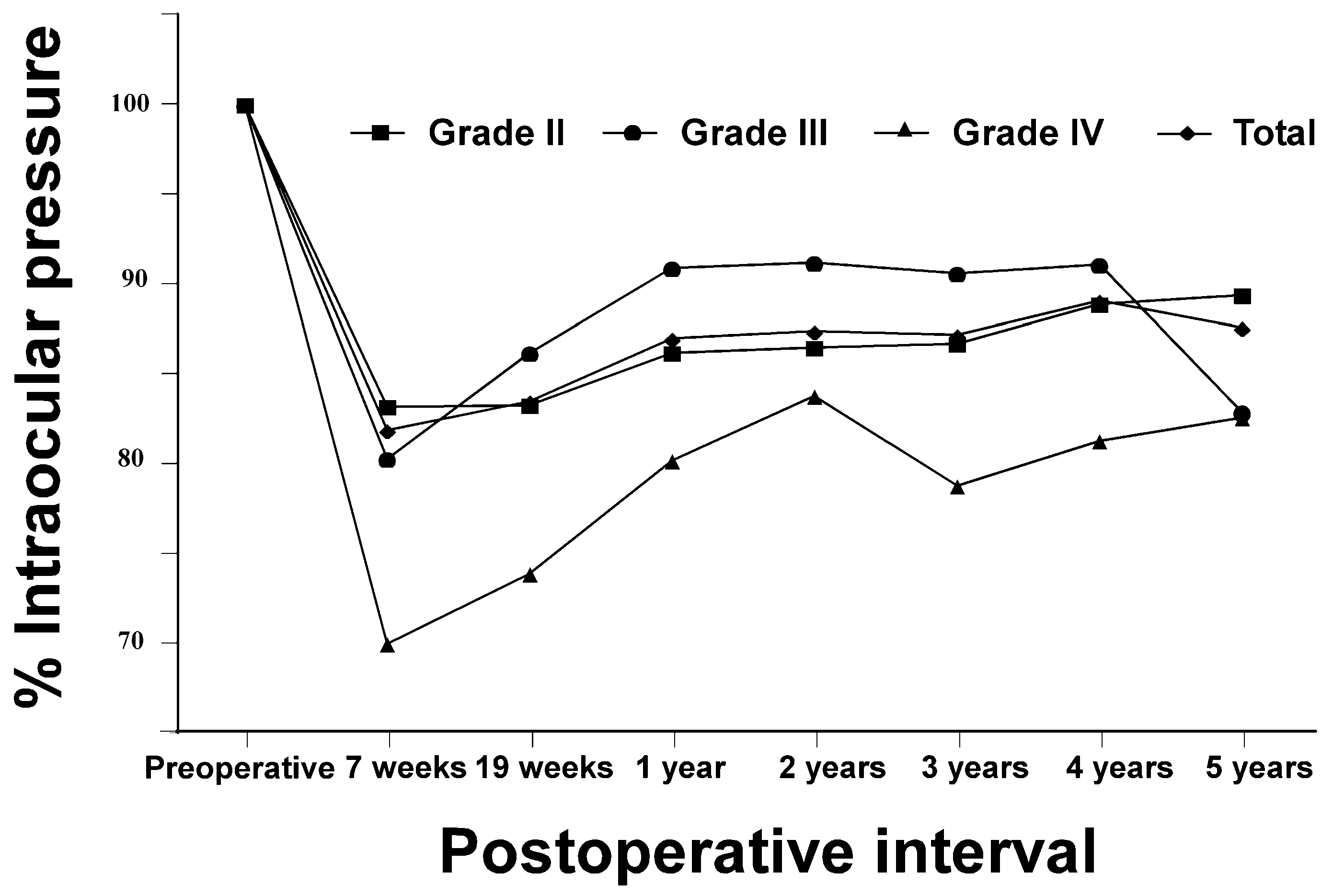

Table 1.
Preoperative characteristics and intraoperative parameters.
| Characteristics/Parameters | Grade II | Grade III | Grade IV | Total | P-value |
| Number of eyes | 62 (72%) | 20 (23%) | 4 (5%) | 86 | |
| Age (y) | 71.1 ± 7.8 | 73.4 ± 7.4 | 75.3 ± 5.7 | 71.8 ± 7.7 | 0.308⁎, 0.829⁎, 0.438⁎ |
| Sex Male | 22 (81%) | 4 (15%) | 1 (4%) | 27 | |
| Female | 40 (68%) | 16 (27%) | 3 (5%) | 59 | |
| Operative time (min) | 4.63 ± 0.88 | 5.48 ± 1.52 | 7.77 ± 1.47 | 4.97 ± 1.13 | 0.04†, 0.064⁎, < 0.01† |
| Phaco time (s) | 13.3 ± 4.2 | 18.3 ± 7.7 | 23.9 ± 6.4 | 15.0 ± 6.0 | 0.018†, 0.268⁎, 0.010† |
| Aspiration time (s) | 72.6 ± 14.1 | 84.3 ± 24.5 | 99.3 ± 5.7 | 76.5 ± 18.0 | 0.110⁎, 0.091⁎, 0.011†⁎ |
| CDE | 5.72 ± 1.89 | 8.42 ± 3.13 | 12.50 ± 3.97 | 6.67 ± 2.79 | < 0.01†, 0.109⁎, < 0.01† |
| Volume of fluid used (mL) | 27.8 ± 5.8 | 31.3 ± 9.6 | 39.3 ± 3.8 | 28.9 ± 7.2 | 0.208⁎, 0.091⁎, 0.010† |
Values are expressed as mean ± SD or numbers with percentages unless otherwise mentioned. P-values derived from the Steel-Dwass post hoc test are shown in the following order: Grade II vs. Grade III, Grade III vs. Grade IV, and Grade II vs. Grade IV. CDE, cumulative dissipated energy. ⁎No significant differences among groups (Kruskal-Wallis test followed by the Steel-Dwass post hoc test). †Significant difference among groups (Kruskal-Wallis test followed by Steel-Dwass post hoc test).
Table 2.
Pre- and post-operative BCVA and CECD values.
| Preoperative | 7 weeks postoperatively | 19 weeks postoperatively | P-value | |
| BCVA (logMAR) (n = 86) | 0.18 ± 0.10 | -0.065 ± 0.035 | -0.066 ± 0.034 | < 0.01†, 0.963⁎, < 0.01† |
| CECD (cells/mm2) (n = 32) | 2546.7 ± 261.9 | 2520.0 ± 238.7 | 2489.9 ± 284.3 | 0.894⁎, 0.983⁎, 0.741⁎ |
| CECD loss (%) | - | 0.6 ± 8.7 | 1.9 ± 8.3 | - |
Table 3.
Mean IOP (mmHg) and mean decrease in IOP (mmHg) over time.
| Mean IOP (Decrease) (n = 86) | ||||||||||
| Examination | Grade II | Grade III | Grade IV | Total | P-value | |||||
| Preoperative | 14.7 ± 2.5 | - | 14.5 ± 2.1 | - | 14.6 ± 1.5 | - | 14.6 ± 2.4 | - | 0.972⁎ | |
| 7 weeks | 12.2 ± 2.6 | (2.5 ± 1.7) | 11.6 ± 2.3 | (2.9 ± 1.9) | 10.3 ± 2.4 | (4.3 ± 1.1) | 12.0 ± 2.5 | (2.6 ± 1.7) | < 0.01† | 0.274⁎ |
| 19 weeks | 12.2 ± 2.5 | (2.4 ± 1.4) | 12.4 ± 1.9 | (2.1 ± 1.9) | 10.8 ± 2.2 | (3.7 ± 0.9) | 12.2 ± 2.3 | (2.4 ± 1.5) | < 0.01† | 0.466⁎ |
| 1 year | 12.6 ± 2.4 | (2.0 ± 1.0) | 13.2 ± 2.3 | (1.3 ± 1.5) | 11.7 ± 2.2 | (2.9 ± 1.5) | 12.7 ± 2.4 | (1.9 ± 1.2) | < 0.01† | 0.476⁎ |
| 2 years | 12.7 ± 2.5 | (2.0 ± 1.4) | 13.1 ± 1.7 | (1.4 ± 1.4) | 12.2 ± 1.3 | (2.4 ± 0.3) | 12.8 ± 2.3 | (1.9 ± 1.4) | < 0.01† | 0.646⁎ |
| 3 years | 12.7 ± 2.6 | (1.9 ± 1.2) | 13.1 ± 2.3 | (1.4 ± 1.6) | 11.5 ± 2.6 | (3.0 ± 1.9) | 12.8 ± 2.5 | (1.9 ± 1.4) | < 0.01† | 0.501⁎ |
| 4 years | 13.1 ± 2.7 | (1.6 ± 1.3) | 13.2 ± 2.1 | (1.4 ± 1.6) | 11.9 ± 1.6 | (2.7 ± 0.7) | 13.0 ± 2.6 | (1.6 ± 1.4) | < 0.01† | 0.639⁎ |
| 5 years | 13.1 ± 2.5 | (1.6 ± 1.2) | 12.0 ± 2.0 | (2.5 ± 1.7) | 12.0 ± 0.6 | (2.6 ± 1.0) | 12.8 ± 2.4 | (1.8 ± 1.4) | < 0.01† | 0.146⁎ |
Values represent mean ± SD. ⁎No significant differences among groups (one-way ANOVA). †Significant difference between preoperative and the corresponding postoperative time point (paired t-test).
Table 4.
Mean IOP (mmHg) and mean decrease in IOP (mmHg) of the two groups (preoperative IOP < 15 mmHg or > 15 mmHg).
Table 4.
Mean IOP (mmHg) and mean decrease in IOP (mmHg) of the two groups (preoperative IOP < 15 mmHg or > 15 mmHg).
| Mean IOP (Decrease) | ||||||
| Time point | Preoperative IOP > 15 mmHg group (n = 45) | P-value | Preoperative IOP < 15 mmHg group (n = 41) | P-value | ||
| Preoperative | 16.7 ± 1.2 | - | 12.7 ± 1.1 | - | ||
| 7 weeks | 13.9 ± 1.8 | (2.8 ± 1.9) | < 0.01† | 10.2 ± 2.3 | (2.5 ± 1.6) | < 0.01† |
| 19 weeks | 13.9 ± 1.6 | (2.8 ± 1.7) | < 0.01† | 10.6 ± 1.6 | (2.1 ± 1.3) | < 0.01† |
| 1 year | 14.7 ± 1.3 | (2.0 ± 1.2) | < 0.01† | 10.9 ± 1.7 | (1.7 ± 1.2) | < 0.01† |
| 2 years | 14.6 ± 1.5 | (2.2 ± 1.2) | < 0.01† | 11.1 ± 1.6 | (1.6 ± 1.4) | < 0.01† |
| 3 years | 14.6 ± 1.9 | (2.1 ± 1.4) | < 0.01† | 11.1 ± 1.7 | (1.6 ± 1.3) | < 0.01† |
| 4 years | 15.0 ± 1.7 | (1.7 ± 1.3) | < 0.01† | 11.2 ± 1.7 | (1.5 ± 1.4) | < 0.01† |
| 5 years | 14.6 ± 2.0 | (2.1 ± 1.6) | < 0.01† | 11.1 ± 1.1 | (1.6 ± 1.2) | < 0.01† |
Values represent mean ± SD. †Significant difference between preoperative and the corresponding time point (paired t-test).
Disclaimer/Publisher’s Note: The statements, opinions and data contained in all publications are solely those of the individual author(s) and contributor(s) and not of MDPI and/or the editor(s). MDPI and/or the editor(s) disclaim responsibility for any injury to people or property resulting from any ideas, methods, instructions or products referred to in the content. |
© 2025 by the authors. Licensee MDPI, Basel, Switzerland. This article is an open access article distributed under the terms and conditions of the Creative Commons Attribution (CC BY) license (http://creativecommons.org/licenses/by/4.0/).
Copyright: This open access article is published under a Creative Commons CC BY 4.0 license, which permit the free download, distribution, and reuse, provided that the author and preprint are cited in any reuse.


