

Submitted:
07 December 2025
Posted:
09 December 2025
You are already at the latest version
Abstract
Silent gastroesophageal reflux disease (GERD) may present without classic symptoms and instead manifest through respiratory and laryngeal signs. We report the case of a 27-year-old Middle Eastern Arab male with acute dry cough and hoarseness, unresponsive to antitussive therapy, ultimately diagnosed with silent GERD after significant improvement with proton pump inhibitor (PPI) therapy.
Keywords:
GERD
; cough
; silent GERD
; gastroesophageal reflux
Introduction
GERD typically presents with heartburn and regurgitation; however, some patients develop “silent GERD,” where extra-esophageal symptoms predominate [1]. Laryngopharyngeal reflux (LPR) can cause cough, hoarseness, and throat irritation without gastrointestinal complaints [2,3]. Misdiagnosis is common because such presentations resemble viral or allergic conditions. This case highlights the diagnostic value of recognizing reflux-related triggers and using a therapeutic PPI trial when GERD is suspected.
Case Presentation
A 27-year-old male presented with a one-week history of acute dry cough, worse at night and after meals. After three days, he developed hoarseness. He also reported mild rhinorrhea consistent with a viral upper respiratory infection.
He denied heartburn, regurgitation, abdominal pain, nausea, vomiting, or dysphagia.
The patient reported that eating chocolate consistently worsened his cough, especially nocturnally—consistent with chocolate’s known effect of relaxing the lower esophageal sphincter (LES) [4].
Prior Treatment
He used:
- Tussivan® antitussive syrup, 125 mL × 3 bottles,
- With no improvement.
Examination & Investigations
- Vitals: normal
- ENT: hoarseness, mild pharyngeal erythema
- Lungs: clear
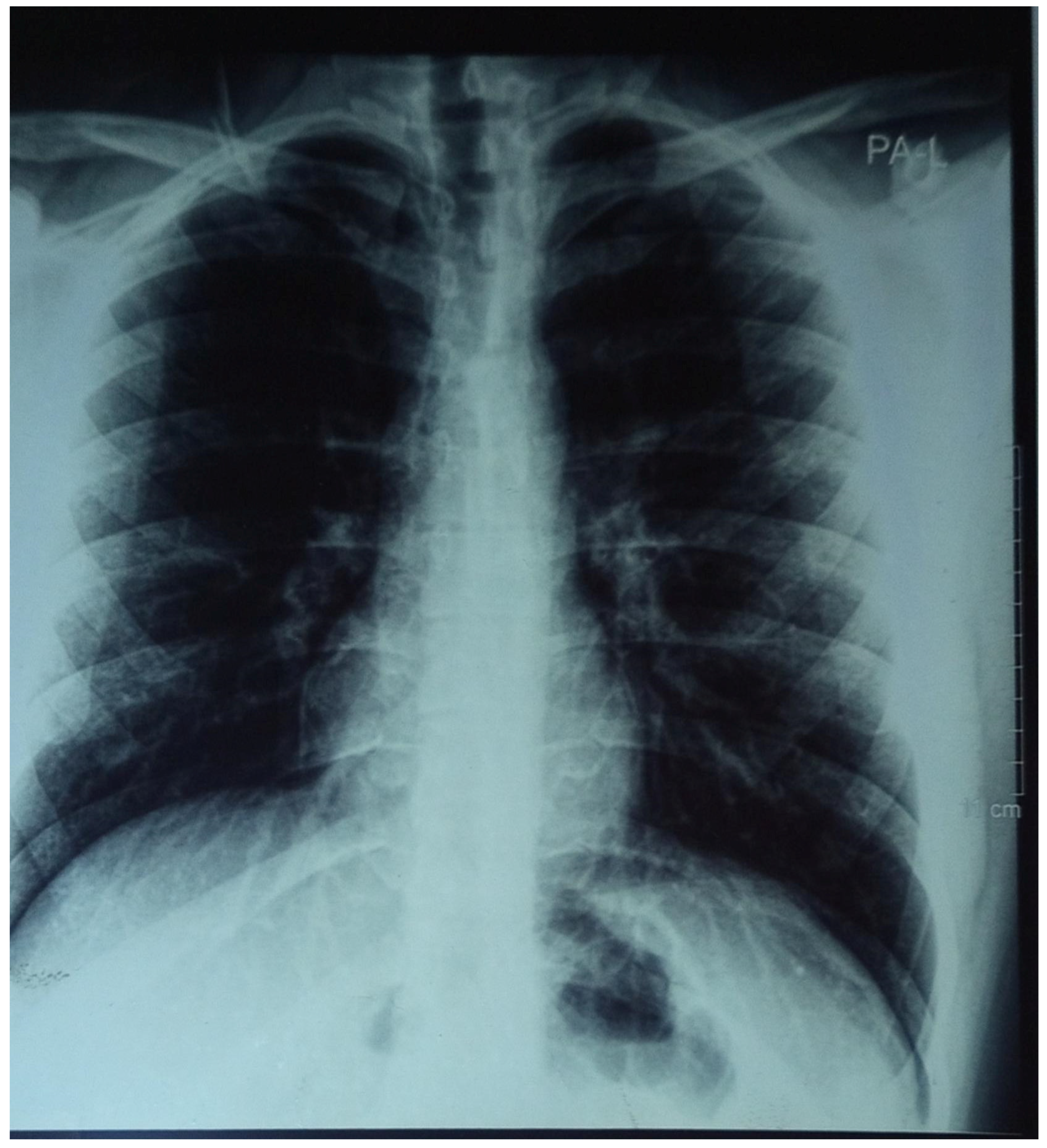
- Chest X-ray: normal ( see Figure 1)
- CBC: mild lymphocytosis
- CRP: normal
Given nighttime coughing, meal-related worsening, hoarseness, and no infectious findings, silent GERD / laryngopharyngeal reflux was suspected.
Management
Treatment consisted of:
Lansoprazole 30 mg once daily
Lifestyle modifications:
Avoid chocolate, caffeine, spicy foods
No meals 2–3 hours before sleep
Elevate head of bed
Smaller, frequent meals
Outcome
Within a few days of starting Lansoprazole, the patient reported:
- Marked improvement in cough
- Significant reduction of hoarseness
Complete symptom resolution occurred within 4 weeks. Poor response to antitussives and rapid improvement with PPI therapy strongly supported the diagnosis of silent GERD with LPR.
Discussion
Silent GERD may present exclusively with extra-esophageal symptoms, such as cough, throat clearing, or voice changes [2,5]. Reflux reaching the larynx causes mucosal inflammation, while esophago-bronchial reflexes also trigger cough [6].
Key diagnostic clues in this patient included:
- Nocturnal worsening
- Post-meal aggravation
- Chocolate-induced symptoms
- Hoarseness without infection
- Poor response to cough medications
- Dramatic response to PPI therapy
- Poor response to cough medications
Current evidence supports the use of empirical PPI trials for suspected extra-esophageal GERD, especially when other causes are excluded [7].
Conclusion
Silent GERD should be considered in patients with acute or subacute dry cough and hoarseness, particularly when symptoms are post-prandial or nocturnal and unresponsive to standard antitussive therapy. Therapeutic PPI trials are useful both diagnostically and therapeutically.
Patient Consent
The patient provided informed consent for publication.
Conflict of Interest
None declared.
References
- Vakil N, van Zanten SV, Kahrilas P, Dent J, Jones R; Global Consensus Group. The Montreal definition and classification of gastroesophageal reflux disease: a global evidence-based consensus. Am J Gastroenterol. 2006 Aug;101(8):1900-20; quiz 1943. PMID: 16928254. [CrossRef]
- Koufman JA. The otolaryngologic manifestations of gastroesophageal reflux disease (GERD): a clinical investigation of 225 patients using ambulatory 24-hour pH monitoring and an experimental investigation of the role of acid and pepsin in the development of laryngeal injury. Laryngoscope. 1991 Apr;101(4 Pt 2 Suppl 53):1-78. PMID: 1895864. [CrossRef]
- Cui N, Dai T, Liu Y, Wang YY, Lin JY, Zheng QF, Zhu DD, Zhu XW. Laryngopharyngeal reflux disease: Updated examination of mechanisms, pathophysiology, treatment, and association with gastroesophageal reflux disease. World J Gastroenterol. 2024 Apr 28;30(16):2209-2219. PMID: 38690022; PMCID: PMC11056915. [CrossRef]
- Zhang M, Hou ZK, Huang ZB, Chen XL, Liu FB. Dietary and Lifestyle Factors Related to Gastroesophageal Reflux Disease: A Systematic Review. Ther Clin Risk Manag. 2021 Apr 15;17:305-323. PMID: 33883899; PMCID: PMC8055252. [CrossRef]
- Irwin RS, Baumann MH, Bolser DC, Boulet LP, Braman SS, Brightling CE, Brown KK, Canning BJ, Chang AB, Dicpinigaitis PV, Eccles R, Glomb WB, Goldstein LB, Graham LM, Hargreave FE, Kvale PA, Lewis SZ, McCool FD, McCrory DC, Prakash UBS, Pratter MR, Rosen MJ, Schulman E, Shannon JJ, Hammond CS, Tarlo SM. Diagnosis and management of cough executive summary: ACCP evidence-based clinical practice guidelines. Chest. 2006 Jan;129(1 Suppl):1S-23S. PMID: 16428686; PMCID: PMC3345522. [CrossRef]
- Harding SM. Recent clinical investigations examining the association of asthma and gastroesophageal reflux. Am J Med. 2003 Aug 18;115 Suppl 3A:39S-44S. PMID: 12928073. [CrossRef]
- Vaezi MF, Hicks DM, Abelson TI, Richter JE. Laryngeal signs and symptoms and gastroesophageal reflux disease (GERD): a critical assessment of cause and effect association. Clin Gastroenterol Hepatol. 2003 Sep;1(5):333-44. PMID: 15017651.. [CrossRef]
Figure 1.
Posteroanterior (PA) chest radiograph showing clear lung fields with no evident focal consolidations, masses, or pleural effusion. The cardiac silhouette appears within normal size limits, and the mediastinal contours are unremarkable. The diaphragm is well defined bilaterally with no signs of subdiaphragmatic air. Overall, the radiograph demonstrates no acute cardiopulmonary abnormalities.
Figure 1.
Posteroanterior (PA) chest radiograph showing clear lung fields with no evident focal consolidations, masses, or pleural effusion. The cardiac silhouette appears within normal size limits, and the mediastinal contours are unremarkable. The diaphragm is well defined bilaterally with no signs of subdiaphragmatic air. Overall, the radiograph demonstrates no acute cardiopulmonary abnormalities.
Disclaimer/Publisher’s Note: The statements, opinions and data contained in all publications are solely those of the individual author(s) and contributor(s) and not of MDPI and/or the editor(s). MDPI and/or the editor(s) disclaim responsibility for any injury to people or property resulting from any ideas, methods, instructions or products referred to in the content. |
© 2025 by the authors. Licensee MDPI, Basel, Switzerland. This article is an open access article distributed under the terms and conditions of the Creative Commons Attribution (CC BY) license (http://creativecommons.org/licenses/by/4.0/).
Copyright: This open access article is published under a Creative Commons CC BY 4.0 license, which permit the free download, distribution, and reuse, provided that the author and preprint are cited in any reuse.


