Submitted:
04 December 2025
Posted:
05 December 2025
You are already at the latest version
Abstract
Nutraceuticals such as curcumin, resveratrol, lycopene, lutein, and coenzyme Q10 pos-sess strong antioxidant and anti-inflammatory activities but their practical use is hin-dered by poor solubility and bioavailability. Traditional nanocarriers like liposomes, nanoemulsions, and polymeric nanoparticles often rely on surfactants and synthetic or-ganic solvents that limit safety, scalability, and regulatory acceptance. The present study evaluated the Facilitated Self-Assembling Technology (FAST) platform as a clean-label al-ternative for generating bioavailable nutraceutical nanoparticles. Using only food-grade facilitating medium, FAST enabled spontaneous formation of stable, amorphous nano-particles with strong negative surface charge and high colloidal stability. Hybrid nano-particles combining epigallocatechin-3-gallate-palmitates (EC16), curcumin, and resvera-trol further improved surface charge, reduced size range, and exhibited enhanced stability under simulated gastric conditions. All formulations demonstrated excellent biocompati-bility in XTT assays, with no reduction in viability compared to control. Fluorescent im-aging of EC16/Cy5 fluorescent hybrid nanoparticles confirmed nanoparticle–cell surface interactions without cytotoxicity. Compared with chemical conjugation and lipid-based nanoencapsulation, FAST offered faster, surfactant-free, and energy-efficient production, fully compliant with FDA generally recognized as safe (GRAS) standards. These results support the FAST platform as an efficient, economical, and scalable nanotechnology for next-generation functional beverages and oral nutraceutical delivery systems that meet both regulatory and consumer demands for natural, sustainable innovation.
Keywords:
nutraceuticals
; beverages
; FAST
; EGCG-palmitate
; curcumin
; CoQ10
; lycopene
; lutein
; resveratrol
; rapamycin
1. Introduction
Nutraceuticals, defined as biologically active compounds derived from foods or botanical sources, have become a cornerstone of modern preventive medicine, offering diverse health benefits ranging from antioxidant, anticancer, and anti-inflammatory protection to neuro- and cardiometabolic support [1,2,3]. Nutraceuticals are differentiated from nutrients by providing therapeutic or prophylactic benefits beyond basic nutrition. Epidemiological and clinical studies consistently demonstrate that diets rich in bioactive phytochemicals correlate with reduced risk of cardiovascular disease, metabolic syndrome, cancer, and neurodegenerative disorders [1,2,3,4]. Driven by consumer awareness of wellness and the shift from treatment to prevention, the global nutraceutical market exceeded USD 540 billion in 2022 and is projected to surpass USD 1 trillion by 2030 [3,4,5]. Among the many studied bioactives, epigallocatechin-3-gallate (EGCG), curcumin, resveratrol, lycopene, lutein, and coenzyme Q10 (CoQ10) are particularly notable for their antioxidant, anti-inflammatory, and signaling-modulatory effects [5,6,7,8,9,10].
However, many nutraceuticals are hydrophobic, and the practical application of most lipophilic nutraceuticals, such as curcumin, resveratrol, lycopene, lutein, and coenzyme Q10 (CoQ10), is hindered by physicochemical and pharmacokinetic limitations, including poor aqueous solubility, rapid metabolism, and instability under physiological condition, resulting in limited intestinal absorption following oral consumption and rapid systemic clearance.
Curcumin in its native form, for example, yields plasma concentrations below 50 ng/mL even after gram-level doses, because of rapid metabolism [11,12,13,14]. Resveratrol exhibits high intestinal absorption but <1 % systemic bioavailability due to first-pass metabolism [6,7,8]. Lycopene typically achieves Cₘₐₓ values of only 0.6–1.2 µM [15,16], while lutein and CoQ10 remain in the nanomolar range even after supplementation [17,18,19,20,21]. Consequently, striking in-vitro efficacy seldom translates in vivo, underscoring the need for formulations that enhance solubility, stability, and permeability.
Recently, nanotechnology has emerged as a transformative approach for improving the pharmacokinetics and functionality of nutraceuticals, and nanoscale delivery systems now underpin many strategies to enhance dispersion, stability, and uptake of hydrophobic active biologics. These systems provide a larger surface area and improved protection against degradation, extending biological residence time and increasing absorption [22,23,24]. Various nano systems—including solid lipid nanoparticles (SLN), nanostructured lipid carriers (NLC), liposomes, and nanoemulsions—have been widely explored to increase dispersion, protect against degradation, and promote absorption. SLNs are composed of solid lipid matrices that encapsulate lipophilic compounds and enable controlled release; they are fabricated through hot or cold high-pressure homogenization, ultrasonication, or microemulsion techniques [4,22,23]. NLCs combine solid and liquid lipids to increase drug loading and minimize expulsion during crystallization [24]. Liposomes, formed from phospholipid bilayers, can entrap both hydrophilic and hydrophobic compounds and are typically produced by thin-film hydration, reverse-phase evaporation, ethanol injection, or lipid-film rehydration [22]. Nanoemulsions—surfactant-stabilized oil-in-water dispersions—are obtained via high-speed shearing, ultrasonication, phase-inversion temperature, or high-pressure homogenization [22,25]. Collectively these approaches have advanced nutraceutical delivery by several orders of magnitude, enabling inclusion of otherwise insoluble active ingredients in food, beverages, and supplements.
Nevertheless, conventional nanotechnologies rely heavily on synthetic surfactants and organic solvents, and therefore suffer from inherent weaknesses that constrain large-scale, food-grade deployment. Their dependence on synthetic surfactants (e.g., polysorbates, Spans, sodium dodecyl sulfate) and organic solvents (i.e. acetone, chloroform) introduces potential toxicity, irritancy, and environmental concerns [4,22,23]. Certain solvent residues are incompatible with “clean-label” expectations and may violate regulatory limits. Energy-intensive manufacturing processes such as ultrasonication and high-pressure homogenization can also degrade heat- or oxidation-sensitive nutraceuticals, lowering bioactivity. Stability remains another major issue: SLNs and NLCs undergo polymorphic transitions and aggregation, liposomes require precise lipid ratios and hydration control, and nanoemulsions experience Ostwald ripening and phase separation. These complications could shorten shelf life and inflate production costs. From a regulatory perspective, agencies such as the Food and Drug Administration (FDA) and European Food Safety Authority (EFSA) require dedicated safety assessments and explicit labeling of engineered nanomaterials, limiting commercial acceptance of systems employing non-GRAS excipients [1,3].
Recognizing these obstacles, researchers have sought greener and simpler nanotechnologies employing fully food-grade materials. The Facilitated Self-Assembling Technology (FAST) platform represents one such innovation [26]. FAST is a surfactant-free nanotechnology that enables spontaneous formation of nanoscale complexes (i.e. micelles) entirely from Generally Recognized As Safe (GRAS) material. Through finely balanced non-covalent interactions such as hydrogen bonding, hydrophobic association, and van der Waals forces, FAST promotes molecular self-organization in liquid media under mild conditions. Unlike conventional systems that demand mechanical energy or surfactants, FAST relies on intrinsic amphiphilicity and charge distribution of its components to drive assembly into uniform nanoparticles typically ranging between 50–200 nm in diameter.
This self-assembly is conceptually analogous to micelle formation yet proceeds without surfactants or significant external energy. The resulting nanoparticles possess narrow size distribution and remarkable colloidal stability, making them well suited for nutraceutical, cosmetic, and pharmaceutical use. As the process occurs entirely in a liquid environment and all constituents are food-grade, the platform eliminates the toxicological and environmental concerns associated with traditional nanocarriers.
Figure 1 illustrates the self-assembly mechanism of the FAST process, in which amphiphilic nutraceutical molecules and GRAS material(s) interact via hydrophobic and hydrogen-bonding forces to form nanoscale particle/micelle structures under liquid, surfactant-free conditions. The pathway involves initial molecular association, nucleation of nanoclusters, and spontaneous growth into stable particles without high-energy input. This gentle, economical yet efficient mechanism preserves bioactivity and allows reproducible large-scale production using simple mixing steps under either isothermal or polythermal conditions.
We hypothesize that the FAST platform provides a safe, economical, and efficient method for converting nutraceutical compounds into aqueous-miscible, bioavailable nanoparticles with broad application. In this study, we evaluated the suitability of FAST-generated nanoparticles prepared from widely studied nutraceuticals—including EC16, curcumin, resveratrol, lycopene, lutein, and CoQ10—and their hybrid formulations for potential oral delivery. The nanoparticles were characterized for particle size, density, surface charge (zeta potential), crystalline structure, cytotoxicity, stability under acidic conditions, and cellular interaction by fluorescent imaging.
2. Materials and Methods
2.1. Compounds and Reagents
Epigallocatechin-3-gallate-palmitate (EC16) was obtained from Camellix, LLC (Evans, GA, USA). Curcumin (98%) was purchased from Belle Chemical (Billings, MT, USA). CoQ10, lycopene, and lutein were obtained from Bulksupplements.com (Henderson, NV, USA). Resveratrol was purchased from CurEase (McEwen, TN, USA). Rapamycin (sirolimus, >99%) was supplied by MedKoo Biosciences (Durham, NC, USA). Cy5 hydrazide (non-sulfonated) was purchased from ApexBio Technology (Houston, TX, USA).
2.2. Cells and Culture Medium
The RAW264.7 murine macrophage cell line (ATCC TIB-71™) was obtained from the American Type Culture Collection (Manassas, VA, USA). Dulbecco’s Modified Eagle’s Medium (DMEM; ATCC 30-2002) was used for culture, supplemented with 10% fetal bovine serum (Neuromics, Edina, MN, USA) and 1% penicillin-streptomycin-amphotericin B solution (100×; Corning, Glendale, AR, USA).
2.3. Preparation of Nanoparticles
Nanoparticles of individual and hybrid nutraceutical compounds were prepared using the proprietary FAST (Facilitated Self-Assembling Technology) method (patent pending). Nanoparticle stock suspensions (0.1–1%) were stabilized in pure glycerol and subsequently diluted with sterile, double-distilled water.
EC16/Cy5 hybrid fluorescent nanoparticles were generated by mixing 100 mg EC16 with 1 mg Cy5 to yield a hybrid nanoparticle stock containing 2% EC16 and 0.02% Cy5. The stock suspension in glycerol was diluted 50-fold to 0.01% EC16 and 0.0001% Cy5 for evaluation. The suspension was purified by centrifugation at 13,400 rpm for 45 minutes, washed twice with purified water, and centrifuged again before resuspension in cell culture medium. All other hybrid nanoparticles were prepared by pre-mixing two or three nutraceuticals prior to the FAST process.
2.4. Dynamic Light Scattering (DLS) Analysis
Nanoparticle tracking analysis was performed according to a method described previously [26,27]. Nanoparticle size distribution and concentration were measured using the ZetaView ×20 Nanoparticle Tracking Analyzer (Particle Metrix, Meerbusch, Germany). The measuring range for particle diameter is 10–2000 nm. Samples were diluted in 1× PBS and analyzed at 23 °C, sensitivity 70, frame rate 30 fps, and shutter speed 100. Measurements were collected from 11 positions with two reading cycles. Post-acquisition parameters were: minimum brightness 20, area range 10–1000, and trace length 15. The ZetaView potential was profiled from 11-position measurements at 23 °C by diluting the nanoparticles in 5% PBS with low conductivity (< 1000 μS/cm).
2.5. X-Ray Diffraction
Characterization of Semi-liquid samples was performed by Bruker D6 Phaser X-ray diffraction using an X-ray diffractometer with CuKα radiation (λ = 0.154060 nm), measured (10-30◦) range of 2θ angle, operating at 20 kV accelerating voltage with 15 sample rotation/min and 166.5 mm radius of the compact goniometer. The Cu source was operated at 40kV/30mA, while the divergence was controlled with a 0.2 mm slit and a 2.5° axial Soller. The LYNXEYE XE-T detector was used in high count rate mode with a 5° detector opening. Additionally, a 0.2 mm Ni filter to surpress Kß diffraction peaks was positioned in the beam path. The Universal stage was selected to mount the sample. The measurement was planned using the WIZARD plugin of DIFFRAC.MEASUREMENT.
2.6. Cell Viability (XTT) Assay
One day before each experiment, RAW264.7 cells were seeded in 96-well plates containing 100 μL/well of cell culture medium and the cells incubated overnight in a 37 °C incubator with 5% CO2. The NPs of the nutraceuticals were diluted with cell culture medium (no serum) and applied in triplicate to the wells. After incubation for 60 minutes, the XTT assay was performed using CyQUANT™ XTT Cell Viability Assay kit according to the manufacturer’s protocol (Thermo Fisher Scientific Inc., Waltham, MA).
2.7. Stability Under Acidic Conditions
To test the stability of the nanoparticles in acidic conditions similar to stomach acid (pH 2), The nanoparticles were diluted from the stock to 0.1% phosphate buffer saline (PBS) suspension. The nanoparticles suspension was then added to a HCl/water solution with pH 1.84 at 1:9 ratio, and incubated at 37 oC for 0 and 60 min before neutralization with 10 X PBS. The resulting 0.001% nanosuspensions and untreated 0.001% nanoparticles in PBS were subjected to ZetaView evaluation of particle size and density.
2.8. Fluorescent Imaging of EC16/Cy5 Nanoparticle-Cell Interaction
EC16/Cy5 hybrid nanoparticles (1 ml, 1% EC16, 0.01% Cy5) were subjected to two rounds of centrifugation at 13,400 rpm for 45 minutes each and washed with distilled water. The final pellets were resuspended 1:9 (0.1% EC16, 0.001% Cy5) in DMEM and applied to RAW264.7 monolayers grown on chamber slides. After 1 or 2 hours of incubation, cells were washed twice with cold PBS, fixed in formalin, and imaged using a KEYENCE BZ-X810 all-in-one fluorescence microscope (KEYENCE Corp., Osaka, Japan).
For control preparation, Cy5 nanoparticles (1 ml, 0.01% Cy5) were processed using the identical centrifugation and washing protocol. Because of their low density, Cy5 nanoparticles were effectively removed during the wash steps, resulting in no visible pellet. One tube containing the post-wash Cy5 sample was diluted in complete DMEM as a negative control. This control confirmed that only EC16/Cy5 hybrid nanoparticles—free of isolated Cy5 nanoparticles—were applied to the cell culture. Any Cy5 nanoparticles formed during hybrid nanoparticle preparation were eliminated through differential centrifugation and washing.
3. Results
3.1. Nanoparticles Formation
All five nutraceuticals (Curcumin, CoQ10, lycopene, lutein, Resveratrol) were successfully converted into nanoparticles using the FAST method (Table 1). Nutraceutical nanoparticles in size and density, as well as Zeta potential at 0.01% in distilled water. EC16 and rapamycin (a pharmaceutical) nanoparticles were used as references. Two nutraceutical nanoparticles, procyanidin B2 and quercetin, are listed as high surface charge nanoparticles.
To improve the stability of the top five nutraceutical nanoparticles, an adjusted formulation with a commonly used food additive was applied to increase the surface charges. Adjusted formulations improved surface charge uniformity, with Zeta potentials ranging from −32.6 to −45.7 mV (Table 2), compared that of the initial batch from -24.54 to -33.72 (Table 1).
To determine if the nutraceuticals could form stable hybrid nanoparticles, dry powder of EC16, curcumin, and resveratrol were pre-mixed to form EC16-curcumin, EC16-resveratrol, and EC16-curcumin-resveratrol hybrid nanoparticles by FAST method. In addition, the three independently formed nanoparticle stocks were mixed as combination nanoparticle stocks. Table 3 shows the size, density and Zeta potential of the hybrid nanoparticles and the combination of the independent nanoparticles. The EC16/Cy5 fluorescent hybrid nanoparticles were listed as a reference.
3.2. X-ray Diffraction (XRD)
A total of nine nanoparticle stocks (semi-liquid) underwent X-ray diffraction analysis to determine if the nanoparticles form crystals. XRD patterns confirmed that all nine tested nanoparticle formulations were amorphous (Figure 2), indicating absence of crystalline structures, consistent with successful nanoparticle formation by the FAST process.
3.3. Cell Viability
To determine potential cytotoxicity of the nutraceutical nanoparticles, nine different nanoparticles were incubated with RAW264.7 cells for 60 minutes before XTT assay. The results indicated there was no significant cytotoxicity in RAW264.7 cells exposed to nine different nanoparticle formulations for 60 minutes (Figure 3). Absorbance at 450–660 nm demonstrated cell viability comparable to controls, supporting the biocompatibility of the FAST-generated nanoparticles.
3.4. Stability under Acidic Conditions
Under acidic conditions but without incubation, the curcumin nanoparticle suspension has a median size was 165.0 ± 111 nm. The concentration was measured at 2.0 x 10⁷ particles/ml. After 60 minutes incubation the concentration of curcumin nanoparticles decreased to 8.4 x 10⁶, with a median size of 130 ± 78. For resveratrol nanoparticles, a 60 minute incubation reduced the concentration from 1.1 x 10⁹ to 5.6 x 10⁸. In contrast, the EC16-curcumin-resveratrol trio hybrid nanoparticle concentration was only reduced from 3.0 x 10⁸ to 2.6 x 10⁸, and the mixed nanoparticles were reduced from 1.3 x 10⁸ to 3.1 x 10⁷. The median size of the nanoparticles remained similar before and after incubation (Table 4). That is, after 60 minutes of exposure to pH 1.84, curcumin, resveratrol, hybrid, and mixed nanoparticles retained similar median sizes, though particle densities decreased by approximately 30–60% (Table 4).
3.5. EC16/Cy5 and Cy5 Nanoparticles
The hybrid nanoparticles have a mean size of 141 ± 86.8, range from 40 to 157 nm. The nanoparticle density is 6.1 x 10¹⁰/ml, with Zeta potential of -43.1 ± 1.13 mV. In comparison, the EC16 nanoparticles has a mean size of 152.5 nm, range from 95 to 218 nm, with Zeta potential of -60.11 mV [26]. The results suggest that EC16/Cy5 nanoparticles are different from EC16 nanoparticles in particle size, range, and Zeta potential. Thus, EC16/Cy5 nanoparticles are a new class of nanoparticles with fluorescence. The stability appeared less than that of EC16 nanoparticles but was still considered stable (>30 mV).
The Cy5 nanoparticles were made by using 1 mg of Cy5 powder, resulting in 1 mg of Cy5 in 5 ml glycerol stock (0.02%). The glycerol stock was diluted in water 50 x to make 0.0005% water suspension, before particle evaluation. At this concentration, the particle density is 2.5 x 10⁸/ml with Zeta potential of -20.42 ± 1.10 mV. The mean particle size is 204.6 ± 201.3 nm. It suggests that Cy5 nanoparticles are significantly bigger in size and low in surface charge compared to EC16/Cy5 nanoparticles.
3.6. EC16/Cy5 Hybrid Nanoparticles Incubated with RAW264.7 Cells
To evaluate the suitability of EC16/Cy5 hybrid nanoparticles for fluorescent labeling and their potential use in in vitro and in vivo studies, purified EC16/Cy5 nanoparticles—free of any residual Cy5 nanoparticles generated during hybrid nanoparticle formation—were incubated with RAW264.7 cells for 1 and 2 hours. As shown in Figure 3, the red fluorescence intensity associated with the cells increased markedly from 1 to 2 hours. Although some diffuse extracellular fluorescence was observed, most of the signal localized adjacent to the cell surface. In contrast, the control sample exhibited no detectable Cy5 fluorescence, confirming that Cy5 nanoparticles were effectively removed during the centrifugation and washing steps.
4. Discussion
Nanotechnology has emerged as a transformative approach to improve the bioavailability, stability, and targeted delivery of nutraceuticals such as curcumin, EGCG, carotenoids, and omega-3 fatty acids. Various nanostructures—micelles, nanoliposomes, and nanoemulsions—can encapsulate bioactives, protect them from oxidation and degradation, and enhance absorption efficiency, thereby converting everyday products such as yogurts, juices, and dairy-based drinks into precision-nutrition platforms [28,29].
In beverage formulations, nanotechnology enables the incorporation of poorly soluble or unstable bioactives into transparent, stable dispersions. Nanoemulsions and lipid nanoparticles can deliver antioxidants, essential oils, and vitamins with superior solubility and stability [29]. For instance, curcumin nanoemulsions protect against oxidative degradation in dairy beverages, while nanoliposomes stabilize omega-3 fatty acids in fortified juices. Future trends point to “smart beverages” that employ nanocarriers for controlled nutrient release tuned to metabolic or circadian rhythms—integrating wellness, personalization, and convenience into a single platform.
Despite the potential, key challenges persist. The nanoscale size of encapsulated particles raises safety questions regarding gastrointestinal absorption, systemic bioaccumulation, and long-term toxicity [29]. Current evidence on chronic exposure remains limited, and the metabolic fate of many emulsifiers and lipids used in nanoencapsulation is not fully understood. Global regulations for food nanomaterials remain inconsistent and lack standardized testing protocols, slowing commercialization. GRAS-based methods such as FAST may overcome these barriers by ensuring biocompatibility and transparency in labeling.
The results from the current study confirmed that all key nutraceuticals successfully formed stable nanoparticles (Table 1, Table 2 and Table 3). While this was anticipated, the initial preparations showed that the surface charges of the five key nutraceuticals—curcumin, resveratrol, lycopene, lutein, and CoQ10—were relatively modest, with Zeta potentials ranging from −24.54 mV to −33.72 mV (Table 1). After formulation optimization, the Zeta potentials improved to a stronger range of −32.6 mV to −45.7 mV without significant changes in median particle size (Table 2).
Furthermore, these nutraceuticals were capable of self-assembling into hybrid nanoparticles composed of two or three compounds (Table 3). Interestingly, hybridization resulted in further increases in surface charge magnitude and reductions in median size to below 100 nm (Table 3). At 0.01%, particle densities ranged from 5.7 × 10⁹ to 2.5 × 10¹⁰ particles/ml, suggesting a significant increase in efficiency. In addition, the surface charge of the hybrid nanoparticles exceeded -50 mV (Table 3). Although combined nanoparticles also exhibited enhanced surface charges, their particle densities remained within the 10⁹ range (Table 3). These findings indicate that formulation optimization can be readily achieved through multiple simple strategies.
X-ray diffraction analysis demonstrated that all nutraceutical nanoparticles, their hybrids, and rapamycin nanoparticles were amorphous, with no detectable crystalline structures (Figure 2). This amorphous state suggests the absence of potential toxicity associated with crystalline aggregates. To further assess cytocompatibility, XTT cell viability assays were performed. As shown in Figure 3, the mock-treated control (media changes) produced an XTT absorbance of 3.1, whereas all nanoparticle and hybrid nanoparticle treatments yielded values between 3.3 and 3.8. No significant differences were observed among treatments, except that rapamycin and EC16/curcumin hybrid nanoparticles showed significantly higher XTT values than the control (Figure 3), indicating increased metabolic activity from mitochondrial dehydrogenase. These results are consistent with our in vivo findings in mice fed 0.02% EC16 as the sole water source for 3 or 6 weeks (data not shown, manuscript in preparation). Despite the GRAS status of all tested nutraceutical compounds, additional acute and chronic safety evaluations are warranted.
We previously evaluated the stability of EC16 nanoparticles under acidic conditions mimicking gastric pH ≈ 1.9 [30]. The present acidic-stability test on curcumin, resveratrol, and EC16/curcumin/resveratrol hybrid nanoparticles showed that post-treatment particles (60 min incubation at 37 °C) retained comparable sizes to controls (time 0) in distilled water (pH 1.84; Table 4). However, 42% of curcumin and 51% of resveratrol nanoparticles remained intact after 60 min, while 87% of hybrid nanoparticles retained their integrity. When pre-mixed, non-hybridized nanoparticles were exposed to the same acidic environment, only 24% remained intact (Table 4). These results suggest that individual nutraceutical nanoparticles exhibit distinct release rates during gastric transit under empty stomach conditions, whereas hybrid nanoparticles provide enhanced stability and deliver the most intact particles to the small intestine for absorption. This observation supports the potential of FAST-generated hybrid nanoparticles as optimized oral delivery systems for multi-nutrient formulations.
Therefore, the GRAS FAST-generated curcumin nanoparticles could achieve higher systemic exposure while avoiding surfactant-related gastrointestinal irritation [11. 14, 23]. Similarly, FAST-generated resveratrol nanoparticles could maintain sustained plasma levels without glucuronidation-driven rapid clearance [6,8,19,20], and FAST-generated CoQ10 nanoparticles could display enhanced absorption and tissue retention compared to oil suspensions [19,31]. Such outcomes highlight the FAST technology’s capacity to overcome both dissolution and permeability barriers through purely physical nano-structuring and warrant in vivo testing.
Successful self-assembly of EC16/Cy5 fluorescent hybrid nanoparticles avoided the need for covalent dye conjugation, which could alter EC16 bioactivity. During the self-assembly process, Cy5 may have formed minor nanoparticle aggregates, which were removed using differential centrifugation; the larger, lower-density Cy5 aggregates were separated during washing (Figure 3, Control). Fluorescence microscopy revealed time-dependent increases in Cy5 fluorescence surrounding RAW264.7 cells between 1 h and 2 h incubation (Figure 4), indicating nanoparticle–cell surface interaction without compromising cell integrity, consistent with the cytotoxicity results (Figure 3). This finding also explains our separate observation of persistent fluorescence in mouse brain tissue 24 hours following oral gavage of EC16/Cy5 hybrid nanoparticles (data not shown, manuscript in preparation). The FAST-based self-assembling hybrid method therefore provides a versatile and non-covalent labeling tool for studying biodistribution and cellular interactions of nutraceutical or pharmaceutical nanoparticles.
In comparison, traditional nanocarriers—such as liposomes, nanoemulsions, engineered micelles, and polymeric nanoparticles—often require synthetic polymers, surfactants, emulsifiers, and toxic organic solvents, which increase toxicity risks and manufacturing costs. In contrast, the FAST platform uses only natural, food-grade ingredients in a single-step, low-energy liquid process, producing nanoparticles that are stable, amorphous, and highly bioavailable.
A recently published clinical pharmacokinetic study comparing four curcumin formulations—AOV (801 and 811, >600-mg capsules), Longvida (SLN), and NovaSOL (polysorbate-based micelle nanoparticles)—demonstrated that all formulations produced serum curcumin conjugate levels exceeding 10 nM. However, serum concentrations of unconjugated (biologically active) curcumin remained below 2 nM (0.74 ng/mL) for all products except NovaSOL, which reached up to 38 nM (13.1 ng/mL). Even so, these levels were still approximately 100-fold lower than the concentrations typically required to elicit biological effects in vitro [32]. Thus, additional efforts are needed to identify more effective delivery strategies.
Table 5 provides a side-by-side comparison of three representative methods for producing curcumin nanoparticles as a model for nutraceutical nanoparticle preparation. This comparative analysis clearly demonstrates the multiple advantages of the FAST platform, including:
(1) avoidance of toxic reagents and production of biodegradable nanoparticles with no chemical residues, fully compliant with FDA GRAS standards;
(2) strong surface charge and high stability under acidic and thermal conditions, making it suitable for incorporation into beverages and functional foods;
(3) a simpler, faster, and more economical process requiring only conventional laboratory or food-processing equipment, thereby minimizing energy consumption and waste; and
(4) a natural, surfactant-free process that enhances regulatory acceptance and consumer trust.
As a result, the FAST design can be seamlessly integrated into existing beverage production lines without specialized equipment, offering excellent scalability, reproducibility, and sustainability for global functional food and nutraceutical manufacturing.
In summary, FAST aligns with contemporary consumer and regulatory trends emphasizing “clean-label” and sustainable production. Its surfactant and emulsifier-free, GRAS-based process drastically reduces the environmental footprint compared with surfactant-dependent methods. The one-step, low-energy process also facilitates industrial scalability and cost control. From a materials-science perspective, FAST’s self-assembly demonstrates a biomimetic paradigm, mimicking natural colloidal structures such as casein micelles and lipoprotein complexes while preserving complete biocompatibility.
Collectively, these characteristics position FAST as a bridge between pharmaceutical-grade nanodelivery and food-grade nutraceutical manufacturing. We reported previously that FAST platform generated EC16 nanoparticles can be applied to antimicrobial formulations against respiratory virus and norovirus infections [30,35,36,37]. By achieving nanoscale micelle structures through natural self-organization, the platform unites efficiency, safety, and regulatory simplicity in a single system. Its adaptability allows formulations of diverse hydrophobic, amphiphilic, and even hydrophilic actives, such as dopamine and gamma-aminobutyric acid (GABA), opening opportunities for multifunctional formulations addressing oxidative stress, inflammation, metabolic imbalance, and cognitive decline with significantly lower dosages (milligrams vs. grams). Furthermore, the modular nature of FAST supports co-delivery of synergistic compounds or controlled release through hybrid technique in assembly conditions.
5. Conclusions
In conclusion, the development of all GRAS FAST platform marks a significant evolution in nutraceutical formulation science. Through gentle GRAS self-assembly of hydrophobic nutraceuticals, FAST overcomes long-standing challenges of solubility, stability, and bioavailability while meeting modern safety and environmental expectations. Continued research should focus on molecular-level characterization of the self-assembly process, structure–function correlations, in vivo biodistribution profiles, acute and chronic toxicity studies, and comparative clinical pharmacokinetics against existing nanocarrier data. Such investigations will establish FAST as a universal, scalable platform capable of transforming the efficacy, sustainability, and consumer acceptance of next-generation nutraceuticals.
Author Contributions
Conceptualization, S.H. and D.D.; methodology, S.H., J.C., A.K.S., M.C., M. K., C.D., A.M., A.J., W.J.; graphics, S.H.; validation, D.G., A.K.S., and M.K.; formal analysis, D.D.; investigation, S.H.; resources, X.X.; data curation, X.X.; writing—original draft preparation, S.H.; writing—review and editing, D.D.; visualization, S.H.; supervision, S.H.; project administration, S.H. All authors have read and agreed to the published version of the manuscript.
Funding
This research received no external funding.
Data Availability Statement
The data presented in this study are available on request from the corresponding author due to pending patent applications.
Acknowledgments
None.
Conflicts of Interest
The authors declare no conflicts of interest.
Abbreviations
The following abbreviations are used in this manuscript:
| GRAS FDA |
Generally Recognized as Safe Food and Drug Administration |
| FAST | Facilitated Self-assembling Technology |
| EC16 | Epigallocatechin-3-Gallate-Palmitate |
| CoQ10 Cy5 SLN NLC DLS EFSA DMSO HCl EDC DMAP |
Coenzyme Q10 Cy5 hydrazide (non-sulfonated) Solid lipid nanoparticles Nanostructured lipid carriers Dynamic Light Scattering European Food Safety Authority Dimethyl Sulfoxide Hydrochloric Acid 1-Ethyl-3-(3-dimethylaminopropyl)carbodiimide 4-Dimethylaminopyridine |
References
- Jampilek J, Kos J, Kralova K. Potential of nanomaterial applications in dietary supplements and foods for special medical purposes. Nanomaterials. 2019, 9, 296. [Google Scholar] [CrossRef]
- Vesely O, Baldovska S, Kolesarova A. Enhancing bioavailability of nutraceutically used resveratrol and other stilbenoids. Nutrients. 2021, 13, 3095. [Google Scholar] [CrossRef]
- Puri V, Nagpal M, Singh I, et al. A comprehensive review on nutraceuticals: therapy support and formulation challenges. Nutrients. 2022, 14, 4637. [Google Scholar] [CrossRef]
- Ashfaq R, Rasul A, Asghar S, et al. Lipid nanoparticles: an effective tool to improve the bioavailability of nutraceuticals. Int J Mol Sci. 2023, 24, 15764. [Google Scholar] [CrossRef] [PubMed]
- Hoti G, Matencio A, Rubin Pedrazzo A, et al. Nutraceutical concepts and dextrin-based delivery systems. Int J Mol Sci. 2022, 23, 4102. [Google Scholar] [CrossRef] [PubMed]
- Almeida L, Vaz-da-Silva M, Falcão A, et al. Pharmacokinetic and safety profile of trans-resveratrol in a rising multiple-dose study in healthy volunteers. Mol Nutr Food Res. 2009, 53, S7–S15. [Google Scholar]
- Vaz-da-Silva M, Loureiro AI, Falcão A, et al. Effect of food on the pharmacokinetic profile of trans-resveratrol. Int J Clin Pharmacol Ther. 2008, 46, 564–570. [Google Scholar] [CrossRef]
- Nunes T, Almeida L, Rocha JF, et al. Pharmacokinetics of trans-resveratrol following repeated administration in healthy elderly and young subjects. J Clin Pharmacol. 2009, 49, 1477–1482. [Google Scholar] [CrossRef]
- Ramírez-Garza SL, Laveriano-Santos EP, Marhuenda-Munguía D, et al. Health effects of resveratrol: results from human intervention trials. Nutrients. 2018, 10, 1892. [Google Scholar] [CrossRef] [PubMed]
- Brown K, Deighton N, Moncayo-Calero M, et al. Resveratrol for the management of human health. Int J Mol Sci. 2024, 25, 747. [Google Scholar] [CrossRef]
- Tabanelli R, Brogi S, Calderone V, et al. Improving curcumin bioavailability: current strategies and future perspectives. Pharmaceutics. 2021, 13, 1715. [Google Scholar]
- Bertoncini-Silva C, Vlad A, Ricciarelli R, et al. Enhancing the bioavailability and bioactivity of curcumin: promises and challenges. Antioxidants (Basel). 2024, 13, 331. [Google Scholar]
- Shoba G, Joy D, Joseph T, et al. Influence of piperine on the pharmacokinetics of curcumin in animals and human volunteers. Planta Med. 1998, 64, 353–356. [Google Scholar] [CrossRef] [PubMed]
- Klickovic U, Doberer D, Gouya G, et al. Human pharmacokinetics of high-dose oral curcumin and its effect on heme oxygenase-1 expression in healthy male subjects. Biomed Res Int. 2014, 2014, 458592. [Google Scholar]
- Kapała A, Lange E, Piórecka B. The anti-cancer activity of lycopene: a systematic review of human and animal studies. Nutrients. 2022, 14, 5152. [Google Scholar] [CrossRef]
- Arballo J, Amengual J, Erdman JW Jr. Lycopene: a critical review of digestion, absorption, metabolism, and excretion. Antioxidants (Basel). 2021, 10, 342. [Google Scholar] [CrossRef]
- Kotagiri SR, Morde A, Rai D, et al. Superior bioavailability of a novel lutein and zeaxanthin formulation in healthy human subjects. Ophthalmol Ther. 2022, 11, 1463–1477. [Google Scholar] [CrossRef]
- Thürmann PA, Schalch W, Aebischer JC, et al. Plasma kinetics of lutein, zeaxanthin, and 3-dehydro-lutein after multiple oral doses of a lutein supplement. Am J Clin Nutr. 2005, 82, 88–97. [Google Scholar] [CrossRef]
- Mantle D, Dybring A. Bioavailability of coenzyme Q10: an overview of the absorption process and subsequent metabolism. Antioxidants (Basel). 2020, 9, 386. [Google Scholar] [CrossRef]
- Maciejewska-Stupska K, Czarnecka K, Szymański P. Bioavailability enhancement of coenzyme Q10: an update of novel approaches. Arch Pharm (Weinheim). 2024, 357, e2300676. [Google Scholar] [CrossRef]
- López-Lluch G, del Pozo-Cruz J, Sánchez-Cadena M, et al. Bioavailability of coenzyme Q10 supplements depends on carrier lipids and solubilization. Nutr Hosp. 2019, 36, 996–1002. [Google Scholar]
- Choi SJ, McClements DJ. Nanoemulsions as delivery systems for lipophilic nutraceuticals: strategies for improving their formulation, stability, functionality, and bioavailability. Food Sci Biotechnol. 2020, 29, 149–168. [Google Scholar] [CrossRef]
- Subramanian P, Nandhini M, Balusamy SR, et al. Lipid-based nanocarrier system for the effective delivery of nutraceuticals. Nanomaterials (Basel). 2021, 11, 2439. [Google Scholar]
- Blanco-Llamero C, Porto-Sánchez A, Vicente-Díez I, et al. Nutraceuticals and food-grade lipid nanoparticles. Foods. 2022, 11, 2318. [Google Scholar] [CrossRef]
- Teixé-Roig J, Flores-Talavera A, Megías-Romero J, et al. Emulsion-based delivery systems to enhance the bioavailability of plant bioactives in functional foods. Foods. 2023, 12, 1502. [Google Scholar] [CrossRef] [PubMed]
- Frank N, Dickinson D, Liu Y, Yu H, Cai J, Hsu S. Facilitated Self-Assembling Technology (FAST) for the preparation of nanoparticles to increase the solubility and bioavailability of hydrophobic molecules. Fortune J Health Sci. 2025, 8, 283–295. [Google Scholar]
- Helwa I, Cai J, Drewry MD, et al. A comparative study of serum exosome isolation using differential ultracentrifugation and three commercial reagents. PLoS One. 2017, 12, e0170628. [Google Scholar]
- Cuffari, B. Nanotechnology and the future of the beverage industry. AZO Nano. Oct 22, 2020.Available at: https://www.azonano.com/article.aspx?ArticleID=5573.
- Su Q, Zhao X, Zhang X, et al. Nano functional food: opportunities, development, and future perspectives. Int J Mol Sci. 2022, 24, 234. [Google Scholar] [CrossRef]
- Frank N, Dickinson D, Dudish C, et al. Potential therapeutic use of EGCG-palmitate nanoparticles for norovirus infection. Biomed J Sci Tech Res. 2024; 59.
- Garcia-Becerra C, Méndez-García LA, González-Santiago O, et al. Characterization and bioavailability of a novel coenzyme Q10 oil-in-water nanoemulsion designed for infant formula. Int J Pharm. 2023, 631, 122576. [Google Scholar]
- Kroon MAGM, van Laarhoven HWM, Swart EL, et al. A pharmacokinetic study and critical reappraisal of curcumin formulations enhancing bioavailability. iScience. 2025, 28, 112575. [Google Scholar] [CrossRef] [PubMed]
- Chen S, Wu J, Tang Q, et al. Nano-micelles based on hydroxyethyl starch–curcumin conjugates for improved stability, antioxidant and anticancer activity of curcumin. Carbohydr Polym. 2020; 228.
- Gonçalves RFS, Vicente AA, Pinheiro AC. Incorporation of curcumin-loaded lipid-based nano delivery systems into food: release behavior in food simulants and a case study in a beverage. Food Chem. 2023;405(Pt A):134740.
- Frank N, Dickinson D, Garcia W, et al. Evaluation of aqueous nanoformulations of epigallocatechin-3-gallate–palmitate (EC16) against human coronavirus as a potential intervention drug. Biomed J Sci Tech Res. 2023;50(1.
- Frank N, Dickinson D, Garcia W, et al. Feasibility study of developing a saline-based antiviral nanoformulation containing lipid-soluble EGCG: a potential nasal drug to treat long COVID. Viruses. 2024, 16, 196. [Google Scholar] [CrossRef] [PubMed]
- Frank N, Dickinson D, Lovett G, et al. Evaluation of novel nasal mucoadhesive nanoformulations containing lipid-soluble EGCG for long COVID treatment. Pharmaceutics. 2024, 16, 791. [Google Scholar] [CrossRef] [PubMed]
Figure 1.
Schematic representation of the Facilitated Self-Assembling Technology (FAST) mechanism forming hybrid nanoparticles under aqueous conditions.
Figure 1.
Schematic representation of the Facilitated Self-Assembling Technology (FAST) mechanism forming hybrid nanoparticles under aqueous conditions.
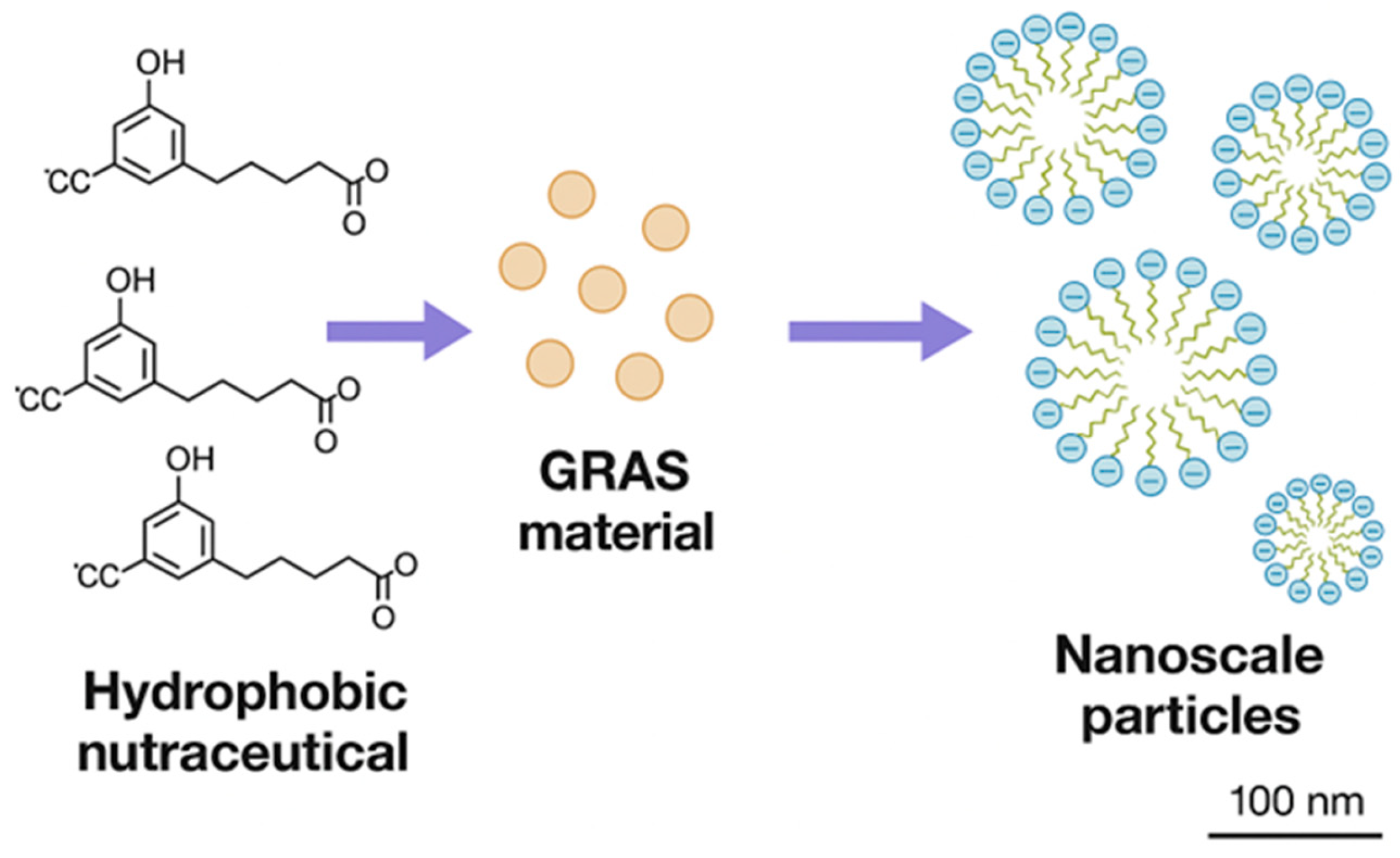
Figure 2.
Combined X-ray diffraction results of nine nutraceutical nanoparticles. ER: EC16-resveratrol, EC: EC16-curcumin, ERC: EC16-resveratrol-curcumin hybrid nanoparticles.
Figure 2.
Combined X-ray diffraction results of nine nutraceutical nanoparticles. ER: EC16-resveratrol, EC: EC16-curcumin, ERC: EC16-resveratrol-curcumin hybrid nanoparticles.
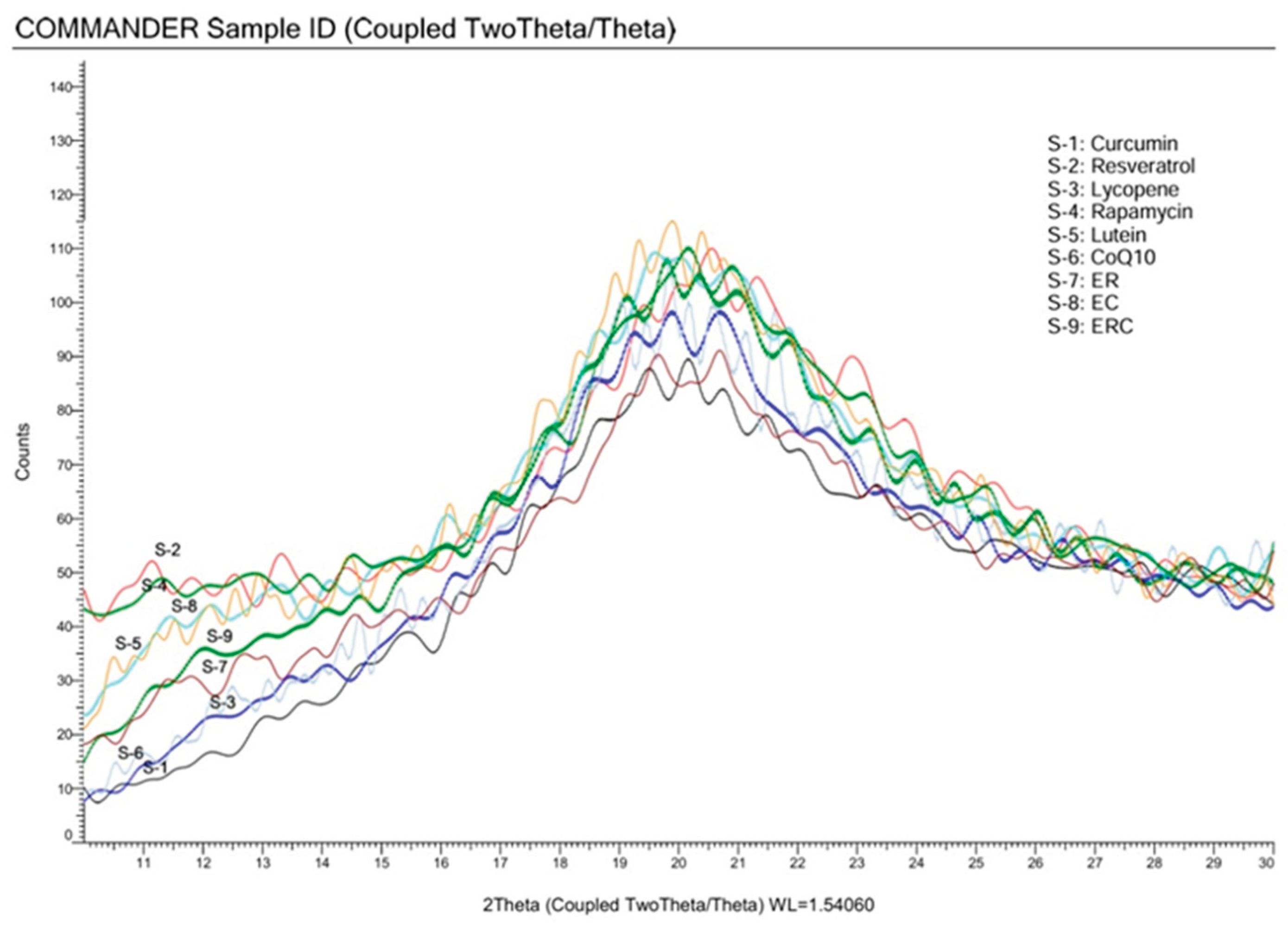
Figure 3.
Results of XTT assay of nine different nanoparticle preparations. Control used cell culture medium incubation. AV represents the average values of absorption of 450 nm minus absorption of 660 nm. SD represents the standard deviations from three samples. Only rapamycin and EC16/curcumin hybrid nanoparticles showed increased cell viability (measured by mitochondrial dehydrogenase activity) with p values <0.05, compared to the control. Other nanoparticles have no significant differences in cell viability compared to the control (p>0.05, one-way ANOVA).
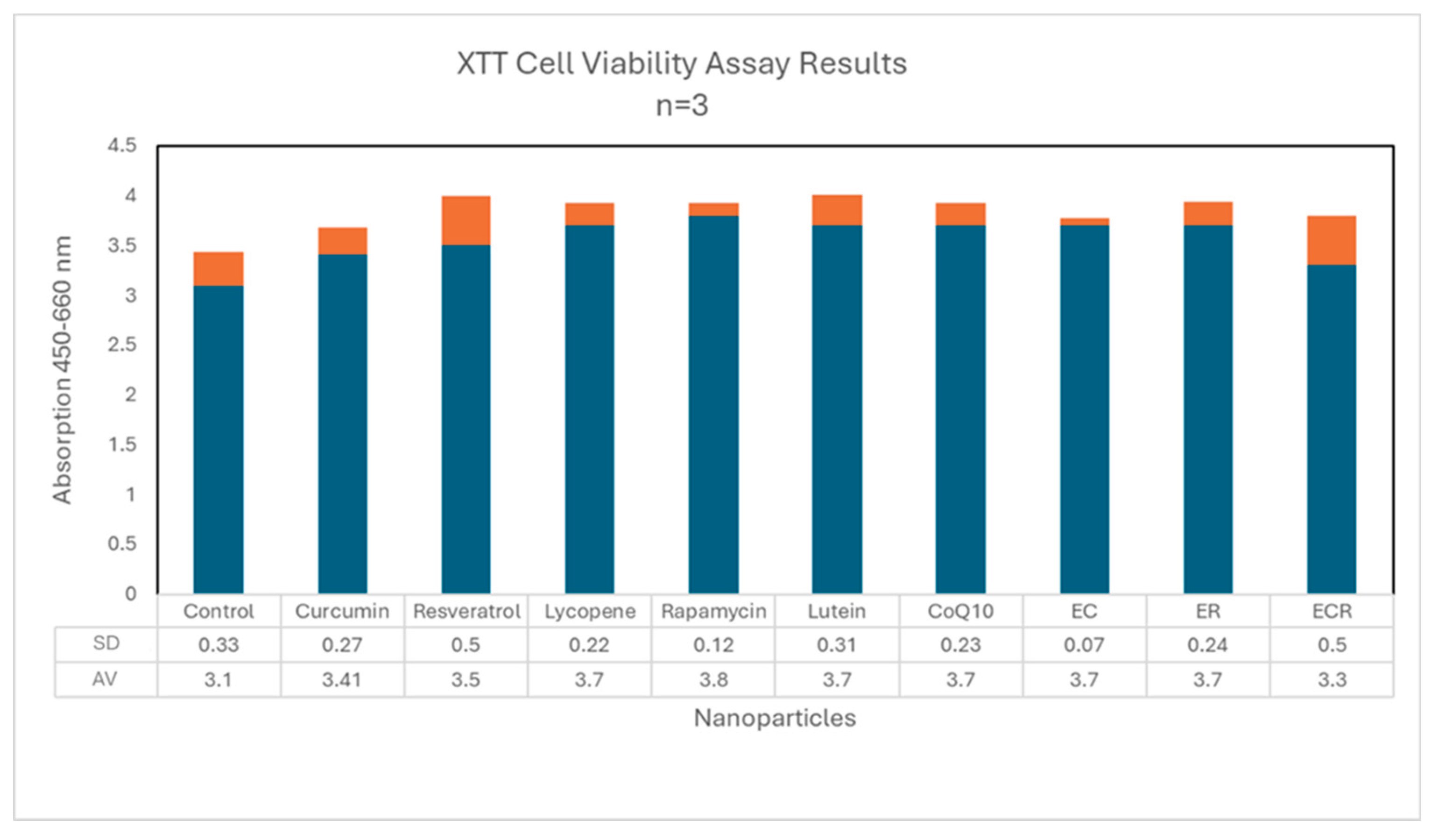
Figure 4.
The fluorescent images demonstrate that the Cy5/EC16 hybrid nanoparticles (red) attached on RAW264.7 cells (blue, DAPI stained) with increased intensity over incubation time. The control shows no Cy5 NPs present.
Figure 4.
The fluorescent images demonstrate that the Cy5/EC16 hybrid nanoparticles (red) attached on RAW264.7 cells (blue, DAPI stained) with increased intensity over incubation time. The control shows no Cy5 NPs present.
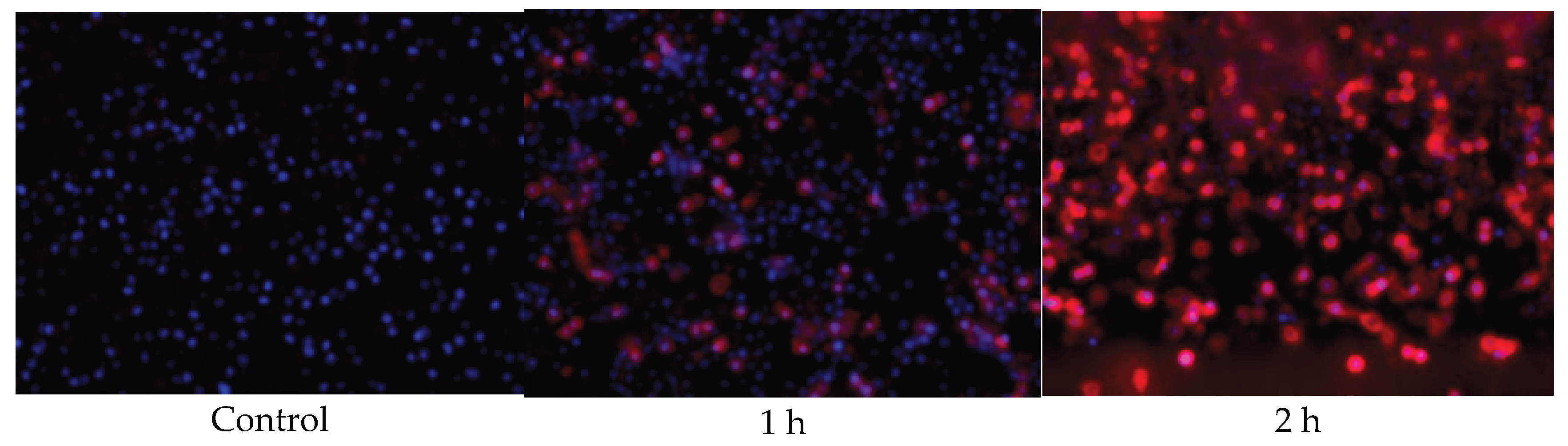

Table 1.
Initial nanoparticle size, Zeta potential and density.
| Compound | Median Size nm | Zeta Potential mV | Density /ml, 0.01% |
|---|---|---|---|
| Curcumin | 136.6 ± 104.9 | -33.72 ± 0.93 | 4.4x 10⁸ |
| Resveratrol | 174.0 ± 76.76 | -31.75 ± 1.86 | 0.8x10⁸ |
| Lycopene | 184.9 ± 156.2 | -20.50 ± 0.87 | 1.7x10⁷ |
| Lutein | 141.7 ± 53.4 | -24.54 ± 1.18 | 1.3x10⁹ |
| CoQ10 | 315.3 ± 131.8 | -26.8 ± 1.11 | 1.2x10⁷ |
| Procyanidin B2 | 144.2 ± 120.4 | -71.59 ± 0 | 4.5x10⁸ |
| Quercetin | 163.8 ± 102.2 | -62 ± 1.0 mV | 1.6 x10⁹ |
| EGCG-palmitate (EC16) | 152.5 ± 78.8 | -60.11± 0.59 mV | 3.2 x10⁹ |
| Rapamycin | 147.7 ± 89.1 | -44.28 ± 1.79 mV | 2.8 x 10⁹ |
Table 2.
Improved surface charges of nutraceutical nanoparticles.
| Compound | Median Size nm | Zeta Potential mV | Density /ml, 0.01% |
|---|---|---|---|
| Curcumin | 166.4 ± 57.3 | -45.71 ± 1.53 | 4.2x 10⁸ |
| Resveratrol | 134.2 ± 55.8 | -44.43 ± 1.89 | 1.3x10⁸ |
| Lycopene | 149 ± 74.9 | -38.86 ± 0.87 | 2.7x10⁸ |
| Lutein | 154.2 ± 66.7 | -32.63 ± 0.13 | 3.0x10⁸ |
| CoQ10 | 232.3 ± 151.3 | -42.81 ± 1.71 | 2.3x10⁷ |
Table 3.
Size, Zeta potential and density of hybrid and combination of nutraceuticals.
| Compounds | Type of NPs | Median Size nm | Zeta Potential mV | Density /ml, 0.01% |
|---|---|---|---|---|
| EC16-Resveratrol | Hybrid | 96.2 ± 64.1 | -53.29 ± 0.67 | 1x 10¹⁰ |
| EC16-Curcumin | Hybrid | 85.0 ± 36.9 | -57.22 ± 0.87 | 2.5x10¹⁰ |
| EC16-Res-Cur | Hybrid | 99.9 ± 79.8 | -51.23 ± 1.25 | 5.7x10⁹ |
| EC16/Resveratrol | Mix | 180.0 ± 155.2 | -57.02 ± 1.37 | 2.3x10⁹ |
| EC16/Curcumin | Mix | 198.8 ± 115.8 | -67.45 ± 1.17 | 5.0x10⁹ |
| EC16/Res/Cur | Mix | 201.4 ± 117.7 | -62.32 ± 0.92 | 4.7x10⁹ |
| EC16-Cy5 (Fluorescent) | Hybrid | 141.9 ± 86.8 | -43.1 ± 1.13 mV | 6.1 x10¹⁰ |
Table 4.
Nanoparticle density and size before and after 60 minute incubation at 37 oC in HCl solution at pH 1.84. Hybrid: hybrid of nanoparticles of EC16-curcumin-resveratrol. Mix: a mix of individually generated EC16, curcumin, and resveratrol nanoparticles.
Table 4.
Nanoparticle density and size before and after 60 minute incubation at 37 oC in HCl solution at pH 1.84. Hybrid: hybrid of nanoparticles of EC16-curcumin-resveratrol. Mix: a mix of individually generated EC16, curcumin, and resveratrol nanoparticles.
| NPs/Incubation time (min) | Density/ml | Median Size (nm) |
| Curcumin 0 | 2.0 x 10⁷ | 165 ± 111 |
| Curcumin 60 | 8.4 x 10⁶ | 130 ± 78 |
| Resveratrol 0 | 1.1 x 10⁹ | 189 ± 647 |
| Resveratrol 60 | 5.6 x 10⁸ | 182 ± 810 |
| Hybrid 0 | 3.0 x 10⁸ | 158 ± 165 |
| Hybrid 60 | 2.6 x 10⁸ | 154 ± 361 |
| Mix 0 | 1.3 x 10⁸ | 160 ± 125 |
| Mix 60 | 3.1 x 10⁷ | 156 ± 220 |
Table 5.
Comparative Analysis of Curcumin Nanoparticle Preparation Methods.
| Parameter | HES–CUR–COOH Chemical Conjugation [33] | Lipid-Based Nano Delivery Systems (NE, SLN, NLC) [34] | FAST (Facilitated Self-Assembling Technology, Current Study) |
| Main Purpose | Covalent synthesis of curcumin–HES micelles for drug delivery | Encapsulation of curcumin in lipid nanoparticles for beverage and food applications | Physical self-assembly of curcumin and natural amphiphiles for nutraceutical and beverage delivery |
| Chemicals Used | Pyridine, glutaric anhydride, EDC, DMAP, DMSO, HCl, ethyl acetate | Beeswax, lecithin, Tween 80, MCT oil, ethanol, acetic acid, sodium benzoate, ascorbic acid | Only GRAS ingredients |
| Reaction Type | Multi-step covalent conjugation (ester and amide bond formation) | Physical emulsification, high-pressure homogenization, and ultrasonication | Spontaneous self-assembly through hydrophobic and hydrogen-bond interactions |
| Processing Time | > 1 week including synthesis and purification | 4–6 h (multiple homogenization and cooling cycles) | < 60 min from powder to nanoparticle stock readily for use |
| Energy Requirement | High—solvent reflux, evaporation, and lyophilization | Very high—ultrasonication and homogenization at ~20,000 psi | Low |
| Byproducts / Waste | Pyridine and DMSO residues; requires dialysis and lyophilization | Residual surfactants and organic solvents | None (zero waste) |
| Potential Toxicity | High — due to pyridine, DMSO, and EDC residues; not suitable for ingestion without rigorous removal | Low to moderate; Tween 80 and lecithin are food-grade but may affect gut permeability | Minimal—fully GRAS and biodegradable materials |
| Particle Composition | Curcumin–HES conjugate polymers, irregular particles | SLN and NLC (~150 nm) lipid carriers, curcumin encapsulated in wax/oil core | Native curcumin nanoparticles (median 150 nm), amorphous and colloidally stable |
| Manufacturing Complexity | Multi-step, laboratory scale | Multi-stage homogenization; high-energy and temperature sensitive | One-step scalable process; compatible with beverage manufacturing lines |
| Scalability / Industrial Feasibility | Low—requires organic solvent handling | Moderate—food-grade but high-cost energy equipment | Excellent—simple, aqueous, eco-friendly, and low-cost |
| Regulatory Classification | Pharmaceutical; requires toxicology approval | Food-grade but limited by surfactant and process energy requirements | Nutraceutical / food-grade, GRAS-compliant, and clean-label |
| Environmental Impact | High solvent use and waste | Moderate—requires heating and surfactant disposal | Near-zero waste, solvent-free, and energy-efficient |
Disclaimer/Publisher’s Note: The statements, opinions and data contained in all publications are solely those of the individual author(s) and contributor(s) and not of MDPI and/or the editor(s). MDPI and/or the editor(s) disclaim responsibility for any injury to people or property resulting from any ideas, methods, instructions or products referred to in the content. |
© 2025 by the authors. Licensee MDPI, Basel, Switzerland. This article is an open access article distributed under the terms and conditions of the Creative Commons Attribution (CC BY) license (http://creativecommons.org/licenses/by/4.0/).
Copyright: This open access article is published under a Creative Commons CC BY 4.0 license, which permit the free download, distribution, and reuse, provided that the author and preprint are cited in any reuse.


