Preprint
Article

This version is not peer-reviewed.

An Appraisal of the Understanding Pressure Effects on Structural, Optical, and Magnetic Properties of CsMnF4 and Other 3dn Compounds

Submitted:

27 November 2025

Posted:

28 November 2025

You are already at the latest version

Abstract

A recent theoretical study of CsMnF4 under pressure [Inorg. Chem. 2024, 63(29), 13231] presents conclusions on its structural, optical, and magnetic behavior that conflict with established experimental evidence. Crucially, this work omits key prior experimental results on CsMnF4 and related Mn3+ fluorides under pressure. This perspective examines the resulting discrepancies, arguing that the omissions of this data undermines the theoretical estimates and methodological validity of Ref. [1]. This paper provides a critical overview centered on two main points: the contested nature of the pressure-induced high-spin to low-spin transition observed in CsMnF4 at ~37 GPa and a detailed discussion of Jahn-Teller physics in this archetypal system. By reconciling the existing literature with the new theoretical claims, this work aims to clarify the high-pressure behavior of CsMnF4.

Keywords: 
;  ;  ;  ;  ;  

1. Introduction

CsMnF₄ is a foundational example of a cooperative Jahn-Teller (JT) system, whose structural distortions mediate its distinctive optical and magnetic properties. While the effects of pressure on this material have been extensively studied [1,2,3,4,5,6,7,8,9,10,11,12], its high-pressure structural evolution and electronic ground state remain subjects of active debate [1,7]. A recent publication [1] has intensified this controversy by presenting theoretical calculations that contradict interpretations from prior experimental work. That study, however, omits several key experimental findings on CsMnF₄ and other Mn3+ fluorides under pressure [2,3,4,5,6,7,8,9,10,11]. This perspective aims to critically reexamine the structural, optical, and magnetic properties of CsMnF₄ under pressure, specifically to address the interpretation in Ref. [1] that conflict with the established literature. A primary focus is the disputed assignment of the high-spin to low-spin transition observed in CsMnF4 near 37 GPa [7].
At ambient pressure, CsMnF₄ crystallizes in a tetragonal structure (space group P4/n), forming a layered perovskite. The MnF63- octahedra are subject to a cooperative JT distortion, resulting in a locally elongated complex with a D2h symmetry. The crystal structure is characterized by an antiferrodistortive arrangement where the in-plane equatorial ligands of one MnF63- unit act as the axial ligands for two adjacent complexes (Figure 1). The in-plane Mn-F bond distances are 2 × 2.168 Å and 2 × 1.817 Å, while the out-of-plane distances are 2 × 1.854 Å [12]. This specific connectivity, with Mn-F-Mn bond angles clase to 180º, favors ferromagnetic superexchange within the layers, successfully explaining the emergence of long-range ferromagnetic order below Tc = 19 K [2,3,4,5].
This paper provides an overview that contrasts the findings of Ref. [1] with the body of existing research. We will first delineate the fundamental Jahn-Teller distortions in this system before critically assessing the high-pressure behavior, with particular emphasis on the debated spin-state transition [1,7]. Our goal is to reconcile the theoretical predictions with experimental evidence and clarify the current understanding of this important material.

2. Results and Discussions

The main scientific concerns to the theoretical work about the structural, electronic and magnetic properties of CsMnF4 published elsewhere [1] are summarized in the six following sections.

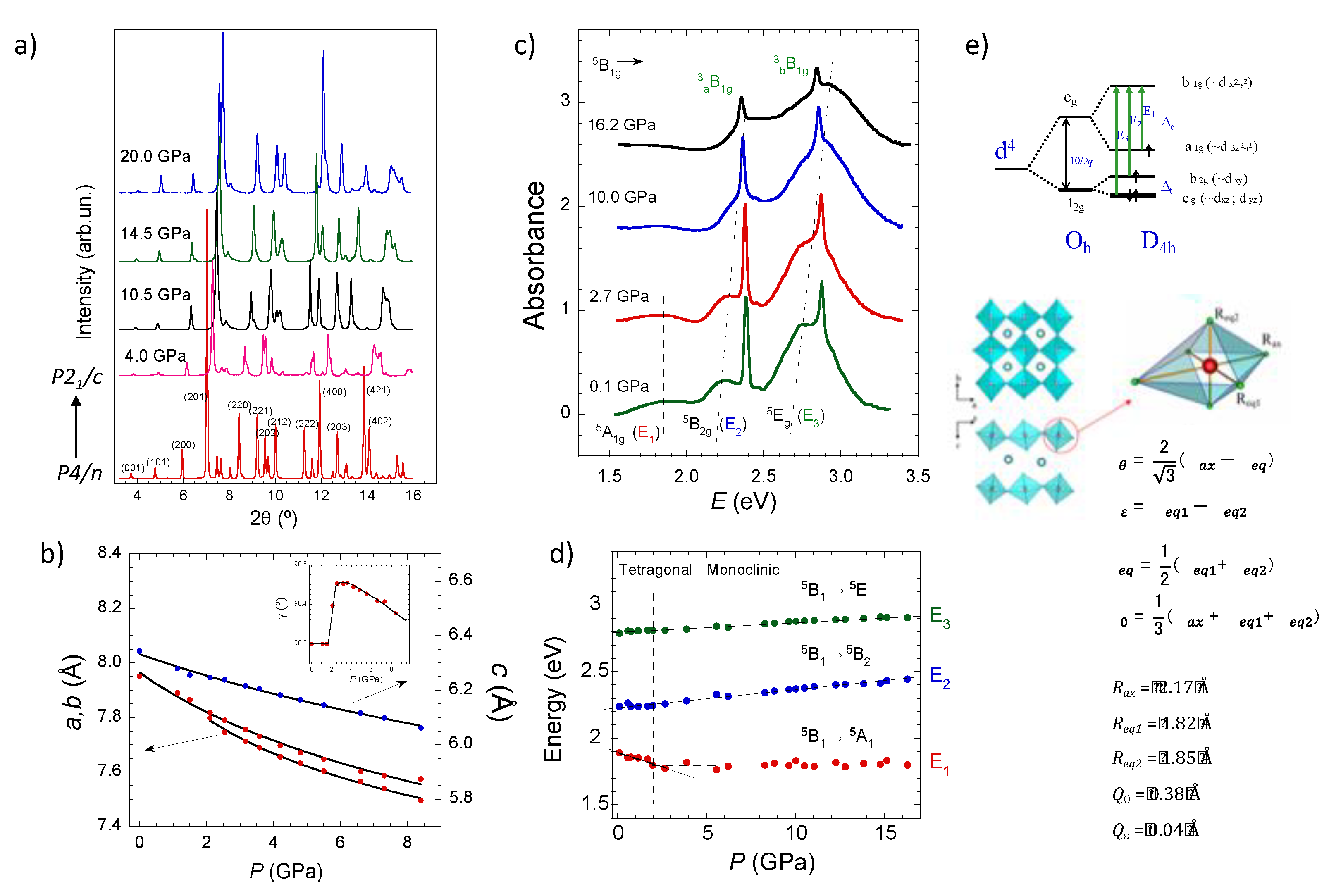
2.1. Structure of CsMnF4

The authors theoretically describe the structural evolution of CsMnF4 in terms of a tetragonal structure P4/n (0-40 GPa), which experiences a structural phase transition at 37.5 GPa to another tetragonal phase (P4) that is stable up to at least 50 GPa. In this structural evolution, the ground state of Mn3+ is high spin (S = 2). The point is that there are three publications [2,3,4] dealing with the structural evolution of CsMnF4 with pressure by x-ray diffraction (XRD): energy dispersive XRD at the Daresbury Synchrotron, UK [2,3], and angle dispersive XRD at the European Synchrotron Radiation Facility (ESRF), France [4]. These independent works concluded that the tetragonal structure P4/n of CsMnF4 is unstable above 2 GPa, transitioning to a monoclinic structure stable up to at least 16 GPa [4] (Figure 1). Energy dispersive XRD data [2,3] conclude that a tetragonal to orthorhombic phase transition at 1.4 GPa followed by a monoclinic transition above 6 GPa (see Orthorhombic to Monoclinic section of Ref. 3). None of these publications were discussed in [1] in relation to the experimentally observed structural evolution –Refs. 2 and 4 were not cited– and therefore, this low-pressure structural transition calls into question all results derived from a tetragonal structure above 2 GPa. Although Ref. 3 was cited in the article, it literally states that “No signs of a structural phase transition around 1.4 GPa, early suggested by Moron et al. [22], have been found in the optical measurements on CsMnF4” (page 13233 in Ref. 1). Besides the recognition of a structural phase transition around 1.4 GPa earlier reported by Moron et al. [3], the authors ignored two other publications [2,4] not cited in Ref. 1, which confirm the tetragonal to monoclinic structural phase transition (around 2 GPa in Ref. 4). In addition to the XRD results, reference 4 also reports precise optical data in the 0-16 GPa range, which provides clear evidence of the influence of the phase transition at 2 GPa in the optical spectra of CsMnF4 (Figure 1).
The measured spectra show that the pressure shift rate of the 5B15A1 transition energy at 1.89 eV changes from -42 meV/GPa to +2 meV/GPa at 2 GPa, coinciding with the tetragonal to monoclinic phase transition [4] (Figure 1). It must be emphasized that Refs. 2-4 are the only ones dealing with the crystal structure of CsMnF4 under high pressure conditions, and were not considered, or cited [2,3,4] in the reported work [1], although one of the authors cited Ref. 4 in a previous publication [6]. Therefore, the structural phase transition detected by XRD by two different research groups, at two different synchrotron facilities, between 1-2 GPa was neither considered in the article1 nor detected by using the authors’ DFT methodology. Furthermore, the structural evolution of CsMnF4 with increasing pressure reported in reference 4 coincides with the structural evolution in the series AMnF4 (A: Cs, Rb, Tl, K, Na) with decreasing the unit cell volume [2,3,4,5] as it can be seen in Figure 2 and Figure 3.

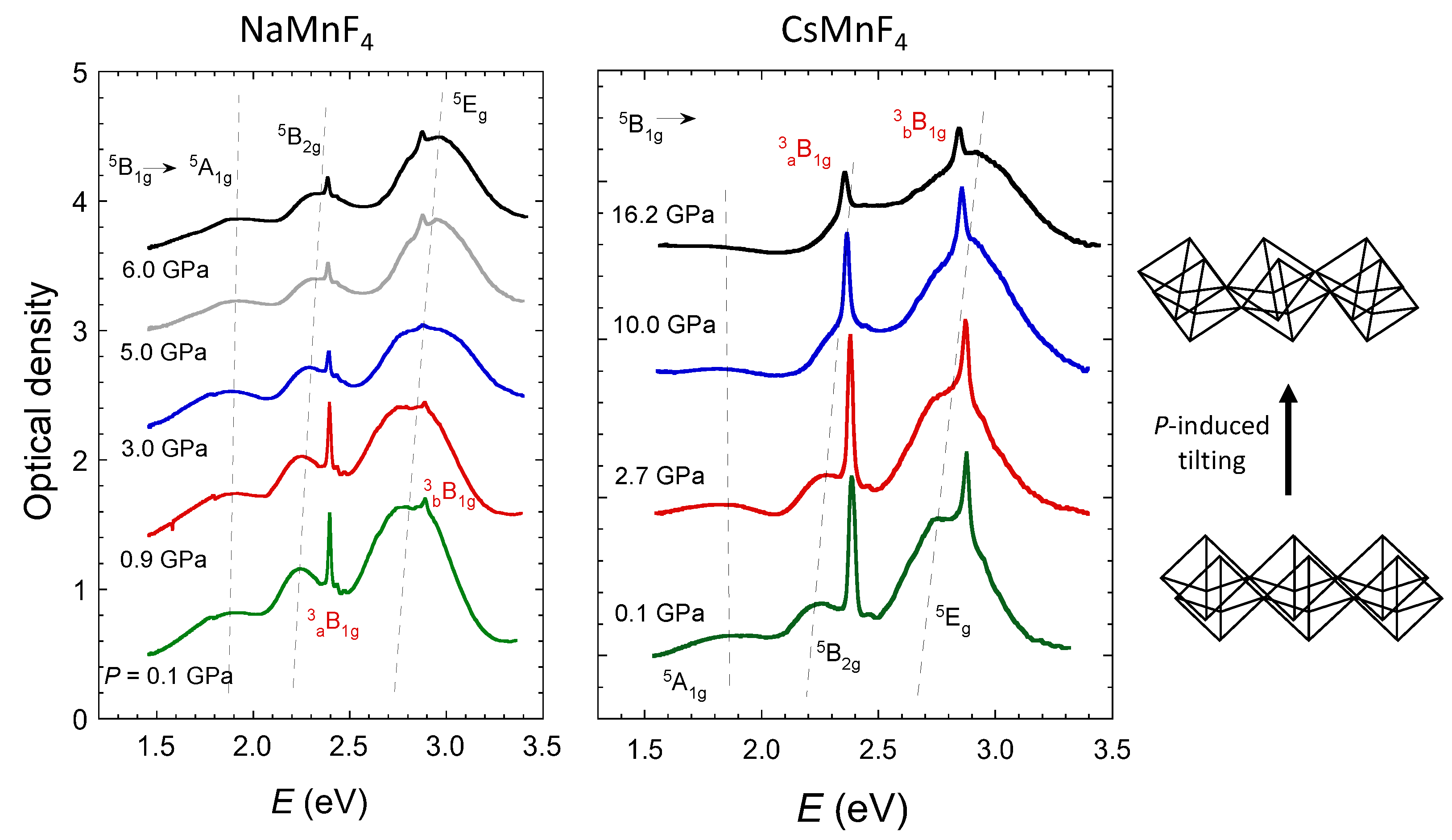
2.2. Pressure Dependence of the Optical and Electronic Properties

The pressure dependence of the optical and electronic properties derived theoretically from the tetragonal phase [1] are unable to explain the experimental results by optical spectroscopy published previously [4,7,8,9]. CsMnF4 in the tetragonal phase is a uniaxial crystal instead of a biaxial crystal attained in the monoclinic structure (P > 2 GPa). Furthermore, the structure of the Mn3+ one-electron d-orbitals obtained theoretically [1], predicts that the energy of the first absorption band associated with the 5B15A1 (denoted by the one-electron orbital transition 3y2-r2→x2-z2 in Figure 4 of Ref. 1), decreases continuously from 1.92 eV to 1.43 eV in the 0-40 GPa range, while experimentally it changes from 1.89 to 1.81 eV in the 0-2 GPa range (tetragonal phase) but blueshifts by only 0.03 eV in the 2-16 GPa range (monoclinic phase) [4,7] giving a total shift of +0.07 eV at 37 GPa [4,8].
It means that the band shifts from 1.89 eV at ambient pressure to 1.88 eV at 37 GPa, which is two orders of magnitude lower than the shift predicted theoretically of -0.49 eV in the same pressure range (Figure 1). Neither the magnitude of the pressure band shift, nor the two opposite pressure rates below and above 2 GPa were accounted for theoretically [1]. References 4 and 8 were not cited in Ref. 1, and their experimental results could have guided the authors to search the actual structural evolution of CsMnF4 as well as its associated properties. It must be noted that the pressure-induced blueshift of the first absorption band experimentally observed in the CsMnF4 monoclinic phase above 2 GPa is also observed in NaMnF4 in the 0-6 GPa range, which has a monoclinic structure at ambient conditions [8,9] (Figure 3). It seems that the preservation of the tetragonal structure in the whole explored pressure range1 is unable to explain the observed optical properties.

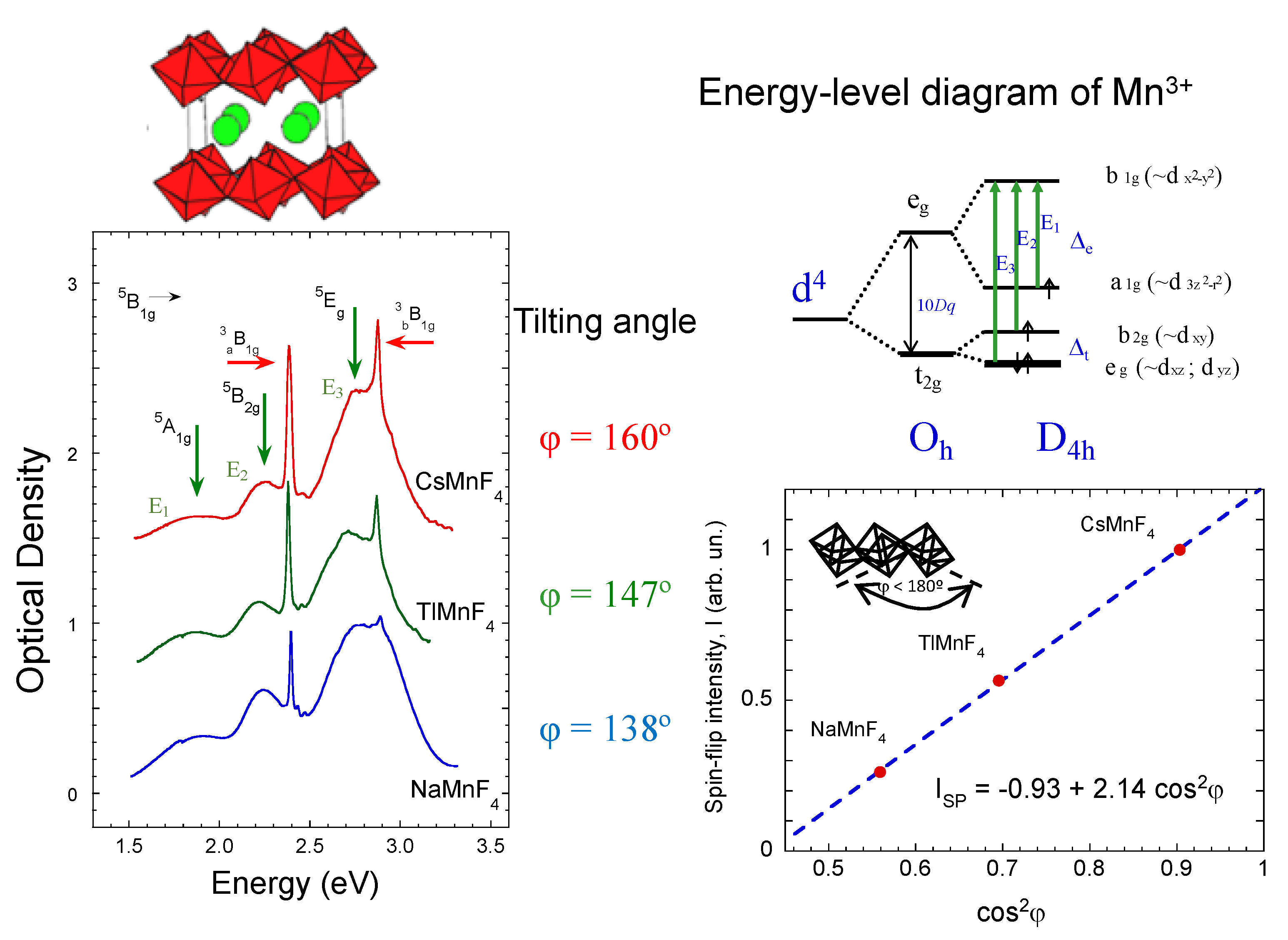
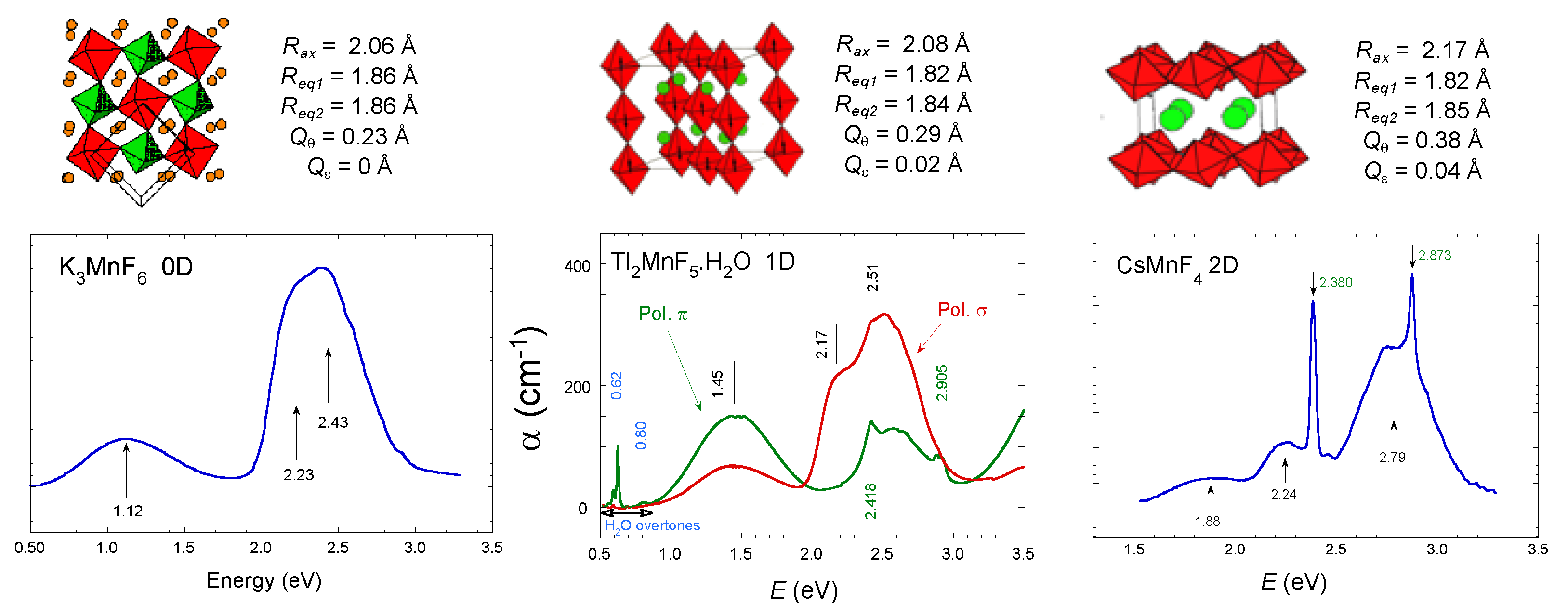
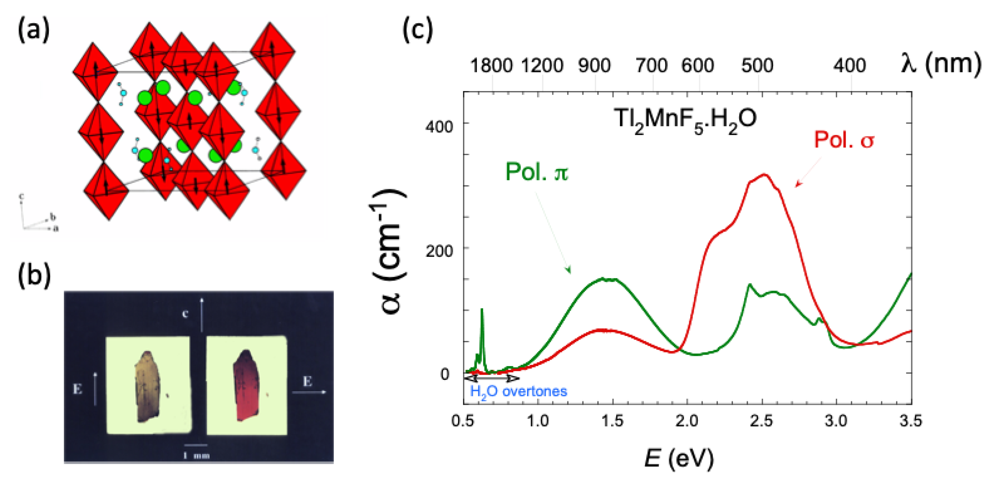
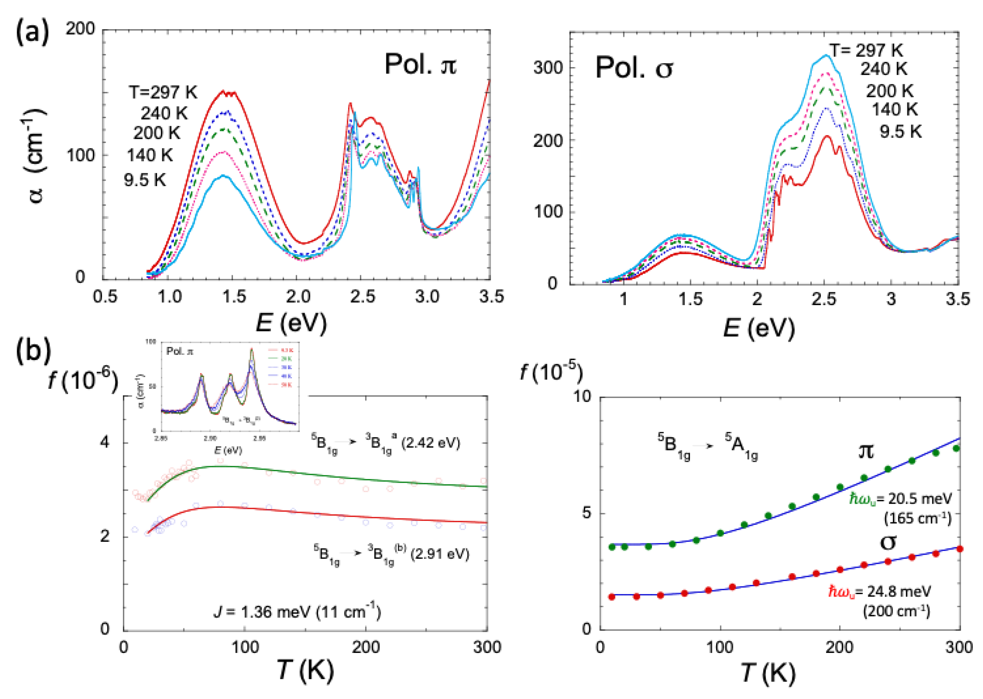
2.3. Optical Band Assignment of Mn3+ Fluorides

The band assignment of the crystal-field spectra related to d-d transitions in CsMnF4 as well as the high-spin (HS) to low-spin (LS) transition detected by optical spectroscopy at 37 GPa reported elsewhere [7] (Figure 4), the spectra of which are reproduced in Figure 4 of Ref. 1, are questioned in the article [1]. Although no alternative interpretation to the assignment of Ref. 7 is given in the article [1], it must be noted that the optical absorption band assignment of Mn3+ fluorides is based on spectroscopic results reported elsewhere [9]. This work, which is not cited in Ref. 1, deals with single-crystal polarized optical absorption spectroscopy and the temperature dependence of the various bands appearing in the optical spectra. Based on their transition energy, spectral shape and oscillator strength, as well as their temperature dependence (Figure 5, Figure 6 and Figure 7), it was concluded that there are two types of bands in the optical spectra: three broad bands associated with single-electron transitions within the d-orbitals of Mn3+ in an almost D4h local environment, and two spin-flip narrow peaks arising from the 3d4 electronic configuration of Mn3+ (Figure 5).
It is experimentally shown that the former ones are electric-dipole spin-allowed transitions, whose oscillator strength increases with increasing temperature by coupling to odd parity vibrations within the (MnF6)3- complex. The spin-flip transitions are shown to be electric-dipole spin-forbidden transitions within a single Mn3+ ion but are activated by the spin-effective mechanism in exchange-coupled systems via the Mn-F-Mn superexchange pathway [9] (Figure 7). The temperature dependence of their oscillator strength demonstrates the spin-flip nature of these narrow peaks observed in the optical spectra of exchange-coupled systems [7,8,9]. In addition, these spin-flip transitions can be easily identified in the optical spectra because, unlike the spin-allowed broad bands whose energies are strongly dependent on the (MnF6)3- octahedral distortion, their energy is not as sensitive to the distortion [7,8,9] (Figure 5). Spin-flip transitions are within 0.05 eV at the same position in all series of manganese (III) fluorides: ESP1 = 2.380 eV and ESP2 = 2.873 eV in 2D CsMnF4 [7], 2.397 and 2.890 eV in 2D NaMnF4 [8,9], 2.380 and 2.860 eV in 2D TlMnF4 (2D fluorides) [8] and, 2.418 and 2.914 eV in the 1D Tl2MnF5.H2O [9].
In this respect, the d-orbital electronic structure derived theoretically for CsMnF4 in the article,1 does not agree with the measured spectra [7] (Figure 4), which are reproduced in Figure 4 of Ref. 1. The spectra show three broad bands associated with single-electron transitions from 3z2-r2, xy, and the nearly degenerate (xz, yz) to x2-y2, in order of increasing energy (note that the local z-axis in Ref. 7 refers to the long Mn-F bond in CsMnF4 of the nearly D4h (MnF6)3- complex). In the article [1], the xy, xz, and yz d-orbitals have different energies with respect to the x2-y2 level: 2.2, 2.5 and 2.8 eV (Figure 7 of Ref. 1). Except for the first absorption band 3z2-r2 → x2-y2 at about 1.9 eV, the other three transition energies cannot give rise to a two-broad-band structure in the optical spectra as observed experimentally (Figure 5), but to three almost equally spaced broad bands, which would probably give rise to a structureless broad band instead of the two well-resolved bands observed experimentally [7]. Furthermore, the calculations performed in the article deal with one-electron d-orbitals and not with the states arising from the d4 electronic configuration, and thus the observed spin-flip transitions are missed in the reported calculations [1]. This is an important point as the authors should know that the spin-flip transitions observed in the spectra of CsMnF4 (Refs. 4,7) are crucial to explain the spin transition at 37 GPa on the basis of optical spectroscopy [7] (Figure 4 and Figure 5).
f T = 2 f ( 0 ) ( 2 S 1 ) S U 2 1 + 4 S U S + 1 U V 2 V
Where f(0) is the 0 K oscillator strength, U=cothV-1/V, V=2JS(S+1)/kBT , S = 2 and J (< 0) is the intrachain exchange constant [9] (Left side). Temperature dependence of the oscillator strength for the 5B1g5A1g broad band (1.45 eV) in π and σ polarizations. Solid lines are fits to f T = f 0   c o t h ( ω u / 2 k B T ) (vibronic mechanism, parity-forbidden electric-dipole transitions activated by odd parity vibrations ω u ). The different temperature dependences and polarization reveal distinct activation mechanisms: exchange for spin-flip peaks and vibronic for spin-allowed broad bands (Right side). Adapted with permission from Reference 9. Copyright 1994 American Chemical Society

2.4. Magnetic Properties of CsMnF4

Regarding the magnetic properties, it is theoretically found that CsMnF4 undergoes an abrupt change from ferromagnetic to antiferromagnetic at 10 GPa within the tetragonal phase [1]. However, there is a misunderstanding with the magnetic measurements under high pressure conditions reported elsewhere [11]. This paper reports a singular magnetic behavior above 2 GPa with a reduction of the Curie temperature and a sharp decrease of the saturation magnetization of CsMnF4 above 3.3 GPa, which the authors explain in terms of a progressive transition from ferromagnetic to antiferromagnetic with a permanent ferromagnetic component, which the authors attribute to canting antiferromagnetism [11]. This behavior is overlooked in the article [1] and therefore, properties observed in the monoclinic phase are not predicted in the tetragonal phase. In fact, this behavior has been explained along the AMnF4 series of layered perovskites, where the only tetragonal member of the series is the ferromagnetic CsMnF4, the remaining compounds having a monoclinic structure are all antiferromagnetic [2,5,11,12]. Moreover, Refs. 2 and 3 show that the AFM Neel temperature correlates with the Mn-F-Mn tilting angle in the monoclinic phase, a figure which cannot be reproduced in the same way using a tetragonal phase due to structural constrains.

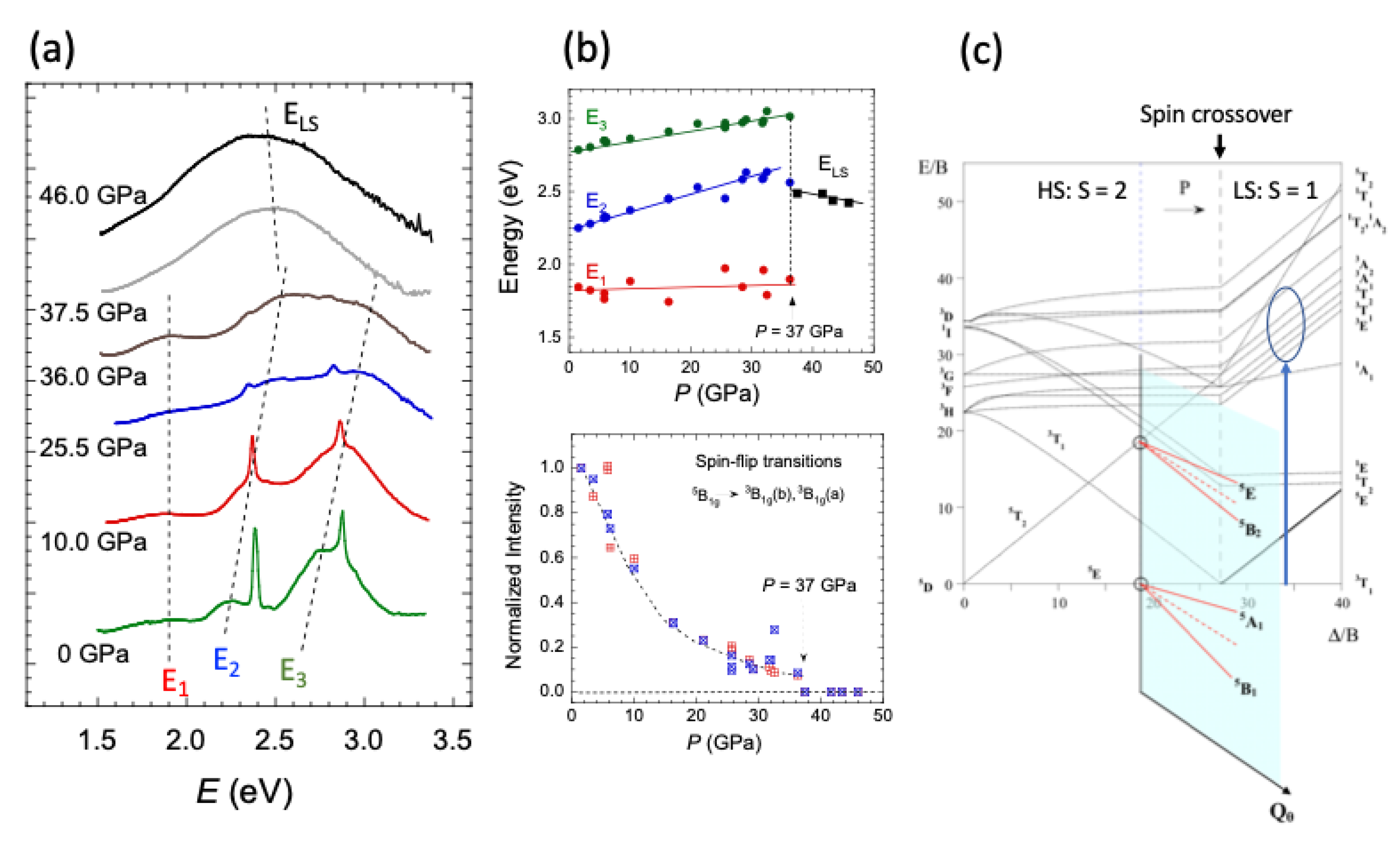
2.5. High-Spin to Low-Spin Transition at 37 GPa

The change in the optical spectra (Figure 4 of Ref. 1; or Figure 2 of Ref. 7) associated with a HS→LS crossover transition at 37 GPa and room temperature in the original paper [7] (Figure 4) is now questioned and associated with a tetragonal to tetragonal phase transition P4/nP4 at 37.5 GPa, both phases being HS (S=2). However, this interpretation lacks the spectral evolution at the crossover transition point. There are three main reasons for retaining the HS to LS transition: 1) The three broad bands observed below 30 GPa, associated with the strong octahedral distortion due mainly to the E⊗e JT effect, transform into a structureless broad band characteristic of a nearly octahedral coordination. The T⊗e JT effect in the LS 3T1 ground state (Oh) is a factor of 4 weaker than the E⊗e HS 5E ground state strongly distorted by the JT effect [7,13]; its stabilization energy in LS is an order of magnitude smaller than in HS. 2) The spin-flip transitions ESP1 and ESP2, which are characteristic of a HS ground state, disappear completely above 37 GPa. According to the Tanabe-Sugano diagrams for d4 ions [13,14], these transitions are absent in a LS ground state (3T1), making them a precise spectroscopic probe for layered AMnF4 (A: Cs, Na, Tl) perovskites in HS [7,9] (Figure 4, Figure 5 and Figure 6). 3) The LS broad band peaking at 2.5 eV (Figure 4) coincides with the centroid of the spin-allowed crystal-field bands at the spin crossover transition (S= 2→1), which corresponds to a crystal-field splitting 10Dq of 27B (C/B = 4.6), where B and C are the Racah parameters for (MnF6)3− [7,13,14]. On the other hand, there is an argument given in Ref. 1 to support a structural transition at 37 GPa instead of a HS→LS crossover transition, which is not properly justified. The authors state that the 10Dq required to stabilize the LS state should be higher than 3 eV, without any further justification. The value of 10Dq derived from the TS diagram at the transition point requires accurate knowledge of the Racah parameter B, which is known to be B = 0.097 eV (780 cm-1) in (MnF6)3- at ambient conditions [10]. This value can also be obtained from the spin-flip transition energies, since the first observed spin-flip transition depends on pressure as ESP2 = 2.397 – 0.0018 P (in eV and GPa units, respectively) [7,9]. From the TS diagram [13,14] (Figure 4) its energy varies as 24.6 B in HS up to the spin crossover point. This means that B slightly decreases from 0.097 eV at ambient pressure to about 0.094 eV at 37 GPa, which is considered to be the spin crossover pressure as stated elsewhere [7]. This means that 10Dq should be about 2.54 eV –lower than 3 eV–, and considering that the single-electron t2g4 eg0t2g3 eg1 transition actually gives rise to several spin-allowed transitions within the d4 electronic configuration, from the LS 3T1 ground state to the 3Γi excited states, whose energies spread over the 0.4 eV range: 2.39 eV (3T1), 2.48 eV (3T2), 2.65 eV (3A2), and 2.76 eV (3A2) [13,14]. All these transitions give rise to a broad band whose centroid peaks at about 2.54 eV, in agreement with the observed spectra of CsMnF4 above 37 GPa in LS [7] (Figure 4). 4) The argument given to rule out spin transition about the enthalpy difference between HS and LS states in tetragonal symmetry is not supported by any information or evidence even in the Supporting Information [1]. It is well known that the estimation of a spin crossover pressure from ab initio calculations is subtle and delicate since it requires a good knowledge of the actual HS and LS structures and the calculations are very sensitive to parameters such as the Hubbard U among others [15,16]. It is therefore scientifically irrelevant to state that the enthalpies for LS and HS are a few tenths of eV, based on an inadequate HS structure and an unknown LS structure, without providing details of the performed calculations. However, optical spectroscopy offers a more suitable means of identifying spin crossover phenomena. This technique can effectively distinguish between the distinct spectra associated with each spin state, irrespective of the specific crystal structures involved in the spin transition. This has been well established for Fe2+ compounds and extensively documented elsewhere [17,18,19].

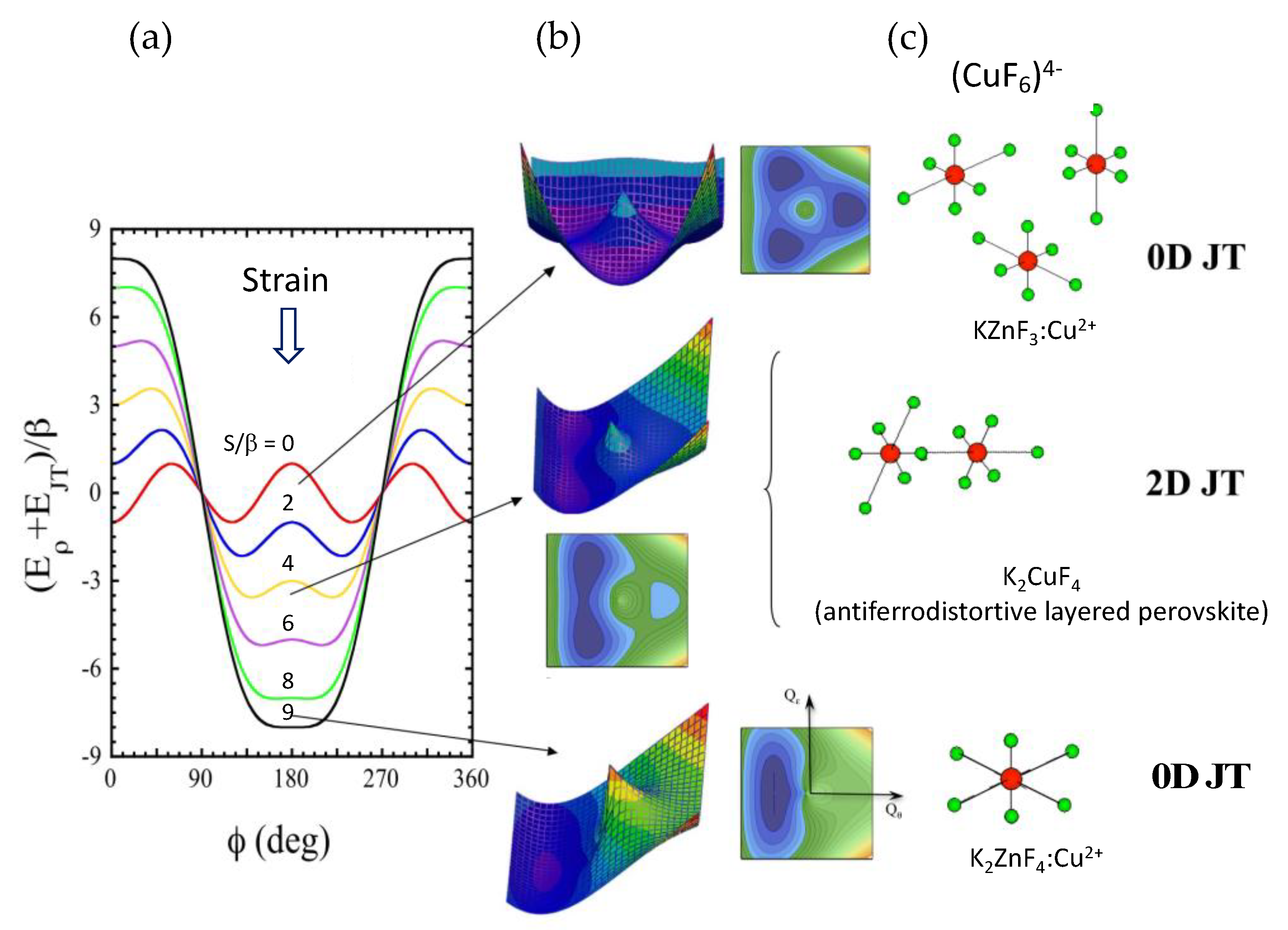
2.6. The Jahn-Teller Effect: Theorem, Theory, and Molecular Distortion

Finally, some concepts about the JT effect need to be clarified in order to avoid misunderstandings among scientists working with systems that exhibit JT distortions. This follows from sentences (i-iii) on page 13234 of Ref. 1, which state: (i) “The existence of a JT effect requires a degenerate electronic state in the initial geometry”, (ii) “CsMnF4 is a layered compound where layers are perpendicular to the crystal c axis (Figure 1). Accordingly, one would expect that the axis of the MnF63− unit perpendicular to the layer plane plays a singular role, a fact seemingly not consistent with the JT assumption.”, and (iii) “The local equilibrium geometry for MnF63− in CsMnF4 is not tetragonal. Indeed, even assuming Y as the main axis (Figure 1) the symmetry would be at most orthorhombic because RXRZ = 0.037 Å. Accordingly, one should expect four and not only three d−d transitions with ΔS = 0 for CsMnF4.”
Because of these three sentences, it is convenient to distinguish between the JT theorem, the JT theory, and the JT distortion when dealing with the JT effect. The JT theorem states that a nonlinear polyatomic molecule with an orbital degenerate ground state is unstable and must distort to a lower energy configuration, or literally “All nonlinear configurations are therefore unstable for an orbital degenerate electronic state” [20]. Based on the JT theorem, the JT theory goes further and explores the distortions of the molecule under different structural perturbations. In particular, for highly distorted E⊗e JT systems such as octahedral MX6 complexes with M: Cr2+, Cu2+, Mn3+, and X: Cl-, F-, most of which exhibit elongated octahedral distortions [21,22,23,24,25], the JT theory shows that the equilibrium geometry caused by a tetragonal symmetry perturbation of the octahedron corresponds to the JT distorted geometries of the octahedron (three degenerate minima associated with tetragonally elongated octahedra along the x, y and z axes) slightly modified by the axial perturbation [21,22,23,24,25] (Appendix A). The JT theory shows that the equilibrium coordination geometry of the JT ion varies from elongated tetragonal along the tensile perturbation direction, to two equivalent minima corresponding to the JT elongated octahedra with a slight orthorhombic distortion for a compressive D4h perturbation of the site, to a tetragonally compressed geometry if the compressive perturbation is large enough (Appendix A). In general, a compressive D4h perturbation results in two equivalent elongated octahedra associated with the two JT distorted geometries with the long axes perpendicular to the D4h axis of the compressed site. The two short M-X bonds are generally different; their difference, which is zero for an Oh site (three equivalent D4h elongated geometries), increases with the D4h compressive perturbation of the site, yielding local JT distortions of D2h symmetry. Thus, E⊗e JT theory shows that slight tetragonal distortions of the initial octahedral symmetry lead to equilibrium geometries corresponding to the JT distortions obtained in Oh slightly perturbed by the D4h compressive strain of the site. Therefore, JT theory shows that it is possible to have equilibrium geometries of the MX6 complex corresponding to JT distortions when the JT ion is placed in a D4h compressed –near Oh– symmetry, resulting in two out of three JT equivalent D2h elongated equilibrium geometries with their elongated M-X bonds perpendicular to each other and both perpendicular to the D4h axis of the compressive perturbation with an initial non-degenerate ground state [22,23,24,25].
This implies a reduction in local symmetry relative to the D₄h symmetry of the site, although the average of the two orthorhombic local symmetries retains the D₄h symmetry of the site (Appendix A). This result contradicts the authors' assertion that the local symmetry of Mn³⁺ or Cu²⁺ in a compressed D₄h symmetry must remain unchanged, contrary to the JT theory.
Other structural perturbations such as non-centrosymmetric strains may be present, but they have no effect on the JT distortion since such distortion modes are not coupled to the parent-octahedral degenerate electronic eg levels in first-order perturbation.

3. Conclusions

In conclusion, JT theory shows that JT ions placed in Oh-perturbed low-symmetry sites, where electronic degeneracy is left, exhibit JT distortions different from the initial site symmetry in which it is placed, making the exclusion of such distortions as not being JT distortions doubtful. The authors in Ref. [1] conclude that reasons for not considering the formation of low-symmetry complexes in Cu2+ and Mn3+ as a JT effect are that “They are also behind the so-called plasticity property of compounds of Cu2+ and Mn3+ ions”, citing reference 84 (Reference 26 herein). In this context, the term plasticity appears to be a euphemism, as Ref. 26, Chapter E is entitled “The Jahn-Teller and pseudo-Jahn-Teller origin of the plasticity of CuII coordination sphere and distortion isomerism”.
Finally, it is crucial to emphasize that translating the behavior of a complex like (MnF6)3− into any fluoride lattice, such as CsMnF4, is plausible. This is because the phenomenon is inherently local, and the JT model parameters can effectively mimic the real structural conditions of the complex within the lattice, including crystal anisotropy, cooperative effects, and electron-lattice coupling. This approach, often referred to as cluster model, significantly simplifies the problem and provides a general framework for understanding distortions induced by the JT or pseudoJT effect [27].

Funding

This research received no external funding.

Data Availability Statement

The data presented in this study are available upon reasonable request from the corresponding author.

Acknowledgments

The author acknowledges the members of the High Pressure and Spectroscopy Group of the University of Cantabria for their collaboration and fruitful discussions.

Conflicts of Interest

The author declares no conflicts of interest.

Abbreviations

The following abbreviations are used in this manuscript:
JT Jahn-Teller
XRD X-ray diffraction
LD Linear dichroism
HS High Spin
LS Low Spin
nD n Dimensional

Appendix A

Jahn-Teller and crystal anisotropy (strain) effects on the local structure of MX6 (M: Cu2+, Mn3+; X: F-, Cl-) in ABX3: M perovskite-type structures.
Preprints 187083 i001
(a) Effect of crystal anisotropy induced by axial strain, characterized by the parameter QθS = ρS cos φS ; QεS = ρS sin φS (tensile for φS = 0 and compressive for φS = 180° along the Qθ-axis), on the local structure of a (CuF6)4- or (MnF6)3- Jahn-Teller (JT) complex in a cubic crystal. The curves represent the ground-state energy in (Qθ,Qε)-space obtained from the Ee JT theory. Qθ and Qε are the octahedral normal coordinates (Qθ = ρ cos φ; Qε = ρ sin φ), representing tetragonal and rhombic distortions, respectively. The three minima for ρS = 0 (Oh) correspond to three locally elongated complexes of D4h symmetry, with the axial distortions along x, y, and z (φ = 0°, 120°, 240°) and an equilibrium geometry given by ρ0. In this model, the parameter β (> 0 for elongated geometry minima) incorporates anharmonic and second-order JT interactions, resulting in warping of the Mexican-hat-type energy surface in (Qθ,Qε)-space. 2β is related to the energy barrier for transitions among energy minima in OhS = 0). Increasing compressive strain at the JT-ion site (ρS << ρ0) destabilizes the elongated complex along the z-axis, while the complexes elongated along x and y axes become topologically degenerate equilibrium geometries of D2h symmetry, near D4h, with distortion ρ0. The larger the tetragonal axial strain of the site (ρS), the greater the local rhombic distortion (Qε). The two degenerate wells collapse into a compressed geometry when Ecrit = AeρS > 9β. The strain energy is introduced in the JT model as Ecrit = ±AeρS , where Ae and ρS are the electron-lattice coupling constant associated with the Ee JT effect and the low-symmetry coordinate at the host site, respectively [12]. The plus or minus signs represent the tensile or compressive strain energy associated with the tetragonal distortion of the site, respectively.
The φ–dependence of the JT energy at the equilibrium geometry (ρ0) is [21,22,23,24]:
E ρ ( ϕ ) = E J T β cos 3 ϕ S cos ϕ ϕ S
Where E J T = A e ρ 0 2 ;     ρ 0 A e k ;     β = A J T ρ 0 2 + A Anh ρ 0 3 ; S = A e ρ S cos ϕ S , k is the force constant of the coupled vibration (Eg), A J T the second-order JT interaction, and A A n h (< 0) the anharmonic term. The effect of strain on the energy minima are calculated for various S / β ratios. Note that a local D4h symmetry is attained for φ = 0°, 120°, 240° ( ρ S = 0 ; S = 0 ) corresponding to elongated distortions, or S > 9 β (compressed distortions). Any other intermediate geometry (0 < S < 9 β ) corresponds to local orthorhombic D2h symmetry.
(b) Ground-state energy surface E(ρ,φ) in (Qθ,Qε)-space for various crystal anisotropies (S/ β ) of D4h symmetry. Observe the transition from elongated to compressed coordination geometry as the axial compressive distortion of the site increases.
(c) Schematic views of the (CuF6)4- equilibrium local structures predicted by the JT model. These predictions replicate the experimentally observed local geometries of (CuF6)4- in different fluoride lattices.
Adapted with permission from Ref. 12. Copyright 2020 American Chemical Society.

References

  1. Santamaría, G. , Fernández-Ruiz, T., García-Lastra, J. M., García-Fernández, P., Sánchez-Movellán, I., Moreno, M., & Aramburu, J. A. Understanding Pressure Effects on Structural, Optical, and Magnetic Properties of CsMnF4 and Other 3dn Compounds. Inorg. Chem. 2024, 63, 13231–13243. [Google Scholar] [CrossRef] [PubMed]
  2. Morón, M. C. , Palacio, F., Clark, S. M., & Paduan-Filho, A. Structural and magnetic behavior of the S= 2 layered ferromagnet CsMnF4 under hydrostatic pressure. Phys. Rev. B (Rapid Comm.) 1995, 51, 8660. [Google Scholar] [CrossRef]
  3. Morón, M. C. , Palacio, F., & Clark, S. M. Pressure-induced structural phase transitions in the AMnF4 series (A= Cs, Rb, K) studied by synchrotron x-ray powder diffraction: Correlation between hydrostatic and chemical pressure. Phys. Rev. B 1996, 54, 7052. [Google Scholar] [CrossRef]
  4. Rodríguez, F. , Aguado, F., Itie, J. P., & Hanfland, M. Structural Correlation in Jahn–Teller Systems of Cu2+ and Mn3+ under Pressure. J. Phys. Soc. Jpn. 2007, 76, 1–4. [Google Scholar] [CrossRef]
  5. Aguado, F. , Rodríguez, F., Valiente, R., Señas, A., Goncharenko, I. Three-dimensional magnetic ordering in the Rb2CuCl4 layer perovskite—structural correlations. J. Phys.: Cond. Matter 2004, 16, 1927. [Google Scholar] [CrossRef]
  6. Santamaría Fernández, G. Propiedades estructurales, magnéticas y ópticas del material en capas CsMnF₄ reanalizadas a la luz de simulaciones de primeros principios, Bachelor Thesis, University of Cantabria, 2021. http://hdl.handle.net/10902/23584. 1090. [Google Scholar]
  7. Aguado, F. , Rodriguez, F., Núñez, P. Pressure-induced Jahn-Teller suppression and simultaneous high-spin to low-spin transition in the layered perovskite CsMnF4. Phys. Rev. B 2007, 76, 094417. [Google Scholar] [CrossRef]
  8. Rodríguez, F. , Aguado, F. Correlations between structure and optical properties in Jahn–Teller Mn3+ fluorides: A study of TlMnF4 and NaMnF4 under pressure. J. Chem. Phys. 2003, 118, 10867–10875. [Google Scholar] [CrossRef]
  9. Aguado, F. , Rodríguez, F., & Núñez, P. Pressure effects on NaMnF4: Structural correlations and Jahn-Teller effect from crystal-field spectroscopy. Phys. Rev. B 2003, 67, 205101. [Google Scholar] [CrossRef]
  10. Rodríguez, F. , Núñez, P., & De Lucas, M. Polarized optical absorption spectroscopy of the Tl2MnF5.H2O 1D manganese(III) single crystal. J. Sol. St. Chem. 1994, 110, 370–383. [Google Scholar] [CrossRef]
  11. Ishizuka, M. , Henmi, S., Endo, S., Morón, M. C., & Palacio, F. Magnetic behavior of CsMnF4 under high pressure. J. Magn. Magn. Mat. 1999, 196, 440–442. [Google Scholar] [CrossRef]
  12. Candela, M. T. , Jara, E., Aguado, F., Valiente, R., & Rodríguez, F. Structural Correlations in Jahn–Teller Systems of Mn3+ and Cu2+: Unraveling Local Structures through Spectroscopic Techniques. J. Phys. Chem. C 2020, 124, 22692–22703. [Google Scholar] [CrossRef]
  13. Tanabe, Y. , & Sugano, S. On the Absorption Spectra of Complex Ions. I. J. Phys. Soc. Jpn. 1954, 9, 753–766. [Google Scholar] [CrossRef]
  14. Griffith, J. S. The Theory of Transition Metal Ions, Cambridge University Press, 1980, pp. 261, 412, 413.
  15. Tsuchiya, T. , Wentzcovitch, R. M., Da Silva, C. R., De Gironcoli, S., & Tsuchiya, J. Pressure induced high spin to low spin transition in magnesiowüstite. physica status solidi (b) 2006, 243, 2111–2116. [Google Scholar] [CrossRef]
  16. Hsu, H. , Umemoto, K., Cococcioni, M., & Wentzcovitch, R. First-principles study for low-spin LaCoO3 with a structurally consistent Hubbard U. Phys. Rev. B 2009, 79, 125124. [Google Scholar] [CrossRef]
  17. Hauser, A. Ligand field theoretical considerations. Spin Crossover in Transition Metal Compounds I, Gütlich, P., & Goodwin, H. A. (Eds.), Springer Science & Business Media, 2004, pp. 49-58.
  18. Gütlich, P. , Gaspar, A. B., & Garcia, Y. Spin state switching in iron coordination compounds. Beilstein J. Org. Chem. 2013, 9, 342–391. [Google Scholar] [CrossRef] [PubMed]
  19. Seredyuk, M.L. , Znovjyak, K.O., & Fritsky, I.O. Influence of Cooperative Interactions on the Spin Crossover Phenomenon in Iron(II) Complexes: A Review. Theor. Exp. Chem. 2022, 58, 75–89. [Google Scholar] [CrossRef]
  20. Jahn, H. A. , & Teller, E. Stability of Polyatomic Molecules in Degenerate Electronic States I. Orbital Degeneracy. Proc. R. Soc. A 1937, 161, 220–235. [Google Scholar] [CrossRef]
  21. Öpik, U. , & Pryce, M. H. L. Studies of the Jahn-Teller effect. I. A survey of the static problem. Proc. Roy. Soc. A. 1957, 238, 425–447. [Google Scholar] [CrossRef]
  22. Hitchman, M. A. The influence of vibronic coupling on the spectroscopic properties and stereochemistry of simple 4- and 6- coordinate copper (II) complexes. Comments Inorg. Chem. 1994, 15, 197–254. [Google Scholar] [CrossRef]
  23. Riley, M. J. Geometric and Electronic Information from the Spectroscopy of Six-Coordinate Copper(II) Compounds. In Transition Metal and Rare Earth Compounds; Yersin, H., Ed.; Springer: 2001; pp 57–80.
  24. Reinen, D. The modulation of Jahn-Teller coupling by elastic and binding strain perturbations – a novel view on an old phenomenon and examples from solid-state chemistry. Inorg. Chem. 2012, 51, 4458–4472. [Google Scholar] [CrossRef]
  25. Rodríguez, F. Unveiling the Local Structure of Cu2+ Ions from d-Orbital Splitting. Application to K2ZnF4: Cu2+ and KZnF3: Cu2+. Inorg. Chem. 2017, 56, 2029–2036. [Google Scholar] [CrossRef] [PubMed]
  26. Gaažo, J. , Bersuker, I.B., Garaj, J., Kabesova, M., Kohout, J., Langfelderova, H., Melnik, M., Serator, M., & Valach, F. Plasticity of the coordination sphere of copper(II) complexes, its manifestation and causes. Coord. Chem. Rev. 1976, 19, 253–297. [Google Scholar]
  27. Polinger, V. , & Bersuker, I. B. (2024). Orientational polarizability of solids induced by the Jahn-Teller and pseudo-Jahn-Teller effects. Phys. Rev. B 2024, 109, 224207. [Google Scholar] [CrossRef]
Figure 1. (a) Powder x-ray diffraction (XRD) of CsMnF4 as a function of pressure, revealing a structural phase transition from tetragonal to monoclinic phase at 2 GPa. (b) Pressure dependence of the unit cell parameters a, b, c and the monoclinic angle γ (shown in the inset), as determined from XRD data. (c) Evolution of the optical absorption spectrum of CsMnF4 within the pressure range the 0–16 GPa. The labeled peaks correspond to electronic transitions associated with the axially elongated D4h symmetry of the (MnF6)3- complex. The dotted lines serve as visual guides to track the pressure-induced shifts of the Jahn-Teller-related three-band structure, which originates from the Jahn-Teller effect. Observe the decrease in the intensity of the narrow peaks with increasing pressure. (d) Detailed pressure dependence of the transition energies E1, E2 and E3 with pressure in CsMnF4. The straight lines represent linear fits to the data, with slopes indicating the pressure shifts. The slopes are -42 meV/GPa (0-2 GPa) and +2 meV/GPa (2-16 GPa) for E1, and +14 meV/GPa and +9 meV/GPa for E2 and E3, respectively, in the 2-16 GPa range: ∆t = 556 - 5 P (meV/GPa). e) Schematic diagram illustrating the splitting of the of the Mn3+ d-levels in octahedral (Oh) and elongated tetragonal (D4h) coordination environments, showing the Ee Jahn-Teller effect. The bottom right panel depicts the ambient-pressure crystal structure of the layered perovskite CsMnF4 (space group: P4/n), including in-layer and intralayer views, the elongated (MnF6)3− complex with axial (Rax,) and equatorial (Req1 and Req2) Mn-F bond distances, and the normal coordinates, Qθ and Qε which represent tetragonal and rhombic distortions, respectively. Note the antiferrodistortive structure shown by the (MnF6)3− octahedra within the a,b layer. Adapted from Reference 4. ©2007 The Physical Society of Japan.
Preprints 187083 g001
Figure 2. Optical absorption spectra of NaMnF4, TlMnF4 and CsMnF4 single crystals. The Mn-F bond distances (Rax and averaged Req) are 2.15, 1.82 Å for TlMnF4 and, 2.17, 1.84 Å for NaMnF4 and CsMnF4. The Mn-F-Mn bond angle (ϕ) is indicated on the right. Green (vertical) arrows represent spin-allowed crystal-field transitions (E1, E2, E3), while red (horizontal) arrows indicate spin-flip peaks (ESP1, ESP2). The integrated intensity of the spin-flip peaks decreases with the tilting angle, θ or (180−ϕ), where ϕ is the Mn-F-Mn bond angle. This variation shows a linear dependence with cos2θ = cos2ϕ, which is proportional to the exchange constant J [5], demonstrating the exchange-induced electric-dipole mechanism for the spin-flip transitions [7]. Errors are 0.05 for relative intensity and 0.005 for cos2 ϕ. Reprinted from Reference 7. Copyright 2007 American Physical Society.
Preprints 187083 g002
Figure 3. Variation of the optical absorption spectrum with pressure of NaMnF4 (0-6 GPa) and CsMnF4 (0-16 GPa) single crystals at room temperature (upstroke). Broken lines illustrate the pressure-induced shift for the three broadbands. Note that the spin-flip transition oscillator strengths decrease with pressure, while the absorbance of the three broad bands, which reflect the low symmetry Jahn-Teller D2h (nearly D4h) splitting, varies slightly. This variation is interpreted in terms of pressure-induced (MnF6)3- tilting decreasing the in-plane Mn-F-Mn exchange interaction. Reprinted from References 7, 8 and 9. Copyright 2003, 2007 American Physical Society.
Preprints 187083 g003
Figure 4. (a) Pressure-dependent optical absorption spectrum of CsMnF4 single crystal (0–46 GPa, room temperature, upstroke). Broken lines illustrate the pressure-induced shifts for the three broadbands. A sharp spectral change occurs at 37 GPa. The right panel shows the Tanabe-Sugano diagram for Mn3+ (3d4) in elongated-D4 (blue shaded) and O symmetries, with corresponding crystal-field excitations. (b) Top row: pressure dependence of E1, E2, and E3. Note that E2 and E3 exhibit a significant blueshift, while E1 remains nearly constant (energy error: 10 meV). Bottom row: Decrease in intensity of spin-flip transitions 5B1g3B1g (a and b) at 2.380 and 2.873 eV, which vanish above 37 GPa, indicating pressure-induced tilting. Up to 36 GPa, the Jahn-Teller related broadband structure and spin-flip peaks are observed, undergoing sudden falls at the critical pressure PC=37 GPa. Above this pressure, the spectrum becomes a structureless broadband that can be interpreted in terms of spin-allowed transitions from a low-spin (S=1) ground state 3T1g (within an Oh coordination) to various excited states 3 Γi (encircled in the Tanabe-Sugano diagram). It suggests a high-spin to low-spin crossover in CsMnF4 at 37 GPa. (c) Tanabe-Sugano diagram for a d4 ion in octahedral symmetry. The blueish area denotes the state splitting due to a tetragonal crystal-field component characterized by the normal coordinate Q. Reprinted from Reference 7. Copyright 2007 American Physical Society.
Preprints 187083 g004
Figure 5. Optical absorption spectra of single-crystal Mn3+ fluorides with different dimensionality: K3MnF6 (0D), Tl2MnF5.H2O (1D), and CsMnF4 (2D). Top row: Schematic view of the crystal structure and local structural parameters for (MnF6)3-. Bottom row: Optical absorption spectra of the three compounds measured at ambient conditions. Transition energies at the band maximum are indicated- The polarized spectra of 1D Tl2MnF5.H2O were measured with the light electric vector parallel (π) and perpendicular (σ) to the edge-sharing MnF4F2/2 chains. All spectra exhibit three broad bands (spin-allowed transitions in nearly D4h (MnF6)3- complex, energies in eV) and two narrow peaks (2.4 and 2.9 eV, spin-flip transitions in 3d4). Broad band energies and associated 3d splitting depend on D4h distortion Qο(and D2h rhombic distortion Qε), while spin-flip peaks are less sensitive to pressure or local Mn3+ distortion. Spin-flip peak intensity, unlike broad band intensity, strongly depends on the exchange interaction: maximum in 2D, weaker in 1D, not observed in 0D. These transitions are polarized along the Mn-F-Mn superexchange direction, consistent with selection rules for exchange-induced electric-dipole transitions.
Preprints 187083 g005
Figure 6. (a) Schematic of the 1D Tl2MnF5.H2O crystallographic and magnetic structures. It is orthorhombic (Cmcm space group) with lattice parameters a = 9.688 (2) Å, b = 8.002 (1) Å, and c = 8.339 (1) Å at room temperature. The structure consists of linear chains of trans-connected [MnF4F2/2] octahedra along the c axis. These octahedra exhibit near-D4h symmetry, elongated along the chain due to the Jahn-Teller effect and crystal anisotropy. The Mn-F-Mn bond angle (β) is 179.2˚, close to 180˚. Magnetically, the crystal is antiferromagnetic with an intrachain exchange constant (J) of 15 cm-1. A 3D magnetic ordering occurs below the Néel temperature (TN) of 28 K, with spins aligned parallel to the c -axis [9]. (b) Dichroism of a Tl2MnF5.H2O single-crystal. The crystal appears olive green under polarized illumination with the light electric vector (E) parallel to the c-axis, and red with E perpendicular. (c) Polarized optical absorption spectra. Spectra are shown for E parallel (π) and perpendicular (σ) to the chain (c-axis). The σ–polarized spectrum displays three broad bands (E1, E2 and E3), similar to other AnMnFm (n = m-3 = 1-3) compounds (Figure 5). The π–polarized spectrum shows two additional narrow peaks (ESP1, ESP2) and two broad bands (E1 and E3); the E2 band is absent [9]. The polarization and temperature dependence (Figure 7) confirm E1, E2 and E3 as crystal-field transitions between one-electron d orbitals in elongated D2h, nearly D4h, symmetry, and ESP1 and ESP2 as spin-flip transitions within the 3d4 configuration. Adapted with permission from Reference 9. Copyright 1994 American Chemical Society.
Preprints 187083 g006
Figure 7. (a) Temperature-dependent π- and σ-polarized absorption spectra of Tl2MnF5.H2O (9.5-297 K). (b) Temperature dependence of the oscillator strength for the spin-flip transitions 5B1g3B1g(a,b) in π-polarization. Solid lines are fits to the Equation1 (exchange-induced transition mechanism) [9] with J =1.36 meV, f(0) = 2.0 x 10⁻⁶ (ESP1= 2.42 eV) and 1.5 x 10⁻⁶ (ESP2= 2.91 eV). The inset shows a magnified view of the ESP2 temperature dependence. Note that these peaks are π-polarized and thus are not observed in σ-polarization (Figure 6 and Figure 7a).
Preprints 187083 g007
Disclaimer/Publisher’s Note: The statements, opinions and data contained in all publications are solely those of the individual author(s) and contributor(s) and not of MDPI and/or the editor(s). MDPI and/or the editor(s) disclaim responsibility for any injury to people or property resulting from any ideas, methods, instructions or products referred to in the content.
Copyright: This open access article is published under a Creative Commons CC BY 4.0 license, which permit the free download, distribution, and reuse, provided that the author and preprint are cited in any reuse.
Prerpints.org logo

Preprints.org is a free preprint server supported by MDPI in Basel, Switzerland.

Subscribe

Disclaimer

Terms of Use

Privacy Policy

Privacy Settings

© 2025 MDPI (Basel, Switzerland) unless otherwise stated