
Submitted:
26 November 2025
Posted:
28 November 2025
You are already at the latest version
Abstract
Ifalmin®, an oral extract of Channa micropeltes (Toman fish) that have registered on Indonesian FDA (BPOM RI) for herbal medicine. Toman fish is known for anti-inflammatory, antidiabetic, wound-healing, and antioxidant activities, but its safety profile requires verification; therefore, this study evaluated the in vivo subchronic toxicity of Channa micropeltes extract. Male and female Rattus norvegicus were allocated to control (0 mg/kg BW), three treatment groups receiving 270, 540, or 1000 mg/kg BW, and a satellite group given 1000 mg/kg BW, with all doses administered orally once daily for 14 consecutive days; animals were observed and assessed on day 15 after dosing cessation. Subchronic toxicity endpoints included clinical signs, body-weight changes, macropathology, relative organ weights, histopathology of heart, liver, kidneys, spleen, and lungs, and standard biochemical and hematological parameters. Across all dose levels, no treatment related abnormalities or organ damage were detected, and physiological and laboratory measures remained comparable to controls. The no-observed-adverse-effect level (NOAEL) was 1000 mg/kg BW. These findings indicate that Ifalmin derived Channa micropeltes extract is relatively non-toxic under the tested subchronic oral exposure conditions.
Keywords:
Ifalmin®
; Channa micropeltes extract
; subchronic toxicity
; NOAEL
; Rattus norvegicus
; histopathology
; biochemical parameters
; hematological parameters
1. Introduction
Ifalmin is a standardized preparation containing Channa micropeltes (Toman fish) extract, a freshwater species indigenous to Southeast Asia with a long record of ethnomedicinal use. Pharmacological evidence indicates that Channa micropeltes exhibits anti inflammatory, antidiabetic, wound healing, and antioxidant activities [1,2], supporting its potential as a health promoting natural product. In addition to these bioactivities, the extract is characterized by a high albumin content. Albumin is a major plasma protein involved in regulation of oncotic pressure, transport of endogenous and exogenous compounds, immune modulation, and tissue regeneration, so its presence may contribute to the proposed therapeutic value of fish derived supplements [3]. However, despite the expanding utilization and commercialization of fish based nutraceuticals such as Ifalmin, their toxicological characterization under repeated dose exposure remains limited.
Subchronic toxicity testing is a key component of preclinical safety assessment for products intended for continuous intake. Repeated oral administration for approximately 14 consecutive days is designed to detect early signs of cumulative or organ specific toxicity, including changes in body weight, gross and relative organ weights, histopathological lesions, and alterations in biochemical and hematological homeostasis, especially within hepatic, renal, cardiovascular, splenic, pulmonary, and gastrointestinal systems [4]. Importantly, natural origin does not guarantee safety, because bioactive constituents may elicit adverse effects at high doses or with prolonged exposure [5]. Rattus norvegicus (Wistar rats) is an established model in toxicology due to its reproducible physiological responses and translational relevance for predicting human safety outcomes [6].
Most prior investigations on Channa micropeltes have concentrated on elucidating its beneficial bioactive properties [1,2], whereas evidence delineating its subchronic safety profile is limited. This evidentiary gap is clinically and regulatory relevant given the increasing prevalence of daily supplement consumption and the risk of unintended toxicity from inappropriate dosing or duration of use. Therefore, the present study aimed to evaluate the subchronic oral toxicity of Ifalmin in male and female Wistar rats following 14 days of graded-dose administration, employing comprehensive clinical, morphometric, histopathological, biochemical, and hematological endpoints [4]. The outcomes of this study are expected to provide objective safety data to support the responsible development and use of Ifalmin within healthcare contexts.
Additionally, Ifalmin is a traditional medicine product from Indonesia, bearing the Jamu logo, and it is registered with the Indonesian National Agency of Drug and Food Control (BPOM RI). The selection of a 14-day period for the subchronic toxicity study is in accordance with the regulations of BPOM RI, which stipulate this duration for subchronic testing on traditional medicine products [4]
2. Materials and Methods
The Ifalmin product, which contains Channa micropeltes extract, is produced by PT. Ismut Fitomedika Indonesia (PT. IFI) in Takalar Regency, South Sulawesi. It has been registered as herbal medicine with the Indonesian National Agency of Drug and Food Control (BPOM) on April 26, 2023, under Registration Number TR183313911. The product contains Channa micropeltes extract as its active ingredient.
2.1. Preparation of Animals:
Thirty male and thirty female Rattus norvegicus, with body weights of 120-130 grams for males and 110-120 grams for females, were obtained and acclimatized for one week prior to the commencement of the study. The animal care and study protocols followed the guidelines established by the Indonesian National Agency of Drug and Food Control (BPOM). The study protocols were reviewed and received ethical clearance (Registration RG.02.05.42.423.01.8.2024.1450 and Protocol No.: A02/PUPK/A04/VIII/2024/Rev.3).
2.2. Subcronic Toxicity Testing:
Following acclimatization, the rats were randomly selected and divided into six groups, each consisting of five animals. The groups included: the control group with a dose of 0 mg/kg body weight (BW) (Group I), the 270 mg/kg BW dose group (Group II), the 540 mg/kg BW dose group (Group III), the 1000 mg/kg BW dose group (Group IV), the satellite control group with a dose of 0 mg/kg BW (Group V), and the satellite control group with a dose of 1000 mg/kg BW (Group VI). The extract was administered orally for 14 days. On day 15, all rats were subjected to necropsy for analysis, which included body weight, organ macropathology, relative organ weight, histology, blood hematology, and blood biochemistry.
2.3. Body Weight Monitoring:
The body weight male and female of Rattus norvegicus was monitored by weighing the animals once a week throughout the treatment period.
2.4. Organ Macropathology Analysis:
On day 14, the main organs of male and female Rattus norvegicus were collected and cleaned using a saline solution. The organs were then visually examined for any changes in shape, size, color, texture, or the presence of hemorrhages. Each organ was photographed for macropathological analysis.
2.5. Measurement of Relative Organ Weight:
The main organs, including the heart, lungs, liver, kidneys, and testes (for males), were evaluated for relative organ weight percentage using the following formula:
Relative Organ Weight (%) = (Organ Weight (g)/Body Weight (g)) ×100
2.6. Histopathological Analyses:
The main organs of male and female Rattus norvegicus were collected post-euthanasia, cleaned with a saline solution, and fixed in 10% buffered formalin for 24-48 hours to preserve tissue structure. The fixed tissues were processed through graded ethanol series for dehydration, cleared with xylene, and embedded in paraffin. Thin tissue sections (3–5 µm) were prepared using a microtome, mounted on glass slides, and stained with Hematoxylin and Eosin (H&E). The stained slides were examined under a light microscope to identify histopathological changes such as necrosis, inflammation, fibrosis, or degeneration. Microscopic images were captured for documentation and analysis [7].
2.7. Serum Biochemical Analyses:
Blood samples were collected from male and female Rattus norvegicus via venipuncture post-euthanasia. The blood samples were collected into vacutainer tubes and subsequently centrifuged at 3000 rpm for 20 minutes to separate the serum from the cellular components. An aliquot of 100 μL of blood serum was combined with 100 μL of buffer and thoroughly mixed, then incubated at 37°C for five minutes. Following this, 250 μL of the kit substrate was added to the mixture, homogenized, and incubated at 37°C for one minute, and various biochemical parameters were analyzed using a Humalyzer 3500 spectrophotometer (Human, Germany) were read at 340 nm [7]. Parameters tested included liver function (AST/SGOT, ALT/SGPT), kidney function (creatinine, and BUN), lipid profile (total cholesterol, and triglycerides), and glucose levels. Data were analyzed statistically to compare control and treatment groups, and results were interpreted in the context of potential physiological or toxicological effects.
2.5. Hematological Analysis
Blood samples were collected from male and female Rattus norvegicus via venipuncture into tubes containing EDTA as an anticoagulant. Hematological parameters, including red blood cell count (RBC), hemoglobin (Hb) concentration, hematocrit (Hct), white blood cell count (WBC), platelet count (PLT), mean corpuscular volume (MCV), mean corpuscular hemoglobin (MCH), Red Cell Distribution Widht – Standard Deviation (RDW-SD), Neutrophils (%), Lymphocytes (%), and Monocytes (%) were analyzed using an automated hematology analyzer. Data were analyzed statistically to compare treatment and control groups [9].
3. Results
3.1. Body Weight Monitoring
In this subchronic toxicity study, body weight changes in both male and female rats were monitored weekly to assess the potential impact of Channa micropeltes extract are shown in Table 1. No substantial differences in relative organ weights were observed across the treatment and control groups, indicating the absence of organ-specific toxicity in this study,although the body weight of the rats tended to increase naturally over the study period, there were no statistically significant differences between the control group and the groups administered the extract at various doses [5,10].
3.2. Toxicological and Clinical Observations
During the 14-day observation period, all treatment groups (G1-G6) administered with toman fish extract exhibited consistent results, with no signs of toxicity observed in the clinical parameters assessed. These parameters, including skin condition, fur, mucous membranes, secretions, excretions, behavior, and seizures, remained normal across all groups throughout the observation period. No significant physiological or behavioral changes were detected in the groups receiving toman fish extract compared to the control group [5].

Table 2.
Toxicological and clinical observations over 14 days.
| Criteria | Group | |||||
|---|---|---|---|---|---|---|
| G1 | G2 | G3 | G4 | G5 | G6 | |
| Skin | Normal | Normal | Normal | Normal | Normal | Normal |
| Fur | Normal | Normal | Normal | Normal | Normal | Normal |
| Mucosus | Normal | Normal | Normal | Normal | Normal | Normal |
| Secretion | Normal | Normal | Normal | Normal | Normal | Normal |
| Excretion | Normal | Normal | Normal | Normal | Normal | Normal |
| Behavior | Normal | Normal | Normal | Normal | Normal | Normal |
| Seizure | Normal | Normal | Normal | Normal | Normal | Normal |
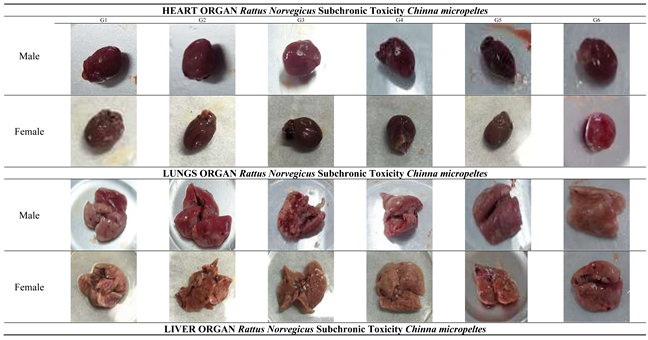
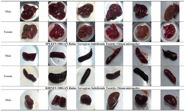
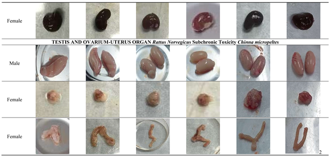
3.3. Organ Macropathology Analysis
In this study, the macropathology of the heart, liver, spleen, kidneys, lungs, and reproductive organs was assessed for both male and female rats following the 14-day administration of Channa micropeltes. The macroscopic examination of vital organs in treated animals showed no abnormalities in color or texture compared to the control group are shown in Table 3 [5].
3.4. Measurement of Relative Organ Weight
Relative organ weights of 14-day treated rats are shown in Table 4 for Male and Table 5 for Female. The relative organ weight analysis revealed no statistically significant differences between the control group and the treated groups in both male and female rats. In male rats, the heart's relative weight in the control group (0.42 ± 0.83%) remained comparable to that of the highest dose group (0.32 ± 0.07%), showing no signs of hypertrophy or atrophy. Similarly, the relative weights of other organs, including the lungs, liver (control: 3.48 ± 0.83%, highest dose: 2.94 ± 0.73%), spleen, kidneys, and testes remained stable across all groups. The satellite group (1000 mg/kg) also showed no significant changes, confirming no residual effects post-treatment. For female rats, the liver's relative weight in the control group (3.70 ± 0.57%) was comparable to that of the highest dose group (3.27 ± 0.30%), indicating no hepatotoxicity. Other organs, including the spleen, kidneys, heart, ovaries, and uterus, displayed no significant alterations in relative weights, further confirming the extract's non-toxic nature in females. The satellite group exhibited similar results, reinforcing the absence of long-term toxicity after the 14-day treatment period. The relative organ weight of each organ recorded at necropsy in the treatment groups did not show a significant difference (P > 0.05) compared to the control [12].
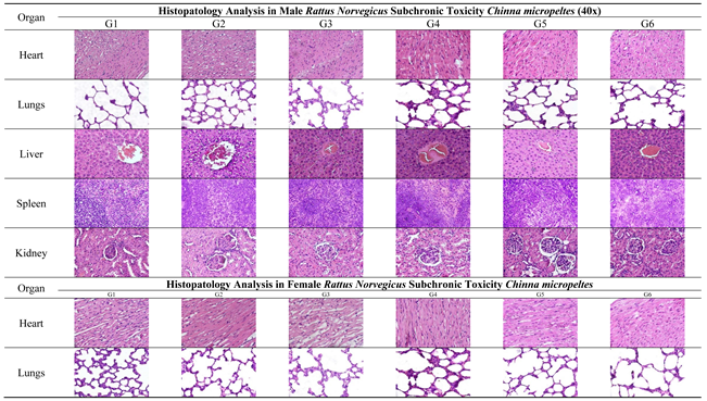
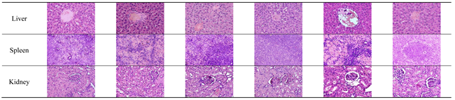
3.5. Histopathological Analyses
The light microscopy analysis of tissue sections from rats treated with Channa micropeltes extract, as well as from the control group, is shown in Table 5 and Table 6. Subchronic studies evaluate the potential adverse effects of prolonged or repeated exposure to plant extracts or compounds over a significant portion of the average lifespan of experimental animals, such as rodents. These studies specifically aim to provide information on target organ toxicity and are designed to determine the no-observed-adverse-effect level (NOAEL) [13]. The histopathological evaluation of man organs (heart, lungs, liver, spleen, kidneys, and reproductive organs) in both male and female rats treated with Channa micropeltes extract revealed no abnormalities or lesions. In this study, a histopathological score of 0 was assigned to all examined organs across treatment groups Histopathological evaluation indicated that both the control and treated groups displayed normal tissue structure, with no signs of pathological lesions in any of the organs examined.
Table 5.
Measurement of Relative Organ Weight 14 Days Subchronic Toxicity.
|
3.6. Serum Biochemical Analyses
The administration of Channa micropeltes for 14 days resulted in no significant changes in ALT, AST, Creatinine, Urea, Glucose, Total Cholesterol, and Triglicerides levels compared to the control group for both male and female rats (Table 6 and Table 7). Likewise, the satellite group, which received 1000 mg/kg of Channa micropeltes, showed no significant differences in any biochemical parameters when compared to the control group in both male and female rats. Based on the blood biochemical analysis of Channa micropeltes, the results indicate no significant differences between the control and treatment groups. Key liver function indicators, Alanine Aminotransferase (ALT) and Aspartate Aminotransferase (AST), showed no significant increase. The ALT levels in the control group were 25.3 ± 4.1 U/L, while in the group receiving the highest dose (1000 mg/kg), ALT was 26.1 ± 3.8 U/L. Similarly, the AST levels in the control group were 18.5 ± 3.2 U/L, compared to 19.2 ± 3.0 U/L in the highest-dose group. The reference range for ALT and AST is typically 35-85 U/L and 30-380 U/L. Nevertheless, the data can still be considered valid if they clearly show that the ALT values are not lower than 10% of the control, which would indicate no significant difference [14]. These results suggest that the extract did not cause significant liver damage. Additionally, kidney function parameters, such as creatinine and urea, remained stable. In the control group, creatinine levels were 0.67 ± 0.10 mg/dL, and in the highest-dose group, they were 0.71 ± 0.09 mg/dL. Urea levels were 17.4 ± 2.3 mg/dL in the control group and 18.0 ± 2.1 mg/dL in the highest-dose group. This is consistent with the reference data for creatinine and urea, which are 0.2-0.7 mg/dL and 12.33-77.6 mg/dL, respectively, indicating that the extract did not affect kidney function. The kidneys' ability to filter waste products remained intact, suggesting no nephrotoxic effects [15,16].
Glucose levels in the blood also remained within the normal range. The control group recorded a glucose level of 90.2 ± 12.4 mg/dL, while the highest-dose group had a level of 92.7 ± 11.8 mg/dL. Based on the reference glucose range of 50-160 mg/dL, the absence of significant fluctuations in glucose levels indicates that the extract did not disrupt metabolism in Channa micropeltes. Finally, the analysis of total cholesterol and triglycerides also showed no significant changes. Total cholesterol in the control group was 150.3 ± 14.6 mg/dL, and in the highest-dose group, it was 151.8 ± 13.9 mg/dL. Triglyceride levels were 80.7 ± 10.1 mg/dL in the control group and 79.5 ± 9.8 mg/dL in the highest-dose group. Based on the reference values for total cholesterol and triglycerides, which are 41-126 mg/dL and 20-114 mg/dL, respectively, the stability in cholesterol and triglyceride levels indicates that the extract did not impair lipid metabolism [16,17].
3.7. Hematological Analyses
Hematological analyses were performed using blood plasma to evaluate potential changes in blood chemistry and to detect any alterations related to Channa micropeltes extract treatment compared to the control group. The results of the hematological analysis did not show significant variations at any dose compared to the control group in both female and male rats, as presented in Table 8. Similarly, the satellite group showed no significant differences between the Channa micropeltes treated groups and the control group across all parameters [18]
4. Discussion
Repeated-dose toxicity studies are designed to reveal cumulative adverse effects, identify potential target organs, and provide a scientific basis for determining the NOAEL prior to longer-term exposure trials. Indonesian FDA Regulation PerBPOM No. 10/2022 emphasize integrated evaluation of clinical signs, growth, hematology, serum biochemistry, organ coefficients, and histopathology to capture both functional and structural toxicity across organ systems. In the context of natural-product development, subacute/subchronic safety data are especially important because bioactive extracts may exert subtle adaptive responses before overt lesions emerge [5,13].
However, this approach differs from subchronic testing outlined in the OECD Guidelines, primarily due to the nature of the product being tested, traditional medicine that will later be registered as a fitofarmaka (phytopharmaceutical product) [5,19]. The classification of traditional medicines in Indonesia is divided into three categories: jamu (traditional herbal medicine), OHT (standaritation traditional medicines), and fitofarmaka [19]. Each category is subject to different regulatory requirements and safety evaluations. BPOM RI provides guidelines indicating that subchronic toxicity testing can be conducted within a 14-day period, specifically for traditional medicines being developed for future fitofarmaka registration. This distinction highlights the regulatory framework's flexibility in accommodating traditional medicine products as they transition into more standardized health products, while ensuring thorough safety evaluation during development [5]
Oral administration in repeated-dose studies is particularly relevant because it mimics the real-world route of exposure for dietary supplements and functional foods. The clean safety profile observed here may reflect limited systemic accumulation and effective metabolic handling of the extract’s constituents. Although pharmacokinetic data were not assessed in the present work, the lack of clinical signs, stable organ coefficients, and absent histopathologic lesions suggest that repeated oral exposure did not result in bioaccumulation or persistent tissue injury. Future studies incorporating ADME or PK profiling would be valuable to confirm gastrointestinal stability, absorption extent, and clearance kinetics, thereby strengthening mechanistic understanding of the extract’s safety [20].
Body weight is a key indicator of systemic health in subchronic toxicity assessments. Throughout the 14-day oral administration, both male and female rats in all treated groups showed a progressive weight gain comparable to controls, consistent with normal physiological growth. The absence of statistically significant differences in body-weight change among dose groups indicates that Channa micropeltes extract did not interfere with growth or energy balance. Importantly, no animals exhibited weight loss or growth retardation, which are typical early signs of toxicity, and food-consumption patterns remained stable, suggesting no adverse effects on appetite, metabolism, or nutrient utilization. Given that a change of approximately ≥10% body weight is often considered biologically relevant for subchronic toxicity [21], the normal weight trajectories observed here support the conclusion that the extract is non-toxic within the tested dose range, including 1000 mg/kg [22].
The macropathological examination of major organs, including the heart, liver, spleen, kidneys, lungs, and reproductive organs, revealed no visible abnormalities in either male or female rats across all treated and satellite groups. The heart, liver, spleen, kidneys, and lungs maintained normal size, color, and texture, with no signs of necrosis, lesions, or structural damage, indicating the absence of cardiotoxic, hepatotoxic, nephrotoxic, or pulmonary effects. Similarly, the reproductive organs (testes, ovaries, and uterus) showed no signs of toxicity or interference with reproductive health. These findings suggest that Channa micropeltes extract does not induce any organ-specific toxicity, even at the highest dose of 1000 mg/kg, confirming its overall safety. The macroscopic description of specimens constitutes a critical element of the pathology report, requiring precision and clinical relevance. Excessive or redundant details can unnecessarily extend the report, thereby elevating the risk of significant clinical information being overlooked by the attending clinician. Employing standardized templates for gross pathological reporting can enhance efficiency and mitigate these risks [23].
Relative organ weight is a sensitive endpoint in repeated-dose toxicity studies because organ mass can change before clear gross or microscopic lesions are detectable; altered organ-to-body-weight ratios may reflect hypertrophy, atrophy, congestion, or edema, especially in liver, kidneys, heart, and spleen [24,25] Thus, a lack of significant organ-weight shifts is generally interpreted as no organ-specific toxic burden under the tested conditions. In this study, relative organ weights of male and female rats remained comparable between control and Channa micropeltes extract groups up to 1000 mg/kg, including the satellite cohort. The stability of major detoxification and target organs (liver, kidneys, heart, spleen, lungs) and reproductive organs suggests no adaptive enlargement or degenerative shrinkage attributable to treatment, a pattern consistent with absence of subchronic cardiotoxicity, nephrotoxicity, hepatotoxicity, or reproductive toxicity [26]. The unchanged organ weights in the recovery (satellite) group further indicate no delayed or residual organ remodeling after cessation of dosing.
Organ-weight changes are often interpreted as early adaptive or adverse responses; however, their toxicological meaning becomes strongest when concordant with histopathologic findings. Large databases have shown that increased liver weight may accompany enzyme induction or hypertrophy, while decreases may indicate atrophy or chronic injury, and the presence or absence of histological correlate helps distinguish adaptation from toxicity. In the present study, the absence of organ-weight shifts together with uniformly normal histology indicates not only a lack of morphological injury but also an absence of subclinical adaptive stress responses in detoxification organs. This concordance across endpoints substantially reduces the likelihood of undetected target-organ toxicity at the tested doses [26].
Subchronic toxicity studies are intended to detect adverse effects arising from repeated exposure over a substantial portion of the rodent lifespan and to identify potential target-organ toxicity, thereby supporting the determination of the no-observed-adverse-effect level (NOAEL) [11,13]. In the present study, histopathological examination of major organs—including the heart, lungs, liver, spleen, kidneys, and reproductive organs showed normal tissue architecture in both male and female rats treated with Channa micropeltes extract. All organs received a histopathological score of 0, and no microscopic evidence of necrosis, inflammation, fibrosis, or cellular degeneration was observed, indicating preserved structural integrity and function [27]. The absence of lesions across detoxification, highly perfused, and reproductive organs provides strong support that the extract does not induce organ-specific toxicity under subchronic exposure. Evaluating both sexes is important because hormonal and metabolic differences may influence toxic responses to natural products; however, the comparable histological findings in males and females suggest no sex-related susceptibility within the tested dose range [28]. Collectively, these results contribute valuable safety data for selecting dose levels in longer-term studies and for refining safe exposure limits in future translational assessments [16]. The inclusion of a satellite (recovery) cohort enhances interpretation by assessing reversibility or delayed-onset toxicity after dosing cessation. In this work, recovery animals displayed organ weights, histology, and clinical profiles comparable to controls, suggesting no residual tissue remodeling or late-emerging toxicity. Such findings are important for natural extracts, where metabolite-driven effects might hypothetically appear after prolonged processing in vivo. The normal recovery profile therefore supports that any exposure-related changes, if present, are unlikely to persist or progress once dosing stops [29]
Serum biochemistry provides functional evidence of target-organ integrity during repeated-dose toxicity, particularly for the liver and kidneys. In this study, Channa micropeltes extract did not produce statistically significant changes in key hepatic enzymes (ALT, AST) or renal biomarkers (creatinine, urea) compared with controls, and all values remained within physiological limits. The absence of enzyme elevation indicates no hepatocellular injury or leakage, while stable creatinine and urea suggest preserved glomerular filtration and no nephrotoxic burden under subchronic exposure [30]. Metabolic parameters also remained unaffected. Blood glucose stayed within normal range, indicating that the extract did not disrupt systemic energy homeostasis. Likewise, total cholesterol and triglycerides showed no treatment-related shifts, supporting the conclusion that lipid metabolism was not impaired [16]. Taken together with the normal histopathology and organ-weight findings, the biochemical profile consistently demonstrates that repeated oral administration of Channa micropeltes extract up to 1000 mg/kg does not elicit liver, kidney, or metabolic toxicity in rats within the study duration.
Hematological parameters are sensitive indicators of systemic and immuno-hematopoietic toxicity in repeated-dose studies. In the present work, Channa micropeltes extract did not induce statistically significant alterations in major hematological indices in either sex, including WBC, RBC, hemoglobin, hematocrit, platelet count, erythrocyte indices (MCV, MCH, RDW-SD), and differential leukocyte profiles. These findings indicate preserved hematopoiesis, oxygen-carrying capacity, and immune homeostasis under subchronic exposure [31]. Minor reductions in platelet and hematocrit values observed in a single female subgroup were not statistically significant, showed no dose-response pattern, and therefore are most consistent with normal biological variability rather than a treatment-related effect. Although reference intervals may vary slightly across sources, the overall pattern of values remaining within physiological limits supports the conclusion that the extract does not elicit hematological toxicity up to 1000 mg/kg [13]. When interpreted together with the normal organ weights, macropathology, histopathology, and serum biochemistry, the hematological profile corroborates the favorable safety of Channa micropeltes extract in this subchronic model.
An important element of toxicological interpretation is the safety margin between the highest tested dose and the expected efficacious or intended human-use dose. In this study, the top dose of 1000 mg/kg represents a multiple of the projected therapeutic exposure, thereby providing a conservative safety buffer. The absence of adverse findings at this level indicates a wide margin of safety, which is desirable for nutraceutical or biopharmaceutical candidates derived from food-based sources. Such a safety window strengthens the feasibility of advancing Channa micropeltes extract into longer duration studies and supports its practical use without requiring exposure near the toxicological threshold.
Overall, the findings of this study strengthen the translational positioning of Channa micropeltes extract as a safe bioactive candidate for further development in Ifalmin®. The consistent absence of toxic effects across systemic indicators (clinical signs and growth), functional parameters (hematology and serum biochemistry), and structural endpoints (macropathology, organ coefficients, and histopathology), including in the recovery cohort, provides a strong weight-of-evidence that short-term repeated exposure does not elicit target-organ toxicity. The uniform safety profile up to 1000 mg/kg bw/day offers a sound basis for selecting dose ranges in longer subchronic studies aligned with OECD/BPOM guidelines (14-day or 90-day studies) and for applying a more conservative approach to human-equivalent exposure estimation. From a product-development perspective, this early safety confirmation is essential to support longer-term efficacy testing and serves as a prerequisite for eventual clinical evaluation, particularly because Ifalmin® is intended as an orally administered natural-product preparation with potential for repeated use [5].
This study has several limitations, primarily related to the lack of literature on 14-day subchronic toxicity testing for traditional medicine products, which hinders the search for references and comparison with other studies. Additionally, Ifalmin, which contains Channa micropeltes (Toman fish), also has limited available literature, making it challenging to confirm safety and efficacy data in comparison with similar products. Further research will be conducted through clinical trials in humans to confirm these findings and assess the safety of the product for long-term use. It will also be registered as a fitofarmaka product in accordance with the guidelines set by BPOM RI.
5. Conclusions
This study demonstrates that a 14-day repeated-dose (subchronic) oral toxicity assessment of Channa micropeltes extract produced no treatment-related adverse effects at 270, 540, or 1000 mg/kg bw/day in either sex. No statistically significant changes were observed in body-weight gain, relative organ weights, serum biochemical parameters, or hematological indices compared with controls. Gross necropsy and histopathological evaluation of major organs showed normal architecture across control, treated, and recovery (satellite) cohorts, indicating no evidence of target-organ toxicity or delayed effects after dosing cessation. Therefore, the NOAEL is considered to be 1000 mg/kg bw/day for both males and females under the present 14-day exposure conditions. These findings provide robust preclinical safety support for the Channa micropeltes extract used in Ifalmin® and justify further development and longer-term studies to confirm chronic safety.
6. Patents
Ifalmin® is protected by Indonesian patent registration No. TR183313911, effective from 26 April 2023 to 26 April 2028. The patent is held by PT. Ismut Fitomedika Indonesia, with Dr. Mansur Ibrahim, M.Kes. listed as the inventor.
Supplementary Materials
The following supporting information can be downloaded at: https://www.mdpi.com/article/doi/s1, Figure S1: title; Table S1: title; Video S1: title.
Author Contributions
Conceptualization, M.I., M.R, M.N.M; methodology, M.I., A.W.J., and M.R.; investigation, A.W.J., N.D.L., and M.R.; resources, M.I, M.R, A.W.J, M.N.M. and N.D.L.; data curation, M.I., A.W.J., and N.D.L.; formal analysis, M.R, N.D.L, A.W.J.; validation, M.I., M.R., and M.N.M.; writing—original draft preparation, A.W.J.; writing—review and editing, M.I., A.W.J., M.R., and M.N.M.; visualization, M.I., M.R., and M.N.M.; supervision, M.I.; project administration, A.W.J.; funding acquisition, M.I. All authors have read and agreed to the published version of the manuscript.
Funding
This research was funded by PT. Ismut Fitomedika Indonesia, with funding oversight provided by Dr. Mansur Ibrahim, M.Kes.
Institutional Review Board Statement
The animal study protocol was approved by the Ethics Committee/Institutional Review Board of the Indonesian Food and Drug Authority (Badan POM RI). The study protocols were reviewed and obtained ethical clearance (Registration RG.02.05.42.423.01.8.2024.1450; Protocol No. A02/PUPK/A04/VIII/2024/Rev.3), with the date of approval 01 September 2024.
Informed Consent Statement
Not applicable
Data Availability Statement
The data presented in this study are available on request from the corresponding author. The data are not publicly available due to proprietary considerations related to product development.
Acknowledgments
The authors gratefully acknowledge PT. Ismut Fitomedika Indonesia and Dr. Mansur Ibrahim, M.Kes. for providing funding support for this study. The authors also thank the Molecular Biology Laboratory, Department of Biology, Faculty of Mathematics and Natural Sciences, Universitas Brawijaya, and all staff involved for their technical assistance and support throughout the research. During the preparation of this manuscript, the authors used ChatGPT 5.1 (OpenAI) for the purpose of polishing the grammar and fluency of the text.
Conflicts of Interest
The authors declare a potential conflict of interest because this study was funded by PT. Ismut Fitomedika Indonesia (developer/patent holder of Ifalmin®) and Dr. Mansur Ibrahim, M.Kes. (inventor). The funders contributed to conceptualization, methodology, resources, data curation, validation, review/editing, visualization, and supervision. They had no role in data collection, primary statistical analysis, or the final decision to publish.
Abbreviations
The following abbreviations are used in this manuscript:
| ADME | Absorption, Distribution, Metabolism, and Excretion |
| ALT | Alanine Aminotransferase. |
| AST | Aspartate Aminotransferase |
| BUN | Blood Urea Nitrogen |
| BPOM RI | Badan Pengawas Obat dan Makanan (Indonesian Food and Drug Authority). |
| BW | Body Weight |
| EDTA | Ethylenediaminetetraacetic Acid. |
| FDA | Food and Drug Authority |
| g | gram |
| G1-G6 | Group 1–Group 6 |
| H&E | Hematoxylin and Eosin stain. |
| Hb | Hemoglobin. |
| Hct | Hematocrit. |
| PT.IFI | Perseroan Terbatas Ismut Fitomedika Indonesia ( |
| IRB | Institutional Review Board. |
| Kg | kilogram. |
| MCV | Mean Corpuscular Volume. |
| MCH | Mean Corpuscular Hemoglobin. |
| mg | milligram. |
| mg/kg | milligram per kilogram. |
| µL | microliter. |
| µm | micrometer. |
| nm | nanometer. |
| NOAEL | No-Observed-Adverse-Effect Level. |
| OECD | Organisation for Economic Co-operation and Development. |
| PK | Pharmacokinetics. |
| PLT | Platelet Count. |
| RBC | Red Blood Cell count. |
| RDW-SD | Red Cell Distribution Width—Standard Deviation. |
| rpm | revolutions per minute. |
| SGOT | Serum Glutamic Oxaloacetic Transaminase |
| SGPT | Serum Glutamic Pyruvic Transaminase |
| TG 407 | Test Guideline 407 |
| TR | Tanda Registrasi (Registration Code) |
| WBC | White Blood Cell count. |
| % | percent. |
References
- Apriasari, M.L.; Puspitasari, D.; Utami, J.P. Anti-Inflammatory Effect of Channa micropeltes Extract in Angiogenesis of Diabetes Mellitus Wound Healing. Rev. Diabet. Stud. 2022, 18, 166–176. [CrossRef]
- Apriasari, M.L.; Ainah, Y.; Febrianty, E.; Carabelly, A.N. Antioxidant Effect of Channa micropeltes in Diabetic Wound of Oral Mucosa. Int. J. Pharmacol. 2019, 15, 137–143. [CrossRef]
- Jităreanu, A.; Trifan, A.; Vieriu, M.; Caba, I.-C.; Mârt, I.; Agoroaei, L. Current Trends in Toxicity Assessment of Herbal Medicines: A Narrative Review. Processes 2023, 11, 83. [CrossRef]
- Szpirer, C. Rat Models of Human Diseases and Related Phenotypes: A Systematic Inventory of the Causative Genes. J. Biomed. Sci. 2020, 27, 84. [CrossRef]
- BPOM (Badan Pengawas Obat dan Makanan Republik Indonesia). Peraturan Badan Pengawas Obat dan Makanan Nomor 10 Tahun 2022 tentang Pedoman Uji Toksisitas Praklinik secara In Vivo; BPOM RI: Jakarta, Indonesia, 2022. Available online: https://peraturan.bpk.go.id/Home/Details/223969/peraturan-bpom-no-10-tahun-2022 (accessed on 24 November 2025).
- Carabelly, A.N.; Firdaus, I.W.A.K.; Nurmardina, P.C.; Putri, D.A.; Apriasari, M.L. The Effect of Topical Toman Fish (Channa micropeltes) Extract on Macrophages and Lymphocytes in Diabetes Mellitus Wound Healing. J. Phys.: Conf. Ser. 2019, 1374, 012028. [CrossRef]
- Lestiariani, L.; Djabir, Y.Y.; Rahim, A. Subacute Toxicity Effects of Physalis angulata Leaf Extract on Kidneys and Liver of Female Wistar Rats. Iran. J. Toxicol. 2023, 17, 19–26. [CrossRef]
- Patel, S.; Patel, S.; Kotadiya, A.; Patel, S.; Shrimali, B.; Joshi, N.; Patel, T.; Trivedi, H.; Patel, J.; Joharapurkar, A.; Jain, M. Age-Related Changes in Hematological and Biochemical Profiles of Wistar Rats. Lab. Anim. Res. 2024, 40, 7. [CrossRef]
- Lazic, S.; Semenova, E.; Williams, D. Determining Organ Weight Toxicity with Bayesian Causal Models: Improving on the Analysis of Relative Organ Weights. Sci. Rep. 2020, 10, 6625. [CrossRef]
- Kwan, Y.P.; Darah, I.; Chen, Y.; Sreeramanan, S.; Sasidharan, S. Acute and Subchronic Toxicity Study of Euphorbia hirta L. Methanol Extract in Rats. BioMed Res. Int. 2013, 2013, 182064. [CrossRef]
- National Research Council (NRC). Toxicity Testing for Assessing Environmental Agents: Interim Report; National Academies Press: Washington, DC, USA, 2006.
- Chiranthanut, N.; Lertprasertsuke, N.; Srisook, E.; Srisook, K. Acute and Subchronic Oral Toxicity Assessment of Extract from Etlingera pavieana Rhizomes. Toxicol. Rep. 2022, 9, 1472–1483. [CrossRef]
- OECD. Test No. 407: Repeated Dose 28-day Oral Toxicity Study in Rodents; OECD Guidelines for the Testing of Chemicals, Section 4; OECD Publishing: Paris, France, 2025. [CrossRef]
- Verma, T.; Singh, J.; Aggarwal, A. Optimization of Soya Enriched Burfi by Using Response Surface Methodology and Its Impact on Biochemical Parameters in Albino Rats. Food Chem. Adv. 2024, 5, 100791. [CrossRef]
- Loeb, W.F.; Quimby, F.W. The Clinical Chemistry of Laboratory Animals, 2nd ed.; Taylor & Francis: Philadelphia, PA, USA, 1999.
- Delwatta, S.L.; Gunatilake, M.; Baumans, V.; Seneviratne, M.D.; Dissanayaka, M.L.; Batagoda, S.S.; Udagedara, A.H.; Walpola, P.B. Reference Values for Selected Hematological, Biochemical and Physiological Parameters of Sprague-Dawley Rats at the Animal House, Faculty of Medicine, University of Colombo, Sri Lanka. Anim. Model. Exp. Med. 2018, 1, 250–254. [CrossRef]
- Muharni; Ferlinahayati; Fitrya; Eliza; Yohandini, H.; Cenora, C. Subchronic Toxicity Study of Ethanol Extract of Poronema canescens Jack. Leaves on White Rats (Rattus noverticus, Wistar Strain). J. Sains Farm. Klin. 2023, 10, 211–217.
- Dewi, I.G.A.M.A.; Adi, A.A.A.M.; Setiasih, N.L.E. Fluctuations in the Hematological Profile of the White Rat Animal Model of Benzo(a)pyrene-Induced Fibrosarcoma. Indones. Med. Vet. 2022, 11, 267–281. [CrossRef]
- Badan Pengawas Obat dan Makanan Republik Indonesia. Keputusan Kepala Badan Pengawas Obat dan Makanan Republik Indonesia Nomor HK.00.05.4.2411/2004 tentang Ketentuan Pokok Pengelompokan dan Penandaan Obat Bahan Alam Indonesia. Badan POM RI; 2004. Available online: https://jdih.pom.go.id/download/rule/905/HK.00.05.4.2411/2004 (accessed on 25 November 2025).
- ECETOC (European Centre for Ecotoxicology and Toxicology of Chemicals). ECETOC Guidance on Dose Selection. Technical Report No. 138; ECETOC: Brussels, Belgium, 2021. Available online: https://www.ecetoc.org/wp-content/uploads/2021/10/ECETOC-TR-138-Guidance-on-Dose-Selection_Final.pdf (accessed on 24 November 2025).
- Organization for Economic Cooperative Development (OECD). OECD Guideline for the Testing of Chemicals, No. 408: Repeated Dose 90-Day Oral Toxicity Study in Rodents. OECD, 1998.
- Rosidah, I.; Renggani, T.N.; Firdausi, N.; Ningsih, S.; Yunianto, P.; Permatasari, D.; Pongtuluran, O.B.; Bahua, H.; Efendi, J.; Kusumastuti, S.A.; Nuralih; El Muttaqien, S.; Nizar; Kusumaningrum, S.; Agustini, K. Acute and Subchronic Toxicological Study of the Cocktail Extract from Curcuma xanthorrhiza Roxb, Phyllanthus niruri L., and Morinda citrifolia L. J. Toxicol. 2024, 2024, 9445226. [CrossRef]
- Varma, M.; Collins, L.C.; Chetty, R.; Karamchandani, D.M.; Talia, K.; Dormer, J.; Vyas, M.; Conn, B.; Guzmán-Arocho, Y.D.; Jones, A.V.; Pring, M.; McCluggage, W.G. Macroscopic Examination of Pathology Specimens: A Critical Reappraisal. J. Clin. Pathol. 2024, 77, 164–168. [CrossRef]
- Piao, Y.; Liu, Y.; Xie, X. Change trends of organ weight background data in Sprague Dawley rats at different ages. J. Toxicol. Pathol. 2013, 26, 29–34. [CrossRef]
- Sellers, R.S.; Morton, D.; Michael, B.; et al. Society of Toxicologic Pathology Position Paper: Organ Weight Recommendations for Toxicology Studies. Toxicol. Pathol. 2007, 35, 751–755. [CrossRef]
- Mezencev, R.; Feshuk, M.; Kolaczkowski, L.; Peterson, G.C.; Zhao, Q.J.; Watford, S.; Weaver, J.A. The association between histopathologic effects and liver weight changes induced in mice and rats by chemical exposures: an analysis of the data from Toxicity Reference Database (ToxRefDB). Toxicol. Sci. 2024, 200, 404–413. [CrossRef]
- Murugan, S.; Solanki, H.; Purusothaman, D.; Bethapudi, B.; Ravalji, M.; Mundkinajeddu, D. Safety Evaluation of Standardized Extract of Curcuma longa (NR-INF-02): A 90-Day Subchronic Oral Toxicity Study in Rats. BioMed Res. Int. 2021, 2021, 6671853. [CrossRef]
- Schilter, B.; Andersson, C.; Anton, R.; Constable, A.; Kleiner, J.; O'Brien, J.; Renwick, A.G.; Korver, O.; Smit, F.; Walker, R. Guidance for the Safety Assessment of Botanicals and Botanical Preparations for Use in Food and Food Supplements. Food Chem. Toxicol. 2003, 41, 1625–1649. [CrossRef]
- Healey, L.; et al. Recovery Animals in Toxicology Studies. Toxicol. Pathol. 2024, 52, Article 10915818241243350. [CrossRef]
- Mary, L.A.; Charles, B.C. Clinical Laboratory Parameters for Crl:WI (Han). Charles River, 2008.
- Jasper, R.; Locatelli, G.O.; Pilati, C.; Locatelli, C. Evaluation of biochemical, hematological and oxidative parameters in mice exposed to the herbicide glyphosate-Roundup(®). Interdiscip. Toxicol. 2012, 5, 133–140. [CrossRef]
Table 1.
Body Weight Monitoring 14 Days Subchronic Toxicity.
| Group | Male | Female |
|---|---|---|
| G1 | 128 ± 1.91 | 120.7 ± 2.28 |
| G2 | 136 ± 8.24 | 132.9 ±12.82 |
| G3 | 137.5 ± 9.83 | 118.98 ± 2.04 |
| G4 | 132.7 ± 5.95 | 120.98 ± 2.12 |
| G5 | 133.5 ± 7.46 | 125.9 ± 6.51 |
| G6 | 129.38 ± 5.39 | 124.98 ± 5.86 |
Table 3.
Organ macropathology Analysis 14 Days Subchronic Toxicity.
|
Table 4.
Measurement of Relative Organ Weight 14 Days Subchronic Toxicity.
| Male | ||||||
| Organ | Group | |||||
| G1 | G2 | G3 | G4 | G5 | G6 | |
| Heart | 0.42 ± 0.83 | 0.35 ± 0.04 | 0.28 ± 0.15 | 0.32 ± 0.07 | 0.31 ± 0.06 | 0.37 ± 0.07 |
| Lung | 1.12 ± 0.65 | 0.91 ± 0.26 | 0.76 ± 0.06 | 0.76 ± 0.14 | 2.03 ± 3.54 | 0.79 ± 0.55 |
| Liver | 3.48 ± 0.83 | 3.34 ± 1.15 | 3.26 ± 0.56 | 2.94 ± 0.73 | 2.93 ± 0.58 | 3.47 ± 0.68 |
| Spleen | 0.32 ± 0.11 | 0.43 ± 0.33 | 0.44 ± 0.29 | 0.47 ± 0.31 | 0.35 ± 0.22 | 0.43 ± 0.28 |
| Kidney | 0.81 ± 0.22 | 0.76 ± 0.17 | 0.74 ± 0.16 | 0.74 ± 0.16 | 0.74 ± 0.24 | 0.69 ± 0.16 |
| Testis | 2.11 ± 1.43 | 1.37 ± 0.98 | 1.43 ± 0.24 | 1.26 ± 0.47 | 1.43 ± 0.35 | 1.37 ± 0.47 |
| Female | ||||||
| Organ | Group | |||||
| G1 | G2 | G3 | G4 | G5 | G6 | |
| Heart | 0.37 ± 0.10 | 0.35 ± 0.05 | 0.34 ± 0.05 | 0.36 ± 0.04 | 0.04 ± 0.12 | 0.38 ± 0.10 |
| Lung | 0.93 ± 0.28 | 0.81 ± 0.08 | 1.10 ± 0.71 | 1.01 ± 0.46 | 0.94 ± 0.18 | 0.89 ± 0.36 |
| Liver | 3.70 ± 0.57 | 3.06 ± 0.87 | 3.72 ± 0.62 | 3.27 ± 0.30 | 3.51 ± 0.59 | 3.78 ± 1.04 |
| Spleen | 0.61 ± 0.16 | 0.82 ± 0.31 | 0.55 ± 0.34 | 0.50 ± 0.42 | 0.75 ± 0.30 | 0.75 ± 0.23 |
| Kidney | 0.84 ± 0.13 | 0.67 ± 0.14 | 0.86 ± 0.19 | 0.82 ± 0.05 | 0.78 ± 0.16 | 0.79 ± 0.11 |
| Ovarium | 0.12 ± 0.04 | 0.06 ± 0.06 | 0.28 ± 0.53 | 0.25 ± 0.36 | 0.06 ± 0.04 | 0.06 ± 0.02 |
| Uterus | 0.15 ± 0.04 | 0.22 ± 0.10 | 0.27 ± 0.14 | 0.18 ± 0.10 | 0.33 ± 0.21 | 0.15 ± 0.07 |
Table 7.
Serum Biochemical Analyses in Rattus novergicus 14 Days Subchronic Toxicity.
| Male | ||||||
| Parameter | Group | |||||
| G1 | G2 | G3 | G4 | G5 | G6 | |
| Trigliserida | 49.4 ± 18.17 | 48.2 ± 28.80 | 36 ± 8.15 | 39.2 ± 8.35 | 39.2 ± 19.77 | 33.4 ± 8.17 |
| Glucose | 68.4 ± 17.27 | 66.2 ± 26.21 | 75 ± 9.87 | 98.4 ± 2.35 | 122.4 ± 5.50 | 95.6 ± 0.45 |
| Colesterol | 76.4 ± 12.70 | 53.6 ± 12.10 | 62.4 ± 6.80 | 57.6 ± 5.77 | 59.8 ± 10.33 | 62.4 ± 10.24 |
| Creatinin | 0.64 ± 0.05 | 0.64 ± 0.11 | 0.64 ± 0.05 | 0.58 ± 0.04 | 0.54 ± 0.05 | 0.48 ± 0.04 |
| Urea BUN | 32.06 ± 3.06 | 29.54 ± 9.27 | 23.18 ± 1.30 | 26.96 ± 13.53 | 29.78 ± 2.90 | 25.38 ± 1.84 |
| AST/SGOT | 182.8 ± 37.41 | 192.6 ± 54.90 | 140.4 ± 25.37 | 157.2 ± 41.32 | 244.8 ± 22.48 | 190.2 ± 24.46 |
| ALT/SGPT | 50 ± 14.46 | 93 ± 43.92 | 43.2 ± 1.92 | 45.2 ± 14.32 | 65.2 ± 7.85 | 53 ± 14.62 |
| Female | ||||||
| Organ | Group | |||||
| G1 | G2 | G3 | G4 | G5 | G6 | |
| Trigliserida | 47.2 ± 15.25 | 75.2 ± 28.67 | 41 ± 18.37 | 56.2 ± 31.58 | 56.6 ± 28.95 | 63.4 ± 19.50 |
| Glucose | 99.6 ± 17. 85 | 76.6 ± 16.20 | 140.2 ± 36.48 | 98.8 ± 27.18 | 82.8 ± 13.20 | 89.2 ± 14.45 |
| Colesterol | 70.6 ± 14.70 | 59.4 ± 14.72 | 56.4 ± 9.07 | 55.4 ± 8.20 | 74.4 ± 11.63 | 63.6 ± 2.07 |
| Creatinin | 0.52 ± 0.04 | 0.64 ± 0.05 | 0.58 ± 0.08 | 0.58 ± 0.08 | 0.6±0.2 | 0.54±0.09 |
| Urea BUN | 35.48 ± 7.36 | 34.62 ± 6.40 | 36.46 ± 10.36 | 39.48 ± 6.05 | 32.46 ± 9.88 | 31.64 ± 9.66 |
| AST/SGOT | 245 ± 141.83 | 213 ± 63.59 | 224 ± 40.09 | 180.6 ± 23.20 | 171 ± 40.67 | 199.2 ± 144.53 |
| ALT/SGPT | 88.8 ± 48.90 | 53.8 ± 27.46 | 77.8 ± 25.96 | 69.2 ± 36.93 | 48.8 ± 33.03 | 81.6 ± 74.74 |
Table 8.
Measurement of Relative Organ Weight 14 Days Subchronic Toxicity.
| Male | ||||||
| Parameter | Group | |||||
| G1 | G2 | G3 | G4 | G5 | G6 | |
| WBC | 11.16 ± 3.82 | 7.56 ± 2.16 | 9.28 ± 4.66 | 4.95 ± 2.14 | 11.16 ± 3.83 | 6.2 ± 1.40 |
| RBC | 6.97 ± 0.67a | 7.94 ± 1.29 | 7.43 ± 0.56 | 7.69 ± 0.89 | 7.27 ± 0.70a | 6.80 ± 0.73 |
| Hb | 13.74 ± 2.02 | 14.74 ± 2.20 | 14.04 ± 1.14 | 13.9 ± 1.77 | 14.18 ± 1.74 | 14.58 ± 0.87 |
| Hct | 37.76 ± 3.89 | 41.66 ± 6.50 | 39.2 ± 3.12 | 39.92 ± 6.41 | 39.38 ± 5.16 | 40.5 ± 2.44 |
| PLT | 919.6 ± 31.67 | 865.2 ± 113.84 | 926 ± 104.86 | 853.5 ± 86.38 | 770.6 ± 138.09 | 779.6 ± 268.08 |
| MCV | 53 ± 2.12 | 51.2 ± 1.48 | 51 ± 2.94 | 51.8 ± 0.84 | 50 ± 1 | 52.6 ± 4.03 |
| RDW-SD | 18.24 ± 1.55 | 17.66 ± 1.22 | 17.98 ± 1.38 | 17.02 ± 0.96 | 17.32 ± 1.83 | 18.6 ± 2.73 |
| MCH | 19.6 ± 1.08 | 18.88 ± 0.70 | 18.62 ± 0.59 | 18.07 ± 0.99 | 19.48 ± 1.47 | 21.54 ± 1.72 |
| Neutrophils | 27.6 ± 16.34 | 20.8 ± 9.57 | 18.4 ± 7.92 | 28.5 ± 7.93 | 25.2 ± 6.14 | 28 ± 11.64a |
| Lymphocytes | 66.4 ± 16.02 | 73.8 ± 9.33 | 75.8 ± 9.09 | 65.5 ± 10.26 | 68.8 ± 5.76 | 65.4 ± 11.08 |
| Monocytes | 6 ± 1.22 | 5.4 ± 1.14 | 5.8 ± 1.78 | 6 ± 2.55 | 6 ± 0.82 | 6.4 ± 1.26 |
| Female | ||||||
| Organ | Group | |||||
| G1 | G2 | G3 | G4 | G5 | G6 | |
| WBC | 12.48 ± 7.20 | 11.4 ± 6.68 | 7.02 ± 4.49 | 4.26 ± 2.16 | 8.7 ± 5.29 | 10.34 ± 2.23 |
| RBC | 6.41 ± 1.32 | 6.91 ± 0.58 | 6.86 ± 0.54 | 7.47 ± 0.63 | 5.83 ± 0.62 | 5.21 ± 1.73 |
| Hb | 12.4 ± 3.38 | 13.57 ± 1.13 | 13.2 ± 1.43 | 13.96 ± 1.00 | 13.44 ± 1.65 | 12.08 ± 3.96 |
| Hct | 37.04 ± 3.82 | 37.84 ± 2.57 | 37.24 ± 2.65 | 39.44 ± 2.50 | 34.18 ± 8.96 | 33.56 ± 10.13 |
| PLT | 889.8 ± 425.98 | 667.83 ± 173.90 | 707.6 ± 190.97 | 704.4 ± 169.94 | 809.4 ± 370.46a | 713.4 ± 146.22 |
| MCV | 51.4 ± 3.71 | 54.6 ± 1.67 | 53.6 ± 1.81 | 52.6 ± 2.50 | 56.8 ± 5.80 | 57.8 ± 5.54 |
| RDW-SD | 37.04 ± 3.82 | 37.84 ± 1.58 | 37.24 ± 1.85 | 39.44 ± 1.11 | 19.02 ± 2.19 | 33.56 ± 1.93 |
| MCH | 19.94 ± 3.12 | 18.24 ± 1.58 | 18.46 ± 1.85 | 16.64 ± 1.11 | 19.02 ± 2.19 | 19.56 ± 1.93 |
| Neutrophils | 23.6 ± 7.02a | 17.2 ± 3.11 | 26.6 ± 9.72 | 24 ± 11.95 | 22 ± 4.63 | 16 ± 7.61 |
| Lymphocytes | 72.6 ± 7.12 | 76.6 ± 4.44 | 68.6 ± 10.18 | 70 ± 11.74 | 72.2 ± 5.63a | 78.6 ± 8.59 |
| Monocytes | 3.8 ± 1.30 | 6.2 ± 2.28 | 4.8 ± 0.83 | 6 ± 2.0 | 5.8 ± 2.38 | 5.4 ± 2.19 |
Disclaimer/Publisher’s Note: The statements, opinions and data contained in all publications are solely those of the individual author(s) and contributor(s) and not of MDPI and/or the editor(s). MDPI and/or the editor(s) disclaim responsibility for any injury to people or property resulting from any ideas, methods, instructions or products referred to in the content. |
© 2025 by the authors. Licensee MDPI, Basel, Switzerland. This article is an open access article distributed under the terms and conditions of the Creative Commons Attribution (CC BY) license (http://creativecommons.org/licenses/by/4.0/).
Copyright: This open access article is published under a Creative Commons CC BY 4.0 license, which permit the free download, distribution, and reuse, provided that the author and preprint are cited in any reuse.


