Preprint
Article

This version is not peer-reviewed.

Pseudomonas aeruginosa Phage Cocktails: Rational Design and Efficacy against Mouse Wound and Systemic Infection

Submitted:

13 November 2025

Posted:

26 November 2025

You are already at the latest version

Abstract

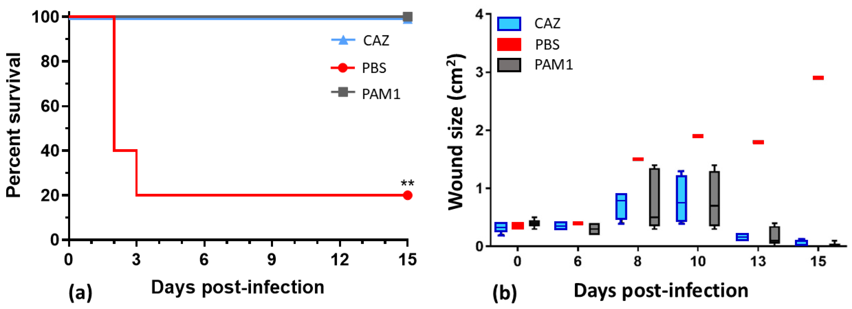
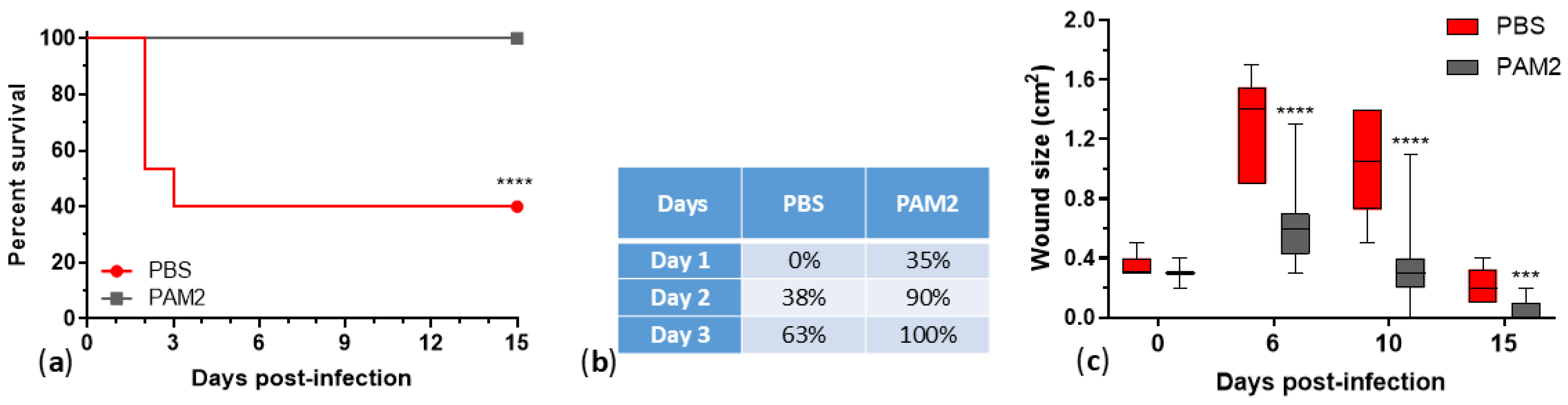
Phages show efficacy against multidrug-resistant Pseudomonas aeruginosa, but limited host ranges require combining them in cocktails. In this work, we characterized 25 P. aeruginosa phages, developed therapeutic cocktails active against diverse clinical isolates, and tested phage efficacy in a mouse incisional wound model. These phages represent seven genera, and genomic and phenotypic analyses indicate that 24/25 are lytic and suitable for phage therapy. Phage host ranges on a diversity panel of 156 P. aeruginosa strains that included 106 sequence types varied from 8% to 54%, and together the 24 lytic phages were active against 133 strains (85%). All of the phages reduced bacterial counts in biofilms. A cocktail of five lytic phages, WRAIR_PAM1, covered 56% of the strain panel, protected 100% of mice from lethal systemic infection (vs. 20% survival in the saline-treated group) and accelerated healing of infected wounds. An improved 5-phage cocktail, WRAIR_PAM2, was formulated by a rational design approach (using phages with broader host ranges, more complementing activity, relatively low resistance background, and compatibility in mixes). WRAIR_PAM2 covered 76% of highly diverse clinical isolates and demonstrated significant efficacy against topical and systemic P. aeruginosa infection, indicating that it is a promising therapeutic candidate.

Keywords: 
;  ;  ;  ;  ;  ;  ;  ;  ;  ;  ;  

1. Introduction

Pseudomonas aeruginosa is a ubiquitous and globally spread gram-negative bacterium and important opportunistic human pathogen that causes >7% of nosocomial infections [1,2]. P. aeruginosa is a major causative agent of soft tissue infections including burns [3-7], acute traumatic [8-11] and chronic wounds [12-15]. These infections are typically difficult to treat because of P. aeruginosa frequent multidrug resistance [3,9,11,16] and robust biofilm formation associated with enhanced antimicrobial tolerance and wound infection persistence [6,9,12,14,17,18]. The common occurrence of multidrug-resistant (MDR), extensively drug-resistant (XDR) or even pandrug-resistant (PDR) P. aeruginosa infections leaves clinicians rapidly diminishing options for antibiotic therapy, which warrants the urgent development of new antimicrobials to augment antibiotics [2,16,19].
A promising class of alternative antibacterials is lytic bacteriophages (phages) [20-25]. Phages are capable of dispersing P. aeruginosa biofilms and killing bacteria in biofilms [26-28]. A number of review articles have described significant efficacy of lytic phages against P. aeruginosa infections in different animal models [2,29-33]. Notable efficacy of phages has been shown against P. aeruginosa infections of acute traumatic [34,35] and burn [36-38] wounds in mice, incisional wounds in rats [39] and rabbits [40], as well as chronic diabetic foot infection in rats [41].
Several clinical cases indicated the efficacy of compassionate phage treatment against P. aeruginosa infections. For example, a phage cocktail applied intravenously had a remarkable therapeutic effect in a patient with septicemia caused by an XDR strain of P. aeruginosa [42]. Phage in combination with ceftazidime completely cured chronic P. aeruginosa infection of an aortic Dacron graft [43]. While antibiotics alone were not effective, positive outcomes of phage treatment combined with antibiotics were observed in patients with severe or refractory XDR P. aeruginosa infections, including pneumonia, recurrent bacteremia [44], intravascular stent [45] and left ventricular assist device driveline infection [46,47], osteomyelitis in different bones, mastoiditis, otitis media, sinusitis, joint and prosthetic joint infections [47].
However, human clinical trials testing phage therapeutics against P. aeruginosa infections have shown more modest results. For example, single-dose phage therapy for chronic otitis caused by drug-resistant P. aeruginosa with a phage cocktail showed safety but resulted in significant clinical improvement and reduction of bacterial burden in only 6/12 patients, and only three patients had bacterial burden under the detectable level by day 42 [48]. A double-blind, randomized Phase 1/2 trial called PhagoBurn compared the efficacy of topical application of a 12-phage cocktail with standard of care (SOC: 1% sulfadiazine silver emulsion cream) treatment against P. aeruginosa infection of burn wounds [49]. In this study, participants in the phage treatment group were less extensively burned and older than those in the SOC group. Prior to the study, only two of the 12 phages were tested for stability in storage, and none of the phages were evaluated for compatibility in mixes. Instability of the cocktail drug product appeared to cause a drop in phage titer by seven logs, from 109 to 102 plaque-forming units (PFU) per mL, from the time of manufacture to the time of application. Despite this significant reduction in potency, there was still a reduction in bacterial burden in 9/17 (53%) of subjects in the phage-treated group, compared with 13/17 (76%) of subjects in the SOC-treated group [49]. Eight human clinical trials that involved 9-96 patients treated with phages against chronic otitis, skin ulcers and wounds, septicemia, and systemic infections caused by P. aeruginosa produced mixed results that varied from trial to trial and from case to case. While phage therapy demonstrated efficacy in most of the cases in five trials, two trials showed no evidence of a therapeutic effect, and in one trial phage efficacy was lower than that of a SOC antibiotic [22]. There are still no FDA-licensed phage therapeutics in the USA [50].
Most P. aeruginosa phages have relatively limited host ranges, with up to 50% of strains susceptible to a single phage [51-58]. Of course, measurements of host range are dependent upon the diversity of the strain panel used for testing. Some phages reported with broader activity were tested against small P. aeruginosa strain panels [26,39,59-63] and/or bacterial isolates with limited or undefined diversity [39,59,60,63-66], e.g., isolated from the same hospital [63,65]. Moreover, P. aeruginosa commonly yields phage-resistant mutants, sometimes at high rates [58,65-74]. Therefore, therapeutic phage mixes (cocktails) need to be developed to cover the majority of clinical isolates and to limit the emergence of phage resistance in the target pathogen [26,35,36,49,55,63,66,67,75-79]. The purpose of this work was to characterize a panel of diverse P. aeruginosa phages, develop phage cocktails with broad activity against MDR clinical isolates, and evaluate their therapeutic efficacy against P. aeruginosa wound infection in mice. We developed two 5-phage cocktails that showed efficacy against topical and systemic infection in mice. One of these cocktails, formulated by rational design (using phages with broader and complementary host ranges, compatible in the mix and binding to different host receptors associated with virulence) showed broader coverage, 76% (119/156) of highly diverse P. aeruginosa strains.

2. Results

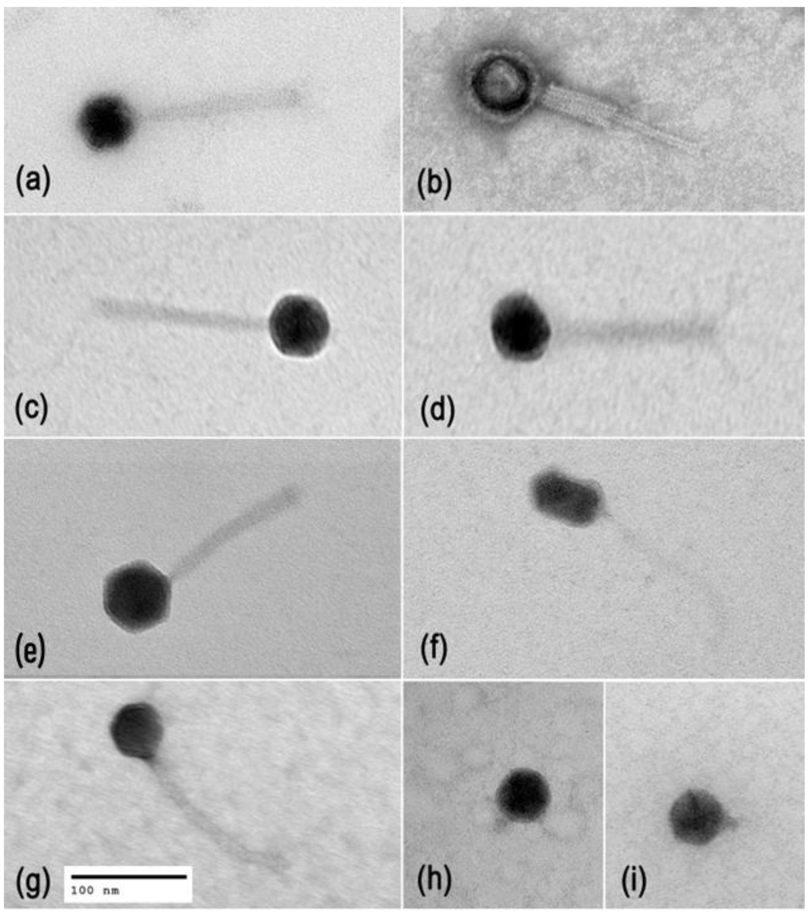
2.1. Phage Diversity, Lifestyles, and Morphology

Twenty-five P. aeruginosa phages (Table 1) were previously isolated, and their genomes were sequenced and published as genome announcements [80,81]. Here, we verified phage taxonomy and lifestyles. Mash alignment [82] against the INPHARED database [83] and verification in the ICTV Taxonomy Browser (https://ictv.global/taxonomy, MSL39.v4, accessed 10 November 2025) allowed classification of the phages into seven genera, including myophages (Pbunavirus and Nankokuvirus), siphophages (Epaquintavirus, Yuavirus, and Septimatrevirus), and podophages (Bruynoghevirus and Hollowayvirus) (Table1, Figure 1). The first four genera represent three recently established families (Lindbergviridae, Vandenendeviridae and Mesyanzhinovviridae), while Septimatrevirus, Bruynoghevirus and Hollowayvirus phages are currently unclassified at the family level. Phage virion morphology was studied by transmission electron microscopy (TEM), which confirmed what the genomic similarities indicated. Typical images of phage virions are shown in Figure 2.
Phage lifestyle predicting classifier BACPHLIP [86] scored 24 of 25 phages between 75.00% (EPa43) and 99.95% (EPa4), suggesting a virulent lifestyle. The only exception was EPa33, which was scored 7.50%, a representative of the genus Hollowayvirus, which includes a number of lysogenic phages similar to F116, a generalized transducer [87]. BLASTn analysis of EPa33 genome against the standard nr/nt database displayed many regions of up to 99% identity to P. aeruginosa chromosomal DNA, specifically, to entire or partial prophage genomes, suggesting that EPa33 is a lysogenic phage and a potential transducer that cannot be used for therapy. For all of the other phages, no significant homology with bacterial DNA was found, and no significant homology was observed between putative proteins of these 24 phages and products associated with lysogenicity, gene transfer, or bacterial proteins, including antibiotic resistance determinants [88] and virulence factors [89]. Therefore, all phages characterized in this work except Hollowayvirus phage EPa33 appear to be lytic and safe for therapeutic use.
Most of the phages were isolated from three different sewage fractions collected on the same day. Exceptions were EPa38, EPa39 and EPa43 obtained from lake water, and EPa40 that was isolated from soil. The most prevalent isolates belonged to the genus Pbunavirus: 13/25 (52%), see Table 1 and Figure 1.

2.2. Phage Host Range Testing against Highly Diverse P. aeruginosa Strains

Phage host ranges were tested by plaque assays against a diversity panel of 156 P. aeruginosa strains, which included mostly drug-resistant isolates from multiple military hospitals located in different U.S. states, Guam and Afghanistan. The sources of strains included various specimens from patients and hospital environmental swabs (Table S1). The strains were kindly provided by the Multidrug-Resistant Organism Repository and Surveillance Network (MRSN) at Walter Reed Army Institute of Research (WRAIR). The overall panel was established using a core genome multilocus sequence typing (MLST) approach and contains 106 sequence types (STs). This larger panel includes a published 100-strain diversity panel recommended for research and testing new drugs [90] (highlighted in blue in Table S1) that represents 91 STs and 71 distinct drug resistance profiles, including high-risk and epidemic lineages widely spread in the U.S. and globally: ST235, ST111, ST244, ST357, ST175, and ST654 [91-94]. All of the phages produced clear plaques on at least some of the strains. Phage host ranges varied from 7.7% (EPa38) to 54.5% (EPa15) (Table 1). Altogether, the 24 lytic phages were active against 133/156 (85.3%) of the strains. Twenty-three P. aeruginosa strains were resistant to all 25 phages.

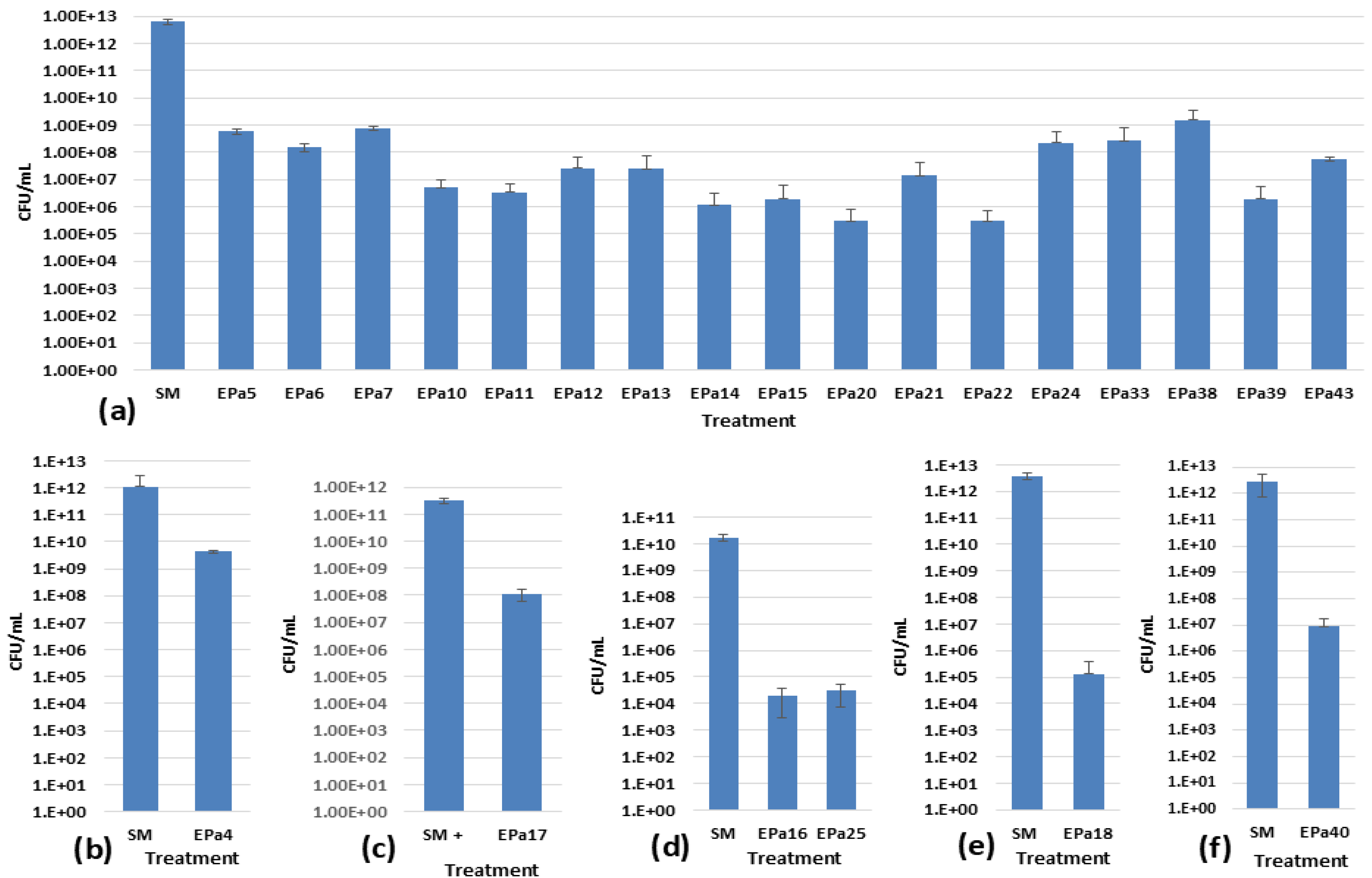
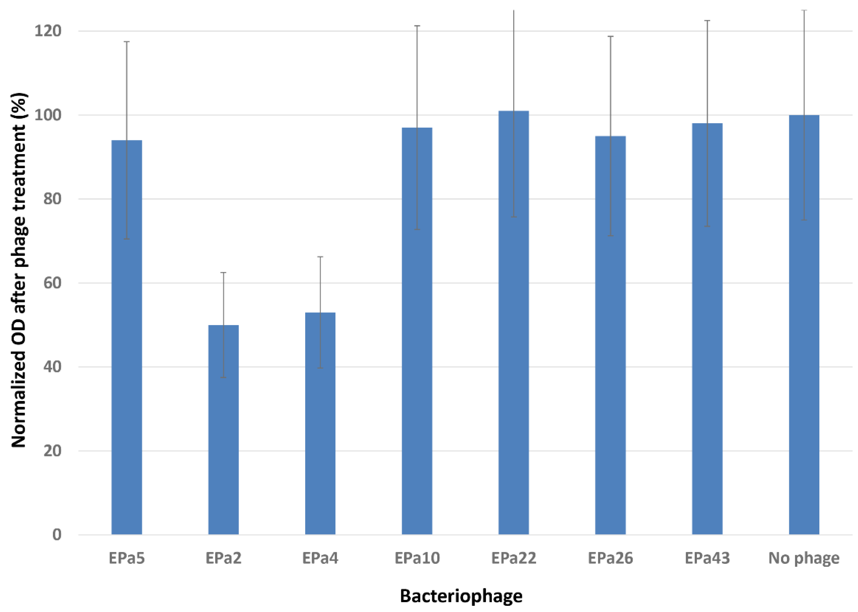
2.3. Phage Anti-biofilm Activity

The ability of phages to degrade biofilm was tested using the crystal violet method with a 96-well MBEC inoculator, as described in Materials and Methods. These experiments performed in sextuplicate showed that only two phages, EPa2 and EPa4, partially dispersed P. aeruginosa biofilms (Table 1, Figure 3). Visualization of phage plaque morphology using a dark-field colony counter showed that each of the 25 phages produces plaques with halos, which are indicative of phage polysaccharide depolymerase and anti-biofilm activity [95]. However, the width and intensity of halos varied and depended upon the P. aeruginosa strain used (data not shown). This might suggest an underestimation of the number of phages with anti-biofilm activity. Therefore, bactericidal activity of each phage in P. aeruginosa biofilms was assessed using six P. aeruginosa strains that were permissive for different phages, MRSN 1409 (for EPa17), MRSN 1680 (for EPa16 and EPa25), MRSN 1899 (for EPa2 and EPa4), MRSN 8130 (for EPa18), ATCC 10145 (for EPa40), and PAO1::lux (for all other phages). The results demonstrated that all phages effectively kill P. aeruginosa in biofilms, reducing bacterial counts by 2-7 logs (Figure 4).
The high counts of P. aeruginosa (n×1012 CFU/mL) observed in our 42-h biofilms have been previously reported by others for several bacterial species grown under different conditions [96-105].

2.4. Phage Cocktail WRAIR_PAM1 and Its Efficacy in a Mouse Wound Model

The first iteration of P. aeruginosa therapeutic 5-phage cocktail, WRAIR_PAM1 (PAM1), was formulated before the phages were fully characterized. Its composition was based entirely on phage anti-biofilm activity. In addition to phages EPa2 and EPa4 that showed some biofilm dispersal activity (Figure 3), PAM1 included EPa5, EPa6, and EPa17 that were first tested for killing P. aeruginosa in biofilms and demonstrated a bactericidal effect. This cocktail was lytic against 87/156 strains and thus covered 55.8% of the diversity panel. PAM1 was tested for efficacy in a mouse dorsal full-thickness wound infection model (Figure 5). Three groups of five BALB/c mice each were challenged with P. aeruginosa strain PAO1::lux and treated for five days. Group 1 was treated with 109 PFU of PAM1, once a day, both topically and intraperitoneally (IP). Group 2 was treated with phosphate- buffered saline (PBS), once a day, topically and IP (negative control). Group 3 was treated with standard-of-care antibiotic ceftazidime (CAZ, positive control, given only IP twice a day). Both PAM1 and CAZ protected 100% of mice from lethal systemic P. aeruginosa infection compared to 20% survival in the negative control group treated with PBS (see Figure 5a) and accelerated healing of infected dorsal wounds. Wound area in phage-treated and ceftazidime-treated mice at day 16 was 0.0 cm2 vs. necrotizing, expanding wound of 2.9 cm2 in the PBS-treated, surviving mouse (Figure 5b). Bacterial burden at day 15 in phage- and antibiotic-treated mice was below the limit of detection (104 photons/second) compared to 106 in the PBS-treated survivor (data not shown).

2.5. Using Rational Design for Developing an Improved Phage Cocktail

The purpose of further experiments was to develop an improved therapeutic phage cocktail with broader coverage of MDR P. aeruginosa clinical isolates and low bacterial resistance. We endeavored to use phages with broader and more complementary host ranges, that bind to different receptors (such as virulence determinants, to promote emergent phage-resistant phenotypes with virulence attenuation trade-offs), have relatively low resistance frequencies, and do not show antagonism to each other in mixes.

2.6. Identification of Phage Receptors on Bacterial Cell Surface

Panels of P. aeruginosa PAO1 knockout mutants with defects in the biosynthesis of different LPS components, type IV pili, and flagella were used for plaque assays to identify phage host receptors (Table S2). All of the phages lytic against the strain PAO1 required type IV pili (T4P), but differed in their secondary binding receptor, utilizing various LPS moieties, including core, A, or B bands, especially long and very long B bands (Tables 1 and S2).

2.7. Predicted Pairwise Host Ranges and Selection of Promising Phage Candidates

Lytic phages of five genera (Pbunavirus, Nankokuvirus, Bruynoghevirus, Epaquintavirus, and Septimatrevirus) except EPa5 and EPa25 that had relatively narrow lytic spectra (see Table 1) were analyzed for predicted host range complementarity in pairs. Seven phages (EPa11, EPa16, EPa17, EPa22, EPa24, EPa40, and EPa43) appeared to have good complementarity in all 21 pairwise combinations, with predicted broader activity in mixes than as individual phages (Table 2). These seven phages, which belong to four different genera, have relatively broad host ranges and bind to at least four different receptors (Table 1), were selected for further characterization as promising candidates for a new phage cocktail with expanded activity. The mix of these seven phages (Mix-7) covered 78.2% of the P. aeruginosa strain panel, and a mix of five of them (EPa11, EPa17, EPa22, EPa24, and EPa43, Mix-5) covered 76.3% of the strains (Table 2).

2.8. Testing Stability of Single Phages and Their Pairs.

The PhagoBurn clinical trial suggested instability of some P. aeruginosa phages in mixes [49]. Therefore, the seven selected phages and their pairwise combinations were tested for stability in storage at 4°C. No significant reduction of viability of each of the seven phages and their 21 pairwise mixes was observed for nine months (Figure S1). Moderate fluctuations of the titers were within the error of the method.

2.9. Measuring Frequencies of Host Resistance Mutations

Determination of frequencies of P. aeruginosa resistance mutations towards each of the seven phages and their pairwise combinations confirmed the lack of phage-phage antagonism (Table 3). Moreover, combinations of phage EPa17 with EPa11, EPa22, EPa24, and EPa40 provided roughly 1-log lower phage resistance frequencies than expected based on resistance frequencies to individual phages. Resistance frequencies to single phages varied approximately from 10–5 to 10–8. EPa43 demonstrated the lowest resistance frequency that was also observed in all pairwise mixes with this phage. Much slower growth of phage-resistant mutants of P. aeruginosa in comparison with the parental strains was observed (data not shown). This is another indication of the poor fitness of phage-resistant mutants.

2.10. Improved Cocktail WRAIR_PAM2 and Its Efficacy against P. aeruginosa Infection in Mice

A new iteration of a 5-phage cocktail, WRAIR_PAM2 (PAM2: EPa11, EPa17, EPa22, EPa24, and EPa43, see Table 4), was formulated by rational design (using phages with broader and complementary host ranges, compatible in the mix and binding to different host receptors). PAM2 covered 76.3% of diverse MDR clinical isolates (see Mix-5 in Table 2) and demonstrated 100% survival and significant acceleration of wound size healing in PAM2-treated mice compared to the saline-treated group (Figure 6), indicating that this cocktail is a promising therapeutic candidate. Table 4 shows the difference between cocktails PAM1 and PAM2.

3. Discussion

P. aeruginosa is a ubiquitous and non-fastidious bacterium prone to multidrug resistance and robust biofilm formation and one of the most significant causes of hospital-acquired infections, including traumatic and burn wound infections [1-5,7,10-12,15-19]. Even though phages have been shown to be effective antibacterials for the treatment of experimentally infected animals, as well as humans with severe MDR P. aeruginosa infections via the expanded access approach, there is still no FDA-licensed phage therapeutic in the USA [20-25,50]. The purpose of this work was to characterize a panel of lytic phages and design and preclinically test a fixed phage cocktail with broad activity against diverse MDR clinical isolates of P. aeruginosa.
Twenty-five P. aeruginosa phages isolated previously [80,81] were characterized here for virion morphology, taxonomy, lifestyles, host ranges, complementarity, anti-biofilm activity, receptors, resistance frequencies, stability, and compatibility. They included myo-, sipho- and podophages (Figure 2) and belonged to seven genera (Table 1, Figure 1), of which the representatives of Pbunavirus, Nankokuvirus, Epaquintavirus, Septimatrevirus, Yuavirus, and Bruynoghevirus had relatively high BACPHLIP [86] virulence scores, did not contain any putative determinants of transduction, nor showed significant identity to bacterial genomes, and thus were considered lytic and safe for treatment purposes. Data suggesting lytic lifestyles of Pbunavirus [55,60,65,80,81,106], Nankokuvirus [55,80,81,107], Septimatrevirus [64,81], and Bruynoghevirus [52,55,80,81] phages have been reported previously. Siphophages EPa5, EPa38 and EPa43 belong to the family Mesyanzhinovviridae (Table 1), the representatives of which are sometimes thought to be temperate based on plaque morphology, lytic potential, and misannotation of their genes as integrase and repressor genes [61,108-111]. However, Mesyanzhinovviridae phages demonstrated only lytic cycle infection in P. aeruginosa specifically and failed to produce stable lysogenic strains [109,112,113]. Their genomes lacked bacterial DNA, integrase, excisionase or recombinase genes. The putative “integrase” described before was later identified as a DNA primase/helicase, and the repressor seems to play a role unrelated to lysogeny [80,81,112,113]. Thus, Epaquintavirus phages EPa5 and EPa43 and Yuavirus phage EPa38 (Table 1, Figure 1) also appear to be lytic. Only Hollowayvirus phage EPa33 was considered lysogenic, based on the very low BACPHLIP score (7.5%), homology to Pseudomonas DNA, presence of putative integrase and repressor genes in its genome, and high whole genome identity to F116 [85], MD8 [114] and other lysogenic phages from this genus. Thus, EPa33 cannot be used for phage therapy.
The 25-phage panel was tested for host ranges against 156 highly diverse P. aeruginosa strains, mostly drug-resistant (136/156, including MDR, XDR and PDR) isolates from multiple hospitals located in different U.S. states, Guam and Afghanistan. The sources of strains included various specimens from patients (swabs from traumatic, burn and surgical wounds, abscesses, respiratory, urine, blood, cerebrospinal fluid, and tissue samples), as well as hospital environmental swabs (Table S1). The MLST-based diversity strain panel contains 104 known STs and two novel STs, combinations of alleles not currently known in the PubMLST database (https://pubmlst.org). These 156 strains include a highly diverse 100-strain panel previously recommended for basic and applied research [90]. The 100-strain panel (highlighted in blue in Table S1) represents 91 STs and 71 distinct antibiotic susceptibility profiles, including high-risk MDR/XDR/PDR and global epidemic lineages ST235, ST111, ST244, ST357, ST175, and ST654 [91-94]. Using such diversity strain panels is important for the development of broad-range therapeutic phage cocktails active against the majority of MDR clinical isolates circulating in a certain area, country, or even globally.
Phage host ranges varied from 7.7% to 54.5% (Table 1). Given the high diversity of P. aeruginosa strains tested, coverage of ≥30% by any single phage was considered a broad host range. Several publications describing P. aeruginosa phages with host ranges significantly greater than 50% used small strain panels [26,39,59-63] and/or bacterial isolates with limited or undefined diversity [39,59,60,63-66], for example, isolated from one hospital [63,65]. Overall, both the entire 25-phage panel and 24 lytic phages tested in this work were active against 133/156 (85.3%) of the strains, with only 23 strains resistant to all 25 phages. All these 23 strains were drug-resistant and a potential correlation with sequence types was observed: 2/2 ST17, 3/6 ST621 and 4/11 ST235 strains were pan-phage-resistant (Table S1). Correlation of ST235 with broader phage resistance has been noted by our team previously [115].
All 25 phages displayed bactericidal effects in P. aeruginosa biofilms, reducing live bacterial counts by two to seven logs (Figure 4), and two of them, EPa2 and EPa4, were able to disrupt biofilms (Figure 3). They both belong to the genus Bruynoghevirus and are closely related to phage LUZ24, which produces several proteins with anti-biofilm activity [116]. Many other P. aeruginosa phages have previously demonstrated different levels of anti-biofilm effects, including bactericidal action and dispersal of biofilm [26-28,34,38,58,65,66,75,117]. The first 5-phage cocktail designed in this work, PAM1, was entirely based on anti-biofilm activity of its components. This cocktail was active against 55.8% of diverse P. aeruginosa clinical isolates and displayed significant efficacy in a mouse incisional wound model by protecting mice from lethal systemic infection and accelerating wound healing (Figure 5). The efficacy of phage cocktails against P. aeruginosa infections of acute traumatic wounds in mice [34,35,118] and rabbits [40] was demonstrated previously, and even single phages effectively killed the bacterium and accelerated wound healing in rats [39,119]. A feature of our animal experiment evaluating the efficacy of phage cocktail PAM1 was the use of a relatively high infectious dose of P. aeruginosa PAO1::lux that resulted in high mortality in the control saline-treated mice.
Rational design of durable fixed therapeutic phage cocktails includes the use of diverse, strictly lytic phages with safe genomic properties (no transduction determinants and bacterial DNA), with broad and complementary host ranges tested in strain diversity panels, demonstrating potent anti-biofilm activity, binding to different receptors, exhibiting relatively low host resistance rates, stable in storage, compatible in mixes with other phages, and showing efficacy in vivo using different animal models [21,66, 74-79]. In this work, we used all these criteria to develop PAM2, a novel broad-range therapeutic phage cocktail against MDR P. aeruginosa.
Five putative receptors for the 25 phages were identified in T4P and different parts of LPS, i.e. all phages representing seven genera, utilize T4P and some LPS moiety(ies) for their binding. In addition to T4P, phage receptors may include LPS core, A, or B bands, the most frequent being long and very long B bands (Tables 1 and S2). There is an extensive history of P. aeruginosa LPS and T4P recognition as major phage receptors [120,121]. It is known that LPS is used as a receptor by lytic phages now assigned to the genera Pbunavirus [65,121-125], Septimatrevirus [126], Pakpunavirus [54,69,127], Luzseptimavirus [124,128], and a temperate, cytotoxin-converting phage ɸCTX (genus Citexvirus) [129]. T4P of P. aeruginosa has been described as a receptor for lytic Yuavirus [122,123], Phikmvvirus [130,131], Phikzvirus [124,132], Abidjanvirus, Zicotriavirus [133], and Pepevirus [120,134] phages, as well as temperate phages F116 [87], Pf1 [135], D3112, and B3 [136] that currently belong to the genera Hollowayvirus, Primolicivirus, Casadabanvirus, and Beetrevirus, respectively. A jumbo Wroclawvirus phage PA5oct required both LPS and T4P for P. aeruginosa lysis [58]. Our data suggest a broader simultaneous involvement of P. aeruginosa LPS and T4P in phage binding. Both LPS [69,137] and T4P [138] are major P. aeruginosa virulence factors. This suggests that receptor-dependent phage-resistant mutants arising during phage therapy would have reduced virulence and thus could be more easily cleared by the immune system.
Temperate phage EPa33 (Hollowayvirus) and lytic phages EPa5 (Epaquintavirus) and EPa25 (Pbunavirus) with relatively narrow lytic spectra were excluded from further analysis. Twenty-two lytic phages of five genera (Pbunavirus, Nankokuvirus, Bruynoghevirus, Epaquintavirus, and Septimatrevirus) (see Table 1) were analyzed for predicted host range complementarity in pairs. Seven phages (EPa11, EPa16, EPa17, EPa22, EPa24, EPa40, and EPa43) represented four genera (Pbunavirus, Nankokuvirus, Epaquintavirus, and Septimatrevirus), bound at least to four different receptors and had relatively broad host ranges. In addition, they complemented each other in all 21 pairwise combinations: the mixes had broader activity than each single phage (see Table 2). These seven phages were selected for further evaluation as likely candidates for an improved phage cocktail.
Phages show different stability in storage, and it is important to maintain phage activity in therapeutic preparations [139]. In addition, a phenomenon termed phage interference can occur during coinfection, as evidenced by reduced burst size of some phage when mixed with another phage [140]. Lytic P. aeruginosa phages can compete when, for example, a phage with a shorter lysis time depletes bacteria quickly, reducing the number of host cells for the slower growing phage [141]. An apparent case of phage-phage antagonism was described when mixing 12 lytic phages for the treatment against P. aeruginosa in burn wounds resulted in reduction of viable phage in the cocktail by seven orders of magnitude [49]. Thus, we tested the selected seven phages and their 21 pairwise combinations for stability in storage at +4°C for nine months. No significant reduction in titers of individual phages or their pairwise mixes was evident (Figure S1).
There is great variation observed in bacterial resistance rates for different phages in different host bacteria, ranging between 10–1 [73] and <10–10 [142]. Phages with lower resistance rates are preferable for therapeutic use. We determined the frequencies of P. aeruginosa resistance mutations for each of the seven selected phages and their pairwise combinations (Table 3). Resistance frequencies to individual phages varied roughly between 10–5 to 10–8. Phage EPa43 demonstrated the lowest resistance frequency, a trend that was also observed in all pairwise mixes with this phage. These experiments not only confirmed the lack of phage-phage antagonism in pairs, but even suggested some potential synergy. The combinations of phage EPa17 with EPa11, EPa22, EPa24, and EPa40 demonstrated one log lower resistance frequencies than each single phage with lower resistance from the pair. P. aeruginosa phage-resistant mutants displayed very slow growth compared to the parental strains. This confirmed a reduced fitness cost typical for phage-resistant mutants that can be beneficial for therapeutic use of these phages [142,143].
Seven phages (EPa11, EPa16, EPa17, EPa22, EPa24, EPa40, and EPa43) together covered 78.2% of the P. aeruginosa strain panel, and five of them (EPa11, EPa17, EPa22, EPa24, and EPa43) covered as much as 76.3% of the strains tested (Table 2). These five phages made up the new cocktail, PAM2, that was developed using rational design. The design included exploiting diverse lytic phages with broader and complementary host ranges, robust anti-biofilm activity, with relatively low rates of bacterial resistance, binding to different host receptors associated with virulence, and stable and compatible in mixes. PAM2 demonstrated efficacy against both local and systemic P. aeruginosa infection. Phage treatment rescued 100% of mice from lethal systemic infection, accelerated wound healing and promoted nest building activity (Figure 6).
Therefore, we developed rational design approach and established a pipeline to produce phage cocktails with broad activity against highly diverse MDR P. aeruginosa clinical isolates. Phage cocktail PAM2 developed in this effort is a promising therapeutic candidate.

4. Materials and Methods

4.1. Bacterial Strains, Media, and Storage of P. aeruginosa and Phage Stocks

One hundred fifty-six P. aeruginosa strains used in this work, mostly multidrug-resistant isolates from military hospitals were received from the Multidrug-resistant Organism Repository and Surveillance Network (MRSN, WRAIR) and are listed in supplemental Table S1. Cultures were grown in Heart Infusion Broth (HIB, Becton, Dickinson and Co., Franklin Lakes, New Jersey, USA) or on plates of 1.5% HIB agar. For phage plating, 0.7% semisolid HIB agar was overlaid on 1.5% HIB agar. P. aeruginosa cultures were stored at –80°C in HIB containing 15% glycerol. Phages were filter sterilized through a 0.22 µm membrane and stored at 4°C in HIB or sodium chloride-magnesium sulfate (SM) buffer (Teknova, Hollister, California, USA).

4.2. Phage Propagation, Purification, Titration and Plaque Assays

Phage dilutions were prepared in SM buffer. Phage titration and plaque assays were performed by the double-layer agar method as described earlier [144], with overnight incubation at 37°C. Phages were propagated on the enrichment strains as follows. Phage stock lysate was added to 150 ml of an early exponential phase bacterial culture grown in HIB at a multiplicity of infection (MOI) of 0.01 and incubated in a 250-ml plastic Erlenmeyer flask at 37°C at 200 rpm until visible lysis occurred (for 3-5 h). The phage lysate was treated with chloroform added to final concentration of 5% (V/V), to destroy all remaining live bacteria. Bacterial debris was removed by 15-min centrifugation at 5,000 g. The supernatant was filtered through a sterile 0.22-μm membrane. Endotoxin levels in phage suspensions were measured using an Endosafe nexgen-PTS device (Charles River Laboratories, Wilmington, Massachusetts, USA). Phage stocks for animal experiments were purified using CsCl gradient centrifugation [144], octanol extraction [145] and chromatography with EndoTrap bulk resin (Hyglos GmbH, Bernried am Starnberger See, Germany), as per the manufacturer’s protocol, to ensure that the endotoxin level was below 500 EU per 109 PFU.

4.3. Phage Host Range Testing

Phage host ranges were tested by a modified efficiency of plating assay [146]. Briefly, ten-fold serial dilutions of the phage suspensions were prepared in a sterile flat bottom 96-well plate. Two microliters of each phage dilution ranging from 10-1 to 10-8 were spotted using a multichannel pipette on semisolid HIB agar overlay infused with P. aeruginosa culture and incubated overnight at 37°C. The results were scored, and the morphology of individual plaques was evaluated. The efficiency of plating (EOP) of phage was calculated as phage titer on the test strain divided into the titer of the same phage on P. aeruginosa strain PAO1.

4.4. Biofilm Degradation Assay Using an MBEC™ Biofilm Inoculator

Biofilm degradation assays were performed as described [147], with modifications. Briefly, overnight cultures of P. aeruginosa were grown in HIB, diluted in a tube with sterile HIB+10% human serum (Sigma-Aldrich Corporation, St. Louis, Missouri, USA) to adjust to an approximate concentration of cell density of 100 CFU/mL, vortexed for approximately 10 seconds and used as an inoculum. One hundred-fifty microliters of the inoculum were placed into each well of the 96-well uncoated polystyrene plate of a MBEC™ Biofilm Inoculator (Innovotech Inc., Edmonton, Alberta, Canada). The peg lid was placed on the inoculator base, sealed with Parafilm and incubated at 37°C for 24 h with shaking at 200 rpm on the plate shaker. A series of 10-fold phage dilutions along with no-phage control was prepared in the 96 well plate. Loosely attached cells were removed from the biofilms formed on the pegs by placing the peg lid onto the 96-well plate filled with sterile normal saline for approximately 10 seconds, the lid with biofilms was transferred into the challenge plate with phage dilutions and incubated at 37°C for 4 h without shaking. The lid was removed from the incubation plate, rinsed twice in saline, the biofilm on the pegs was fixed in 99% methanol for 15 min, air dried, and stained with 0.1% crystal violet (CV) in 12% ethanol for 15 min at room temperature. The pegs were washed in tap water to remove unbound CV, air dried and the dye bound to biofilm was extracted with 200 µl of 100% ethanol in a 96 well plate. The absorbance of the eluted dye was measured at 595 nm on a plate reader. The experiment for each phage was repeated five times and average values of optical density for six experiments were determined.

4.5. Phage Bactericidal Activity in Biofilms

Strains were streaked out from glycerol stocks on HIB agar plates two days prior to the experiment. Overnight bacterial cultures were prepared the day before the experiment. Two milliliters of LB broth (Becton, Dickinson and Co.) were added to a 14-mL Falcon tube. A single isolated bacterial colony was picked using a sterile inoculation loop and suspended in LB broth. This culture was allowed to shake (200 rpm) at 37°C for 14-18 hours. To prepare the biofilm culture, overnight cultures were diluted 1:100 by volume in Tryptic Soy Broth (Becton, Dickinson and Co.) with 10% human plasma collected from whole blood in Na heparin (Valley Biomedical Products and Services, Winchester, Virginia, USA), and 200 µl of diluted culture was added to interior wells of a 96-well plate. Exterior wells were filled with 200 µL of phosphate-buffered saline (PBS, Sigma-Aldrich Corporation) to counteract the edge effect. Inoculated plate was incubated at 37°C for 18 h. Phages were diluted down to 5×10⁹ PFU/ml, and 50 µL of diluted phage was added to the well (four wells were treated with each phage). SM buffer was used as a control (50 µL in four wells). All treatments were added slowly to the back corner of the plate to avoid disrupting the biofilm. The plate was returned to the incubator for 24 hours at 37°C. After treatment and incubation, four wells from each treatment row were plated for CFU as follows. The biofilm plate was flipped and given a top-down shake to remove the excess media and planktonic bacteria, and the lid was replaced. Two hundred microliters of PBS with 40 mM sodium citrate were added to each well and the biofilm resuspended by pipetting 150 µL 10 times. A deep 96-well plate with 900 µL PBS in each well was prepared. One hundred microliters of sample from each well were transferred to the first row of the deep well plate, mixed five times before continuing to dilute 1:10 down the plate. Five microliters of sample from each well were plated on HIB agar in square grid Petri dishes, in duplicate, and incubated overnight at 37°C. When determining CFU, the most concentrated dilutions that provided easily countable colonies were used.

4.6. Transmission Electron Microscopy of Phage Particles

Phages were prepared for transmission electron microscopy as described previously [148], with modifications. Briefly, phage suspensions were washed twice with 0.1% ammonium acetate by centrifugation for 3 h at 13,250×g, phage titers were adjusted to 109 PFU/mL, phage preparations were deposited on 300 mesh carbon-coated copper grids (Electron Microscopy Sciences, Hatfield, Pennsylvania, USA), stained with 2% uranyl acetate for 1 min, and assessed under a JEM-1400 electron microscope (JEOL Ltd., Tokyo, Japan) at 80 kv. The phage particle images were analyzed with ImageJ software v1.53 (National Institutes of Health, Bethesda, Maryland, USA).

4.7. Phage Receptor Identification Using P. aeruginosa PAO1 Isogenic Mutants

Preliminary mapping of phage receptors was performed by plating phages on two panels of P. aeruginosa PAO1 knockout mutants with defects in the synthesis of different parts of LPS, type IV pili and flagella (Table 5), compared to the parental strain PAO1.

4.8. Phage Genome Analysis

The genomes of all 25 Pseudomonas phages have previously been sequenced and published [80,81]. Here, these 25 genomes and 12 reference phage genomes were downloaded from NCBI, and phage annotation was conducted using the Pharokka pipeline [149] v 1.7.0, classified using mash [82] v2.2.2 against the INPHARED database [83] and verified using BLASTn+ [150] v2.16.0 alignments and the ICTV Taxonomy Browser (https://ictv.global/taxonomy, MSL40.v1, accessed 10 November 2025). A phylogenetic tree was created using the VICTOR pipeline [84]. Pairwise comparisons of the amino acid sequences were conducted using the Genome-BLAST Distance Phylogeny (GBDP) method [151] under settings recommended for prokaryotic viruses [84]. The resulting intergenomic distances were used to infer a balanced minimum evolution tree with branch support via FASTME 2.0 [152] using the D4 formula. Branch support was inferred from 100 pseudo-bootstrap replicates each. Trees were rooted at the midpoint [153]. The phylogenetic tree was visualized using iTOL v6 [85]. Lifestyle scoring was conducted in BACPHLIP [86]. Protein homology detection was performed using MMseqs2 [154] against CARD [88] and VFDB [89] databases, and searches were conducted using HHpred [155].

4.9. Testing Stability of Single Phages and Their Pairs

The seven single CsCl-purified phages EPa11, EPa16, EPa17, EPa22, EPa24, EPa40, and EPa43 were diluted in SM buffer and used in different titers varying from 109 to 7×10¹⁰ PFU/mL, to be able to discriminate viability curves in graphs. Similarly, 21 pairwise combinations of these phages were mixed in a ratio of 1:1, total titers ranging between 109 to 8×10¹⁰ PFU/mL. Single phages and pairwise mixes were stored for nine months at 4°C in 1.8-mL NuncTM cryogenic tubes (ThermoFisher Scientific Inc., Waltham, Massachusetts, USA) wrapped in aluminum foil and plated on the culture of each P. aeruginosa strain susceptible to a specific phage) in semisolid HIB agar overlays at 3, 10, 23, 30, and 85 days, 6 and 9 months.

4.10. Determination of P. aeruginosa Resistance Mutation Frequencies to Single Phages and Phage Pairs

One hundred microliters of overnight culture of P. aeruginosa strain MRSN 321 (susceptible to each of phages EPa11, EPa16, EPa17, EPa22, EPa24, and EPa40) diluted in PBS and containing >108 CFU were plated on semisolid HIB agar overlays containing 109 PFU of an individual phage, or each of 21 pairwise mixes of these phages (5×10⁸ + 5×10⁸ PFU). In parallel, the bacterial culture was titrated in PBS down to 10–6 and the 10–5 and 10–6 dilutions (100 µL) were plated on HIB agar plates in triplicate to calculate phage resistance frequencies.

4.11. Testing of Phage Cocktail Efficacy in a Mouse Wound P. aeruginosa Infection Model

Treatment with PAM1 and PAM2 was evaluated in our previously developed mouse dorsal wound model [35], with modifications. Briefly, 6-8-week-old female BALB/c mice (18-22 g, substrain BALB/cAnNCr, Charles River Laboratories, Wilmington, MA, USA) received irradiated food and water ad libitum. They were housed in groups in sanitized solid-bottom individually ventilated cages on sterile paper bedding and were provided with environmental enrichment. The animals were maintained under a 12:12 hour light/dark cycle, with 10–15 air changes per hour, relative humidity of 40–60%, and room temperature 23–24°C. Prior to the start of the study, all animals were monitored twice daily during routine animal health rounds. On Day 0, mice were anesthetized by intraperitoneal (IP) injection of Ketamine (100 mg/kg) and Xylazine (10 mg/kg), their backs were shaved, the surgery area was sterilized with iodine and 70% alcohol, and a 6-mm, full-thickness wound was created on their dorsal side using a sterile biopsy punch. Each wound was inoculated with bioluminescent P. aeruginosa strain PAO1::lux in 25 µL of PBS (approximately 5×10⁷ CFU in the PAM1 experiment and 1×10⁷ CFU in the PAM2 experiment) and covered with a TegadermTM bandage (3M, Maplewood, Minnesota, USA). Mice were single-housed from day 0 (inoculation) through day 15 and then returned to group housing. The analgesic Buprenorphine was given to mice intramuscularly at 0.05 mg/kg once on day 0, immediately following surgery, when the animals were still sedated. Additional Buprenorphine was administered to mice demonstrating clinical signs indicative of pain and distress. There were three treatment groups of five mice each in the PAM1 experiment (phage treatment, PBS – negative control, and ceftazidime – positive control). There were two treatment groups in the PAM2 experiment (phage treatment, 20 mice, and PBS treatment, 15 mice). For phage treatment, mice received 1×10⁹ PFU of a cocktail (2×10⁸ PFU of each phage in the cocktail) in 25 µL of PBS topically under the Tegaderm dressing and the same dose in 200 µL of PBS IP, once a day. Mice not receiving phage treatment instead received doses of PBS topically and IP once a day or ceftazidime IP, in a dose of 410 mg/kg, at 5 µL/g, twice a day. Treatments were administered at 4 h (day 0), and then once daily on days 1-4 (PAM1 experiment) or 1-3 (PAM2 experiment), for a total of five or four treatments, respectively. On day 7 post-infection, TegadermTM dressings were removed. To assess the efficacy of treatments, multiple parameters were measured, including survival, body weight, clinical scores, wound size and healing, and bioluminescent signal in the wound. Mouse weights and clinical scores were monitored and recorded daily. An in vivo imaging system (IVIS; PerkinElmer, Waltham, Massachusetts, USA) was used to measure the bioluminescent signal of PAO1::lux as a means to visualize and perform relative quantification of bacterial burden in the wound beds over the course of the PAM1 experiment. Wound size was measured and compared for all mice in all treatment groups using a Silhouette wound measurement device (Aranz Medical Ltd., Christchurch, New Zealand) on days 0, 6, 8, 10, 13, and 15 post-infection (PAM1 experiment) and days 0, 6, 10, and 15 (PAM2 experiment). Mice determined to have reached the experimental endpoint were humanely euthanized. Survival data were recorded and presented as survival graphs. Statistical analyses were completed using a Gehan-Breslow-Wilcoxon test (for survival) and Mann-Whitney t-test (for wound healing). Significance was established at p < 0.05.

Ethics Approval Statement

Research was conducted in an AAALACi accredited program in compliance with the Animal Welfare Act and other federal statutes and regulations relating to animals and experiments involving animals and adheres to principles stated in the Guide for the Care and Use of Laboratory Animals, NRC Publication, 2011 edition. The study protocol was reviewed and approved by the Walter Reed Army Institute of Research/Naval Medical Research Center Institutional Animal Care and Use Committee in compliance with all applicable Federal regulations governing the protection of animals in research.

Supplementary Materials

The following supporting information can be downloaded at the website of this paper posted on Preprints.org. Table S1: P. aeruginosa strains used in this work; Table S2: Identification of phage receptors by plating on P. aeruginosa PAO1 mutants; Figure S1: Stability of single P. aeruginosa phages (a) and their pairwise mixes (b) for 9 months at 4°C.

Author Contributions

Conceptualization, M.P.N., A.C.J., and A.A.F.; methodology, A.C.J., Y.A.A., T.L.P., K.V.S., N.M., C.M.W., H.R.F., O.A.K., E.E., and A.A.F.; validation, M.P.N., A.C.J., Y.A.A., T.L.P., K.V.S., N.M., C.M.W., H.R.F., E.E., and A.A.F.; formal analysis, M.P.N., A.C.J., Y.A.A., T.L.P., K.V.S., N.M., C.M.W., H.R.F., O.A.K., E.E., and A.A.F.; resources, M.P.N., A.C.J., Y.A.A.; T.L.P., B.E.S., M.P.S., S.M.N., D.W.E., and A.A.F.; data curation, M.P.N., A.C.J., T.L.P., K.V.S., N.M., C.M.W., M.P.S., D.W.E., and A.A.F.; writing—original draft preparation, A.A.F; writing—review and editing, M.P.N., A.C.J., Y.A.A., T.L.P., N.M., K.V.S., N.M., C.M.W., H.R.F., O.A.K., E.E., B.E.S., M.P.S., S.M.N., D.W.E., and A.A.F.; supervision, M.P.N., A.C.J., Y.A.A., C.M.W., B.E.S., M.P.S., S.M.N., D.W.E., and A.A.F.; project administration, M.P.N., A.C.J., M.P.S., and A.A.F.; funding acquisition, M.P.N., A.C.J., M.P.S., and A.A.F. All authors have read and agreed to the published version of the manuscript.

Funding

This research was funded by the Military Infectious Diseases Program (MIDRP), grant numbers W0228_17_WR and W0252_18_WR, and DoD Congressionally Directed Medical Research Programs, Peer Reviewed Medical Research Program, grant PR182667.

Institutional Review Board Statement

Not applicable.

Informed Consent Statement

Not applicable.

Data Availability Statement

Not applicable.

Acknowledgments

Material has been reviewed by the Walter Reed Army Institute of Research. There is no objection to its presentation. The opinions or assertions contained herein are the private views of the authors, and are not to be construed as official, or as reflecting true views of the Department of the Army or the Department of Defense. Research was conducted under an IACUC-approved animal use protocol in an AAALAC International-accredited facility with a Public Health Services Animal Welfare Assurance and in compliance with the Animal Welfare Act and other federal statutes and regulations relating to laboratory animals. We thank Yunxiu He and Amanda M. Ward (Bacterial Diseases Branch, WRAIR), as well as Joshua A. Stanbro (Infectious Diseases Directorate, Naval Medical Research Command) for their help in performance of microbiology experiments. Nathan M. Brown (Department of Infection, Immunity and Inflammation, University of Leicester, Leicester, United Kingdom) and Cédric Lood (Katholieke Universiteit Leuven, Leuven, Belgium) contributed to phage genome analysis. The Multidrug Resistant Organism Repository and Surveillance Network (MRSN) at the WRAIR provided the P. aeruginosa clinical isolates used in this work. We also thank Rob Lavigne (Katholieke Universiteit Leuven, Leuven, Belgium) and Karen Maxwell (University of Toronto, Toronto, ON, Canada) for kindly providing mutants of P. aeruginosa strain PAO1 used for phage receptor identification.

Conflicts of Interest

The authors declare no conflict of interest.

Abbreviations

The following abbreviations are used in this manuscript:
CAZ Ceftazidime
CFU Colony-forming unit
EOP Efficiency of plating
HIB Heart Infusion Broth
PAM Pseudomonas aeruginosa phage mix
LPS Lipopolysaccharide
MDR Multidrug-resistant
MLST Multilocus sequence typing
MOI Multiplicity of infection
MRSN Multidrug-Resistant Organism Repository and Surveillance Network
PDR Pandrug-resistant
PFU Plaque-forming unit
ST Sequence type
T4P Type IV pili
WRAIR Walter Reed Army Institute of Research
XDR Extensively drug-resistant

References

  1. Reynolds, D.; Kollef, M. The epidemiology and pathogenesis and treatment of Pseudomonas aeruginosa infections: an update. Drugs 2021, 81, 2117–2131. [Google Scholar] [CrossRef]
  2. Wood, S.J; Kuzel, T.M.; Shafikhani, S.H. Pseudomonas aeruginosa: infections, animal modeling, and therapeutics. Cells 2023, 12, 199. [Google Scholar] [CrossRef] [PubMed]
  3. Keen, E.F., 3rd; Murray, C.K.; Robinson, B.J.; Hospenthal, D.R.; Co, E.M.; Aldous, W.K. Changes in the incidences of multidrug-resistant and extensively drug-resistant organisms isolated in a military medical center. Infect. Control Hosp. Epidemiol. 2010, 31, 728–732. [Google Scholar] [CrossRef] [PubMed]
  4. Azzopardi, E.A.; Azzopardi, E.; Camilleri, L.; Villapalos, J.; Boyce, D.E.; Dziewulski, P.; Dickson, W.A.; Whitaker, I.S. Gram negative wound infection in hospitalised adult burn patients – systematic review and metanalysis. PLoS One 2014, 9, e95042. [Google Scholar] [CrossRef]
  5. Norbury, W.; Herndon, D.N.; Tanksley, J.; Jeschke, M.G.; Finnerty, C.C. Infection in burns. Surg. Infect. (Larchmt) 2016, 17, 250–255. [Google Scholar] [CrossRef]
  6. Maslova, E.; Eisaiankhongi, L.; Sjöberg, F.; McCarthy, R.R. Burns and biofilms: priority pathogens and in vivo models. NPJ Biofilms Microbiomes 2021, 7, 73. [Google Scholar] [CrossRef]
  7. Ghasemian, S.; Karami-Zarandi, M.; Heidari, H.; Khoshnood, S.; Kouhsari, E.; Ghafourian, S. Maleki, A.; Kazemian, H. Molecular characterizations of antibiotic resistance, biofilm formation, and virulence determinants of Pseudomonas aeruginosa isolated from burn wound infection. J. Clin. Lab. Anal. 2023, 37, e24850. [Google Scholar] [CrossRef]
  8. Burns, T.C.; Stinner, D.J.; Mack, A.W.; Potter, B.K.; Beer, R.; Eckel, T.T.; Possley, D.R.; Beltran, M.J.; Hayda, R.A.; Andersen, R.C.; et al. Microbiology and injury characteristics in severe open tibia fractures from combat. J. Trauma Acute Care Surg. 2012, 72, 1062–1067. [Google Scholar] [CrossRef] [PubMed]
  9. Akers, K.S.; Mende, K.; Cheatle, K.A.; Zera, W.C.; Yu, X.; Beckius, M.L.; Aggarwal, D.; Li, P.; Sanchez, C.J.; Wenke, J.C.; et al. Biofilms and persistent wound infections in United States military trauma patients: a case-control analysis. BMC Infect. Dis. 2014, 14, 190. [Google Scholar] [CrossRef]
  10. Tribble, D.R.; Krauss, M.R.; Murray, C.K.; Warkentien, T.E.; Lloyd, B.A.; Ganesan, A.; Greenberg, L.; Xu, J.; Li, P.; Carson, M.L.; et al. Epidemiology of trauma-related infections among a combat casualty cohort after initial hospitalization: the trauma infectious disease outcomes study. Surg. Infect. (Larchmt) 2018, 19, 494–503. [Google Scholar] [CrossRef]
  11. Mende, K.; Akers, K.S.; Tyner, S.D.; Bennett, J.W.; Simons, M.P.; Blyth, D.M.; Li, P.; Stewart, L.; Tribble, D.R. Multidrug-resistant and virulent organisms trauma infections: Trauma Infectious Disease Outcomes Study Initiative. Mil. Med. 2022, 187, 42–51. [Google Scholar] [CrossRef]
  12. Serra, R.; Grande, R.; Butrico, L.; Rossi, A.; Settimio, U.F.; Caroleo, B.; Amato, B.; Gallelli, L.; de Franciscis, S. Chronic wound infections: the role of Pseudomonas aeruginosa and Staphylococcus aureus. Expert Rev. Anti-Infect. Ther. 2015, 13, 605–613. [Google Scholar] [CrossRef]
  13. Ruffin, M.; Brochiero, E. Repair process impairment by Pseudomonas aeruginosa in epithelial tissues: major features and potential therapeutic avenues. Front. Cell. Infect. Microbiol. 2019, 9, 182. [Google Scholar] [CrossRef]
  14. Sachdeva, C.; Satyamoorthy, K.; Murali, T.S. Microbial interplay in skin and chronic wounds. Curr. Clin. Microbiol. Rep. 2022, 9, 21–31. [Google Scholar] [CrossRef]
  15. Phan, S.; Feng, C.H.; Huang, R.; Lee, Z.X.; Moua, Y.; Phung, O.J.; Lenhard, J.R. Relative abundance and detection of Pseudomonas aeruginosa from chronic wound infections globally. Microorganisms 2023, 11, 1210. [Google Scholar] [CrossRef]
  16. Langendonk, R.F.; Neill, D.R.; Fothergill, J.L. The building blocks of antimicrobial resistance in Pseudomonas aeruginosa: implications for current resistance-breaking therapies. Front. Cell. Infect. Microbiol. 2021, 11, 665759. [Google Scholar] [CrossRef]
  17. Ciofu, O.; Tolker-Nielsen, T. Tolerance and resistance of Pseudomonas aeruginosa biofilms to antimicrobial agents – how P. aeruginosa can escape antibiotics. Front. Microbiol. 2019, 10, 913. [Google Scholar] [CrossRef]
  18. Thi, M.T.T.; Wibowo, D.; Rehm, B.H.A. Pseudomonas aeruginosa biofilms. Int. J. Mol. Sci. 2020, 21, 8671. [Google Scholar] [CrossRef]
  19. Kamal, S.; Varshney, K.; Uayan, D.J.; Tenorio, B.G.; Pillay, P.; Sava, S.T. Risk factors and clinical characteristics of pandrug-resistant Pseudomonas aeruginosa. Cureus 2024, 16, e58114. [Google Scholar] [CrossRef]
  20. Krylov, V.N. Bacteriophages of Pseudomonas aeruginosa: long-term prospects for use in phage therapy. Adv. Virus Res. 2014, 88, 227. [Google Scholar] [CrossRef]
  21. Nikolich, M.P.; Filippov, A.A. Bacteriophage therapy: developments and directions. Antibiotics (Basel) 2020, 9, 135. [Google Scholar] [CrossRef]
  22. Holger, D.; Kebriaei, R.; Morrisette, T.; Lev, K.; Alexander, J.; Rybak, M. Clinical pharmacology of bacteriophage therapy: a focus on multidrug-resistant Pseudomonas aeruginosa infections. Antibiotics (Basel) 2021, 10, 556. [Google Scholar] [CrossRef]
  23. Strathdee, S.A.; Hatfull, G.F.; Mutalik, V.K.; Schooley, R.T. Phage therapy: from biological mechanisms to future directions. Cell 2023, 186, 17–31. [Google Scholar] [CrossRef]
  24. Cui, L.; Watanabe, S.; Miyanaga, K.; Kiga, K.; Sasahara, T.; Aiba, Y.; Tan, X.E.; Veeranarayanan, S.; Thitiananpakorn, K.; Nguyen, H.M.; et al. A comprehensive review on phage therapy and phage-based drug development. Antibiotics (Basel) 2024, 13, 870. [Google Scholar] [CrossRef]
  25. Alipour-Khezri, E.; Skurnik, M.; Zarrini, G. Pseudomonas aeruginosa bacteriophages and their clinical applications. Viruses 2024, 16, 1051. [Google Scholar] [CrossRef]
  26. Alves, D.R.; Perez-Esteban, P.; Kot, W.; Bean, J.E.; Arnot, T.; Hansen, L.H.; Enright, M.C.; Jenkins, A.T.A. A novel bacteriophage cocktail reduces and disperses Pseudomonas aeruginosa biofilms under static and flow conditions. Microb. Biotechnol. 2016, 9, 61–74. [Google Scholar] [CrossRef]
  27. Chegini, Z.; Khoshbayan, A.; Moghadam, M.T.; Farahani, I.; Jazireian, P.; Shariati, A. Bacteriophage therapy against Pseudomonas aeruginosa biofilms: a review. Ann. Clin. Microbiol. Antimicrob. 2020, 19, 45. [Google Scholar] [CrossRef]
  28. Amankwah, S.; Adisu, M.; Gorems, K.; Abdella, K.; Kassa, T. Assessment of phage-mediated inhibition and removal of multidrug-resistant Pseudomonas aeruginosa biofilm on medical implants. Infect. Drug Resist. 2022, 15, 2797–2811. [Google Scholar] [CrossRef]
  29. Saussereau, E.; Debarbieux, L. Bacteriophages in the experimental treatment of Pseudomonas aeruginosa infections in mice. Adv. Virus Res. 2012, 83, 123–141. [Google Scholar] [CrossRef]
  30. Soothill, J. Use of bacteriophages in the treatment of Pseudomonas aeruginosa infections. Expert Rev. Anti Infect. Ther. 2013, 11, 909–915. [Google Scholar] [CrossRef]
  31. Pires, D.P.; Vilas Boas, D.; Sillankorva, S.; Azeredo, J. Phage therapy: a step forward in the treatment of Pseudomonas aeruginosa infections. J. Virol. 2015, 89, 7449–7456. [Google Scholar] [CrossRef] [PubMed]
  32. Melo, L.D.R.; Oliveira, H.; Pires, D.P.; Dabrowska, K.; Azeredo, J. Phage therapy efficacy: a review of the last 10 years of preclinical studies. Crit. Rev. Microbiol. 2020, 46, 78–99. [Google Scholar] [CrossRef]
  33. Khan, F.M.; Manohar, P.; Gondil, V.S.; Mehra, N.; Oyejobi, G.K.; Odiwuor, N.; Ahmad, T.; Huang, G. The applications of animal models in phage therapy: an update. Hum. Vaccin. Immunother. 2023, 19, 2175519. [Google Scholar] [CrossRef]
  34. Basu, S.; Agarwal, M.; Kumar Bhartiya, S.; Nath, G.; Kumar Shukla, V. An in vivo wound model utilizing bacteriophage therapy of Pseudomonas aeruginosa biofilms. Ostomy Wound Manage. 2015, 61, 16–23. [Google Scholar] [PubMed]
  35. Engeman, E.; Freyberger, H.R.; Corey, B.W.; Ward, A.M.; He, Y.; Nikolich, M.P.; Filippov, A.A.; Tyner, S.D.; Jacobs, A.C. Synergistic killing and re-sensitization of Pseudomonas aeruginosa to antibiotics by phage-antibiotic combination treatment. Pharmaceuticals (Basel) 2021, 14, 184. [Google Scholar] [CrossRef]
  36. McVay, C.S.; Velásquez, M.; Fralick, J.A. Phage therapy of Pseudomonas aeruginosa infection in a mouse burn wound model. Antimicrob. Agents Chemother. 2007, 51, 1934–1938. [Google Scholar] [CrossRef]
  37. Kumari, S.; Harjai, K.; Chhibber, S. Bacteriophage treatment of burn wound infection caused by Pseudomonas aeruginosa PAO in BALB/c mice. Am. J. Biomed. Sci. 2009, 1, 385–394. [Google Scholar] [CrossRef]
  38. Holguín, A.V.; Rangel, G.; Clavijo, V.; Prada, C.; Mantilla, M.; Gomez, M.C.; Kutter, E.; Taylor, C.; Fineran, P.C.; González Barrios, A.F.; et al. Phage ΦPan70, a putative temperate phage, controls Pseudomonas aeruginosa in planktonic, biofilm and burn mouse model assays. Viruses 2015, 7, 4602–4623. [Google Scholar] [CrossRef] [PubMed]
  39. Kamer, A.M.A.; Abdelaziz, A.A.; Nosair, A.M.; Al-Madboly, L.A. Characterization of newly isolated bacteriophage to control multi-drug resistant Pseudomonas aeruginosa colonizing incision wounds in a rat model: in vitro and in vivo approach. Life Sci. 2022, 310, 121085. [Google Scholar] [CrossRef]
  40. Wang, J.; Meng, W.; Zhang, K.; Wang, J.; Lu, B.; Wang, R.; Jia, K. Topically applied bacteriophage to control multi-drug resistant Pseudomonas aeruginosa-infected wounds in a New Zealand rabbit model. Front. Microbiol. 2022, 13, 1031101. [Google Scholar] [CrossRef]
  41. Ghanaim, A.M.; Foaad, M.A.; Gomaa, E.Z.; El Dougdoug, K.A.; Mohamed, G.E.; Arisha, A.H.; Khamis, T. Bacteriophage therapy as an alternative technique for treatment of multidrug-resistant bacteria causing diabetic foot infection. Int. Microbiol. 2023, 26, 343–359. [Google Scholar] [CrossRef]
  42. Jennes, S.; Merabishvili, M.; Soentjens, P.; Pang, K.W.; Rose, T.; Keersebilck, E.; Soete, O.; François, P.-M.; Teodorescu, S.; Verween, G.; et al. Use of bacteriophages in the treatment of colistin-only-sensitive Pseudomonas aeruginosa septicaemia in a patient with acute kidney injury – a case report. Crit. Care 2017, 21, 129. [Google Scholar] [CrossRef]
  43. Chan, B.K.; Turner, P.E.; Kim, S.; Mojibian, H.R.; Elefteriades, J.A.; Narayan, D. Phage treatment of an aortic graft infected with Pseudomonas aeruginosa. Evol. Med. Public Health. 2018, 60–66. [Google Scholar] [CrossRef] [PubMed]
  44. Aslam, S.; Lampley, E.; Wooten, D.; Karris, M.; Benson, C.; Strathdee, S.; Schooley, R.T. Lessons learned from the first 10 consecutive cases of intravenous bacteriophage therapy to treat multidrug-resistant bacterial infections at a single center in the United States. Open Forum Infect. Dis. 2020, 7, ofaa389. [Google Scholar] [CrossRef] [PubMed]
  45. Otava, U.E.; Tervo, L.; Havela, R.; Vuotari, L.; Ylänne, M.; Asplund, A.; Patpatia, S.; Kiljunen, S. Phage-antibiotic combination therapy against recurrent Pseudomonas septicaemia in a patient with an arterial stent. Antibiotics (Basel) 2024, 13, 916. [Google Scholar] [CrossRef]
  46. Green, S.I.; Clark, J.R.; Santos, H.H.; Weesner, K.E.; Salazar, K.C.; Aslam, S.; Campbell, J.W.; Doernberg, S.B.; Blodget, E.; Morris, M.J.; et al. A retrospective, observational study of 12 cases of expanded-access customized phage therapy: production, characteristics, and clinical outcomes. Clin. Infect. Dis. 2023, 77, 1079–1091. [Google Scholar] [CrossRef]
  47. Onallah, H.; Hazan, R. ; Nir-Paz,R.; PASA16 study group,; Brownstein, M.L.; Fackler, J.R.; Horne, B.; Hopkins, R.; Basu, S.; Yerushalmy, O. et al. Refractory Pseudomonas aeruginosa infections treated with phage PASA16: a compassionate use case series. Med. [CrossRef]
  48. Wright, A.; Hawkins, C.H.; Anggård, E.E.; Harper, D.R. A controlled clinical trial of a therapeutic bacteriophage preparation in chronic otitis due to antibiotic-resistant Pseudomonas aeruginosa; a preliminary report of efficacy. Clin. Otolaryngol. 2009, 34, 349–357. [Google Scholar] [CrossRef]
  49. Jault, P.; Leclerc, T.; Jennes, S.; Pirnay, J.P.; Que, Y.A.; Resch, G.; Rousseau, A.F.; Ravat, F.; Carsin, H.; Le Floch, R.; et al. Efficacy and tolerability of a cocktail of bacteriophages to treat burn wounds infected by Pseudomonas aeruginosa (PhagoBurn): a randomised, controlled, double-blind phase 1/2 trial. Lancet Infect. Dis. 2019, 19, 35–45. [Google Scholar] [CrossRef]
  50. Yang, Q.; Le, S.; Zhu, T.; Wu, N. Regulations of phage therapy across the world. Front. Microbiol. 2023, 14, 1250848. [Google Scholar] [CrossRef] [PubMed]
  51. Ceyssens, P.-J.; Lavigne, R.; Mattheus, W.; Chibeu, A.; Hertveldt, K.; Mast, J.; Robben, J.; Volckaert, G. Genomic analysis of Pseudomonas aeruginosa phages LKD16 and LKA1: establishment of the ɸKMV subgroup within the T7 supergroup. J. Bacteriol. 2006, 188, 6924–6931. [Google Scholar] [CrossRef] [PubMed]
  52. Ceyssens, P.-J.; Hertveldt, K.; Ackermann, H.-W.; Noben, J.-P.; Demeke, M.; Volckaert, G.; Lavigne, R. The intron-containing genome of the lytic Pseudomonas phage LUZ24 resembles the temperate phage PaP3. Virology 2008, 377, 233–238. [Google Scholar] [CrossRef]
  53. Debarbieux, L.; Leduc, D.; Maura, D.; Morello, E.; Criscuolo, A.; Grossi, O.; Balloy, V.; Touqui, L. Bacteriophages can treat and prevent Pseudomonas aeruginosa lung infections. J. Infect. Dis. 2010, 201, 1096–1104. [Google Scholar] [CrossRef] [PubMed]
  54. Garbe, J.; Bunk, B.; Rohde, M.; Schobert, M. Sequencing and characterization of Pseudomonas aeruginosa phage JG004. BMC Microbiol. 2011, 11, 102. [Google Scholar] [CrossRef] [PubMed]
  55. Essoh, C.; Blouin, Y.; Loukou, G.; Cablanmian, A.; Lathro, S.; Kutter, E.; Thien, H.V.; Vergnaud, G.; Pourcel, C. The susceptibility of Pseudomonas aeruginosa strains from cystic fibrosis patients to bacteriophages. PLoS One 2013, 8, e60575. [Google Scholar] [CrossRef] [PubMed]
  56. Kwiatek, M.; Mizak, L.; Parasion, S.; Gryko, R.; Olender, A.; Niemcewicz, M. Characterization of five newly isolated bacteriophages active against Pseudomonas aeruginosa clinical strains. Folia Microbiol. (Praha) 2015, 60, 7–14. [Google Scholar] [CrossRef]
  57. Tang, C.; Deng, C.; Zhang, Y.; Xiao, C.; Wang, J.; Rao, X.; Hu, F.; Lu, S. Characterization and genomic analyses of Pseudomonas aeruginosa podovirus TC6: establishment of genus Pa11virus. Front. Microbiol. 2018, 9, 2561. [Google Scholar] [CrossRef]
  58. Olszak, T.; Danis-Wlodarczyk, K.; Arabski, M.; Gula, G.; Maciejewska, B.; Wasik, S.; Lood, C.; Higgins, G.; Harvey, B.J.; Lavigne, R.; et al. Pseudomonas aeruginosa PA5oct jumbo phage impacts planktonic and biofilm population and reduces its host virulence. Viruses 2019, 11, 1089. [Google Scholar] [CrossRef]
  59. Knezevic, P.; Kostanjsek, R.; Obreht, D.; Petrovic, O. Isolation of Pseudomonas aeruginosa specific phages with broad activity spectra. Curr. Microbiol. 2009, 59, 173–180. [Google Scholar] [CrossRef]
  60. Garbe, J.; Wesche, A.; Bunk, B.; Kazmierczak, M.; Selezska, K.; Rohde, C.; Sikorski, J.; Rohde, M.; Jahn, D.; Schobert, M. Characterization of JG024, a Pseudomonas aeruginosa PB1-like broad host range phage under simulated infection conditions. BMC Microbiol. 2010, 10, 301. [Google Scholar] [CrossRef]
  61. Essoh, C.; Latino, L.; Midoux, C.; Blouin, Y.; Loukou, G.; Nguetta, S.-P.A.; Lathro, S.; Cablanmian, A.; Kouassi, A.K.; Vergnaud, G.; et al. Investigation of a large collection of Pseudomonas aeruginosa bacteriophages collected from a single environmental source in Abidjan, Côte d'Ivoire. PLoS One 2015, 10, e0130548. [Google Scholar] [CrossRef]
  62. Mattila, S.; Ruotsalainen, P.; Jalasvuori, M. On-demand isolation of bacteriophages against drug-resistant bacteria for personalized phage therapy. Front. Microbiol. 2015, 6, 1271. [Google Scholar] [CrossRef] [PubMed]
  63. Latz, S.; Krüttgen, A.; Häfner, H.; Buhl, E.M.; Ritter, K.; Horz, H.-P. Differential effect of newly isolated phages belonging to PB1-like, ɸKZ-like and LUZ24-like viruses against multi-drug resistant Pseudomonas aeruginosa under varying growth conditions. Viruses 2017, 9, 315. [Google Scholar] [CrossRef]
  64. Karumidze, N.; Thomas, J.A.; Kvatadze, N.; Goderdzishvili, M.; Hakala, K.W.; Weintraub, S.T.; Alavidze, Z.; Hardies, S.C. Characterization of lytic Pseudomonas aeruginosa bacteriophages via biological properties and genomic sequences. Appl Microbiol Biotechnol 2012, 94, 1609–1617. [Google Scholar] [CrossRef]
  65. Danis-Wlodarczyk, K.; Olszak, T.; Arabski, M.; Wasik, S.; Majkowska-Skrobek, G.; Augustyniak, D.; Gula, G.; Briers, Y.; Jang, H.B.; Vandenheuvel, D.; et al. Characterization of the newly isolated lytic bacteriophages KTN6 and KT28 and their efficacy against Pseudomonas aeruginosa biofilm. PLoS One. 2015, 10, e0127603. [Google Scholar] [CrossRef]
  66. Forti, F.; Roach, D.R.; Cafora, M.; Pasini, M.E.; Horner, D.S.; Fiscarelli, E.V.; Rossitto, M.; Cariani, L.; Briani, F.; Debarbieux, L.; et al. Design of a broad-range bacteriophage cocktail that reduces Pseudomonas aeruginosa biofilms and treats acute infections in two animal models. Antimicrob. Agents Chemother. 2018, 62, e02573–17. [Google Scholar] [CrossRef] [PubMed]
  67. Hall, A.R.; De Vos, D.; Friman, V.P.; Pirnay, J.-P.; Buckling, A. Effects of sequential and simultaneous applications of bacteriophages on populations of Pseudomonas aeruginosa in vitro and in wax moth larvae. Appl. Environ. Microbiol. 2012, 78, 5646–5652. [Google Scholar] [CrossRef]
  68. Feary, T.W.; Fisher, E.Jr.; Fisher, T.N. Lysogeny and phage resistance in Pseudomonas aeruginosa. Proc. Soc. Exp. Biol. Med. 1963, 113, 426–430. [Google Scholar] [CrossRef]
  69. Le, S.; Yao, X.; Lu, S.; Tan, Y.; Rao, X.; Li, M.; Jin, X.; Wang, J.; Zhao, Y.; Wu, N.C.; et al. Chromosomal DNA deletion confers phage resistance to Pseudomonas aeruginosa. Sci. Rep. 2014, 4, 4738. [Google Scholar] [CrossRef] [PubMed]
  70. Friman, V.P.; Soanes-Brown, D.; Sierocinski, P.; Molin, S.; Johansen, H.K.; Merabishvili, M.; Pirnay, J.-P.; De Vos, D.; Buckling, A. Pre-adapting parasitic phages to a pathogen leads to increased pathogen clearance and lowered resistance evolution with Pseudomonas aeruginosa cystic fibrosis bacterial isolates. J. Evol. Biol. 2016, 29, 188–198. [Google Scholar] [CrossRef]
  71. Pinto, A.M.; Faustino, A.; Pastrana, L.M.; Bañobre-López, M.; Sillankorva, S. Pseudomonas aeruginosa PAO1 in vitro time-kill kinetics using single phages and phage formulations – modulating death, adaptation, and resistance. Antibiotics (Basel) 2021, 10, 877. [Google Scholar] [CrossRef]
  72. Wright, R.C.T.; Friman, V.-P.; Smith, M.C.M.; Brockhurst, M.A. Resistance evolution against phage combinations depends on the timing and order of exposure. mBio 2019, 10, e01652–19. [Google Scholar] [CrossRef] [PubMed]
  73. Boon, M.; Holtappels, D.; Lood, C.; van Noort, V.; Lavigne, R. Host range expansion of Pseudomonas virus LUZ7 is driven by a conserved tail fiber mutation. Phage (New Rochelle) 2020, 1, 87–90. [Google Scholar] [CrossRef]
  74. Vaitekenas, A.; Tai, A.S.; Ramsay, J.P.; Stick, S.M.; Kicic, A. Pseudomonas aeruginosa resistance to bacteriophages and its prevention by strategic therapeutic cocktail formulation. Antibiotics (Basel) 2021, 10, 145. [Google Scholar] [CrossRef]
  75. Fu, W.; Forster, T.; Mayer, O.; Curtin, J.J.; Lehman, S.M.; Donlan, R.M. Bacteriophage cocktail for the prevention of biofilm formation by Pseudomonas aeruginosa on catheters in an in vitro model system. Antimicrob. Agents Chemother. 2010, 54, 397–404. [Google Scholar] [CrossRef]
  76. Chan, B.K.; Abedon, S.T.; Loc-Carrillo, C. Phage cocktails and the future of phage therapy. Future Microbiol. 2013, 8, 769–783. [Google Scholar] [CrossRef]
  77. Merabishvili, M.; Pirnay, J.-P. , De Vos, D. Guidelines to compose an ideal bacteriophage cocktail. Methods Mol. Biol. 2018, 1693, 99–110. [Google Scholar] [CrossRef]
  78. Kovacs, C.J.; Rapp, E.M.; Rankin, W.R.; McKenzie, S.M.; Brasko, B.K.; Hebert, K.E.; Bachert, B.A.; Kick, A.R.; Burpo, F.J.; Barnhill, J.C. Combinations of bacteriophage are efficacious against multidrug-resistant Pseudomonas aeruginosa and enhance sensitivity to carbapenem antibiotics. Viruses 2024, 16, 1000. [Google Scholar] [CrossRef] [PubMed]
  79. Marchi, J.; Minh, C.N.N.; Debarbieux, L.; Weitz, J.S. Multi-strain phage induced clearance of bacterial infections. PLoS Comput. Biol. 2025, 21, e1012793. [Google Scholar] [CrossRef] [PubMed]
  80. Farlow, J.; Freyberger, H.R.; He, Y.; Ward, A.M.; Rutvisuttinunt, W.; Li, T.; Campbell, R.; Jacobs, A.C.; Nikolich, M.P.; Filippov, A.A. Complete genome sequences of 10 phages lytic against multidrug-resistant Pseudomonas aeruginosa. Microbiol. Resour. Announc. 9. [CrossRef]
  81. Campbell, R.A.; Farlow, J.; Freyberger, H.R.; He, Y.; Ward, A.M.; Ellison, D.W.; Getnet, D.; Swierczewski, B.E.; Nikolich, M.P.; Filippov, A.A. Genome sequences of 17 diverse Pseudomonas aeruginosa phages. Microbiol. Resour. Announc. 2021, 10, e00031–21. [Google Scholar] [CrossRef]
  82. Ondov, B.D.; Treangen, T.J.; Melsted, P.; Mallonee, A.B.; Bergman, N.H.; Koren, S.; Phillippy, A.M. Mash: fast genome and metagenome distance estimation using MinHash. Genome Biol. 2016, 17, 132. [Google Scholar] [CrossRef]
  83. Cook, R.; Brown, N.; Redgwell, T.; Rihtman, B.; Barnes, M.; Clokie, M.; Stekel, D.J.; Hobman, J.; Jones, M.A.; Millard, A. INfrastructure for a PHAge REference Database: identification of large-scale biases in the current collection of cultured phage genomes. Phage (New Rochelle) 2021, 2, 214–223. [Google Scholar] [CrossRef]
  84. Meier-Kolthoff, J.P.; Göker, M. VICTOR: genome-based phylogeny and classification of prokaryotic viruses. Bioinformatics 2017, 33, 3396–3404. [Google Scholar] [CrossRef]
  85. Letunic, I.; Bork, P. Interactive Tree of Life (iTOL) v6: recent updates to the phylogenetic tree display and annotation tool. Nucleic Acids Res. 2024, 52, W78–W82. [Google Scholar] [CrossRef]
  86. Hockenberry, A.J.; Wilke, C.O. BACPHLIP: predicting bacteriophage lifestyle from conserved protein domains. PeerJ 2021, 9, 11396. [Google Scholar] [CrossRef]
  87. Byrne, M.; Kropinski, A.M. The genome of the Pseudomonas aeruginosa generalized transducing bacteriophage F116. Gene 2005, 346, 187–194. [Google Scholar] [CrossRef] [PubMed]
  88. Alcock BP, Raphenya AR, Lau TTY, Tsang KK, Bouchard M, Edalatmand A, Huynh W, Nguyen A-LV, Cheng AA, Liu S. et al. CARD 2020: antibiotic resistome surveillance with the comprehensive antibiotic resistance database. Nucleic Acids Res. 2019, 48, D517–D525. [CrossRef]
  89. Chen, L. VFDB: a reference database for bacterial virulence factors. Nucleic Acids Res. 2004, 33, D325–D328. [Google Scholar] [CrossRef] [PubMed]
  90. Lebreton, F.; Snesrud, E.; Hall, L.; Mills, E.; Galac, M.; Stam, J.; Ong, A.; Maybank, R.; Kwak, Y.I.; Johnson, S.; et al. A panel of diverse Pseudomonas aeruginosa clinical isolates for research and development. JAC Antimicrob. Resist. 2021, 3, dlab179. [Google Scholar] [CrossRef]
  91. Treepong, P.; Kos, V.N.; Guyeux, C.; Blanc, D.S.; Bertrand, X.; Valot, B.; Hocquet, D. Global emergence of the widespread Pseudomonas aeruginosa ST235 clone. Clin. Microbiol. Infect. 2018, 24, 258–266. [Google Scholar] [CrossRef] [PubMed]
  92. Horcajada, J.P.; Montero, M.; Oliver, A.; Sorlí, L.; Luque, S.; Gómez-Zorrilla, S.; Benito, N.; Grau, S. Epidemiology and treatment of multidrug-resistant and extensively drug-resistant Pseudomonas aeruginosa infections. Clin. Microbiol. Rev. 2019, 2019 32, e00031–19. [Google Scholar] [CrossRef]
  93. Del Barrio-Tofiño, E.; López-Causapé, C.; Oliver, A. Pseudomonas aeruginosa epidemic high-risk clones and their association with horizontally-acquired β-lactamases: 2020 update. Int. J. Antimicrob. Agents 2020, 56, 106196. [Google Scholar] [CrossRef]
  94. Stribling, W.; Hall, L.R.; Powell, A.; Harless, C.; Martin, M.J.; Corey, B.W.; Snesrud, E. ; Ong, A; Maybank, R; Stam, J. et al. Detecting, mapping, and suppressing the spread of a decade-long Pseudomonas aeruginosa nosocomial outbreak with genomics. eLife 2024, 1, 93181.1. [Google Scholar] [CrossRef]
  95. Hughes, K.A.; Sutherland, I.W.; Jones, M.V. Biofilm susceptibility to bacteriophage attack: the role of phage-borne polysaccharide depolymerase. Microbiology 1998, 144, 3039–3047. [Google Scholar] [CrossRef]
  96. Kim, H.-J.; Kim, J.-H.; Son, J.H; Seo, H.-J.; Park, S.-J.; Paek, N.-S.; Kim, S.-K. Characterization of bacteriocin produced by Lactobacillus bulgaricus. J. Microbiol. Biotechnol. 2004, 14, 503–508. [Google Scholar]
  97. Liao, C.; Liu, X.; Shan, L. Optimization of liquid media and biosafety assessment for algae-lysing bacterium NP23. Can. J. Microbiol. 2014, 60, 593–597. [Google Scholar] [CrossRef]
  98. Othman, M.; Ariff, A.B.; Wasoh, H.; Kapri, M.R.; Halim, M. Strategies for improving production performance of probiotic Pediococcus acidilactici viable cell by overcoming lactic acid inhibition. AMB Express 2017, 7, 215. [Google Scholar] [CrossRef] [PubMed]
  99. Boukhris, I.; Smaoui, S.; Ennouri, K.; Morjene, N.; Farhat-Khemakhem, A.; Blibech, M.; Alghamdi, O.A.; Chouayekh, H. Towards understanding the antagonistic activity of phytic acid against common foodborne bacterial pathogens using a general linear model. PLoS One 2020, 15, e0231397. [Google Scholar] [CrossRef]
  100. Tang, H.W.; Abbasiliasi, S.; Murugan, P.; Tam, Y.J. Ng, H.S.; Tan J.S. Influence of freeze-drying and spray-drying preservation methods on survivability rate of different types of protectants encapsulated Lactobacillus acidophilus FTDC 3081. Biosci. Biotechnol. Biochem. 2020, 84, 1913–1920. [Google Scholar] [CrossRef]
  101. Purnomo, A.S.; Sariwati, A.; Kamei, I. Synergistic interaction of a consortium of the brown-rot fungus Fomitopsis pinicola and the bacterium Ralstonia pickettii for DDT biodegradation. Heliyon 2020, 6, e04027. [Google Scholar] [CrossRef]
  102. Freddi, L.; Djokic, V.; Petot-Bottin, F.; Girault, G.; Perrot, L.; Vicente, A.F.; Ponsart, C. The use of flocked swabs with a protective medium increases the recovery of live Brucella spp. and DNA detection. Microbiol. Spectr. 2021, 9, e0072821. [Google Scholar] [CrossRef]
  103. Chen, Y.; Chau, J.; Yoon, J.; Hladky, J. Rapid, label-free pathogen identification system for multidrug-resistant bacterial wound infection detection on military members in the battlefield. PLoS One 2022, 17, e0267945. [Google Scholar] [CrossRef]
  104. Huang, L.; Shui, X.; Wang, H.; Qiu, H.; Tao, C.; Yin, H.; Wang, P. Effects of Bacillus halophilus on growth, intestinal flora and metabolism of Larimichthys crocea. Biochem. Biophys. Rep. 2023, 35, 101546. [Google Scholar] [CrossRef]
  105. Alharbi, M.G.; Al-Hindi, R.R.; Alotibi, I.A.; Azhari, S.A.; Farsi, R.M.; Teklemariam, A.D. Evaluation of phage-antibiotic combinations in the treatment of extended-spectrum β-lactamase-producing Salmonella enteritidis strain PT1. Heliyon 2023, 9, e13077. [Google Scholar] [CrossRef] [PubMed]
  106. Lavigne, R.; Darius, P.; Summer, E.J.; Seto, D.; Mahadevan, P.; Nilsson, A.S.; Ackermann, H.W.; Kropinski, A.M. Classification of Myoviridae bacteriophages using protein sequence similarity. BMC Microbiol. 2009, 9, 224. [Google Scholar] [CrossRef]
  107. Uchiyama, J.; Rashel, M.; Takemura, I.; Kato, S.-I.; Ujihara, T.; Muraoka, A.; Matsuzaki, S.; Daibata, M. Genetic characterization of Pseudomonas aeruginosa bacteriophage KPP10. Arch. Virol. 2012, 157, 733–738. [Google Scholar] [CrossRef]
  108. Lohr, J.E.; Chen, F.; Hill, R.T. Genomic analysis of bacteriophage ɸJL001: insights into its interaction with a sponge-associated alpha-proteobacterium. Appl. Environ. Microbiol. 2005, 71, 1598–1609. [Google Scholar] [CrossRef] [PubMed]
  109. Ceyssens, P.-J.; Mesyanzhinov, V.; Sykilinda, N.; Briers, Y.; Roucourt, B.; Lavigne, R.; Robben, J.; Domashin, A.; Miroshnikov, K.; Volckaert, G.; et al. The genome and structural proteome of YuA, a new Pseudomonas aeruginosa phage resembling M6. J. Bacteriol. 2007, 190, 1429–1435. [Google Scholar] [CrossRef] [PubMed]
  110. Dyson, Z.A.; Seviour, R.J.; Tucci, J.; Petrovski, S. Genome sequences of Pseudomonas oryzihabitans phage POR1 and Pseudomonas aeruginosa phage PAE1. Genome Announc. 2016, 4, e01515–15. [Google Scholar] [CrossRef]
  111. Wang, X.; Tang, J.; Dang, W.; Xie, Z.; Zhang, F.; Hao, X.; Sun, S.; Liu, X.; Luo, Y.; Li, M.; et al. Isolation and characterization of three Pseudomonas aeruginosa viruses with therapeutic potential. Microbiol. Spectr. 2023, 11, e0463622. [Google Scholar] [CrossRef]
  112. Evseev, P.V.; Gorshkova, A.S.; Sykilinda, N.N.; Drucker, V.V.; Miroshnikov, K.A. Pseudomonas bacteriophage AN14—a Baikal-borne representative of Yuavirus. Limnol. Freshw. Biol. 2020, 5, 1055–1066. [Google Scholar] [CrossRef]
  113. Troshin, K.; Sykilinda, N.; Shuraleva, S.; Tokmakova, A.; Tkachenko, N.; Kurochkina, L.; Miroshnikov, K.; Suzina, N.; Brzhozovskaya, E.; Petrova, K.; et al. Pseudomonas phage Lydia and the evolution of the Mesyanzhinovviridae family. Viruses 2025, 17, 369. [Google Scholar] [CrossRef]
  114. Evseev, P.; Lukianova, A.; Sykilinda, N.; Gorshkova, A.; Bondar, A.; Shneider, M.; Kabilov, M.; Drucker, V.; Miroshnikov, K. Pseudomonas phage MD8: genetic mosaicism and challenges of taxonomic classification of lambdoid bacteriophages. Int. J. Mol. Sci. 2021, 22, 10350. [Google Scholar] [CrossRef]
  115. Burke, K.A.; Urick, C.D.; Mzhavia, N.; Nikolich, M.P.; Filippov, A.A. Correlation of Pseudomonas aeruginosa phage resistance with the numbers and types of antiphage systems. Int. J. Mol. Sci. 2024, 25, 1424. [Google Scholar] [CrossRef] [PubMed]
  116. Wagemans, J.; Delattre, A.S.; Uytterhoeven, B.; De Smet, J.; Cenens, W.; Aertsen, A.; Ceyssens, P.J.; Lavigne, R. Antibacterial phage ORFans of Pseudomonas aeruginosa phage LUZ24 reveal a novel MvaT inhibiting protein. Front. Microbiol. 2015, 6, 1242. [Google Scholar] [CrossRef]
  117. Namonyo, S.; Weynberg, K.D.; Guo, J.; Carvalho, G. The effectiveness and role of phages in the disruption and inactivation of clinical P. aeruginosa biofilms. Environ. Res. 2023, 234, 116586. [Google Scholar] [CrossRef]
  118. Kheljan, F.S.; Hesari, F.S.; Aminifazl, M.S.; Skurnik, M.; Goladze, S.; Zarrini, G. Design of phage-cocktail-containing hydrogel for the treatment of Pseudomonas aeruginosa-infected wounds. Viruses 2023, 15, 803. [Google Scholar] [CrossRef]
  119. Rezk, N.; Abdelsattar, A.S.; Elzoghby, D.; Agwa, M.M.; Abdelmoteleb, M.; Aly, R.G.; Fayez, M.S.; Essam, K.; Zaki, B.M.; El-Shibiny, A. Bacteriophage as a potential therapy to control antibiotic-resistant Pseudomonas aeruginosa infection through topical application onto a full-thickness wound in a rat model. J. Genet. Eng. Biotechnol. 2022, 20, 133. [Google Scholar] [CrossRef]
  120. Bradley, D.E. The structure and infective process of a Pseudomonas aeruginosa bacteriophage containing ribonucleic acid. J. Gen. Microbiol. 1966, 45, 83–96. [Google Scholar] [CrossRef]
  121. Jarrell, K.; Kropinski, A.M. Identification of the cell wall receptor for bacteriophage E79 in Pseudomonas aeruginosa strain PAO. J. Virol. 1977, 23, 461–466. [Google Scholar] [CrossRef] [PubMed]
  122. Ceyssens, P.-J.; Lavigne, R. Bacteriophages of Pseudomonas. Future Microbiol. 2010, 5, 1041–1055. [Google Scholar] [CrossRef]
  123. Forti, F.; Bertoli, C.; Cafora, M.; Gilardi, S.; Pistocchi, A.; Briani, F. Identification and impact on Pseudomonas aeruginosa virulence of mutations conferring resistance to a phage cocktail for phage therapy. Microbiol. Spectr. 2023, 11, e01477–23. [Google Scholar] [CrossRef] [PubMed]
  124. Markwitz, P.; Lood, C.; Olszak, T.; van Noort, V.; Lavigne, R.; Drulis-Kawa, Z. Genome-driven elucidation of phage-host interplay and impact of phage resistance evolution on bacterial fitness. ISME J. 2022, 16, 533–542. [Google Scholar] [CrossRef]
  125. García-Cruz, J.C.; Rebollar-Juarez, X.; Limones-Martinez, A.; Santos-Lopez, C.S.; Toya, S.; Maeda, T.; Ceapă, C.D.; Blasco, L.; Tomás, M.; Díaz-Velásquez, C.E.; et al. Resistance against two lytic phage variants attenuates virulence and antibiotic resistance in Pseudomonas aeruginosa. Front. Cell Infect. Microbiol. 2024, 13, 1280265. [Google Scholar] [CrossRef] [PubMed]
  126. Allan, B.J.; Davies, P.; Carstens, E.B.; Kropinski, A.M. Characterization of the genome of Pseudomonas aeruginosa bacteriophage ɸPLS27 with particular reference to the ends of the DNA. J. Virol. 1989, 63, 1587–1594. [Google Scholar] [CrossRef]
  127. Pan, X.; Cui, X.; Zhang, F.; He, Y.; Li, L.; Yang, H. Genetic evidence for O-specific antigen as receptor of Pseudomonas aeruginosa phage K8 and its genomic analysis. Front. Microbiol. 2016, 7, 252. [Google Scholar] [CrossRef]
  128. Ceyssens, P.-J.; Brabban, A.; Rogge, L.; Lewis, M.S.; Pickard, D.; Goulding, D.; Dougan, G.; Noben, J.-P.; Kropinski, A.; Kutter, E.; et al. Molecular and physiological analysis of three Pseudomonas aeruginosa phages belonging to the “N4-like viruses”. Virology 2010, 405, 26–30. [Google Scholar] [CrossRef] [PubMed]
  129. Nakayama, K; Kanaya, S. ; Ohnishi, M.; Terawaki, Y.; Hayashi, T. The complete nucleotide sequence of ɸCTX, a cytotoxin-converting phage of Pseudomonas aeruginosa: implications for phage evolution and horizontal gene transfer via bacteriophages. Mol. Microbiol. 1999, 31, 399–419. [Google Scholar] [CrossRef]
  130. Chibeu, A.; Ceyssens, P.J.; Hertveldt, K.; Volckaert, G.; Cornelis, P.; Matthijs, S.; Lavigne, R. The adsorption of Pseudomonas aeruginosa bacteriophage ɸKMV is dependent on expression regulation of type IV pili genes. FEMS Microbiol. Lett. 2009, 296, 210–218. [Google Scholar] [CrossRef]
  131. Yang, L.; Zhang, T.; Li, L.; Zheng, C.; Tan, D.; Wu, N.; Wang, M.; Zhu, T. Characterization of Pseudomonas aeruginosa bacteriophage L5 which requires type IV pili for infection. Front. Microbiol. 2022, 13, 907958. [Google Scholar] [CrossRef]
  132. Danis-Wlodarczyk, K.; Vandenheuvel, D.; Jang, H.B.; Briers, Y.; Olszak, T.; Arabski, M.; Wasik, S.; Drabik, M.; Higgins, G.; Tyrrell, J.; et al. A proposed integrated approach for the preclinical evaluation of phage therapy in Pseudomonas infections. Sci. Rep. 2016, 6, 28115. [Google Scholar] [CrossRef]
  133. Martins, L.F.; Dos Santos Junior, A.P.; Nicastro, G.G.; Scheunemann, G.; Angeli, C.B.; Rossi, F.P.N.; Quaggio, R.B.; Palmisano, G.; Sgro, G.G.; Ishida, K.; et al. Phages ZC01 and ZC03 require type-IV pilus for Pseudomonas aeruginosa infection and have a potential for therapeutic applications. Microbiol. Spectr. 2024, 12, e0152724. [Google Scholar] [CrossRef]
  134. Olsthoorn, R.C.; Garde, G.; Dayhuff, T.; Atkins, J.F.; Van Duin, J. Nucleotide sequence of a single-stranded RNA phage from Pseudomonas aeruginosa: kinship to coliphages and conservation of regulatory RNA structures. Virology 1995, 206, 611–625. [Google Scholar] [CrossRef]
  135. Holland, S.J.; Sanz, C.; Perham, R.N. Identification and specificity of pilus adsorption proteins of filamentous bacteriophages infecting Pseudomonas aeruginosa. Virology 2006, 345, 540–548. [Google Scholar] [CrossRef] [PubMed]
  136. Roncero, C.; Darzins, A.; Casadaban, M.J. Pseudomonas aeruginosa transposable bacteriophages D3112 and B3 require pili and surface growth for adsorption. J. Bacteriol. 1990, 172, 1899–1904. [Google Scholar] [CrossRef] [PubMed]
  137. Cryz, S.J.Jr.; Pitt, T.L.; Fürer, E.; Germanier, R. Role of lipopolysaccharide in virulence of Pseudomonas aeruginosa. Infect. Immun. 1984, 44, 508–513. [Google Scholar] [CrossRef]
  138. Comolli, J.C.; Hauser, A.R.; Waite, L.; Whitchurch, C.B.; Mattick, J.S.; Engel, J.N. Pseudomonas aeruginosa gene products PilT and PilU are required for cytotoxicity in vitro and virulence in a mouse model of acute pneumonia. Infect. Immun. 1999, 67, 3625–3630. [Google Scholar] [CrossRef]
  139. Jończyk-Matysiak, E.; Łodej, N.; Kula, D.; Owczarek, B.; Orwat, F.; Międzybrodzki, R.; Neuberg, J.; Bagińska, N.; Weber-Dąbrowska, B.; Górski, A. Factors determining phage stability/activity: challenges in practical phage application. Expert Rev. Anti-Infect. Ther. 2019, 17, 583–606. [Google Scholar] [CrossRef]
  140. Delbruck, M. Interference between bacterial viruses: III. The mutual exclusion effect and the depressor effect. J. Bacteriol. 1945, 50, 151–170. [Google Scholar] [CrossRef] [PubMed]
  141. Bürkle, M.; Korf, I.H.E.; Lippegaus, A.; Krautwurst, S.; Rohde, C.; Weissfuss, C.; Nouailles, G.; Tene, X.M.; Gaborieau, B.; Ghigo, G.-M. Phage-phage competition and biofilms affect interactions between two virulent bacteriophages and Pseudomonas aeruginosa. ISME J. 2025, 19, wraf065. [Google Scholar] [CrossRef]
  142. Filippov, A.A.; Sergueev, K.V.; He, Y.; Huang, X.Z.; Gnade, B.T.; Mueller, A.J.; Fernandez-Prada, C.M.; Nikolich, M.P. Bacteriophage-resistant mutants in Yersinia pestis: identification of phage receptors and attenuation for mice. PLoS One 2011, 6, e25486. [Google Scholar] [CrossRef]
  143. Mangalea, M.R.; Duerkop, B.A. Fitness trade-offs resulting from bacteriophage resistance potentiate synergistic antibacterial strategies. Infect. Immun. 2020, 88, e00926–19. [Google Scholar] [CrossRef]
  144. Sambrook, J.; Fritsch, E.F.; Maniatis, T. Molecular Cloning: A Laboratory Manual, 2nd ed.; Cold Spring Harbor Laboratory Press: Cold Spring Harbor, New York, NY, USA, 1989. [Google Scholar]
  145. Bichet, M.C.; Patwa, R.; Barr, J.J. Protocols for studying bacteriophage interactions with in vitro epithelial cell layers. STAR Protoc. 2021, 2, 100697. [Google Scholar] [CrossRef]
  146. Sergueev, K.V.; Filippov, A.A.; Farlow, J.; Su, W.; Kvachadze, L.; Balarjishvili, N.; Kutateladze, M.; Nikolich, M.P. Correlation of host range expansion of therapeutic bacteriophage Sb-1 with allele state at a hypervariable repeat locus. Appl. Environ. Microbiol. 2019, 85, e01209–19. [Google Scholar] [CrossRef] [PubMed]
  147. Sambanthamoorthy, K.; Gokhale, A.A.; Lao, W.; Parashar, V.; Neiditch, M.B.; Semmelhack, M.F.; Lee, I.; Waters, C.M. . Identification of a novel benzimidazole that inhibits bacterial biofilm formation in a broad-spectrum manner. Antimicrob. Agents Chemother. 2011, 55, 4369–4378. [Google Scholar] [CrossRef]
  148. Ackermann, H.W. Basic phage electron microscopy. Methods Mol. Biol. 2009, 501, 113–126. [Google Scholar] [CrossRef]
  149. Bouras, G.; Nepal, R.; Houtak, G.; Psaltis, A.J.; Wormald, P.-J.; Vreugde, S. Pharokka: a fast scalable bacteriophage annotation tool. Bioinformatics 2023, 39, btac776. [Google Scholar] [CrossRef] [PubMed]
  150. Camacho, C.; Coulouris, G.; Avagyan, V.; Ma, N.; Papadopoulos, J.; Bealer, K.; Madden, T.L. BLAST+: architecture and applications. BMC Bioinformatics 2009, 10, 421. [Google Scholar] [CrossRef]
  151. Meier-Kolthoff, J.P.; Auch, A.F.; Klenk, H.-P.; Göker, M. Genome sequence-based species delimitation with confidence intervals and improved distance functions. BMC Bioinformatics 2013, 14, 60. [Google Scholar] [CrossRef] [PubMed]
  152. Lefort, V.; Desper, R.; Gascue, O. FastME 2.0: a comprehensive, accurate, and fast distance-based phylogeny inference program. Mol. Biol. Evol. 2015, 32, 2798–800. [Google Scholar] [CrossRef]
  153. Farris, J.S. Estimating phylogenetic trees from distance matrices. Am. Nat. 1972, 106, 645–668. [Google Scholar] [CrossRef]
  154. Steinegger, M.; Söding, J. MMseqs2 enables sensitive protein sequence searching for the analysis of massive data sets. Nat. Biotechnol. 2017, 35, 1026–1028. [Google Scholar] [CrossRef] [PubMed]
  155. Söding, J.; Biegert, A.; Lupas, A.N. The HHpred interactive server for protein homology detection and structure prediction. Nucl. Acids Res. 2005, 33, W244–W248. [Google Scholar] [CrossRef] [PubMed]
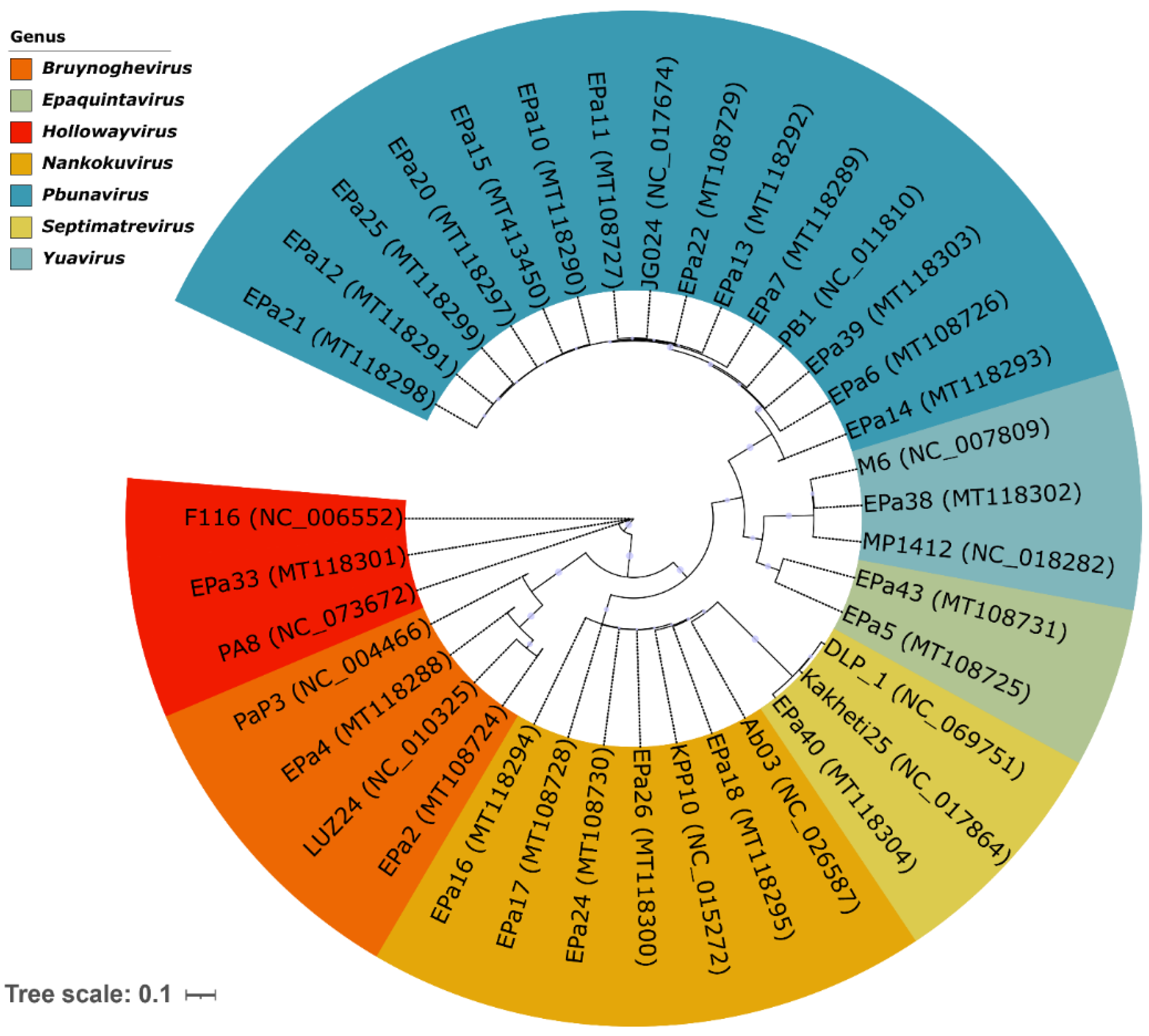
Figure 1. Phylogenetic tree demonstrating genetic diversity and genus designation of 25 P. aeruginosa phages, with a predominance of Pbunavirus phages. Each genus cluster contains two representative reference phages, selected from the ICTV master species list 2025, except the recently established genus Epaquintavirus that was named after EPa5. The tree was constructed by whole genome comparison using VICTOR pipeline [84] with the D4 formula and visualized with iTOL [85].
Preprints 185098 g001
Figure 2. Transmission electron microscopy images of P. aeruginosa phage particles. Myovirus morphology: (a) EPa6, (b) EPa7, (c) EPa10, (d) EPa13 (genus Pbunavirus), and (e) EPa18 (genus Nankokuvirus). Siphovirus morphology: (f) EPa5 (genus Epaquintavirus) and (g) EPa40 (genus Septimatrevirus). Podovirus morphology: (h) EPa2 and (i) EPa4 (genus Bruynoghevirus).
Preprints 185098 g002
Figure 3. Partial dispersal of P. aeruginosa biofilms by phages EPa2 and EPa4 in a crystal violet assay.
Preprints 185098 g003
Figure 4. Bactericidal effect of phages against P. aeruginosa biofilms. Average colony-forming units per mL (CFU/mL) in biofilms formed by strains (a) PAO1::lux, (b) MRSN 1899, (c) MRSN 1409, (d) MRSN 1680, (e) MRSN 8130, and (f) ATCC 10145. SM: SM buffer without phage (negative control).
Preprints 185098 g004
Figure 5. Efficacy of phage cocktail WRAIR_PAM1 (PAM1) against P. aeruginosa topical and systemic infection in mice: (a) mouse survival; (b) wound healing dynamics. CAZ: mice treated with standard-of-care antibiotic ceftazidime (positive control); PBS: mice treated with phosphate-buffered saline (negative control); PAM1: mice treated with the 5-phage cocktail. Mouse groups were compared for survival using Gehan-Breslow-Wilcoxon test. **P=0.0046. N=5 for each mouse group.
Preprints 185098 g005
Figure 6. Efficacy of phage cocktail WRAIR_PAM2 (PAM2) against mouse topical and generalized P. aeruginosa infection: (a) mouse survival (mouse groups were compared using Gehan-Breslow-Wilcoxon test, ****P=<0.0001); (b) comparison of mouse nest building activity; (c) wound healing dynamics (groups were compared at each time point using Mann-Whitney t-test, ****P<0.0001, ***P<0.001). PBS, mice treated with placebo, phosphate-buffered saline (negative control); PAM2, mice treated with the phage cocktail. N=15 for PBS-treated mouse group; N=20 for phage-treated group.
Preprints 185098 g006
Table 1. Characteristics of 25 P. aeruginosa phages used in this work.
Phage ID Genome size, bp Family* Genus Host range Anti-biofilm effect Phage receptor
Dispersal Killing
EPa2 43,299 Unclassified Bruynoghevirus 32.7% + + T4P, A band (1)
EPa4 45,439 Unclassified Bruynoghevirus 31.4% + + T4P, A band (1)
EPa5 64,096 Mesyanzhinovviridae Epaquintavirus 18.6% + T4P, LPS core (2)
EPa6 66,031 Lindbergviridae Pbunavirus 46.2% + T4P, LPS core (2)
EPa7 65,629 Lindbergviridae Pbunavirus 44.2% + T4P, LPS core (2)
EPa10 66,774 Lindbergviridae Pbunavirus 40.4% + T4P, L-B, VL-B (3)
EPa11 66,800 Lindbergviridae Pbunavirus 51.3% + T4P, L-B, VL-B (3)
EPa12 66,520 Lindbergviridae Pbunavirus 46.2% + T4P, L-B, VL-B (3)
EPa13 65,680 Lindbergviridae Pbunavirus 42.3% + T4P, A, B, L-B, VL-B (4)
EPa14 65,797 Lindbergviridae Pbunavirus 42.3% + T4P, A, B, L-B, VL-B (4)
EPa15 66,002 Lindbergviridae Pbunavirus 54.5% + T4P, L-B, VL-B (3)
EPa16 88,727 Vandenendeviridae Nankokuvirus 35.9% + Unknown**
EPa17 88,859 Vandenendeviridae Nankokuvirus 30.8% + Unknown**
EPa18 88,109 Vandenendeviridae Nankokuvirus 37.8% + Unknown**
EPa20 66,505 Lindbergviridae Pbunavirus 42.3% + T4P, L-B, VL-B (3)
EPa21 66,764 Lindbergviridae Pbunavirus 39.7% + T4P, L-B, VL-B (3)
EPa22 65,897 Lindbergviridae Pbunavirus 51.9% + T4P, A, B, L-B, VL-B (4)
EPa24 88,728 Vandenendeviridae Nankokuvirus 46.8% + T4P, LPS core (2)
EPa25 66,811 Lindbergviridae Pbunavirus 28.2% + T4P, L-B, VL-B (3)
EPa26 88,805 Vandenendeviridae Nankokuvirus 42.9% + T4P, LPS core (2)
EPa33 64,021 Unclassified Hollowayvirus 30.1% + T4P, A, B (5)
EPa38 61,775 Mesyanzhinovviridae Yuavirus 7.7% + T4P, LPS core (2)
EPa39 66,708 Lindbergviridae Pbunavirus 45.5% + T4P, L-B, VL-B (3)
EPa40 42,788 Unclassified Septimatrevirus 32.7% + T4P, A band (1)
EPa43 64,323 Mesyanzhinoviridae Epaquintavirus 14.1% + T4P, LPS core (2)
Notes: *Phage taxonomy at the family level is in the process of updating by the International Committee on Taxonomy of Viruses (ICTV). Until recently, genera Bruynoghevirus and Hollowayvirus belonged to the family Podoviridae, and genus Septimatrevirus belonged to the family Siphoviridae formerly established based on virion morphology; now these phages are not classified at the family level. T4P, type IV pili; A band, common polysaccharide antigen (CPA) chain of lipopolysaccharide (LPS); B band, O-specific antigen (OSA) polysaccharide chain of the LPS; L-B, long B band; VL-B, very long B band. **These phages are not active against P. aeruginosa strain PAO1 and its derivatives.
Table 2. Predicted complementary host ranges of P. aeruginosa phages for pairwise mixes.
Phage Host range (%) Phage Host range (%) Phage Host range (%)
EPa11 51.3 EPa11+EPa24 59.0 EPa17+EPa40 51.3
EPa16 35.9 EPa11+EPa40 53.8 EPa17+EPa43 34.0
EPa17 30.8 EPa11+EPa43 55.8 EPa22+EPa24 62.8
EPa22 51.9 EPa16+EPa17 51.3 EPa22+EPa40 62.2
EPa24 46.8 EPa16+EPa22 60.9 EPa22+EPa43 55.8
EPa40 32.7 EPa16+EPa24 51.3 EPa24+EPa40 48.7
EPa43 14.1 EPa16+EPa40 44.2 EPa24+EPa43 51.3
EPa11+EPa16 57.1 EPa16+EPa43 42.3 EPa40+EPa43 37.2
EPa11+EPa17 64.1 EPa17+EPa22 69.2 Mix-7* 78.2
EPa11+EPa22 62.2 EPa17+EPa24 57.7 Mix-5** 76.3
Notes: *Mix-7: EPa11, EPa16, EPa17, EPa22, EPa24, EPa40, and EPa43. **Mix-5: EPa11, EPa17, EPa22, EPa24, and EPa43.
Table 3. Frequencies of P. aeruginosa resistance to single phages and their pairwise mixes.
Phage Resistance frequency Phage Resistance frequency
To single To mix To single To mix
EPa11 7.54×10⁻⁶ 5.70×10⁻⁶ EPa11 7.54×10⁻⁶ 2.63×10⁻⁸
EPa16 1.33×10⁻⁶ EPa17 2.19×10⁻⁷
EPa11 7.54×10⁻⁶ 6.40×10⁻⁶ EPa11 7.54×10⁻⁶ 8.07×10⁻⁶
EPa22 3.42×10⁻⁶ EPa24 9.39×10⁻⁶
EPa11 7.54×10⁻⁶ 1.58×10⁻⁶ EPa11 7.54×10⁻⁶ <8.77×10⁻⁹
EPa40 2.53×10⁻⁷ EPa43 <8.77×10⁻⁹
EPa16 1.33×10⁻⁶ 1.67×10⁻⁷ EPa16 1.33×10⁻⁶ 2.98×10⁻⁶
EPa17 2.19×10⁻⁷ EPa22 3.42×10⁻⁶
EPa16 1.33×10⁻⁶ 5.35×10⁻⁶ EPa16 1.33×10⁻⁶ 1.01×10⁻⁶
EPa24 9.39×10⁻⁶ EPa40 2.53×10⁻⁷
EPa16 1.33×10⁻⁶ <8.77×10⁻⁹ EPa17 2.19×10⁻⁷ 4.39×10⁻⁸
EPa43 <8.77×10⁻⁹ EPa22 3.42×10⁻⁶
EPa17 2.19×10⁻⁷ 3.51×10⁻⁸ EPa17 2.19×10⁻⁷ 8.77×10⁻⁸
EPa24 9.39×10⁻⁶ EPa40 2.53×10⁻⁷
EPa17 2.19×10⁻⁷ <8.77×10⁻⁹ EPa22 3.42×10⁻⁶ 6.93×10⁻⁶
EPa43 <8.77×10⁻⁹ EPa24 9.39×10⁻⁶
EPa22 3.42×10⁻⁶ 1.19×10⁻⁵ EPa22 3.42×10⁻⁶ <8.77×10⁻⁹
EPa40 2.53×10⁻⁷ EPa43 <8.77×10⁻⁹
EPa24 9.39×10⁻⁶ 1.19×10⁻⁵ EPa24 9.39×10⁻⁶ <8.77×10⁻⁹
EPa40 2.53×10⁻⁷ EPa43 <8.77×10⁻⁹
EPa40 2.53×10⁻⁷ <8.77×10⁻⁹ 11+16+17+22+24+40+43 <8.77×10⁻⁹
EPa43 <8.77×10⁻⁹
Table 4. Phage cocktails PAM1 and PAM2.
Cocktail Phage Family Genus Host
range (%)
Host receptor Cocktail
activity (%)
PAM1 EPa2 Unclassified Bruynoghevirus 32.7 T4P, A band (1) 55.8
EPa4 Unclassified Bruynoghevirus 31.4 T4P, A band (1)
EPa5 Mesyanzhinovviridae Epaquintavirus 18.6 T4P, LPS core (2)
EPa6 Lindbergviridae Pbunavirus 46.2 T4P, LPS core (2)
EPa17 Vandenendeviridae Nankokuvirus 30.8 Unknown
PAM2 EPa11 Lindbergviridae Pbunavirus 51.3 T4P, L-B, VL-B (3) 76.3
EPa17 Vandenendeviridae Nankokuvirus 30.8 Unknown
EPa22 Lindbergviridae Pbunavirus 51.9 T4P, A, B, L-B, VL-B (4)
EPa24 Vandenendeviridae Nankokuvirus 46.8 T4P, LPS core (2)
EPa43 Mesyanzhinovviridae Epaquintavirus 14.1 T4P, LPS core (2)
Table 5. Twelve knockout mutants of P. aeruginosa PAO1 used for phage receptor identification.
Mutant Phenotype Source Mutant Phenotype Source
rmd No A band LPS Dr. Rob Lavigne (Katholieke Universiteit Leuven, Leuven, Belgium) wzz1 No long B band Dr. Karen Maxwell (University of Toronto, Toronto, Canada
wbpL A/B band synthesis initiation defect wzz2 No very long B band
rmlC* Truncated LPS core, no A, B bands wzz1,2 No long/very long B band
fliA No flagella wapQ No phosphate in LPS inner core
pilA No type IV pili wbpM No B band
fliA algC pilA No flagella, A band, type IV pili rmlC* Truncated LPS core, no A, B bands
*Two rmlC mutants acquired from two different sources were tested.
Disclaimer/Publisher’s Note: The statements, opinions and data contained in all publications are solely those of the individual author(s) and contributor(s) and not of MDPI and/or the editor(s). MDPI and/or the editor(s) disclaim responsibility for any injury to people or property resulting from any ideas, methods, instructions or products referred to in the content.
Copyright: This open access article is published under a Creative Commons CC BY 4.0 license, which permit the free download, distribution, and reuse, provided that the author and preprint are cited in any reuse.
Prerpints.org logo

Preprints.org is a free preprint server supported by MDPI in Basel, Switzerland.

Subscribe

Disclaimer

Terms of Use

Privacy Policy

Privacy Settings

© 2025 MDPI (Basel, Switzerland) unless otherwise stated