Preprint
Review

This version is not peer-reviewed.

Key Criteria of Hemostatic Efficiency of Highly Porous Cellulose-Based Scaffolds: A Review

A peer-reviewed article of this preprint also exists.

Submitted:

19 November 2025

Posted:

21 November 2025

You are already at the latest version

Abstract

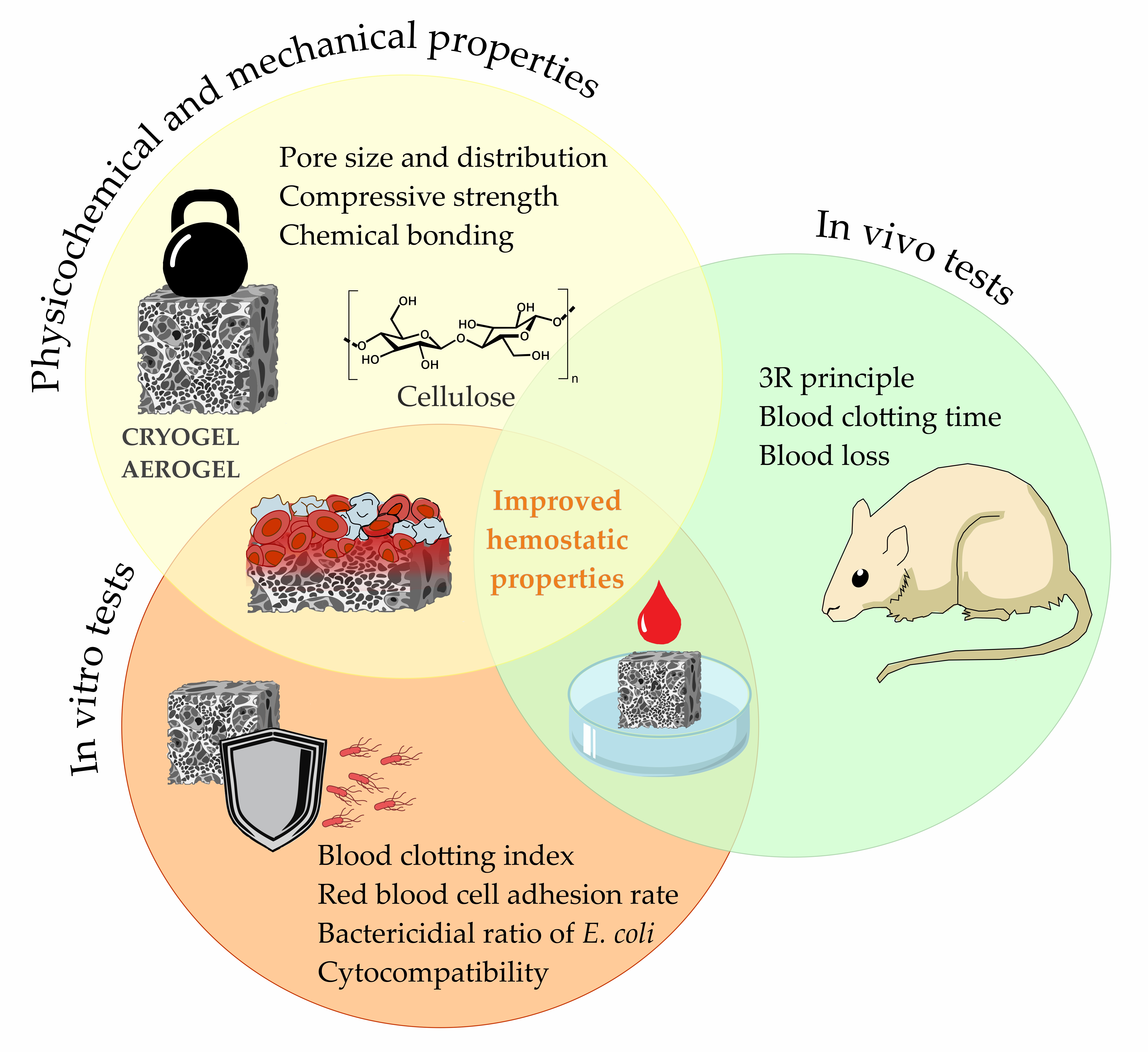
Introduction. Severe bleeding and uncompensated blood loss remain some of the most serious complications associated with trauma and surgical interventions that can threaten life. In recent years, intensive research has been directed toward developing hemostatic materials based on natural and synthetic polymers. Among them, cellulose and its modified derivatives represent one of the most promising sources for creating effective hemostatic systems. The aim of this review was to identify key criteria for the efficiency of cellulose-based gels with hemostatic activity. Methods. Experimental studies aimed at evaluating new hemostatic materials were analyzed based on international sources using the PRISMA methodology. Results. A total of 111 publications were identified. Following the identification and screening stages, 20 articles were selected for the final qualitative synthesis. The analyzed publications include experimental studies focused on the development and analysis of highly porous cellulose-based scaffolds in the form of aerogels and cryogels. The type and origin of cellulose, as well as the influence of additional components and synthesis conditions on gel formation, were investigated. Discussion and Conclusions. Three major groups of key criteria that should be considered when developing new cellulose-based highly porous scaffolds with hemostatic functionality were identified: (I) physicochemical and mechanical properties (pore size distribution, compressive strength, and presence of functional groups); (II) in vitro tests (blood clotting index, red blood cell adhesion rate, hemolysis, cytocompatibility, and antibacterial activity); (III) in vivo hemostatic efficiency (hemostasis time and blood loss) in compliance with the 3Rs policy (replacement, reduction, refinement).

Keywords: 
;  ;  ;  ;  

1. Introduction

Hemostatic agents significantly reduce the negative consequences of injuries and surgical interventions caused by uncompensated blood loss. However, traditional methods of controlling bleeding have several limitations, including side effects, low adhesion, slow hemostasis, or complexity of use [1]. Therefore, medical devices with hemostatic properties must combine safety, high efficiency, versatility, and usability. This is especially important in emergency medicine, where rapid and reliable achievement of hemostasis can save lives [2].
Promising sources for the development of hemostatic agents are natural polysaccharides, such as cellulose-based materials. Due to their biocompatibility, biodegradability, high blood absorption capacity, cost-effectiveness, and renewability, they represent a component in the development of new hemostatic compositions [3]. Traditional cotton gauze is widely used as a hemostatic material because of its advantages—safety, flexibility, wound conformability, hypoallergenicity, and low cost. However, its limited pro-coagulant activity, insufficient red blood cells adhesion and low platelet activation reduce its clinical hemostatic efficacy [4].
Recently, cellulose derivatives have been widely used in the development of new agents for stopping bleeding. Purified cellulose undergoes physical treatment (size reduction, nanostructuring) or chemical modification (acetylation and etherification, crosslinking, particle hydrophobization), which significantly improves its properties: increasing surface area, enhancing water retention, and others. Materials with hemostatic effects are often obtained by forming complexes from cellulose and sodium alginate salts, starches, or activated biopolymers [5].
A special popularity is gaining the chemical transformation of natural cellulose to produce highly porous scaffolds — aerogels and cryogels. Aerogels are ultra-lightweight porous materials produced by dissolution, solvent exchange, and drying with supercritical CO2. [6]. The preparation of cellulose-based cryogels involves freezing aqueous solutions of amorphized linear polymers or monomers followed by freeze-drying. There are several biomedical studies exploring cellulose and polysaccharide-based cryogels, in which authors note with large and interconnected pores structure that facilitates gas exchange in the wound area, absorbs wound exudate, and simultaneously provides a suitable environment for cell viability. Therefore, cryogels show significant potential for hemostatic agents, wound dressings and applications in regenerative biomedicine and tissue engineering [7].
Despite the active development of novel hemostatic devices, there is a major issue in selecting evaluation criteria that objectively reflect hemostatic performance. Thus, the objective of this systematic review was to identify key efficiency criteria for highly porous cellulose-based scaffolds exhibiting hemostatic activity.

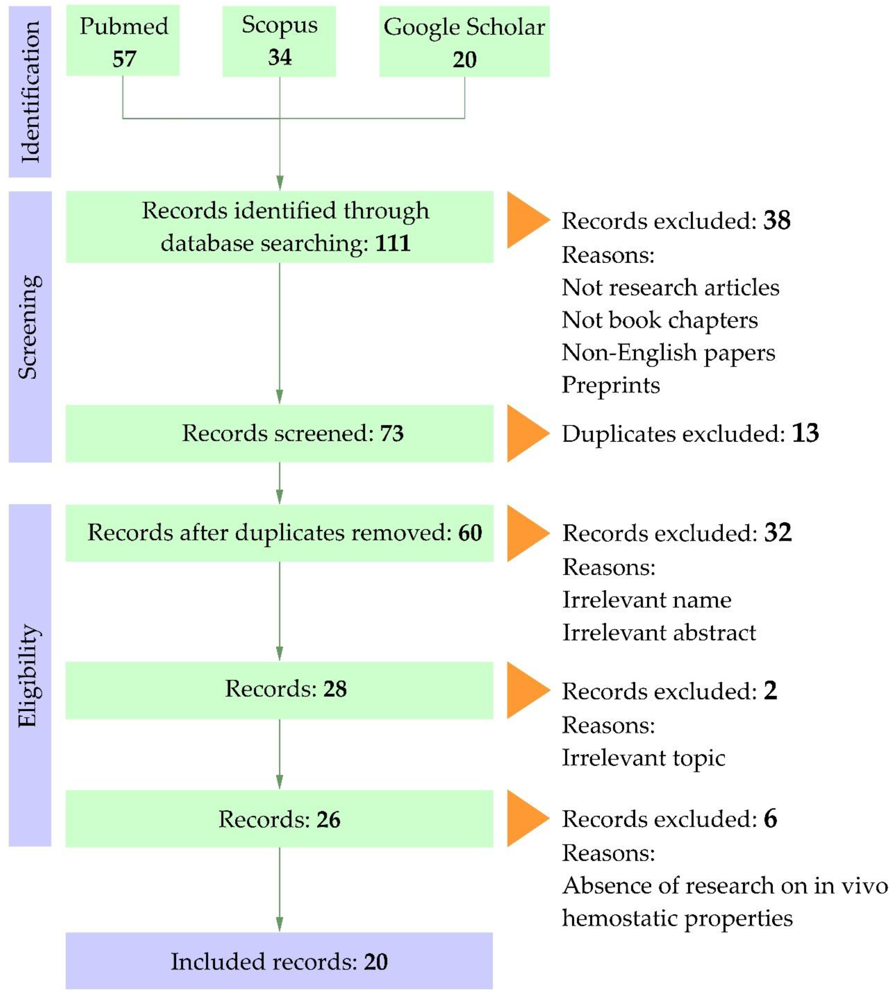
2. Materials and Methods

2.1. Search Method

The PRISMA Extension for Scoping Reviews guidelines were applied for study selection and data presentation [8]. The literature search was conducted in Scopus, PubMed, and Google Scholar using the following terms and combinations: (“haemosta*” OR “hemosta*”) AND (“platelets” OR “blood” OR “erythrocyte” OR “plasma” OR “RBC”) AND “cellulose” AND (“aerogel” OR “cryogel” OR “xerogel”).

2.2. Paper Selection

All identified studies were screened based on their titles and abstracts. Acceptable articles were experimental studies or book chapters in English. Preprints and duplicates were excluded. Publications were considered if they contained information on the development of cellulose-based aerogels and cryogels, their physicochemical properties, and results from in vitro and in vivo experiments evaluating hemostatic properties. Xerogels are a new type of cellulose modification, but no information has been found regarding their use as a hemostatic agent. Studies that included only one of the selected groups were excluded. The selection flow is presented in Figure 1.

2.3. Data Extraction

Information regarding author affiliations, publication years, study design, raw material sources for aerogel/cryogel preparation, fabrication methodology, physicochemical characterization, hemostatic evaluation approaches, laboratory animal species and primary outcomes was extracted by one author (N.A.) into an Excel spreadsheet. The data were independently verified by authors (A.R., K.A., L.L.). Disagreements were resolved by consensus or, when required, consultation with a fourth reviewer (A.S.).

3. Results

A total of 111 publications were identified in the databases: Scopus–34, PubMed—57, and Google Scholar—20. Following the identification and screening stages, 20 articles were selected for the final qualitative synthesis (Table 1). The analyzed publications include experimental studies focused on the development and analysis of highly porous cellulose-based scaffolds in the form of aerogels (13 studies) and cryogels (7 studies).
Researchers used various types of cellulose for the creation of hemostatic materials. Specifically, samples of plant and bacterial origin, as well as their modifications (TEMPO-oxidized, carbonized, microcrystalline, phosphorylated, carboxymethyl cellulose, hydroxyethyl cellulose), are employed. Notably, Wan et al. (2024) used waste pomelo peel as a cellulose feedstock [13]. In many studies, aerogels or cryogels are multi-component systems; instead, auxiliary matrices and modifiers such as chitosan, collagen, gelatin, agar, and alginates are commonly used. Additionally, halloysite, bioactive glass, organic acids, dopamine derivatives, silver nanoparticles, and zeolites are considered promising components for the formation of cellulose-based composite systems.
Parameters for obtaining highly porous cellulose-based scaffolds determine their mechanical and physicochemical properties, including porosity, pore size, and pore size distribution. Particular attention was paid to the initial components (concentration during gelation, type of cellulose used, and its modifications), methods of forming the porous structure (dissolution time, water replacement techniques), parameters for solvent removal, and the type of drying employed.
An important feature of the examined publications is the multifactorial analysis of variations in scaffolds that differ in the qualitative and quantitative composition of their main components. For example, in the study [18] two types of scaffolds are investigated: nanoporous zeolite-anchored cellulose nanofibers (nZ@CNFs) and Ca2+-exchanged nZ@CNFs (Ca-nZ@CNF). In the research by Cao et al. [22], a complex of carboxymethyl cellulose and dopamine (CMC/DA) with various concentrations of EDC/NHS additives (12.5/6.25, 25/12.5, 50/25, 100/50 mg/mg) is analyzed, as well as CMC/DA enriched with silver (CMC/DA/Ag). It should be noted that in many studies, some of the parameters analyzed are not presented in the main text but are contained in supplementary information. Considering the variability in composition and the multi-component nature of the studied samples, analyzing the efficiency of such systems requires a structured approach to classification the criteria used to evaluate hemostatic activity, which formed the basis for the subsequent structuring of results in this review.

4. Discussion`

The highly porous scaffolds used in studies of hemostatic properties were predominantly produced via freezing followed by freeze-drying. This method typically results in partial structural collapse and the formation of large pores with a relatively low specific surface area [31]. Such effects become particularly evident when water-based solvents are used, as crystallization of solvents with high freezing points promotes the development of large, irregular cavities [32]. In several studies [9,18,19,20,24,25,26,27], aerogels with hemostatic functionality were obtained through freezing and drying techniques consistent with cryogelation. The replacement of water by solvents such as ethanol or methanol, which possess lower freezing points [12,13,15,16], mitigates this effect on pore formation. However, the exposure to low temperatures and vacuum pressure during freeze-drying creates surface tension within the pores, which alters the internal morphology of the scaffolds. These conditions often lead to pore wall thickening and partial collapse, increasing the overall density and decreasing the specific surface area of the resulting material.
Pore size is known to play a crucial role in stimulating vascularization and regulating cellular infiltration, depending on the cell type involved [33]. Consequently, insufficient attention to the physicochemical and mechanical characterization of gels limits the understanding of their structure–property relationships and hinders a comprehensive assessment of hemostatic performance. In general, freeze-drying offers significant advantages for obtaining mechanically stable and elastic highly porous materials. The ability to control the freezing rate allows adjustment of pore size and overall porosity [34], which in turn provides optimal conditions for blood contact and blood clot formation [14].
Based on the reviewed literature, three primary groups of criteria were identified for assessing cellulose-based hemostatic materials.

4.1. Physicochemical and Mechanical Properties of Cellulose-Based Hemostatic Scaffolds

Most studies include scanning electron microscopy (SEM) or atomic force microscopy (AFM) imaging with analysis of pore size and distribution (Table 2). According to the IUPAC classification, pores are divided into three categories: micropores (<2 nm), mesopores (2–50 nm), and macropores (>50 nm) [29].
When it comes to hemostatic materials, particular attention is given to macro- and mesoporous structures, as they largely determine the interaction between the scaffold and blood components. Macropores (>50 nm), typically formed during cryogelation, enable the penetration of cellular components such as erythrocytes and platelets into the matrix, increasing the effective contact area and enhancing adhesion, which accelerates hemostasis. This has been confirmed in multiple studies demonstrating that macroporous cryogels exhibit more pronounced cellular fixation and faster platelet plug formation [11,21,23].
Mesopores (2–50 nm), in contrast, are too small to allow cell penetration since blood cells are several orders of magnitude larger (erythrocytes ~6–8 µm, platelets ~2–4 µm). The role of mesoporous structures lies primarily in the adsorption of plasma proteins (fibrinogen, albumin, complement components), which influences platelet adhesion and activation. Thus, mesopores regulate hemocompatibility through protein–surface interactions rather than serving as physical channels for cellular passage [35,36].
It should be noted that in several studies, exact pore sizes or ranges were not reported, allowing only approximate estimation based on SEM micrographs. The majority of analyzed scaffolds can be classified as macroporous gels (12 of 20 publications), though some exhibit combined macro- and mesoporous architectures without quantitative assessment of their ratio [11,16,18,23,28].
Porosity values reported selected studies range from 70.0% to 97.5%. It is evident that porosity affects mechanical strength, whereas pore size has a more pronounced impact on hemostatic efficiency. Compressive strength is a key parameter, as it determines the structural stability of the gel upon contact with aqueous media or blood. Only about half of the reviewed studies provide quantitative compressive strength data, which vary widely (0.002–0.700 MPa), reflecting differences in testing methods and preventing direct comparison of results. In our view, a suitable hemostatic scaffold should exhibit sufficient mechanical integrity to prevent structural collapse during application while maintaining efficient interaction with the wound surface.
Many authors also analyze the chemical bonding and functional groups within the gels using Fourier-transform infrared spectroscopy (FTIR) or nuclear magnetic resonance (NMR, 1H or 13C), though these data are not summarized in this review. Some researchers additionally consider the presence of coagulation factors—most notably Ca2+ ions—within the gel matrix, which play a pivotal role in the coagulation cascade by promoting the conversion of prothrombin to thrombin [18,30].
Hence, the key criteria in this first group include pore size determination, compressive strength, characterization of reactive functional groups, and evaluation of coagulation-related components such as calcium ions.

4.2. In Vitro Tests

The second group of parameters includes studies of the interaction between the developed gels and biological media in vitro (Table 3). The blood clotting index (BCI) is one of the most widely used metrics for assessing hemostatic materials. In approximately 70% of the reviewed works, the BCI values varied from 0.05 to 89.76%. Lower BCI values indicate faster and more efficient coagulation, identifying these materials as more promising hemostatic systems. This correlation has been consistently confirmed for both aerogels and cryogels [10,12,13,14,15,18,19,20,22,24,25,28].
Another key parameter is the adhesion rate of red blood cells and platelets to the gel surface, which depends on the presence of reactive functional groups. The adhesion of these cellular components plays a critical role in initiating and maintaining hemostasis. However, not all studies account for both cell types; in several works [10,12,13], only erythrocyte adhesion was evaluated (52.91%, 80.45%, and 40.00%, respectively).
The hemolysis ratio of erythrocytes was reported in most publications [10,11,12,13,14,15,16,17,19,20,21,22,23,24,25,26,27,28]. According to the American Society for Testing and Materials standard (ASTM F756–2000), hemolysis should not exceed 5%. Most authors confirmed that their measured values were below this threshold; several studies provided specific values ranging from 0.66% to 2.30% [12,14,17,19,21,25,28]. These results indicate negligible cytotoxic effects and validate the biocompatibility of direct blood contact.
Additionally, cytocompatibility analyses revealed high cell viability (>90% in 15 of 20 studies), consistent with ISO 10993–5 requirements. Several studies also assessed the antibacterial activity of the gels, mainly against S. aureus (Gram-positive) and E. coli (Gram-negative) [10,11,14,17,23,25,26,27,28], with bactericidal ratios against E. coli ranging from 70% to 100%. The incorporation of antibacterial agents via ionic, covalent, or hydrogen bonding can prevent microbial contamination of wounds and reduce the risk of infection-related complications.
Compared with the first group, in vitro tests—covering BCI, erythrocyte and platelet adhesion, hemolysis, cytocompatibility, and antibacterial activity—provide insight into the interaction mechanisms between porous scaffolds and biological systems, confirming their safety prior to in vivo testing.

4.3. In Vivo Hemostatic Efficiency

This group is the most clinically relevant since it directly determines the therapeutic potential of hemostatic materials (Table 4). Most studies employed rats as experimental animals, and less frequently rabbits, consistent with the 3Rs policy (replacement, reduction, refinement) and international biomedical ethics [17]. The most common experimental models included liver punch biopsy wounds (3–5 mm in diameter, 1–5 mm deep) and distal tail amputations (1–2 cm). A femoral artery bleeding model was used only once [12].
The general in vivo testing protocol comprised anesthesia, surgical field preparation, wound induction, immediate gel application, and measurement of hemostasis time and blood loss. Cotton or double-layer gauze was used as negative controls, while commercial gelatin sponges served as positive controls. We consider gauze and cotton inappropriate reference materials, as they exhibit minimal procoagulant activity and therefore poor hemostatic performance. The absence of a standard reference material (SRM) remains a major obstacle for comparing in vivo results.
Meng Wang et al. [16] did not perform direct hemostatic tests but instead used the aerogel as a skin graft material in animal experiments, which can be regarded as a modified form of in vivo assessment.
The tail amputation model is faster and simpler than the liver bleeding model and is often used as an auxiliary test for evaluating coagulation parameters before or after the main experiment. In contrast, noncompressible wounds of parenchymal organs such as the liver are considered more objective, as they mimic severe surgical trauma involving both venous and arterial bleeding [37]. The choice of model should depend on the material’s intended use: for cellulose-based gels as dressing materials, surface compressible bleeding models (tail) are appropriate; for adhesive porous scaffolds, internal noncompressible bleeding models (liver) are preferable.
Post-hemostasis morphological evaluation using scanning electron microscopy [27,28] revealed dense fixation of erythrocytes within the gel matrix and a developed fibrin network, confirming that bleeding cessation occurs through blood–cellulose interaction, local thrombus formation, and the development of a platelet–fibrin matrix on the gel surface.
On one hand, published data indicate that highly porous cellulose-based cryo- and aerogels possess significant potential in biomedical applications due to their rapid hemostatic response, effective water absorption, enhanced erythrocyte and platelet adhesion, and activation of the coagulation cascade [10,25]. Their efficiency is attributed to abundant hydroxyl groups, hydrophilicity, and high porosity. Upon blood contact, the scaffolds rapidly absorb liquid into the porous channels, triggering autocatalytic coagulation activation, platelet aggregation, and erythrocyte adhesion, ultimately leading to thrombus formation [9]. The criteria identified in this review should be considered essential for the rational design of new hemostatic biomaterials.
On the other hand, current studies reveal several limitations. Most focus solely on in vitro hemostatic performance, with insufficient evaluation of blood–cell interactions and tissue responses in vivo. Data on biocompatibility and potential cytotoxicity are often fragmentary, complicating the assessment of clinical safety and efficacy. Furthermore, systematic comparisons of porosity parameters, scaffold density, and gel preparation methods are lacking, hindering understanding of hemostatic mechanisms. Long-term effects such as material degradation, inflammation, and microbial adhesion remain insufficiently explored.
Therefore, future research should adopt an integrated approach, including standardized hemostasis models, validated reference materials, biocompatibility and immune response evaluation, and systematic correlation of structural and functional characteristics. Highly porous cellulose-based scaffolds demonstrate strong hemostatic performance due to their optimized porosity and hydrophilic nature; however, for clinical translation, additional studies on long-term stability, immune effects, and tissue interactions are required.

5. Conclusions

In this review, we identified three major groups of key criteria that should be considered when developing new cellulose-based highly porous scaffolds with hemostatic functionality:
I. Physicochemical and mechanical properties (pore size distribution, compressive strength, and presence of functional groups).
II. In vitro tests (blood clotting index, red blood cell adhesion rate, hemolysis, cytocompatibility, and antibacterial activity).
III. In vivo hemostatic efficiency (hemostasis time and blood loss) in compliance with the 3Rs policy (replacement, reduction, refinement).
The systematization of published data shows that modern cellulose modifications represent a promising basis for the development of safe and effective hemostatic materials. However, the absence of a standard reference material (SRM)—with gauze or cotton still being used as controls—as well as the lack of unified evaluation methods and the high variability of experimental designs, significantly complicate the comparability of results across studies.
According to the proposed criteria, a complete and comprehensive characterization of the developed highly porous scaffolds is missing in all analyzed publications. The studies by Ahmad Mahmoodzadeh et al. (2021) and Zhan Xu et al. (2022) appear to be the most complete in terms of structural and functional characterization.
In conclusion, we propose several practical recommendations for the design and characterization of cellulose-based gels:
When preparing gels according to the methodologies described in this review, with minimal modification of synthesis parameters, analysis of the first group of criteria is sufficient.
When modifying the synthesis process while using similar raw materials, physicochemical and mechanical characterization should be supplemented by in vitro tests to confirm the effect of changes on hemostatic response.
When developing a fundamentally new gel, it is necessary to analyze all groups of criteria, define the intended hemostatic application, and select an appropriate in vivo bleeding model.
Future research should focus on the standardization of methodological research protocols, justification of efficacy metrics, and a more detailed investigation of structure–property–effect relationships.

Author Contributions

Conceptualization, S.N.A. and A.S.A.; methodology, S.N.A. and M.K.A.; data curation, S.N.A., S.L.L.; writing—original draft preparation, S.N.A. and S.A.R.; writing—review and editing, S.A.R., A.S.A. and S.L.L.; visualization, M.K.A; supervision, A.S.A.; project administration, S.A.R. All authors have read and agreed to the published version of the manuscript.

Funding

The Russian Science Foundation supported preparation of this review article (project 25-24-20063).

Institutional Review Board Statement

Not applicable.

Informed Consent Statement

Not applicable.

Data Availability Statement

Not applicable.

Acknowledgments

The authors are grateful to the Northern (Arctic) Federal University named after M.V. Lomonosov and Northern State Medical University (Arkhangelsk) for technical support.

Conflicts of Interest

The authors declare no conflicts of interest.

References

  1. Hickman, D. A.; Pawlowski, C. L.; Sekhon, U. D.; Marks, J.; Gupta, A. S. Biomaterials and advanced technologies for hemostatic management of bleeding. Adv. Mater. 2018, 30(4), 1700859. [CrossRef]
  2. Fu, F.; Zuo, X.; Wang, Y.; Zhao, F.; Li, C.; Zeng, Y.; Wang.; L.; Wang, F. Centrifugal Spinning-Derived Biomimetic Aerogel for Rapid Hemostasis with Minimal Blood Loss. Nano Lett. 2025, 25(15), 6040-6050. [CrossRef]
  3. Bukatuka, C. F.; Mbituyimana, B.; Xiao, L.; Qaed Ahmed, A. A.; Qi, F.; Adhikari, M.; Shi, Zhijun; Yang, G. Recent Trends in the Application of Cellulose-Based Hemostatic and Wound Healing Dressings. J. Funct. Biomater. 2025, 16(5), 151. [CrossRef]
  4. Cidreira, A. C. M.; de Castro, K. C.; Hatami, T.; Linan, L. Z.; Mei, L. H. I. Cellulose nanocrystals-based materials as hemostatic agents for wound dressings: a review. Biomed. Microdevices 2021, 23(4), 43. [CrossRef]
  5. Rezvani Ghomi, E.; Niazi, M.; Ramakrishna, S. The evolution of wound dressings: from traditional to smart dressings. Polym. Advan. Technol. 2023, 34(2), 520-530. [CrossRef]
  6. Budtova, T.; Aguilera, D. A.; Beluns, S.; Berglund, L.; Chartier, C.; Espinosa, E.; Gaidukovs, S.; Klimek-Kopyra, A.; Kmita, A.; Lachowicz, D.; Liebner, F.; Platnieks, O.; Rodrigues, A.; Navarro, L.K.T.; Zou, F.; Buwalda, S. J. Biorefinery approach for aerogels. Polymers 2020, 12(12), 2779. [CrossRef]
  7. Tyshkunova, I. V.; Poshina, D. N.; Skorik, Y. A. Cellulose cryogels as promising materials for biomedical applications. Int. J. Mol. Sci. 2022, 23(4), 2037. [CrossRef]
  8. Pochinkova, P. A.; Gorbatova, M. A.; Narkevich, A. N.; Grjibovski, A. M. Updated brief recommendations onwriting and presenting systematic reviews: what’s new in PRISMA-2020 guidelines? Marine Medicine 2022, 8(2), 88-101. [CrossRef]
  9. Long, M.; Yang, X.; Shi, T.; Yang, Y. All-natural aerogel of nanoclay/cellulose nanofibers with hierarchical porous structure for rapid hemostasis. Int. J. Biol. Macromol. 2024, 278, 134592. [CrossRef]
  10. Li, M.; Dai, Q.; Zhu, S.; Feng, Q.; Qin, Z.; Gao, H.; Cao, X. An ultrafast water absorption composite cryogel containing Iron-doped bioactive glass with rapid hemostatic ability for non-compressible and coagulopathic bleeding. Chem. Eng. J. 2023, 469, 143758. [CrossRef]
  11. Zhu, Y.; Chen, F.; Wu, M.; Xiang, J.; Yan, F.; Xie, Y.; Tong, Z.; Chen, Y.; Cai, L. Biocompatible and antibacterial Flammulina velutipes-based natural hybrid cryogel to treat noncompressible hemorrhages and skin defects. Front. Bioeng. and Biotech. 2022, 10, 960407. [CrossRef]
  12. Mahmoodzadeh, A.; Moghaddas, J.; Jarolmasjed, S.; Kalan, A. E.; Edalati, M.; Salehi, R. Biodegradable cellulose-based superabsorbent as potent hemostatic agent. Chem. Eng. J. 2021, 418, 129252. [CrossRef]
  13. Wan, W.; Feng, Y.; Tan, J.; Zeng, H.; Jalaludeen, R. K.; Zeng, X.; Zheng, B.; Song, J.; Zhang, X.; Chen, S.; Pan, J. Carbonized cellulose aerogel derived from waste pomelo peel for rapid hemostasis of trauma--induced bleeding. Adv. Sci. 2024, 11(19), 2307409. [CrossRef]
  14. Xu, Z.; Tian, W.; Wen, C.; Ji, X.; Diao, H.; Hou, Y.; Fan, J.; Liu, Z.; Ji, T.; Sun, F.; Wu, D.; Zhang, J. Cellulose-based cryogel microspheres with nanoporous and controllable wrinkled morphologies for rapid hemostasis. Nano Lett. 2022, 22(15), 6350-6358. [CrossRef]
  15. Wang, B.; Li, X.; Wei, L.; Ma, S.; Wang, J.; Zhu, W.; Wang, H. Engineering high-performance composite cellulose materials for fast hemostasis. ACS Biomater. Sci. Eng. 2024, 10(8), 5313-5326. [CrossRef]
  16. Wang, M.; Sun, P.; Zhang, J.; Li, D.; Liu, Y.; Xia, Y.; Shao, L.; Jia, M. Intelligent and biocompatible cellulose aerogels featured with high-elastic and fast-hemostatic for epistaxis and wound healing. Int. J. Biol. Macromol. 2024, 277, 134239. [CrossRef]
  17. Mendes, B. B.; Gómez-Florit, M.; Araújo, A. C.; Prada, J.; Babo, P. S.; Domingues, R. M.; Reis, R.L.; Gomes, M. E. Intrinsically bioactive cryogels based on platelet lysate nanocomposites for hemostasis applications. Biomacromolecules 2020, 21(9), 3678-3692. [CrossRef]
  18. Chen, L.; Jin, Z.; Kamiya, T.; Liu, X.; Tian, Y.; Xia, R.; Wang, G.; Li, T.; Zhang, Q. Nanoporous zeolite anchored cellulose nanofiber aerogel for safe and efficient hemostasis. Small 2025, 21(21), 2500696. [CrossRef]
  19. Tripathi, G.; Park, M.; Lim, H.; Lee, B. T. Natural TEMPO oxidized cellulose nano fiber/alginate/dSECM hybrid aerogel with improved wound healing and hemostatic ability. Int. J. Biol. Macromol. 2023, 243, 125226. [CrossRef]
  20. Wang, Y.; Guo, Y.; Liu, Y.; Zhao, X.; Huang, Y.; Zhang, X.; Hu, X.; Mequanint, K.; Luo, G.; Xing, M. Platelet vesicles synergetic with biosynthetic cellulose aerogels for ultra--fast hemostasis and wound healing. Adv. Healthc. Mater. 2024, 13(17), 2304523. [CrossRef]
  21. Shakya, K. R.; Nigam, K.; Sharma, A.; Jahan, K.; Tyagi, A. K.; Verma, V. Preparation and assessment of agar/TEMPO-oxidized bacterial cellulose cryogels for hemostatic applications. J. Mater. Chem. B 2024, 12(14), 3453-3468. [CrossRef]
  22. Cao, S.; Li, Q.; Zhang, S.; Liu, Z.; Lv, X.; Chen, J. Preparation of biodegradable carboxymethyl cellulose/dopamine/Ag NPs cryogel for rapid hemostasis and bacteria-infected wound repair. Int. J. Biol. Macromol. 2022, 222, 272-284. [CrossRef]
  23. Li, Y.; Chu, C.; Chen, C.; Sun, B.; Wu, J.; Wang, S.; Ding, W.; Sun, D. Quaternized chitosan/oxidized bacterial cellulose cryogels with shape recovery for noncompressible hemorrhage and wound healing. Carbohyd. Polym. 2024, 327, 121679. [CrossRef]
  24. Pan, S.; Li, Y.; Tong, X.; Chen, L.; Wang, L.; Li, T.; Zhang, Q. Strongly-adhesive easily-detachable carboxymethyl cellulose aerogel for noncompressible hemorrhage control. Carbohyd. Polym. 2023, 301, 120324. [CrossRef]
  25. Chen, L.; Pan, S.; Wang, L.; Liu, X.; Jin, Z.; Xia, R.; Zhang, Q. Superhydrophobic cellulose nanofiber aerogels for efficient hemostasis with minimal blood loss. ACS Appl. Mater. Inter. 2024, 16(36), 47294-47302. [CrossRef]
  26. Liu, L.; Liu, L.; Chen, L.; Chen, G.; Wei, Y.; Hong, F. F. Synthesis of hemostatic aerogel of TEMPO-oxidized cellulose nanofibers/collagen/chitosan and in vivo/vitro evaluation. Mater. Today Bio 2024, 28, 101204. [CrossRef]
  27. Wang, L.; Zhang, C.; Zhao, W.; Li, W.; Wang, G.; Zhou, X.; Zhang, Q. Water-swellable cellulose nanofiber aerogel for control of hemorrhage from penetrating wounds. ACS Appl. Bio Mater. 2022, 5(10), 4886-4895. [CrossRef]
  28. Jin, Z.; Chen, L.; Liu, X.; Xia, R.; Li, W.; Wang, G.; Zhang, Q. Zeolite firmly anchored regenerated cellulose aerogel for efficient and biosafe hemostasis. Int. J. Biol. Macromol. 2025, 304, 140743. [CrossRef]
  29. Lawrence, M., Jiang, Y. (2017). Porosity, pore size distribution, micro-structure. In Bio-aggregates Based Building Materials. RILEM State-of-the-Art Reports; Amziane, S.; Collet, F. Еds.; vol 23. Dordrecht: Springer, Netherlands, 2017; pp. 39-71. [CrossRef]
  30. Schimper, C. B.; Pachschwöll, P.; Maitz, M. F.; Werner, C.; Rosenau, T.; Liebner, F. Hemocompatibility of cellulose phosphate aerogel membranes with potential use in bone tissue engineering. Front. Bioeng. and Biotech. 2023, 11, 1152577. [CrossRef]
  31. Bouras, H.; Zou, F.; Tillier, Y.; Buwalda, S.; Budtova, T. Starch aerogels, cryogels and xerogels for biomedical applications. In Advanced Functional Polymers for Medicine; Utrecht, Netherlands, 2024.
  32. Rodríguez-Dorado, R.; López-Iglesias, C.; García-González, C. A.; Auriemma, G.; Aquino, R. P.; Del Gaudio, P. Design of aerogels, cryogels and xerogels of alginate: Effect of molecular weight, gelation conditions and drying method on particles’ micromeritics. Molecules 2019, 24(6), 1049. [CrossRef]
  33. Croll, T. I.; Gentz, S.; Mueller, K.; Davidson, M.; O’Connor, A. J.; Stevens, G. W.; Cooper-White, J. J. Modelling oxygen diffusion and cell growth in a porous, vascularising scaffold for soft tissue engineering applications. Chem. Eng. Sci. 2005, 60(17), 4924-4934. [CrossRef]
  34. Buchtová, N.; Budtova, T. Cellulose aero-, cryo-and xerogels: towards understanding of morphology control. Cellulose 2016, 23(4), 2585-2595. [CrossRef]
  35. Yeo, J. H.; Lam, Y. W.; Fraser, S. T. Cellular dynamics of mammalian red blood cell production in the erythroblastic island niche. Biophys. Rev. 2019, 11(6), 873-894. [CrossRef]
  36. Zhao, X.; Guo, B.; Wu, H.; Liang, Y.; Ma, P. X. Injectable antibacterial conductive nanocomposite cryogels with rapid shape recovery for noncompressible hemorrhage and wound healing. Nat. Comm. 2018, 9(1), 2784. [CrossRef]
  37. Darya, G.; Mohammadi, H.; Dehghan, Z.; Nakhaei, A.; Derakhshanfar, A. Animal models of hemorrhage, parameters, and development of hemostatic methods. Lab. Anim. Res. 2025, 41(1), 5. [CrossRef]
Figure 1. PRISMA flow chart.
Preprints 185826 g001
Table 1. Brief description of highly porous cellulose-based scaffolds.
Form of the
hemostatic agent
Type and origin
of cellulose
Additional component Conditions for obtaining
highly porous scaffolds
Ref
Aerogel TEMPO-oxidized
cellulose nanofibers (ScienceK Co., Ltd.)
Halloysite Concentration 1%
Dissolution for 2 h
Freezing temperature -60 °C for 24 h
Vacuum freeze dryer
[9]
Cryogel Hydroxyethyl
cellulose (Aladdin Chemistry)
Quaternized chitosan and Iron-doped bioactive glass Concentration 0.1%, 0.2%, and 0.4%
Freezing temperature -20 °C for 36-48 h
Freeze-dryer
[10]
Cryogel Hydroxyethyl
cellulose (Shandong Head Reagent Co., Ltd.)
Flammulina velutipes extract Concentration 2%
Freezing temperature -20 °C for 12 h
Washing in deionized water, 24 h
Freeze dryer -50 °C, 24 h
[11]
Aerogel Carboxymethyl
cellulose and hydroxyl ethyl cellulose (Sigma-Aldrich)
Tranexamic acid Substitution of the solvent (water)
with isopropyl alcohol and hexane
1 method of drying: in Petri dishes
in the oven for 15 h
1 method of drying: freezing
temperature -20 °C for 24 h
Freeze dryer
[12]
Aerogel Carbonized cellulose from pomelo peel waste (Jiangyong Xiangyou, Yongzhou) - Substitution of the solvent (water)
with ethanol
Freeze dryer
[13]
Cryogel
microspheres
Microcrystalline
cellulose (Innochem Beijing)
Polydopamine Substitution of the solvent (water)
with butanol 50%
Freeze dryer
[14]
Aerogel Microcrystalline
cellulose (Anhui Shanhe
Pharmaceutical Co., Ltd.)
Gelatin, Diatomite Substitution of the solvent (water)
with ethanol
Freezing temperature -20 °C for 12 h
Freeze dryer for 18 h
[15]
Aerogel Carboxymethyl
cellulose and
TEMPO-oxide
cellulose nanofibers
- Concentration 0.1-0.5%
Substitution of the solvent (water)
with butyldehydrodiketone ethylene glycol for 24 h
Freezing temperature -75 °C, 12 h
Freeze dryer for 48 h
[16]
Cryogel Microcrystalline
cellulose powder (Sigma-Aldrich)
Platelet lysate Concentration 1.2-2.4%
Freezing temperature -80 °C
Freeze dryer
[17]
Aerogel Carboxymethyl
cellulose nanofibers, dry bleached wood pulp powders
Zeolite powder Concentration 1%
Freezing temperature -20 °C for 12 h
Freeze dryer -50 °C.
[18]
Aerogel TEMPO oxidized
cellulose nano fiber (TOCNF)
Alginate and
decellularized pig skin fragments
Concentration 1% alginate, 1% TOCNF
Freeze dryer
[19]
Aerogel Oxidized bacterial cellulose G. xylinum Platelet extracellular vesicles Concentration 0.2-1.2%;
Freezing temperature -20 °C
(3 freeze-thaw cycles of 15 h each)
Substitution of the solvent (water)
with tert-butanol for 12 h
Freeze dryer
[20]
Cryogel TEMPO oxidized bacterial cellulose (BC)
K. xylinus
Agar Concentration 1% agar and 20, 30, 40% OBC w/w of agar
Substitution of the solvent (water)
with methanol
Freezing temperature -80 °C;
Freeze dryer for 24 h
[21]
Cryogel Carboxymethyl
cellulose
(Macklin Co., Ltd.)
Dopamine, silver nanoparticles Concentration 2%
Freezing temperature -20 °C for 36 h
Freeze dryer
[22]
Cryogel Oxidized bacterial cellulose A. xylinum Quaternized chitosan Concentration 5%
Freezing temperature − 80 °C;
Freeze dryer
[23]
Aerogel Carboxymethyl
cellulose (Ever Bright Enterprise Development Co., Ltd.)
N-hydroxysuccinimide ether Concentration 2%
Freezing temperature -80 °C;
Freeze dryer
Heating for cross-linking − 80 °C for 1 h
[24]
Aerogel Bacterial cellulose (Hainan Yeguo Foods Co., Ltd.) Polydophamine and modified fluoroalkyl chains Concentration 1%
Freezing
Freeze dryer -50 °C
[25]
Aerogel TEMPO-oxidized cellulose nanofibers Сollagen/chitosan Concentration 1%
Freezing temperature -80 °C, 12 h
Freeze dryer, 48 h
[26]
Aerogel Carboxymethyl
cellulose nanofibers, bleached wood pulp
Сitric acid Concentration 1%
Freezing temperature 4 °C for 2 h and
-80 °C for 6 h
Freeze dryer
Heating for cross-linking − 80 °C for 1 h
[27]
Aerogel Carboxymethyl
cellulose (Shandong Senxin Environmental Protection Technology Co., Ltd.)
Zeolite Concentration 7%;
Dissolution in ethanol and acid for 24 h;
Washing with water
Freeze dryer
[28]
Table 2. Physicochemical and mechanical properties of cellulose-based hemostatic scaffolds.
Pore size Porosity, % Compression stress, MPa Ref
Macropores, µm Mesopores, nm
7.0-19.0 ∼13.00 0.070 [9]
0.070 [10]
100–200 0.020 [11]
151.6 ± 8.6 4.53–16.87 70.0 0.082 [12]
80.0 [13]
16.00–55.00 96.9 [14]
94.9 [15]
15.0–25.0 50-300 0.018 [16]
88.9 ± 1.5 [17]
110 21.37 ± 1.81 [18]
0.002 [19]
30.0 97.4 ± 0.4 [20]
6.3 ± 0.3 1.50–2.50 0.700 [21]
> 80.0 [22]
∼100-200 30.08 0.004 [23]
[24]
50-200 0.013 [25]
94.8 0.097 [26]
30-100 0.065 [27]
∼100 5.10 [28]
Table 3. In vitro analysis of cellulose-based hemostatic gels.
BCI, % Red blood cell
adhesion rate, %
Hemolysis, % Cytocompatibility, % Bactericidal ratio
of E. coli, %
Ref
59.47 ± 4.92 [9]
11.50 ± 0.87 52.9 < 5.00 80.12 95.0 [10]
< 5.00 100 100 [11]
0.80 80.0 0.66 ± 0.05 100 [12]
10.0 55.0 < 5.0 100 [13]
3.80 3.00 >99.3 34.6 [14]
12.3 < 5.00 >94 [15]
< 5.00 >90 + [16]
2.30 91.2 ± 8.2 [17]
8.2 [18]
4.0 2.68 ± 2.03 > 90 [19]
< 0.05 < 5.00 100 [20]
89.76 ± 0.49 3.20 ± 0.43 [21]
< 1.0 < 5.00 100 [22]
< 5.00 70.0 [23]
0.025 < 5.00 + [24]
7.4 ± 2.5 7.50 ± 4.00 > 95 88.2 [25]
< 4.00 > 80 100 [26]
< 1.00 > 90 95.0 [27]
10.0 1.37 > 90 > 95.0 [28]
Table 4. In vivo analysis of cellulose-based hemostatic gels.
Anesthetic Blood clotting time, seconds Blood loss, g Ref
liver tail femoral artery
Pentobarbital 98.0 ± 24.0 2.100 ± 0.870 [9]
Pentobarbital 42.9 ± 2.2 0.300 ± 0.070 [10]
Isoflurane ∼70 0.150 [11]
Ketamine
and xylazine
179 0.800 [12]
Pentobarbital 91.6 ± 5.5 0.293 ± 0.032 [13]
+ 43.5 ± 9.8 0.740 ± 0.250 [14]
Tribromoethanol 37.4 ± 5.3 0.330 ± 0.006 [15]
[16]
Medetomidine [17]
+ 131.8 ± 10.2 0.210 ± 0.490 [18]
Isoflurane 25.0 10.0 [19]
98.6 ± 17.3 0.760 ± 0.090 [20]
38.0 0.15 [21]
47.00 ± 4.2 0.065 ± 0.018 [22]
Isoflurane 20.0 0.08 [23]
+ 0.025 ± 0.010 [24]
106.2 ± 37.3 0.013 ± 0.010 [25]
Chloral hydrate
solution
41.0 0.039 [26]
Tribromoethanol 349.8 0.380 [27]
Avertin 162.0 ± 6.0 0.286 ± 0.014 [28]
Note: “+” indicates that results are present, but without qualitative or quantitative values.
Disclaimer/Publisher’s Note: The statements, opinions and data contained in all publications are solely those of the individual author(s) and contributor(s) and not of MDPI and/or the editor(s). MDPI and/or the editor(s) disclaim responsibility for any injury to people or property resulting from any ideas, methods, instructions or products referred to in the content.
Copyright: This open access article is published under a Creative Commons CC BY 4.0 license, which permit the free download, distribution, and reuse, provided that the author and preprint are cited in any reuse.
Prerpints.org logo

Preprints.org is a free preprint server supported by MDPI in Basel, Switzerland.

Subscribe

Disclaimer

Terms of Use

Privacy Policy

Privacy Settings

© 2026 MDPI (Basel, Switzerland) unless otherwise stated