Preprint
Article

This version is not peer-reviewed.

The Blowfly Chrysomya megacephala as a Vector of Pathogens Associated with Infectious Diseases

Submitted:

19 November 2025

Posted:

20 November 2025

You are already at the latest version

Abstract

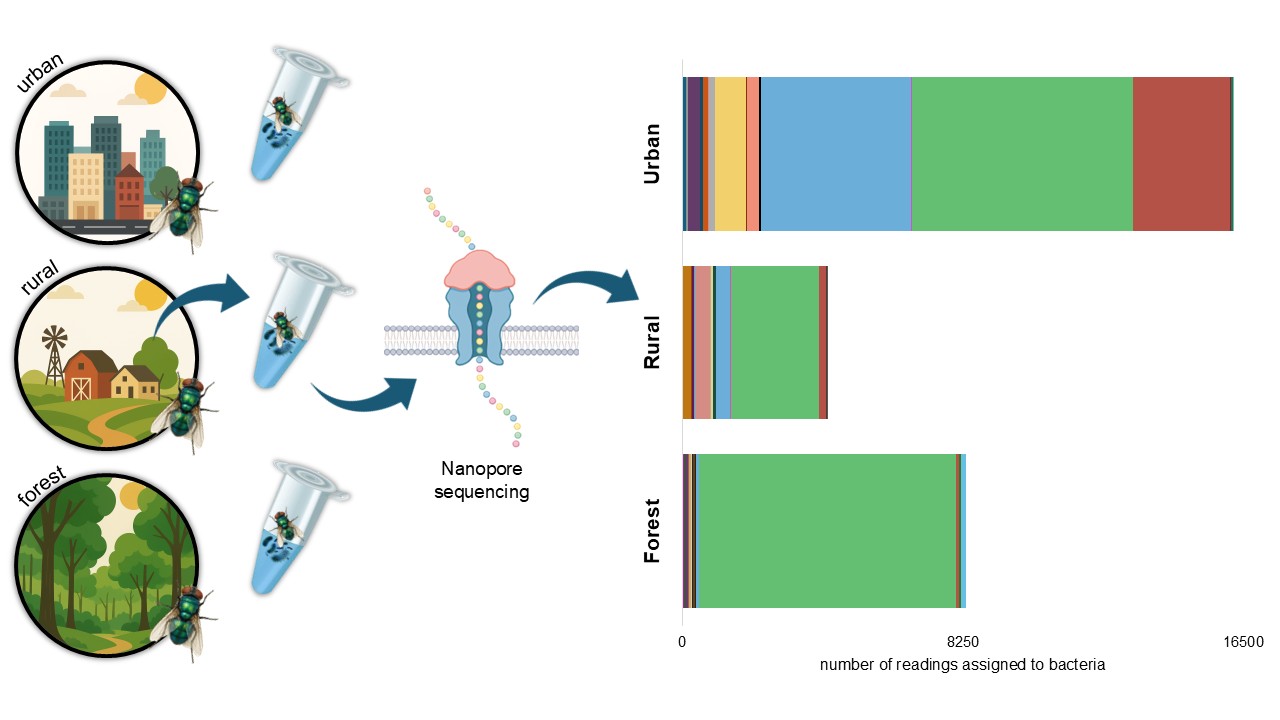
Chrysomya megacephala is a synanthropic fly with a high potential to act as a mechanical vector of pathogenic bacteria, surpassing Musca domestica in both bacterial load and diversity. Native to Asia and Africa, it has become a cosmopolitan species, successfully adapting to a wide range of environments, including natural ecosystems. In Colombia, studies on its role as a vector are limited and have largely relied on traditional culturing methods. This study aimed to characterize the pathogenic bacterial microbiota associated with C. megacephala using 16S rRNA gene sequencing in urban, rural, and forest settings of a coastal tourist city. Flies were collected using Van Someren Rydon traps with attractants and sterile materials. Bacterial identification was performed through Oxford Nanopore MinION sequencing. A total of 49 bacterial species were identified, with urban environments showing the highest richness and abundance. In forest environments, Vagococcus carniphilus was the dominant species. Notably, 20 bacterial species of public health relevance were detected, including Clostridium botulinum, Clostridium perfringens, Ignatzschineria ureiclastica, Escherichia coli, and Streptococcus agalactiae. These findings indicate that bacterial community composition varies by environment and underscore the potential role of C. megacephala as a mechanical vector, highlighting the importance of surveillance for its public health implications.

Keywords: 
;  ;  ;  ;  ;  ;  ;  ;  ;  

1. Introduction

Chrysomya megacephala (Fabricius, 1794) has been categorized as a synanthropic species because of its close association with human settlements, and like Musca domestica Linnaeus, 1758, it has been widely recognized as a mechanical vector of pathogens, largely owing to its feeding and oviposition habits [1]. Various studies have shown that C. megacephala poses a greater epidemiological risk. This species was reported to carry bacteria in 87.7% of the analyzed samples, whereas it was found in 66.2% of the M. domestica samples, indicating a greater diversity of bacterial species [1,2]. Additionally, in urban environments, C. megacephala was found to be not only more abundant than M. domestica but also to exhibit bacterial positivity rates ranging from 96.4% to 100%, harboring bacterial loads up to 11–12-fold higher [2]. Although both species constitute more than 50% of their microbiome, C. megacephala presents greater diversity and a greater load of pathogenic bacteria, including clinically relevant species such as Escherichia coli, Enterobacter cloacae, and Pseudomonas spp. [3].
Chrysomya megacephala belongs to the family Calliphoridae (Diptera) and is commonly known as the oriental latrine fly. It is an exotic species that is native to Asia and Africa and was introduced into South America in the late 1970s [4]. Its remarkable dispersal ability, high fecundity, and ecological adaptability have allowed it to be rapidly established in various environments [5]. It is now considered a cosmopolitan species and is currently widely distributed across the globe [6,7]. It is a necrophagous and saprophagous species that is strongly associated with urban environments but is also capable of colonizing natural habitats. The presence of this species has been recorded in fragments of tropical dry forest—an ecosystem with minimal human intervention—indicating that it does not rely exclusively on urban or degraded environments to thrive [5].
In Colombia, research on bacteria associated with synanthropic flies has been limited and has focused mostly on M. domestica [8,9]. A single study including multiple fly species reported that C. megacephala had the highest mechanical vector risk index (MVRI), indicating a high capacity to transport pathogenic bacteria from contaminated sources to surfaces in contact with humans [10].
All of these studies employed conventional culturing techniques, representing a significant limitation, as it is estimated that only ~1% of the bacterial microbiome is cultivable. In contrast, next-generation sequencing (NGS) can characterize the entire microbial diversity of a sample and compare it to known nucleotide sequences in databases, enabling the identification of all sequenced organisms [11]. Despite methodological constraints, seven different bacterial species have been identified in C. megacephala, including Escherichia coli, Providencia rettgeri, Pasteurella pneumotropica, Kluyvera sp., Serratia odorifera, Chryseobacterium meningosepticum, and Enterobacter sakazakii, highlighting the underestimated epidemiological relevance of this species in the Colombian context [10].
Recent studies using metagenomic techniques have shown that the external surfaces of this fly—particularly the legs and wings—harbor a greater quantity and diversity of bacteria than its internal structures, including numerous clinically important pathogens [3]. This elevated microbial load is facilitated by morphological structures on its exoskeleton, such as microvilli, setae, and hairs, which enhance microbial retention [9].
Given this evidence, the present study aims to identify the pathogenic bacteria associated with C. megacephala in a coastal tourist city with a high influx of national and international visitors by examining its external microbiota in urban, rural, and forest environments through 16S rRNA gene sequencing. This approach will not only expand the knowledge of its vector potential across gradients of anthropogenic disturbance but also provide critical insights into the health risks posed by this species in global tourist settings, where interactions between humans and synanthropic vectors may facilitate the spread of emerging pathogens.

2. Materials and Methods

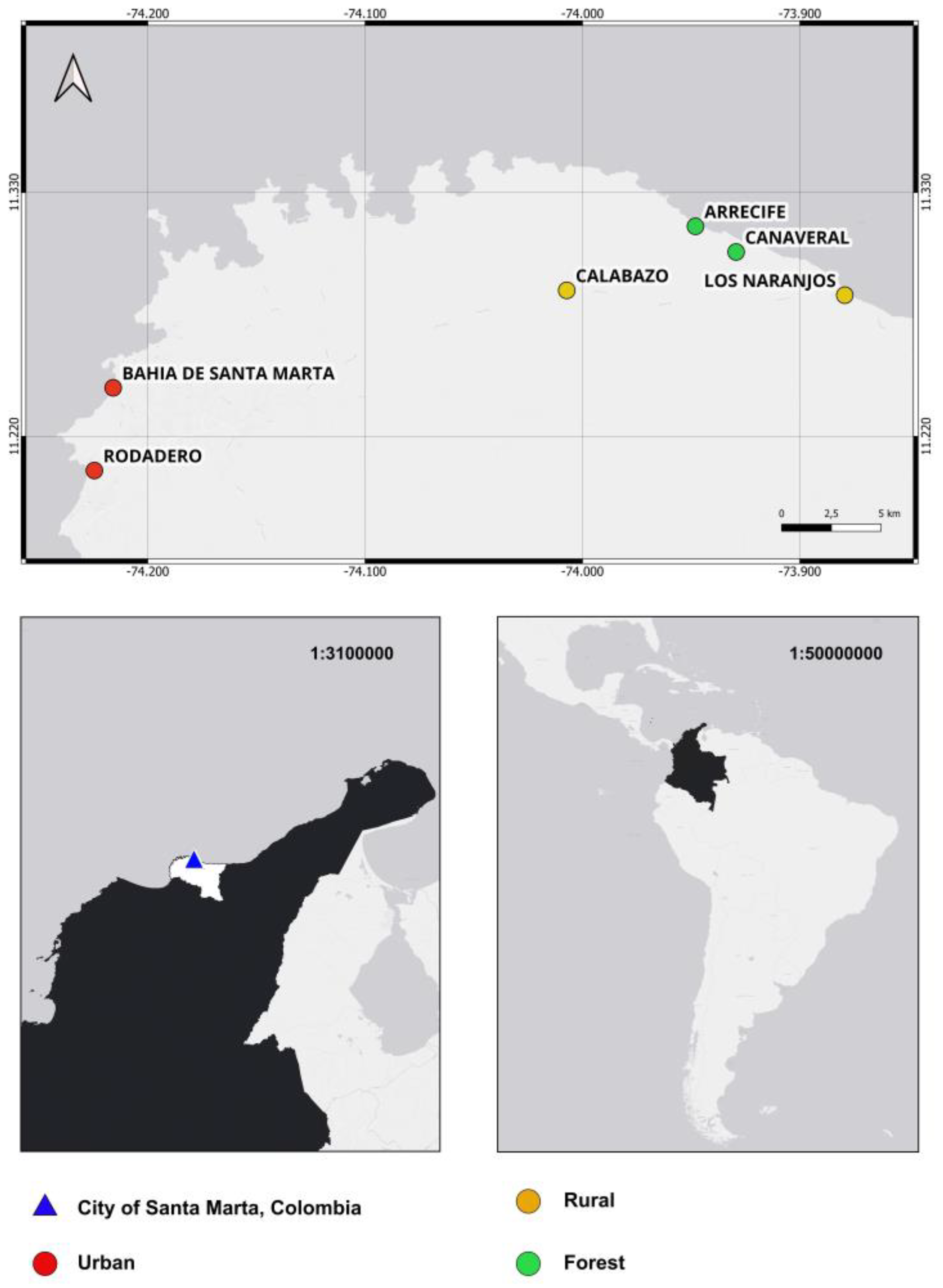
2.1. Study Area and Fly Collection

The study was conducted in the northern region of South America, along the Caribbean coast, in the city of Santa Marta, Colombia (Figure 1). This city is one of the main tourist destinations in the Caribbean region and is surrounded by beaches of high recreational and ecological value that attract thousands of national and international visitors year-round. It has a tropical dry climate, with average annual temperatures between 27 and 30 °C and annual rainfall ranging from 500 to 1,000 mm. Six zones with distinct environmental characteristics were selected. The urban zones of El Rodadero and Bahía de Santa Marta are located within the city core and are characterized by high population density, intense tourist activity, and extensive infrastructure. In contrast, the rural zones of Calabazo and Los Naranjos feature a landscape dominated by secondary vegetation and mixed land use combining agriculture and ecotourism, with scattered, low-density populations. Finally, the areas of Cañaveral and Arrecife represent forested zones located within Tayrona National Natural Park, a tropical dry forest ecosystem with high biological diversity and varying degrees of conservation. Together, these sites represent an ecological gradient with marked variations in vegetation cover, anthropogenic disturbance, and the availability of decomposing organic matter.
Field collection was carried out in January, April, July, and October 2024. Each sampling effort lasted 12 hours, during which three Van Someren Rydon traps were installed 50 m apart at a height of 1.5 m. The traps were baited alternately with decomposing fish, fermented fruit, and human feces to simulate the diversity of organic matter present in these environments. The attractants were placed in plastic containers covered with tulle mesh to prevent direct contact between the flies and the bait. Inside each trap, sterile muslin fabric mesh was used to capture live samples, which were euthanized by freezing at −80 °C to avoid cross-contamination. Additionally, 1.5-ml Eppendorf tubes containing saline solution were placed near the traps as environmental controls.
The collected and frozen flies were individually placed into Eppendorf tubes with saline solution for external washing. To dislodge surface bacteria, each sample was vortexed for 5 minutes at 1,000 rpm. After this procedure, the flies were removed and taxonomically identified using the Whitworth key [12] to confirm the identity of Chrysomya megacephala.
The supernatants from ten flies per environment were pooled to create one sample per environment (urban, rural, or forest). Each pooled sample was centrifuged at 10,000 rpm for 5 minutes to concentrate the bacteria at the bottom of the tube, from which the sample was used for bacterial DNA extraction.

2.2. Molecular Procedures

Genomic DNA was extracted using the HiPurA Multi-Sample HiGenoMB Kit, with key modifications to the manufacturer's protocol. Microcentrifuge tubes containing the bacterial suspension were first centrifuged at 12,000 g for 8 minutes at 4 °C. The resulting pellet was resuspended in 200 μl of lysozyme solution (45 mg/ml) and incubated at 37 °C for 40 minutes in a thermomixer at 300 rpm. Next, 20 μl of proteinase K (20 mg/ml) and 200 μl of lysis solution (C1) were added, followed by vortexing for 10 seconds and incubation at 55 °C for 30 minutes. The extracted DNA was preserved in 50 μl of DNase- and pyrogen-free water at −80 °C. DNA concentration and quality were assessed using a Qubit 3 fluorometer (INVITROGEN), and purity was evaluated via the A260/A280 ratio [13].
Amplification of the 16S rRNA gene was performed using TaKaRa LA Taq polymerase [14] with the universal primers 27F (5′-AGAGTTTGATCMTGGCTCAG-3′) and 1492R (5′-GGTTACCTTGTTACGACTT-3′) [15]. PCRs were conducted using 4 μl of extracted DNA under the following cycling conditions: initial denaturation at 94 °C for 2 minutes, followed by 30 cycles of 94 °C for 30 seconds, 55 °C for 30 seconds, and 72 °C for 90 seconds, with a final extension at 72 °C for 10 minutes.
Library preparation was performed using the Native Barcode Kit 96 V14 (SQK-NBD114.96), and sequencing was carried out on a MinION FLO-MIN114 flow cell (R10 version) at the Centro de Genética y Biología Molecular, Universidad del Magdalena.

2.3. Bioinformatic Analyses

The raw data were processed via Dorado v0.9.1 for basecalling, employing the SUP (super accuracy basecalling) algorithm with the dna_r10.4.1_e8.2_400 bps_sup@v5.0.0 model, retaining reads with a Phred quality score ≥ 10. Demultiplexing was performed under strict parameters, retaining only reads with valid barcodes at both ends. Read quality was assessed using NanoPlot v1.44.1 [16], and adapters and chimeric sequences were removed using Porechop v0.2.4 [17]. The tool fastp v0.24.0 [18] was used to retain reads between 1,400 and 1,700 bp, and Cutadapt v3.5 [19] was applied to trim primers with a 25% error tolerance. Reads containing internal primers were discarded. Final quality checks were performed using NanoPlot and MultiQC [20].
Taxonomic classification and read curation were conducted via the PRONAME pipeline [21] with the SILVA 138 SSURef N99 database [22]. The reads were clustered into OTUs using VSEARCH v2.22.1 [23] at a 98.7% similarity threshold [24]. Consensus sequences (CSs) were generated by selecting centroids and 100 random reads per cluster using Seqkit v2.3.0 [25], followed by error correction using Medaka v2.0.1. Chimeric sequences were detected with VSEARCH using SILVA as a reference.
After contaminant removal, a second validation of the consensus sequences was performed via BLASTn v2.15.0 [26] against the NCBI nonredundant database, considering alignments valid if they met the following criteria: identity ≥ 98.7%, coverage ≥ 99%, E value ≤ 0, and bit score ≥ 50 [24]. The final results were exported in a QIIME2-compatible format for visualization via QIIME2 View.

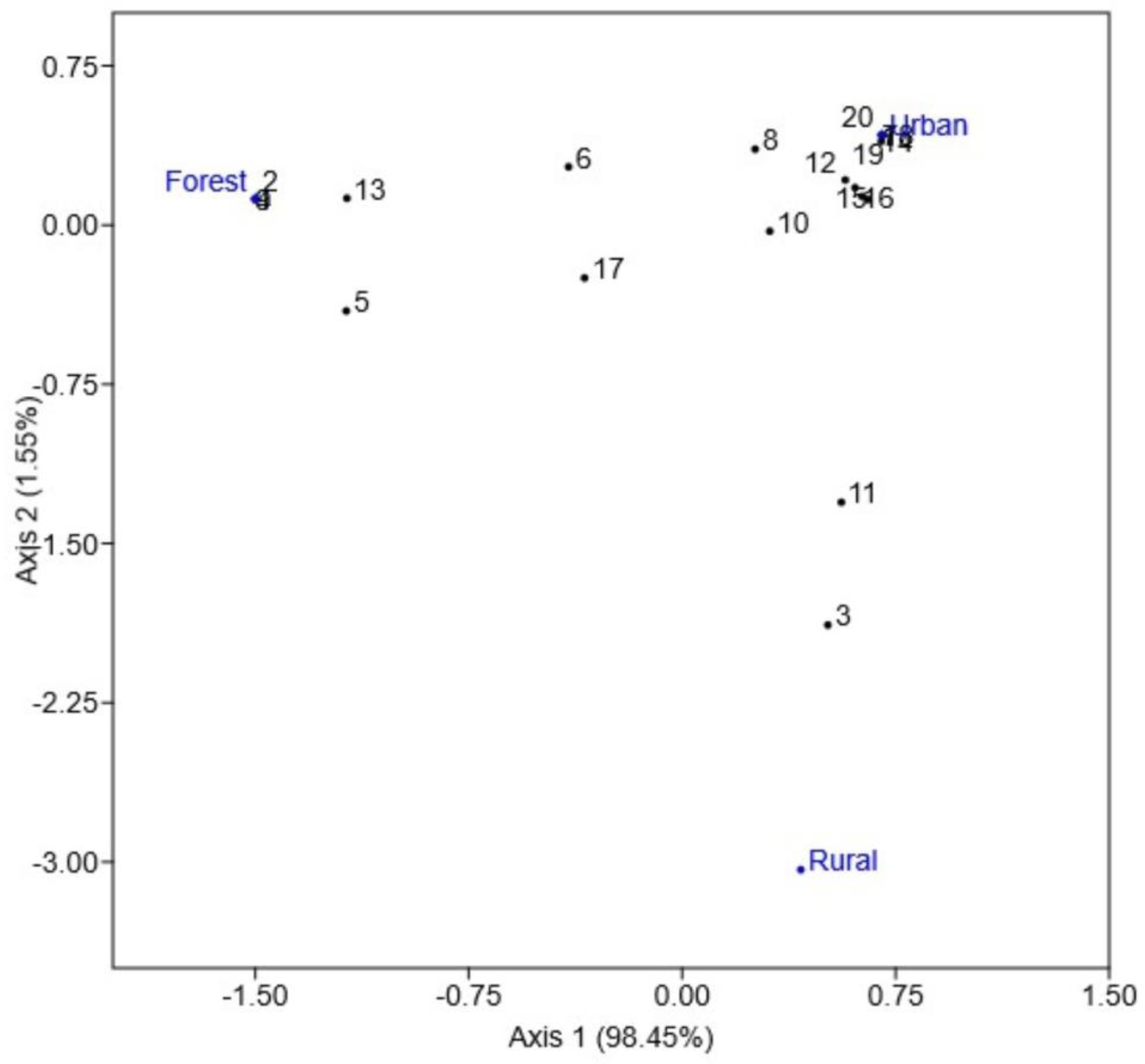
2.4. Bacterial Diversity Analyses

Taxa detected in negative controls, both laboratory and environmental, as well as reads not classified at the species level, were excluded from further analysis. Statistical analyses were based on relative abundance data. Using PAST software version 4.12 [27], diversity indices such as the Shannon (H′) index and dominance (D) index were calculated. An analysis of similarities (ANOSIM) was performed using the Bray‒Curtis index to assess compositional differences among environments.
Finally, correspondence analysis (CA) was conducted using only medically and veterinary-relevant bacterial species to explore and visualize specific associations between taxa and the environments where C. megacephala was collected.

3. Results

A total of 49 bacterial species were identified, among which Vagococcus carniphilus was the most abundant (16,596 reads; 57.65%), followed by Streptococcus infantarius (4,916; 17.08%) and Weissella cibaria (3,185; 11.06%) (Table 1). The urban environment not only presented the highest species richness, with 32 taxa but also presented a more even bacterial community structure (H′ = 1.63; D = 0.27). The rural environment presented slightly lower richness, with 30 species, and intermediate values of diversity and evenness (H′ = 1.49; D = 0.39). In contrast, the forest environment, although it presented the same richness as the rural environment, was strongly dominated by a single species that accounted for more than 90% of the reads, resulting in significantly reduced diversity (H′ = 0.56; D = 0.82). The ANOSIM revealed significant differences in bacterial composition between environments (R = 1, p = 0.003), indicating that the bacterial communities associated with C. megacephala are completely distinct depending on whether they originated from urban, rural, or forest settings.
The identification of medically and veterinary-relevant bacteria on the external surface of C. megacephala underscores the potential of this species to act as a mechanical vector of microorganisms that may impact human and animal health. To explore the clinical relevance of the identified taxa, a literature review was conducted, and the findings are summarized in Table 2. Several of the detected bacterial species, although commonly found in the environment or in food products, have also been implicated in opportunistic infections and serious diseases. These results highlight the importance of considering the environment in which C. megacephala is found when assessing its potential role in the transmission of pathogenic bacteria.
The correspondence analysis (CA) enabled visualization of associations between medically and veterinary-relevant bacterial taxa and the sampled environments (Figure 2). The results revealed that several pathogenic species were preferentially associated with the urban environment, whereas others clustered around forest samples. Moreover, the rural environment presented a distinct bacterial profile, separated from the urban and forest environments in the ordination space. The first axis explained 98.45% of the total variability, capturing most of the differences in the bacterial‒environment associations.
Overall, , these results demonstrate that C. megacephala harbors a diverse bacterial community on its external surface, including taxa of medical and veterinary importance, and that these communities vary depending on the environment. These findings raise new questions regarding the potential role of this species in the dispersal of microorganisms across different ecological contexts.

4. Discussion

Chrysomya megacephala is a synanthropic species strongly associated with environments rich in decomposing organic matter, such as carcasses, excreta, and household waste [1,2]. In this study, the bacterial community associated with the surface of C. megacephala was dominated by the phylum Firmicutes, followed by notable representatives of Proteobacteria and Actinobacteria, reflecting a microbial structure shaped by the insect’s ecological environment and food sources [48]. These results are consistent with previous findings reporting high microbial diversity on both the external surface and internal organs of C. megacephala and M. domestica, including numerous genera with pathogenic potential [3].
One of the most notable findings was the high prevalence of Vagococcus carniphilus, a gram-positive coccobacillus rarely reported in clinical contexts but documented in substrates such as raw meat, animal intestines, and fermentative environments [49]. Its consistent presence in all the samples suggests a stable ecological association with the fly, which is potentially symbiotic. V. carniphilus has been associated with skin lesions and hemorrhages in fish, with the potential to infect mammals, highlighting its relevance in veterinary health [44]. Although it has not been clearly linked to human pathogenicity, its high abundance in these flies positions it as a potential emerging pathogen of public health concern.
The detection of Streptococcus infantarius, a microorganism traditionally associated with fermented foods but emerging as a cause of musculoskeletal infections in humans [43], accounting for 17% of the total isolated bacteria, reinforces the role of C. megacephala as a mechanical vector in urban environments. Likewise, the identification of Streptococcus agalactiae, a well-known human pathogen implicated in infections such as septicemia, meningitis, and pneumonia, underscores the risk of indirect transmission from contaminated surfaces [41]. These findings are in line with previous reports of potentially pathogenic streptococci isolated from flies collected in Colombian hospitals, further supporting the hypothesis of cross-transmission via nontraditional vectors [9].
Another important observation was the detection of Weissella cibaria, which accounted for more than 11% of the isolates. This bacterium, which is commonly used in food fermentation and recognized for its probiotic potential, has also been reported as an opportunistic pathogen in immunocompromised patients [46]. Its presence on the surface of C. megacephala may indicate the transfer of fermentative microbiota from food waste to clinical environments, a pattern previously suggested in other studies [3]. This dual role—commensal/probiotic versus pathogenic—requires contextual evaluation of each strain’s ecological and genetic background to assess its potential risk or benefit to human health.
In parallel, the identification of Ignatzschineria ureiclastica, a genus associated with myiasis and bacteremia secondary to dipteran larval infestations [35], is clinically relevant. Although this study focused on adult flies, the persistence of this genus throughout the dipteran life cycle suggests potential risk in urban areas where contact between flies, open wounds, and organic waste is prevalent [2,50]. Therefore, controlling urban fly populations not only prevents infestations but also may serve as an indirect strategy to reduce the spread of emerging bacterial pathogens.
Although in lower abundance, clinically relevant species such as Morganella morganii, Clostridium botulinum, Clostridium perfringens, and Escherichia coli have also been identified, all of which are associated with opportunistic infections and antimicrobial resistance [29,30,31,33,40]. Previous studies in Colombia [10] reported the presence of multidrug-resistant enterobacteria in flies collected from hospitals and urban markets, which may explain the detection of these bacteria in our samples, particularly in densely populated areas. The ability of C. megacephala to act as a temporary reservoir for these pathogens underscores its importance in urban microbial ecology.
Our findings align with previous observations of differences in bacterial load between M. domestica and C. megacephala, with the latter exhibiting greater versatility in terms of bacterial diversity [1]. Our results confirm that C. megacephala can harbor species of high medical relevance, especially in tropical regions where it is abundant, synanthropic, and frequently encountered in human environments [3]. The consistency of these findings with studies conducted in other geographic regions suggests that ecological factors, such as high availability of organic waste, frequent contact with excreta, and unregulated urbanization, are common drivers shaping its microbiome.

5. Conclusions

Considering these findings, the importance of C. megacephala as a mechanical vector of pathogens is reinforced, emphasizing its potential role in the indirect transmission of infectious diseases in urban settings. The identified bacteria not only pose immediate risks through contamination of food and surfaces but also may serve as indicators of the environmental circulation of opportunistic and emerging microorganisms. We propose that this species be incorporated into vector-borne disease (VBD) surveillance programs, particularly in tropical regions where it is prevalent and where sanitary conditions are often limited.

Author Contributions

Conceptualization, C.V.C., A.L.P.B. and M.T.M.; methodology C.V.C. and A.L.P.B.; software, C.V.C.; investigation, , C.V.C., A.L.P.B. and M.T.M.; resources, , C.V.C. and M.T.M.; data curation, A.L.P.B.; writing—original draft preparation, C.V.C. and A.L.P.B.; writing—review and editing, C.V.C. and A.L.P.B.; project administration, M.T.M.; funding acquisition, C.V.C. and M.T.M. All authors have read and agreed to the published version of the manuscript.

Funding

This work was supported by the Science Fund of the Universidad del Magdalena (Fonciencias) and the Ministerio de Ciencia, Tecnología e Innovación de Colombia (Minciencias), through the project “Evaluación de la dinámica espacio-temporal de la comunidad de moscas de importancia médico-sanitaria y caracterización metagenómica de sus patógenos asociados en destinos turísticos de Santa Marta”, funded under Call 890 of 2020.

Institutional Review Board Statement

Not applicable.

Informed Consent Statement

Not applicable.

Data Availability Statement

We FASTQ files are deposited in the Sequence Read Archive (SRA) databases (https://www.ncbi.nlm.nih.gov/sra) under BioProject PRJNA1292279.

Acknowledgments

We acknowledge the Centro de Genética y Biología Molecular at the Universidad del Magdalena for their laboratory support. We are also grateful to the local communities who granted access to their territories during fieldwork, as well as to Colombia's National Natural Parks for authorizing entry and sample collection in protected áreas.

Conflicts of Interest

The authors declares that they have no competing interests.

References

  1. Sukontason, K.L.; Bunchoo, M.; Khantawa, B.; Piangjai, S.; Rongsriyam, Y.; Sukontason, K. Comparison between Musca domestica and Chrysomya megacephala as carriers of bacteria in northern Thailand. Southeast Asian J Trop Med Public Health 2007, 38(1), 38. [Google Scholar]
  2. Chaiwong, T.; Srivoramas, T.; Sueabsamran, P.; Sukontason, K.; Sanford, M.R.; Sukontason, K.L. The blow fly, Chrysomya megacephala, and the house fly, Musca domestica, as mechanical vectors of pathogenic bacteria in Northeast Thailand. Trop Biomed. 2014, 31(2), 336–46. [Google Scholar] [PubMed]
  3. Junqueira, A.C.M.; Ratan, A.; Acerbi, E.; Drautz-Moses, D.I.; Premkrishnan, B.N.; Costea, P.I.; et al. The microbiomes of blowflies and houseflies as bacterial transmission reservoirs. Sci Rep. 2017, 7(1), 16324. [Google Scholar] [CrossRef] [PubMed]
  4. Guimarães, J.H.; Prado, A.P.; Linhares, A.X. Three newly introduced blowfly species in southern Brazil (Diptera: Calliphoridae). Rev Bras Entomol. 1978, 22, 53–60. [Google Scholar]
  5. Vasconcelos, S.D.; Salgado, R.L. First record of six Calliphoridae (Diptera) species in a seasonally dry tropical forest in Brazil: evidence for the establishment of invasive species. Fla Entomol. 2014, 97(2), 814–6. [Google Scholar] [CrossRef]
  6. Wolff, M.; Kosmann, C. Families Calliphoridae and Mesembrinellidae. Zootaxa. 2016, 4122(1), 856–75. [Google Scholar] [CrossRef]
  7. Wells, J.D. Chrysomya megacephala (Diptera: Calliphoridae) has reached the continental United States: review of its biology, pest status, and spread around the world. J Med Entomol. 1991, 28(3), 471–3. [Google Scholar] [CrossRef]
  8. Quiceno, J.; Bastidas, X.; Rojas, D.; Bayona, M. La mosca doméstica como portador de patógenos microbianos en cinco cafeterías del norte de Bogotá. Rev UDCA Actual Divulg Cienc 2010, 13(2), 23–29, Español. [Google Scholar] [CrossRef]
  9. Estrada, S.; Ceballos, M.T.; Vanegas, C.; Yepes, S.; Estrada, M.S.; Roncancio, G. Bacterias identificadas en la superficie de Musca domestica y su potencial patogenicidad para el humano. Hechos Microbiol. 2011, 2(2), 55–62, Español. [Google Scholar] [CrossRef]
  10. Cadavid-Sánchez, I.C.; Amat, E.; Gómez-Piñerez, L.M. Enterobacteria isolated from synanthropic flies (Diptera, Calyptratae) in Medellín, Colombia. Caldasia 2015, 37(2), 319–32. [Google Scholar] [CrossRef]
  11. Goodwin, S.; McPherson, J.D.; McCombie, W.R. Coming of age: ten years of next-generation sequencing technologies. Nat Rev Genet. 2016, 17(6), 333–51. [Google Scholar] [CrossRef] [PubMed]
  12. Whitworth, T. Keys to the genera and species of blow flies (Diptera: Calliphoridae) of the West Indies and description of a new species of Lucilia Robineau-Desvoidy. Zootaxa 2010, 2663(1), 1–35. [Google Scholar] [CrossRef]
  13. Khare, P.; Raj, V.; Chandra, S.; Agarwal, S. Quantitative and qualitative assessment of DNA extracted from saliva for its use in forensic identification. J Forensic Dent Sci. 2014, 6(2), 81–5. [Google Scholar] [CrossRef] [PubMed]
  14. Shimada, A.; Takeda, O. Site-Directed Mutagenesis with LA-PCR™ Technology. In The Nucleic Acid Protocols Handbook; Rapley, R., Ed.; Humana Press: Totowa, NJ, USA, 2000; p. 845. [Google Scholar] [CrossRef]
  15. Yeo, K.; Connell, J.; Bouras, G.; Smith, E.; Murphy, W.; Hodge, J.C.; et al. A comparison between full-length 16S rRNA Oxford nanopore sequencing and Illumina V3-V4 16S rRNA sequencing in head and neck cancer tissues. Arch Microbiol. 2024, 206(6), 248. [Google Scholar] [CrossRef] [PubMed]
  16. De Coster, W.; D'Hert, S.; Schultz, D.T.; Cruts, M.; Van Broeckhoven, C. NanoPack: Visualizing and processing long-read sequencing data. Bioinformatics 2018, 34(15), 2666–9. [Google Scholar] [CrossRef]
  17. Wick, R.R. Porechop [Software]. GitHub. 2017. https://github.com/rrwick/Porechop.
  18. Chen, S.; Zhou, Y.; Chen, Y.; Gu, J. fastp: An ultra-fast all-in-one FASTQ preprocessor. Bioinformatics 2018, 34(17), i884–90. [Google Scholar] [CrossRef]
  19. Martin, M. Cutadapt removes adapter sequences from high-throughput sequencing reads. EMBnet.journal 2011, 17(1), 3. [Google Scholar] [CrossRef]
  20. Ewels, P.; Magnusson, M.; Lundin, S.; Käller, M. MultiQC: Summarize analysis results for multiple tools and samples in a single report. Bioinformatics 2016, 32(19), 3047–8. [Google Scholar] [CrossRef]
  21. Dubois, B.; Delitte, M.; Lengrand, S.; Bragard, C.; Legrève, A.; Debode, F. PRONAME: a user-friendly pipeline to process long-read nanopore metabarcoding data by generating high-quality consensus sequences. Front Bioinform. 2024, 4, 1483255. [Google Scholar] [CrossRef]
  22. Quast, C.; Pruesse, E.; Yilmaz, P.; Gerken, J.; Schweer, T.; Yarza, P.; et al. The SILVA ribosomal RNA gene database project: Improved data processing and web-based tools. Nucleic Acids Res. 2013, 41(D1), D590–6. [Google Scholar] [CrossRef]
  23. Rognes, T.; Flouri, T.; Nichols, B.; Quince, C.; Mahé, F. VSEARCH: A versatile open-source tool for metagenomics. PeerJ 2016, 4, e2584. [Google Scholar] [CrossRef] [PubMed]
  24. Kim, M.; Oh, H.S.; Park, S.C.; Chun, J. Towards a taxonomic coherence between average nucleotide identity and 16S rRNA gene sequence similarity for species demarcation of prokaryotes. Int J Syst Evol Microbiol. 2014, 64 Pt 2, 346–51. [Google Scholar] [CrossRef] [PubMed]
  25. Shen, W.; Le, S.; Li, Y.; Hu, F. SeqKit: a cross-platform and ultrafast toolkit for FASTA/Q file manipulation. PLoS One 2016, 11, e0163962. [Google Scholar] [CrossRef]
  26. Camacho, C.; Coulouris, G.; Avagyan, V.; Ma, N.; Papadopoulos, J.; Bealer, K.; et al. BLAST+: Architecture and applications. BMC Bioinformatics 2009, 10, 421. [Google Scholar] [CrossRef]
  27. Hammer, Ø.; Harper, D.A.T.; Ryan, P.D. PAST: Paleontological statistics software package for education and data analysis. Palaeontol Electron. 2001, 4, 1–9, http://palaeo-electronica.org/2001_1/past/issue1_01.htm. [Google Scholar]
  28. Heinäsmäki, T.; Kerttula, T. First report of bacteremia by Asaia bogorensis in a patient with a history of intravenous-drug abuse. J Clin Microbiol. 2006, 44(8), 3048–50. [Google Scholar] [CrossRef]
  29. Rawson, A.M.; Dempster, A.W.; Humphreys, C.M.; Minton, N.P. Pathogenicity and virulence of Clostridium botulinum. Virulence 2023, 14(1), 2205251. [Google Scholar] [CrossRef] [PubMed]
  30. Mehdizadeh Gohari, I.; Navarro, M.A.; Li, J.; Shrestha, A.; Uzal, F.A.; McClane, B.A. Pathogenicity and virulence of Clostridium perfringens. Virulence 2021, 12(1), 723–53. [Google Scholar] [CrossRef]
  31. Samul, D.; Worsztynowicz, P.; Leja, K.; Grajek, W. Beneficial and harmful roles of bacteria from the Clostridium genus. Acta Biochim Pol. 2013, 60(4), 515–21. [Google Scholar] [CrossRef]
  32. Rostamian, M.; Rahmati, D.; Akya, A. Clinical manifestations, associated diseases, diagnosis, and treatment of human infections caused by Erysipelothrix rhusiopathiae: a systematic review. Germs. 2022, 12(1), 16. [Google Scholar] [CrossRef]
  33. Pakbin, B.; Brück, W.M.; Rossen, J.W. Virulence factors of enteric pathogenic Escherichia coli: a review. Int J Mol Sci. 2021, 22(18), 9922. [Google Scholar] [CrossRef]
  34. Mada, P.K.; Khan, M.H. Hathewaya limosa empyema: a case report. Cureus 2024, 16(2), e55156. [Google Scholar] [CrossRef]
  35. Reed, K.; Reynolds, S.B.; Smith, C. The first case of Ignatzschineria ureiclastica/larvae in the United States presenting as a myiatic wound infection. Cureus 2021, 13(4), e14518. [Google Scholar] [CrossRef] [PubMed]
  36. Pot, B.; Felis, G.E.; De Bruyne, K.; Tsakalidou, E.; Papadimitriou, K.; Leisner, J.; et al. The genus Lactobacillus. In Lactic Acid Bacteria; Holzapfel, W.H., Wood, B.J.B., Eds.; Wiley-Blackwell: Hoboken, NJ, USA, 2014; Chapter 19. [Google Scholar] [CrossRef]
  37. Karaaslan, A.; Soysal, A.; Kepenekli Kadayifci, E.; Yakut, N.; Ocal Demir, S.; Akkoc, G.; et al. Lactococcus lactis spp. lactis infection in infants with chronic diarrhea: two case reports and literature review in children. J Infect Dev Ctries. 2016, 10(3), 304–7. [Google Scholar] [CrossRef]
  38. Motta, R.G.; Martinez, A.C.; Junior, O.C.M.P.; Martins, L.D.S.A.; da Paz, J.P.; Bello, T.S.; et al. Atypical mandibular osteomyelitis in an ewe caused by coinfection of Pseudomonas aeruginosa and Lactococcus raffinolactis. Acta Sci Vet. 2022, 50. [Google Scholar] [CrossRef]
  39. Simoiu, M.; Filipescu, M.C.; Omer, M.; Borcan, A.M.; Olariu, M.C. Leuconostoc pseudomesenteroides bacteremia in an immunocompromised patient with hematological comorbidities—case report. Microorganisms 2024, 12(11), 2215. [Google Scholar] [CrossRef]
  40. Alsaadi, A.; Alghamdi, A.A.; Akkielah, L.; Alanazi, M.; Alghamdi, S.; Abanamy, H.; et al. Epidemiology and clinical characteristics of Morganella morganii infections: a multicenter retrospective study. J Infect Public Health 2024, 17(3), 430–4. [Google Scholar] [CrossRef]
  41. Bobadilla, F.J.; Novosak, M.G.; Cortese, I.J.; Delgado, O.D.; Laczeski, M.E. Prevalence, serotypes and virulence genes of Streptococcus agalactiae isolated from pregnant women with 35–37 weeks of gestation. BMC Infect Dis. 2021, 21, 1–11. [Google Scholar] [CrossRef]
  42. Oliver, S.P.; Pighetti, G.M.; Almeida, R.A. Environmental pathogens. In Encyclopedia of Dairy Sciences, 2nd ed.; Fuquay, J.W., Ed.; Academic Press: Amsterdam, Netherlands, 2011. [Google Scholar] [CrossRef]
  43. Zellner, A.A.; Voss, J.; Franz, A.; Roos, J.; Hischebeth, G.T.R.; Molitor, E.; et al. Musculoskeletal infections caused by Streptococcus infantarius–a case series and review of literature. Int Orthop. 2025, 1–10. [Google Scholar] [CrossRef] [PubMed]
  44. Anifowose, O.; Oyebola, O.O.; Ajala, A.C. Haemato-biochemical and histopathological effects of Vagococcus carniphilus infection in Clarias gariepinus (Burchell 1822) juveniles. SSRN 2024. [Google Scholar] [CrossRef]
  45. Cobo, F.; Pérez-Carrasco, V.; García-Salcedo, J.A.; Navarro-Marí, J.M. Bacteremia caused by Veillonella dispar in an oncological patient. Anaerobe 2020, 66, 102285. [Google Scholar] [CrossRef] [PubMed]
  46. Kamboj, K.; Vasquez, A.; Balada-Llasat, J.M. Identification and significance of Weissella species infections. Front Microbiol. 2015, 6, 1204. [Google Scholar] [CrossRef] [PubMed]
  47. Björkroth, J.; Dicks, L.M.T.; Endo, A. The genus Weissella. In Lactic Acid Bacteria: Biodiversity and Taxonomy; Holzapfel, W.H., Wood, B.J.B., Eds.; Wiley: Hoboken, NJ, USA, 2014; pp. 417–428. [Google Scholar] [CrossRef]
  48. Liu, S.; Xi, B.D.; Qiu, Z.P.; He, X.S.; Zhang, H.; Dang, Q.L.; et al. Succession and diversity of microbial communities in landfills with depths and ages and its association with dissolved organic matter and heavy metals. Sci Total Environ. 2019, 651, 909–16. [Google Scholar] [CrossRef] [PubMed]
  49. Shewmaker, P.L.; Steigerwalt, A.G.; Morey, R.E.; Carvalho, M.D.G.S.; Elliott, J.A.; Joyce, K.; et al. Vagococcus carniphilus sp. nov., isolated from ground beef. Int J Syst Evol Microbiol. 2004, 54 Pt 5, 1505–10. [Google Scholar] [CrossRef]
  50. Ngando, F.J.; Tang, H.; Zhang, X.; Yang, F.; Shang, Y.; Zhang, C.; et al. Effects of feeding sources and different temperature changes on the gut microbiome structure of Chrysomya megacephala (Diptera: Calliphoridae). Insects 2025, 16(3), 283. [Google Scholar] [CrossRef]
Figure 1. Map of sampling points in Santa Marta, Colombia.
Preprints 185887 g001
Figure 2. Correspondence Analysis (CA) between bacteria of medical-veterinary importance and environments. The codes represent the 20 species of bacteria (see table 2).
Preprints 185887 g002
Table 1. Bacteria isolated from the outer surface of Chrysomya megacephala, in Santa Marta*
Taxa Urban Rural Forest Total
No (%) No (%) No (%) No (%)
Acinetobacter nectaris 109 (0.67) 1 (0.02) 22 (0.26) 132 (0.46)
Asaia bogorensis 21 (0.13) 0 (0) 0 (0) 21 (0.07)
Bacteroides xylanisolvens 12 (0.07) 2 (0.05) 0 (0) 14 (0.05)
Brochothrix thermosphacta 0 (0) 0 (0) 2 (0.02) 2 (0.01)
Candidatus Kinetoplastibacterium sp. 0 (0) 0 (0) 22 (0.26) 22 (0.08)
Catenibacterium mitsuokai 1 (0.01) 262 (6.16) 0 (0) 263 (80.91)
Clostridium botulinum 0 (0) 0 (0) 1 (0.01) 1 (0)
Clostridium perfringens 2 (0.01) 4 (0.09) 0 (0) 6 (0.02)
Clostridium sp. 0 (0) 0 (0) 4 (0.05) 4 (0.01)
Collinsella stercoris 0 (0) 4 (0.09) 0 (0) 4 (0.01)
Dorea formicigenerans 2 (0.01) 53 (1.25) 0 (0) 55 (0.19)
Enterococcus termitis 1 (0.01) 6 (0.14) 18 (0.22) 25 (0.09)
Erysipelothrix rhusiopathiae 0 (0) 1 (0.02) 5 (0.06) 6 (0.02)
Escherichia coli 1 (0.01) 0 (0) 1 (0.01) 2 (0.01)
Faecalitalea cylindroides 0 (0) 0 (0) 1 (0.01) 1 (0)
Hathewaya limosa 1 (0.01) 0 (0) 0 (0) 1 (0)
Ignatzschineria ureiclastica 362 (2.23) 1 (0.02) 92 (1.1) 455 (1.58)
Lactobacillus animalis 0 (0) 4 (0.09) 0 (0) 4 (0.01)
Lactobacillus brevis 90 (0.56) 0 (0) 0 (0) 90 (0.31)
Lactobacillus floricola 153 (0.94) 0 (0) 20 (0.24) 173 (0.6)
Lactobacillus gasseri 0 (0) 0 (0) 2 (0.02) 2 (0.01)
Lactobacillus helveticus 0 (0) 0 (0) 4 (0.05) 4 (0.01)
Lactobacillus kunkeei 0 (0) 0 (0) 4 (0.05) 4 (0.01)
Lactobacillus pontis 0 (0) 5 (0.12) 1 (0.01) 6 (0.02)
Lactobacillus sakei 30 (0.19) 449 (10.56) 0 (0) 479 (1.66)
Lactococcus lactis 154 (0.95) 25 (0.59) 35 (0.42) 214 (0.74)
Leuconostoc pseudomesenteroides 935 (5.77) 60 (1.41) 54 (0.65) 1049 (3.64)
Ligilactobacillus ruminis 1 (0.01) 1 (0.02) 0 (0) 2 (0.01)
Limosilactobacillus reuteri 4 (0.02) 32 (0.75) 6 (0.07) 42 (0.15)
Lonsdalea britannica 0 (0) 0 (0) 18 (0.22) 18 (0.06)
Morganella morganii 11 (0.07) 1 (0.02) 69 (0.83) 81 (0.28)
Neokomagataea thailandica 2 (0.01) 0 (0) 9 (0.11) 11 (0.04)
Olsenella sp. 0 (0) 37 (0.87) 0 (0) 37 (0.13)
Parolsenella catena 0 (0) 1 (0.02) 0 1 (0)
Pseudolactococcus raffinolactis 1 (0.01) 1 (0.02) 0 2 (0.01)
Ruminococcus sp. 3 (0.02) 27 (0.64) 4 (0.05) 34 (0.12)
Streptococcus agalactiae 343 (2.12) 3 (0.07) 0 (0) 346 (1.2)
Streptococcus equinus 83 (0.51) 8 (0.19) 1 (0.01) 92 (0.32)
Streptococcus infantarius 4406 (27.19) 405 (9.53) 105 (1.26) 4916 (17.08)
Streptococcus parauberis 0 (0) 0 (0) 1 (0.01) 1 (0)
Streptococcus sp. 37 (0.23) 44 (1.04) 0 (0) 81 (0.28)
Turicibacter sp. 0 (0) 6 (0.14) 0 (0) 6 (0.02)
Vagococcus carniphilus 6479 (39.98) 2575 (60.59) 7542 (90.54) 16596 (57.65)
Veillonella dispar 7 (0.04) 0 (0) 0 (0) 7 (0.02)
Weissella cibaria 2858 (17.64) 220 (5.18) 107 (1.28) 3185 (11.06)
Weissella confusa 2 (0.01) 0 (0) 0 (0) 2 (0.01)
Weissella ghanensis 15 (0.09) 1 (0.02) 1 (0.01) 17 (0.06)
Wolbachia endosymbiont sp. 79 (0.49) 11 (0.26) 50 (0.6) 140 (0.49)
Zymobacter palmae 1 (0.01) 0 (0) 129 (1.55) 130 (0.45)
Total 16206 (100) 4250 (100) 8330 (100) 28786 (100)
* No: number of readings assigned, % relative abundance by environment.
Table 2. Bacteria of medical-veterinary importance associated with Chrysomya megacephala*.
Code Taxa Environment Association
1 As. bogorensis U Bacteremia in immunocompromised patients (28).
2 Cl. botulinum F It produces botulinum neurotoxin, which causes botulism (29).
3 Cl. perfringens U, R Infections in humans and livestock, such as gas gangrene and enterotoxemia (30).
4 Clostridium sp. F Some species are associated with various human and veterinary diseases (31).
5 Er. rhusiopathiae R, F Skin and systemic diseases in humans (32).
6 E. coli U, F Diarrhea, hemorrhagic colitis, urinary tract infection; infections in fish (33).
7 H. limosa U Human infections and empiema (34).
8 I. ureiclastica U, R, F Bacteremia associated with myiasis (infested wounds) (35).
9 Lb. gasseri F Infections in immunocompromised patients (36).
10 Lc. lactis U, R, F Infections in immunocompromised patients (37).
11 Ps. raffinolactis U, R Lethal coinfection associated with mandibular pyogranulomatous osteomyelitis in sheep (38).
12 Le. pseudomesenteroides U, R, F Bacteremia in immunocompromised patients (39).
13 M. morganii U, R, F Human infections (40).
14 S. agalactiae U, R Serious infections such as sepsis, pneumonia, and meningitis (41).
15 S. equinus U, R, F Bovine mastitis and septicemia (42).
16 S. infantarius U, R, F Bacteremia, endocarditis, and musculoskeletal infections (43).
17 Va. carniphilus U, R, F Skin lesions and hemorrhages in fish (44).
18 Ve. dispar U Severe infections in humans (45).
19 We. cibaria U, R, F Opportunistic pathogen (46).
20 We. confusa U Bacteremia, endocarditis, and abscess cases in humans (47).
* U: urban, R: rural, F: forest.
Disclaimer/Publisher’s Note: The statements, opinions and data contained in all publications are solely those of the individual author(s) and contributor(s) and not of MDPI and/or the editor(s). MDPI and/or the editor(s) disclaim responsibility for any injury to people or property resulting from any ideas, methods, instructions or products referred to in the content.
Copyright: This open access article is published under a Creative Commons CC BY 4.0 license, which permit the free download, distribution, and reuse, provided that the author and preprint are cited in any reuse.
Prerpints.org logo

Preprints.org is a free preprint server supported by MDPI in Basel, Switzerland.

Subscribe

Disclaimer

Terms of Use

Privacy Policy

Privacy Settings

© 2025 MDPI (Basel, Switzerland) unless otherwise stated