Preprint
Article

This version is not peer-reviewed.

Transgenic Citrus sinensis Expressing the Pepper Bs2 R-Gene Shows Broad Transcriptional Activation of Defense Responses to Citrus Canker

Submitted:

18 November 2025

Posted:

19 November 2025

You are already at the latest version

Abstract

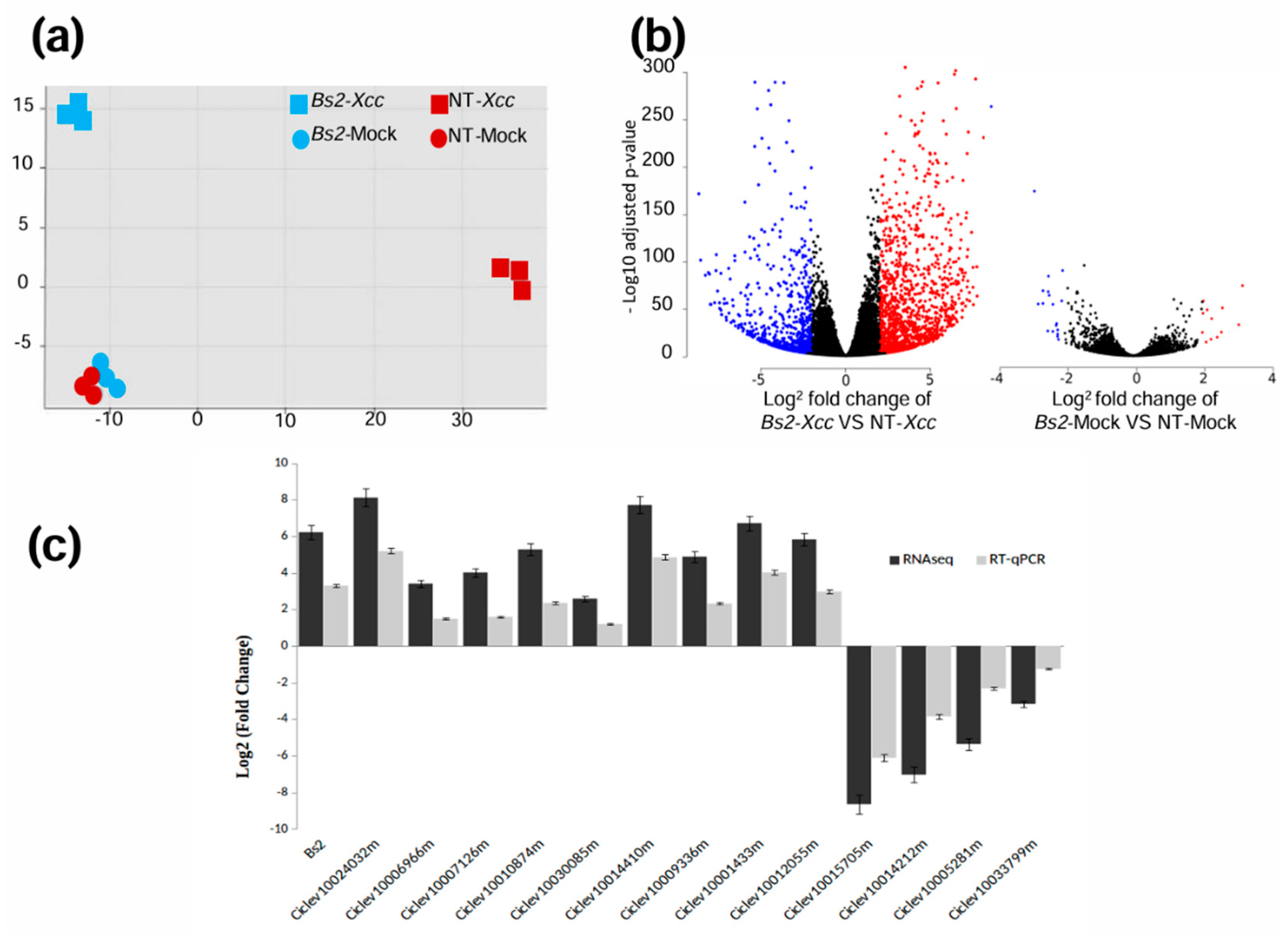
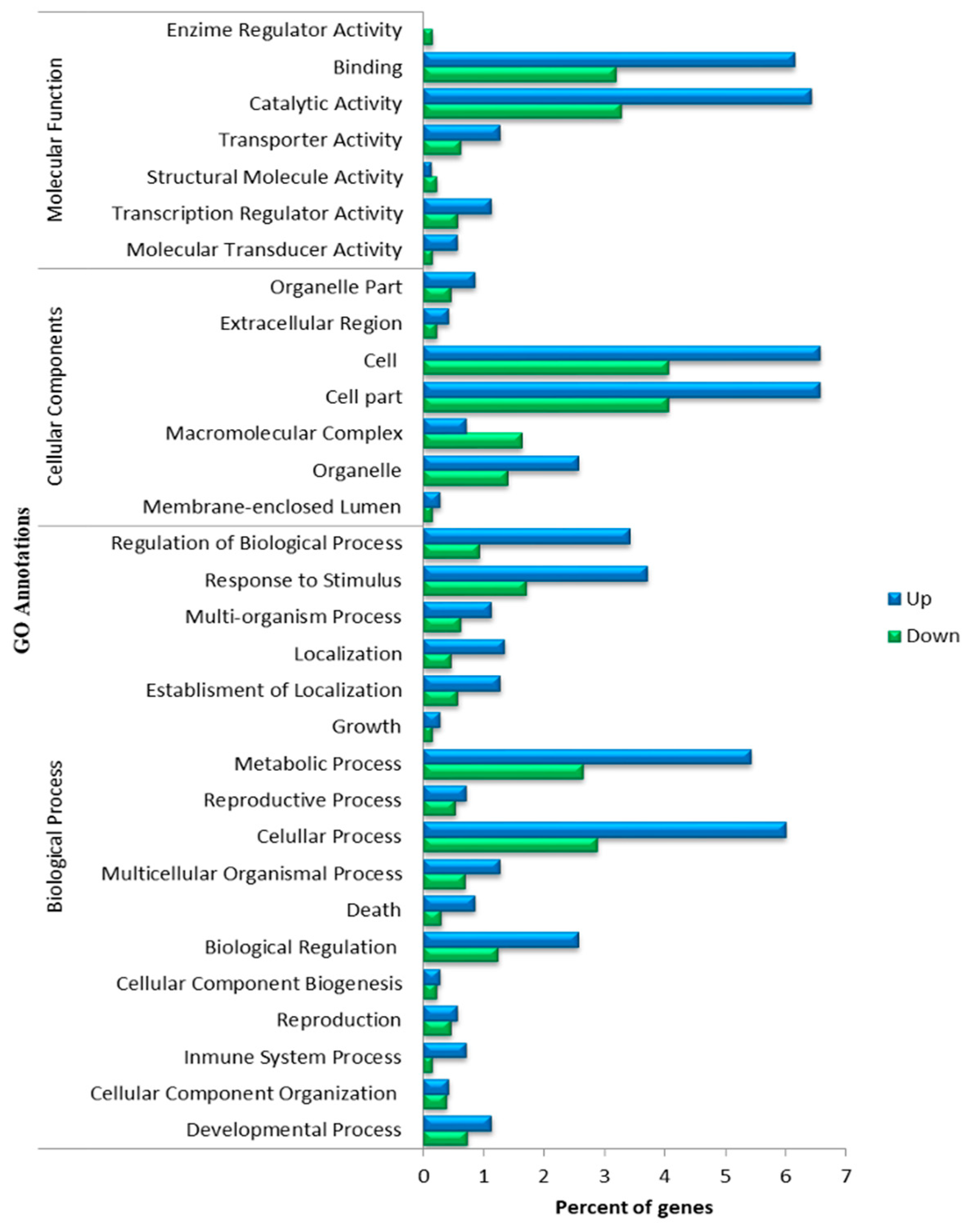
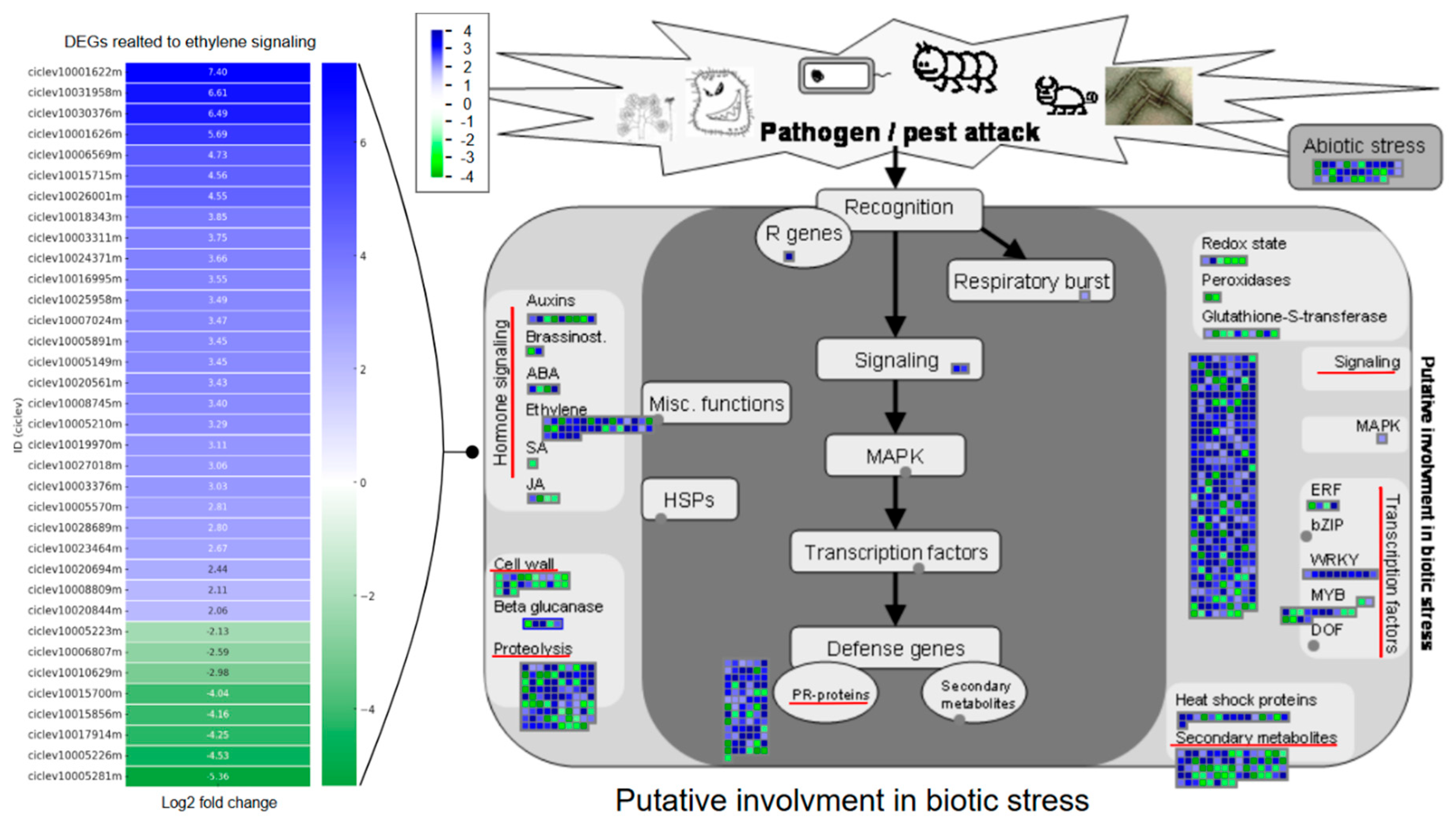
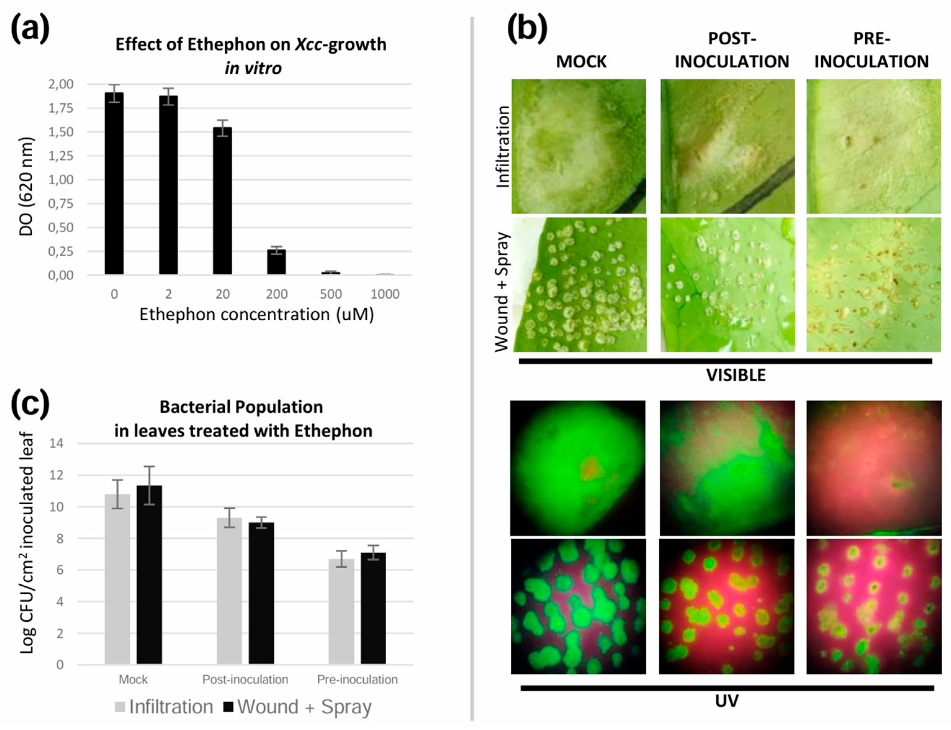
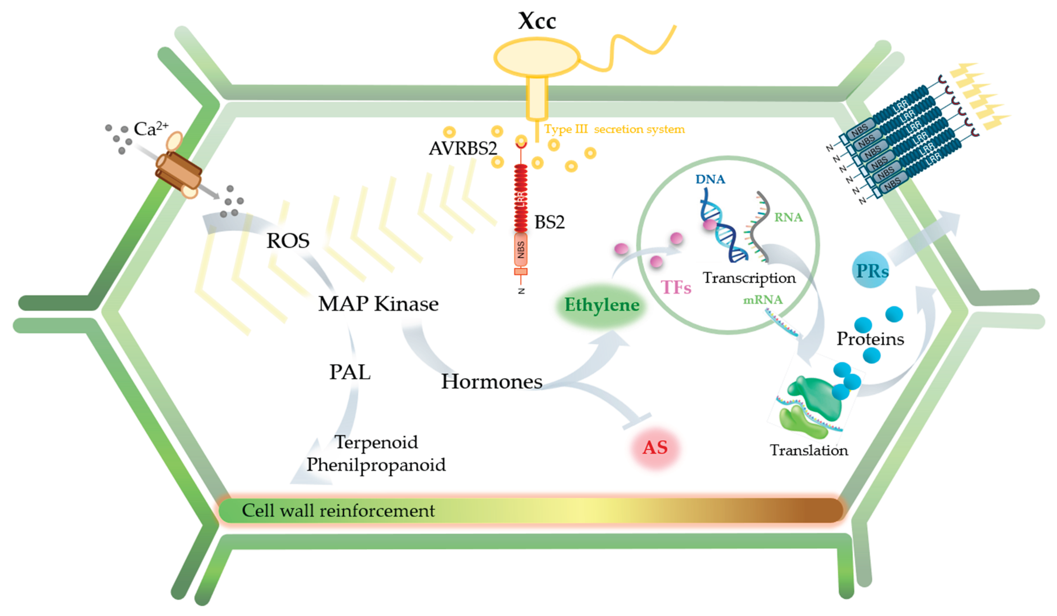
The pepper Bs2 resistance gene confers resistance to susceptible Solanaceae plants against pathogenic strains of Xanthomonas campestris pv. vesicatoria carrying the avrBs2 avirulence gene. Previously, we generated Bs2-transgenic Citrus sinensis plants that exhibited enhanced resistance to citrus canker caused by Xanthomonas citri subsp. citri (Xcc), although the underlying mechanisms remained unknown. To elucidate the molecular basis of the early defense response, we performed a comparative transcriptomic analysis of Bs2-expressing and non-transgenic plants 48 hours after Xcc inoculation. A total of 2,022 differentially expressed genes (DEGs) were identified, including 1,356 up-regulated and 666 down-regulated genes. In Bs2-plants, 36.8% of the up-regulated DEGs were associated with defense responses and biotic stress. Functional annotation revealed major changes in genes encoding receptor-like kinases, transcription factors, hormone biosynthesis enzymes, pathogenesis-related proteins, secondary metabolism, and cell wall modification. Among hormone-related pathways, genes linked to ethylene biosynthesis and signaling were the most strongly regulated. Consistently, endogenous ethylene levels increased in Bs2-plants following Xcc infection, and treatment with an ethylene-releasing compound enhanced resistance in non-transgenic plants. Overall, our results indicate the Bs2 expression activates a complex defense network in citrus and may represent a valuable strategy for controlling canker and other Xanthomonas-induced diseases.

Keywords: 
;  ;  

1. Introduction

Citrus canker, caused by Xanthomonas citri pv. citri (Xcc), remains one of the most significant phytosanitary threats to the global citrus industry. Its management is particularly challenging, relying on early detection, eradication of infected trees, and strict quarantine measures that rarely achieve complete control [1,2]. Given the limitations of conventional control strategies, increasing efforts have focused on developing biotechnological approaches to enhance Citrus resistance. Among these, Agrobacterium tumefaciens–mediated genetic transformation has become an important tool in citrus improvement programs, offering multiple opportunities to complement the development of resistant cultivars [3]. Such approaches are especially relevant for the sustainable management of diseases in perennial crops like Citrus sp.
Several genetic strategies have been explored to mitigate citrus canker, including the expression of antimicrobial peptides [4,5,6], genes that enhance broad-spectrum defense mechanisms [7,8,9], and the introduction of heterologous resistance (R) genes [10,11]. R genes typically act as race-specific pathogen receptors and most belong to the large nucleotide-binding site–leucine-rich repeat (NBS-LRR) family [12]. Many R genes have been successfully transferred across genera through genetic engineering, resulting in effective disease control. For instance, constitutive expression of the rice Xa21 gene in banana (Musa × paradisiaca) conferred resistance to Xanthomonas campestris pv. musacearum (Xcm) [13], and expression of Xa21 gene in transgenic C. sinensis enhanced resistance to X. citri pv. citri [10]. Similarly, the maize Rxo1 gene conferred resistance to Xanthomonas oryzae pv. oryzicola in rice [14]. However, resistance mediated by race-specific receptors is often short-lived, as pathogens can evolve mechanisms to overcome it [13].
In contrast, some R genes recognize conserved, nonredundant effectors that are essential for pathogen virulence and thus represent potentially durable sources of resistance [13]. One example is the Bs2 gene from pepper (Capsicum annuum cv. Early Calwonder), which encodes an NBS-LRR resistance protein that recognizes the corresponding avrBs2 avirulence gene and confers resistance to Xanthomonas campestris pv. vesicatoria (Xcv) [15]. Transgenic expression of Bs2 enhances resistance to Xcv not only in susceptible pepper genotypes but also in other Solanaceae species, including tomato and tobacco [15]. Additionally, we previously demonstrated that the avrBs2 gene is highly conserved among Xanthomonas species, including X. citri pv. citri (Xcc), whose avrBs2 sequence shares 96% identity with that of Xcv [16]. Based on this finding, transgenic C. sinensis cv. Pineapple (sweet orange) plants expressing the Bs2 gene under the control of a pathogen-inducible glutathione S-transferase (gst1) promoter from potato were generated, exhibiting enhanced resistance to citrus canker [11]. These Bs2-transgenic plants showed higher production of reactive oxygen species and increased expression of pathogenesis-related (PR) genes after Xcc inoculation compared with non-transgenic controls, suggesting that the reduced canker symptoms were the consequence of defense mechanisms triggered by recognition of the conserved avrBs2 effector. Nevertheless, the downstream molecular and signaling pathways activated by Bs2 in citrus remain poorly understood.
In the present study, we analysed early transcriptomic changes in C. sinensis plants expressing the pepper Bs2 gene following Xcc inoculation to elucidate the molecular mechanisms induced by this gene. Because effective plant defense depends on rapid perception and early signaling, we focused our analysis on the early stages of infection. The transcriptome comparison revealed differentially expressed genes (DEGs) between Bs2 and non-transgenic plants, which correlated with anatomical, biochemical, and molecular evidence consistent with the activation of defense responses in the transgenic plants. This study provides insights into the defense mechanisms triggered by the heterologous Bs2 gene in citrus, offering valuable implications for improving citrus canker management through direct deployment of Bs2 or by leveraging candidate genes identified in this work.

2. Materials and Methods

Plant material and bacterial culture
Seven-month-old Bs2-transgenic (Bs2-plants) [11] and non-transgenic (NT) C. sinensis cv. Pineapple were grown in 10-L pots containing GrowMix® Multipro commercial substrate (Terrafertil S.A., Argentina) under controlled environmental conditions (28–30 °C, 16-h photoperiod).
Xanthomonas citri pv. citri (Xcc) expressing GFP [17] was cultured at 28 °C with shaking at 200 rpm in PYM medium [19]. Bacterial cells were harvested by centrifugation at 4,000 rpm for 15 min and resuspended in sterile 10 mM MgCl₂ to a final density of either 10⁸ CFU mL⁻¹ (for spray inoculation assays) or 10⁴ CFU mL⁻¹ (for infiltration assays).
Challenge assays with Xcc
For the RNA-seq experiment, young leaves were inoculated by infiltration to ensure bacterial entry and minimize variability among biological replicates. Inoculated plants were maintained at 28–30 °C and 70% relative humidity. At 48 h post-inoculation (hpi), leaf samples were collected using a cork borer. From the inoculated area, two leaf discs (1 cm²) were taken from each of five leaves per biological replicate (plant). A total of ten discs per treatment were pooled, immediately frozen in liquid nitrogen, and stored at –80 °C until RNA extraction. Three biological replicates were collected per treatment.
To evaluate in planta bacterial growth, Xcc was inoculated using two different methods: infiltration with a needle (without syringe) and spraying. Inoculated plants were kept at 26 °C and 70% relative humidity. Mock inoculations were performed with 10 mM MgCl₂ solution as a control in all experiments. Each assay was repeated three times, using at least five leaves per plant.
Determination of Xcc population
Symptom development and disease progression were monitored phenotypically and documented using a Leica MZ6 stereomicroscope under both white and UV light (520 nm). The GFP-tagged Xcc strain enabled visualization of live bacterial colonies as bright green fluorescent foci under UV illumination, allowing accurate detection of infection sites and active bacterial proliferation. Representative images were taken at different time points, and bacterial growth was quantified as previously described [11]. Six leaf discs (1 cm²) from inoculated leaves were ground in 0.2 mL of sterile distilled water, and serial dilutions (100 µL) of the homogenate were plated onto PYM nutrient medium [18]. The bacterial population was expressed as colony-forming units (CFU) per cm² and monitored up to 14 days post-inoculation (dpi).
RNA extraction and library preparation
Total RNA was extracted from three biological replicates per treatment: Bs2-transgenic (Bs2-plants) and non-transgenic (NT) plants, each either Xcc-inoculated or mock-treated. RNA samples were isolated and purified using the TURBO DNA-free™ Kit (Thermo Fisher Scientific). Four libraries were constructed for paired-end sequencing: (1) inoculated Bs2-plants (Bs2-Xcc); (2) mock-inoculated Bs2-plants (Bs2-Mock); (3) inoculated NT-plants (NT-Xcc); and (4) mock-inoculated NT-plants (NT-Mock).
RNA integrity was verified using a 2100 Bioanalyzer (Agilent Technologies). One microgram of total RNA was used to construct each library with the Illumina TruSeq Stranded mRNA Library Prep Kit (Illumina, RS-122-2102). Library construction was performed at the University of Florida ICBR Gene Expression and Genotyping Core Facility (RRID:SCR_019145). The resulting libraries displayed a broad fragment distribution (200–2000 bp) with a peak around 500 bp. Library quantification was performed by qPCR using the KAPA Library Quantification Kit (Kapa Biosystems, KK4824). The libraries were then pooled at equimolar concentrations and sequenced on an Illumina HiSeq 3000 platform (2 × 100 bp paired-end reads) at the University of Florida ICBR NextGen DNA Sequencing Core Facility (RRID:SCR_019152).
Sequencing read mapping and gene expression estimation
Raw reads were trimmed and filtered according to base quality and read length using BBDuk from the BBMap package, version 34.41 [19]. The trimmed paired-end reads were aligned to the Citrus clementina v1.0 reference genome using TopHat v2.0.09 with default parameters [20]. Reads from each biological replicate were mapped independently, and multi-mapped reads were discarded. Gene-level read counts were obtained using htseq-count with the union model. Expression levels were estimated as FPKM (Fragments Per Kilobase of transcript per Million mapped reads) using the DESeq2 package [21].
Identification of differentially expressed genes and functional annotation
Differential expression analysis between transgenic and non-transgenic plants, both inoculated and mock-treated, was conducted using the DESeq2 package (Bioconductor). Genes with an absolute log₂ fold change (|log₂FC| > 2) and a false discovery rate (FDR) < 0.001 were considered significantly differentially expressed. Gene Ontology (GO) terms related to biological processes, molecular functions, and cellular components were assigned using the AgriGO toolkit [22]. Functional categorization of DEGs was carried out using MapMan 3.5.1R2 software [23].
To validate the RNA-seq data, fourteen DEGs were randomly selected for qRT–PCR analysis (Applied Biosystems). Gene-specific primers were designed based on coding sequences using IDT SciTools Web Tools (Supplementary Table S1). The 18S rRNA gene was used as an internal reference [24]. Relative expression levels were calculated using the 2⁻ΔΔCt method [25].
Treatments with exogenous ethephon
To evaluate the effect of ethylene on Xanthomonas citri pv. citri (Xcc) growth, exogenous ethephon (an ethylene-releasing compound) was added to PYM nutrient medium at concentrations ranging from 1000 to 2 µM. Each glass tube contained 5 mL of PYM medium supplemented with ethephon, and 100 µL of an Xcc culture (OD₆₀₀ = 0.1) was inoculated into each tube. Cultures were incubated at 28 °C with shaking at 200 rpm for 24 h, and bacterial growth was measured by optical density at 600 nm.
To assess the effect of ethylene on plant defense, a sub-inhibitory concentration of ethephon (1 µM) was applied to C. sinensis NT-plants by spraying either 48 h before or 48 h after inoculation with Xcc (10⁸ CFU mL⁻¹ for spray inoculation and 10⁴ CFU mL⁻¹ for infiltration). Control plants were sprayed with sterile water. Disease symptoms were recorded at 14 days post-inoculation (dpi). Each treatment included three independent plants and three leaf replicates per plant.
Quantification of endogenous ethylene
Endogenous ethylene production was measured following Zou et al. [26]. After infiltration with 200 µL of Xcc suspension, leaves from Bs2- and NT-plants were placed in sealed 30 mL vials and incubated for 48 h in the dark at 25 °C. Gas samples (1 mL) were withdrawn using a gas-tight syringe and analyzed by gas chromatography (Agilent 7890A, USA). Each treatment included three biological replicates.
Antibacterial activity and phenolic compound content (PCC).
Leaf discs (0.1 g) from NT- and Bs2-plants were collected at 0, 24, 48, and 72 h post-inoculation (hpi) with Xcc. Samples were ground and extracted in 2 mL of 80% methanol:20% water for 24 h at 30 °C. Extracts were centrifuged, lyophilized, and re-suspended in distilled water to a final concentration of 0.1 g FW mL⁻¹.
Phenolic compound content (PCC) was quantified using the Folin–Ciocalteu method [27]. Gallic acid was used to construct a calibration curve, and results were expressed as milligrams of gallic acid equivalents per gram of fresh weight (mg GAE g⁻¹ FW).
Antibacterial activity was evaluated by the agar well-diffusion method. Xcc suspension (OD₆₀₀ = 1.0; 150 µL) was mixed with 10 mL of 0.7% PYM agar and poured into sterile Petri dishes. Eight wells (0.5 mm diameter) were made using a cork borer, and 50 µL of each methanolic extract (0.1 mg FW mL⁻¹) were added per well. Plates were incubated at 30 °C for 24 h, and inhibition zones were measured. All assays were performed by triplicate.

3. Results

3.1. Differentially Expressed Genes (DEGs) in Citrus Sinensis Bs2-Plants

To gain insight into the early molecular mechanisms involved in the response of Bs2-plants to Xcc infection, a global transcriptional analysis was conducted by comparing Bs2- and non-transgenic (NT) plants at 48 h post-inoculation (hpi) using RNA-seq technology. Four datasets of 100-bp paired-end raw reads were generated. Raw reads were subjected to quality control using SeqQC, with more than 95% of bases exhibiting a quality score above Q20 (data not shown). After mapping the high-quality reads to the C. clementina v1.0 reference genome, 82–88% of reads were uniquely aligned and used for downstream expression analyses.
Pairwise comparisons among biological replicates showed high consistency in estimated gene expression levels. Principal component analysis (PCA) revealed that Bs2-Mock and NT-Mock samples clustered closely together, indicating minimal transcriptional differences between them under non-inoculated conditions. In contrast, Bs2-Xcc and NT-Xcc samples were clearly separated from each other and from their respective mock treatments, demonstrating distinct transcriptional reprogramming induced by the presence of the Bs2 gene (Figure 1a).
A volcano plot showing fold-change values versus the –log₁₀ of adjusted p-values illustrated the overall differential expression patterns between treatments (Figure 1b). Under mock conditions, only 31 DEGs were detected between Bs2- and NT-plants (12 up-regulated and 19 down-regulated). In contrast, under Xcc-inoculated conditions, a total of 2,022 DEGs were identified, with the number of up-regulated genes (1,356) more than doubling the number of down-regulated ones (666). As expected, the Bs2 gene was exclusively detected in Bs2-plants.
In NT-plants, comparison of NT-Xcc vs. NT-Mock revealed 2,037 DEGs, with similar proportions of up-regulated (958) and down-regulated (1,079) genes. Conversely, comparison of Bs2-Xcc vs. Bs2-Mock identified only 574 DEGs; however, the number of up-regulated genes (488) was nearly sixfold higher than that of down-regulated ones (86). These results indicate that expression of the Bs2 gene reprograms the transcriptional response of C. sinensis to Xcc infection, biasing gene regulation toward activation rather than suppression—unlike the response observed in NT-plants (data not shown).
The accuracy of RNA-seq data was validated by quantitative real-time PCR (qRT-PCR) analysis of a randomly selected subset of 14 DEGs (Figure 1c). The expression trends obtained by both platforms were highly consistent, with a strong positive correlation (Spearman’s ρ = 0.985) between RNA-seq and qRT-PCR fold-change values, confirming the reliability of the transcriptomic results.

3.2. Functional Analysis of DEGs

Gene Ontology (GO) classification and KEGG pathway enrichment analyses were conducted to gain insight into the biological functions of the differentially expressed genes (DEGs). GO terms were assigned to the predicted genes using the C. clementina GO annotation integrated into the DESeq2 analysis pipeline. Of the 2,022 DEGs identified between Bs2- and NT-plants, 1,717 were successfully annotated with at least one GO term.
The annotated DEGs were categorized according to GO Level 2 into 30 functional groups for up-regulated genes and 31 groups for down-regulated genes (Figure 2). Within the Molecular Function category, the most represented terms were catalytic activity (131 up-regulated and 64 down-regulated genes) and binding (126 up-regulated and 60 down-regulated genes). In the Cellular Component ontology, the predominant terms were cell and cell part, followed by organelle. Within the Biological Process category, the largest groups were cellular process and metabolic process, followed by response to stimulus and regulation of biological process (Figure 2).
These results indicate that Bs2-mediated responses in C. sinensis involve extensive transcriptional reprogramming of genes associated with core cellular metabolism and stress-related biological processes.

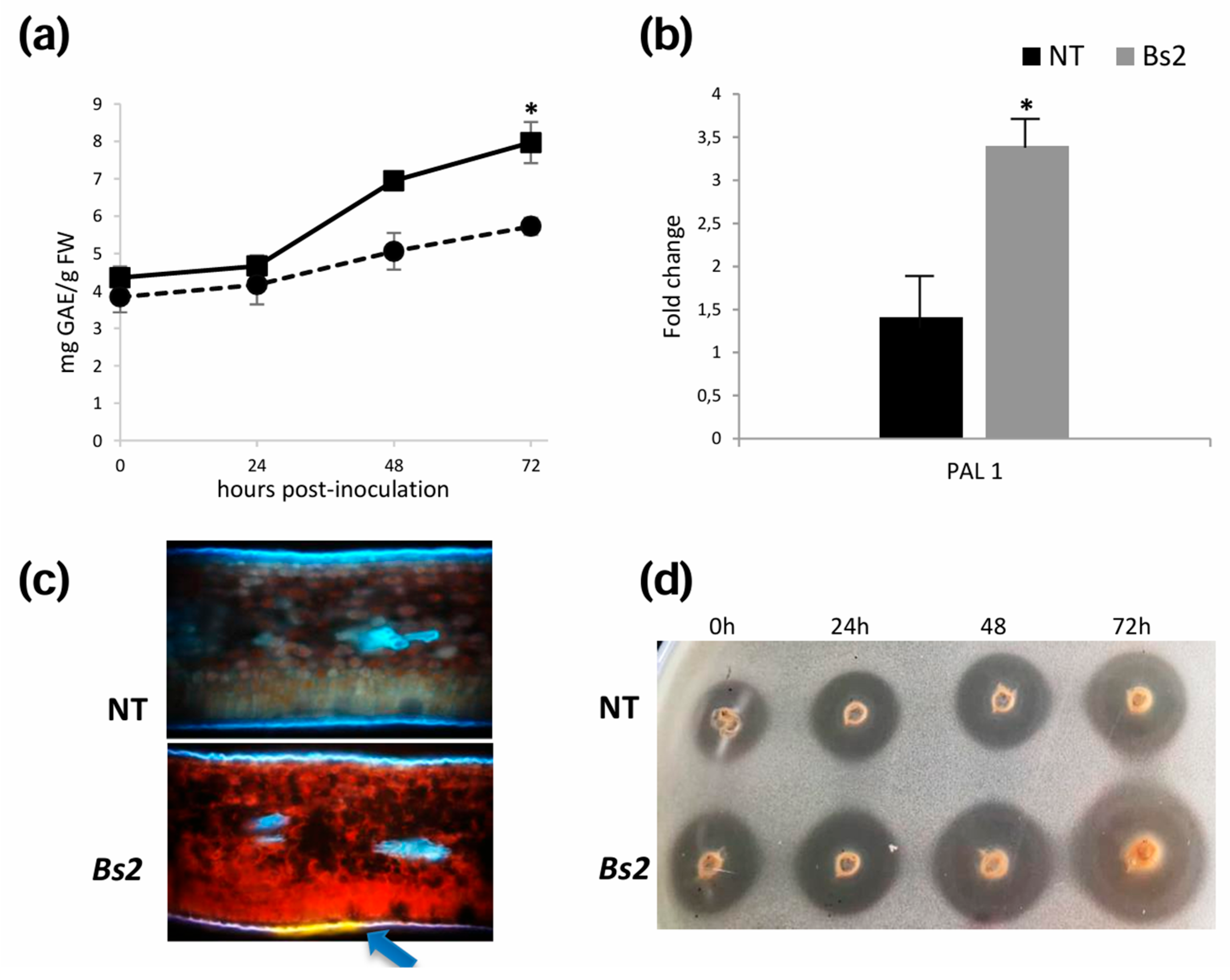
3.3. Plant Defense Is Induced in Bs2-Plants After Xcc Inoculation

Functional classification and visualization of DEGs were performed using the MapMan software. The biotic stress category was the most enriched, comprising 752 DEGs (36.8%) in Bs2-Xcc plants (Figure 3). The principal subcategories included signaling, proteolysis, pathogenesis-related (PR) proteins, secondary metabolism, hormone signaling, transcription factors, and cell wall organization.
A more detailed analysis (Supplementary Table S2) revealed that approximately 43% of DEGs were related to signal perception and transduction, including receptor-like kinases (RLKs), calcium signaling, and phytohormone-related pathways. A total of 324 DEGs corresponded to RLKs and wall-associated kinases (WAKs), which are transmembrane receptors located in the cell wall that participate in broad-spectrum, elicitor-induced defense responses [28]. Among these, 170 DEGs belonged to the leucine-rich repeat receptor-like kinase (LRR-RLK) subfamily, of which 150 (88%) were up-regulated. Additionally, 79 DEGs (68 up-regulated) encoded receptor-like kinases containing a Domain of Unknown Function 26 (DUF26), also known as cysteine-rich receptor-like kinases (CRKs), which are known to regulate defense signaling and programmed cell death. A smaller group of 11 DEGs encoded wall-associated kinases (WAKs), 10 of which were up-regulated.
Following pathogen recognition, a signaling cascade is triggered in which calcium ions (Ca²⁺) act as key secondary messengers. In Bs2-plants, 19 of the 22 Ca²⁺-related DEGs were up-regulated, including 12 genes encoding calmodulin or calmodulin-like proteins, which function as intracellular calcium sensors. Consistent with these results, two DEGs encoding ABC-2-type domain-containing proteins were notably induced (6.89- and 6.22-fold, respectively) in Bs2-plants. Certain ABC transporters have been reported to interact with calmodulin and other Ca²⁺-binding proteins to modulate signal transduction [29].
Intracellular nucleotide-binding site–leucine-rich repeat (NBS-LRR) genes play central roles in the recognition of pathogen effectors, mediating effector-triggered immunity (ETI). Most of these genes constitute a class of plant resistance (R) genes [30]. In the present study, 79 DEGs encoding NBS-LRR proteins were identified in Bs2-plants, 62 of which were up-regulated (Supplementary Table S2). Pathogenesis-related (PR) proteins are hallmark components of plant defense that accumulate following pathogen perception [30]. Although most PR genes exhibit low basal expression under normal growth conditions, they are rapidly induced after infection through signaling pathways mediated by salicylic acid (SA), jasmonic acid (JA), and ethylene (ET), contributing to systemic acquired resistance (SAR) [31]. In Bs2-plants, 10 PR-related DEGs were identified, eight of which were up-regulated at 48 hpi.
Transcription factors (TFs) play key regulatory roles in orchestrating the transcriptional reprogramming that underlies plant defense responses. In this study, 30 DEGs corresponding to three major TF families associated with biotic stress—WRKY, MYB, and ERF—were differentially expressed in Bs2-plants, of which 21 were up-regulated. Notably, all WRKY TFs were up-regulated, with WRKY70 (ciclev10012055) showing a 5.84-fold induction in Bs2-plants.

3.4. Bs2 Induces Major Regulation of the Ethylene Pathway

Within the Plant hormone signaling subcategory, 56 genes were differentially expressed in Bs2-plants, with 35 up-regulated and 20 down-regulated (Supplementary Table S2). The most abundant and differentially expressed hormone-related genes were associated with the biosynthesis, degradation, or signal transduction of ethylene. This group included 35 DEGs (27 up-regulated), such as senescence-related gene 1 (SRG1), oxygenase family proteins, ethylene-responsive element binding proteins (EREBPs), and ethylene-responsive factors (ERFs). In contrast, only one gene related to salicylic acid (SA) synthesis or degradation (ciclev10017993m) was identified, and it was down-regulated. Additionally, two DEGs corresponding to NPR1 were down-regulated (ciclev10033799m and ciclev10030929m, with fold changes of 3.17 and 2.36, respectively), whereas one NPR1 suppressor gene was up-regulated (2.89-fold change) in Bs2-plants at 48 hpi. Regarding jasmonic acid (JA), three DEGs were down-regulated and only one was up-regulated. Moreover, one methyl esterase 1 (ciclev10033393m), an enzyme involved in the methylation of compounds such as SA and JA and associated with systemic acquired resistance (SAR) [32], was significantly induced (5.3-fold) in Bs2-plants.
The high proportion of DEGs related to the ethylene (ET) pathway compared with those associated with SA or JA suggests a predominant role of ET signaling, at least during the early defense stages triggered by the Bs2 gene. To confirm this hypothesis, ethylene content was quantified in Bs2- and NT-plants at 48 hpi. Bs2-plants showed significantly higher ET levels than NT-plants (5.4 ± 0.32 µg g⁻¹ FW vs. 3.1 ± 0.12 µg g⁻¹ FW, respectively).
To determine whether ethylene directly affects Xcc growth, an in vitro assay using ethephon (an ethylene-releasing compound) revealed that Xcc growth was inhibited at concentrations ranging from 1000 to 10 µM (Figure 4A). Subsequently, to assess whether ethylene modulates plant defense against Xcc, a subinhibitory concentration of ethephon (1 µM) was applied to NT-plants either 48 h before or after Xcc inoculation, using both infiltration (10⁴ CFU mL⁻¹) and wound + spray (10⁸ CFU mL⁻¹) methods. At 14 dpi, ethephon-treated plants exhibited fewer canker symptoms than untreated controls, with the greatest reduction observed when ethephon was applied prior to bacterial inoculation, regardless of the inoculation method (Figure 4B). Quantification of bacterial populations at 14 dpi was consistent with the observed symptom reduction (Figure 4C).

3.5. Changes in Secondary Metabolic Pathways and Cell Wall

A total of 70 DEGs were involved in the synthesis of secondary metabolites in Bs2-plants (Supplementary Table S2), of which 41 were up-regulated. One of the major enriched categories was phenylpropanoid biosynthesis, which includes numerous secondary metabolites associated with plant signaling and defense against biotic and abiotic stress [33]. Among the most highly up-regulated DEGs in Bs2-plants was phenylalanine ammonia-lyase (PAL, 5.28-fold change, ciclev10010874m), which catalyzes the first step in the phenylpropanoid pathway. In addition, isoflavone-7-O-methyltransferase, cinnamyl-alcohol dehydrogenase, chalcone synthase, and several transferase family genes were up-regulated.
The expression of genes related to phenolic compound biosynthesis was consistent with the increased total soluble phenolic content (Figure 5A) and with qRT–PCR validation of PAL1 expression (Figure 5B), both of which were significantly higher in Bs2-plants. Histological analyses showed the accumulation of bright green autofluorescence corresponding to polyphenolic compounds, particularly on the abaxial side of Bs2 leaves at 48 hpi (Figure 5C). Moreover, methanolic extracts from Bs2 leaves exhibited higher phenolic compound content and in vitro antimicrobial activity against Xcc growth (Figure 5D), suggesting reinforcement of chemical defenses that restrict pathogen progression.
Additionally, 60 DEGs (43 up- and 17 down-regulated) were annotated as cytochrome P450 monooxygenases. This large enzyme family participates in multiple reactions, including secondary metabolite biosynthesis, and in higher plants plays crucial roles in plant–microbe interactions as well as in the biosynthesis of antioxidants, phytohormones, and callose [34].
In the Cell Wall category, 23 DEGs were identified in Bs2-plants (Supplementary Table S2), 14 of which were down-regulated. These included genes associated with cell wall modification, degradation, and loosening, such as xyloglucan:xyloglucosyl transferase and pectinesterase.
Other DEGs contributing to cell wall weakening, such as cellulase, endoglucanase, polygalacturonase, and expansin, were also down-regulated in Bs2-plants. Expansin promotes rapid tissue expansion and cell wall loosening, processes required for hypertrophy and hyperplasia during canker pustule formation [35]. Consistent with this, three DEGs encoding SAUR (Small Auxin Up RNA) auxin response proteins, which promote cell expansion [39], were also down-regulated in Bs2-plants.

4. Discussion

The current study presents a comparative transcriptional analysis between Bs2 transgenic and non-transgenic plants to identify differentially expressed genes after inoculation with Xcc, in an attempt to understand the nature of the defense mechanism induced by the pepper Bs2 gene in C. sinensis. Our results indicate that the resistance of Bs2-plants to Xcc is linked to extensive transcriptional reprogramming across multiple functional pathways. We propose a regulatory model connecting key signaling networks with the expression of defense-associated genes (Figure 6).
A remarkable feature of the resistance response in Bs2-plants is that the number of up-regulated genes is approximately twice that observed in the susceptible response (NT–Xcc). A similar trend has been reported in canker-resistant transgenic C. sinensis overexpressing a spermidine synthase gene [36], in Meiwa kumquat (Fortunella crassifolia), which is immune to Xcc [37], and in C. limon following inoculation with an avirulent Xcc variant [38]. Therefore, the greater number of up-regulated genes is likely associated with resistance to Xcc infection. In susceptible genotypes, Xanthomonas pathogenesis involves the suppression of plant innate immune responses through molecular suppressors such as cyclic glucans and xanthan, resulting in a predominance of down-regulated genes [17,18].
Although the downstream signaling events following avr–R protein interactions are not yet fully understood, several characteristic responses have been described, including extensive transcriptomic reprogramming, strong activation of mitogen-activated protein kinases (MAPKs), increased production of reactive oxygen species (ROS), and localized programmed cell death or hypersensitive response (HR) [39]. Activation of the MAPK cascade subsequently triggers the regulation of downstream transcription [40]. In our study, Bs2-plants exhibited a stronger MAPK activation profile: 68.9% of the differentially expressed genes (DEGs) within the “Biotic Stress” MapMan category corresponded to enzymes, receptor kinases, and transcription factors (TFs), of which 70% were up-regulated. Moreover, several transcription factors belonging to four major families—MYB, WRKY, bHLH, and NAC—known as key regulators of defense responses, were induced. All MYB- and WRKY-related DEGs were up-regulated. Although they are multifunctional, both TFs orchestrate cellular strategies essential for plant defense against biotic stresses [41].
In previous work, we demonstrated that Bs2-transgenic plants exhibited maximum accumulation of H₂O₂ at 48 hours after Xcc inoculation. ROS are among the earliest and most important signaling molecules in plant defense, preceding HR and contributing to cell wall reinforcement, accumulation of phenolic compounds, and activation of defense-related genes [42]. Moreover, ROS and calcium have been proposed to act as interconnected messengers in systemic signal transduction, facilitating communication from local tissues to the entire plant [10]. In this context, several genes associated with calcium signaling were up-regulated, including calmodulin genes—whose proteins sense cytosolic Ca²⁺ fluctuations—as well as interacting partners such as ABC transporter proteins [29]. Consistently, two DEGs encoding ABC-2 type domain–containing proteins were highly up-regulated (6.89- and 6.22-fold, respectively) in Bs2Xcc plants.
The phenylpropanoid pathway represents a key component of plant defense because it contributes to both mechanical barriers that restrict pathogen invasion and chemical defense through the production of antimicrobial metabolites [43]. Phenolic deposits have also been documented around HR lesions triggered by Xcc in resistant cultivars such as calamondin and kumquat [44]. In this context, the differential induction of key genes from the flavonoid and phenylpropanoid pathways, including PAL1 and CHS1, suggests their involvement in Bs2-activated defense responses in C. sinensis. Phenolic compounds additionally serve as precursors for important molecules such as phytohormones. For example, PAL1 participates in salicylic acid (SA) biosynthesis, a central component of R-gene-mediated resistance and effector-triggered immunity (ETI) [45]. In this study, however, no evidence of SA involvement was detected at 48 hpi, as no DEGs related to SA signaling were identified. Furthermore, two NPR1-encoding DEGs—NPR1 being a master regulator of SA signaling—were down-regulated, whereas one NPR1 suppressor gene was up-regulated. In contrast, Chiesa et al. [46] reported activation of the SA signaling pathway in C. limon, but in a nonhost interaction with X. campestris pv. campestris and at earlier time points (3 and 24 hpi).
By contrast, transcriptome analysis revealed that ethylene-responsive genes were distinctly induced in Bs2-plants upon Xcc infection. Ethylene is a multifunctional phytohormone involved in numerous plant processes, including responses to pathogen attack, where it can either promote resistance or susceptibility depending on the host–pathogen system [47]. Our results suggest that ethylene may contribute to Bs2-mediated resistance. Similar patterns have been reported in other plant–Xanthomonas interactions. For example, we previously observed induction of ACC oxidase transcripts—a key enzyme in ethylene biosynthesis—in C. limon at 48 hpi after inoculation with the avirulent variant Xcc AT [48]. Likewise, Cernada et al. [49] reported significant transcriptional changes associated with ethylene signaling during the interaction between C. sinensis and X. axonopodis pv. aurantifolii pathotype C (Xaa), which causes disease only in Mexican lime. In resistant C. sinensis plants, Xaa triggered a MAPK signaling cascade and activated WRKY and ethylene-responsive TFs at 6 and 48 hpi, leading the authors to propose a central role for ethylene in resistance.
Another major group of up-regulated genes in Bs2-plants consists of those involved in pathogen perception and signal transduction, including receptor-like protein kinases (RLKs), predominantly those containing leucine-rich repeat (LRR) domains, as well as lectin- and cysteine-rich RLKs. RLK genes function as membrane-localized receptors that recognize pathogen-associated molecular patterns (PAMPs) or damage signals and subsequently activate plant immune responses. Members of the RLK family, particularly LRR-RLKs, are key components of plant disease resistance. Their evolutionary expansion has broadened recognition specificity, enhancing the plant’s ability to detect and respond to diverse pathogens [50]. Therefore, it is possible that Bs2 may also contribute to expanding the plant’s pathogen recognition capacity—an intriguing hypothesis for future investigation.
The reduced development of canker symptoms in Bs2-transgenic plants was also supported by the repression of 69% of DEGs related to cell wall modification, breakdown, or degradation. The down-regulation of genes associated with cell wall metabolism has also been observed in citrus plants challenged with an avirulent strain of Xcc [38] and in HLB-tolerant citrus trees [51]. Consistent with our results, one of the most strongly down-regulated DEGs corresponded to an expansin gene, whose suppression has been reported to promote pathogen resistance [10].
In conclusion, this study shows that the inducible expression of the pepper Bs2 resistance gene in C. sinensis activates a complex defense program against Xcc. The results reveal the regulation of multiple resistance-related signaling pathways that together form an interconnected defense network, and they highlight ethylene signaling as a key component of this response. This work confirms the functionality of the Bs2 gene from pepper in C. sinensis, thereby extending its taxonomic range of activity beyond the Solanaceae family. Overall, these findings broaden the potential applications of the Bs2 gene for developing durable resistance against Xanthomonas spp. in citrus crops

Supplementary Materials

The following supporting information can be downloaded at the website of this paper posted on Preprints.org. Table S1: Gene-specific primers to validate RNAseq; Table S2: DEGs details.

Author Contributions

Sendín, Lorena Noelia: Investigation; Results Interpretation, Supervision; Writing, Review & Editing. lorenasendin@gmail.com; Ledesma, Verónica Andrea: Performed Experiments. vledesma@eeaoc.org.ar; Gómez, Rocío Liliana: Performed Experiments, Data Analysis, Writing Original Draft Preparation. rociog433@gmail.com; Yu, Qibin: Data Analysis. qibin@ufl.edu; Albornoz, Patricia: Data Analysis. albornoz@csnat.unt.edu.ar; Pardo, Esteban Mariano: Visualization; Results Interpretation; Review & Editing. mpardokarate@gmail.com; Enrique, Ramón: Writing, Review & Editing. renrique@eeaoc.org.ar; Gmitter, Frederick: Resources; Supervision. fgmitter@ufl.edu; Castagnaro, Atilio Pedro: Conceptualization; Supervision. atilio@eeaoc.org.ar; Maria Paula Filippone: Conceptualization; Supervision, Funding acquition, Writing, Review & Editing. paulafilippone2018@gmail.com

Funding

This project was supported by Estación Experimental Agroindustrial Obispo Colombres (EEAOC), Consejo Nacional de Investigaciones Científicas y Tecnológicas (CONICET), the Agencia Nacional de Promoción Científica y Tecnológica (ANPCyT, FITR-30; PICTO-2016-0130), BEC.AR program, sponsored by Ministerio de Modernización –Presidencia de la Nación Argentina, the University of Florida ICBR Gene Expression and Genotyping Core Facility (RRID:SCR_019145) and NextGen DNA Sequencing Core Facility (RRID:SCR_019152).

Acknowledgments

We especially thank the University of Florida ICBR Gene Expression and Genotyping Core Facility (RRID: SCR_019145) and NextGen DNA Sequencing Core Facility (RRID: SCR_019152).

Conflicts of Interest

there is not conflict of interest.

Abbreviations

The following abbreviations are used in this manuscript:
avrBs2 avrBs2 avirulence gene
Bs2 Bs2 resistance gene from Capsicum annuum
Bs2-plants Transgenic plants carrying the Bs2 gene
Ca²⁺ Calcium ion
CFU Colony Forming Units
CHS1 Chalcone Synthase 1
CRKs Cysteine-Rich Receptor-Like Kinases
CT / Ct Cycle Threshold (ΔΔCt method)
DEGs Differentially Expressed Genes
DESeq2 Differential expression analysis package
dpi Days Post-Inoculation
DUF26 Domain of Unknown Function 26
ERFs Ethylene-Responsive Factors
EREBPs Ethylene-Responsive Element Binding Proteins
ET Ethylene
ETI Effector-Triggered Immunity
FDR False Discovery Rate
FPKM Fragments Per Kilobase of transcript per Million mapped reads
FW Fresh Weight
GAE Gallic Acid Equivalents
GFP Green Fluorescent Protein
GO Gene Ontology
HLB Huanglongbing
hpi Hours Post-Inoculation
HR Hypersensitive Response
ICBR Interdisciplinary Center for Biotechnology Research
IDT Integrated DNA Technologies
JA Jasmonic Acid
LRR Leucine-Rich Repeat
LRR-RLK Leucine-Rich Repeat Receptor-Like Kinase
MAPK / MAPKs Mitogen-Activated Protein Kinase(s)
MDPI Multidisciplinary Digital Publishing Institute
MgCl₂ Magnesium Chloride
NBS-LRR Nucleotide-Binding Site – Leucine-Rich Repeat
NPR1 Nonexpressor of Pathogenesis-Related Genes 1
NT Non-Transgenic
OD600 Optical Density at 600 nm
PAL Phenylalanine Ammonia-Lyase
PAL1 Phenylalanine Ammonia-Lyase 1
PAMPs Pathogen-Associated Molecular Patterns
PCA Principal Component Analysis
PCC Phenolic Compound Content
PCR Polymerase Chain Reaction
PR Pathogenesis-Related
PYM Peptone–Yeast–Malt Extract Medium
qRT-PCR Quantitative Real-Time Polymerase Chain Reaction
R Resistance
RLKs Receptor-Like Kinases
RNA-seq RNA sequencing
ROS Reactive Oxygen Species
rpm Revolutions Per Minute
SA Salicylic Acid
SAR Systemic Acquired Resistance
SAUR Small Auxin Up RNA
SD Standard Deviation
SRG1 Senescence-Related Gene 1
TFs Transcription Factors
UV Ultraviolet
WAKs Wall-Associated Kinases
Xaa Xanthomonas axonopodis pv. Aurantifolii
Xcc Xanthomonas citri pv. Citri
Xcm Xanthomonas campestris pv. Musacearum
Xcv Xanthomonas campestris pv. vesicatoria

References

  1. Ali, S.; Hameed, A.; Muhae-Ud-Din, G.; Ikhlaq, M.; Ashfaq, M.; Atiq, M.; Ali, F.; Zia, Z.U.; Naqvi, S.A.H.; Wang, Y. Citrus Canker: A Persistent Threat to the Worldwide Citrus Industry—An Analysis. Agronomy 2023, 13, 1112. [Google Scholar] [CrossRef]
  2. Behlau, F. An Overview of Citrus Canker in Brazil. Trop. Plant Pathol. 2021, 46, 1–12. [Google Scholar] [CrossRef]
  3. Sun, Y.; Niu, Y.; He, B.; Ma, L.; Li, G.; Tran, V.-T.; Zeng, B.; Hu, Z. A Dual Selection Marker Transformation System Using Agrobacterium tumefaciens for the Industrial Aspergillus oryzae 3.042. J. Microbiol. Biotechnol. 2019, 29, 230–234. [Google Scholar] [CrossRef] [PubMed]
  4. Stover, E.; Stange, R.R.; McCollum, T.G.; Jaynes, J.; Irey, M.; Mirkov, E. Screening Antimicrobial Peptides In Vitro for Use in Developing Transgenic Citrus Resistant to Huanglongbing and Citrus Canker. J. Am. Soc. Hortic. Sci. 2013, 138, 142–148. [Google Scholar] [CrossRef]
  5. Furman, N.; Kobayashi, K.; Zanek, M.C.; Calcagno, J.; García, M.L.; Mentaberry, A. Transgenic Sweet Orange Plants Expressing a Dermaseptin Coding Sequence Show Reduced Symptoms of Citrus Canker Disease. J. Biotechnol. 2013, 167, 412–419. [Google Scholar] [CrossRef]
  6. Conti, G.; Gardella, V.; Vandecaveye, M.A.; Gómez, C.A.; Joris, G.; et al. Transgenic Citrange troyer Rootstocks Overexpressing Antimicrobial Potato Snakin-1 Show Reduced Citrus Canker Symptoms. J. Biotechnol. 2020, 324, 99–102. [Google Scholar] [CrossRef]
  7. Zhang, X.; Francis, M.I.; Dawson, W.O.; Graham, J.H.; Orbović, V.; Triplett, E.W.; Zhonglin, M. Over-Expression of the Arabidopsis NPR1 Gene in Citrus Increases Resistance to Citrus Canker. Eur. J. Plant Pathol. 2010, 128, 91–100. [Google Scholar] [CrossRef]
  8. Fu, X.-Z.; Liu, J.-H. Transcriptional Profiling of Canker-Resistant Transgenic Sweet Orange Constitutively Overexpressing a Spermidine Synthase Gene. Biomed. Res. Int. 2013, 2013, 918136. [Google Scholar] [CrossRef]
  9. Hao, G.; Pitino, M.; Duan, Y.; Stover, E. Reduced Susceptibility to Xanthomonas citri in Transgenic Citrus Expressing the FLS2 Receptor from Nicotiana benthamiana. Mol. Plant-Microbe Interact. 2016, 29, 132–142. [Google Scholar] [CrossRef]
  10. Ding, L.; Xuan, X.; Wenwu, G. Production of Transgenic ‘Anliucheng’ Sweet Orange (Citrus sinensis Osbeck) with Xa21 Gene for Potential Canker Resistance. J. Integr. Agric. 2014, 13, 2370–2377. [Google Scholar] [CrossRef]
  11. Sendín, L.N.; Orce, I.G.; Gómez, R.L.; Enrique, R.; Grellet Bournonville, C.F.; Noguera, A.S.; Vojnov, A.A.; Marano, M.R.; Castagnaro, A.P.; Filippone, M.P. Inducible Expression of Bs2 R Gene from Capsicum chacoense in Sweet Orange (Citrus sinensis L. Osbeck) Confers Enhanced Resistance to Citrus Canker Disease. Plant Mol. Biol. 2017, 93, 607–621. [Google Scholar] [CrossRef] [PubMed]
  12. Marone, D.; Russo, M.A.; Laidò, G.; De Leonardis, A.M.; Mastrangelo, A.M. Plant Nucleotide Binding Site–Leucine-Rich Repeat (NBS-LRR) Genes: Active Guardians in Host Defense Responses. Int. J. Mol. Sci. 2013, 14, 7302–7326. [Google Scholar] [CrossRef] [PubMed]
  13. Tripathi, J.N.; Lorenzen, J.; Bahar, O.; Ronald, P.; Tripathi, L. Transgenic Expression of the Rice Xa21 Pattern-Recognition Receptor in Banana Confers Resistance to Xanthomonas campestris pv. musacearum. Plant Biotechnol. J. 2014, 12, 663–673. [Google Scholar] [CrossRef] [PubMed]
  14. Zhao, B.; Lin, X.; Poland, J.; Trick, H.; Leach, J.; Hulbert, S. A Maize Resistance Gene Functions against Bacterial Streak Disease in Rice. Proc. Natl. Acad. Sci. USA 2005, 102, 15383–15388. [Google Scholar] [CrossRef]
  15. Tai, T.H.; Dahlbeck, D.; Clark, E.T.; Gajiwala, P.; Pasion, R.; Whalen, M.C.; Stall, R.E.; Staskawicz, B.J. Expression of the Bs2 Pepper Gene Confers Resistance to Bacterial Spot Disease in Tomato. Proc. Natl. Acad. Sci. USA 1999, 96, 14153–14158. [Google Scholar] [CrossRef]
  16. Sendín, L.; Filippone, M.; Orce, I.; Rigano, L.; Enrique, R.; Peña, L.; Vojnov, A.; Marano, M.; Castagnaro, A. Transient Expression of Pepper Bs2 Gene in Citrus limon as an Approach to Evaluate Its Utility for Management of Citrus Canker Disease. Plant Pathol. 2012, 61, 648–657. [Google Scholar] [CrossRef]
  17. Rigano, L.A.; Siciliano, F.; Enrique, R.; Sendín, L.; Filippone, P.; Torres, P.S.; Qüesta, J.; Dow, J.M.; Castagnaro, A.P.; Vojnov, A.A. Biofilm Formation, Epiphytic Fitness, and Canker Development in Xanthomonas axonopodis pv. citri. Mol. Plant-Microbe Interact. 2007, 20, 1222–1230. [Google Scholar] [CrossRef]
  18. Siciliano, F.; Torres, P.; Sendín, L.; Bermejo, C.; Filippone, P.; Vellice, G.; Ramallo, J.; Castagnaro, A.; Vojnov, A.; Marano, M.R. Analysis of the Molecular Basis of Xanthomonas axonopodis pv. citri Pathogenesis in Citrus limon. Electron. J. Biotechnol. 2006, 9, 0–0. [Google Scholar] [CrossRef]
  19. Chaisson, M.J.; Tesler, G. Mapping Single-Molecule Sequencing Reads Using Basic Local Alignment with Successive Refinement (BLASR): Application and Theory. BMC Bioinform. 2012, 13, 238. [Google Scholar] [CrossRef]
  20. Trapnell, C.; Roberts, A.; Goff, L.; Pertea, G.; Kim, D.; Kelley, D.R.; Pimentel, H.; Salzberg, S.L.; Rinn, J.L.; Pachter, L. Differential Gene and Transcript Expression Analysis of RNA-Seq Experiments with TopHat and Cufflinks. Nat. Protoc. 2012, 7, 562–578. [Google Scholar] [CrossRef]
  21. Love, M.I.; Huber, W.; Anders, S. Moderated Estimation of Fold Change and Dispersion for RNA-Seq Data with DESeq2. Genome Biol. 2014, 15, 550. [Google Scholar] [CrossRef] [PubMed]
  22. Du, Z.; Zhou, X.; Ling, Y.; Zhang, Z.; Su, Z. agriGO: A GO Analysis Toolkit for the Agricultural Community. Nucleic Acids Res. 2010, 38, W64–W70. [Google Scholar] [CrossRef] [PubMed]
  23. Thimm, O.; Bläsing, O.; Gibon, Y.; Nagel, A.; Meyer, S.; Krüger, P.; Selbig, J.; Müller, L.A.; Rhee, S.Y.; Stitt, M. MAPMAN: A User-Driven Tool to Display Genomics Data Sets onto Diagrams of Metabolic Pathways and Other Biological Processes. Plant J. 2004, 37, 914–939. [Google Scholar] [CrossRef] [PubMed]
  24. Yang, L.; Hu, C.; Li, N.; Zhang, J.; Yan, J.; Deng, Z. Transformation of Sweet Orange [Citrus sinensis (L.) Osbeck] with pthA-nls for Acquiring Resistance to Citrus Canker Disease. Plant Mol. Biol. 2011, 75, 11–23. [Google Scholar] [CrossRef]
  25. Schmittgen, T.D.; Livak, K.J. Analyzing Real-Time PCR Data by the Comparative CT Method. Nat. Protoc. 2008, 3, 1101–1108. [Google Scholar] [CrossRef]
  26. Zou, Z.; Long, X.; Zhao, Q.; Zheng, Y.; Song, M.; Ma, S.; Jing, Y.; Wang, S.; He, Y.; Esteban, C.R. A Single-Cell Transcriptomic Atlas of Human Skin Aging. Dev. Cell 2021, 56, 383–397. [Google Scholar] [CrossRef]
  27. Meyers, K.J.; Watkins, C.B.; Pritts, M.P.; Liu, R.H. Antioxidant and Antiproliferative Activities of Strawberries. J. Agric. Food Chem. 2003, 51, 6887–6892. [Google Scholar] [CrossRef]
  28. Kohorn, B.D.; Kohorn, S.L. The Cell Wall-Associated Kinases, WAKs, as Pectin Receptors. Front. Plant Sci. 2012, 3, 88. [Google Scholar] [CrossRef]
  29. Campe, R.; Langenbach, C.; Leissing, F.; Popescu, G.V.; Popescu, S.C.; Goellner, K.; Beckers, G.J.; Conrath, U. ABC Transporter PEN3/PDR8/ABCG36 Interacts with Calmodulin, Which, Like PEN3, Is Required for Arabidopsis Nonhost Resistance. New Phytol. 2016, 209, 294–306. [Google Scholar] [CrossRef]
  30. Durrant, W.E.; Dong, X. Systemic Acquired Resistance. Annu. Rev. Phytopathol. 2004, 42, 185–209. [Google Scholar] [CrossRef]
  31. van Loon, L.C.; Rep, M.; Pieterse, C.M. Significance of Inducible Defense-Related Proteins in Infected Plants. Annu. Rev. Phytopathol. 2006, 44, 135–162. [Google Scholar] [CrossRef]
  32. Klessig, D.F.; Choi, H.W.; Dempsey, D.A. Systemic Acquired Resistance and Salicylic Acid: Past, Present, and Future. Mol. Plant-Microbe Interact. 2018, 31, 871–888. [Google Scholar] [CrossRef]
  33. Tuladhar, P.; Sasidharan, S.; Saudagar, P. Role of Phenols and Polyphenols in Plant Defense Response to Biotic and Abiotic Stresses. In Biocontrol Agents and Secondary Metabolites; Elsevier: Amsterdam, The Netherlands, 2021; pp. 419–441. [Google Scholar]
  34. Minerdi, D.; Savoi, S.; Sabbatini, P. Role of Cytochrome P450 Enzyme in Plant–Microorganism Communication: A Focus on Grapevine. Int. J. Mol. Sci. 2023, 24, 4695. [Google Scholar] [CrossRef]
  35. Spartz, A.K.; Lee, S.H.; Wenger, J.P.; Gonzalez, N.; Itoh, H.; Inzé, D.; Peer, W.A.; Murphy, A.S.; Overvoorde, P.J.; Gray, W.M. The SAUR19 Subfamily of Small Auxin Up RNA Genes Promote Cell Expansion. Plant J. 2012, 70, 978–990. [Google Scholar] [CrossRef] [PubMed]
  36. Fu, X.Z.; Liu, J.H. Transcriptional Profiling of Canker-Resistant Transgenic Sweet Orange (Citrus sinensis Osbeck) Constitutively Overexpressing a Spermidine Synthase Gene. Biomed. Res. Int. 2013, 2013, 1–13. [Google Scholar]
  37. Wang, Y.; Fu, X.Z.; Liu, J.H.; Hong, N. Differential Structure and Physiological Response to Canker Challenge Between ‘Meiwa’ Kumquat and ‘Newhall’ Navel Orange with Contrasting Resistance. Sci. Hortic. 2011, 128, 115–123. [Google Scholar] [CrossRef]
  38. Roeschlin, R.A.; Favaro, M.A.; Chiesa, M.A.; Alemano, S.; Vojnov, A.A.; Castagnaro, A.P.; Filippone, M.P.; Gmitter, F.G.; Gadea, J.; Marano, M.R. Resistance to Citrus Canker Induced by a Variant of Xanthomonas citri ssp. citri Is Associated with a Hypersensitive Cell Death Response Involving Autophagy-Associated Vacuolar Processes. Mol. Plant Pathol. 2017, 18, 1267–1281. [Google Scholar] [CrossRef]
  39. Cui, H.; Tsuda, K.; Parker, J.E. Effector-Triggered Immunity: From Pathogen Perception to Robust Defense. Annu. Rev. Plant Biol. 2015, 66, 487–511. [Google Scholar] [CrossRef]
  40. Wani, S.H.; Anand, S.; Singh, B.; Bohra, A.; Joshi, R. WRKY Transcription Factors and Plant Defense Responses: Latest Discoveries and Future Prospects. Plant Cell Rep. 2021, 40, 1071–1085. [Google Scholar] [CrossRef]
  41. Abdullah-Zawawi, M.R.; Ahmad-Nizammuddin, N.F.; Govender, N.; Maon, S.N.; Abu-Bakar, N.; Mohamed-Hussein, Z.-A. Comparative Genome-Wide Analysis of WRKY, MADS-Box and MYB Transcription Factor Families in Arabidopsis and Rice. Sci. Rep. 2021, 11, 19678. [Google Scholar] [CrossRef]
  42. Singh, A.K.; Kumar, S.R.; Dwivedi, V.; Rai, A.; Pal, S.; Shasany, A.K.; Nagegowda, D.A. A WRKY Transcription Factor from Withania somnifera Regulates Triterpenoid Withanolide Accumulation and Biotic Stress Tolerance through Modulation of Phytosterol and Defense Pathways. New Phytol. 2017, 215, 1115–1130. [Google Scholar] [CrossRef] [PubMed]
  43. Ramaroson, M.-L.; Koutouan, C.; Helesbeux, J.-J.; Le Clerc, V.; Hamama, L.; Geoffriau, E.; Briard, M. Role of Phenylpropanoids and Flavonoids in Plant Resistance to Pests and Diseases. Molecules 2022, 27, 8371. [Google Scholar] [CrossRef] [PubMed]
  44. Chen, P.S.; Wang, L.Y.; Chen, Y.J.; Tzeng, K.C.; Chang, S.C.; Chung, K.R.; Lee, M.H. Understanding Cellular Defence in Kumquat and Calamondin to Citrus Canker Caused by Xanthomonas citri subsp. citri. Physiol. Mol. Plant Pathol. 2012, 79, 1–12. [Google Scholar] [CrossRef]
  45. Jones, J.D.; Dangl, J.L. The Plant Immune System. Nature 2006, 444, 323–329. [Google Scholar] [CrossRef]
  46. Chiesa, M.A.; Roeschlin, R.A.; Favaro, M.A.; Uviedo, F.; Campos-Beneyto, L.; D’Andrea, R.; Marano, M.R. Plant Responses Underlying Nonhost Resistance of Citrus limon Against Xanthomonas campestris pv. campestris. Mol. Plant Pathol. 2019, 20, —. [Google Scholar] [CrossRef]
  47. Broekaert, W.F.; Delauré, S.L.; De Bolle, M.F.; Cammue, B.P. The Role of Ethylene in Host–Pathogen Interactions. Annu. Rev. Phytopathol. 2006, 44, 393–416. [Google Scholar] [CrossRef]
  48. Orce, I.G.; Debes, M.; Sendín, L.; Luque, A.; Arias, M.; Vojnov, A.; Marano, M.; Castagnaro, A.; Filippone, M.P. Closely Related Xanthomonas citri subsp. citri Isolates Trigger Distinct Histological and Transcriptional Responses in Citrus limon. Sci. Agric. 2016, 73, 552–558. [Google Scholar] [CrossRef]
  49. Cernadas, R.A.; Camillo, L.R.; Benedetti, C.E. Transcriptional Analysis of the Sweet Orange Interaction with Citrus Canker Pathogens Xanthomonas axonopodis pv. citri and X. axonopodis pv. aurantifolii. Mol. Plant Pathol. 2008, 9, 609–631. [Google Scholar] [CrossRef]
  50. Bhat, A.; Haney, C.H. The Role of Plant Receptor-Like Kinases in Sensing Extrinsic and Host-Derived Signals and Shaping the Microbiome. Cell Host Microbe 2025, 33, 1233–1240. [Google Scholar] [CrossRef]
  51. Wang, Y.; Zhou, L.; Yu, X.; Stover, E.; Luo, F.; Duan, Y. Transcriptome Profiling of Huanglongbing (HLB)-Tolerant and -Susceptible Citrus Plants Reveals the Role of Basal Resistance in HLB Tolerance. Front. Plant Sci. 2016, 7, 933. [Google Scholar] [CrossRef]
Figure 1. (a) Principal component analysis (PCA) of regularized log-transformed count data showing the first two principal components for Citrus sinensis Bs2- and non-transgenic (NT) plants at 48 h post-inoculation (hpi) with Xanthomonas citri pv. citri (Xcc) or mock-inoculated with 10 mM MgCl₂. (b) Volcano plots showing up-regulated (red) and down-regulated (blue) genes in Bs2-plants relative to NT-plants at 48 hpi under Xcc-inoculated (left) and mock (right) conditions. Coloured points represent differentially expressed genes (DEGs) with α = 0.05 and log₂FC > 2. The y-axis represents the negative log₁₀ of the false discovery rate (–log₁₀ FDR), and the x-axis shows the log₂ fold change (log₂FC) derived from RNA-seq data of three independent biological replicates. FC, fold change; FDR, false discovery rate. (c) Comparative analysis between qRT–PCR and RNA-seq expression profiles. Log₂-transformed relative mRNA levels of DEGs obtained by RNA-seq were validated by qRT–PCR for defense-related genes in C. sinensis Bs2-plants at 48 hpi with Xcc. The Citrus β-actin transcript was used as an internal reference gene, and non-inoculated Bs2-plants served as calibrators. Values represent means ± standard deviation (SD) from three independent biological replicates. Ciclev IDs and their corresponding annotations are listed in Supplementary Table S1.
Preprints 185700 g001
Figure 2. Gene Ontology (GO) annotation of differentially expressed genes (DEGs) in Citrus sinensis Bs2- and non-transgenic (NT) plants at 48 h post-inoculation (hpi) with Xanthomonas citri pv. citri (Xcc). Level 2 GO classification of up-regulated and down-regulated genes is shown. The DEGs were grouped into three major GO categories: Biological Process, Cellular Component, and Molecular Function. Blue and green bars represent the percentage of up-regulated and down-regulated genes, respectively.
Preprints 185700 g002
Figure 3. Biotic stress responses in Citrus sinensis Bs2-plants at 48 h post-inoculation (hpi) with Xanthomonas citri pv. citri (Xcc). Log₂ fold-change in gene expression was visualized using MapMan. Blue squares indicate up-regulated genes, green squares indicate down-regulated genes, and grey circles represent non-differentially expressed genes. Colour intensity corresponds to fold-change values >4 or <−4. On the left, a heat map displays the expression profiles of DEGs related to ethylene signaling.
Preprints 185700 g003
Figure 4. Effect of ethylene on Xanthomonas citri pv. citri (Xcc) population and symptom development in Citrus sinensis leaves. (a) In vitro growth of Xcc in PYM medium supplemented with different concentrations of ethephon (equivalent to 1000–0 µM of ethylene). Bacterial growth after 24 h was determined by optical density at 600 nm (OD₆₀₀). (b) Canker symptoms in a representative non-transgenic (NT) leaf treated with ethephon before (pre-inoculation) or after (post-inoculation) Xcc inoculation, or with 10 mM MgCl₂ as a control. The right and left halves of each leaf were inoculated by infiltration with an Xcc suspension (10⁴ CFU mL⁻¹) and by spraying (10⁸ CFU mL⁻¹), respectively. Symptoms were recorded at 14 days post-inoculation (dpi) under visible and UV light. (c) Viable Xcc cells were quantified from inoculated leaves of each treatment at 14 dpi and expressed as CFU cm⁻². Values represent the mean ± standard deviation (SD) of three independent biological replicates.
Preprints 185700 g004
Figure 5. Evaluation of phenolic compound accumulation in the response of Citrus sinensis Bs2-plants to Xanthomonas citri pv. citri (Xcc). (a) Total phenolic compound content in Bs2 and non-transgenic (NT) leaves at 0, 24, 48, and 72 hours post-inoculation (hpi). Values represent means ± standard deviation (SD) of three independent biological replicates. (b) qRT–PCR analysis of phenylalanine ammonia-lyase (PAL1) mRNA levels in Bs2-plants at 48 hpi with Xcc. Relative gene expression was calculated using Bs2 mock-inoculated plants (10 mM MgCl₂) as reference samples and normalized to β-actin expression. Bs2-plants without inoculation were used as calibrators. Data are presented as means ± SD from three independent biological replicates. Asterisks indicate significant differences according to Tukey’s test (P < 0.05). (c) Light and UV microscopic images of C. sinensis (Bs2 and NT) leaves inoculated with Xcc. Images were taken at 48 hpi under white and UV light. Bright green fluorescence indicates the accumulation of polyphenolic compounds (arrows), and red fluorescence corresponds to chlorophyll. Scale bar = 10 mm. (d) Representative inhibition zone assay showing the in vitro antibacterial activity of methanolic extracts from Bs2 and NT leaves collected at different time points after inoculation with Xcc.
Preprints 185700 g005
Figure 6. Diagrammatic representation of putative defense mechanisms in transgenic Citrus sinensis expressing the Bs2 gene against Xanthomonas citri pv. citri (Xcc), based on the functional categories of defense-related genes identified in the RNA-seq analysis. The model integrates the following major defense responses: perception, signaling, transcription factors, defense regulators, hormone signaling, cell wall modification, and secondary metabolism.
Preprints 185700 g006
Disclaimer/Publisher’s Note: The statements, opinions and data contained in all publications are solely those of the individual author(s) and contributor(s) and not of MDPI and/or the editor(s). MDPI and/or the editor(s) disclaim responsibility for any injury to people or property resulting from any ideas, methods, instructions or products referred to in the content.
Copyright: This open access article is published under a Creative Commons CC BY 4.0 license, which permit the free download, distribution, and reuse, provided that the author and preprint are cited in any reuse.
Prerpints.org logo

Preprints.org is a free preprint server supported by MDPI in Basel, Switzerland.

Subscribe

Disclaimer

Terms of Use

Privacy Policy

Privacy Settings

© 2025 MDPI (Basel, Switzerland) unless otherwise stated