Preprint
Article

This version is not peer-reviewed.

Efficacy of Antitumor Drugs Against Uterine Leiomyosarcoma Obtained Through the Analysis of LMP2-Deficient Mice and the Result from a Cancer Gene Panel at Cancer Genomic Medicine

Submitted:

16 November 2025

Posted:

17 November 2025

You are already at the latest version

Abstract

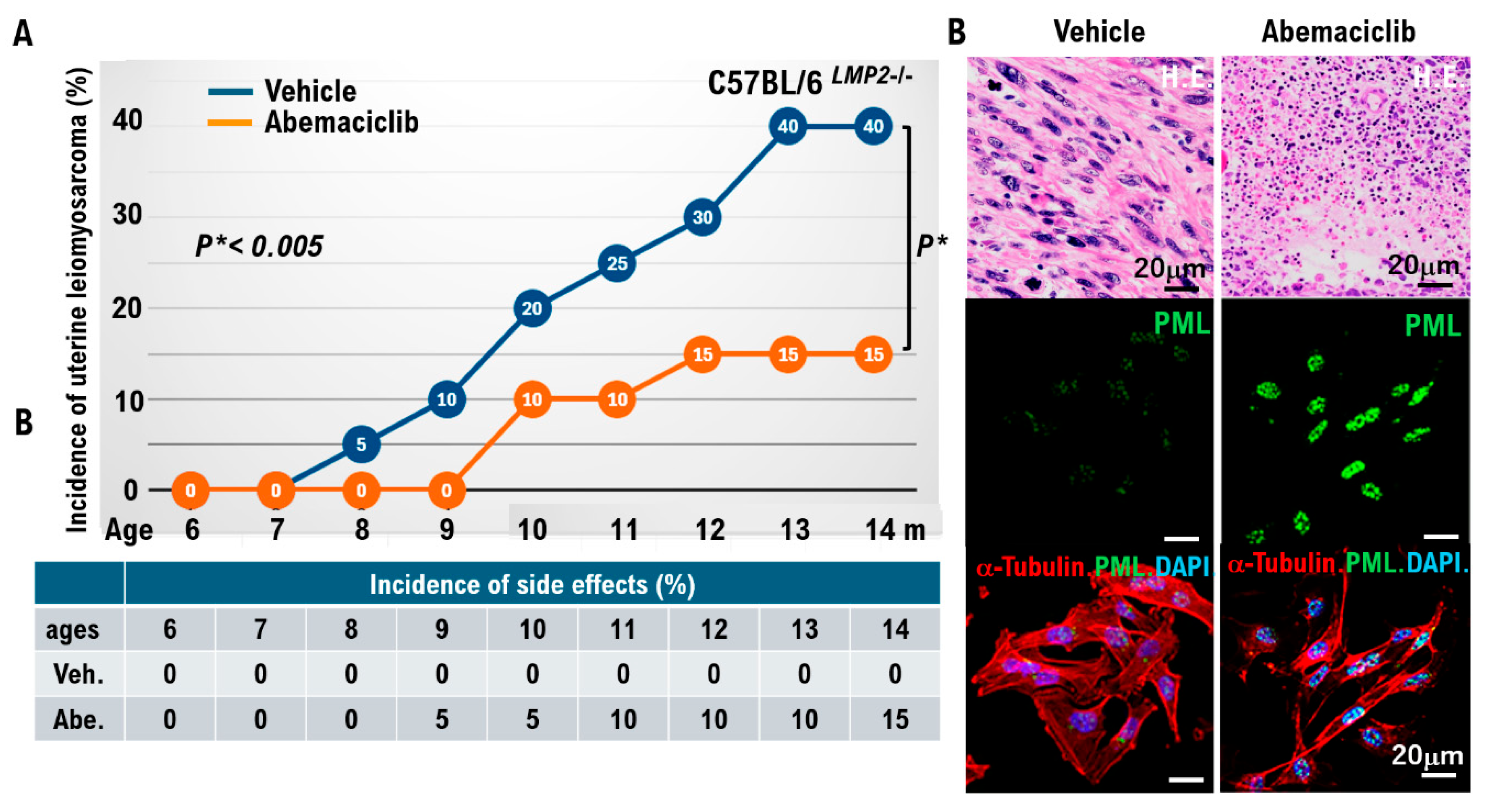
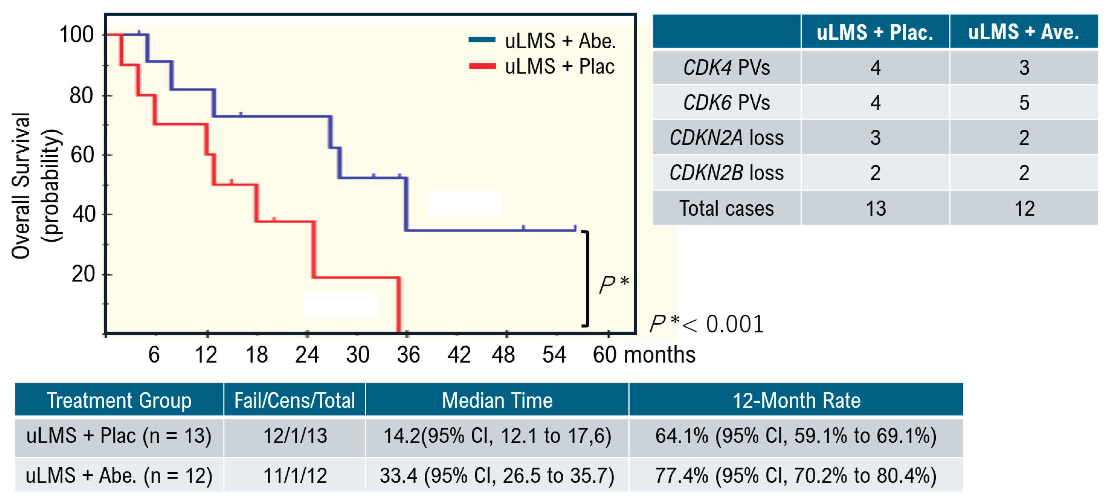
Uterine smooth muscle cells form the uterine framework. The myometrium is the middle layer of the uterine wall, consisting primarily of uterine smooth muscle cells (also called uterine myocytes) but also supports stromal and vascular tissues. Smooth muscle cells differentiate from stem cells. Broadly, two types of tumor cells derived from uterine smooth muscle cells can arise in the uterine smooth muscle layer. Benign uterine leiomyomas and malignant uterine leiomyosarcomas arise from the uterine smooth muscle layer. Because uterine leiomyoma cells display diverse morphologies, differential diagnosis from other uterine smooth muscle tumors can be challenging. Uterine leiomyosarcoma also has a poor prognosis due to frequent metastasis and recurrence. Overseas clinical trials have shown limited efficacy and noted adverse effects of existing antitumor agents. Therefore, we analyzed molecular pathological characteristics of uterine leiomyosarcoma in a spontaneous mouse model (C57BLACK/6LMP2-/-) that we previously reported in collaboration with Professor Susumu Tonegawa of the MIT–Picower Institute. We identified activation of cell-cycle G0/G1 inducers. Intraperitoneal abemaciclib, which targets cell cycle G0/G1 inducers (CDK4/cyclin D, CDK2/cyclin E), in C57BLACK/6LMP2-/- mice reduced uterine leiomyosarcoma incidence in C57BLACK/6LMP2-/- mice. We then enrolled 25 patients with advanced metastatic uterine leiomyosarcoma who had activating pathogenic variants in G0/G1-inducing factors identified by cancer gene panel testing. They were assigned to the abemaciclib or placebo groups, and median treatment time and 12-month treatment rate were compared. Results demonstrated efficacy in patients with advanced metastatic uterine leiomyosarcoma with activating cell-cycle variants. Because this trial involved a small cohort, larger studies are warranted.

Keywords: 
;  ;  ;  ;  

Introduction

Smooth muscle cells, a type of mesenchymal cells, form the framework of the uterine body and both adnexa (fallopian tubes and ovaries [1,2]. Uterine leiomyomas are benign tumors that develop in the smooth muscle layer [3]. Uterine leiomyomas respond well to the female hormone estrogen and grow [4]. Therefore, in many cases, uterine leiomyomas grow and decrease in size depending on the menstrual cycle [4]. Therefore, the treatment of uterine leiomyomas involves hormone therapy using a Gonadotropin releasing hormone (GnRH) antagonist, an estrogen antagonist, to induce regression [5]. Uterine leiomyosarcomas are rare malignant uterine mesenchymal tumors that arise in the smooth muscle layers of the uterine corpus and ovaries. Uterine leiomyosarcomas are highly malignant and progressive, making them difficult to treat. Although uterine leiomyosarcomas have a low incidence rate in Japan, they develop when smooth muscle cells differentiate and become malignant. Unlike benign uterine leiomyomas, uterine leiomyosarcomas are unresponsive to female hormones such as estrogen [6]. As such, the incidence of uterine leiomyosarcomas peaks in postmenopausal women in their 50s and 60s, and it has also been observed in women in their 80s [7]. Furthermore, owing to these characteristics, the effectiveness of hormone therapy for uterine leiomyosarcomas remains unconfirmed. Uterine leiomyosarcoma cells undergo active cell division, which makes them prone to infiltrate the surrounding tissue and form hematogenous pulmonary metastases [8]. Uterine leiomyosarcomas are detected in many women aged >40 years. However, they can also occur in younger people, although the incidence rate is extremely low.
The clinical symptoms of uterine leiomyosarcomas include abnormal vaginal bleeding, lumps or pain in the lower abdomen, menstrual irregularities, and general fatigue, which can also be observed in patients with other gynecological tumors, such as uterine leiomyomas, endometrial cancer, and cervical cancer; therefore, uterine leiomyosarcomas have no specific symptoms [9]. As the disease progresses, weight loss and rapid tumor growth occur. The symptoms of uterine leiomyosarcomas are similar to those of uterine fibroids; however, because uterine leiomyosarcomas respond to estrogen, the tumor size increases or decreases during the menstrual cycle [4,9]. Furthermore, because uterine leiomyosarcomas grow without responding to estrogen, they can also occur in postmenopausal women. Hence, uterine leiomyosarcomas commonly develop in women in their 80s. Benign uterine leiomyosarcomas should be carefully distinguished from malignant ones [10,11].

Diagnostic Methods 

Uterine leiomyosarcomas are diagnosed through medical interviews, physical examinations, and imaging diagnostics, such as ultrasound, contrast-enhanced magnetic resonance imaging (MRI), and contrast-enhanced computed tomography (CT) [12]. However, distinguishing between malignant and benign uterine leiomyosarcomas using imaging diagnostics alone is often difficult, and tissue testing (biopsy or pathological diagnosis of surgically removed specimens) is essential for a definitive diagnosis [11,12].

Prognosis and Treatment 
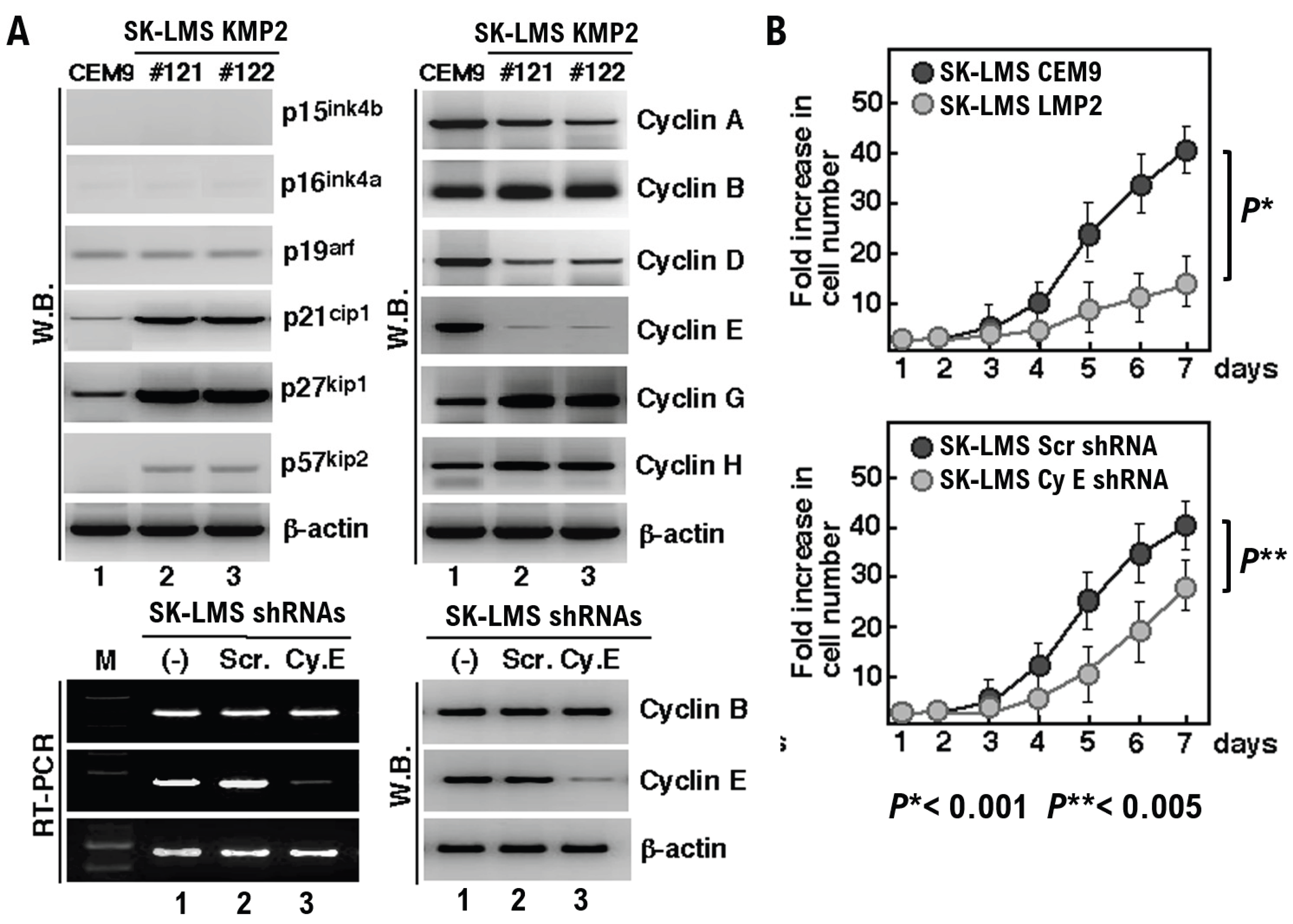
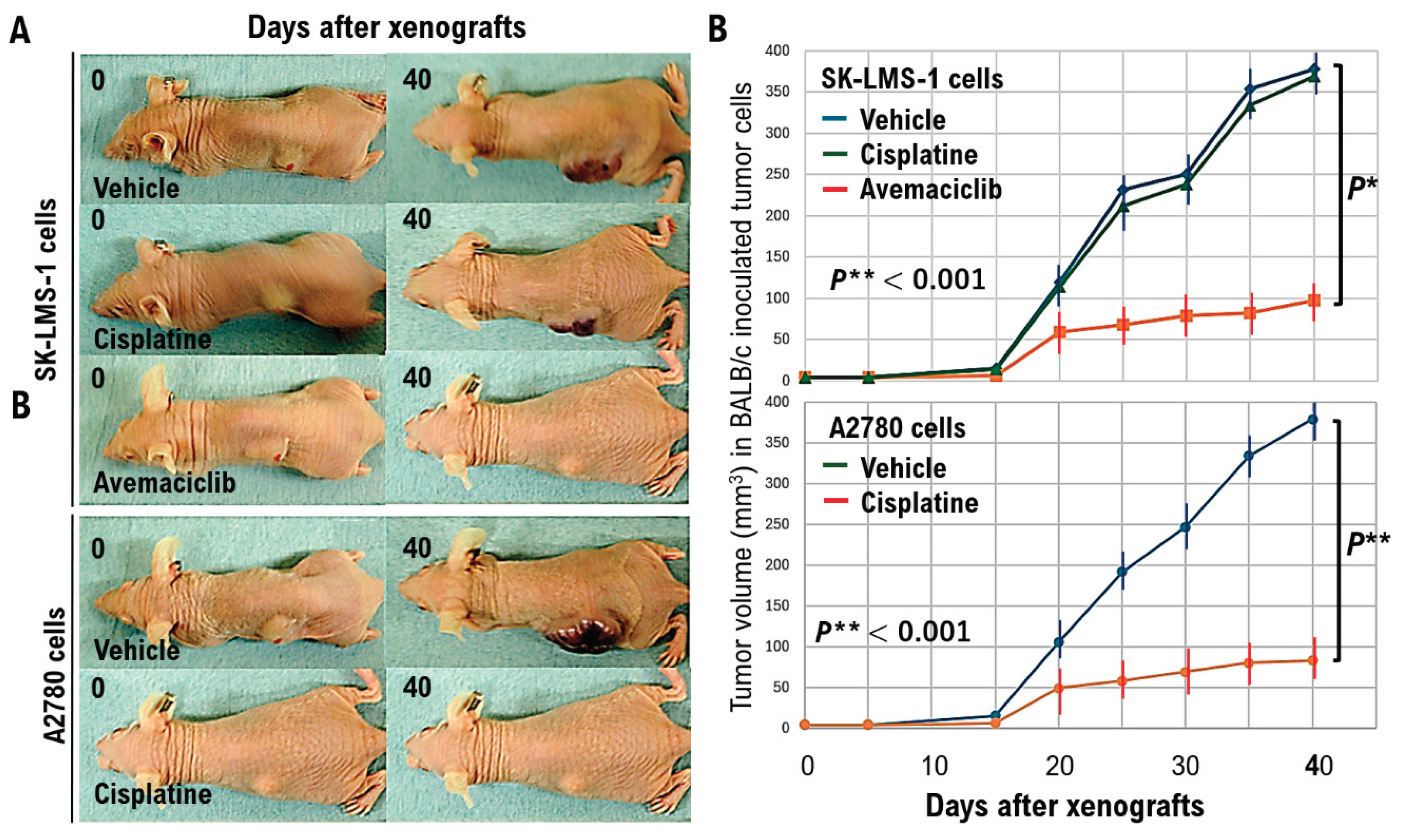
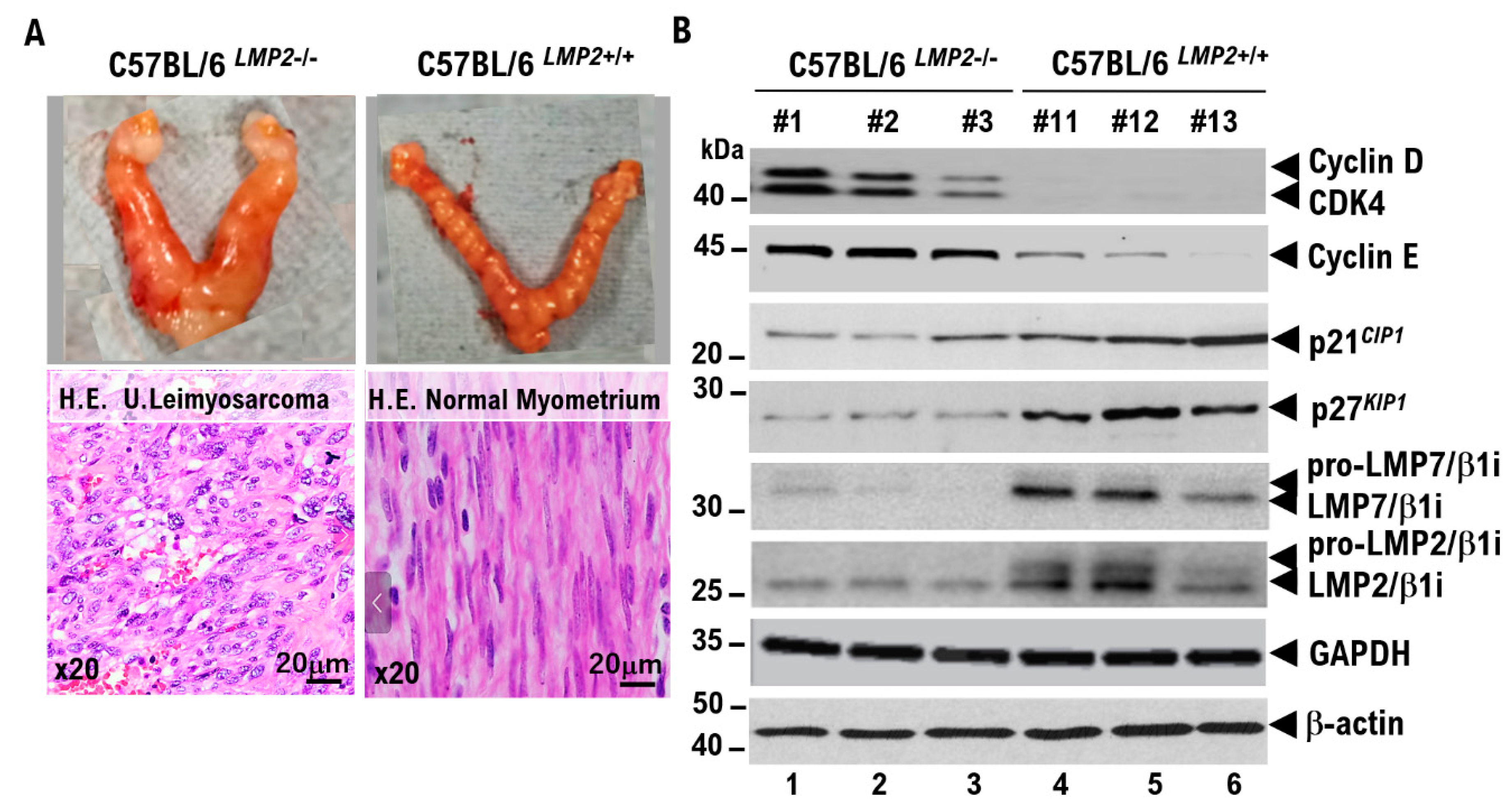
Uterine leiomyosarcomas grow rapidly and have a high recurrence and metastasis rate, resulting in a poor prognosis [13,14]. Surgical resection is the first-choice treatment for uterine leiomyosarcomas. Generally, a total hysterectomy is performed to treat them. For advanced uterine leiomyosarcomas, total hysterectomy, bilateral removal of the ovaries and salpingotubes, and removal of the surrounding tissues may be performed. Chemotherapy and radiation therapy may be performed as adjuvant treatments after surgical removal; however, their effectiveness is limited, and the recurrence rate is high. The median postoperative time to recurrence is 1.8 years. Furthermore, the 5-year survival rate of patients with uterine leiomyosarcomas is <20%. However, no standard treatment has been established for uterine leiomyosarcomas. The efficacy of doxorubicin alone, doxorubicin and bevacizumab combination therapy, gemcitabine alone, and immune checkpoint inhibitors for uterine leiomyosarcomas has been investigated [15]. However, no efficacy or toxicity has been observed in these clinical trials.
We have reported a genetically engineered mouse (C57BLACK/6Lmp2-/-) that spontaneously developed a uterine leiomyosarcoma after 6 months of age [16,17]. LMP2/β1i mice do not develop common malignant tumors other than uterine leiomyosarcomas [16]. Thus, the loss of LMP2/β1i, a subunit of the immunoproteasome, is associated with the development of uterine leiomyosarcomas. Therefore, we performed cancer genome profiling (CGP) of primary cultured cells established from human uterine leiomyosarcomas (SK-LMS) and SK-LMS cells-LMP2/β1i, which constitutively express LMP2/β1i. SK-LMS cells-LMP2/β1i cells exhibited G1 arrest in the cell cycle in conjunction with LMP2/β1i expression. SK-LMS cells strongly express cell cycle inducers (cyclins D and E) that act during the G0/G1 phase. Furthermore, uterine leiomyosarcoma cells developed in LMP2/β1i-deficient mice also strongly express cell cycle inducers (cyclins D and E) that act at the G0/G1 phase. Abemaciclib [18], which inhibits the activity of CDK4/6/cyclin D and CDK2/cyclin E, also inhibited the proliferation of SK-LMS cells. Therefore, we administered abemaciclib (100 mM) intraperitoneally once weekly to LMP2/β1i-deficient mice aged 6–14 months. Compared to LMP2/β1i-deficient mice that received saline intraperitoneally until 14 months of age, the incidence of uterine leiomyosarcoma was significantly lower in LMP2/β1i-deficient mice that received abemaciclib (100 mM) intraperitoneally once weekly until 14 months of age.
Our clinical research team, working in the cancer genome medicine department of a Japanese university, conducted cancer gene panel testing on approximately 6,900 patients with advanced or metastatic cancer from February 2019 to September 2025 and investigated new treatments for these patients based on the results of CGP. To date, cancer genomic medicine has investigated new treatments for 153 cases of advanced/metastatic uterine leiomyosarcoma using cancer gene panel testing. Of the 153 cases, pathogenic variants (PVs) were detected in CDK4 in seven cases, CDK6 in nine cases, CDKN2A in five cases, and CDKN2B in four cases. Our research team divided these 25 cases into 2 cohorts and examined the overall survival (OS) rates in the abemaciclib and placebo groups. The median OS rate (50%) was 33.4 months in the abemaciclib group and 14.2 months in the placebo group. Abemaciclib was effective in patients with advanced or metastatic uterine leiomyosarcomas in which PVs were detected as factors that induce the G0/G1 phase of the cell cycle. Thus, antitumor agents targeting the PVs detected in each type of uterine leiomyosarcoma may become a central treatment strategy for advanced or metastatic uterine leiomyosarcoma. Although uterine leiomyosarcomas are rare, they are rapidly growing malignant tumors with a poor prognosis. Because the symptoms are similar to those of uterine fibroids, a thorough examination is necessary for diagnosis. Surgical resection is the first choice of treatment; however, postoperative follow-up is important because of the high risks of recurrence and metastasis. Accurate knowledge and regular checkups are essential for gynecological diseases that require particular attention. Future studies should examine the therapeutic effects of antitumor agents targeting the PVs within each molecule in patients with advanced and metastatic uterine leiomyosarcoma.

Results

LMP2/β1i deletion was observed in uterine leiomyosarcoma cells, resulting in active G0/G1-inducing factors. Previously, we reported genetically engineered mice (C57BLACK/6Lmp2-/-) that spontaneously developed a uterine leiomyosarcoma after 6 months of age [16,17]. Mice up to 14 months of age did not develop common malignancies other than uterine leiomyosarcomas [16]. Thus, deletion of LMP2/β1i, a subunit of the immunoproteasome, is associated with the development of uterine leiomyosarcomas in mice [16,17]. Therefore, we investigated the molecular characteristics of human uterine leiomyosarcoma cells. LMP2/β1i expression loss was observed in uterine leiomyosarcoma cells (SK-LMS-1) established from surgically removed human uterine leiomyosarcomas, as well as in mouse uterine leiomyosarcoma cells in which LMP2/β1i deletion has been observed [16,17]. Thus, LMP2/β1i expression loss may be involved in the development of human uterine leiomyosarcoma [19]. When SK-LMS cells-LMP2/β1i, in which LMP2/β1i is constitutively expressed, were created, the constitutive expression of LMP2/β1i induced a phenotypic change in the SK-LMS cells to flat-revertant, slowing their cell proliferation rate (Figure 1). Therefore, we performed CGP on primary cultured cells established from human uterine leiomyosarcoma (SK-LMS cells) and SK-LMS cells-LMP2/β1i, in which LMP2/β1i is constitutively expressed (Figure 1A, SFigure 1). Compared to SK-LMS-LMP2 cells, SK-LMS cells showed a weaker expression of factors that suppress cell proliferation (p21cip1, p27kip1, and p57kip2). However, constitutive expression of LMP2/β1i enhanced the expression of these factors (p21cip1, p27kip1, and p57kip2) (Figure 1A). By contrast, SK-LMS cells showed strong expression of factors that enhance cell proliferation (cyclins D and E). The proliferation rate of cyclin E-deficient mouse fibroblasts is slower than that of parental mouse fibroblasts [20]. Therefore, to examine whether reducing cyclin E expression also reduces cell proliferation in human uterine leiomyosarcoma cells, we generated SK-LMS-sh cyclin E mRNA cells in which cyclin E expression was inhibited by inducing sh cyclin E mRNA expression. When we examined the proliferation rate of each cell type, we confirmed that SK-LMS-LMP2/β1 cell proliferation was reduced (Figure 1A,C). Reducing cyclin E expression in SK-LMS cells reduced cell proliferation (Figure 1A,C). Consequently, LMP2/β1i expression induced G1 arrest in the cell cycle of SK-LMS cells-LMP2/β1i cells (Figure 1C).
Cell cycle analysis is essential for elucidating the mechanisms of cell proliferation. Flow cytometry measures the fluorescence intensity of individual cells stained with a DNA detection reagent, making it possible to calculate the percentage of cells in each phase of the cell cycle according to their DNA content (G0/G1 phase: quiescence and cell growth [nuclear phase is 2n]; S phase: DNA synthesis [nuclear phase increases from 2n to 4n]; and G2/M phase: division preparation and division [nuclear phase is n]) [21]. Therefore, we investigated the effects of LMP2/β1i expression or cyclin E expression on the cell cycle in cultured human uterine leiomyosarcoma cells (SK-LMS cells) using cell cycle analysis with propidium iodide (PI) staining using the flow cytometer CyFlow Cube Becton Dickinson Immunocytometry System, Mountain View, CA, USA) [21]. In SK-LMS CEM9s cells, which were transfected with an empty vector, the number of cells in the C0/G1 phase was 46.1% of the total number of cells within 24 h (SFigure 2A). By contrast, in SK-LMS-LMP2 cells constitutively expressing LMP2/β1i, the number of cells in the C0/G1 phase was 64.9% of the total cell number within 24 h (SFigure 2A). Therefore, LMP2/β1i expression induced G0/G1 arrest in cultured uterine leiomyosarcoma cells. SK-LMS CEM9-Scr.shRNA cells transfected with a Scr.shRNA vector expressing a nonspecific mRNA accounted for 46.4% of the total cell population in the C0/G1 phase within 24 h (SFigure 2B). By contrast, in SK-LMS CEM9-cyclin E.shRNA cells transfected with a cyclin E shRNA vector that inhibits cyclin E translation from mRNA, the number of cells remaining in the C0/G1 phase accounted for 63.8% of the total cell population within 24 h (SFigure 2B). Thus, cyclin E expression induced G0/G1 transition in cultured uterine leiomyosarcoma cells.
Cyclin E activity is important for tumorigenicity in vivo. SK-LMS CEM9-cyclin E shRNA reduced cyclin E activity in uterine leiomyosarcoma cells. Two types of uterine leiomyosarcoma cells (1.0 × 10⁵) were plated in a petri dish and cultured for 2 weeks at 37 °C in a 5.0% CO2 atmosphere. After 2 weeks of cell culture, cell proliferation was measured under a microscope using trypan blue staining. The proliferation rate of SK-LMS CEM9-Scr.shRNA cells was approximately 102-fold higher than that of SK-LMS CEM9-cyclin E.shRNA cells (Figure 2A).
Therefore, we investigated the role of cyclin E in tumorigenesis in human uterine leiomyosarcoma cells. SK-LMS CEM9-Scr.shRNA cells were exanografted intradermally into the left side of the back of immunodeficient BALB/c Slc-nu/nu mice, and SK-LMS CEM9-cyclin E.shRNA cells were exanografted intradermally into the right side (Figure 2A). Our research team measured the size (w × d × h/2) of tumors growing at each exanografting site every 5 days after intradermal implantation of cultured human uterine leiomyosarcoma cells into the left and right sides of the backs of BALB/c Slc-nu/nu mice. Consequently, 40 days after each human uterine leiomyosarcoma cell line was intradermally implanted into the left and right sides of the back of BALB/c Slc-nu/nu mice, tumor size in SK-LMS CEM9-Scr.shRNA cells significantly increased compared to that in SK-LMS CEM9-cyclin E.shRNA cells (Figure 2B).
Therefore, intradermal tumors formed by each type of human uterine leiomyosarcoma cell line on the left and right sides of the back of BALB/c Slc-nu/nu mice were excised and sectioned using standard methods. Sections of each tumor tissue were stained by standard immunohistochemistry (IHC) using 4’,6-diamidino-2-phenylindole (DAPI), Ki-67/MIB1, anti-human cyclin E antibody, anti-promyelocytic leukemia (PML) antibody, and anti-α-tubulin antibody (Figure 2C). Compared to the sections of tumors grown with SK-LMS, CEM9-Scr.shRNA cells and sections of tumors grown with SK-LMS CEM9-cyclin E.shRNA cells showed similar α-tubulin levels (red). The proliferation marker Ki-67/MIB1 was strongly expressed in tumor sections grown with SK-LMS CEM9-Scr.shRNA cells. However, the cell proliferation marker Ki-67/MIB1 was barely detected in tumor sections from SK-LMS CEM9-cyclin E.shRNA cells, demonstrating the importance of cyclin E expression for tumor formation in human uterine leiomyosarcoma cells. Furthermore, the apoptosis marker PML [22,23] was absent in SK-LMS CEM9-cyclin E.shRNA cells, indicating that the inhibition of cyclin E expression did not induce cell death by apoptosis.
Regarding the antitumor effect of antitumor agents targeting G0/G1-inducing factors on uterine leiomyosarcoma, CGP showed that CDK4/6, which acts in the G0/G1 phase, was activated in cultured human uterine leiomyosarcoma cells (SK-LMS cells). Therefore, abemaciclib, which exerts antitumor effects by targeting CDK4/6, may also exert antitumor effects on SK-LMS cells. Cisplatin has strong antitumor effects against gynecological malignancies (ovarian, endometrial, and cervical cancers); however, it does not exhibit antitumor effects against uterine mesenchymal tumors. The mechanism of action of cisplatin involves strong DNA binding. SK-LMS cells were cultured at a density of 2.0 × 10⁵ in a 10 cm petri dish, with vehicle (phosphate-buffered saline [PBS]), cisplatin (5.0 mg/kg; Nippon Kayaku Co.,Ltd. Chiyodaku, Tokyo, Japan), and abemaciclib (20 mg/kg; Eli Lilly and Company, Kobe, Hyogo, Japan) to each culture medium, and cultured the cells for 3 h at 37 °C in a CO2 5.0% culture environment. After the addition of each agent, the total cell number was counted every 12 h using trypan blue (Figure 3). Addition of abemaciclib to the culture medium decreased the number of SK-LMS cells over time (Figure 3A). However, the addition of the platinum compound cisplatin to the culture medium, as well as the vehicle (PBS), resulted in the proliferation of SK-LMS cells (Figure 3A). As shown in Figure 2B, after 72 h of culture in abemaciclib, SK-LMS cells stained blue with b-galactosidase [24,25] (Figure 3B). Thus, abemaciclib (20 mg/kg) caused SK-LMS cells to die by apoptosis (Figure 3B). The addition of cisplatin to the culture medium resulted in a time-dependent decrease in the number of high-grade serous ovarian carcinoma cells (A2780 cells) (Figure 3C). However, when PBS was added to the culture medium, the A2780 cells continued to proliferate (Figure 3C). As shown in Figure 3D, after 72 h of culture in cisplatin, A2780 cells stained blue with b-galactosidase [24,25] (Figure 3D), indicating that cisplatin caused SK-LMS cells to die by apoptosis (Figure 3D).
SK-LMS cells strongly express cell cycle inducers (cyclins D and E) that act during the G0/G1 phase. Abemaciclib, which exerts antitumor effects by targeting cell cycle inducers (CDK4/6/cyclin D and CDK2/cyclin E) that act in the G0/G1 phase, demonstrated antitumor effects in human uterine leiomyosarcoma cells (SK-LMS cells) (Figure 3A,B). Therefore, we xenografted SK-LMS cells (1.0 × 10⁶) intradermally into the left side of the back of immune-deficient BALB/c Slc-nu/nu mice and examined whether abemaciclib had an antitumor effect on tumors formed by SK-LMS cells that had grown intradermally. Specifically, we xenografted SK-LMS cells (1.0 × 10⁶) intradermally into the left side of the back of immune-deficient BALB/c Slc-nu/nu mice. After xenografting, SK-LMS cells (1.0 × 10⁶), vehicle (PBS), abemaciclib, or cisplatin were administered intraperitoneally every 5 days, and the size (w × d × h/2) of the tumors formed at the xenografting site was measured. Intraperitoneal administration of abemaciclib inhibited SK-LMS cell proliferation, resulting in the formation of small tumors (Figure 3). Intraperitoneal administration of vehicle (PBS) or cisplatin did not inhibit SK-LMS cell proliferation, and the tumors formed by SK-LMS cells at the transplantation site grew in size over time (Figure 3).
Furthermore, our research team xenografted high-grade serous ovarian carcinoma cells (A2780 cells) (1.0 × 10⁶) intradermally into the left back of immune-deficient BALB/c Slc-nu/nu mice. After xenografting A2780 cells (1.0 × 10⁶), vehicle (PBS) or cisplatin was administered intraperitoneally every 5 days, and the size (w × d × h/2) of tumors formed at the xenografting site owing to the proliferation of A2780 cells was measured. Intraperitoneal administration of cisplatin (5.0 mg/kg) inhibited the proliferation of A2780 cells, resulting in the formation of small tumors (Figure 3). Intraperitoneal administration of the vehicle (PBS) (100 mL) did not inhibit the proliferation of A2780 cells, and the tumors formed by the A2780 cells at the exnografting site grew in size over time (Figure 3).
Regarding the antitumor effects of abemaciclib on uterine leiomyosarcoma in LMP2/β1i-deficient mice, SK-LMS cells exhibited significantly reduced expression of LMP2/β1i and strong expression of cell cycle inducers (cyclins D and E) that act during the G0/G1 phase. The antitumor effect of abemaciclib, which targets the cell cycle inducers CDK4/6/cyclin D and CDK2/cyclin E that act at the G0/G1 phase, was observed in SK-LMS cells. Therefore, we examined the gene expression patterns in uterine leiomyosarcoma cells that developed in LMP2/β1i-deficient mice (Figure 4). In uterine leiomyosarcoma cells that developed in LMP2/β1i-deficient mice, we observed a strong expression of cell cycle inducers (CDK4/cyclin D and cyclin E) that act in the G0/G1 phase, and reduced expression of p21CIP1 and p27KIP1 (Figure 4). Therefore, we intraperitoneally administered abemaciclib (20 mice) or vehicle (PBS) (20 mice) once a week to LMP2/β1i-deficient mice aged 6–14 months. From 5 to 14 months of age, once a month, all LMP2/β1i-deficient mice in the abemaciclib group (20 mice) and vehicle (PBS) group (20 mice) underwent laparotomy to monitor the size of the uterus and ovaries and the presence or absence of tumors, and then the abdomen was closed. In the LMP2/β1i-deficient mice group that received intraperitoneal administration of vehicle (PBS) from 5 to 14 months of age, 40% (8/20) of the mice developed uterine leiomyosarcoma by 14 months of age (Figure 5). In the LMP2/β1i-deficient mice group that received intraperitoneal administration of abemaciclib (20 mg/kg) from 5 to 14 months of age, 15% (3/20) of the mice developed uterine leiomyosarcoma by 14 months of age (Figure 5). The incidence of uterine leiomyosarcoma was significantly reduced in LMP2/β1i-deficient mice administered abemaciclib intraperitoneally once weekly until 14 months of age (Figure 5A). We performed a histopathological analysis of mouse uterine leiomyosarcoma developed in LMP2/β1i-deficient mice, in which tumor regression was observed with abemaciclib, and no antitumor effect was observed with vehicle (PBS) administration.
In tissue sections of mouse uterine leiomyosarcoma that showed tumor regression with abemaciclib, leiomyosarcoma cells shrank and were stained for PML (green) (Figure 5B). This demonstrated that abemaciclib induced apoptotic cell death in mouse uterine leiomyosarcoma cells (Figure 5B). By contrast, in tissue sections of mouse uterine leiomyosarcoma cells in which vehicle (PBS) administration showed no antitumor effect, the leiomyosarcoma cells maintained their skeletal structure, and no PML (green) staining was observed (Figure 5B). Thus, vehicle (PBS) administration did not induce apoptotic cell death in the mouse uterine leiomyosarcoma cells (Figure 5B).
To investigate the antitumor effect of abemaciclib on uterine leiomyosarcoma with PVs, which is a factor acting on the G0/G1 phase of the cell cycle, the department of a Japanese university conducted cancer gene panel testing on approximately 6,900 patients with advanced or metastatic cancer between February 2019 and September 2025. Based on the CGP results, our clinical research team is currently investigating new treatment options for these patients. To date, new treatments have been investigated in 153 cases of advanced or metastatic uterine leiomyosarcoma using cancer gene panel testing in our cancer genomics department (SFigure 3). Of the 153 cases of advanced or metastatic uterine leiomyosarcoma in which cancer gene panel testing was performed, PVs were detected in the CDK4 molecule in seven cases, the CDK6 molecule in nine cases, the CDKN2 A molecule in five cases, and the CDKN2B molecule in four cases (SFigure 3). Our clinical research team divided the 25 patients into two cohorts and examined the OS rates in the abemaciclib and placebo groups (Figure 6). In many uterine leiomyosarcoma cases, lung metastases are observed within 2 years after surgical removal. Thus, patients with advanced/metastatic uterine leiomyosarcoma were those in whom lung metastases were detected on contrast-enhanced CT (SFigure 4). Hence, we describe cases in which lung metastases can no longer be detected after the administration of antitumor agents as having demonstrated a response to antitumor drug treatment (SFigure 4). The median OS rate (50%) was 33.4 months in the abemaciclib group and 14.2 months in the placebo group (Figure 6). The 12-month survival rates in the placebo and abemaciclib groups were 64.1% and 77.4%, respectively. Although this was a clinical study with a small cohort, abemaciclib was effective in patients with advanced/metastatic uterine leiomyosarcoma, in which PVs were considered a factor that could induce the G0/G1 phase of the cell cycle. Thus, antitumor agents targeting the PVs detected in each type of uterine leiomyosarcoma may become a central treatment strategy for advanced/metastatic uterine leiomyosarcoma. No serious adverse events were observed in this clinical study (SFigure 5).

Discussion

Tumors that develop in the uterine smooth muscle layer have diverse cytoskeletons and cell nuclei; therefore, differentiating between benign and malignant uterine leiomyosarcomas in many cases is often difficult. Surgical resection is the only first-line treatment for uterine leiomyosarcoma. Approximately 1.8 months after surgical removal, many patients develop hematogenous lung metastases. Unfortunately, no established treatment has proven effective for uterine leiomyosarcoma. The 5-year survival rate of patients with uterine leiomyosarcoma is approximately 20%. Therefore, establishing treatment for uterine leiomyosarcoma is an important aspect of gynecological care. Our research team has previously reported a genetically modified mouse model (C57BLACK/6Lmp2-/-) that spontaneously developed uterine leiomyosarcoma at 6 months of age. LMP2/β1i mice do not develop common malignant tumors other than uterine leiomyosarcoma. Thus, the loss of LMP2/β1i, a subunit of the immunoproteasome, is associated with the development of uterine leiomyosarcoma. Therefore, we investigated the molecular characteristics of the human uterine leiomyosarcoma cells. Similar to mouse uterine leiomyosarcoma cells, human uterine leiomyosarcoma cells lost LMP2/β1i expression. Therefore, we performed CGP to compare primary cultured human uterine leiomyosarcoma (SK-LMS) cells with SK-LMS cells-LMP2/β1i, which constitutively express LMP2/β1i. G1 cell cycle arrest in SK-LMS cells-LMP2/β1i cells was linked with LMP2/β1i expression. SK-LMS cells strongly expressed cell cycle inducers (cyclins D and E) that act during the G0/G1 phase. Furthermore, cell cycle inducers (cyclin D and cyclin E) that act at the G0/G1 phase were also strongly expressed in uterine leiomyosarcoma cells developed in C57BLACK/6Lmp2-/- mice. Abemaciclib, which inhibits the activity of CDK4/6/cyclin D and CDK2/cyclin E, inhibited the proliferation of SK-LMS cells. Therefore, we administered abemaciclib (20 mg/kg) intraperitoneally once weekly to C57BLACK/6Lmp2-/-/- mice at 6 months of age until they reached 14 months of age. Compared with C57BLACK/6Lmp2-/- mice that received intraperitoneal saline up to 14 months of age, C57BLACK/6Lmp2-/- mice that received intraperitoneal abemaciclib once weekly up to 14 months of age had a significantly lower incidence of uterine leiomyosarcoma. However, the antitumor activity of LMP2/β1i was not observed in cervical cancer cells (SFigure 6). This is likely because the activity of the immunoproteasome and its subunits are organ-, tissue-, and substrate-dependent [26].
Our clinical research team at the Cancer Genomics Department of a Japanese university conducted cancer gene panel testing (FoundationOne CDx and FoundationOne Cdx liquid) [27,28] on approximately 6,900 patients with advanced or metastatic cancer from February 2019 to September 2025. Based on the results of the CGP, we considered new treatments for these patients with cancer. To date, our Cancer Genomics Department has used cancer gene panel testing to identify new treatments for 153 patients with advanced or metastatic uterine leiomyosarcoma. Of the 153 cases of uterine leiomyosarcoma, PVs were detected within the CDK4 molecule in seven cases, within the CDK6 molecule in nine cases, within the CDKN2A molecule in five cases, and within the CDKN2B molecule in four cases. Our research team divided these 25 cases into two cohorts and examined the OS rates in the abemaciclib and placebo groups. The median OS rate (50%) was 33.4 months in the abemaciclib group and 14.2 months in the placebo group. Abemaciclib proved effective in patients with advanced or metastatic uterine leiomyosarcoma, whose G0/G1 cell cycle phase-inducing factors are PVs. No serious adverse events were observed in this clinical study (SFigure 5).
Our clinical research team, working in the cancer genomics department of a Japanese university, will perform cancer gene panel testing (FoundationOne CDx, FOundationOne CDx liquid) on approximately 6,900 patients with advanced or metastatic cancer from February 2019 to September 2025 and will consider new treatments for these patients based on the results of CGP. To date, new treatments have been considered for 153 cases of advanced or metastatic uterine leiomyosarcoma using cancer gene panel testing in our cancer genomics department (SFigure 3). For example, cancer gene panel testing detected PVs within the breast cancer susceptibility gene I (BRCA1) (five cases) or BRCA2 (eight cases). Therefore, poly (ADP-ribose) polymerase (PARP) inhibitors [29], such as olaparib, niraparib, and talazoparib, have been orally administered to patients with uterine leiomyosarcoma with PVs within the BRCA1 or BRCA2 molecule. However, PARP inhibitors are ineffective in some cases. When the variant allele frequency (VAF) of BRCA1 or BRCA2 PVs is low (<30%), the BRCA1 or BRCA2 VAFs are likely somatic mutations. Clinical trials have shown that the antitumor effect of PARP inhibitors is better when BRCA1 or BRCA2 VAFs are germline rather than somatic mutations. Therefore, compared with other progressive/metastatic malignant tumors, the detection rate of variant allele frequency (VAFs) in surgically removed tissue or liquid specimens is low in uterine leiomyosarcoma, and tumors with high VAF values are more likely to respond well to antitumor drugs.
Medical researchers worldwide have conducted clinical trials using existing antitumor agents to investigate their antitumor effects on uterine leiomyosarcoma. Adding trabectedin to doxorubicin as a first-line treatment for patients with advanced leiomyosarcoma, followed by maintenance therapy with trabectedin, may offer superior efficacy compared with doxorubicin alone [30,31]. In patients with metastatic or surgically unresectable uterine or soft tissue leiomyosarcoma, the combination of doxorubicin and trabectedin induction, followed by trabectedin maintenance therapy, was associated with improved OS and progression-free survival compared with doxorubicin alone [30,31]. This combination therapy significantly prolongs progression-free survival with manageable, albeit high, toxicity, and may be considered a first-line treatment option for metastatic leiomyosarcoma [31]. In a nonrandomized clinical trial, the combination of doxorubicin and pembrolizumab was well tolerated [32]. Although the primary endpoint of objective response rate (ORR) was not met, progression-free survival (PFS) and OS were favorable compared to those reported in previous studies. Further research focusing on undifferentiated pleomorphic sarcomas and dedifferentiated liposarcomas is required. Therefore, Japanese medical staff administered a combination of doxorubicin and trabectedin to patients with advanced and metastatic uterine leiomyosarcoma; however, the tumors did not decrease in size. When considering side effects and efficacy, the medical staff prioritized the benefits. Furthermore, results from clinical trials conducted overseas have shown that the combination of gemcitabine and docetaxel is well-tolerated and highly effective in both treated and untreated patients with uterine leiomyosarcoma [33]. However, gemcitabine and docetaxel combination therapy has shown antitumor effects in approximately 30% of all advanced metastatic uterine leiomyosarcoma cases. Successful treatment with the gemcitabine and docetaxel combination therapy is extremely rare in Japanese patients with uterine leiomyosarcoma. Many cases of uterine leiomyosarcoma develop hematogenous pulmonary metastases within 2 years of surgical removal; however, lymphatic metastasis is extremely rare. Vascular endothelial growth factor (VEGF) may be involved in cancerous neovascularization. Therefore, the molecular-targeted drug anti-VEGF antibody (bevacizumab) may inhibit the formation of hematogenous pulmonary metastases, which are observed in many cases of uterine leiomyosarcoma. However, adding bevacizumab to gemcitabine/docetaxel as first-line treatment for metastatic uterine leiomyosarcoma does not improve PFS, OS, or ORR [34].
Uterine leiomyosarcoma is a rare malignant tumor that grows rapidly and has a poor prognosis. As the symptoms are similar to those of uterine fibroids, thorough testing is required for diagnosis. Surgical removal is the first choice of treatment; however, owing to the high risk of recurrence and metastasis, postoperative follow-up is important. It is a gynecological disease that requires particular attention; therefore, accurate knowledge and regular checkups are important. Currently, the effectiveness of new cancer treatments, including antitumor agents that target PVs, as detected by cancer gene panel testing, is being investigated worldwide. Although establishing a new treatment that demonstrates efficacy is difficult, remarkable antitumor effects have been observed in some cases. The clinical trial reported by our research team was a small-scale cohort study. Because uterine leiomyosarcoma is a rare malignancy, recruiting participants for clinical trials may be difficult. Future clinical trials with larger cohorts must be conducted to re-examine the results of the clinical trials conducted by our research team. Furthermore, the therapeutic effects of antitumor agents targeting the PVs in each molecule in patients with advanced/metastatic uterine leiomyosarcoma should be investigated.

Material and Methods

1. 
DNA Transfection and Isolation of Flat Revertants
pCEM9-LMP2, pCEM9, pCEM9 Cyclin E.shRNA, pCEM9 Scr.shRNA-control (Santa Cruz Biotechnology Inc. Dallas, Texas, U.S.A.), or its empty vector pCEM9 were transfected with a FuGENE6 transfection reagent (Roche, IN, USA, Madison, WI USA) according to the manufacturer’s recommendations with 5 μg of plasmid DNA and 5 × 10⁵ SK-LMS cells (HTB-88: purchased from ATCC, Manassas, VA, USA) and 5 × 10⁵ HeLa cells (CRM-CCL-2: purchased from ATCC, Manassas, VA, USA) plated into 6-well tissue culture dishes (#CLS353046: Corning NY, USA) on the previous day. At 48 h after transfection, the cells were treated with trypsin and replated onto 100-mm dishes with 15 ml of growth medium containing 1 mg of G418 per ml (SIGMA-Aldrich, MO, USA). The cells were incubated at 37 °C for an additional 6–8 days, with medium changes on days 1, 3, or 4. The number of G418-resistant colonies was counted at this stage.
2. 
Reverse Transcription-polymerase Chain Reaction Analysis (RT-PCR)
The expressions of cyclin B, cyclin E, and β-actin transcripts were examined using RT-PCR. Total RNA was prepared from human uterine LMS tissues and normal myometrial tissues using TRIzol reagent according to the manufacturer’s protocol (Invitrogen Co., CA, USA). RNA was reverse-transcribed using Superscript II enzyme (Invitrogen), and single-stranded cDNA was used for amplification. LMP2 and β-actin transcripts were subjected to PCR with the appropriate primer sets following a program of 35 cycles at 94 °C for 30 s, 60 °C for 30 s, and 72 °C for 1.5 min with an additional 5 min for the extension of transcripts (15). The primers were used as follows: cyclin B1-hum-for, 5′-AAGAGCTTTAAACTTTGGTCTGGG-3′; cyclin B1-hum-rev, 5′-CTTTGTAAGTCCTTGATTTACCATG-3′; cyclin E1-human-for, 5′-GTCCTGGCTGAATGTATACATGC-3′; cyclin E1-human-rev, 5N-CCCTATTTTGTTCAGACAACATGGC-3′, and β-Actin (F 5′-TCCGGAGACGGGGTCA-3′ and R 5′-CCTGCTTGCTGATCCA-3′). These experiments were conducted at Shinshu University in accordance with the local guidelines (approval nos. 4737, 150, and M192).
3. 
Flow Cytometry
Cells were incubated in culture dishes in F-12 medium (GIBCO, NY, USA) without a flow cytometry standard (FCS) to allow for analysis of the cell cycle. After overnight culture to allow cellular attachment to the plate, the medium was removed, and fresh medium containing 15% FCS was added. Adherent cells were collected and washed with PBS thrice at 16, 24, and 36 h. The cells were fixed in 70% ethanol and stored at 4 °C for 20 min, and then resuspended in a DNA-stain solution containing PI (20 mg/ml; Calbiochem, CA, USA) and RNase (20 mg/ml; Novagen, Darmstadt, Germany). The cells were analyzed with a FACScan (Fluorescence Activated Cell Sorter) flow cytometer in combination with the BD lysis II software (488 nm; Becton Dickinson Immunocytometry System, Mountain View, CA, USA).
4. 
Xenograft Studies
Nude mice (7–8 week old female BALB/c Slc-nu/nu mice, Japan SLC, Shizuoka, Japan) were xenografted intracutaneously with 1 × 10⁷ cells of the SK-LMS-CEM9 (Transform type) clones, SK-LMS-LMP2/b1i (Flatrevertant type) clones, SK-LMS-cyclinE/shRNA control (F type) clones, or SK-KMS-Scr/shRNA (T type) clones, and A2780 cells with BD Matrigel Matrix (BD Biosciences, MA, USA) in 5 mg/ml of culture medium containing 15% FCS plus SmGM-2 SingleQuots (CAMBREX, MD, USA) at 100 μl. Nude mice (7–8 week old female BALB/c Slc-nu/nu mice, Japan SLC) were also xenografted intracutaneously with 1 × 10⁷ cells of the HeLa-CEM9 (T type) clones, HeLa-LMP2wt (T type) clones, and HeLa-LMP2K33A (T type) clones with BD Matrigel Matrix (BD Biosciences, MA, USA) in 5 mg/ml of culture medium containing 15% FCS at 100 μl.
6. 
IHC
IHC staining for cyclin E, aTublin, PML, and Ki-67 was performed on tissue sections obtained from the xenografted SK-LMS Scr-sh RNA clone, KS-LMS cyclinE sh RNA clone and the tissue sections obtained from mice uterine leiomyosarcoma developed in c57BLCKLMP2-/-mice.
7. 
b-Galactosidase staining
Induction of cellular senescence not only resulted in various phenotypic changes, but also increased the expression of β-galactosidase, a pH-dependent enzyme in lysosomes. Various enzymes were activated and measured when the cells were cultured in culture dishes. Therefore, as cells senesce, β-galactosidase activity increased. To visualize cellular senescence, a senescence-associated β-galactosidase staining kit (#602010) was used, and β-galactosidase activity is displayed as a blue color using the manufacture’s procedure.
8. 
Ethics approval and consent to participate
Shinshu University approved the experiments (Approval No. M192). All experiments using human tissues were conducted at the National Hospital Organization, Kyoto Medical Center (approval no. NHO H31-02), in accordance with the institutional guidelines issued on August 17, 2019, by the Central Ethics Review Board of the National Hospital Organization Headquarters (Tokyo, Japan) and Shinshu University (Nagano, Japan). The authors attended educational lectures on medical ethics in 2020 and 2021, supervised by the Japanese government (completion numbers AP0000151756, AP0000151757, AP0000151769, and AP000351128). Consent for participation in this clinical study was obtained from all the patients. After being briefed on the clinical study and agreeing to the clinical research objectives, the participants signed consent forms. The authors attended seminars on the ethics of experimental research using small animals on July 2, 2020, and July 20, 2021. The code number for ethical approval of experiments with small animals was KMC R02-0702.
9. 
Ethical compliance with human study
This study involves research with human participants and was approved by the institutional ethics committee(s) and IRBs. This manuscript contains personal and/or medical information and a case report/case history about an identifiable individual; therefore, it has been sufficiently anonymized in line with our anonymization policy. The authors obtained direct consent from the patient.
The authors attended research ethics education through the Education for Research Ethics and Integrity (APRIN e-learning program (eAPRIN)) agency. The completion numbers for the authors are AP0000151756, AP0000151757, AP0000151769, and AP000351128.
This study involves research with animal materials and was approved by the institutional ethics committee(s) and IRBs (Ethics Committee for research with animals in National Hospital Organization Headquarter; Meguro, Tokyo, Japan).
Ethics committee name: IRB of the National Hospital Organization Headquarters (approval code: H31-1-2; approval date: November 09, 2019, and June 17, 2023, approved code: R07-1-2).

ARRIVE Checklist Documentation

We use live animals, the protocol of our research involves any live animals.

Supplementary Materials

The following supporting information can be downloaded at the website of this paper posted on Preprints.org. The details of materials and methods are indicated in the supplementary file online.

Author Contributions

TH, KS, MO, and MM were involved in the study design, data collection, data review and interpretation, and manuscript writing. TH, KS, and OM were involved in the literature search, study design, data collection, data interpretation, and manuscript writing. TH, KS, and OM were involved in data collection and interpretation. TH and OM were involved in data collection and interpretation and manuscript writing. TH, KS, OM, and MM were involved in the study conception and design, data analysis and interpretation, and manuscript writing. TH and OM were involved in patient recruitment, data analysis and interpretation, and manuscript writing. TH and IK were the medical leads for anti-tumor agents, and they participated in the data collection and evaluation and manuscript writing and editing. TH and IK were the lead physicians and were involved in the study design and conduct, data analysis and interpretation, and manuscript review.

Funding

This clinical research was performed using research funding from the following: Japan Society for Promoting Science for TH (grant no. 19K09840), START-program Japan Science and Technology Agency (JST) for TH (grant no. STSC20001), National Hospital Organization Multicenter Clinical Study for TH (grant no. 2019-Cancer in general-02), and Japan Agency for Medical Research and Development (AMED) (grant no. 22ym0126802j0001), Tokyo, Japan. The funders had no role in the study design, data collection and analysis, decision to publish, or preparation of the manuscript.

Data Availability Statement

Data are available on various websites and have also been made publicly available. More information can be found in the first paragraph of the Results section. The transparency document associated with this article can be found in the online version at
https://kyoto.hosp.go.jp/html/guide/medicalinfo/ clinical research/expand/gan.html (accessed on 15 March 2025).

Acknowledgments

The authors want to thank Prof. and Dr. Susumu Tonegawa at Mass. Inst. Tech. and Picower Institution for scientific discussion and providing C57BLACK/6LMP2-/- and Dr. Kohji Moriyoshi at the pathology division, National Hospital Organization, Kyoto Medical Center. The authors also want to acknowledge all medical staff for clinical research at Kyoto University School of Medicine and the National Hospital Organization Kyoto Medical Center. We also appreciate Dr. Keita Idegami, Chugai Pharma Manufacturing Co., Ltd. (Kitaku, Tokyo, Japan) and Sysmex Corporation. (Kobe, Hyogo, Japan) for providing medical imformation.

Conflicts of Interest

Authors have nothing to disclose.

References

  1. Aguilar HN, Mitchell BF. Physiological pathways and molecular mechanisms regulating uterine contractility. Hum Reprod Update. 2010 Nov-Dec;16(6):725-44. [CrossRef]
  2. NCI Dictionary of Cancer Terms”. National Cancer Institute. Retrieved 2017-12-27.
  3. Gu W, Hong X, Le Bras A, Nowak WN, Issa Bhaloo S, Deng J, Xie Y, Hu Y, Ruan XZ, Xu Q. Smooth muscle cells differentiated from mesenchymal stem cells are regulated by microRNAs and suitable for vascular tissue grafts. J Biol Chem. 2018 May 25;293(21):8089-8102. [CrossRef]
  4. Swartz CD, Afshari CA, Yu L, Hall KE, Dixon D. Estrogen-induced changes in IGF-I, Myb family and MAP kinase pathway genes in human uterine leiomyoma and normal uterine smooth muscle cell lines. Mol Hum Reprod. 2005 Jun;11(6):441-50. [CrossRef]
  5. Preoperative medical therapy before surgery for uterine fibroids. Puscasiu L, Vollenhoven B, Nagels HE, Melinte IM, Showell MG, Lethaby A. Cochrane Database Syst Rev. 2025 Apr 4;4(4):CD000547. [CrossRef]
  6. Leitao MM, Soslow RA, Nonaka D, Olshen AB, Aghajanian C, Sabbatini P, Dupont J, Hensley M, Sonoda Y, Barakat RR, Anderson S. Tissue microarray immunohistochemical expression of estrogen, progesterone, and androgen receptors in uterine leiomyomata and leiomyosarcoma. Cancer. 2004 Sep 15;101(6):1455-62. [CrossRef]
  7. De Bruyn C, Ceusters J, Vanden Brande K, Timmerman S, Froyman W, Timmerman D, Van Rompuy AS, Coosemans A, Van den Bosch T. Ultrasound features using MUSA terms and definitions in uterine sarcoma and leiomyoma: cohort study. Ultrasound Obstet Gynecol. 2024 May;63(5):683-690. [CrossRef]
  8. Callegaro D, Fiore M, Gronchi A. Personalizing surgical margins in retroperitoneal sarcomas. Expert Rev Anticancer Ther. 2015 May;15(5):553-67. [CrossRef]
  9. leiomyosarcoma symptoms Sarcoma; information for leiomyosarcoma families. https://www.leiomyosarcoma.org/symptoms/.
  10. Adams CL, Dimitrova I, Post MD, Gibson L, Spillman MA, Behbakht K, Bradford AP. Identification of a novel diagnostic gene expression signature to discriminate uterine leiomyoma from leiomyosarcoma. Exp Mol Pathol. 2019 Oct;110:104284. [CrossRef]
  11. Matsumoto Y, Hayashi T, Amano Y, Abiko K, Konishi I. DEVELOPMENT OF ENDOSALPINGIOSIS IN PATIENTS WITH A HISTORY OF BREAST CANCER. Georgian Med News. 2024 Oct;(355):72-76.
  12. Xu X, Zhao S, Xue L. Leiomyosarcomas of the inferior vena cava: diagnostic features on contrast-enhanced CT, ultrasonography and MRI. Front Oncol. 2025 Feb 4;15:1442674. eCollection 2025. [CrossRef]
  13. Klein CR, Koob S, Tischler V, Heine A, Brossart P, Feldmann G, Mayer K. Chronification of metastatic leiomyosarcoma in 9 lines of therapy by precision oncology: a case report and review of the literature. Front Oncol. 2025 Sep 2;15:1626478. eCollection 2025. [CrossRef]
  14. unç M, Akıllı H, Kuşçu E, Ayhan A. Timing and survival benefits of cytoreduction in patients with recurrent leiomyosarcoma. T Arch Gynecol Obstet. 2025 Oct 17. [CrossRef]
  15. Gadducci A, Guerrieri ME. Pharmacological treatment for uterine leiomyosarcomas. Expert Opin Pharmacother. 2015 Feb;16(3):335-46. [CrossRef]
  16. Hayashi T, Faustman DL. Development of spontaneous uterine tumors in low molecular mass polypeptide-2 knockout mice. Cancer Res. 2002 Jan 1;62(1):24-7.
  17. Hayashi T, Horiuchi A, Sano K, Hiraoka N, Kanai Y, Shiozawa T, Tonegawa S, Konishi I. Mice-lacking LMP2, immuno-proteasome subunit, as an animal model of spontaneous uterine leiomyosarcoma. Protein Cell. 2010 Aug;1(8):711-7. [CrossRef]
  18. Abemaciclib; https://en.wikipedia.org/wiki/Abemaciclib.
  19. Hayashi T, Horiuchi A, Sano K, Hiraoka N, Kasai M, Ichimura T, Sudo T, Tagawa Y, Nishimura R, Ishiko O, Kanai Y, Yaegashi N, Aburatani H, Shiozawa T, Konishi I. Potential role of LMP2 as tumor-suppressor defines new targets for uterine leiomyosarcoma therapy. Sci Rep. 2011;1:180. [CrossRef]
  20. Geng, Y., Yu, Q., Sicinska, E., Das, M., Schneider, J. E., Bhattacharya, S., Rideout, W. M., III, Bronson, R. T., Gardner, H., Sicinski, P. Cyclin E ablation in the mouse. Cell 114: 431-443, 2003.
  21. Diermeier-Daucher S, Brockhoff G. Dynamic proliferation assessment in flow cytometry. Curr Protoc Cell Biol. 2010 Sep;Chapter 8:Unit 8.6.1-23. [CrossRef]
  22. Wang ZG, Ruggero D, Ronchetti S, Zhong S, Gaboli M, Rivi R, Pandolfi PP. PML is essential for multiple apoptotic pathways. Nat Genet. 1998 Nov;20(3):266-72. [CrossRef]
  23. Bernardi R, Papa A, Pandolfi PP. Regulation of apoptosis by PML and the PML-NBs. Oncogene. 2008 Oct 20;27(48):6299-312. [CrossRef]
  24. Cai Y, Zhou H, Zhu Y, Sun Q, Ji Y, Xue A, Wang Y, Chen W, Yu X, Wang L, Chen H, Li C, Luo T, Deng H. Elimination of senescent cells by beta-galactosidase-targeted prodrug attenuates inflammation and restores physical function in aged mice. Cell Res. 2020 Jul;30(7):574-589. [CrossRef]
  25. Valieva Y, Ivanova E, Fayzullin A, Kurkov A, Igrunkova A. Senescence-Associated beta-Galactosidase Detection in Pathology. Diagnostics (Basel). 2022 Sep 25;12(10):2309. [CrossRef]
  26. Van Kaer L, Ashton-Rickardt PG, Eichelberger M, Gaczynska M, Nagashima K, Rock KL, Goldberg AL, Doherty PC, Tonegawa S. Altered peptidase and viral-specific T cell response in LMP2 mutant mice. Immunity. 1994 Oct;1(7):533-41. [CrossRef]
  27. 276. https://www.foundationmedicine.com/test/foundationone-cdx.
  28. 287. poly (ADP-ribose) polymerase (PARP) inhibitors; https://www.cancer.gov/publications/dictionaries/cancer-terms/def/poly-adp-ribose-polymerase-inhibitor.
  29. Han Y, Wei L. Novel clinical potential of poly (ADP-ribose) polymerase inhibitors in triple-negative breast cancer: Mechanistic insights and clinical applications (Review). Oncol Lett. 2025 Mar 4;29(5):215. [CrossRef]
  30. Pautier P, Italiano A, Piperno-Neumann S, Chevreau C, Penel N, Firmin N, Boudou-Rouquette P, Bertucci F, Lebrun-Ly V, Ray-Coquard I, Kalbacher E, Bompas E, Collard O, Isambert N, Guillemet C, Rios M, Le Cesne A, Balleyguier C, Archambaud B, Duffaud F; French Sarcoma Group. Doxorubicin-Trabectedin with Trabectedin Maintenance in Leiomyosarcoma. N Engl J Med. 2024 Sep 5;391(9):789-799. [CrossRef]
  31. 31. Pautier P, Italiano A, Piperno-Neumann S, Chevreau C, Penel N, Firmin N, Boudou-Rouquette P, Bertucci F, Balleyguier C, Lebrun-Ly V, Ray-Coquard I, Kalbacher E, Bardet A, Bompas E, Collard O, Isambert N, Guillemet C, Rios M, Archambaud B, Duffaud F; French Sarcoma Group. Doxorubicin alone versus doxorubicin with trabectedin followed by trabectedin alone as first-line therapy for metastatic or unresectable leiomyosarcoma (LMS-04): a randomised, multicentre, open-label phase 3 trial. Lancet Oncol. 2022 Aug;23(8):1044-1054. [CrossRef]
  32. Pollack SM, Redman MW, Baker KK, Wagner MJ, Schroeder BA, Loggers ET, Trieselmann K, Copeland VC, Zhang S, Black G, McDonnell S, Gregory J, Johnson R, Moore R, Jones RL, Cranmer LD. Assessment of Doxorubicin and Pembrolizumab in Patients With Advanced Anthracycline-Naive Sarcoma: A Phase 1/2 Nonrandomized Clinical Trial. JAMA Oncol. 2020 Nov 1;6(11):1778-1782. [CrossRef]
  33. Hensley ML, Maki R, Venkatraman E, Geller G, Lovegren M, Aghajanian C, Sabbatini P, Tong W, Barakat R, Spriggs DR. Gemcitabine and docetaxel in patients with unresectable leiomyosarcoma: results of a phase II trial. J Clin Oncol. 2002 Jun 15;20(12):2824-31. [CrossRef]
  34. Hensley ML, Miller A, O’Malley DM, Mannel RS, Behbakht K, Bakkum-Gamez JN, Michael H. Randomized phase III trial of gemcitabine plus docetaxel plus bevacizumab or placebo as first-line treatment for metastatic uterine leiomyosarcoma: an NRG Oncology/Gynecologic Oncology Group study.J Clin Oncol. 2015 Apr 1;33(10):1180-5. [CrossRef]
Figure 1. The G0/G1 phase of the cell cycle is activated in uterine leiomyosarcoma cultured cells. A. LMP2/β1i expression is significantly reduced in uterine leiomyosarcoma cultured cells (SK-LMS). Decreased expression of LMP2/β1i may be involved in the development of uterine leiomyosarcoma. To address this issue, stable transformant SK-LMS-LMP2 cells, which constitutively express LMP2/β1i, were established in uterine leiomyosarcoma cultured cells (SK-LMS). SK-LMS-CEM9 cells were established by transfecting uterine leiomyosarcoma cultured cells (SK-LMS) with the empty vector CEM9. Significant reduction in LMP2/β1i expression in uterine leiomyosarcoma cells results in rapid cell proliferation. Therefore, to investigate the differences in gene expression between the SK-LMS-CEM9 cell clones and the SK-LMS-LMP2 cell clones #121 and #122, total RNA obtained from each clone was purified and subjected to microarray analysis using an Agilent 3.1 chip. LMP2/β1i expression increased the p21cip1, p27kip1, and p57kip2 expressions, which negatively regulate the cell cycle. LMP2/β1i expression significantly reduced the expression of cyclins D and E, which are active in the G0/G1 phase of the cell cycle. LMP2/β1i expression resulted in the greatest reduction in cyclin E expression. Thus, we established uterine leiomyosarcoma cultured cells (SK-LMS) by expressing cyclin E antisense mRNA and reducing cyclin E expression, SK-LMS-cy E. We established uterine leiomyosarcoma cultured cells (SK-LMS-Scr.) by transfecting SK-LMS with the empty vector Scr. B. The proliferation rates of SK-LMS-CEM9 cells, SK-LMS-LMP2 cell clone #121, SK-LMS-Scr cells, and SK-LMS-cy E cells were compared. Each day, the nuclei of each cell clone was stained with trypan blue, and viable cells were counted under a microscope. The cell counts for each clone were plotted on a graph. When the proliferation rates of four types of uterine leiomyosarcoma cell (SK-LMS) clones were examined, constitutive expression of LMP2/β1i significantly reduced cell proliferation. Furthermore, reduced expression of cyclin E reduced cell proliferation. Compared to SK-LMS-cy E cells, the proliferation rate of SK-LMS-LMP2 cell clones was slower, suggesting that LMP2/β1i expression may also affect other factors besides cyclin E expression.
Preprints 185343 g001
Figure 2. Cell cycle G0/G1 inducer cyclin E mediates tumor growth. A. SK-LMS-Scr. shRNA (1.0 × 10⁶), a uterine leiomyosarcoma cell line (SK-LMS) transfected with the empty vector Scr., was xenografted intradermally into the left side of the back of immune-deficient BALB/c Slc-nu/nu mice. SK-LMS-cyE shRNA (1.0 × 10⁶), a uterine leiomyosarcoma cell line (SK-LMS) transfected with cyclin E antisense mRNA to reduce cyclin E expression, was xenografted intradermally into the right side of the back of immune-deficient BALB/c Slc-nu/nu mice. After intradermal xenografting into the left and right sides of the back of immunodeficient mice, the size of the tumors formed at the xenografting sites of each cell type was measured every five days. B. The volume of tumors formed when each cell clone was grown intracutaneously in immune-deficient mice (BALB/c Slc-nu/nu mice) was plotted on a graph. Forty days after intradermal cell xenografting, tumors measuring approximately 370 mm3 were observed in SK-LMS-Scr. shRNA cells. By contrast, small tumors measuring approximately 30 mm3 were observed in SK-LMS-cy E shRNA cells. C. To confirm whether the significant decrease in cyclin E expression in SK-LMS uterine leiomyosarcoma cells was attributable to cell senescence/cell death, tumors formed 40 days after intradermal cell transplantation were excised and the expression of Ki-67/MIB1, a cell proliferation marker, and PML, a cell senescence/apoptosis marker, in each tumor was examined using immunohistochemical staining. Consequently, Ki-67, which was detected in SK-LMS-Scr.shRNA cells, was not detected in SK-LMS-cyE shRNA cells. Furthermore, PML, which was not detected in SK-LMS-Scr.shRNA cells, was also not detected in SK-LMS-cyE shRNA cells. Thus, the smaller tumors were caused solely by slow cell proliferation caused by reduced cyclin E expression, and that cellular senescence/apoptosis had not occurred.
Preprints 185343 g002
Figure 3. Antitumor Effect of Abemaciclib on SK-LMS A. Uterine leiomyosarcoma cells SK-LMS uterine leiomyosarcoma cells exhibit G0/G1 cell cycle activation, and SK-LMS-cyE shRNA cells, which downregulate cyclin E expression, did not form tumors after intradermal xenografting in immunodeficient BALB/c Slc-nu/nu mice. Since cyclin E is a G0/G1 activator of the cell cycle, G0/G1 inhibitors may inhibit tumor formation by SK-LMS uterine leiomyosarcoma cells in immunodeficient BALB/c Slc-nu/nu mice. Therefore, abemaciclib, a cell cycle G0/G1 inhibitor, is prescribed in clinical practice for other types of cancer. SK-LMS uterine leiomyosarcoma cells (1.0 × 10⁶) were xenografted intradermally on the left side of the back of immunodeficient mice (BALB/c Slc-nu/nu mice). Vehicle (PBS 100 μL), cisplatin (50 mg/kg), or abemaciclib (20 mg/kg) were administered intraperitoneally every 5 days, and the size of tumors formed by the SK-LMS uterine leiomyosarcoma cells was measured. Cisplatin has an antitumor effect against high-grade serous ovarian cancer. High-grade serous ovarian cancer cells A2780 (1.0 × 10⁶) were xenografted intradermally on the left side of the back of immunodeficient mice (BALB/c Slc-nu/nu mice). Vehicle (PBS) or cisplatin (50 mg/kg) was administered intraperitoneally every 5 days, and the size of tumors formed by high-grade serous ovarian cancer cells A2780 was measured. Photographs of the entire mouse taken on day 0 and day 40 after xenografting of SK-LMS uterine leiomyosarcoma cells or A2780 high-grade serous ovarian cancer cells into immunodeficient mice (BALB/c Slc-nu/nu mice) are shown. B. SK-LMS uterine leiomyosarcoma cells (1.0 × 10⁶) were xenografted intradermally on the left side of the back of immunodeficient BALB/c Slc-nu/nu mice. Vehicle (PBS), cisplatin (50 mg/kg), or abemaciclib (20 mg/kg) were administered intraperitoneally every 5 days. The tumor size formed by the SK-LMS uterine leiomyosarcoma cells was measured. The measured tumor volume values were plotted on a graph. High-grade serous ovarian cancer cells A2780 (1.0 × 10⁶) were xenografted intradermally on the left side of the back of immunodeficient mice (BALB/c Slc-nu/nu mice). Vehicle (PBS) or cisplatin (50 mg/kg) was administered intraperitoneally every 5 days. The tumor size formed by the high-grade serous ovarian cancer cells A2780 was measured. The measured tumor volumes were plotted on a graph.
Preprints 185343 g003
Figure 4. Expression of G0/G1-inducing factors in spontaneously developing uterine leiomyosarcoma in a c/57BLLMP2-/- mouse. A. Macroscopic findings of the uterus and ovaries in a c/57BLLMP2-/- mouse with spontaneously developing uterine leiomyosarcoma, and macroscopic findings of normal uterus and ovaries in a c/57BLLMP2+/+ mouse. The photographs of hematoxylin and eosin (HE)-stained pathological findings of spontaneously developing uterine leiomyosarcoma in a c/57BLLMP2-/- mouse. The photographs of HE-stained pathological findings of normal uterus and ovaries in a c/57BLLMP2+/+ mouse. B. The expression of cell cycle factors and immune protease subunits in normal uterine tissue and uterine leiomyosarcoma was examined by Western blotting. Molecular biological analysis using uterine leiomyosarcoma cultured cells (SK-LMS) revealed that cell cycle G0/G1-inducing factors were activated by loss of LMP2/β1i. Therefore, a strong expression of cell cycle G0/G1-inducing factors was observed in spontaneously developing uterine leiomyosarcoma in c/57BLLMP2-/- mice. Therefore, we examined the expression of cell cycle G0/G1 inducers (cyclin D, CDK4, cyclin E, p21CIP1, and p27KIP1), immunoproteasome subunits (LMP2 and LMP7), and GAPDH and α-actin as internal controls in uterine leiomyosarcoma tissues excised from three randomly selected c/57BLLMP2-/- mice and uterine corpus tissues excised from three c/57BLLMP2+/+ mice by western blot analysis.
Preprints 185343 g004
Figure 5. Antitumor effect of abemaciclib on spontaneously occurring uterine leiomyosarcoma in c/57BLLMP2-/- mice. A. G0/G1 inducers (cyclin D, CDK4, and cyclin E) were activated in spontaneously occurring uterine leiomyosarcoma tissue in c/57BLLMP2-/- mice. Therefore, abemaciclib, an antitumor agent targeting G0/G1 inducers (cyclin D, CDK4, and cyclin E), may be effective against spontaneously occurring uterine leiomyosarcoma in c/57BLLMP2-/- mice. Forty c/57BLLMP2-/- mice were divided into two groups: 20 treated with vehicle (PBS) and 20 treated with abemaciclib (20 mg/kg) to examine the antitumor effect of abemaciclib on spontaneously developing uterine leiomyosarcoma in c/57BLLMP2-/- mice. From 5 to 14 months of age, c/57BLLMP2-/- mice were intraperitoneally administered abemaciclib (20 mg/kg) or vehicle (PBS; 100 μL) every week. From the age of 6 months, laparotomy was performed every month to observe the uterus, and then the abdomen was closed. Diarrhea has been reported as a side effect of abemaciclib, so the anus was observed before laparotomy to monitor for symptoms of diarrhea. B. 14-month-old c/57BLLMP2-/- mice were selected from the vehicle (PBS) and abemaciclib (20 mg/kg)-treated groups, and tumor tissue was excised to confirm the antitumor effect of abemaciclib using immunohistochemical staining. Vehicle (PBS) administration revealed spindle-shaped, variegated cell morphology, and variegated nuclear morphology in the uterine smooth muscle layer, confirming the typical histopathological findings of uterine leiomyosarcoma. Abemaciclib administration revealed pathological findings of atrophy and death of uterine smooth muscle cells in the uterine smooth muscle layer. In the abemaciclib group, the nuclei were stained positive for PML, indicating senescence/apoptosis. Meanwhile, the vehicle (PBS) group was negative for PML.
Preprints 185343 g005
Figure 6. Abemaciclib efficacy in uterine leiomyosarcoma with pathogenic variants (PVs) detected in cell cycle G0/G1 inducers. Twenty-five patients with advanced or metastatic uterine leiomyosarcoma with PVs detected in cell cycle G0/G1 inducers (CDK4, CDK6, CDKN2A, and CDK2B) using a cancer gene panel test (FoundationOne CDx and FoundationOne CDx liquid) were divided into two cohorts: placebo (13 patients) and abemaciclib (12 patients). Survival curves from the start of abemaciclib treatment to 60 months of age. The median overall survival rates (50%) were 33.4 months in the abemaciclib group and 14.2 months in the placebo group. The 12-month survival rates in the placebo and abemaciclib groups were 64.1% and 77.4%, respectively.
Preprints 185343 g006
Disclaimer/Publisher’s Note: The statements, opinions and data contained in all publications are solely those of the individual author(s) and contributor(s) and not of MDPI and/or the editor(s). MDPI and/or the editor(s) disclaim responsibility for any injury to people or property resulting from any ideas, methods, instructions or products referred to in the content.
Copyright: This open access article is published under a Creative Commons CC BY 4.0 license, which permit the free download, distribution, and reuse, provided that the author and preprint are cited in any reuse.
Prerpints.org logo

Preprints.org is a free preprint server supported by MDPI in Basel, Switzerland.

Subscribe

Disclaimer

Terms of Use

Privacy Policy

Privacy Settings

© 2026 MDPI (Basel, Switzerland) unless otherwise stated