Submitted:
07 November 2025
Posted:
12 November 2025
You are already at the latest version
Abstract
Hepatoblastoma (HB), the most common pediatric liver malignancy, is associated with high cure rates although patients with advanced or recurrent disease have less favorable outcomes. Because patients are invariably <4 years of age, chemotherapies can cause significant long-term morbidities. Immortalized HB cell lines could be of great utility for drug screening, for the identification of novel therapeutic susceptibilities, and for studies requiring highly regulated and/or rapidly changing in vitro environments. However, HB research is hampered by a paucity of established cell lines that could be used for such purposes, with only two human cell lines being readily available and neither of which is representative of the most common HB molecular subtypes. Recently, immortalized cell lines have been derived from murine HBs that are driven by several of the most common oncogenes associated with human tumors. These comprise five distinct groups associated with the deregulation of each of the possible combinations of oncogenic forms of the bcatenin, YAP and NRF2 transcription factors or the over-expression of MYC. All five groups share many of the attributes and molecular signatures of actual human HBs. In addition, they have been used for purposes as diverse as identifying novel molecular targets through the use of CRISPR-based screens and the demonstration that some HB cells can trans-differentiate into endothelial cells that support more robust tumor growth. The experience gained from these models and advances in the propagation of human hepatocytes in mice suggests that it will soon be possible to generate bespoke human HBs and immortalized cell lines.
Keywords:
β-catenin
; CDKN2A
; MYC
; NRF2
; Wnt
; YAP
; hepatocellular carcinoma
1. Introduction
Despite being the most common liver cancer of childhood, hepatoblastoma (HB) remains a rare tumor, even by pediatric standards, with fewer than 100 cases per year occurring in the United States and comprising ~1% of all pediatric cancers world-wide [1]. However, its incidence (~1.8 cases/million) is increasing more rapidly than that of any other pediatric cancer [1]. Fortunately, it is highly curable, with current overall survival rates approaching ~90% for lower stage disease, following treatment with a combination of surgery and chemotherapy [2,3,4]. In contrast, the success rate declines markedly for more advanced stages (~50-60% survival). Recurrent/refractory disease also tends to be resistant to traditional therapies such that orthotopic liver transplantation is currently considered the treatment of choice for this subset of individuals, despite its expense, its limited availability, and its associated subjugation to lifelong immunosuppression and its own long-term morbidities [3,5,6].
Despite the generally positive outcomes associated with current HB treatments, they must be employed judiciously and with the long-term goal of achieving higher cure rates while also reducing side effects. This is particularly important given the above-noted increase in HB’s incidence and the increasing number of individuals who are receiving therapy [1,7]. A better understanding of HB’s underlying pathogenesis and biology is therefore necessary to allow the most specific and least toxic drugs and drug combinations to be identified and tested, particularly in vitro where such studies can be conducted rapidly and efficiently. Although current chemotherapeutic modalities are effective, well-understood and initially well-tolerated, their long-term side effects can be substantial and are at least in part a consequence of the particularly young age of HB patients, the vast majority of whom are <4 years [8,9]. For these reasons, new and less toxic forms of therapy, more rapid and efficient ways of drug testing and a better understanding of potential molecular targets are essential.
Although improved chemotherapeutic success and long-term survival have long been associated with certain clinical, histologic and biochemical features of HB and, more recently, with its transcriptional signature, all patients still tend to receive the same drugs [10,11,12,13,14]. This is quite unlike the case for most other cancers-particularly those of adult origin-for which therapies are now increasingly “personalized” as tumors are classified according to their gene expression profiles and the presence of targetable oncogenic driver mutations [15,16,17,18]. For HB, clinical trials of new agents based upon gene expression patterns are hampered by the small number of new cases and the already high cure rates as noted above. Thus, both the testing of novel chemotherapeutic agents and the identification of new targets using unbiased techniques such as CRSPR screening are best done in cell lines [19,20,21,22,23,24]. In this review we discuss how, until quite recently, the paucity of readily attainable and well-characterized HB cell lines has impeded drug discovery, slowed the identification of novel and specific susceptibilities and likely prevented us from improving cure rates and reducing long-term morbidities. We also describe how this has restricted our ability to answer basic questions concerning HB biology. We then discuss in some detail the recent derivation of several different types of murine HB cell lines of defined molecular origin and how this has improved our ability to address long-standing questions in the field of HB research while providing valuable new reagents with which to pursue drug discovery. The advantages and disadvantages of the various murine cell lines are also discussed. Finally, we speculate as to how the knowledge gained from murine HB cell lines points the way toward the ultimate goal of developing genetically defined human HB cell lines without the need for actual tumors.
2. Materials and Methods
2.1. Computational Work
All RNA-seq raw data were downloaded from the GEO database under accession numbers GSE157623 (BYN dataset), GSE193124 (ABC MYC dataset) and GSE303515 (Tet-MYC dataset). Raw FASTQ files were processed using the nf-core/rnaseq v3.12.0 pipeline. Reads were aligned to the GRCm38 reference genome (annotation: Ensembl release 86 .gtf). Raw read counts were used as input for differential expression analysis with the DESeq2 R package (version 1.42.1). Log2 fold-change values relative to normal liver samples were used to generate heatmaps. using the ComplexHeatmap R package. To enable cross-dataset comparisons of Prkdc transcripts, mean expression levels in normal livers were manually set to 1 for each dataset. Tumor sample expression values were then normalized and expressed relative to liver expression.
2.2. Tumor Cell Fractionation and Immunoblotting.
All procedures have been described previously and were performed on samples of tumors or control livers that had previously been flash frozen in liquid nitrogen and then maintained at -80C [25]. Nuclear and cytoplasmic fractions were isolated using a Subcellular Protein Fractionation Kit for Tissues according to the directions provided by the vendor (Thermo Fisher, Inc., Waltham, MA). After quantification of each fraction, 10 μg of each sample was subjected to SDS-PAGE and western blotting using PVDF membranes (Thermo Fisher) [25,26]. The following antibodies were used for the detection of proteins of interest (β-catenin: Abcam, Cambridge, UK, cat. no. ab16051, [1:4000]; YAP: Cell Signaling Technology, Danvers, MA, cat. no. 4912, [1:500]; NRF2: Sigma-Aldfrich, St. Louis, MO, cat. no.SAB2701989, [1:500]; Histone H3: Cell Signaling Technology, cat. no. 9715, [1:1000]; GAPDH: Sigma-Aldrich, cat. no. G8795, [1:10,000]. Immunoblots were developed using a Thermo Scientific Pierce ECL Western Blotting Substrate Kit (Thermo Fisher, Waltham, MA).
2.3. Statistical Analyses
To enable cross-platform comparisons in Fig. 1B, the mean expression levels of control liver samples were manually set to 1 in each dataset. Tumor values were then normalized and expressed relative to liver expression within each group. Statistical analysis was performed using GraphPad Prism v10.5.0. An ordinary one-way ANOVA was applied to compare expression values across all groups, assuming a Gaussian distribution. Post-hoc comparisons were conducted using Fisher’s Least Significant Difference (LSD) test.
3. Human HB Cell Lines: Still a Dismal State of Affairs
3.1. Background
The establishment of cell lines from primary cancers has never been easy or reliable despite the primary tumor cells already having cleared key barriers to achieving immortalization and unlimited replication that should have allowed them to thrive in vitro [27]. Many non-mutually-exclusive and tissue-specific factors likely account for the inability to make this transition. These include genetic and/or epigenetic backgrounds that are either lacking or are poorly adapted for maintaining viability or proliferation in vitro, the absence of or imbalance among essential nutrients or growth factors provided by surrounding normal tissues, and the loss of 3-dimensional relationships and associated mechanical forces that impact matrix stiffness [28,29,30,31,32]. Given that essentially no progress has been made in establishing human HB cell lines since this topic was last reviewed a decade ago [33], researchers have recently shifted their attention to alternative approaches, namely, the generation of murine cell lines that will be discussed in greater detail below [20,34,35].
3.2. The Current Status of Human HB Cell Lines
Fewer than a dozen human HB cell lines have been developed, although the vast majority have been neither well-characterized, widely reported nor made available to the wider research community (Table 1). Indeed, a survey of several major commercial vendors of mammalian cell lines (Accegen, Applied Biological Materials, ATCC, Biohippo, Cellosaurus, The Corriell Institute, Cytion, and LifeLine Cell Technology) shows only the HepG2 and Huh6 cells to be available. A number of other cell lines that have been reported such as H7D7A and DPX2 are clonal derivatives of HepG2 cells (https://www.cellosaurus.org/CVCL_1T06, https://www.cellosaurus.org/CVCL_UK01). Thus only eight of these HB cell lines truly derive from independent tumors. Moreover, a PubMed search conducted in Aug 2025 using the names of the human HB cell lines shown in Table 1, with or without the term “hepatoblastoma”, revealed over 43,000 references for HepG2 cells, 250 references for Huh6 cells and fewer than 60 references for all the other cell lines combined. Thus, >99% of studies employing HB cell lines have been performed with the same cell line (HepG2) or its clonal derivatives.
3.3. HepG2 Cells
HepG2 cells were originally established from a liver tumor that was mis-identified as a well-differentiated hepatocellular carcinoma (HCC) [36,37] This incorrect diagnosis may have been partly informed by the patient’s age (15 year), which was much older than that of the typical child with HB, even though HB is occasionally seen in older children and even adults of considerable age [36,38]. The initial confusion was compounded by a subsequent genomic analysis of the cell line that reported nearly 100 mutant forms of various oncogenes and tumor suppressors (TSs), none of which involved the genes that are commonly associated with HB [39,40]. Subsequent re-examination of the original histologic sections of this tumor along with molecular profiling and comparative genomic hybridization led to the conclusion that HepG2 cells did in fact originate from a classical epithelial-type HB [41,42]. This cell line has subsequently undergone numerous in vivo and in vitro selections, adaptations and sub-clonings and has likely experienced considerable genetic drift although formal comparisons among the different lines are lacking. It is also highly aneuploid and carries numerous trisomies, translocations and deletions [43]. Attesting to the notion that considerable genetic drift has occurred is the fact that independent studies have described different sizes of in-frame deletions involving exons 3 and 4 of the CTNNB1 gene that encodes β-catenin, the major oncogenic driver of HB [10,43,44,45,46,47,48,49,50]. Despite the corrected attribution of this cell line, the damage has nonetheless been done; 30% of the above-mentioned references have utilized HepG cells in the context of HCC, including nearly 1200 references since 2023.

Table 1.
Summary of human HB cell lines.
| Name of cell line | Patient origin | Relevant mutations, de-regulated oncogenes and/or tumor suppressors |
Comments | Reference(s) |
|---|---|---|---|---|
| HepG2 | 15 yo male | β−cat (in-frame deletion), Tert promoterG222A | [41,42,45,49,50] | |
| Huh6 | 1 yo male | β−cateninG34V | [37,49,51,52] | |
| HepT1 | 3 yo female | [53] | ||
| HepT3 | 9 mo male | [54] | ||
| Hep293TT | 5 yo female | β−catenin (in-frame deletion) | [55] | |
| HepU1, HepU2 | 3 yo male | 2 lines from same HB | [56] | |
| HB1 | 6 mo male | High MYC & H-RAS expression | [57] | |
| OHR | 4 mo male | TP53, β−cateninR281H | [58] |
3.4. Huh6 Cells
Huh6 cells represent the next most commonly used human HB cell line and the only other one that is commercially available. Like HepG2 cells, at least two clonal derivatives of this cell line, HLM-3 and Clone 5 have been derived that show increased metastatic behavior in immuno-compromised mice [51,59]. Huh6 cells also possess a mutant form of β-catenin, albeit a missense (G34V) rather than a deletion mutation [45].
3.5. Molecular Heterogeneity of Human HB Cell Lines
The fact that >99% of published studies with human HB cell lines have used either HepG2 or Huh6 cells further underscores the dire need for additional ones as first pointed out by [33]. However, even if these were available, several seemingly insurmountable problems would remain. First, even though the mutational landscape of HBs is rather limited [60,61], it is unlikely that any two tumors would be molecularly identical. Previous analyses of both human and experimental murine HBs have shown that the nature of the underlying oncogenic drivers can profoundly impact survival and the behaviors of both the tumors and the cell lines derived from them [10,11,12,25,35,62]. Indeed, even different β-catenin mutations exert non-identical effects on tumor growth and behavior in murine models of HB [25,63]. This likely reflects the unique stability, nuclear accessibility and transcriptional range and potency of each mutant protein. Second, somatic genetic heterogeneity and ethnicity among different individuals with HB could influence tumor behaviors, although the degree to which this occurs is likely small [64]. Lastly, the utility of all human cell lines or patient-derived HB xenografts for modeling tumor growth in vivo is severely limited by the fact that they require propagation in immuno-deficient mice [65,66,67]. This naturally impacts the ability to study recently appreciated roles of the immune system in HB tumorigenesis and how it can be harnessed for various immunotherapies [68,69,70,71,72].
3.6. The Oncogenic Drivers of Human HB
Since HB is the least genetically complex of all human cancers [60,61], primary murine tumors and cell lines expressing the most common oncogenic drivers will be representative of a larger fraction of human tumors and will thus be of proportionately greater utility. Relatively few cell lines should thus suffice to create a reasonably complete library of the most common HB molecular subtypes.
Consistent with HB’s genetic simplicity, only a handful of recurrent mutations or dysregulated oncogene and tumor TS pathways have been identified in human HBs at frequencies exceeding ~5% (Table 2) [40,73,74,75]. The most common of these involves heterogeneous missense or in-frame deletion mutations in exon 3 of the CTNNB1 gene, which encodes the β-catenin transcription factor (TF). HB-associated β-catenin mutation rates as high as 90% have been identified in some studies although the incidence in Asian populations is reported to be only half that seen in Caucasians [13,25,40,44,46,50,64,73,74,76,77,78]. Despite this variability, the common feature of these mutations is that they abrogate the normal, Wnt ligand-dependent interaction between β-catenin and the “APC” TS complex, which sequesters β-catenin in a transcriptionally inactive cytoplasmic state. Mutant β-catenins, unable to interact with the APC complex instead become Wnt-independent, constitutively translocate to the nucleus, and activate a large number of genes involved in cell cycle control, survival and migration, notably MYC and CCND1 [25,26,40,79,80,81].
The Hippo/YAP/TAZ pathway is perhaps the next most commonly de-regulated one in HB (Table 2) [25,63,80,82,83,84,85]. Although recurrent mutations have not been reported in this pathway, the wild-type YAP (yes-associated protein) TF is over-expressed and constitutively localized to the nucleus in 50-60% of primary HBs [80,82]. Moreover, HBs in mice can be induced following the enforced over-expression in the liver of a patient-derived mutant form of β-catenin (del90) and an engineered, phosphorylation-defective form of YAP (YAPS127A) that constitutively localizes to the nucleus and recapitulates the nuclear localization of wild-type YAP in human HBs [80,82,86]. These studies also established that HB induction requires the cooperation between mutant forms of β-catenin and YAPS127A and that neither one alone is oncogenic [25,26,80].
Table 2.
Most frequent primary oncogenic drivers in human hepatoblastoma.
| Name of driver | Function | % tumors | Form of de-regulation* | References |
|---|---|---|---|---|
| β−catenin | oncogene | ~60-80 | PM, DEL | [10,25,44,46,50,73] |
| Hippo/YAP/TAZ | oncogene | ~50-60 | O | [40,80,82,85] |
| NFR2/NFE2L2 | oncogene | ~50 | A, PM | [13,73,81] |
| TERT | oncogene | 6 | pMut+ | [73,74] |
| MYC | oncogene | ~25-5- | CNV, OE | [10,14,54] |
| APC | TS | 15 | TRUNC | [83,87,88,89] |
| AXIN1/2 | TS | 8 | PM | [48,90,91] |
| ARID1A | TS | 6 | O | [74] |
| CDKN2A | TS | 50 | pMe, O | [34,92,93] |
* PM: point mutation; DEL: deletion mutation; O: other; A: amplification; pMut; promoter mutation; CNV: copy number variation; OE: overexpression; pME; promoter hypermethylation; TRUNC: truncation. + Largely confined to tumors from older individuals (>8 yo)
Deregulation of the NRF2/NFE2L2 (NRF2) gene is observed in about half of human HBs (Table 2). About 90% of these cases involve gene copy number increases as high as 6-fold whereas the remaining ones involve recurrent point mutations, usually in residues residing within or in close proximity to the so-called DLG and ETGE motifs centered around residues 23-31 and 77-82, respectively [13,73,81,94]. Like β-catenin and YAP, NRF2 is a TF and is normally maintained in an inactive state in the cytoplasm via a redox-sensitive association with its inhibitor, KEAP1 [62,95]. Oxidative or other xenobiotic stresses promote NRF2-KEAP1 dissociation leading to NRF2’s translocation to the nucleus and the induction of numerous genes involved in the anti-oxidant response [40,95,96,97,98]. Previous studies had suggested that NRF2 contributes to cancer only indirectly and that this could either accelerate or delay its onset depending upon when during the transformation process its deregulation occurred [40,94,98,99]. For example, mutations acquired early would lead to a chronically activated anti-oxidant response, less oxidative DNA damage, long-term reduction in the acquisition of potentially oncogenic mutations, and a slowing of tumor evolution. In contrast, NRF2 mutations acquired at later times would allow already activated oncogenes that are associated with reactive oxygen species (ROS) production such as MYC and RAS to be expressed at even higher levels and drive more aggressive tumors without succumbing to ROS-mediated toxicity [100,101,102]. However, we have demonstrated that the over-expression of patient-derived mutant forms of NRF2 (specifically NRF2L30P and NRF2R34P) can cooperate with β-catenindel90 or YAPS127A to induce HBs with near 100% efficiency [81]. Each pair-wise combination of β-catenindel90, YAPS127A and NRF2L30P/R34P (BY, BN and YN) generates HBs with distinct biological behaviors and transcriptional profiles with the triple “BYN” combination generating particularly aggressive tumors.
Mutations in the promoter of TERT (telomerase reverse transcriptase), specifically at two mutually exclusive hotspots (positions -124 and -146 upstream of the ATG start site) have been described in about 6% of HBs in contrast to HCCs where such mutations are about ten times more frequent [73,74,103,104]. These C→T transitions generate de novo binding sites for ETS family TFs, leading to TERT reactivation in cells in which it had been previously quiescent. Current models indicate that, while not being directly transforming, TERT reactivation allows cells to overcome the Hayflick limit and achieve immortalization [27,105,106].
The MYC gene is commonly over-expressed in HB [14,26]. Much of this is likely indirect and a reflection of aberrant B,Y and N signaling and the high proliferative rates of the tumor cells [107,108,109,110,111]. However, additional modest up-regulation may result from gains in chromosome 8, which harbors the MYC gene and which occurs in 25-50% of HBs [10,14,54]. On the other hand, a more recent assessment of 112 HBs has reported no such increase in MYC copy number [74].
Mutations in three distinct TS pathways are associated with >5% of HBs (Table 2). The first involves components of the APC TS complex, which are exclusively associated with a subset of HBs that harbor wild-type CTNNB1 loci. Initially thought to comprise a relatively small fraction of HBs, as many as 15% of sporadic tumors likely harbor mutations in APC itself or other members of the APC complex, notably Axin1 and Axin 2 (Table 2) [48,75,83,87,88,89,91]. This would be consistent with HB mouse models showing that sufficiently high levels of wild-type β-catenin (in association with YAPS127A) can overwhelm the capacity of the APC complex, enter the nucleus and deregulate transcription in much the same way as mutant forms do [25]. Collectively, these results suggest that mutations in β-catenin or a component of the APC complex are associated with well over 80-90% of human HBs and that β-catenin is a major driver of the disease. APC mutations are invariably associated with HBs that develop in individuals with familial adenomatous polyposis (FAP); while classically associated with the development of thousands of colonic polyps that have a high rate of malignant transformation, individuals with FAP also have as much as a 7500-fold higher than average risk of developing HB as children [112]. Because of the life-long implications of FAP for both patients and their immediate relatives, it is currently recommended that all children with HB be screened for APC mutations, although it can be argued that this be reserved for those individuals whose tumors harbor wild-type CTNNB1 genes [34,87].
The second TS pathway involves the CDKN2A gene in which promoter hypermethylation, variously sized deletions or other uncharacterized forms of transcriptional silencing have been described in approximately half of human HBs (Table 2) [34,92,93,113]. This intensely studied locus encodes two well-known TSs, p16INK4A and p14ARF (p19ARF in the mouse). Their transcripts are initiated from alternate promoters embedded within different exons, 1 and 1β with the former residing ~20 kb downstream of the latter. Splice acceptor sites for these alternate exon 1 transcripts located at different regions of exon 2 generates mRNAs that are related in sequence but are translated in alternate reading frames such that the two proteins show no relatedness [114]. 16INK4A is an indirect TS in that its up-regulation leads to retinoblastoma protein (Rb)-mediated growth suppression and the induction of senescence [114,115]. p19ARF also exerts its TS effects indirectly via its up-regulation of TP53-mediated growth arrest and apoptotic signaling. Considerable tissue-specific cross-talk and functional overlap exist between these two pathways; which is more important for achieving immortalization when inactivated is thus highly context-dependent although they probably act additively if not synergistically given that their expression is highly correlated and that deletion of the entire CDKN2A locus, rather than just 1 or 1β is a common occurrence in a number of human cancers [116,117,118,119,120].
Finally, nonsense and frameshift mutations in the ARID1A TS gene have been described in about 6% of HBs, which is about one-third the frequency seen in HCCs [74,121,122]. A component of the SWI/SNF chromatin remodeling complex, ARID1A plays a key role in directing SWI/SNF to enhancers and ensuring that the correct epigenetic landscape and optimal levels of target gene expression are achieved [122,123,124]. Recurrent mutations in other members of the SWI/SNF complex resulting in both gain- and loss-of-function have been detected in many cancers although those involving ARID1A are the most common [125].
4. Murine HB Cell Lines: Recent Progress and the Meeting of (Some) Unfulfilled Needs
4.1. Background
As noted above, in vitro studies of HB have, until quite recently, been limited by a paucity of established cell lines and an over-reliance on HepG2 cells. As is true for human cell lines from any cancer, “what you see is what you get” in that researchers must be content with cell lines bearing driver mutations that cannot be pre-selected and may not necessarily be representative of a molecular group of special interest. The use of such cell lines for pre-clinical testing might therefore either under- or over-estimate the actual value and importance of new drugs or drug targets for tumors with different driver mutations [19]. Finally, because their in vivo propagation requires an immuno-compromised host, no human cell line can overcome their inherent inferiority for studies involving tumor-immune system interactions [68,70,71,126]. Consequently, and despite not being of human origin, murine-derived HB cell lines offer a number of positive features relative to their human counterparts. We discuss below the various types of murine HB cell lines that are currently available to investigators and/or that can be readily generated, along with the advantages and disadvantages of each (Table 3).
4.2. Chemically-Induced Murine HB Cell Lines
The MHB-2 “HB” cell line, the first to be described, was generated from a murine liver cancer that arose following treatment with diethylnitrosamine (DEN) and phenobarbital (PB) in which the overall incidence of tumor formation was stated to be 29% but the efficiency of cell line derivation was not reported [62,127,128]. Described as a HB tumor that also expressed biliary markers, this derivative line did not form subcutaneous tumors in the mouse strain of origin. Subsequent studies with tumors induced with only DEN in other mouse strains have shown them to more closely resemble HCCs, which would be consistent with the finding that they do not generally appear before ~20 months of age, i.e. in older mice (Table 3) [129,130]. Interestingly, all tumors examined contained activating missense mutations in HRAS (codon 61), BRAF (codon 584) or EGFR (codon 254), which are not as a rule found in human HBs or even in HCCs, although EGFR is often over-expressed in the latter in the absence of gene amplification [131,132,133]. On the other hand, ~20% of DEN only-induced tumors originating in older animals do contain APC or CTNNB1 mutations. It is not clear whether MHB-2’s original description as arising from a HB is due to a mouse strain difference, a mis-diagnosis of the original tumor, the use of PB as a tumor promoter [134] or simply a rare occurrence in which a HB rather than a HCC was induced. Regardless, because the cell line is not tumorigenic and carries mutations with no known relationship to human HBs, it serves as a questionable model for HB. Moreover, neither it nor other DEN-induced tumors are histologically or molecularly representative of naturally-occurring human HBs or of murine HBs driven by the enforced over-expression of known oncogenes as described below.
4.3. Genetically-Defined Murine HB Cell Lines: Enforced Over-Expression of Mutant Forms of β-Catenin, YAP and NRF2
Tao [80] first showed that murine liver tumors closely resembling the crowded fetal subtype of human HB could be generated using hydrodynamic tail vein injection (HDTVI) to deliver mutant form of β-catenindel90 (B) and YAPS127A (Y) encoded by Sleeping Beauty (SB) plasmid vectors [80]. The high Myc levels expressed by these tumors prompted a subsequent investigation from our group that asked whether and to what extent the initiation and proliferation of these tumors were Myc-dependent [26]. An unequivocal answer to this question was obtained by delivering the same vectors to mice bearing a hepatocyte-specific Myc gene deletion (Myc-/- mice) [135]. In these animals, tumor initiation rates remained constant at >90% although growth rates were moderately reduced. This latter finding contrasted with previous studies showing the regenerative capacity of Myc-/- livers and hepatocytes to be identical to those of their Myc+/+ counterparts [135,136,137,138]. Thus, Myc’s role appears to differ in normal and transformed hepatocytes; in the former case, it plays no role in proliferation, even under the most demanding of circumstances and over long periods of time whereas in the latter case, it maximizes tumor proliferation [135,136,137,138]. The finding that liver cancers can be initiated by the over-expression of a human MYC transgene alone indicates that its cryptic oncogenic tendencies emerge only when it is deregulated, highly over-expressed, and is able to engage low-affinity target genes that are normally not subject to its control [20,139,140,141,142,143].
We subsequently reported that the patient-derived NRF2 mutant NRFL30P (N) as well as another mutant, NRFR34P, were also oncogenic when co-expressed with B or Y. This led to the generation of three distinct tumor types that we designated as BY, BN and YN, based on the identities of the pairwise driver oncoprotein combinations [81]. Unlike the case with β-catenin, in which different mutations in combination with Y generated tumors with distinct biological and transcriptional features [25], virtually no differences were seen between the two NRF mutants. All three pairwise combinations of SB vectors generated tumors with near 100% efficiency although BY tumors grew significantly more rapidly. A fourth tumor group, BYN, generated by a combination of all three mutant oncoproteins, initiated the most rapidly growing tumors that, while having the same crowded fetal histology as BY tumors, also displayed innumerable fluid-filled cysts many of which abutted small, well-defined regions of necrosis [81]. BYN tumors thus resemble a rare human HB variant that can be mistaken radiologically for a cystic hamartoma [144,145]. In >30 attempts, we were unable to generate immortalized cell lines from any of these different HB groups.
p16INK4a and/or p14/19ARF, or their downstream TSs Rb and TP53 are frequently down-regulated, inactivated or mutated in human and murine tumors, including HBs [92,93,115,117,119,146,147]. Similarly, the majority of human HBs that we examined showed undetectable or extremely low-level expression of p16INK4A and p14ARF transcripts [34]. At the same time, other human and all four B/Y/N murine HB subtypes expressed wild-type p16INK4a and p14/19ARF at high levels. Many relevant downstream transcriptional targets of Rb and TP53 were also dysregulated in these latter tumors, albeit in directions opposite to those expected based on the elevated levels of p16INK4A and p14/19ARF. These findings suggested that driver oncogenic proliferative signals were competing with and overriding the pro-senescent, pro-apoptotic and growth-inhibitory ones emanating from the two Cdkn2a-encoded TS pathways. That these pathways remained intact despite their failure to restrict tumor growth was demonstrated by showing that the inclusion of SB vectors encoding p16INK4A or p19ARF in the initial HDTVI inocula was sufficient to completely block de novo tumorigenesis [34,148]. Seemingly paradoxical up-regulation of p16INK4A and/or p14ARF has been described in many different human tumor types, where it is not infrequently associated with more aggressive disease and shorter survival [147,149,150]. We thus reasoned that silencing or mutating Cdkn2a might facilitate in vitro immortalization as it does for other normal and transformed cells [27,114,115,118,151]. This was achieved by in vivo Crspr-mediated targeting of exon 2 of the Cdkn2a gene at the time of HDTVI of the B/Y/N-encoding SB vectors. Using this approach, we were able to generate immortalized HB cell lines in 16 of 16 attempts (Table 3) [34,35].
Because BY and BYN tumors grew so rapidly, their derivative cell lines (eight BY and three BYN) were the first to be characterized and reported [34]. As an initial step to understanding how immortalization could be achieved so efficiently, we showed that the primary tumors arising in Crspr-targeted livers expressed little-no wild-type p16INK4A and p19ARF proteins relative to non-Crspr-targeted control HBs, thus indicating successful in vivo Crspr targeting. These levels remained low-undetectable after cells from the primary tumors were allowed to attach to tissue culture plates but prior to the emergence of immortalized clones. We referred to these heterogeneous, non-proliferating populations as “P0” cells. High-throughput sequencing of P0 Cdkn2a exon 2 amplicons shortly after plating revealed numerous large, non-coding deletions and an average of 37 other mutations capable of encoding various types of missense, truncated or fused p16INK4A and/or p19ARF proteins. Examination of the pooled proliferating clones that finally emerged from the P0 background several weeks later demonstrated an ~40% reduction in the overall complexity of these latter Cdkn2a mutations indicating negative selection for a significant number of the initial mutations. While these clones remained molecularly heterogeneous, as few as one or two of the mutants comprised >50% of the entire population in seven of the nine cell lines that were examined. The proliferation of these cells could also be rapidly and markedly inhibited by re-expressing wild-type p16INK4A or p19ARF [34]. Moreover, in nearly every case, enforcing the expression of some of the more dominant of the above p16INK4A and/or p19ARF mutations also suppressed growth as efficiently as wild-type p16INK4A and p19ARF, if not more so, thus again confirming the intactness of the downstream Rb and TP53 pathways.
BY and BYN cell lines largely retained the characteristics and behaviors of the original tumors from which they were derived. For example, with the exception of the BY2 cell line, all generated rapidly growing subcutaneous tumors resembling primary crowded fetal HBs and did so without the need for exogenous basement membrane preparations such as Matrigel. Although primary tumors never gave rise to obvious metastatic disease, pulmonary “pseudo-metastases” could be readily generated within 3-5 weeks after standard slow tail vein delivery.
Using the approach described above for generating BY and BYN cell lines, two BN and three YN cell lines were next established from primary tumors that grew significantly more slowly that BY or BYN tumors [34,35]. All three YN and one BN cell line could be subsequently propagated as subcutaneous tumors and pulmonary pseudo-metastases following tail vein injection. In each case, they retained the histologic appearance of the original primary tumors, with BN tumors resembling differentiated HBs and YN tumors resembling HBs with variable HCC-like features.
BN and YN tumors also expressed a heterogeneous assortment of p16INK4A and p19ARF mutants, although none were identical to those described in BY and BYN tumors [34,35]. Moreover, enforcing the expression of BY- and BYN-associated p16INK4A and/or p19ARF mutants in BN and YN cells often had quite different consequences. One such example occurred with a BY cell line-derived mutant dubbed “BY2-1“ that comprised >50% of the mutants in the initially plates BY2 P0 cell population and the BY2 cell line that eventually emerged from it [34]. Consisting of p19ARF with two in-frame deletions (del64-70 and del107-124), over-expression of this mutant inhibited the growth of BY cells by 30%, of BN1 cells by 75% and did not affect the growth of YN2 cells at all. Another BY-derived mutant that exerted differential growth inhibitory effects was “BY3-1”, a fusion protein comprised of the N-terminal 63 residues of p19ARF and the C-terminal 76 residues of p16INK4a separated by a 7 amino acid missense linker. In experiments in which various mutants were allowed to compete in different cell types, BY3-1 was gradually lost in BY1 and YN2 cells but continued to be expressed at high levels in BN1 cells. Collectively, these findings strongly suggested that the driver oncoprotein background significantly influences the nature of the Cdkn2a mutant(s) that will ultimately achieve clonal dominance and the degree of growth suppression that each will exert.
An unexpected feature of BY and BN cell lines, but particularly the latter, was discovered while investigating their ability to form and survive as tumor cell spheroids on non-adherent surfaces [152]. Despite developing hypoxic interiors, these spheroids remained highly viable for at least 2-3 weeks and rapidly resumed monolayer growth upon re-plating into standard tissue culture dishes. Because tumor cells can sometimes acquire endothelial cell (EC)-like properties in response to hypoxia [153,154,155], this was investigated for the HB cell lines by stably incorporating into them a vector encoding EGFP under the control of the EC-specific Tie2 promoter [153,154]. Significant EGFP expression was detected in the hypoxic interiors of spheroids within 3-5 days of their formation whereas in monolayer cells up to 5% of the BN cell population showed a strong EGFP signal within 24-48 hr of exposure to 1% oxygen. In the latter case, the ~25 day half-life of EGFP expression upon returning to a normoxic environment provided sufficient time for EGFP+ cell populations to be isolated by fluorescence-activated cell sorting, expanded and examined for various EC attributes. These cells rapidly and efficiently formed EC-like “tubes” following their return to hypoxic conditions and did so without the basement membrane support or EC-specific growth medium that is normally required [156]. EGFP+ BN cells also formed tumors much more rapidly than did control cells maintained under normoxic conditions. Although the former tumors no longer expressed EGFP, they contained 1.7-fold more tumor-associated small blood vessels while maintaining their HB-like histology. This suggested that the more rapid growth of these tumors was a consequence of their more efficient establishment of a vasculature during the earliest stages of tumorigenesis [157].
To determine the relevance of the above findings and to provide in vivo context, the transcriptomes of normal murine livers and each of the four molecular groups of primary HBs were examined for the expression of 1853 non-redundant EC-specific transcripts derived from 15 distinct gene sets in the Molecular Signatures Database (mSigDB) [35]. This collection was obtained from tissues as diverse as pancreas, brain, heart and kidney with over half of the gene sets being from fetal or embryonic tissues and encompassing the well-known anatomic and molecular heterogeneity of EC cells [158,159,160]. Each HB group expressed distinct subsets of the above genes, with BN tumor profiles most closely matching those of normal livers and the other three profiles closely resembling one another while showing a high degree of dissimilarity to the profiles of livers. In these cases, a set of ~600 transcripts that is highly enriched in ECs derived from liver sinusoids was markedly down-regulated and was replaced by EC-specific transcripts from a variety of the other non-liver sources. These findings indicated that, with the exception of BN HBs, the EC-specific gene expression profiles of murine HB groups derive from a variety of non-liver-associated EC-specific genes. Similarly human HBs displayed EC-specific gene expression profiles that were distinct from those expressed by matched livers. The greater heterogeneity of these gene expression profiles relative to murine HBs was consistent with their overall more extensive molecular diversity [11,12].
Differential gene expression was then examined in BN cells maintained under four strictly controlled conditions in vitro: 1. Continuous normoxia (UN); 2. Exposure to hypoxia for two days (UH); 3. The same conditions as in (2) except that EGFP+ cells were then sorted and expanded under normoxic conditions for ~ten days (SN); 4. The same conditions as in (3) except that the cells were returned to a hypoxic environment for two days at the end of the study. RNAseq was then performed on these four groups. Among the more notable findings was that the sorted and expanded cells from groups (3) and (4) continued to express nearly 300 hepatocyte-specific genes in addition to the EC-specific ones. This and the reversibility of the EC-like phenotype strongly suggested that cells with EC-like properties had originated from a subpopulation of transformed hepatocytes rather than from a more specialized EC-like precursor population. Also of note was that each of the above four cell groups expressed distinct subsets of the previously mentioned 1853 unique, EC-specific genes. Together with the results from different primary HBs, these findings indicated that the striking acquisition of EC-like properties by BN cells is dependent upon the ectopic induction of EC-specific genes that are not normally expressed by the liver. The extent and success of the epithelial (hepatocyte)-to-mesenchymal (EC cell) trans-differentiation process thus seemingly reflects the degree to which these genes can cooperate so as to generate a chimeric but nonetheless functional EC population that allows for the formation of a functional neo-vasculature.
Subsequent examination of the behavior of the above EC-specific genes was performed in mice whose hepatocytes harbored a doxycycline-responsive human MYC transgene (Tet-MYC mice). In response to MYC up-regulation, these mice rapidly develop tumors that resemble HCCs with HB-like characteristics [139,141]. Transcriptional profiling of the above 1853 EC-specific genes showed that about one-third of them were expressed in control livers and at early times after MYC induction and prior to the appearance of obvious tumors [34,139,141]. Large neoplasms that arose ~30 days later were associated with a down-regulation of these genes and the up-regulation of a new subset of the original 1853 transcripts. These transcripts also disappeared within three days of the onset of tumor regression and were replaced by a third set of transcripts that persisted until at least day seven. Tumors re-induced three-four months after the original tumors had regressed were associated with the suppression of this regression-specific subset and the re-expression of the genes detected in the original primary tumors. Together with the results from BN cell line studies, these findings indicate that the complement of EC-specific genes associated with primary and recurrent liver tumors is not only very different from that seen in control livers but is also distinct from that seen during the early stages of tumor regression when substantial vascular remodeling is likely occurring. EC-specific genes thus appear to be recruited in response to the specific needs of mature and regressing tumors. The above EC-specific genes were also differentially expressed in eight other cancer types and their corresponding normal tissues. In six of these cases, tumors expressing the highest levels of these transcripts were associated with significantly shorter survival times [35].
Following the above observations, attention then focused on why BN cells, more so than BY, YN or BYN cells transdifferentiated so much more efficiently into ECs [35]. It was speculated that Y was responsible for suppressing the EC program as has been reported to be the case in other cell types [161,162]. Indeed, stably expressing Y in BN cells and thus converting them to BYN cells, suppressed their ability to undergo EC trans-differentiation in response to hypoxia by 20-fold [35].
The final question that was addressed was whether cell lines from the above four molecularly-defined HB groups could be used to demonstrate differential sensitivities to four drugs commonly used to treat HB (vincristine, etoposide, cis-platinum and doxorubicin). For each agent, all cell lines generally showed similar sensitivities thereby suggesting that factors other than B,Y and N combinations determine differential responses to these drugs in vivo.
4.4. Genetically-Defined Murine HB Cell Lines: Enforced Over-Expression of MYC.
Liver cancers can be efficiently induced in the above-described Tet-MYC mice or in those that constitutively express a human MYC transgene under the control of a synthetic “CAG promoter” beginning in mid-gestation (ABC-MYC mice) [20,139,141]. Although tumors in both models show high expression of MYC protein, their levels have not to our knowledge been directly compared. While possibly due to differences in expression levels or mouse strains, the somewhat shorter median survival associated with ABC-MYC tumors more likely reflects the fact that MYC up-regulation is initiated prior to birth (~E7.5-9) whereas Tet-MYC induction has generally been delayed for as long as 4-6 weeks after birth. Fetal livers also contain more of the hepatic progenitor target cells from which HBs are believed to arise and thus likely contain a greater abundance of oncogenically susceptible cells [5,163,164]. These MYC-driven tumors have been variously described as moderately differentiated HCCs with HB-like characteristics (Tet-MYC) or fetal/embryonal HBs (ABC-MYC) [20,139,141]. Similar HCC-like tumors have been described as arising at variable frequencies following HDTVI-mediated delivery of MYC alone or in combinations with activated forms of Akt or YAP [165].
The liver cancers that develop in response to MYC over-expression appear quite rapidly [20,139,141]. This contrasts with the conventional wisdom that a single driver, whether it be an oncogene or a TS, is not tumorigenic unless accompanied by at least one additional “hit” that is provided either experimentally or by chance occurrence, with the latter generally being associated with a longer lag phase [80,166,167,168,169,170,171,172]. Although neither CTNNB1 nor YAP1 is known to be a direct transcriptional target for MYC, both genes are over-expressed in the above-described MYC-driven tumors [20,139]. Functionally, therefore, these tumors may be more related to B/Y/N tumors than initially meets the eye. It may also explain why, at the whole transcriptome level, these tumors resemble HBs [20]. The lack of any known CTNNB1 mutations in these tumors does not rule out an oncogenic role for wild-type β-catenin any more than it does in HBs with mutations involving components of APC complex or in mice that over-express the wild-type protein [25,48,53,83,87,88,89,90,91]. So long as β-catenin is expressed at sufficiently high levels to overwhelm its inhibitory cytoplasmic interaction with APC, it can freely enter the nucleus and assume an oncogenic role [25].
Our own previously mentioned failure to establish immortalized cell lines from any of the four genetic groups of B/Y/N HBs without concurrently targeting the Cdkn2a locus [34,35], raises the question of how to account for the ease with which multiple cell lines could be generated from ABC-MYC-driven HBs [20]. This is a particularly relevant question given that, as suggested above, B/Y/N-induced tumors, which express high levels of Myc appear to be functionally related to ABC-MYC-induced tumors, which express high levels of B,Y and N [20,25,34]. The explanation likely lies with the MYC transgene, which is both aberrantly deregulated and expressed at much higher levels in these tumors than is endogenous MYC in B/Y/N tumors. MYC is a well-known immortalizing factor although the precise mechanisms by which this is achieved are likely multifactorial, tissue-specific, and finely balanced with its promotion of apoptosis and senescence via regulation of p16INK4A, p19ARF and other genes involved in aging and survival [173,174,175,176,177,178,179,180,181].
Fang et al have more fully characterized ABC-MYC tumors and several of the immortalized cell lines originating from them [20]. As was true for all four B/Y/N and Tet-MYC tumor groups, the multi-focal primary ABC-MYC neoplasms resembled human embryonal or mixed embryonal-fetal HBs although focal subpopulations of cells resembling fetal and cholangioblastic HBs and HCC were noted as well and were most consistent with the mixed epithelial subtypes of human HB. Although no individual immuno-histochemical markers can clearly distinguish between HB and HCC, ABC-MYC tumors showed combined staining profiles that were most consistent with HB rather than HCC, i.e. AFP+, glypican-3+, glutamine synthetase+, Sal4+, and arginase-1+. On the other hand, although ABC-MYC tumors showed strong staining for β-catenin, it was largely confined to the cytoplasm, thus indicating that the mechanisms of transformation were distinct from those of B/Y/N-type murine HBs, where β-catenin is nuclear-localized in all cases as it is in the majority of human HBs as well [25,44,46,48,50,80,82]. Because neither the level nor localization of YAP and NRF2 were described, the degree to which these contributed to tumorigenesis could not be ascertained. Fang et al also speculated that the histologic diversity of ABC-MYC tumors could be explained by the degree to which MYC was overexpressed and/or the stage of differentiation of different hepatocyte progenitor target populations [20]. Similar arguments could explain the heterogeneity observed among Tet-MYC and different B/Y/N tumor types.
A better definition of the HB-like properties of ABC-MYC tumors was obtained by unbiased transcriptome profiling and gene set enrichment analysis (GSEA), which revealed a close relatedness to human HBs [20]. This analysis also showed evidence for up-regulation of the β-catenin signaling pathway, despite the fact, as mentioned above, that the tumors did not show immuno-histochemical evidence of nuclear β-catenin accumulation. Overall, 50% of up-regulated genes and 43% of downregulated genes in ABC-MYC tumors overlapped with their counterparts in primary human HBs, indicating a high degree of similarity among neoplasms from these different species and attesting to the high similarity of their HB-like molecular signatures. Functionally, the deregulated genes in murine HBs could be grouped into pathways that are known to be associated with MYC-regulated genes, including those pertaining to cell cycle, DNA replication and repair and RNA splicing [182,183]. Further GSEA profiling showed that ABC-MYC tumors also up-regulated genes associated with cancer stem cells and “undifferentiated cancers” while confirming their membership in the HB molecular category known as “C2” that is associated with unfavorable outcomes [10,11,12,13,20,81,184].
At least six cell lines from ABC-MYC tumors were generated in much the same way as described above for B/Y/N HB tumor groups except that single cell suspensions were first allowed to form tumor cell spheroids on non-adherent surfaces before being transferred to standard tissue culture plates where they were subsequently maintained as monolayer cultures. These cells could again form spheroids when transferred back onto non-adherent surfaces and retained their tumorigenic capacity when propagated subcutaneously or as pulmonary pseudo-metastatic growths (Jun Yang, personal communication) [20].
Genome-wide Crspr-Cas9 screening performed in one of the above ABC-MYC cell lines identified nearly 1600 genes deemed essential for survival and/or proliferation, with 100 of these encoding targets with known inhibitors. Among the most functionally relevant genes were those involved in Hippo/YAP signaling indicating that, as in the case of B/Y/N tumors and cell lines, this pathway, like the Wnt/β-catenin pathway, is deregulated by MYC over-expression. These that were deemed as being essential in this screen included those encoding Yap, Taz, Taok1, Lats1, and Nf2. Importantly, the Cdkn2a locus, which is so important for the establishment of B/Y/N cell lines [34,35], was also among the negatively selected genes. Crspr-Cas9 screens performed in two additional independently-derived ABC-MYC cell lines identified 1346 and 846 essential genes, with 471 being shared among the three cell lines. This indicated that, even with this simple model of HB generation, significant levels of genetic heterogeneity and differential dependencies still existed among cell lines derived from independently generated tumors.
Comparing the three ABC-MYC cell lines from above studies to those previously identified in a similar Crspr-Cas9 screen performed in human Huh6 HB cells [19] revealed a remarkable 72% overlap among the cancer dependency genes, most notably those again involving YAP signaling [20]. This confirmed that oncogenic and therapeutically targetable genes were shared between human and murine HBs. It also provided validation that these cell lines could be of considerable value in the search for novel molecular susceptibilities.
A genome-wide Crspr-Cas9 screen was next performed in one of the cell lines in combination with IC20 and IC90 doses of doxorubicin, a drug that is commonly used in the treatment of HB [185]. Depending on the doxorubicin dose, between 70 and 335 genes were identified in which mutation/inactivation were associated with increased drug sensitivity or resistance. In the latter case, these included Cdkn2a (as well as Tp53), in keeping with the known effects of p16INK4A and p19ARF on senescence and apoptosis noted above for B/Y/N tumors. This also suggested that, unlike the case in which Cdkn2a needed to be deliberately targeted during the generation of B/Y/N cells, the locus remained intact during ABC-MYC cell line derivation and that the basis of their in vitro immortalization was distinct. Other genes noted as modifying doxorubicin sensitivity included those involved in DNA repair; non-homologous end-joining; and mToR, PI3 kinase and MEK signaling. Cdk7 and Aurora A kinase were also identified as potential targets.
The above-mentioned ABC-MYC cell line was next used to test 125 FDA-approved cancer drugs, 51 of which inhibited viability by >50% [20]. Consistent with the findings from the above-mentioned high throughput Crspr-Cas9 screen, active drugs (in addition to doxorubicin) included known inhibitors of mToR, MEK, EGFR, Cdk7 and Aurora kinase. In several cases, these findings were consistent with previously reported small clinical trials suggesting that such inhibitors might be useful in the treatment of either primary or relapsed/refractory HB [78,186,187].
Notable synergy was seen between doxorubicin and the PRKDC inhibitor AZD7648 as well as an anti-PRKDC siRNA. PRKDC (DNA-dependent protein kinase catalytic subunit) plays a critical role in the repair of double-stranded DNA breaks (DSBs) and thus might be expected to participate in the repair of the DSBs induced by a topoisomerase inhibitor such as doxorubicin [188]. Importantly, similar synergy was observed in two other ABC-MYC cell lines as well as with a second, structurally distinct PRKCD inhibitor, NU7441.
5. Relatedness of Murine HB Cell Lines
5.1. Genetic Relatedness
To ascertain the degree to which each of the above-discussed primary groups of tumors differed from one another, we compared their whole transcriptomic profiles. A high degree of similarity was observed across all six groups, thus reaffirming their previously discussed HB-like properties (Figure 1) [20,34,139]. A minor exception was seen with a group of ~400 transcripts that was more markedly down-regulated in ABC-MYC tumors versus all other groups (p<10-15 in all cases using a two sample t-test) and that was weighted in favor of those encoding proteins involved in steroid and glycogen metabolism. Also seen was a highly significant (ca. two-fold) increase in Prkdc transcripts in ABC-MYC tumors that may have accounted for the enhanced sensitivity of their derivative cell lines to inhibitors of this enzyme as noted in the previous section [20]. A more modest (ca. 1.5-fold) increase in Prkdc transcripts was seen in BYN HBs but not in BY, BN or YN tumors (Figure 1 B). Importantly, elevated Prkdc transcripts were not observed in Tet-MYC tumors. The reasons for these inter-group variations are unclear but could be due to non-mutually exclusive differences in mouse strains and/or target cell populations, to the levels and interplay among the B,Y and N drivers, and to differences in the levels of MYC over-expression [34]. These studies, while exciting, should still serve as a cautionary note. Though indicating that murine HB cell lines will likely be highly useful for identifying novel targets and for the screening of new drugs [20], they also strongly imply that the importance of such findings may well depend not only upon the underlying molecular subtype of the cell line(s) as we originally proposed but also on inter-cell line variations over which there is little or no control [35]. Multiple HB cell lines representing a variety of different molecular subtypes are thus likely to be needed in order to gain a comprehensive understanding of which human HBs show the best responses to new chemotherapeutic agents and allow for the creation of more personalized therapies. It is also quite possible that additional independently-derived cell lines of each individual group will be needed to account for unavoidable intra-group differences.
5.2. Post-Translational Relatedness
In human and/or experimental murine HBs driven by various B/Y/N combinations, both wild-type and mutant forms of β-catenin, YAP and NRF2 localize to nuclei although the degree to which the former does so reflects the identity of the mutation [25,47,80,81,189]. In ABC-MYC tumors in contrast, endogenous β-catenin, though over-expressed, was largely confined to the cytoplasm [20]. This raised the question as to how, if at all, β-catenin cooperates with MYC to promote transformation [20]. To extend and expand this observation to the closely related tumors generated in Tet-MYC mice (Figure 1 A), we separated nuclear and cytoplasmic fractions from their tumors and control livers and performed immunoblotting for wild-type β-catenin, Yap and Nrf2 as previously described [25]. All three proteins were highly over-expressed in tumors with Yap and NRF2 being confined exclusively to the cytoplasm (Figure 2). In contrast, whereas substantial amounts of β-catenin could also be identified in the cytoplasm, substantial amounts were also detectable in the nuclear compartment. This was consistent with our previous observation in HBs generated by Y in cooperation with various β-catenin mutants showing that variable amounts of b-catenin can be constitutively nuclear [25]. The above findings suggest that, at least in the case of Tet-MYC tumors, MYC’s role in transformation may require the cooperation of nuclearly-localized and deregulated β-catenin but not YAP or NRF2, which remain largely localized to the cytoplasm despite their over-expression. Together with the transcriptional differences noted in Figure 1, the findings further underscores differences between Tet-MYC and ABC-MYC tumor groups that may are likely related to differences in the levels of MYC that are expressed and/or the underlying identity of the target cell of origin.
6. Caveats to the Use of Murine Cell Lines for Drug Screening
While in vitro immortalization of B/Y/N HBs clearly involves the deliberate mutational disruption of the Cdkn2a locus, it is less clear how this state is achieved in ABC-MYC-derived cell lines, although it is likely more complex [20,81]. As previously mentioned, MYC deregulation is a well-known means by which primary human and murine cells can be immortalized as is the over-expression of TERT and the disabling of CDKN2A locus, which involves inactivation of the RB and TP53 pathways that promote senescence and apoptosis [105,151,173,174,175]. These roads to immortalization connect in circuitous ways by virtue of such features as MYC’s activation of TERT and p19ARF and the ability of RB and TP53 to suppress MYC [174,175,179,183,190]. Activating these immortalization pathways can profoundly affect chemotherapeutic responses in ways that are tumor-, drug- and context-dependent [191,192,193,194,195,196]. Whether or not any of these pathways are altered in primary tumors will thus likely contribute substantially to chemotherapeutic responses that may or may not reflect those of cell lines derived from them.
7. Advantages and Disadvantages of Human and Murine Cell Lines
7.1. Background
As is true for all cell lines, those derived from human and mouse HBs are associated with distinct advantages and disadvantages. Depending upon circumstances, it may be beneficial to favor those derived from one species or to use them in complementary ways such as verifying novel therapeutic susceptibilities or confirming the efficacy of new drugs as previously demonstrated by Fang et. al. [20].
7.2. Human Cell Lines
Perhaps the greatest advantage of human HB cell lines is simply the fact that they are derived from actual human tumors such that any observed behaviors cannot be mistaken for those that are merely the result of species differences. Human cell lines also contain the full complement of the driver mutations and epigenetic changes that are necessary for tumorigenesis and that have been selected over time both in vivo and in vitro. This contrasts with murine cell lines in which genes such as B, Y, N and MYC are likely to represent only the minimal number of factors necessary to achieve transformation. Finally, human HB cell lines are driven by oncoproteins that, despite their mutated and/or over-expressed state generally remain subject to the control of their endogenous promoters. Oncoproteins such as MYC are notorious for regulating target genes in ways that are highly dependent on driver protein levels as a result of many of these targets possessing low-affinity binding sites [143,197,198]. Tumors arising from the exogenous over-expression of MYC, B, Y, and N while technically sharing many of the attributes of actual HBs, might therefore not behave in precisely the same way as their human counterparts, particularly if they regulated a large number of such “pathologic” targets [139,143].
Human HB cell lines also have several disadvantages. First, of course, is their extreme rarity as discussed in section 3.2, this being a state that has remained unchanged for quite some time and that prompted the search for murine substitutes in the first place [20,33,34,35]. Second, due to genetic drift, sub-cloning and selection for certain traits that favor long-term in vitro growth and survival, human HB cell lines are likely to have acquired properties that are less prominent, if they exist at all, in the tumors from which they originated. Third, human HBs offer no control over the nature and mutation of the driver oncoproteins and TSs. Thus, no two cell lines are likely to be genetically identical, a situation that is further compounded by the natural genetic diversity within the human population itself [39]. Fourth, because the cell lines (and the tumors from which they originate) already contain the full complement of all the drivers necessary for transformation and other beneficial functions, the individual contributions of these factors can be difficult to ascertain. Finally, because human cell lines and patient-derived xenografts must be passaged in vivo in immuno-compromised mice, interactions between tumor cells and the immune environment cannot be studied in their natural context [49,67,69,199]. This is a particularly notable weakness given the increasing appreciation for the role played by various immune cell populations in support of HB growth [65,68,70,71,126].
7.3. Murine Cell Lines
In comparison to human cell lines such as HepG2 and Huh6, murine HB cell lines generated by the over-expression of MYC or various combinations of B/Y/N offer a number of distinct advantages (Table 4) [20,34,35]. First, and at least in the case of the latter groups, their generation is both easy and efficient. Second, they are also easy to maintain and require no specialized growth medium or exogenous additives. Third, they are genetically defined, which provides opportunities to discern the effects of individual or additional oncogenes and/or TSs. An example of this was our previous demonstration that the transition of BY to BYN HBs is marked by more rapid growth and the acquisition of a new histologic profile marked by innumerable fluid-filled cysts with adjacent and well-demarcated areas of necrosis [34]. Fourth, aside from the deliberately introduced drivers, they are isogenic, thereby allowing for the identification and study of random, uncontrollable and unanticipated genetic alterations that contribute to tumor behavior. Fifth, the ease with which B/Y/N HBs can be generated suggests that other bespoke cell lines bearing different or novel oncogenic drivers could be as well. These could for example include lines derived using different patient-derived β-catenin mutations or previously uninvestigated driver genes such as Tert or Arid1a. In this manner, novel cooperating oncogenes and TSs might be discovered. Sixth, collections of multiple primary HBs and cell lines derived from them provide the ability to identify common downstream targets whose identities can help to pinpoint the precise means by which transformation is achieved. As an example, we performed such experiments in BY, BN, YN and BYN tumors along with BY tumors expressing several different β-catenin mutations [25]. 22 genes were ultimately identified that were always up- or down-regulated regardless of tumor type and the identities of the drivers [200]. The Gas1 gene (growth arrest-specific 1), which encodes a glycosylphosphatidylinositol (GPI)-linked outer membrane protein that is up-regulated in certain quiescent cell types, was among the most highly down-regulated among these groups. Overriding Gas1 down-regulation by enforcing its expression during the initiation of BYN HBs markedly slowed the growth of tumors and their derivative cell lines in a manner that required GPI-mediated membrane anchoring of the Gas1 protein [200]. Seventh, regardless of the identities of their genetic drivers, murine HB cell lines should be able to be propagated in vivo as either subcutaneous or pseudo-metastatic tumors in immune-competent host mice of the same genetic background from which they arose. This provides unprecedented opportunities to not only study tumor cell-immune cell interactions in immunologically intact animals but also to examine such questions as to whether these interactions differ with respect to the tumor’s anatomic site. Finally, as already noted, the ability to determine how different drivers impact such properties as metabolism and the ability to trans-differentiate into ECs can be studied under highly controlled in vitro conditions [35].
Unlike the case of ABC-MYC tumors, the transformed phenotype of Tet-MYC tumors can be reversed simply by re-instating doxycycline and suppressing the expression of the human MYC transgene [139,141]. Cell lines from these latter tumors have not been described, but we assume that they should be no more difficult to generate than ABC-MYC cell lines. If so, then their transformed state should also be reversible in vitro as well simply by suppressing MYC transgene expression with exogenously-added doxycycline. These cell lines would provide a unique opportunity to study both tumor induction and regression under highly controlled and regulated in vitro conditions.
Having enumerated the advantages of murine HB cell lines, there are of course disadvantages, not the least of which is that they are of murine origin. Also unavoidable is the fact that the driver oncogenes are not under the control of their own promoters and thus would not necessarily be regulated similarly or at the same levels as if they were in their natural context. Because an insufficient number of human HB cell lines have been generated, it is not known whether the means by which they have become immortalized in the process represent the most common pathways and how they relate to the immortalization processes of murine HB cell lines, all of which thus far involve the over-expression of MYC or the partial inactivation of Cdkn2a. As already mentioned, because such pathways often modulate the response to chemotherapeutic agents, caution is warranted when attempting to extrapolate the findings from drug screening campaigns to actual human tumors although this is an issue when using human HB-derived cell lines as well [183,192,194,195].
8. Future Directions
The knowledge that it is both possible and easy to reliably generate murine HB cell lines from primary tumors clearly provides a number of valuable reagents and hold promise for additional ones. Perhaps more importantly, this work also points the way to creating the next generation of murine HB cell lines that relies upon recent advances in in vivo Crspr/Cas base editing to introduce patient-derived B,Y,N or other mutations directly into the endogenous loci of mice so as to precisely recapitulate the more human-like nature and regulation of the driver oncogenes [201,202,203].
The finding that partial inactivation of the Cdkn2a locus is a critical step on the road to in vitro murine HB immortalization suggests that a similar strategy could be used to generate human HB cell lines. At least two approaches to establishing these might be considered. In the first, pre-existing primary tumors could be dissociated into single cell suspensions, plated and immediately transduced with Crispr/Cas9 vectors such as we have used that target the CDKN2A locus [34,35]. This method, while attractive, seemingly straight-forward and directly deriving from our own experience, will need to be deployed with the knowledge that primary human cells tend to be much more refractory to immortalization than murine cells and often require the inactivation of multiple TS pathways along with the up-regulation of TERT [176,204,205]. The targeting of various combinations of these pathways may thus need to be tested initially in order to identify the most reliable and consistent one(s). Additional steps might include the cultivation of tumor cells under hypoxic conditions, which has been shown to enhance the in vitro immortalization of murine cells but has yet to be shown to be benefit human cells [206]. It will also be necessary to ensure that the above approaches do not immortalize cells other than hepatocytes in which cases, cloning or other means of separation may be necessary to select for transformed hepatocytes.
The second potential approach involves the concurrent establishment of genetically defined human HBs and their derivative cell lines. For this, we envision that human embryonal, fetal or neonatal hepatocytes would first be propagated in immuno-deficient mice that lack the enzyme fumarylacetoacetate hydrolase (Fah), and that serve as a model for type 1 hereditary tyrosinemia [207,208,209]. Fah-/- mice (and humans) accumulate upstream hepatotoxic intermediates of tyrosine that lead to the gradual death of hepatocytes and eventual hepatic failure. This can be prevented in both mice and humans with the drug nitisinone (also known as NTBC), which blocks the upstream tyrosine catabolic enzyme 4-hydroxyphenylpyruvate dioxygenase and prevents the accumulation of these intermediates. Fah-/- mice can also be rescued by the intrasplenic injection of Fah+/+ hepatocytes, which gradually replace up to ~70% of the dying Fah-/- cells and allow the animals to achieve nitisinone independence. Mice with such “humanized” livers can serve as recipients for HDTVI-mediated delivery of Sleeping Beauty vectors encoding oncoproteins such as B, Y, N, and MYC along with appropriate immortalizing vectors. As discussed in the previous paragraph, Crspr-mediated base editing could be used to introduce oncogenic mutations into endogenous oncogene loci. An alternative approach would involve the genetic manipulation of human hepatocytes in vitro prior to their introduction into the livers of recipient Fah-/- mice. Finally, pluripotent human stem cells that have been induced to differentiate into hepatocytes could be substituted for the above primary hepatocytes [210].
9. Conclusions
The status of human HB-derived cell lines has remained stagnant over the past decade and overwhelmingly monopolized by the highly atypical HepG2 cell line [33]. In contrast, tremendous progress has recently been made in establishing immortalized cell lines from genetically defined murine HBs, with at least 19 of these having been reported and characterized (Table 3) [20,34,35]. Despite expressing four combinations of oncogenic drivers or the single driver MYC, these cell lines possess a number of biological and histologic similarities to one another and to human HBs while deregulating a surprisingly similar array of downstream targets that closely recapitulate the transcriptomes of human tumors. These lines afford a number of advantages including their use to test and compare differential sensitivities to pre-existing or new chemotherapeutic drugs and to identify new susceptibilities. At the same time, the ease with which they can be derived from primary HBs provides the ability to examine inter-group similarities and differences whereas the multiple independent cell lines that can be generated with each set of driver also provides a means of assessing intra-group differences [20,34,35]. The vast majority of cell lines derived from the four B/Y/N tumor groups are also tumorigenic and can generate pseudo-metastatic growths. This allows different tumor groups to be propagated in vivo in the immuno-competent mice from which they were derived and permitting studies of tumor-immune system interactions that were heretofore impossible to perform with human HB cell lines. Finally, the unexpected finding that BN cell lines can reversibly acquire EC-like properties in response to short-term hypoxia provide a novel and readily quantifiable means by which to study the biochemical and molecular basis for this epithelial-to-mesenchymal transition, the role it plays in promoting tumor growth and the generation of the tumor neovasculature. Overall, the ideas gained from these studies suggest novel ways by which genetically-defined human HB cell lines can be generated in future work.
Supplementary Materials
Supplementary File 1. List of genes that were significantly more down-regulated in ABC-MYC HBs versus Tet-MYC HBs as indicated in Figure 1 A.
Author Contributions
E.V.P., C.H. and H.W. wrote the manuscript. H.W. performed computational analyses
Funding
This work was supported by funding from The UPMC CHP Research Advisory Committee, The UPMC CHP Foundation, an Independent Investigator Award from The RALLY Foundation (Award no. 22IN42) and by Innovation Grant no. 1419786 from The Alex’s Lemonade Stand Foundation.
Acknowledgements
We thank Ms. Jie Lu for performing the immunoblotting studies depicted in Figure 2.
Conflicts of Interest
The authors declare no conflicts of interest.
Abbreviations
| B: | A patient-derived mutant form of the β-catenin transcription factor |
| BN: | Tumor or cell line driven by the combination of B and N oncoproteins |
| BY: | Tumor or cell line driven by the combination of B and Y oncoproteins |
| BYN: | Tumor or cell line driven by the combination of B, Y and N oncoproteins |
| DEN: | dienthylnitrosamine |
| EC: | Endothelial cell |
| Fah: | fumarylacetoacetate hydrolyase |
| HB: | Hepatoblastoma |
| HCC: | Hepatocellular carcinoma |
| N: | A patient-derived constitutively active form of the NRF2 transcription factor |
| PB: | Phenobarbitol |
| TF: | Transcription factor |
| TS: | Tumor suppressor |
| Y: | A constitutively activated form of the yes-associated protein transcription factor (YAPS127A) |
| YN: | Tumor or cell line driven by the combination of Y and N oncoproteins |
References
- Kahla, J.A.; Siegel, D.A.; Dai, S.; Lupo, P.J.; Foster, J.H.; Scheurer, M.E.; Heczey, A.A. Incidence and 5-year survival of children and adolescents with hepatoblastoma in the United States. Pediatr Blood Cancer 2022, 69, e29763. [CrossRef]
- Pio, L.; O'Neill, A.F.; Woodley, H.; Murphy, A.J.; Tiao, G.; Franchi-Abella, S.; Fresneau, B.; Watanabe, K.; Alaggio, R.; Lopez-Terrada, D.; et al. Hepatoblastoma. Nat Rev Dis Primers 2025, 11, 36. [CrossRef]
- Trobaugh-Lotrario, A.D.; Feusner, J.H. Relapsed hepatoblastoma. Pediatr Blood Cancer 2012, 59, 813-817. [CrossRef]
- Trobaugh-Lotrario, A.D.; Katzenstein, H.M. Chemotherapeutic approaches for newly diagnosed hepatoblastoma: past, present, and future strategies. Pediatr Blood Cancer 2012, 59, 809-812. [CrossRef]
- Lim, I.I.P.; Bondoc, A.J.; Geller, J.I.; Tiao, G.M. Hepatoblastoma-The Evolution of Biology, Surgery, and Transplantation. Children (Basel) 2018, 6. [CrossRef]
- Sindhi, R.; Rohan, V.; Bukowinski, A.; Tadros, S.; de Ville de Goyet, J.; Rapkin, L.; Ranganathan, S. Liver Transplantation for Pediatric Liver Cancer. Cancers (Basel) 2020, 12. [CrossRef]
- Hubbard, A.K.; Spector, L.G.; Fortuna, G.; Marcotte, E.L.; Poynter, J.N. Trends in International Incidence of Pediatric Cancers in Children Under 5 Years of Age: 1988-2012. JNCI Cancer Spectr 2019, 3, pkz007. [CrossRef]
- Dembowska-Baginska, B.; Wieckowska, J.; Brozyna, A.; Swieszkowska, E.; Ismail, H.; Broniszczak-Czyszek, D.; Stefanowicz, M.; Grajkowska, W.; Kalicinski, P. Health Status in Long-Term Survivors of Hepatoblastoma. Cancers (Basel) 2019, 11. [CrossRef]
- Hiyama, E.; Hishiki, T.; Watanabe, K.; Ida, K.; Ueda, Y.; Kurihara, S.; Yano, M.; Hoshino, K.; Yokoi, A.; Takama, Y.; et al. Outcome and Late Complications of Hepatoblastomas Treated Using the Japanese Study Group for Pediatric Liver Tumor 2 Protocol. J Clin Oncol 2020, 38, 2488-2498. [CrossRef]
- Cairo, S.; Armengol, C.; De Reynies, A.; Wei, Y.; Thomas, E.; Renard, C.A.; Goga, A.; Balakrishnan, A.; Semeraro, M.; Gresh, L.; et al. Hepatic stem-like phenotype and interplay of Wnt/beta-catenin and Myc signaling in aggressive childhood liver cancer. Cancer Cell 2008, 14, 471-484. [CrossRef]
- Carrillo-Reixach, J.; Torrens, L.; Simon-Coma, M.; Royo, L.; Domingo-Sabat, M.; Abril-Fornaguera, J.; Akers, N.; Sala, M.; Ragull, S.; Arnal, M.; et al. Epigenetic footprint enables molecular risk stratification of hepatoblastoma with clinical implications. J Hepatol 2020, 73, 328-341. [CrossRef]
- Hooks, K.B.; Audoux, J.; Fazli, H.; Lesjean, S.; Ernault, T.; Dugot-Senant, N.; Leste-Lasserre, T.; Hagedorn, M.; Rousseau, B.; Danet, C.; et al. New insights into diagnosis and therapeutic options for proliferative hepatoblastoma. Hepatology 2018, 68, 89-102. [CrossRef]
- Sumazin, P.; Chen, Y.; Trevino, L.R.; Sarabia, S.F.; Hampton, O.A.; Patel, K.; Mistretta, T.A.; Zorman, B.; Thompson, P.; Heczey, A.; et al. Genomic analysis of hepatoblastoma identifies distinct molecular and prognostic subgroups. Hepatology 2017, 65, 104-121. [CrossRef]
- Zhang, Y.; Solinas, A.; Cairo, S.; Evert, M.; Chen, X.; Calvisi, D.F. Molecular Mechanisms of Hepatoblastoma. Semin Liver Dis 2021, 41, 28-41. [CrossRef]
- Malone, E.R.; Oliva, M.; Sabatini, P.J.B.; Stockley, T.L.; Siu, L.L. Molecular profiling for precision cancer therapies. Genome Med 2020, 12, 8. [CrossRef]
- Pleasance, E.; Bohm, A.; Williamson, L.M.; Nelson, J.M.T.; Shen, Y.; Bonakdar, M.; Titmuss, E.; Csizmok, V.; Wee, K.; Hosseinzadeh, S.; et al. Whole-genome and transcriptome analysis enhances precision cancer treatment options. Ann Oncol 2022, 33, 939-949. [CrossRef]
- Singh, M.P.; Rai, S.; Pandey, A.; Singh, N.K.; Srivastava, S. Molecular subtypes of colorectal cancer: An emerging therapeutic opportunity for personalized medicine. Genes Dis 2021, 8, 133-145. [CrossRef]
- Yin, L.; Duan, J.J.; Bian, X.W.; Yu, S.C. Triple-negative breast cancer molecular subtyping and treatment progress. Breast Cancer Res 2020, 22, 61. [CrossRef]
- Dharia, N.V.; Kugener, G.; Guenther, L.M.; Malone, C.F.; Durbin, A.D.; Hong, A.L.; Howard, T.P.; Bandopadhayay, P.; Wechsler, C.S.; Fung, I.; et al. A first-generation pediatric cancer dependency map. Nat Genet 2021, 53, 529-538. [CrossRef]
- Fang, J.; Singh, S.; Cheng, C.; Natarajan, S.; Sheppard, H.; Abu-Zaid, A.; Durbin, A.D.; Lee, H.W.; Wu, Q.; Steele, J.; et al. Genome-wide mapping of cancer dependency genes and genetic modifiers of chemotherapy in high-risk hepatoblastoma. Nat Commun 2023, 14, 4003. [CrossRef]
- Kamiyama, H.; Rauenzahn, S.; Shim, J.S.; Karikari, C.A.; Feldmann, G.; Hua, L.; Kamiyama, M.; Schuler, F.W.; Lin, M.T.; Beaty, R.M.; et al. Personalized chemotherapy profiling using cancer cell lines from selectable mice. Clin Cancer Res 2013, 19, 1139-1146. [CrossRef]
- Katti, A.; Diaz, B.J.; Caragine, C.M.; Sanjana, N.E.; Dow, L.E. CRISPR in cancer biology and therapy. Nat Rev Cancer 2022, 22, 259-279. [CrossRef]
- Mirabelli, P.; Coppola, L.; Salvatore, M. Cancer Cell Lines Are Useful Model Systems for Medical Research. Cancers (Basel) 2019, 11. [CrossRef]
- Wei, F.; Wang, S.; Gou, X. A review for cell-based screening methods in drug discovery. Biophys Rep 2021, 7, 504-516. [CrossRef]
- Zhang, W.; Meyfeldt, J.; Wang, H.; Kulkarni, S.; Lu, J.; Mandel, J.A.; Marburger, B.; Liu, Y.; Gorka, J.E.; Ranganathan, S.; et al. beta-Catenin mutations as determinants of hepatoblastoma phenotypes in mice. J Biol Chem 2019, 294, 17524-17542. [CrossRef]
- Wang, H.; Lu, J.; Edmunds, L.R.; Kulkarni, S.; Dolezal, J.; Tao, J.; Ranganathan, S.; Jackson, L.; Fromherz, M.; Beer-Stolz, D.; et al. Coordinated Activities of Multiple Myc-dependent and Myc-independent Biosynthetic Pathways in Hepatoblastoma. J Biol Chem 2016, 291, 26241-26251. [CrossRef]
- Hanahan, D.; Weinberg, R.A. Hallmarks of cancer: the next generation. Cell 2011, 144, 646-674. [CrossRef]
- Adeshakin, F.O.; Adeshakin, A.O.; Afolabi, L.O.; Yan, D.; Zhang, G.; Wan, X. Mechanisms for Modulating Anoikis Resistance in Cancer and the Relevance of Metabolic Reprogramming. Front Oncol 2021, 11, 626577. [CrossRef]
- Claveria-Cabello, A.; Herranz, J.M.; Latasa, M.U.; Arechederra, M.; Uriarte, I.; Pineda-Lucena, A.; Prosper, F.; Berraondo, P.; Alonso, C.; Sangro, B.; et al. Identification and experimental validation of druggable epigenetic targets in hepatoblastoma. J Hepatol 2023, 79, 989-1005. [CrossRef]
- Gordon, K.; Clouaire, T.; Bao, X.X.; Kemp, S.E.; Xenophontos, M.; de Las Heras, J.I.; Stancheva, I. Immortality, but not oncogenic transformation, of primary human cells leads to epigenetic reprogramming of DNA methylation and gene expression. Nucleic Acids Res 2014, 42, 3529-3541. [CrossRef]
- Liu, Q.; Luo, Q.; Ju, Y.; Song, G. Role of the mechanical microenvironment in cancer development and progression. Cancer Biol Med 2020, 17, 282-292. [CrossRef]
- Pipiya, V.V.; Gilazieva, Z.E.; Issa, S.S.; Rizvanov, A.A.; Solovyeva, V.V. Comparison of primary and passaged tumor cell cultures and their application in personalized medicine. Explor Target Antitumor Ther 2024, 5, 581-599. [CrossRef]
- Rikhi, R.R.; Spady, K.K.; Hoffman, R.I.; Bateman, M.S.; Bateman, M.; Howard, L.E. Hepatoblastoma: A Need for Cell Lines and Tissue Banks to Develop Targeted Drug Therapies. Front Pediatr 2016, 4, 22. [CrossRef]
- Wang, H.; Lu, J.; Chen, K.; Ma, B.; Henchy, C.; Knapp, J.; Ranganathan, S.; Prochownik, E.V. Efficient derivation of immortalized, isogenic cell lines from genetically defined murine hepatoblastomas. Biochem Biophys Res Commun 2025, 780, 152478. [CrossRef]
- Chen, K.; Toksoz, A.; Henchy, C.; Knapp, J.; Lu, J.; Ranganathan, S.; Wang, H.; Prochownik, E.V. Derivation of Genetically Defined Murine Hepatoblastoma Cell Lines with Angiogenic Potential. Cancers 2025, 17. [CrossRef]
- Aden, D.P.; Fogel, A.; Plotkin, S.; Damjanov, I.; Knowles, B.B. Controlled synthesis of HBsAg in a differentiated human liver carcinoma-derived cell line. Nature 1979, 282, 615-616. [CrossRef]
- Doi, I. Establishment of a cell line and its clonal sublines from a patient with hepatoblastoma. Gan 1976, 67, 1-10.
- Zheng, M.H.; Zhang, L.; Gu, D.N.; Shi, H.Q.; Zeng, Q.Q.; Chen, Y.P. Hepatoblastoma in adult: review of the literature. J Clin Med Res 2009, 1, 13-16. [CrossRef]
- NCI. Cancer Disparities. 2025.
- Prochownik, E.V. Reconciling the Biological and Transcriptional Variability of Hepatoblastoma with Its Mutational Uniformity. Cancers (Basel) 2021, 13. [CrossRef]
- Arzumanian, V.A.; Kiseleva, O.I.; Poverennaya, E.V. The Curious Case of the HepG2 Cell Line: 40 Years of Expertise. Int J Mol Sci 2021, 22. [CrossRef]
- Lopez-Terrada, D.; Cheung, S.W.; Finegold, M.J.; Knowles, B.B. Hep G2 is a hepatoblastoma-derived cell line. Hum Pathol 2009, 40, 1512-1515. [CrossRef]
- Zhou, B.; Ho, S.S.; Greer, S.U.; Spies, N.; Bell, J.M.; Zhang, X.; Zhu, X.; Arthur, J.G.; Byeon, S.; Pattni, R.; et al. Haplotype-resolved and integrated genome analysis of the cancer cell line HepG2. Nucleic Acids Res 2019, 47, 3846-3861. [CrossRef]
- Blaker, H.; Hofmann, W.J.; Rieker, R.J.; Penzel, R.; Graf, M.; Otto, H.F. Beta-catenin accumulation and mutation of the CTNNB1 gene in hepatoblastoma. Genes Chromosomes Cancer 1999, 25, 399-402.
- de La Coste, A.; Romagnolo, B.; Billuart, P.; Renard, C.A.; Buendia, M.A.; Soubrane, O.; Fabre, M.; Chelly, J.; Beldjord, C.; Kahn, A.; et al. Somatic mutations of the beta-catenin gene are frequent in mouse and human hepatocellular carcinomas. Proc Natl Acad Sci U S A 1998, 95, 8847-8851. [CrossRef]
- Jeng, Y.M.; Wu, M.Z.; Mao, T.L.; Chang, M.H.; Hsu, H.C. Somatic mutations of beta-catenin play a crucial role in the tumorigenesis of sporadic hepatoblastoma. Cancer Lett 2000, 152, 45-51. [CrossRef]
- Sha, Y.L.; Liu, S.; Yan, W.W.; Dong, B. Wnt/beta-catenin signaling as a useful therapeutic target in hepatoblastoma. Biosci Rep 2019, 39. [CrossRef]
- Taniguchi, K.; Roberts, L.R.; Aderca, I.N.; Dong, X.; Qian, C.; Murphy, L.M.; Nagorney, D.M.; Burgart, L.J.; Roche, P.C.; Smith, D.I.; et al. Mutational spectrum of beta-catenin, AXIN1, and AXIN2 in hepatocellular carcinomas and hepatoblastomas. Oncogene 2002, 21, 4863-4871. [CrossRef]
- Woodfield, S.E.; Shi, Y.; Patel, R.H.; Jin, J.; Major, A.; Sarabia, S.F.; Starosolski, Z.; Zorman, B.; Gupta, S.S.; Chen, Z.; et al. A Novel Cell Line Based Orthotopic Xenograft Mouse Model That Recapitulates Human Hepatoblastoma. Sci Rep 2017, 7, 17751. [CrossRef]
- Koch, A.; Denkhaus, D.; Albrecht, S.; Leuschner, I.; von Schweinitz, D.; Pietsch, T. Childhood hepatoblastomas frequently carry a mutated degradation targeting box of the beta-catenin gene. Cancer Res 1999, 59, 269-273.
- Marayati, R.; Julson, J.R.; Bownes, L.V.; Quinn, C.H.; Hutchins, S.C.; Williams, A.P.; Markert, H.R.; Beierle, A.M.; Stewart, J.E.; Hjelmeland, A.B.; et al. Metastatic human hepatoblastoma cells exhibit enhanced tumorigenicity, invasiveness and a stem cell-like phenotype. J Pediatr Surg 2022, 57, 1018-1025. [CrossRef]
- Tokiwa, T.; Doi, I.; Sato, J. Preparation of single cell suspensions from hepatoma cells in culture. Acta Med Okayama 1975, 29, 147-150.
- Pietsch, T.; Fonatsch, C.; Albrecht, S.; Maschek, H.; Wolf, H.K.; von Schweinitz, D. Characterization of the continuous cell line HepT1 derived from a human hepatoblastoma. Lab Invest 1996, 74, 809-818.
- Weber, R.G.; Pietsch, T.; von Schweinitz, D.; Lichter, P. Characterization of genomic alterations in hepatoblastomas. A role for gains on chromosomes 8q and 20 as predictors of poor outcome. Am J Pathol 2000, 157, 571-578. [CrossRef]
- Chen, T.T.; Rakheja, D.; Hung, J.Y.; Hornsby, P.J.; Tabaczewski, P.; Malogolowkin, M.; Feusner, J.; Miskevich, F.; Schultz, R.; Tomlinson, G.E. Establishment and characterization of a cancer cell line derived from an aggressive childhood liver tumor. Pediatr Blood Cancer 2009, 53, 1040-1047. [CrossRef]
- Scheil, S.; Hagen, S.; Bruderlein, S.; Leuschner, I.; Behnisch, W.; Moller, P. Two novel in vitro human hepatoblastoma models, HepU1 and HepU2, are highly characteristic of fetal-embryonal differentiation in hepatoblastoma. Int J Cancer 2003, 105, 347-352. [CrossRef]
- Manchester, K.M.; Warren, D.J.; Erlandson, R.A.; Wheatley, J.M.; La Quaglia, M.P. Establishment and characterization of a novel hepatoblastoma-derived cell line. J Pediatr Surg 1995, 30, 553-558. [CrossRef]
- Kanno, S.; Tsunoda, Y.; Shibusawa, K.; Ishikawa, M.; Okamoto, S.; Matsumura, M.; Gomi, A.; Suzuki, M.; Iijima, T.; Yatsuzuka, M. [Establishment of a human hepatoblastoma (immature type) cell line OHR]. Hum Cell 1989, 2, 211.
- Hayashi, S.; Fujita, K.; Matsumoto, S.; Akita, M.; Satomi, A. Isolation and identification of cancer stem cells from a side population of a human hepatoblastoma cell line, HuH-6 clone-5. Pediatr Surg Int 2011, 27, 9-16. [CrossRef]
- Grobner, S.N.; Worst, B.C.; Weischenfeldt, J.; Buchhalter, I.; Kleinheinz, K.; Rudneva, V.A.; Johann, P.D.; Balasubramanian, G.P.; Segura-Wang, M.; Brabetz, S.; et al. The landscape of genomic alterations across childhood cancers. Nature 2018, 555, 321-327. [CrossRef]
- Ma, X.; Liu, Y.; Liu, Y.; Alexandrov, L.B.; Edmonson, M.N.; Gawad, C.; Zhou, X.; Li, Y.; Rusch, M.C.; Easton, J.; et al. Pan-cancer genome and transcriptome analyses of 1,699 paediatric leukaemias and solid tumours. Nature 2018, 555, 371-376. [CrossRef]
- Kobayashi, K.; Sakairi, T.; Goto, K.; Tsuchiya, T.; Sugimoto, J.; Mutai, M. Establishment and characterization of a cell line from a chemically-induced mouse hepatoblastoma. J Vet Med Sci 2000, 62, 263-267. [CrossRef]
- Min, Q.; Molina, L.; Li, J.; Adebayo Michael, A.O.; Russell, J.O.; Preziosi, M.E.; Singh, S.; Poddar, M.; Matz-Soja, M.; Ranganathan, S.; et al. beta-Catenin and Yes-Associated Protein 1 Cooperate in Hepatoblastoma Pathogenesis. Am J Pathol 2019, 189, 1091-1104. [CrossRef]
- Liu, J.; Gao, C.; Wang, L.; Jian, X.; Ma, M.; Li, T.; Hao, X.; Zhang, Q.; Chen, Y.; Zhao, J.; et al. Trans-Ancestry Mutation Landscape of Hepatoblastoma Genomes in Children. Front Oncol 2021, 11, 669560. [CrossRef]
- Bondoc, A.; Glaser, K.; Jin, K.; Lake, C.; Cairo, S.; Geller, J.; Tiao, G.; Aronow, B. Identification of distinct tumor cell populations and key genetic mechanisms through single cell sequencing in hepatoblastoma. Commun Biol 2021, 4, 1049. [CrossRef]
- Yang, J.; Davidoff, A.M.; Murphy, A.J. From preneoplastic lesion to heterogenous tumor: recent insights into hepatoblastoma biology and therapeutic opportunities. Mol Cancer 2025, 24, 198. [CrossRef]
- Yang, D.; Chen, F.; Gong, D.; Zeng, L.; Xiang, D.; He, Y.; Chen, L.; Yan, J.; Zhang, S. Establishment of childhood hepatoblastoma xenografts and evaluation of the anti-tumour effects of anlotinib, oxaliplatin and sorafenib. Pediatr Surg Int 2022, 38, 465-472. [CrossRef]
- Hirsch, T.Z. Immune exclusion in hepatoblastoma: Is beta-catenin to blame again? J Hepatol 2025, 83, 286-289. [CrossRef]
- Klein, P.; Guillorit, H.; Mora Charrot, L.; Laborde, R.; Dugot-Senant, N.; Izotte, J.; Rousseau, B.; Grosset, C.F. Design of a Neonatal Orthotopic Metastatic Xenograft Model of Hepatoblastoma in Mice. Oncology 2025, 1-15. [CrossRef]
- Munter, D.; de Faria, F.W.; Richter, M.; Aranda-Pardos, I.; Hotfilder, M.; Walter, C.; Paga, E.; Inserte, C.; Albert, T.K.; Roy, R.; et al. Multiomic analysis uncovers a continuous spectrum of differentiation and Wnt-MDK-driven immune evasion in hepatoblastoma. J Hepatol 2025, 83, 367-382. [CrossRef]
- Ma, M.; Jin, C.; Dong, Q. Intratumoral Heterogeneity and Immune Microenvironment in Hepatoblastoma Revealed by Single-Cell RNA Sequencing. J Cell Mol Med 2025, 29, e70482. [CrossRef]
- Krijgsman, D.; Kraaier, L.; Verdonschot, M.; Schubert, S.; Leusen, J.; Duiker, E.; de Kleine, R.; de Meijer, V.; de Krijger, R.; Zsiros, J.; et al. Hepatoblastoma exhibits a predominantly myeloid immune landscape and reveals opportunities for macrophage targeted immunotherapy. BioRxiv 2023.
- Eichenmuller, M.; Trippel, F.; Kreuder, M.; Beck, A.; Schwarzmayr, T.; Haberle, B.; Cairo, S.; Leuschner, I.; von Schweinitz, D.; Strom, T.M.; et al. The genomic landscape of hepatoblastoma and their progenies with HCC-like features. J Hepatol 2014, 61, 1312-1320. [CrossRef]
- Nagae, G.; Yamamoto, S.; Fujita, M.; Fujita, T.; Nonaka, A.; Umeda, T.; Fukuda, S.; Tatsuno, K.; Maejima, K.; Hayashi, A.; et al. Genetic and epigenetic basis of hepatoblastoma diversity. Nat Commun 2021, 12, 5423. [CrossRef]
- Pire, A.; Hirsch, T.Z.; Morcrette, G.; Imbeaud, S.; Gupta, B.; Pilet, J.; Cornet, M.; Fabre, M.; Guettier, C.; Branchereau, S.; et al. Mutational signature, cancer driver genes mutations and transcriptomic subgroups predict hepatoblastoma survival. Eur J Cancer 2024, 200, 113583. [CrossRef]
- Armengol, C.; Cairo, S.; Fabre, M.; Buendia, M.A. Wnt signaling and hepatocarcinogenesis: the hepatoblastoma model. Int J Biochem Cell Biol 2011, 43, 265-270. [CrossRef]
- Buendia, M.A. Genetic alterations in hepatoblastoma and hepatocellular carcinoma: common and distinctive aspects. Med Pediatr Oncol 2002, 39, 530-535. [CrossRef]
- Lopez-Terrada, D.; Gunaratne, P.H.; Adesina, A.M.; Pulliam, J.; Hoang, D.M.; Nguyen, Y.; Mistretta, T.A.; Margolin, J.; Finegold, M.J. Histologic subtypes of hepatoblastoma are characterized by differential canonical Wnt and Notch pathway activation in DLK+ precursors. Hum Pathol 2009, 40, 783-794. [CrossRef]
- MacDonald, B.T.; Tamai, K.; He, X. Wnt/beta-catenin signaling: components, mechanisms, and diseases. Dev Cell 2009, 17, 9-26. [CrossRef]
- Tao, J.; Calvisi, D.F.; Ranganathan, S.; Cigliano, A.; Zhou, L.; Singh, S.; Jiang, L.; Fan, B.; Terracciano, L.; Armeanu-Ebinger, S.; et al. Activation of beta-catenin and Yap1 in human hepatoblastoma and induction of hepatocarcinogenesis in mice. Gastroenterology 2014, 147, 690-701. [CrossRef]
- Wang, H.; Lu, J.; Mandel, J.A.; Zhang, W.; Schwalbe, M.; Gorka, J.; Liu, Y.; Marburger, B.; Wang, J.; Ranganathan, S.; et al. Patient-Derived Mutant Forms of NFE2L2/NRF2 Drive Aggressive Murine Hepatoblastomas. Cell Mol Gastroenterol Hepatol 2021, 12, 199-228. [CrossRef]
- Gong, W.; Han, Z.; Fang, F.; Chen, L. Yap Expression Is Closely Related to Tumor Angiogenesis and Poor Prognosis in Hepatoblastoma. Fetal Pediatr Pathol 2022, 41, 929-939. [CrossRef]
- Kurahashi, H.; Takami, K.; Oue, T.; Kusafuka, T.; Okada, A.; Tawa, A.; Okada, S.; Nishisho, I. Biallelic inactivation of the APC gene in hepatoblastoma. Cancer Res 1995, 55, 5007-5011.
- Li, H.; Wolfe, A.; Septer, S.; Edwards, G.; Zhong, X.; Abdulkarim, A.B.; Ranganathan, S.; Apte, U. Deregulation of Hippo kinase signalling in human hepatic malignancies. Liver Int 2012, 32, 38-47. [CrossRef]
- Russell, J.O.; Camargo, F.D. Hippo signalling in the liver: role in development, regeneration and disease. Nat Rev Gastroenterol Hepatol 2022, 19, 297-312. [CrossRef]
- Watt, K.I.; Judson, R.; Medlow, P.; Reid, K.; Kurth, T.B.; Burniston, J.G.; Ratkevicius, A.; De Bari, C.; Wackerhage, H. Yap is a novel regulator of C2C12 myogenesis. Biochem Biophys Res Commun 2010, 393, 619-624. [CrossRef]
- Aretz, S.; Koch, A.; Uhlhaas, S.; Friedl, W.; Propping, P.; von Schweinitz, D.; Pietsch, T. Should children at risk for familial adenomatous polyposis be screened for hepatoblastoma and children with apparently sporadic hepatoblastoma be screened for APC germline mutations? Pediatr Blood Cancer 2006, 47, 811-818. [CrossRef]
- Oda, H.; Imai, Y.; Nakatsuru, Y.; Hata, J.; Ishikawa, T. Somatic mutations of the APC gene in sporadic hepatoblastomas. Cancer Res 1996, 56, 3320-3323.
- Yang, A.; Sisson, R.; Gupta, A.; Tiao, G.; Geller, J.I. Germline APC mutations in hepatoblastoma. Pediatr Blood Cancer 2018, 65. [CrossRef]
- Koch, A.; Weber, N.; Waha, A.; Hartmann, W.; Denkhaus, D.; Behrens, J.; Birchmeier, W.; von Schweinitz, D.; Pietsch, T. Mutations and elevated transcriptional activity of conductin (AXIN2) in hepatoblastomas. J Pathol 2004, 204, 546-554. [CrossRef]
- Miao, J.; Kusafuka, T.; Udatsu, Y.; Okada, A. Sequence variants of the Axin gene in hepatoblastoma. Hepatol Res 2003, 25, 174-179. [CrossRef]
- Iolascon, A.; Giordani, L.; Moretti, A.; Basso, G.; Borriello, A.; Della Ragione, F. Analysis of CDKN2A, CDKN2B, CDKN2C, and cyclin Ds gene status in hepatoblastoma. Hepatology 1998, 27, 989-995. [CrossRef]
- Shim, Y.H.; Park, H.J.; Choi, M.S.; Kim, J.S.; Kim, H.; Kim, J.J.; Jang, J.J.; Yu, E. Hypermethylation of the p16 gene and lack of p16 expression in hepatoblastoma. Mod Pathol 2003, 16, 430-436. [CrossRef]
- Kerins, M.J.; Ooi, A. A catalogue of somatic NRF2 gain-of-function mutations in cancer. Sci Rep 2018, 8, 12846. [CrossRef]
- Itoh, K.; Mimura, J.; Yamamoto, M. Discovery of the negative regulator of Nrf2, Keap1: a historical overview. Antioxid Redox Signal 2010, 13, 1665-1678. [CrossRef]
- Fukutomi, T.; Takagi, K.; Mizushima, T.; Ohuchi, N.; Yamamoto, M. Kinetic, thermodynamic, and structural characterizations of the association between Nrf2-DLGex degron and Keap1. Mol Cell Biol 2014, 34, 832-846. [CrossRef]
- Kobayashi, M.; Yamamoto, M. Molecular mechanisms activating the Nrf2-Keap1 pathway of antioxidant gene regulation. Antioxid Redox Signal 2005, 7, 385-394. [CrossRef]
- Shibata, T.; Ohta, T.; Tong, K.I.; Kokubu, A.; Odogawa, R.; Tsuta, K.; Asamura, H.; Yamamoto, M.; Hirohashi, S. Cancer related mutations in NRF2 impair its recognition by Keap1-Cul3 E3 ligase and promote malignancy. Proc Natl Acad Sci U S A 2008, 105, 13568-13573. [CrossRef]
- Glorieux, C.; Enriquez, C.; Gonzalez, C.; Aguirre-Martinez, G.; Buc Calderon, P. The Multifaceted Roles of NRF2 in Cancer: Friend or Foe? Antioxidants (Basel) 2024, 13. [CrossRef]
- Lim, J.K.M.; Leprivier, G. The impact of oncogenic RAS on redox balance and implications for cancer development. Cell Death Dis 2019, 10, 955. [CrossRef]
- Prochownik, E.V.; Li, Y. The ever expanding role for c-Myc in promoting genomic instability. Cell Cycle 2007, 6, 1024-1029. [CrossRef]
- Vafa, O.; Wade, M.; Kern, S.; Beeche, M.; Pandita, T.K.; Hampton, G.M.; Wahl, G.M. c-Myc can induce DNA damage, increase reactive oxygen species, and mitigate p53 function: a mechanism for oncogene-induced genetic instability. Mol Cell 2002, 9, 1031-1044. [CrossRef]
- Nault, J.C.; Zucman-Rossi, J. TERT promoter mutations in primary liver tumors. Clin Res Hepatol Gastroenterol 2016, 40, 9-14. [CrossRef]
- Niu, Z.S.; Niu, X.J.; Wang, W.H. Genetic alterations in hepatocellular carcinoma: An update. World J Gastroenterol 2016, 22, 9069-9095. [CrossRef]
- Bell, R.J.; Rube, H.T.; Xavier-Magalhaes, A.; Costa, B.M.; Mancini, A.; Song, J.S.; Costello, J.F. Understanding TERT Promoter Mutations: A Common Path to Immortality. Mol Cancer Res 2016, 14, 315-323. [CrossRef]
- Elliott, K.; Singh, V.K.; Backerholm, A.; Ogren, L.; Lindberg, M.; Soczek, K.M.; Hoberg, E.; Luijts, T.; Van den Eynden, J.; Falkenberg, M.; et al. Mechanistic basis of atypical TERT promoter mutations. Nat Commun 2024, 15, 9965. [CrossRef]
- Croci, O.; De Fazio, S.; Biagioni, F.; Donato, E.; Caganova, M.; Curti, L.; Doni, M.; Sberna, S.; Aldeghi, D.; Biancotto, C.; et al. Transcriptional integration of mitogenic and mechanical signals by Myc and YAP. Genes Dev 2017, 31, 2017-2022. [CrossRef]
- Ibar, C.; Irvine, K.D. Integration of Hippo-YAP Signaling with Metabolism. Dev Cell 2020, 54, 256-267. [CrossRef]
- Rennoll, S.; Yochum, G. Regulation of MYC gene expression by aberrant Wnt/beta-catenin signaling in colorectal cancer. World J Biol Chem 2015, 6, 290-300. [CrossRef]
- Xiao, W.; Wang, J.; Ou, C.; Zhang, Y.; Ma, L.; Weng, W.; Pan, Q.; Sun, F. Mutual interaction between YAP and c-Myc is critical for carcinogenesis in liver cancer. Biochem Biophys Res Commun 2013, 439, 167-172. [CrossRef]
- Yochum, G.S.; Sherrick, C.M.; Macpartlin, M.; Goodman, R.H. A beta-catenin/TCF-coordinated chromatin loop at MYC integrates 5' and 3' Wnt responsive enhancers. Proc Natl Acad Sci U S A 2010, 107, 145-150. [CrossRef]
- Yen, T.; Stanich, P.P.; Axell, L.; Patel, S.G. APC-Associated Polyposis Conditions. In GeneReviews((R)), Adam, M.P., Feldman, J., Mirzaa, G.M., Pagon, R.A., Wallace, S.E., Amemiya, A., Eds.; Seattle (WA), 1993.
- Shen, G.; Shen, H.; Zhang, J.; Yan, Q.; Liu, H. DNA methylation in Hepatoblastoma-a literature review. Ital J Pediatr 2020, 46, 113. [CrossRef]
- Kim, W.Y.; Sharpless, N.E. The regulation of INK4/ARF in cancer and aging. Cell 2006, 127, 265-275. [CrossRef]
- LaPak, K.M.; Burd, C.E. The molecular balancing act of p16(INK4a) in cancer and aging. Mol Cancer Res 2014, 12, 167-183. [CrossRef]
- Engeland, K. Cell cycle regulation: p53-p21-RB signaling. Cell Death Differ 2022, 29, 946-960. [CrossRef]
- Hamid, A.; Petreaca, B.; Petreaca, R. Frequent homozygous deletions of the CDKN2A locus in somatic cancer tissues. Mutat Res 2019, 815, 30-40. [CrossRef]
- Randle, D.H.; Zindy, F.; Sherr, C.J.; Roussel, M.F. Differential effects of p19(Arf) and p16(Ink4a) loss on senescence of murine bone marrow-derived preB cells and macrophages. Proc Natl Acad Sci U S A 2001, 98, 9654-9659. [CrossRef]
- Wang, H.P.; Zhou, Y.L.; Huang, X.; Zhang, Y.; Qian, J.J.; Li, J.H.; Li, X.Y.; Li, C.Y.; Lou, Y.J.; Mai, W.Y.; et al. CDKN2A deletions are associated with poor outcomes in 101 adults with T-cell acute lymphoblastic leukemia. Am J Hematol 2021, 96, 312-319. [CrossRef]
- Zhou, Y.; Nakajima, R.; Shirasawa, M.; Fikriyanti, M.; Zhao, L.; Iwanaga, R.; Bradford, A.P.; Kurayoshi, K.; Araki, K.; Ohtani, K. Expanding Roles of the E2F-RB-p53 Pathway in Tumor Suppression. Biology (Basel) 2023, 12. [CrossRef]
- Lee, J.S. The mutational landscape of hepatocellular carcinoma. Clin Mol Hepatol 2015, 21, 220-229. [CrossRef]
- Zhang, X.; Zhang, Y.; Zhao, J.; Wu, Y.; Zhang, N.; Shen, W. ARID1A mutations in cancer development: mechanism and therapy. Carcinogenesis 2023, 44, 197-208. [CrossRef]
- Mathur, R. ARID1A loss in cancer: Towards a mechanistic understanding. Pharmacol Ther 2018, 190, 15-23. [CrossRef]
- Xu, S.; Tang, C. The Role of ARID1A in Tumors: Tumor Initiation or Tumor Suppression? Front Oncol 2021, 11, 745187. [CrossRef]
- Orlando, K.A.; Nguyen, V.; Raab, J.R.; Walhart, T.; Weissman, B.E. Remodeling the cancer epigenome: mutations in the SWI/SNF complex offer new therapeutic opportunities. Expert Rev Anticancer Ther 2019, 19, 375-391. [CrossRef]
- Wang, Y.; Xiang, X.; Chen, H.; Zhou, L.; Chen, S.; Zhang, G.; Liu, X.; Ren, X.; Liu, J.; Kuang, M.; et al. Intratumoral erythroblastic islands restrain anti-tumor immunity in hepatoblastoma. Cell Rep Med 2023, 4, 101044. [CrossRef]
- Sakairi, T.; Kobayashi, K.; Goto, K.; Okada, M.; Kusakabe, M.; Tsuchiya, T.; Sugimoto, J.; Sano, F.; Mutai, M.; Morohashi, T. Immunohistochemical characterization of hepatoblastomas in B6C3F1 mice treated with diethylnitrosamine and sodium phenobarbital. J Vet Med Sci 2001, 63, 1121-1125. [CrossRef]
- Sakairi, T.; Okada, M.; Ikeda, I.; Utsumi, H.; Kohge, S.; Sugimoto, J.; Sano, F.; Takagi, S. Evaluation of gene expression related to hepatic cell maturation and differentiation in a chemically induced mouse hepatoblastoma cell line. Exp Mol Pathol 2007, 83, 419-427. [CrossRef]
- Aleksic, K.; Lackner, C.; Geigl, J.B.; Schwarz, M.; Auer, M.; Ulz, P.; Fischer, M.; Trajanoski, Z.; Otte, M.; Speicher, M.R. Evolution of genomic instability in diethylnitrosamine-induced hepatocarcinogenesis in mice. Hepatology 2011, 53, 895-904. [CrossRef]
- Connor, F.; Rayner, T.F.; Aitken, S.J.; Feig, C.; Lukk, M.; Santoyo-Lopez, J.; Odom, D.T. Mutational landscape of a chemically-induced mouse model of liver cancer. J Hepatol 2018, 69, 840-850. [CrossRef]
- Lee, S.C.; Lim, S.G.; Soo, R.; Hsieh, W.S.; Guo, J.Y.; Putti, T.; Tao, Q.; Soong, R.; Goh, B.C. Lack of somatic mutations in EGFR tyrosine kinase domain in hepatocellular and nasopharyngeal carcinoma. Pharmacogenet Genomics 2006, 16, 73-74. [CrossRef]
- Prior, I.A.; Hood, F.E.; Hartley, J.L. The Frequency of Ras Mutations in Cancer. Cancer Res 2020, 80, 2969-2974. [CrossRef]
- Tannapfel, A.; Sommerer, F.; Benicke, M.; Katalinic, A.; Uhlmann, D.; Witzigmann, H.; Hauss, J.; Wittekind, C. Mutations of the BRAF gene in cholangiocarcinoma but not in hepatocellular carcinoma. Gut 2003, 52, 706-712. [CrossRef]
- Klaunig, J.E.; Weghorst, C.M.; Pereira, M.A. Effect of phenobarbital on diethylnitrosamine and dimethylnitrosamine induced hepatocellular tumors in male B6C3F1 mice. Cancer Lett 1988, 42, 133-139. [CrossRef]
- Edmunds, L.R.; Otero, P.A.; Sharma, L.; D'Souza, S.; Dolezal, J.M.; David, S.; Lu, J.; Lamm, L.; Basantani, M.; Zhang, P.; et al. Abnormal lipid processing but normal long-term repopulation potential of myc-/- hepatocytes. Oncotarget 2016, 7, 30379-30395. [CrossRef]
- Baena, E.; Gandarillas, A.; Vallespinos, M.; Zanet, J.; Bachs, O.; Redondo, C.; Fabregat, I.; Martinez, A.C.; de Alboran, I.M. c-Myc regulates cell size and ploidy but is not essential for postnatal proliferation in liver. Proc Natl Acad Sci U S A 2005, 102, 7286-7291. [CrossRef]
- Sanders, J.A.; Schorl, C.; Patel, A.; Sedivy, J.M.; Gruppuso, P.A. Postnatal liver growth and regeneration are independent of c-myc in a mouse model of conditional hepatic c-myc deletion. BMC Physiol 2012, 12, 1. [CrossRef]
- Li, F.; Xiang, Y.; Potter, J.; Dinavahi, R.; Dang, C.V.; Lee, L.A. Conditional deletion of c-myc does not impair liver regeneration. Cancer Res 2006, 66, 5608-5612. [CrossRef]
- Dolezal, J.M.; Wang, H.; Kulkarni, S.; Jackson, L.; Lu, J.; Ranganathan, S.; Goetzman, E.S.; Bharathi, S.S.; Beezhold, K.; Byersdorfer, C.A.; et al. Sequential adaptive changes in a c-Myc-driven model of hepatocellular carcinoma. J Biol Chem 2017, 292, 10068-10086. [CrossRef]
- Levens, D. Cellular MYCro economics: Balancing MYC function with MYC expression. Cold Spring Harb Perspect Med 2013, 3. [CrossRef]
- Shachaf, C.M.; Kopelman, A.M.; Arvanitis, C.; Karlsson, A.; Beer, S.; Mandl, S.; Bachmann, M.H.; Borowsky, A.D.; Ruebner, B.; Cardiff, R.D.; et al. MYC inactivation uncovers pluripotent differentiation and tumour dormancy in hepatocellular cancer. Nature 2004, 431, 1112-1117. [CrossRef]
- Soucek, L.; Evan, G.I. The ups and downs of Myc biology. Curr Opin Genet Dev 2010, 20, 91-95. [CrossRef]
- Wang, H.; Ma, B.; Stevens, T.; Knapp, J.; Lu, J.; Prochownik, E.V. MYC Binding Near Transcriptional End Sites Regulates Basal Gene Expression, Read-Through Transcription, and Intragenic Contacts. Adv Sci (Weinh) 2025, 12, e14601. [CrossRef]
- Miller, J.H. The ultrasonographic appearance of cystic hepatoblastoma. Radiology 1981, 138, 141-143. [CrossRef]
- Singh, A.; Wong, K.; Nathan, P.C.; Shaikh, F.; Ngan, B.Y.; Sayed, B.A.; Doria, A.S. A Malignant Hepatoblastoma Mimicking a Benign Mesenchymal Hamartoma: Lessons Learned. J Pediatr Hematol Oncol 2023, 45, e530-e533. [CrossRef]
- Kreuger, I.Z.M.; Slieker, R.C.; van Groningen, T.; van Doorn, R. Therapeutic Strategies for Targeting CDKN2A Loss in Melanoma. J Invest Dermatol 2023, 143, 18-25 e11. [CrossRef]
- Witkiewicz, A.K.; Knudsen, K.E.; Dicker, A.P.; Knudsen, E.S. The meaning of p16(ink4a) expression in tumors: functional significance, clinical associations and future developments. Cell Cycle 2011, 10, 2497-2503. [CrossRef]
- Kulkarni, S.; Dolezal, J.M.; Wang, H.; Jackson, L.; Lu, J.; Frodey, B.P.; Dosunmu-Ogunbi, A.; Li, Y.; Fromherz, M.; Kang, A.; et al. Ribosomopathy-like properties of murine and human cancers. PLoS One 2017, 12, e0182705. [CrossRef]
- Lagopati, N.; Belogiannis, K.; Angelopoulou, A.; Papaspyropoulos, A.; Gorgoulis, V. Non-Canonical Functions of the ARF Tumor Suppressor in Development and Tumorigenesis. Biomolecules 2021, 11. [CrossRef]
- O'Sullivan, E.A.; Wallis, R.; Mossa, F.; Bishop, C.L. The paradox of senescent-marker positive cancer cells: challenges and opportunities. NPJ Aging 2024, 10, 41. [CrossRef]
- Duncan, E.L.; Wadhwa, R.; Kaul, S.C. Senescence and immortalization of human cells. Biogerontology 2000, 1, 103-121. [CrossRef]
- Ishiguro, T.; Ohata, H.; Sato, A.; Yamawaki, K.; Enomoto, T.; Okamoto, K. Tumor-derived spheroids: Relevance to cancer stem cells and clinical applications. Cancer Sci 2017, 108, 283-289. [CrossRef]
- Elster, J.D.; McGuire, T.F.; Lu, J.; Prochownik, E.V. Rapid in vitro derivation of endothelium directly from human cancer cells. PLoS One 2013, 8, e77675. [CrossRef]
- Sajithlal, G.B.; McGuire, T.F.; Lu, J.; Beer-Stolz, D.; Prochownik, E.V. Endothelial-like cells derived directly from human tumor xenografts. Int J Cancer 2010, 127, 2268-2278. [CrossRef]
- Testa, U.; Pelosi, E.; Castelli, G. Endothelial Progenitors in the Tumor Microenvironment. Adv Exp Med Biol 2020, 1263, 85-115. [CrossRef]
- Islam, S.; Flaherty, P. Assay Methods. Protocol: Endothelial Cell Tube Formation Assay. 2013.
- Jiang, X.; Wang, J.; Deng, X.; Xiong, F.; Zhang, S.; Gong, Z.; Li, X.; Cao, K.; Deng, H.; He, Y.; et al. The role of microenvironment in tumor angiogenesis. J Exp Clin Cancer Res 2020, 39, 204. [CrossRef]
- Aird, W.C. Endothelial cell heterogeneity. Cold Spring Harb Perspect Med 2012, 2, a006429. [CrossRef]
- Becker, L.M.; Chen, S.H.; Rodor, J.; de Rooij, L.; Baker, A.H.; Carmeliet, P. Deciphering endothelial heterogeneity in health and disease at single-cell resolution: progress and perspectives. Cardiovasc Res 2023, 119, 6-27. [CrossRef]
- Zhong, J.; Gao, R.R.; Zhang, X.; Yang, J.X.; Liu, Y.; Ma, J.; Chen, Q. Dissecting endothelial cell heterogeneity with new tools. Cell Regen 2025, 14, 10. [CrossRef]
- Lv, Y.; Kim, K.; Sheng, Y.; Cho, J.; Qian, Z.; Zhao, Y.Y.; Hu, G.; Pan, D.; Malik, A.B.; Hu, G. YAP Controls Endothelial Activation and Vascular Inflammation Through TRAF6. Circ Res 2018, 123, 43-56. [CrossRef]
- Quan, Y.; Shan, X.; Hu, M.; Jin, P.; Ma, J.; Fan, J.; Yang, J.; Zhang, H.; Fan, X.; Gong, Y.; et al. YAP inhibition promotes endothelial cell differentiation from pluripotent stem cell through EC master transcription factor FLI1. J Mol Cell Cardiol 2022, 163, 81-96. [CrossRef]
- Musick, S.R.; Smith, M.; Rouster, A.S.; Babiker, H.M. Hepatoblastoma. In StatPearls; Treasure Island (FL), 2025.
- Schady, D.A.; Roy, A.; Finegold, M.J. Liver tumors in children with metabolic disorders. Transl Pediatr 2015, 4, 290-303. [CrossRef]
- Yamamoto, M.; Xin, B.; Watanabe, K.; Ooshio, T.; Fujii, K.; Chen, X.; Okada, Y.; Abe, H.; Taguchi, Y.; Miyokawa, N.; et al. Oncogenic Determination of a Broad Spectrum of Phenotypes of Hepatocyte-Derived Mouse Liver Tumors. Am J Pathol 2017, 187, 2711-2725. [CrossRef]
- Cheung, A.F.; Carter, A.M.; Kostova, K.K.; Woodruff, J.F.; Crowley, D.; Bronson, R.T.; Haigis, K.M.; Jacks, T. Complete deletion of Apc results in severe polyposis in mice. Oncogene 2010, 29, 1857-1864. [CrossRef]
- Donehower, L.A.; Harvey, M.; Slagle, B.L.; McArthur, M.J.; Montgomery, C.A., Jr.; Butel, J.S.; Bradley, A. Mice deficient for p53 are developmentally normal but susceptible to spontaneous tumours. Nature 1992, 356, 215-221. [CrossRef]
- Pedraza-Farina, L.G. Mechanisms of oncogenic cooperation in cancer initiation and metastasis. Yale J Biol Med 2006, 79, 95-103.
- Pelengaris, S.; Khan, M.; Evan, G.I. Suppression of Myc-induced apoptosis in beta cells exposes multiple oncogenic properties of Myc and triggers carcinogenic progression. Cell 2002, 109, 321-334. [CrossRef]
- Regua, A.T.; Arrigo, A.; Doheny, D.; Wong, G.L.; Lo, H.W. Transgenic mouse models of breast cancer. Cancer Lett 2021, 516, 73-83. [CrossRef]
- Strasser, A.; O'Connor, L.; Huang, D.C.; O'Reilly, L.A.; Stanley, M.L.; Bath, M.L.; Adams, J.M.; Cory, S.; Harris, A.W. Lessons from bcl-2 transgenic mice for immunology, cancer biology and cell death research. Behring Inst Mitt 1996, 101-117.
- Vogelstein, B.; Kinzler, K.W. The multistep nature of cancer. Trends Genet 1993, 9, 138-141. [CrossRef]
- Benanti, J.A.; Wang, M.L.; Myers, H.E.; Robinson, K.L.; Grandori, C.; Galloway, D.A. Epigenetic down-regulation of ARF expression is a selection step in immortalization of human fibroblasts by c-Myc. Mol Cancer Res 2007, 5, 1181-1189. [CrossRef]
- Chalak, M.; Hesaraki, M.; Mirbahari, S.N.; Yeganeh, M.; Abdi, S.; Rajabi, S.; Hemmatzadeh, F. Cell Immortality: In Vitro Effective Techniques to Achieve and Investigate Its Applications and Challenges. Life (Basel) 2024, 14. [CrossRef]
- Gil, J.; Kerai, P.; Lleonart, M.; Bernard, D.; Cigudosa, J.C.; Peters, G.; Carnero, A.; Beach, D. Immortalization of primary human prostate epithelial cells by c-Myc. Cancer Res 2005, 65, 2179-2185. [CrossRef]
- Hahn, W.C. Immortalization and transformation of human cells. Mol Cells 2002, 13, 351-361.
- Ko, A.; Han, S.Y.; Choi, C.H.; Cho, H.; Lee, M.S.; Kim, S.Y.; Song, J.S.; Hong, K.M.; Lee, H.W.; Hewitt, S.M.; et al. Oncogene-induced senescence mediated by c-Myc requires USP10 dependent deubiquitination and stabilization of p14ARF. Cell Death Differ 2018, 25, 1050-1062. [CrossRef]
- Wang, H.; Mannava, S.; Grachtchouk, V.; Zhuang, D.; Soengas, M.S.; Gudkov, A.V.; Prochownik, E.V.; Nikiforov, M.A. c-Myc depletion inhibits proliferation of human tumor cells at various stages of the cell cycle. Oncogene 2008, 27, 1905-1915. [CrossRef]
- Wang, M.L.; Walsh, R.; Robinson, K.L.; Burchard, J.; Bartz, S.R.; Cleary, M.; Galloway, D.A.; Grandori, C. Gene expression signature of c-MYC-immortalized human fibroblasts reveals loss of growth inhibitory response to TGFbeta. Cell Cycle 2011, 10, 2540-2548. [CrossRef]
- Wu, C.H.; van Riggelen, J.; Yetil, A.; Fan, A.C.; Bachireddy, P.; Felsher, D.W. Cellular senescence is an important mechanism of tumor regression upon c-Myc inactivation. Proc Natl Acad Sci U S A 2007, 104, 13028-13033. [CrossRef]
- Zhuang, D.; Mannava, S.; Grachtchouk, V.; Tang, W.H.; Patil, S.; Wawrzyniak, J.A.; Berman, A.E.; Giordano, T.J.; Prochownik, E.V.; Soengas, M.S.; et al. C-MYC overexpression is required for continuous suppression of oncogene-induced senescence in melanoma cells. Oncogene 2008, 27, 6623-6634. [CrossRef]
- Wang, H.; Lu, J.; Stevens, T.; Roberts, A.; Mandel, J.; Avula, R.; Ma, B.; Wu, Y.; Wang, J.; Land, C.V.; et al. Premature aging and reduced cancer incidence associated with near-complete body-wide Myc inactivation. Cell Rep 2023, 42, 112830. [CrossRef]
- Wang, H.; Stevens, T.; Lu, J.; Airik, M.; Airik, R.; Prochownik, E.V. Disruption of Multiple Overlapping Functions Following Stepwise Inactivation of the Extended Myc Network. Cells 2022, 11. [CrossRef]
- Buendia, M.A.; Armengol, C.; Cairo, S. Molecular classification of hepatoblastoma and prognostic value of the HB 16-gene signature. Hepatology 2017, 66, 1351-1352. [CrossRef]
- Katzenstein, H.M.; Langham, M.R.; Malogolowkin, M.H.; Krailo, M.D.; Towbin, A.J.; McCarville, M.B.; Finegold, M.J.; Ranganathan, S.; Dunn, S.; McGahren, E.D.; et al. Minimal adjuvant chemotherapy for children with hepatoblastoma resected at diagnosis (AHEP0731): a Children's Oncology Group, multicentre, phase 3 trial. Lancet Oncol 2019, 20, 719-727. [CrossRef]
- Nielsen, D.; Briem-Richter, A.; Sornsakrin, M.; Fischer, L.; Nashan, B.; Ganschow, R. The use of everolimus in pediatric liver transplant recipients: first experience in a single center. Pediatr Transplant 2011, 15, 510-514. [CrossRef]
- Wagner, F.; Henningsen, B.; Lederer, C.; Eichenmuller, M.; Godeke, J.; Muller-Hocker, J.; von Schweinitz, D.; Kappler, R. Rapamycin blocks hepatoblastoma growth in vitro and in vivo implicating new treatment options in high-risk patients. Eur J Cancer 2012, 48, 2442-2450. [CrossRef]
- Tan, J.; Sun, X.; Zhao, H.; Guan, H.; Gao, S.; Zhou, P.K. Double-strand DNA break repair: molecular mechanisms and therapeutic targets. MedComm (2020) 2023, 4, e388. [CrossRef]
- Park, W.S.; Oh, R.R.; Park, J.Y.; Kim, P.J.; Shin, M.S.; Lee, J.H.; Kim, H.S.; Lee, S.H.; Kim, S.Y.; Park, Y.G.; et al. Nuclear localization of beta-catenin is an important prognostic factor in hepatoblastoma. J Pathol 2001, 193, 483-490. [CrossRef]
- Sachdeva, M.; Mo, Y.Y. p53 and c-myc: how does the cell balance "yin" and "yang"? Cell Cycle 2009, 8, 1303. [CrossRef]
- Fontana, R.; Ranieri, M.; La Mantia, G.; Vivo, M. Dual Role of the Alternative Reading Frame ARF Protein in Cancer. Biomolecules 2019, 9. [CrossRef]
- Hientz, K.; Mohr, A.; Bhakta-Guha, D.; Efferth, T. The role of p53 in cancer drug resistance and targeted chemotherapy. Oncotarget 2017, 8, 8921-8946. [CrossRef]
- Sklar, M.D.; Prochownik, E.V. Modulation of cis-platinum resistance in Friend erythroleukemia cells by c-myc. Cancer Res 1991, 51, 2118-2123.
- Witkiewicz, A.K.; Ertel, A.; McFalls, J.; Valsecchi, M.E.; Schwartz, G.; Knudsen, E.S. RB-pathway disruption is associated with improved response to neoadjuvant chemotherapy in breast cancer. Clin Cancer Res 2012, 18, 5110-5122. [CrossRef]
- Zhang, Z.; Yu, L.; Dai, G.; Xia, K.; Liu, G.; Song, Q.; Tao, C.; Gao, T.; Guo, W. Telomerase reverse transcriptase promotes chemoresistance by suppressing cisplatin-dependent apoptosis in osteosarcoma cells. Sci Rep 2017, 7, 7070. [CrossRef]
- Zhao, T.; Hu, F.; Liu, X.; Tao, Q. Blockade of telomerase reverse transcriptase enhances chemosensitivity in head and neck cancers through inhibition of AKT/ERK signaling pathways. Oncotarget 2015, 6, 35908-35921. [CrossRef]
- Lorenzin, F.; Benary, U.; Baluapuri, A.; Walz, S.; Jung, L.A.; von Eyss, B.; Kisker, C.; Wolf, J.; Eilers, M.; Wolf, E. Different promoter affinities account for specificity in MYC-dependent gene regulation. Elife 2016, 5. [CrossRef]
- Schuhmacher, M.; Eick, D. Dose-dependent regulation of target gene expression and cell proliferation by c-Myc levels. Transcription 2013, 4, 192-197. [CrossRef]
- Bissig-Choisat, B.; Kettlun-Leyton, C.; Legras, X.D.; Zorman, B.; Barzi, M.; Chen, L.L.; Amin, M.D.; Huang, Y.H.; Pautler, R.G.; Hampton, O.A.; et al. Novel patient-derived xenograft and cell line models for therapeutic testing of pediatric liver cancer. J Hepatol 2016, 65, 325-333. [CrossRef]
- Chen, K.; Wang, H.; Ma, B.; Knapp, J.; Henchy, C.; Lu, J.; Stevens, T.; Ranganathan, S.; Prochownik, E.V. Gas1-Mediated Suppression of Hepatoblastoma Tumorigenesis. Am J Pathol 2025, 195, 982-994. [CrossRef]
- Lee, H.K.; Oh, Y.; Hong, J.; Lee, S.H.; Hur, J.K. Development of CRISPR technology for precise single-base genome editing: a brief review. BMB Rep 2021, 54, 98-105. [CrossRef]
- Liang, Y.; Chen, F.; Wang, K.; Lai, L. Base editors: development and applications in biomedicine. Front Med 2023, 17, 359-387. [CrossRef]
- Schweitzer, A.Y.; Adams, E.W.; Nguyen, M.T.A.; Lek, M.; Isaacs, F.J. Precision multiplexed base editing in human cells using Cas12a-derived base editors. Nat Commun 2025, 16, 5061. [CrossRef]
- Salmon, P.; Oberholzer, J.; Occhiodoro, T.; Morel, P.; Lou, J.; Trono, D. Reversible immortalization of human primary cells by lentivector-mediated transfer of specific genes. Mol Ther 2000, 2, 404-414. [CrossRef]
- Wright, W.E.; Pereira-Smith, O.M.; Shay, J.W. Reversible cellular senescence: implications for immortalization of normal human diploid fibroblasts. Mol Cell Biol 1989, 9, 3088-3092. [CrossRef]
- Otero-Albiol, D.; Santos-Pereira, J.M.; Lucena-Cacace, A.; Clemente-Gonzalez, C.; Munoz-Galvan, S.; Yoshida, Y.; Carnero, A. Hypoxia-induced immortalization of primary cells depends on Tfcp2L1 expression. Cell Death Dis 2024, 15, 177. [CrossRef]
- de Jong, Y.P. Mice Engrafted with Human Liver Cells. Semin Liver Dis 2024, 44, 405-415. [CrossRef]
- Grompe, M. Fah Knockout Animals as Models for Therapeutic Liver Repopulation. Adv Exp Med Biol 2017, 959, 215-230. [CrossRef]
- Strom, S.C.; Davila, J.; Grompe, M. Chimeric mice with humanized liver: tools for the study of drug metabolism, excretion, and toxicity. Methods Mol Biol 2010, 640, 491-509. [CrossRef]
- Takeishi, K.; Collin de l'Hortet, A.; Wang, Y.; Handa, K.; Guzman-Lepe, J.; Matsubara, K.; Morita, K.; Jang, S.; Haep, N.; Florentino, R.M.; et al. Assembly and Function of a Bioengineered Human Liver for Transplantation Generated Solely from Induced Pluripotent Stem Cells. Cell Rep 2020, 31, 107711. [CrossRef]
Figure 1.
Transcriptomic comparisons among primary murine HBs generated by Tet-MYC, ABC-MYC and B/Y/N combinations. (A). Heatmap depicting log2-fold differences versus normal control livers using hierarchical clustering (hclust). Although all six of the indicated HB groups are closely related, the Tet-MYC/ABC-MYC tumor groups are distinct from each of the four B/Y/N groups. ABC-MYC HBs also differ from all others by virtue of their more pronounced down-regulation of ~400 genes involved in steroid and glycogen metabolism (red box and Supplementary File 1). (B). Expression of Prkdc transcripts among each of the groups shown in A.
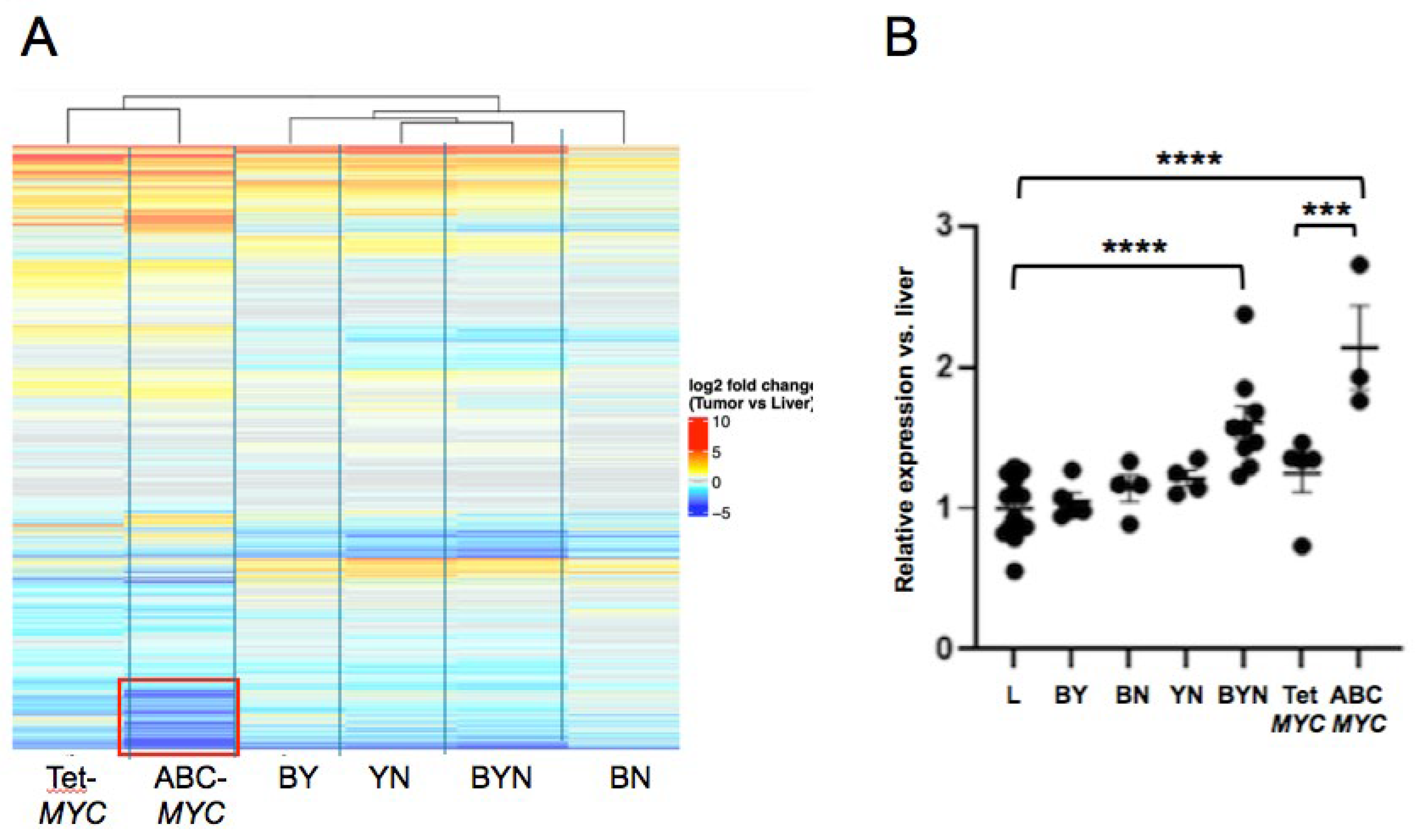
Figure 2.
Nuclear localization of endogenous β-catenin, Yap and NRF2. Nuclear and cytoplasmic fractions from Tet-MYC livers (L) and tumors (T) were resolved by SDS-PAGE and subjected to immuno-blotting for the indicated proteins. Histone H3 and Gapdh were used as controls for protein loading and known nuclear and cytoplasmic localization, respectively.
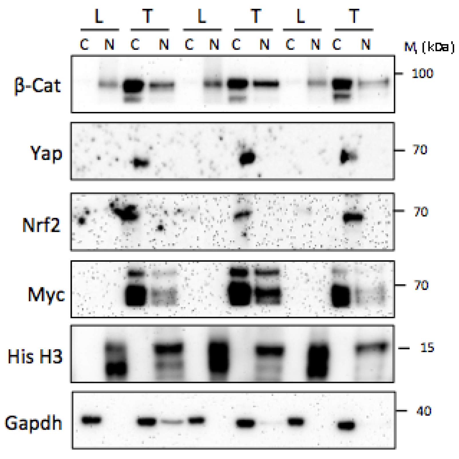
Table 3.
Summary of murine hepatoblastoma cell lines.
| Name of cell lines(s) | Relevant mutations de-regulated oncogenes, and/or tumor suppressors |
Comments | Reference(s) |
|---|---|---|---|
| MHB-2 and others | H-Ras, B-Raf, EGFR ~20%: Ctnnb1 & Apc mutations in older mice |
DEN/PB-treated mouse*, DEN-induced only resemble HCCs, non-tumorigenic | [62,127,128] |
| BY 1-5, BY21 | β−catenindel90, YAPS127A | Immortalized via Crspr mutagenesis of Cdkn2a | [34] |
| BYN 1-3 | β−catenindel90, YAPS127A, NRF2L30P | Immortalized via Crspr mutagenesis of Cdkn2a | [34] |
| BN 1,2 | β−catenindel90, NRF2L30P | Immortalized via Crspr mutagenesis of Cdkn2a | [35] |
| YN1-3 | YAPS127A, NRF2L30P | Immortalized via Crspr mutagenesis of Cdkn2a | [35] |
| NEJF1,2,3,4,5,6,10 | ABC-MYC | [20] |
*DEN = diethylnitrosamine, PB = phenobarbitol.
Table 4.
The advantages and disadvantages of current human and murine HB cell lines.
| Species | Advantages | Disadvantages |
|---|---|---|
| Human |
|
|
| Mouse |
|
|
Disclaimer/Publisher’s Note: The statements, opinions and data contained in all publications are solely those of the individual author(s) and contributor(s) and not of MDPI and/or the editor(s). MDPI and/or the editor(s) disclaim responsibility for any injury to people or property resulting from any ideas, methods, instructions or products referred to in the content. |
© 2025 by the authors. Licensee MDPI, Basel, Switzerland. This article is an open access article distributed under the terms and conditions of the Creative Commons Attribution (CC BY) license (http://creativecommons.org/licenses/by/4.0/).
Copyright: This open access article is published under a Creative Commons CC BY 4.0 license, which permit the free download, distribution, and reuse, provided that the author and preprint are cited in any reuse.


