
Submitted:
09 November 2025
Posted:
12 November 2025
You are already at the latest version
Abstract
Background/Objectives: The cannabidiol (CBD)-rich cannabis extract (CAN296) exhibits potent anti-inflammatory and anticancer activity with a favorable safety profile, making it a promising candidate for the management of inflammatory, precancerous, and malignant conditions, particularly for diseases with limited treatment options such as oral lichen planus (OLP), oral manifestations of graft-versus-host disease (oGVHD), and oral squamous cell carcinoma (OSCC). However, cannabis extracts in general, including CAN296, are characterized by high lipophilicity and poor aqueous solubility, which pose significant challenges to formulation stability and bioavailability. This study aimed to develop a stable Tween-based nanoemulsion of cannabis extract optimized for oral mucosal delivery. Methods:
Ethanol-dissolved cannabis extract was nanoemulsified with Tween 80/Span 80 surfactant blends at varying concentrations up to 4000 µg/mL and 1% total surfactant. Formulations were visually inspected for physical stability over an 8-week period. Selected emulsions were analyzed by dynamic light scattering (DLS) for droplet size and polydispersity and by transmission electron microscopy (TEM) for morphology. Additional assays included static contact angle (SCA) measurements for wettability, temperature-dependent stability testing (at 25 °C vs. 4 °C for 30 days), and an in vitro release study using a dialysis membrane, complemented by scanning electron microscopy (SEM) to visualize droplet deposition on the membrane surface. Results: Nanoemulsions containing ≥80% Tween 80 (1% surfactant) successfully incorporated cannabis extract up to 800 µg/mL, clear at 400 µg/mL, and turbid but homogeneous at 800 µg/mL, forming stable dispersions with improved nanoscale uniformity. DLS and TEM confirmed uniform spherical morphology, while SCA analysis indicated optimal droplet cohesion and wettability. Storage stability was maintained for 30 days at 4 °C, and in vitro dialysis studies revealed strong membrane association with limited diffusion, further supported by SEM visualization of membrane-bound aggregates. These findings suggest a favorable mucoadhesive potential for oral application. Conclusion: A Tween-dominant (≥80%) nanoemulsion stably incorporated CBD-rich extracts up to 800 µg/mL, well above therapeutic levels. The optimized system showed nanoscale uniformity, improved stability at 4 °C, and significant membrane retention, supporting its potential as a mucoadhesive platform for targeted oral delivery in immune-mediated and precancerous conditions.
Keywords:
cannabidiol
; cannabis extract
; nanoemulsion
; oral mucosal delivery
; oral lichen planus
; graft-versus-host disease
; head and neck squamous cell carcinoma
; immunomodulation
1. Introduction:
Cannabis-derived extracts rich in cannabidiol (CBD) possess substantial therapeutic potential for immune-mediated and oncologic oral conditions due to their anti-inflammatory, immunomodulatory, and pro-apoptotic properties [1,2,3,4,5,6]. CBD-rich cannabis extracts can modulate immune responses by various mechanisms. Several studies showed suppression of the activation of Cluster of Differentiation 4 helper T cells (CD4⁺) and Cluster of Differentiation 8 cytotoxic T cells (CD8⁺) T-cells, downregulation of pro-inflammatory cytokines such as Tumor necrosis factor alpha (TNF-α) and Interferon gamma (IFN-γ), and inhibition of the expression of cytotoxic effector molecules, including granzyme B, perforin, and Fas ligand [7,8,9]. Furthermore, CBD-rich cannabis extracts have been shown to promote selective apoptosis in malignant oral epithelial cells in head and neck squamous cell carcinoma (HNSCC) models [10,11,12,13,14,15]. Enrichment with the minor cannabinoid cannabichromene (CBC) further enhanced the cytotoxicity in HNSCC models through synergistic mechanisms [4].
Building on these findings, the biological effects of cannabis extracts are primarily mediated through the endocannabinoid system (ECS), a complex regulatory network that orchestrates critical physiological functions, including immune regulation, inflammation, nociception, and epithelial homeostasis [16,17,18,19,20]. The classical ECS comprises the cannabinoid receptor type 1 (CB1) and 2 (CB2), the endogenous ligands anandamide (AEA) and 2-arachidonoylglycerol (2-AG), and the enzymes responsible for their synthesis and degradation. The extended ECS further encompasses additional molecular targets, such as orphan G–protein–coupled receptors, transient receptor potential (TRP) channels, and peroxisome proliferator–activated receptors (PPARs), including PPARγ [21,22,23,24,25,26,27]. Through these pathways, CBD-rich extracts exert immunomodulatory, anti-inflammatory, and pro-apoptotic effects by modulating ECS receptors, reducing cytokine secretion, and suppressing cytotoxic mediators [7,8,12,13].
In our previous studies, we demonstrated that the CBD-rich cannabis extract (CAN296) and CBD:CBC formulations exert potent immunomodulatory and pro-apoptotic effects, showing therapeutic efficacy in T cell–mediated oral disorders such as oral lichen planus (OLP) and oral graft-versus-host disease (oGVHD), as in HNSCC models [4,28].
Building upon these therapeutic observations, it is essential to understand the clinical relevance of the target diseases. OLP and oGVHD are chronic, immune-mediated disorders that impair quality of life and carry a significant risk of malignant transformation [29,30]. OLP is a T-cell-driven inflammatory disease of the oral mucosa that presents as reticular, erosive, or plaque-like lesions [31], with a global prevalence of 0.5–4% [32]. It is classified as an oral potentially malignant disorder, with transformation to OSCC influenced by lesion type, duration, and patient-specific factors [33,34].
Chronic graft-versus-host disease (GVHD), a significant complication following allogeneic hematopoietic stem-cell transplantation (HSCT), occurs when donor T cells attack host tissues, resulting in persistent inflammation and tissue damage [35,36]. Oral manifestations often resemble OLP, including lichenoid lesions, mucosal atrophy, and ulcerations [37,38], and oGVHD similarly carries a malignant potential [39,40]. Both disorders share a common immunopathological basis characterized by T-cell dysregulation and cytotoxic epithelial injury. Activated CD4⁺ and CD8⁺ T cells secrete IFN-γ and TNF-α, while cytotoxic CD8⁺ T cells express Fas ligand (FasL) and release perforin and granzyme B at the epithelial basement membrane, inducing keratinocyte apoptosis [29,41,42,43,44]. Chronic immune activation, oxidative stress, and epithelial instability contribute to genomic damage and increased susceptibility to malignant transformation [45,46,47]. These mechanisms link OLP and oGVHD to HNSCC, the most common malignancy of the head and neck region [48]. HNSCC arises from the mucosal epithelium of the oral cavity, pharynx, or larynx and represents the sixth most common cancer globally, accounting for approximately 900,000 cases and over 400,000 deaths annually [49,50,51,52].
Current treatment modalities for OLP and oGVHD primarily rely on immunosuppressive agents. Topical corticosteroids (clobetasol, triamcinolone) are the first-line treatment for OLP and effectively reduce lesion severity and pain [53,54]. However, long-term use is associated with adverse effects, including mucosal atrophy, candidiasis, and reduced therapeutic efficacy due to tachyphylaxis [55,56]. Systemic corticosteroids, often used in severe cases of OLP and oGVHD, carry additional risks such as osteoporosis, diabetes, and increased susceptibility to infections [57,58]. Despite their efficacy, these agents do not address the chronicity or malignant potential of these disorders, underscoring the need for safer, targeted therapies capable of modulating immune dysregulation while minimizing toxicity. Calcineurin inhibitors, such as tacrolimus (TAC) and cyclosporine, offer an alternative mechanism of action by inhibiting T-cell activation through the suppression of interleukin-2 (IL-2) transcription [59,60]. While these agents have shown efficacy in managing OLP and oGVHD, their use is limited by systemic toxicity, including nephrotoxicity, hypertension, and neurotoxicity [61,62]. Moreover, the chronic and relapsing nature of both conditions often necessitates prolonged treatment, further exacerbating the risk of adverse effects [63,64]. Notably, conventional therapies fail to address the precancerous and malignant transformation potential of OLP and oGVHD.
Cannabinoids, particularly those found in CBD-type cannabis extracts, have shown potent antitumor effects in preclinical models of various cancers, including HNSCC (59, 60). Among over 500 bioactive compounds in Cannabis sativa, CBD and Δ⁹-tetrahydrocannabinol (THC) are the most studied components, while other cannabinoids and terpenes contribute to therapeutic activity via the “entourage effect” [65,66,67].
Cannabinoids exert anticancer effects through multiple mechanisms, including induction of apoptosis [68,69,70,71], inhibition of angiogenesis [72,73], suppression of tumor proliferation (67, 69), and modulation of the tumor microenvironment [74,75,76]. Notably, CBD-type extracts selectively target malignant cells while sparing healthy tissue [4,5], a property particularly relevant for oral diseases such as OLP and oGVHD, where chronic inflammation heightens carcinogenic risk.
This selectivity, together with the shared pathophysiology of OLP and oGVHD, including chronic inflammation, T-cell dysregulation, and malignant potential, supports the rationale for developing CBD-rich formulations. Accordingly, the present study focused on creating a stable, surfactant-based nanoemulsion system to enable localized mucosal delivery and sustained bioavailability of a CBD-rich extract.
For effective local treatment of oral lesions, formulation strategies must ensure mucosal compatibility, physical stability, and bioavailability. Due to the lipophilicity of cannabinoids in CBD-type extracts, aqueous and ethanol-based systems pose challenges for the delivery of these compounds. Nanoemulsion platforms can overcome these limitations by enhancing solubility and tissue penetration [77,78,79,80,81].
We hypothesized that incorporating the CBD-rich cannabis extract CAN296 into a Tween-dominant nanoemulsion system (≥80% Tween 80) would result in superior physicochemical performance compared with the crude extract oil, specifically by improving nanoscale droplet uniformity, wettability, and mucosal membrane interaction. These properties are expected to enhance local bioavailability and stability, thereby overcoming the limitations associated with the native, lipophilic extract. Accordingly, this study aimed to develop and characterize a stable, ethanol-compatible CBD-rich nanoemulsion optimized for oral mucosal application. The optimized formulation will serve as the basis for subsequent biological assays to evaluate its immunomodulatory and cytotoxic potential in oral inflammatory and precancerous conditions, including OLP, oGVHD, and early-stage HNSCC.
2. Results
2.1. Cannabis Emulsions with Up to 800 µg/mL Load Are Stabilized by 1% Surfactant Containing ≥80% Tween
Initial attempts using the crude, non-formulated cannabis extract (without surfactant) resulted in immediate phase separation, confirming that the native extract cannot form a stable aqueous dispersion. Therefore, subsequent experiments focused on determining whether a surfactant-based system could achieve and maintain nanoscale stability. To define the optimal formulation for long-term physical stability of cannabis emulsions, we systematically evaluated the influence of varying Tween:Span ratios on emulsion stability across a range of cannabis concentrations. Emulsions were prepared using a constant 1% surfactant system, with Tween 80 and Span 80 in different ratios (0:100 to 100:0), and cannabis extract was added at concentrations ranging from 0 to 4000 µg/mL. Each homogenized formulation was stored at room temperature and visually monitored for phase separation and clarity over an 8-week observation period (Figure 1).
Formulations without surfactants exhibited immediate phase separation at all cannabis concentrations, confirming that surface-active agents are essential for dispersion stability. When using mixed-surfactant systems with 35% or 65% Tween, the emulsions exhibited limited short-term stability, characterized by a turbid (milky) appearance. Specifically, formulations containing 400 µg/mL cannabis remained stable for up to 3 weeks, while those with 800 µg/mL remained stable for only 2 weeks before phase separation occurred. In contrast, emulsions containing ≥80% Tween (i.e., 80:20 and 100:0 Tween:Span) demonstrated prolonged physical stability throughout the 8-week observation period. At 400 µg/mL cannabis concentration, these formulations remained optically clear (i.e., transparent or translucent), while emulsions containing 800 µg/mL appeared turbid but homogeneous (milky), with no visible phase separation. None of the emulsions, regardless of the Tween:Span ratio, could stably incorporate cannabis concentrations of ≥ 1200 µg/mL; all such formulations exhibited visible oil phase separation during storage.
These results confirm that a 1% surfactant system requires at least 80% Tween 80 to achieve long-term stability for cannabis concentrations of up to 800 µg/mL. Beyond this threshold, the system's solubilization capacity is exceeded, indicating a formulation limit under the tested conditions. These findings guided our selection of the 80% Tween formulation for subsequent biological assays.
2.2. Dynamic Light Scattering Analysis Confirms Stability Limit of 800 µg/mL Cannabis in 1% Surfactant Emulsions with ≥80% Tween
To characterize emulsion stability in terms of droplet size distribution, we performed DLS analysis on representative formulations containing 1% surfactant (80% Tween, 20% Span) and varying cannabis extract concentrations (400, 800, 2000, and 4000 µg/mL). For comparison, two additional formulations lacking surfactant (0% Tween, 0% Span) were tested at cannabis concentrations of 2000 and 4000 µg/mL (Figure 2).
The formulation containing 400 µg/mL cannabis with 80% Tween exhibited a small and uniform particle size distribution, with a Z-average of 312.6 nm, a primary peak at 195.2 nm, and a moderate polydispersity index (PDI) (0.3647), indicating physical stability. The sample containing 800 µg/mL cannabis remained physically stable but showed larger, more heterogeneous particles (Z-average = 536.3 nm; primary peak = 279.9 nm; PDI = 0.4865). These two concentrations were classified as “Stable.”
At higher cannabis concentrations (2000 and 4000 µg/mL) with 80% Tween, the emulsions were unstable, with Z-averages exceeding 1000 nm and higher PDIs, including 0.8595 at 2000 µg/mL. These were marked as "Unstable". The control formulations without surfactant exhibited smaller average particle sizes (193.1 and 249.0 nm). These values reflect unstable multi-population distributions due to rapid phase separation and a lack of homogeneity; therefore, both samples were categorized as "Highly Unstable."
These findings confirm that surfactants are required to form uniform and stable cannabis emulsions, and that the maximum cannabis load that can be effectively stabilized with 1% Surfactant containing ≥80% Tween is approximately 800 µg/mL. This DLS-based analysis supports and extends the previously reported visual stability observations, providing particle size and polydispersity parameters that define the formulation limits under the tested conditions.
2.3. Transmission Electctron Microscopy (TEM) Reveals Progressive Particle Homogeneity with Increasing Tween Content
To visualize the morphological characteristics of the cannabis emulsions, representative samples containing 400 µg/mL cannabis extract and formulated with varying Tween:Span ratios were imaged using TEM. All emulsions were prepared with 1% total surfactant, except for the control sample, which contained no surfactant (0% Tween, 0% Span). Images were acquired at a fixed scale of 200 nm (Figure 4).
TEM micrographs revealed marked differences in droplet morphology and distribution depending on the surfactant composition. In the absence of surfactant (Panel A), significant, irregularly shaped oil droplets were observed, displaying high heterogeneity and frequent coalescence. Formulations containing low Tween content (35% Tween, Panel B; 65% Tween, Panel C) exhibited smaller droplets but retained notable morphological heterogeneity, with numerous irregular aggregates and variable droplet sizes.
In contrast, emulsions formulated with ≥80% Tween showed progressively improved particle uniformity. The 80% Tween sample (Panel D) produced more spherical, monodisperse droplets with reduced polydispersity. The 100% Tween formulation (Panel E) showed a dense field of uniformly distributed, nanoscale droplets with no visible aggregation. These findings are consistent with the DLS data, confirming that a higher Tween content promotes the formation of homogeneous nanoemulsions at a fixed cannabis concentration of 400 µg/mL.
2.4. Static Contact Angle (SCA) Measurements Reveal Nonlinear Wettability with Maximal Cohesion at 800 µg/mL
To be effective, nanoemulsions should exhibit increased wettability, which reflects improved spreading and adhesion on biological surfaces, thereby enhancing drug delivery and absorption. To assess surface wettability across different concentrations of CBD-rich cannabis, SCA measurements were performed on glass substrates using a drop shape analyzer (DSA). Droplets (5 µL) of formulations containing 0, 400, 800, and 1200 µg/mL cannabis extract were analyzed using the sessile-drop method (Figure 5).
The control formulation (0 µg/mL) exhibited a low SCA (~19°), indicating high spreading. At 400 µg/mL, the angle slightly increased (~23°), while 800 µg/mL yielded the highest value (~28°), reflecting reduced wettability but still within the range of good surface spreading. At 1200 µg/mL, the SCA returned to ~19°, indicating the greatest wettability among the concentrations tested. Statistical analysis confirmed that the 800 µg/mL formulation exhibited significantly higher contact angles compared to both 0 µg/mL and 1200 µg/mL (p < 0.01) (Panel A), indicating reduced wettability and increased droplet cohesion at this concentration. Together, these results demonstrate a non-linear, concentration-dependent relationship between extract content and surface spreading, with maximal droplet cohesion observed at an extract concentration of 800 µg/mL. A representative droplet image of the 800 µg/mL formulation is shown in Panel B.
2.5. Enhanced Stability of Nanoemulsions at 4 °C Compared to Room Temperature After 30 Days
To evaluate the impact of storage temperature on nanoemulsion stability, previously examined stable formulations containing cannabis extract (400 µg/mL and 800 µg/mL) were stored for 30 days at two temperatures. Samples were maintained at room temperature (~25 °C) and under refrigeration (4 °C), and droplet size distributions were analyzed by DLS (Figure 6). The largest detected droplet population (nm) was used as an indicator of aggregation or phase separation.
After 30 days, emulsions stored at 4 °C maintained droplet sizes close to their initial range (up to ~600 nm). In contrast, samples kept at room temperature exhibited a marked increase in size, with droplets expanding to over 4000 nm. Statistical analysis confirmed a significant interaction between storage temperature and cannabis concentration (two-way ANOVA, temperature × concentration; ****P < 0.0001). Pairwise comparisons indicated highly significant differences between storage conditions at 400 µg/mL (****P < 0.0001) and significant differences at 800 µg/mL (*P = 0.0259). Together, these results demonstrate that storage at 4 °C effectively minimized droplet coalescence and aggregation, preserving the nanoemulsion structure across both concentrations.
2.6. Significant In Vitro Retention of Cannabis Extract Nanoemulsion on Dialysis Membrane Suggests Mucoadhesive Potential
Surface adhesion and prolonged residence time can enhance local bioavailability for topical or mucosal drug delivery systems. To assess this property, the interaction of cannabis nanoemulsion with a dialysis membrane in an in vitro diffusion experiment was performed over 168 hours using a formulation containing 800 µg/mL cannabis extract (Figure 7). The extract-loaded nanoemulsion was placed in the donor compartment of the dialysis device, while the blank nanoemulsion was added to the receiver compartment. Eleven sealed capsules were incubated, and samples were destructively collected from both compartments at ten predefined time points and stored frozen. At the end of the experiment, ultra-high-performance liquid chromatography (UHPLC) was used to analyze all samples and quantify the CBD content. Extract concentrations were calculated by adjusting the CBD values based on the extract’s known CBD content (55%).
Over the course of the experiment, the donor compartment exhibited a gradual decrease in cannabis extract concentration, from 800 μg/mL at time zero to 116 μg/mL at 168 hours. In contrast, the receiver compartment showed only a modest cumulative increase, reaching 111.6 µg/mL by day 7. An increasing discrepancy between donor loss and receiver accumulation was observed throughout the time course, indicating that a significant portion of the extract was retained at the dialysis membrane. This membrane-retained fraction was calculated as the difference between donor depletion and receiver gain, and is shown as the purple bars in Figure 7.
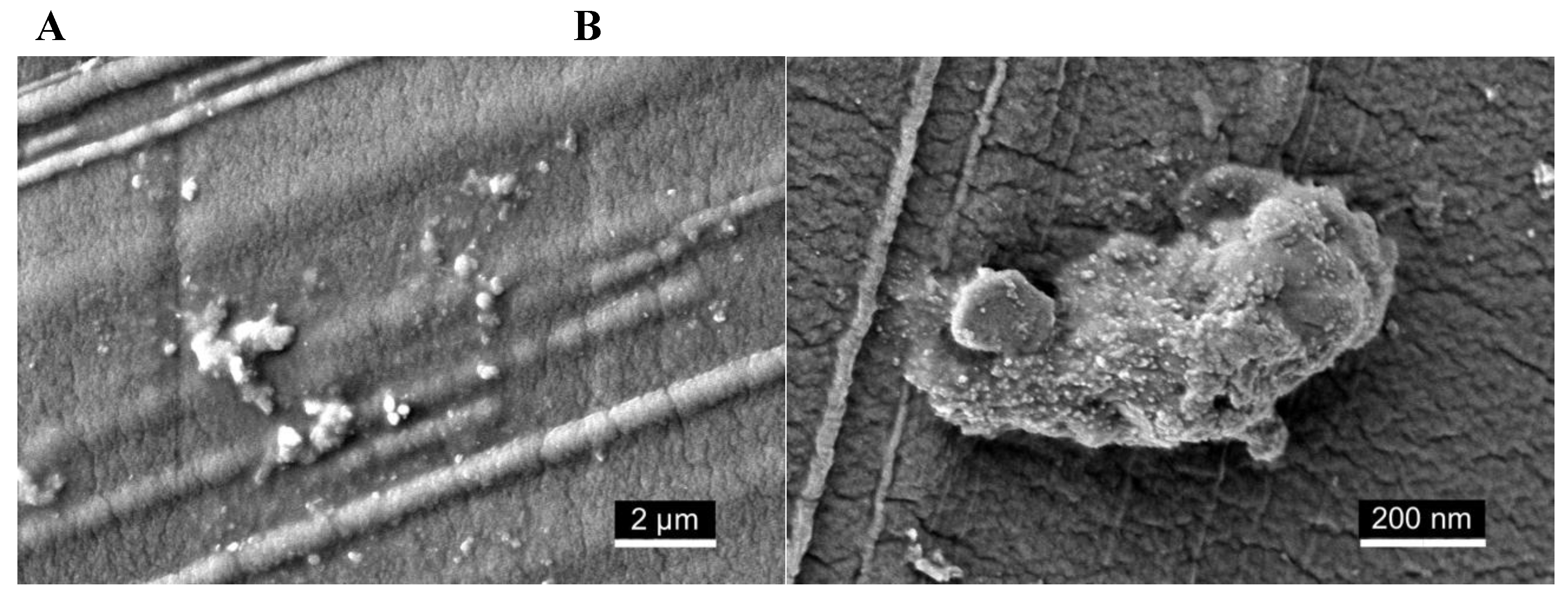
2.7. Scanning Electron Microscopy (SEM) Visualization of Nanoemulsion Aggregates on Dialysis Membrane Surface
To visualize the localization of cannabis extract nanoemulsion on the dialysis membrane surface, an additional dialysis capsule was prepared under the same conditions described in Section 4.8, using formulations containing either 400 or 800 μg/mL of cannabis extract (Figure 8). After 24 hours of incubation, the capsule was disassembled, and the membrane was gently removed. It was then rinsed with distilled water, air-dried, and sputter-coated with a thin layer of iridium (2–3 nm) for SEM imaging.
SEM imaging qualitatively revealed deposition of nanoemulsion material on the membrane surface. At lower magnification (Figure 8A), scattered spherical droplets and small clusters were visible across the surface. At higher magnification (Figure 8B), visually denser regions of submicron aggregates were observed, forming irregular, closely packed structures. The intrinsic pore structure of the membrane was not resolved under the imaging conditions, likely due to dehydration and sputter coating, which may have obscured finer surface features. These observations are descriptive and based on qualitative visual assessment.
3. Discussion
Formulating a CBD-rich cannabis extract into a physically stable nanoemulsion required careful optimization of surfactant composition, extract concentration, and energy input to achieve nanoscale uniformity and mucosal compatibility. The optimized formulation, containing 1% total surfactant with ≥80% Tween 80 and ethanol as a cosolvent, demonstrated long-term physical stability with uniform droplet distribution, favorable wettability, and enhanced membrane interaction, supporting its suitability for oral mucosal delivery.
While our prior research demonstrated the anticancer effects of CBD-type cannabis extracts in HNSCC [4] and their immunomodulatory activity in OLP and oGVHD[28], the present work focused on overcoming the pharmaceutical challenge of formulating a physically stable Tween/Span-based nanoemulsion system incorporating ethanol as a cosolvent, suitable for oral mucosal application of CBD-rich cannabis extracts.
Cannabis-derived formulations are most commonly delivered via oral ingestion, inhalation, or sublingual sprays, each with notable drawbacks. Oral and edible products exhibit poor and variable bioavailability (6–20%) due to extensive first-pass metabolism and the high lipophilicity of cannabinoids [82,83,84]. Inhalation provides rapid systemic absorption but is associated with inconsistent dosing, short duration of effect, and potential respiratory irritation or airway inflammation [85,86]. Sublingual tinctures and sprays partially bypass hepatic metabolism, yet they often rely on ethanol-based vehicles that can cause mucosal irritation and provide limited residence time for local retention [82,87,88]. In contrast, the nanoemulsion platform developed in this study improves solubility, enhances mucosal wetting and adhesion, and enables localized, noninvasive delivery suitable for chronic inflammatory and precancerous oral conditions.
The nanoemulsion in this study was prepared using a MICCRA high-shear disperser, which applies controlled mechanical shear to achieve fine droplet dispersion without the need for ultrasonic or high-pressure homogenization. This moderate-energy method preserved the integrity of the cannabis extract, ensured mucosal biocompatibility, and maintained uniform droplet size and physical stability. In contrast, high-energy techniques such as ultrasonic cavitation and high-pressure homogenization have produced sub-200 nm translucent cannabis nanoemulsions with extended stability, but at the cost of elevated surfactant concentrations, which—although promoting smaller droplets and optical clarity—may be associated with reduced biocompatibility, potential mucosal irritation, and thermal or mechanical degradation of sensitive phytochemicals within the extract [89,90,91,92].
Initial attempts with the crude, non-formulated cannabis extract revealed that it could not be maintained as a stable aqueous dispersion, exhibiting immediate phase separation even under high shear. Consequently, comparative testing between the extract alone and the formulated nanoemulsion was not feasible. The study, therefore, focused on evaluating the physicochemical feasibility and performance of surfactant-based nanoemulsions as a prerequisite for subsequent biological testing.
Our findings demonstrate that emulsifying a cannabis extract with a Tween-dominant surfactant system enables long-term physical stability and the formation of nanoscale particles. A 1% total surfactant concentration containing at least 80% Tween 80 was required to stabilize cannabis emulsions at concentrations up to 800 µg/mL. At 400 µg/mL, the emulsions remained optically clear; however, at 800 µg/mL, they appeared milky but homogeneous, with no phase separation. Concentrations ≥1200 µg/mL exceeded the solubilization capacity of the system, resulting in visible phase separation and establishing a clear formulation limit under the tested conditions.
DLS and TEM corroborated these visual observations. Emulsions containing 400–800 µg/mL cannabis extract with 80% Tween displayed droplet sizes below 600 nm with moderate polydispersity indices, confirming the formation of a nanoscale, relatively uniform dispersion. In contrast, higher cannabis loads produced larger, heterogeneous populations and early aggregation behavior, as reflected in both DLS intensity/volume distributions and TEM images.
In this study, the formulation achieved nanoscale dispersions at extract concentrations of up to 800 µg/mL using low-energy homogenization and only 1% total surfactant, emphasizing ethanol compatibility, mucosal tolerability, and suitability for localized oral application. In contrast, a buccal CBD nanoemulsion system prepared with Tween 80/Labrasol exhibited rapid dissolution and stability in simulated saliva, thereby favoring systemic absorption over local mucosal retention [93].
SCA measurements provided further insight into the formulation’s interfacial behavior on solid surfaces. Both the 400 and 800 µg/mL formulations exhibited SCA within the optimal range for wetting the oral mucosa [94], suggesting favorable surface spreading and adhesion. Wettability followed a concentration-dependent pattern, with slightly increased droplet cohesion observed at 800 µg/mL, indicating altered interfacial dynamics at higher extract loads. These findings support the hypothesis that increasing the concentration of cannabis extract modulates droplet spreading and cohesion, factors that may enhance mucosal adherence and localized retention during buccal application.
Storage studies highlighted the effect of temperature on emulsion stability. After 30 days, emulsions stored at 4 °C maintained droplet sizes close to their initial range (up to ~600 nm). In contrast, samples kept at 25 °C exhibited marked coalescence (>4000 nm). Refrigerated storage mitigates coalescence and ripening in CBD nanoemulsions [95], consistent with colloid theory, which identifies Ostwald ripening as the principal coarsening mechanism in Tween/Span systems and outlines strategies to suppress it [96,97].
Comparable cannabinoid nanoemulsion systems demonstrated enhanced physicochemical stability under refrigerated storage, maintaining sub-200 nm droplet sizes and chemical integrity over extended periods. In contrast, room temperature conditions promoted droplet coalescence and cannabinoid degradation [95]. Refrigeration effectively minimizes kinetic instability and Ostwald ripening phenomena in surfactant-based cannabinoid formulations. This is consistent with stabilization effects also documented in lipid-based oral emulsions and intravenous hemp oil nanoemulsions. Together, these findings establish 4°C storage as a broadly applicable preservation strategy for cannabinoid delivery systems.
In vitro release testing using a dialysis membrane revealed a gradual depletion of the donor over 168 hours, with only modest accumulation in the receiver compartment. The discrepancy between donor loss and receiver gain, quantified as the membrane-retained fraction, indicates that a substantial portion of the extract remained associated with the dialysis membrane under the test conditions. This behavior suggests meaningful interaction between the nanoemulsion droplets and the membrane surface, consistent with mucoadhesive-like retention that could support localized delivery and sustained release on oral mucosal tissues.
Although differing in composition, mucoadhesive oral delivery systems have demonstrated that prolonged mucosal residence and controlled drug release enhance therapeutic outcomes in inflammatory oral diseases [98,99,100]. SEM analysis revealed membrane-associated retention and surface aggregation of the nanoemulsion, consistent with the therapeutic goal of maintaining localized drug presence while minimizing systemic exposure. SEM images showed scattered spherical droplets and densely packed submicron aggregates on the membrane surface, providing clear morphological evidence of deposition and supporting the formulation’s potential for sustained mucosal residence.
4. Materials and Methods
4.1. Phytocannabinoid Extraction and Sample Preparation
Air-dried Type III high-CBD cannabis CAN296 was obtained from a licensed Israeli medical cannabis distributor. Inflorescences were extracted in ethanol and decarboxylated by heating at 130 °C. The resulting extract was dissolved in dimethyl sulfoxide (DMSO; Sigma-Aldrich, St. Louis, MO, USA) and stored at –20 °C.
Phytocannabinoid profiling was performed using an UHPLC system coupled with a Q Exactive Focus Hybrid Quadrupole-Orbitrap mass spectrometer (Thermo Scientific, Bremen, Germany), as previously described [4]. Identification and absolute quantification of cannabinoids were achieved using analytical standards and external calibration curves (Thermo Scientific, Bremen, Germany), following previously established protocols [101].
4.2. Formulation Preparation
Cannabis nanoemulsions were prepared by first dissolving the CAN296 extract in 25% ethanol, which served as a cosolvent to aid in the dispersion of the lipophilic extract. The extract solution was then emulsified with 1% surface-active agent composed of Tween 80 and Span 80 (Sigma-Aldrich, St. Louis, MO, USA) at varying ratios. Cannabis concentrations ranged from 0 to 4000 µg/mL in 400 µg/mL increments. After 5 minutes of stirring, the mixtures were homogenized using a MICCRA D-9 disperser (MICCRA GmbH, Heitersheim, Germany) at 11,000 rpm for 3 minutes.
4.3. Stability Evaluation of Emulsions by Visual Inspection
Emulsion stability was evaluated through systematic visual inspection under consistent ambient lighting conditions. Samples were stored in clear, sealed vials at room temperature (~25°C) and assessed weekly over an 8-week period (days 7, 14, 21, 28, 35, and 42). Each formulation was observed for signs of turbidity, creaming, sedimentation, and phase separation. Emulsions were classified as Stable if they remained uniform (transparent or turbid) with no visible separation; Unstable if they exhibited droplet aggregation or partial separation with increasing turbidity or layering; and Highly Unstable if they underwent complete separation or sedimentation within the first week. These assessments served as a primary tool to evaluate formulation robustness across different surfactant ratios and cannabis concentrations.
4.4. Dynamic Light Scattering Analysis
Droplet size, PDI, and colloidal stability of cannabis nanoemulsions were assessed using DLS. Formulations contained 1% surfactant (80% Tween 80 / 20% Span 80) with 800, 2000, and 4000 µg/mL cannabis, along with surfactant-free controls. Measurements were performed at 25 °C on a Zetasizer ZS XPLORER (Malvern Panalytical, UK) in triplicate. Z-average, PDI, and intensity-weighted distributions were recorded and analyzed with ZS XPLORER software. Results were interpreted in conjunction with visual stability data to classify emulsions as stable, unstable, or highly unstable. It should be noted that DLS provides an intensity-weighted average that can overrepresent larger droplet populations in polydisperse systems; therefore, the measured Z-average and PDI values reflect relative rather than absolute particle sizes.
4.5. TEM Imaging
Morphological analysis of nanoemulsion particles was performed using TEM. Samples were adsorbed onto Formvar-carbon-coated copper grids (EMS, 200 mesh) and negatively stained with 2% uranyl acetate to enhance image contrast. After staining, the grids were allowed to air-dry before imaging. TEM was performed using a JEOL JEM-1400 Plus transmission electron microscope (JEOL Ltd., Tokyo, Japan) operated at 100 kV. High-resolution images were acquired using a Gatan Orius 600 CCD camera, allowing for detailed visualization of droplet morphology, particle size, and aggregation patterns across various surfactant formulations.
4.6. SCA Measurement
Wettability of the CBD-rich cannabis nanoemulsion was evaluated using a KRÜSS Drop Shape Analyzer (DSA; KRÜSS GmbH, Hamburg, Germany) equipped with ADVANCE software (v1.11.2.25901). Glass microscope slides (Thermo Scientific, Germany) were cleaned with ethanol, rinsed with deionized water, and air-dried before use. A 5 µL droplet of each formulation (0, 400, 800, and 1200 µg/mL) was dispensed with a microsyringe, and SCA values were measured immediately using the sessile drop method. Left- and right-angle measurements were automatically averaged, with six replicates per sample under ambient laboratory conditions (~25 °C).
4.7. Phytocannabinoid Identification and Quantification
Phytocannabinoid analyses were performed using a Thermo Scientific UHPLC system coupled with a Q ExactiveTM Focus Hybrid Quadrupole-Orbitrap mass spectrometer (Thermo Scientific, Bremen, Germany). Identification and absolute quantification of phytocannabinoids were performed using analytical phytocannabinoid standards and Cannabis samples at preset concentrations, as previously described [101].
4.8. In Vitro Release Kinetics Using a Dialysis Membrane System
The release of CBD from the nanoemulsion (800 µg/mL) was evaluated using a dialysis kit (Pur-A-Lyzer Mini 6000, 1 kDa molecular weight cutoff; Sigma-Aldrich, Merck KGaA, Darmstadt, Germany) with a 1 kDa pore membrane. The cannabis-loaded nanoemulsion was placed inside the membrane chamber, and a blank nanoemulsion was added to the outer compartment. Twelve sealed vials were incubated at room temperature under constant agitation. At ten predefined time points (0, 0.5, 1, 2, 4, 8, 24, 48, 72 hours, and 7 days), samples were collected separately from the internal (donor) and external (receiver) compartments. UHPLC was used to analyze all samples for CBD concentration (Section 4.7), and release values were corrected based on the extract’s CBD content (~50%).
4.9. SEM Imaging
Samples were examined using an Extra-High-Resolution Scanning Electron Microscope (Magellan 400 L, FEI Company, Hillsboro, OR, USA) after plasma sputter-coating with a thin iridium layer (2–3 nm) to enhance surface conductivity. Each membrane sample was imaged in three representative regions at varying magnifications to assess surface morphology and potential pore obstruction. SEM images were processed using ImageJ software (NIH, USA) for qualitative visualization. Uniform brightness and contrast adjustments were applied to all images to enhance the visibility of surface features. Thresholding was used to improve the contrast between droplets and the membrane background, without compromising image integrity.
4.10. Statistical Analysis
All statistical analyses were conducted using GraphPad Prism version 10.6.1 for macOS (GraphPad Software, San Diego, CA, USA). Inferential statistical analyses were performed where applicable to assess differences between formulations and conditions. SCA data, storage stability measurements (droplet size), and in vitro release profiles were analyzed using one-way or two-way analysis of variance (ANOVA) with appropriate post hoc tests (Tukey’s or Šidák’s multiple comparisons), as specified. Data are presented as mean ± SD of at least three independent measurements unless otherwise noted. Visual inspection, DLS measurements, and TEM imaging were interpreted descriptively to evaluate qualitative trends in emulsion clarity, temporal stability, droplet size distribution, and polydispersity.
5. Conclusion
This research established a Tween-dominant nanoemulsion capable of stabilizing a robust concentration of CBD-rich cannabis extract. This optimized system remains stable under refrigeration, exhibits favorable wettability and membrane retention, and provides a physically stable, ethanol-compatible platform for oral mucosal delivery of cannabis extract. Future research should evaluate mucosal biocompatibility and in vivo retention to advance translational development for OLP, oGVHD, and early-stage HNSCC.
Author Contributions
Conceptualization, K.B., D.M., and O.B.; methodology, K.B.; validation, K.B., D.M., and O.B.; formal analysis, K.B.; investigation, K.B., G.M., and A.S.; resources, D.M. and O.B.; data curation, K.B.; writing—original draft preparation, K.B.; writing—review and editing, K.B., D.M., O.B., and S.P.; visualization, K.B.; supervision, D.M. and O.B.; project administration, K.B.; funding acquisition, D.M. and O.B. All authors have read and agreed to the published version of the manuscript.
Funding
This study was not supported by external funding.
Data Availability Statement
The data presented in this study are available on request from the corresponding author. The data are not publicly available due to ethical restrictions related to donor confidentiality.
Acknowledgments
The authors would like to thank Ohad Guberman for the extraction and sample preparation of the cannabis extract and synthetic cannabinoids, as well as Cannasoul Analytics and the Technion – Israel Institute of Technology for technical support and phytochemical profiling. The authors also gratefully acknowledge Seach Medical Group for providing the high-CBD strain CAN296 used in this study.
Declaration of Generative AI and AI-Assisted Technologies in the Writing Process
During the preparation of this work, the authors used ChatGPT (OpenAI, GPT-5, 2025) to assist in improving the clarity and readability of the text. After using this tool, the authors reviewed and edited the content as needed and take full responsibility for the final manuscript.
Conflicts of Interest
The authors declare no conflict of interest.
Abbreviations
The following abbreviations are used in this manuscript:
| 2-AG | 2-arachidonoylglycerol |
| ANOVA | Analysis of variance |
| AEA | N-arachidonoylethanolamine (anandamide) |
| CAN296 | CBD-rich cannabis extract (Type III strain) |
| CB1 | Cannabinoid receptor type 1 |
| CB2 | Cannabinoid receptor type 2 |
| CBC | Cannabichromene |
| CBD | Cannabidiol |
| CD4⁺ | Cluster of differentiation 4 helper T cells |
| CD8⁺ | Cluster of differentiation 8 cytotoxic T cells |
| DLS | Dynamic light scattering |
| DMSO | Dimethyl sulfoxide |
| DSA | Drop shape analyzer |
| ECS | Endocannabinoid system |
| GVHD | Graft-versus-host disease |
| HNSCC | Head and neck squamous cell carcinoma |
| HSCT | Hematopoietic stem cell transplantation |
| IFN-γ | Interferon gamma |
| IL-2 | Interleukin-2 |
| oGVHD | Oral graft-versus-host disease |
| OLP | Oral lichen planus |
| OSCC | Oral squamous cell carcinoma |
| PDI | Polydispersity index |
| PPAR | Peroxisome proliferator-activated receptor |
| PPARγ | Peroxisome proliferator-activated receptor gamma |
| SCA | Static contact angle |
| SD | Standard deviation |
| SEM | Scanning electron microscopy |
| TAC | Tacrolimus |
| TEM | Transmission electron microscopy |
| THC | Δ⁹-tetrahydrocannabinol |
| TNF-α | Tumor necrosis factor alpha |
| TRP | Transient receptor potential |
| UHPLC | Ultra-high-performance liquid chromatography |
References
- Nichols, J.M.; Kaplan, B.L.F. Immune Responses Regulated by Cannabidiol. Cannabis and cannabinoid research 2020, 5, 12–31. [Google Scholar] [CrossRef]
- Burstein, S. Cannabidiol (CBD) and its analogs: A review of their effects on inflammation. Bioorg Med Chem 2015, 23, 1377–1385. [Google Scholar] [CrossRef] [PubMed]
- Pisanti, S.; Malfitano, A.M.; Ciaglia, E.; Lamberti, A.; Ranieri, R.; Cuomo, G.; Abate, M.; Faggiana, G.; Proto, M.C.; Fiore, D.; Laezza, C.; Bifulco, M. Cannabidiol: State of the art and new challenges for therapeutic applications. Pharmacol Ther 2017, 175, 133–150. [Google Scholar] [CrossRef] [PubMed]
- Blal, K.; Besser, E.; Procaccia, S.; Schwob, O.; Lerenthal, Y.; Abu Tair, J.; Meiri, D.; Benny, O. The Effect of Cannabis Plant Extracts on Head and Neck Squamous Cell Carcinoma and the Quest for Cannabis-Based Personalized Therapy. Cancers (Basel) 2023, 15. [Google Scholar] [CrossRef]
- Massi, P.; Solinas, M.; Cinquina, V.; Parolaro, D. Cannabidiol as potential anticancer drug. British journal of clinical pharmacology 2013, 75, 303–312. [Google Scholar] [CrossRef]
- Billi, M.; Pagano, S.; Pancrazi, G.L.; Valenti, C.; Bruscoli, S.; Di Michele, A.; Febo, M.; Grignani, F.; Marinucci, L. DNA damage and cell death in human oral squamous cell carcinoma cells: The potential biological effects of cannabidiol. Arch Oral Biol 2025, 169, 106110. [Google Scholar] [CrossRef]
- Pertwee, R.G. The diverse CB1 and CB2 receptor pharmacology of three plant cannabinoids: delta9-tetrahydrocannabinol, cannabidiol and delta9-tetrahydrocannabivarin. British journal of pharmacology 2008, 153, 199–215. [Google Scholar] [CrossRef]
- McPartland, J.M.; Duncan, M.; Di Marzo, V.; Pertwee, R.G. Are cannabidiol and Δ(9) -tetrahydrocannabivarin negative modulators of the endocannabinoid system? A systematic review. British journal of pharmacology 2015, 172, 737–753. [Google Scholar] [CrossRef]
- Blal, K.; Rosenblum, R.; Novak-Kotzer, H.; Procaccia, S.; Abu Tair, J.; Casap, N.; Meiri, D.; Benny, O. Immunomodulatory Effects of a High-CBD Cannabis Extract: A Comparative Analysis with Conventional Therapies for Oral Lichen Planus and Graft-Versus-Host Disease. In Preprints, Preprints: 2025. [CrossRef]
- Kozela, E.; Lev, N.; Kaushansky, N.; Eilam, R.; Rimmerman, N.; Levy, R.; Ben-Nun, A.; Juknat, A.; Vogel, Z. Cannabidiol inhibits pathogenic T cells, decreases spinal microglial activation and ameliorates multiple sclerosis-like disease in C57BL/6 mice. British journal of pharmacology 2011, 163, 1507–1519. [Google Scholar] [CrossRef]
- Mecha, M.; Feliú, A.; Iñigo, P.M.; Mestre, L.; Carrillo-Salinas, F.J.; Guaza, C. Cannabidiol provides long-lasting protection against the deleterious effects of inflammation in a viral model of multiple sclerosis: A role for A2A receptors. Neurobiology of disease 2013, 59, 141–150. [Google Scholar] [CrossRef]
- Rieder, S.A.; Chauhan, A.; Singh, U.; Nagarkatti, M.; Nagarkatti, P. Cannabinoid-induced apoptosis in immune cells as a pathway to immunosuppression. Immunobiology 2010, 215, 598–605. [Google Scholar] [CrossRef]
- Vuolo, F.; Abreu, S.C.; Michels, M.; Xisto, D.G.; Blanco, N.G.; Hallak, J.E.; Zuardi, A.W.; Crippa, J.A.; Reis, C.; Bahl, M.; Pizzichinni, E.; Maurici, R.; Pizzichinni, M.M.M.; Rocco, P.R.M.; Dal-Pizzol, F. Cannabidiol reduces airway inflammation and fibrosis in experimental allergic asthma. European journal of pharmacology 2019, 843, 251–259. [Google Scholar] [CrossRef] [PubMed]
- Malfait, A.M.; Gallily, R.; Sumariwalla, P.F.; Malik, A.S.; Andreakos, E.; Mechoulam, R.; Feldmann, M. The nonpsychoactive cannabis constituent cannabidiol is an oral anti-arthritic therapeutic in murine collagen-induced arthritis. Proceedings of the National Academy of Sciences of the United States of America 2000, 97, 9561–9566. [Google Scholar] [CrossRef] [PubMed]
- Ribeiro, A.; Almeida, V.I.; Costola-de-Souza, C.; Ferraz-de-Paula, V.; Pinheiro, M.L.; Vitoretti, L.B.; Gimenes-Junior, J.A.; Akamine, A.T.; Crippa, J.A.; Tavares-de-Lima, W.; Palermo-Neto, J. Cannabidiol improves lung function and inflammation in mice submitted to LPS-induced acute lung injury. Immunopharmacol Immunotoxicol 2015, 37, 35–41. [Google Scholar] [CrossRef] [PubMed]
- Donvito, G.; Nass, S.R.; Wilkerson, J.L.; Curry, Z.A.; Schurman, L.D.; Kinsey, S.G.; Lichtman, A.H. The Endogenous Cannabinoid System: A Budding Source of Targets for Treating Inflammatory and Neuropathic Pain. Neuropsychopharmacology 2018, 43, 52–79. [Google Scholar] [CrossRef]
- Pandey, R.; Mousawy, K.; Nagarkatti, M.; Nagarkatti, P. Endocannabinoids and immune regulation. Pharmacological research 2009, 60, 85–92. [Google Scholar] [CrossRef]
- Pertwee, R.G.; Howlett, A.C.; Abood, M.E.; Alexander, S.P.; Di Marzo, V.; Elphick, M.R.; Greasley, P.J.; Hansen, H.S.; Kunos, G.; Mackie, K.; Mechoulam, R.; Ross, R.A. International Union of Basic and Clinical Pharmacology. LXXIX. Cannabinoid receptors and their ligands: Beyond CB₁ and CB₂. Pharmacological reviews 2010, 62, 588–631. [Google Scholar] [CrossRef]
- Pacher, P.; Bátkai, S.; Kunos, G. The endocannabinoid system as an emerging target of pharmacotherapy. Pharmacological reviews 2006, 58, 389–462. [Google Scholar] [CrossRef]
- McPartland, J.M. Phylogenomic and chemotaxonomic analysis of the endocannabinoid system. Brain Res Brain Res Rev 2004, 45, 18–29. [Google Scholar] [CrossRef]
- De Petrocellis, L.; Ligresti, A.; Moriello, A.S.; Allarà, M.; Bisogno, T.; Petrosino, S.; Stott, C.G.; Di Marzo, V. Effects of cannabinoids and cannabinoid-enriched Cannabis extracts on TRP channels and endocannabinoid metabolic enzymes. British journal of pharmacology 2011, 163, 1479–1494. [Google Scholar] [CrossRef]
- O'Sullivan, S.E. An update on PPAR activation by cannabinoids. British journal of pharmacology 2016, 173, 1899–1910. [Google Scholar] [CrossRef] [PubMed]
- Pertwee, R.G. Pharmacology of cannabinoid CB1 and CB2 receptors. Pharmacol Ther 1997, 74, 129–180. [Google Scholar] [CrossRef] [PubMed]
- Pertwee, R.G. GPR55: A new member of the cannabinoid receptor clan? British journal of pharmacology 2007, 152, 984–986. [Google Scholar] [CrossRef] [PubMed]
- Moreno, E.; Cavic, M.; Krivokuca, A.; Casadó, V.; Canela, E. The Endocannabinoid System as a Target in Cancer Diseases: Are We There Yet? Frontiers in pharmacology 2019, 10, 339. [Google Scholar] [CrossRef]
- Śledziński, P.; Zeyland, J.; Słomski, R.; Nowak, A. The current state and future perspectives of cannabinoids in cancer biology. Cancer Med 2018, 7, 765–775. [Google Scholar] [CrossRef]
- Mangal, N.; Erridge, S.; Habib, N.; Sadanandam, A.; Reebye, V.; Sodergren, M.H. Cannabinoids in the landscape of cancer. Journal of cancer research and clinical oncology 2021, 147, 2507–2534. [Google Scholar] [CrossRef]
- Blal, K.; Rosenblum, R.; Novak-Kotzer, H.; Procaccia, S.; Abu Tair, J.; Casap, N.; Meiri, D.; Benny, O. Immunomodulatory Effects of a High-CBD Cannabis Extract: A Comparative Analysis with Conventional Therapies for Oral Lichen Planus and Graft-Versus-Host Disease. International Journal of Molecular Sciences 2025, 26, 10711. [Google Scholar] [CrossRef]
- Sugerman, P.B.; Savage, N.W.; Walsh, L.J.; Zhao, Z.Z.; Zhou, X.J.; Khan, A.; Seymour, G.J.; Bigby, M. The pathogenesis of oral lichen planus. Crit Rev Oral Biol Med 2002, 13, 350–365. [Google Scholar] [CrossRef]
- Al-Hashimi, I.; Schifter, M.; Lockhart, P.B.; Wray, D.; Brennan, M.; Migliorati, C.A.; Axéll, T.; Bruce, A.J.; Carpenter, W.; Eisenberg, E.; Epstein, J.B.; Holmstrup, P.; Jontell, M.; Lozada-Nur, F.; Nair, R.; Silverman, B.; Thongprasom, K.; Thornhill, M.; Warnakulasuriya, S.; van der Waal, I. Oral lichen planus and oral lichenoid lesions: Diagnostic and therapeutic considerations. Oral Surg Oral Med Oral Pathol Oral Radiol Endod 2007, 103 Suppl, S25.e1-12. [CrossRef]
- Scully, C.; Carrozzo, M. Oral mucosal disease: Lichen planus. Br J Oral Maxillofac Surg 2008, 46, 15–21. [Google Scholar] [CrossRef]
- Manchanda, Y.; Rathi, S.K.; Joshi, A.; Das, S. Oral Lichen Planus: An Updated Review of Etiopathogenesis, Clinical Presentation, and Management. Indian Dermatol Online J 2024, 15, 8–23. [Google Scholar] [CrossRef]
- González-Moles, M.; Ruiz-Ávila, I.; González-Ruiz, L.; Ayén, Á.; Gil-Montoya, J.A.; Ramos-García, P. Malignant transformation risk of oral lichen planus: A systematic review and comprehensive meta-analysis. Oral Oncol 2019, 96, 121–130. [Google Scholar] [CrossRef]
- Warnakulasuriya, S.; Kujan, O.; Aguirre-Urizar, J.M.; Bagan, J.V.; González-Moles, M.; Kerr, A.R.; Lodi, G.; Mello, F.W.; Monteiro, L.; Ogden, G.R.; Sloan, P.; Johnson, N.W. Oral potentially malignant disorders: A consensus report from an international seminar on nomenclature and classification, convened by the WHO Collaborating Centre for Oral Cancer. Oral Dis 2021, 27, 1862–1880. [Google Scholar] [CrossRef] [PubMed]
- Ferrara, J.L.; Levine, J.E.; Reddy, P.; Holler, E. Graft-versus-host disease. Lancet 2009, 373, 1550–1561. [Google Scholar] [CrossRef] [PubMed]
- Blazar, B.R.; Murphy, W.J.; Abedi, M. Advances in graft-versus-host disease biology and therapy. Nat Rev Immunol 2012, 12, 443–458. [Google Scholar] [CrossRef] [PubMed]
- Imanguli, M.M.; Alevizos, I.; Brown, R.; Pavletic, S.Z.; Atkinson, J.C. Oral graft-versus-host disease. Oral Dis 2008, 14, 396–412. [Google Scholar] [CrossRef]
- Kuten-Shorrer, M.; Woo, S.B.; Treister, N.S. Oral graft-versus-host disease. Dent Clin North Am 2014, 58, 351–368. [Google Scholar] [CrossRef]
- Mawardi, H.; Elad, S.; Correa, M.E.; Stevenson, K.; Woo, S.B.; Almazrooa, S.; Haddad, R.; Antin, J.H.; Soiffer, R.; Treister, N. Oral epithelial dysplasia and squamous cell carcinoma following allogeneic hematopoietic stem cell transplantation: Clinical presentation and treatment outcomes. Bone Marrow Transplant 2011, 46, 884–891. [Google Scholar] [CrossRef]
- Curtis, R.E.; Travis, L.B.; Rowlings, P.A.; Socié, G.; Kingma, D.W.; Banks, P.M.; Jaffe, E.S.; Sale, G.E.; Horowitz, M.M.; Witherspoon, R.P.; Shriner, D.A.; Weisdorf, D.J.; Kolb, H.J.; Sullivan, K.M.; Sobocinski, K.A.; Gale, R.P.; Hoover, R.N.; Fraumeni, J.F., Jr.; Deeg, H.J. Risk of lymphoproliferative disorders after bone marrow transplantation: A multi-institutional study. Blood 1999, 94, 2208–2216. [Google Scholar]
- Nosratzehi, T. Oral Lichen Planus: An Overview of Potential Risk Factors, Biomarkers and Treatments. Asian Pacific journal of cancer prevention : APJCP 2018, 19, 1161–1167. [Google Scholar] [CrossRef]
- Lodi, G.; Scully, C.; Carrozzo, M.; Griffiths, M.; Sugerman, P.B.; Thongprasom, K. Current controversies in oral lichen planus: Report of an international consensus meeting. Part 2. Clinical management and malignant transformation. Oral Surg Oral Med Oral Pathol Oral Radiol Endod 2005, 100, 164–178. [Google Scholar] [CrossRef]
- Shlomchik, W.D. Graft-versus-host disease. Nat Rev Immunol 2007, 7, 340–352. [Google Scholar] [CrossRef] [PubMed]
- Zeiser, R.; Blazar, B.R. Pathophysiology of Chronic Graft-versus-Host Disease and Therapeutic Targets. N Engl J Med 2017, 377, 2565–2579. [Google Scholar] [CrossRef] [PubMed]
- Payeras, M.R.; Cherubini, K.; Figueiredo, M.A.; Salum, F.G. Oral lichen planus: Focus on etiopathogenesis. Arch Oral Biol 2013, 58, 1057–1069. [Google Scholar] [CrossRef] [PubMed]
- Warnakulasuriya, S. Global epidemiology of oral and oropharyngeal cancer. Oral Oncol 2009, 45, 309–316. [Google Scholar] [CrossRef]
- Mignogna, M.D.; Lo Muzio, L.; Lo Russo, L.; Fedele, S.; Ruoppo, E.; Bucci, E. Oral lichen planus: Different clinical features in HCV-positive and HCV-negative patients. Int J Dermatol 2000, 39, 134–139. [Google Scholar] [CrossRef]
- Johnson, D.E.; Burtness, B.; Leemans, C.R.; Lui, V.W.Y.; Bauman, J.E.; Grandis, J.R. Head and neck squamous cell carcinoma. Nat Rev Dis Primers 2020, 6, 92. [Google Scholar] [CrossRef]
- Ferlay, J.; Colombet, M.; Soerjomataram, I.; Mathers, C.; Parkin, D.M.; Piñeros, M.; Znaor, A.; Bray, F. Estimating the global cancer incidence and mortality in 2018: GLOBOCAN sources and methods. Int J Cancer 2019, 144, 1941–1953. [Google Scholar] [CrossRef]
- Bray, F.; Ferlay, J.; Soerjomataram, I.; Siegel, R.L.; Torre, L.A.; Jemal, A. Global cancer statistics 2018: GLOBOCAN estimates of incidence and mortality worldwide for 36 cancers in 185 countries. CA Cancer J Clin 2018, 68, 394–424. [Google Scholar] [CrossRef]
- Ferlay, J.; Ervik, M.; Lam, F.; Colombet, M.; Mery, L.; Piñeros, M.; Znaor, A.; Soerjomataram, I.; Bray, F. Global cancer observatory: Cancer today. Lyon, France: International agency for research on cancer 2018, 1-6.
- Ferlay, J.; Colombet, M.; Soerjomataram, I.; Parkin, D.M.; Piñeros, M.; Znaor, A.; Bray, F. Cancer statistics for the year 2020: An overview. Int J Cancer 2021. [Google Scholar] [CrossRef]
- Thongprasom, K.; Carrozzo, M.; Furness, S.; Lodi, G. Interventions for treating oral lichen planus. Cochrane Database Syst Rev 2011, 7, Cd001168. [Google Scholar] [CrossRef]
- Lodi, G.; Carrozzo, M.; Furness, S.; Thongprasom, K. Interventions for treating oral lichen planus: A systematic review. Br J Dermatol 2012, 166, 938–947. [Google Scholar] [CrossRef]
- Carbone, M.; Conrotto, D.; Carrozzo, M.; Broccoletti, R.; Gandolfo, S.; Scully, C. Topical corticosteroids in association with miconazole and chlorhexidine in the long-term management of atrophic-erosive oral lichen planus: A placebo-controlled and comparative study between clobetasol and fluocinonide. Oral Dis 1999, 5, 44–49. [Google Scholar] [CrossRef]
- Sotoodian, B.; Lo, J.; Lin, A. Efficacy of Topical Calcineurin Inhibitors in Oral Lichen Planus. J Cutan Med Surg 2015, 19, 539–545. [Google Scholar] [CrossRef]
- Couriel, D.; Caldera, H.; Champlin, R.; Komanduri, K. Acute graft-versus-host disease: Pathophysiology, clinical manifestations, and management. Cancer 2004, 101, 1936–1946. [Google Scholar] [CrossRef]
- Flowers, M.E.; Martin, P.J. How we treat chronic graft-versus-host disease. Blood 2015, 125, 606–615. [Google Scholar] [CrossRef] [PubMed]
- Kahan, B.D. Cyclosporine: A revolution in transplantation. Transplant Proc 1999, 31, 14s–15s. [Google Scholar] [CrossRef] [PubMed]
- Spencer, C.M.; Goa, K.L.; Gillis, J.C. Tacrolimus. An update of its pharmacology and clinical efficacy in the management of organ transplantation. Drugs 1997, 54, 925–975. [Google Scholar] [CrossRef] [PubMed]
- Nash, R.A.; Antin, J.H.; Karanes, C.; Fay, J.W.; Avalos, B.R.; Yeager, A.M.; Przepiorka, D.; Davies, S.; Petersen, F.B.; Bartels, P.; Buell, D.; Fitzsimmons, W.; Anasetti, C.; Storb, R.; Ratanatharathorn, V. Phase 3 study comparing methotrexate and tacrolimus with methotrexate and cyclosporine for prophylaxis of acute graft-versus-host disease after marrow transplantation from unrelated donors. Blood 2000, 96, 2062–2068. [Google Scholar]
- Martin, P.J.; Rizzo, J.D.; Wingard, J.R.; Ballen, K.; Curtin, P.T.; Cutler, C.; Litzow, M.R.; Nieto, Y.; Savani, B.N.; Schriber, J.R.; Shaughnessy, P.J.; Wall, D.A.; Carpenter, P.A. First- and second-line systemic treatment of acute graft-versus-host disease: Recommendations of the American Society of Blood and Marrow Transplantation. Biology of blood and marrow transplantation : Journal of the American Society for Blood and Marrow Transplantation 2012, 18, 1150–1163. [Google Scholar] [CrossRef]
- Chancellor, M.B. Rationale for the Use of Topical Calcineurin Inhibitors in the Management of Oral Lichen Planus and Mucosal Inflammatory Diseases. Cureus 2024, 16, e74570. [Google Scholar] [CrossRef]
- Lee, S.J.; Vogelsang, G.; Flowers, M.E. Chronic graft-versus-host disease. Biology of blood and marrow transplantation : Journal of the American Society for Blood and Marrow Transplantation 2003, 9, 215–233. [Google Scholar] [CrossRef]
- Grof, C.P.L. Cannabis, from plant to pill. British journal of clinical pharmacology 2018, 84, 2463–2467. [Google Scholar] [CrossRef] [PubMed]
- Izzo, A.A.; Borrelli, F.; Capasso, R.; Di Marzo, V.; Mechoulam, R. Non-psychotropic plant cannabinoids: New therapeutic opportunities from an ancient herb. Trends Pharmacol Sci 2009, 30, 515–527. [Google Scholar] [CrossRef] [PubMed]
- Ben-Shabat, S.; Fride, E.; Sheskin, T.; Tamiri, T.; Rhee, M.H.; Vogel, Z.; Bisogno, T.; De Petrocellis, L.; Di Marzo, V.; Mechoulam, R. An entourage effect: Inactive endogenous fatty acid glycerol esters enhance 2-arachidonoyl-glycerol cannabinoid activity. European journal of pharmacology 1998, 353, 23–31. [Google Scholar] [CrossRef] [PubMed]
- Heider, C.G.; Itenberg, S.A.; Rao, J.; Ma, H.; Wu, X. Mechanisms of Cannabidiol (CBD) in Cancer Treatment: A Review. Biology (Basel) 2022, 11. [Google Scholar] [CrossRef]
- Fu, Z.; Zhao, P.Y.; Yang, X.P.; Li, H.; Hu, S.D.; Xu, Y.X.; Du, X.H. Cannabidiol regulates apoptosis and autophagy in inflammation and cancer: A review. Frontiers in pharmacology 2023, 14, 1094020. [Google Scholar] [CrossRef]
- Mashabela, M.D.; Kappo, A.P. Anti-Cancer and Anti-Proliferative Potential of Cannabidiol: A Cellular and Molecular Perspective. Int J Mol Sci 2024, 25. [Google Scholar] [CrossRef]
- Salum, K.C.R.; Miranda, G.B.A.; Dias, A.L.; Carneiro, J.R.I.; Bozza, P.T.; da Fonseca, A.C.P.; Silva, T. The endocannabinoid system in cancer biology: A mini-review of mechanisms and therapeutic potential. Oncol Rev 2025, 19, 1573797. [Google Scholar] [CrossRef]
- Blázquez, C.; González-Feria, L.; Alvarez, L.; Haro, A.; Casanova, M.L.; Guzmán, M. Cannabinoids inhibit the vascular endothelial growth factor pathway in gliomas. Cancer Res 2004, 64, 5617–5623. [Google Scholar] [CrossRef]
- Ma, L.; Liu, M.; Liu, C.; Zhang, H.; Yang, S.; An, J.; Qu, G.; Song, S.; Cao, Q. Research Progress on the Mechanism of the Antitumor Effects of Cannabidiol. Molecules 2024, 29. [Google Scholar] [CrossRef]
- Bakshi, H.A.; Faruck, H.L.; Ravesh, Z.; Ansari, P.; Hannan, J.M.A.; Hashimoto, R.; Takayama, K.; Farzand, R.; Nasef, M.M.; Mensah, A.; Aljabali, A.A.A.; Mishra, V.; Charbe, N.B.; Goyal, R.; Negi, P.; Serrano-Aroca, Á.; Bahar, B.; El-Tanani, M.; Courtenay, A.J.; McCarron, P.; Jack, I.G.; Tambuwala, M.M. Therapeutic Potential of Cannabinoids on Tumor Microenvironment: A Molecular Switch in Neoplasia Transformation. Integr Cancer Ther 2022, 21, 15347354221096766. [Google Scholar] [CrossRef]
- Iozzo, M.; Sgrignani, G.; Comito, G.; Chiarugi, P.; Giannoni, E. Endocannabinoid System and Tumour Microenvironment: New Intertwined Connections for Anticancer Approaches. Cells 2021, 10. [Google Scholar] [CrossRef]
- Sen, P.; Sadat, S.; Ebisumoto, K.; Al-Msari, R.; Miyauchi, S.; Roy, S.; Mohammadzadeh, P.; Lips, K.; Nakagawa, T.; Saddawi-Konefka, R.; Sharabi, A.B.; Califano, J.A. CBD promotes antitumor activity by modulating tumor immune microenvironment in HPV associated head and neck squamous cell carcinoma. Front Immunol 2025, 16, 1528520. [Google Scholar] [CrossRef] [PubMed]
- Russo, E.B. Taming THC: Potential cannabis synergy and phytocannabinoid-terpenoid entourage effects. British journal of pharmacology 2011, 163, 1344–1364. [Google Scholar] [CrossRef] [PubMed]
- Russo, E.B. The Case for the Entourage Effect and Conventional Breeding of Clinical Cannabis: No "Strain," No Gain. Front Plant Sci 2018, 9, 1969. [Google Scholar] [CrossRef] [PubMed]
- Uziel, A.; Gelfand, A.; Amsalem, K.; Berman, P.; Lewitus, G.M.; Meiri, D.; Lewitus, D.Y. Full-Spectrum Cannabis Extract Microdepots Support Controlled Release of Multiple Phytocannabinoids for Extended Therapeutic Effect. ACS Appl Mater Interfaces 2020, 12, 23707–23716. [Google Scholar] [CrossRef]
- Uziel, A.; Procaccia, S.; Shapira, A.; Gelfand, A.; Cohen, J.; Meiri, D.; Lewitus, D.Y. Pheophytins as bioenhancers: Improving cannabidiol bioavailability and efficacy in cannabis extracts. Int J Pharm 2025, 684, 126194. [Google Scholar] [CrossRef]
- Shreiber-Livne, I.; Sulimani, L.; Shapira, A.; Procaccia, S.; Meiri, D.; Sosnik, A. Poly(ethylene glycol)-b-poly(epsilon-caprolactone) nanoparticles as a platform for the improved oral delivery of cannabidiol. Drug Deliv Transl Res 2023, 13, 3192–3203. [Google Scholar] [CrossRef]
- Stella, B.; Baratta, F.; Della Pepa, C.; Arpicco, S.; Gastaldi, D.; Dosio, F. Cannabinoid Formulations and Delivery Systems: Current and Future Options to Treat Pain. Drugs 2021, 81, 1513–1557. [Google Scholar] [CrossRef]
- Nie, Y.; Kong, Y.; Peng, J.; Sun, J.; Fan, B. Enhanced oral bioavailability of cannabidiol by flexible zein nanoparticles: In vitro and pharmacokinetic studies. Front Nutr 2024, 11, 1431620. [Google Scholar] [CrossRef]
- Hermush, V.; Mizrahi, N.; Brodezky, T.; Ezra, R. Enhancing cannabinoid bioavailability: A crossover study comparing a novel self-nanoemulsifying drug delivery system and a commercial oil-based formulation. J Cannabis Res 2025, 7, 35. [Google Scholar] [CrossRef]
- Palrasu, M.; Wright, L.; Patel, M.; Leech, L.; Branch, S.; Harrelson, S.; Khan, S. Perspectives on Challenges in Cannabis Drug Delivery Systems: Where Are We? Med Cannabis Cannabinoids 2022, 5, 102–119. [Google Scholar] [CrossRef] [PubMed]
- Malvi, A.; Khatib, M.N.; Balaraman, A.K.; Roopashree, R.; Kaur, M.; Srivastava, M.; Barwal, A.; Siva Prasad, G.V.; Rajput, P.; Syed, R.; Sharma, G.; Kumar, S.; Singh, M.P.; Bushi, G.; Chilakam, N.; Pandey, S.; Brar, M.; Mehta, R.; Sah, S.; Gaidhane, A.M.; Shabil, M.; Daniel, A.S. Cannabis consumption and risk of asthma: A systematic review and meta-analysis. BMC Pulm Med 2025, 25, 48. [Google Scholar] [CrossRef]
- Brako, F.; Boateng, J. Transmucosal drug delivery: Prospects, challenges, advances, and future directions. Expert Opin Drug Deliv 2025, 22, 525–553. [Google Scholar] [CrossRef] [PubMed]
- Moniruzzaman, M.; Janjua, T.I.; Martin, J.H.; Begun, J.; Popat, A. Cannabidiol - Help and hype in targeting mucosal diseases. Journal of controlled release : Official journal of the Controlled Release Society 2024, 365, 530–543. [Google Scholar] [CrossRef] [PubMed]
- Leibtag, S.; Peshkovsky, A. Cannabis extract nanoemulsions produced by high-intensity ultrasound: Formulation development and scale-up. Journal of Drug Delivery Science and Technology 2020, 60, 101953. [Google Scholar] [CrossRef]
- Banerjee, A.; Binder, J.; Salama, R.; Trant, J.F. Synthesis, characterization and stress-testing of a robust quillaja saponin stabilized oil-in-water phytocannabinoid nanoemulsion. J Cannabis Res 2021, 3, 43. [Google Scholar] [CrossRef]
- Gawin-Mikołajewicz, A.; Nawrot, U.; Malec, K.H.; Krajewska, K.; Nartowski, K.P.; Karolewicz, B.L. The Effect of High-Pressure Homogenization Conditions on the Physicochemical Properties and Stability of Designed Fluconazole-Loaded Ocular Nanoemulsions. Pharmaceutics 2023, 16. [Google Scholar] [CrossRef]
- O'Sullivan, S.E.; Jensen, S.S.; Kolli, A.R.; Nikolajsen, G.N.; Bruun, H.Z.; Hoeng, J. Strategies to Improve Cannabidiol Bioavailability and Drug Delivery. Pharmaceuticals (Basel) 2024, 17. [Google Scholar] [CrossRef]
- Provenzano, R.; De Caro, C.; Vitiello, A.; Izzo, L.; Ritieni, A.; Ungaro, F.; Quaglia, F.; Russo, E.; Miro, A.; d'Angelo, I. Enhancing transmucosal delivery of CBD through nanoemulsion: In vitro and in vivo studies. Drug Deliv Transl Res 2024, 14, 1648–1659. [Google Scholar] [CrossRef]
- van der Mei, H.C.; White, D.J.; Busscher, H.J. On the wettability of soft tissues in the human oral cavity. Archives of Oral Biology 2004, 49, 671–673. [Google Scholar] [CrossRef]
- Sobczak, A.; Zieliński, P.; Jelińska, A.; Gostyńska-Stawna, A. Novel Intravenous Nanoemulsions Based on Cannabidiol-Enriched Hemp Oil—Development and Validation of an HPLC-DAD Method for Cannabidiol Determination. Molecules 2025, 30, 278. [Google Scholar] [CrossRef]
- Koroleva, M.; Nagovitsina, T.; Yurtov, E. Nanoemulsions stabilized by non-ionic surfactants: Stability and degradation mechanisms. Phys Chem Chem Phys 2018, 20, 10369–10377. [Google Scholar] [CrossRef] [PubMed]
- Tadros, T.; Izquierdo, P.; Esquena, J.; Solans, C. Formation and stability of nano-emulsions. Adv Colloid Interface Sci 2004, 108–109, 303–318. [Google Scholar] [CrossRef] [PubMed]
- Colley, H.E.; Said, Z.; Santocildes-Romero, M.E.; Baker, S.R.; D'Apice, K.; Hansen, J.; Madsen, L.S.; Thornhill, M.H.; Hatton, P.V.; Murdoch, C. Pre-clinical evaluation of novel mucoadhesive bilayer patches for local delivery of clobetasol-17-propionate to the oral mucosa. Biomaterials 2018, 178, 134–146. [Google Scholar] [CrossRef] [PubMed]
- Brennan, M.T.; Madsen, L.S.; Saunders, D.P.; Napenas, J.J.; McCreary, C.; Ni Riordain, R.; Pedersen, A.M.L.; Fedele, S.; Cook, R.J.; Abdelsayed, R.; Llopiz, M.T.; Sankar, V.; Ryan, K.; Culton, D.A.; Akhlef, Y.; Castillo, F.; Fernandez, I.; Jurge, S.; Kerr, A.R.; McDuffie, C.; McGaw, T.; Mighell, A.; Sollecito, T.P.; Schlieve, T.; Carrozzo, M.; Papas, A.; Bengtsson, T.; Al-Hashimi, I.; Burke, L.; Burkhart, N.W.; Culshaw, S.; Desai, B.; Hansen, J.; Jensen, P.; Menné, T.; Patel, P.B.; Thornhill, M.; Treister, N.; Ruzicka, T. Efficacy and safety of a novel mucoadhesive clobetasol patch for treatment of erosive oral lichen planus: A phase 2 randomized clinical trial. J Oral Pathol Med 2022, 51, 86–97. [Google Scholar] [CrossRef]
- Gavin, A.; Pham, J.T.; Wang, D.; Brownlow, B.; Elbayoumi, T.A. Layered nanoemulsions as mucoadhesive buccal systems for controlled delivery of oral cancer therapeutics. Int J Nanomedicine 2015, 10, 1569–1584. [Google Scholar] [CrossRef]
- Berman, P.; Futoran, K.; Lewitus, G.M.; Mukha, D.; Benami, M.; Shlomi, T.; Meiri, D. A new ESI-LC/MS approach for comprehensive metabolic profiling of phytocannabinoids in Cannabis. Scientific reports 2018, 8, 14280. [Google Scholar] [CrossRef]
Figure 1.
Stability of 1% Tween/Span cannabis nanoemulsions at varying Tween ratios and cannabis extract concentrations. Nanoemulsions were formulated with a constant 1% surfactant system composed of Tween 80 and Span 80 at varying ratios, with CAN296 concentrations ranging from 0 to 4000 µg/mL. Samples were visually monitored for physical stability over an 8-week period. Stability duration (weeks) is shown as a function of CAN296 concentration for formulations containing 0% surfactant (control, blue circles), 1% surfactant with 35% Tween / 65% Span (purple diamonds), 65% Tween / 35% Span (pink circles), 80% Tween / 20% Span (green diamonds), and 100% Tween / 0% Span (teal circles).
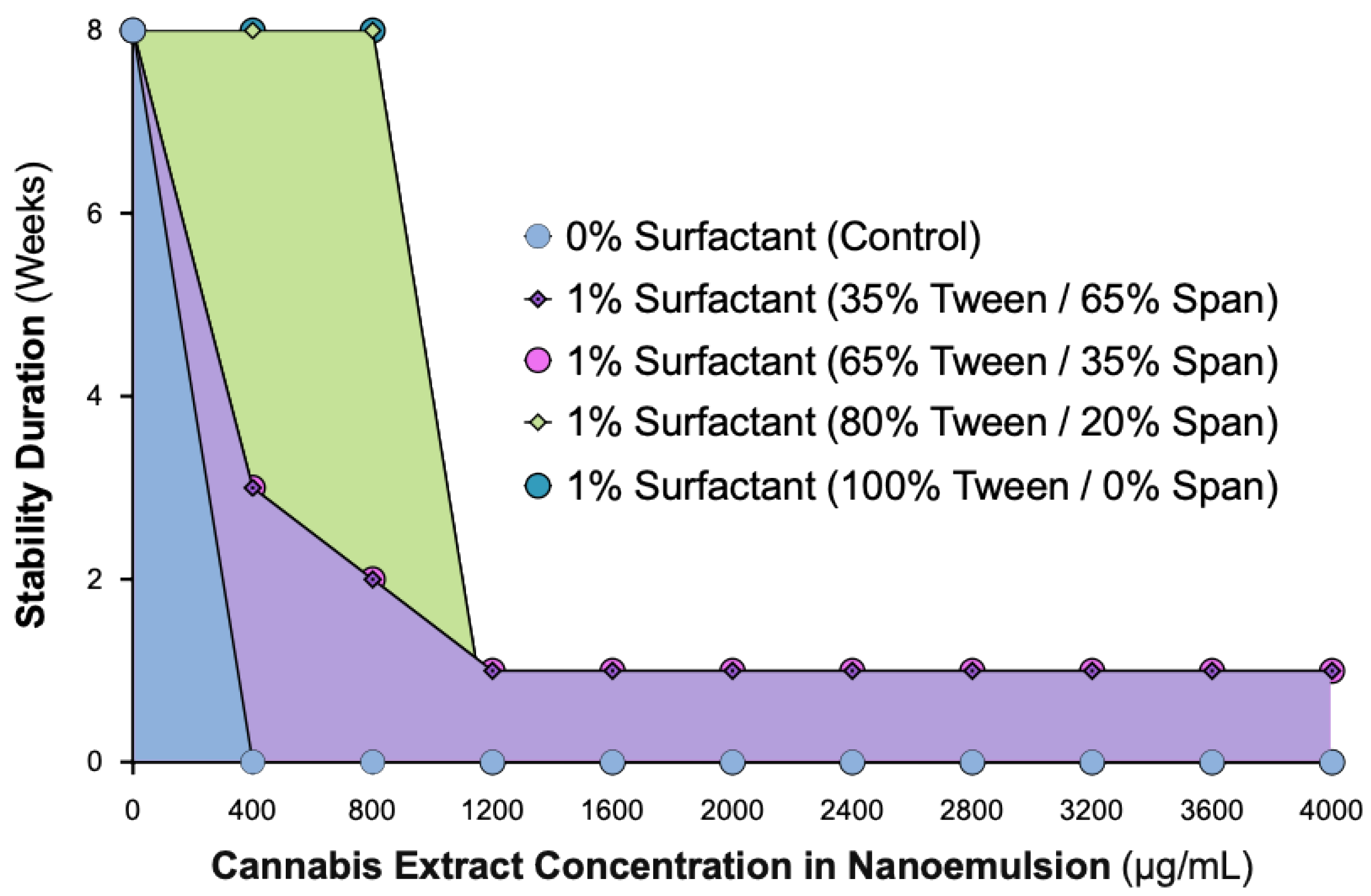
Figure 2.
Effect of CAN296 load on particle size and polydispersity in nanoemulsions with 1% surfactant (80% Tween / 20% Span). Bar plots showing the Z-average size (unfilled bars), primary particle size (horizontal line bars), and PDI (filled pattern bars) of nanoemulsions containing increasing CAN296 concentrations (400–4000 µg/mL). The left Y-axis represents particle size (nm), and the right Y-axis indicates PDI values. Formulation conditions grouped samples into two categories: 1% surfactant with 80% Tween and 20% Span (left) and no surfactant (control; right). Classification zones (“Stable,” “Unstable,” and “Highly Unstable”) are annotated above the plot to reflect formulation groups based on physical appearance and DLS-derived metrics. Abbreviations: CAN296, CBD-rich cannabis extract (Type III strain); PDI, polydispersity index.
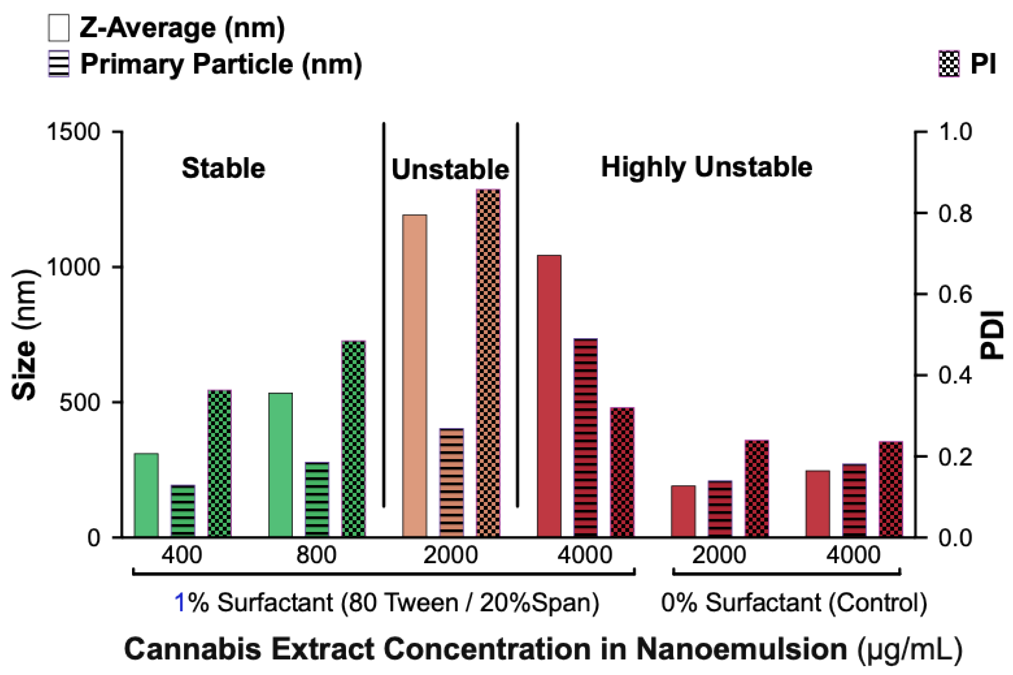
Figure 4.
TEM images showing that ≥80% Tween promotes formation of uniform, monodisperse droplets in CAN296 nanoemulsions (400 µg/mL extract) at varying Tween:Span ratios. Representative TEM micrographs of CAN296 nanoemulsions were prepared with 400 µg/mL CAN296 under the following conditions: (A) 0% surfactant (control), (B) 1% surfactant with 35% Tween / 65% Span, (C) 1% surfactant with 65% Tween / 35% Span, (D) 1% surfactant with 80% Tween / 20% Span, and (E) 1% surfactant with 100% Tween. All samples were negatively stained, deposited on carbon-coated copper grids, and imaged under identical magnification. The white line in each panel represents a 200 nm scale bar. Abbreviations: CAN296, CBD-rich cannabis extract (Type III strain); TEM, transmission electron microscopy.
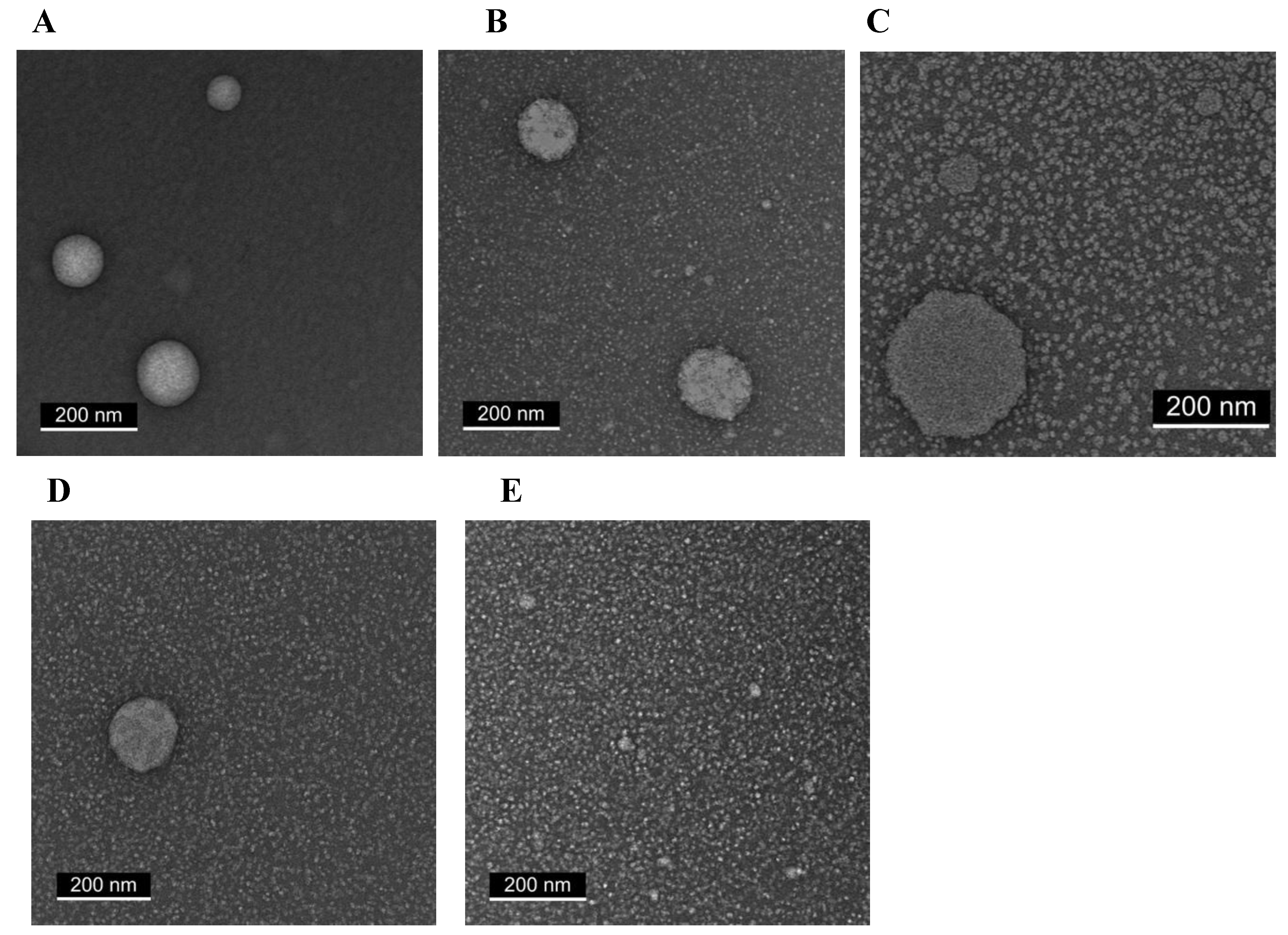
Figure 5.
SCA measurements of CAN296 nanoemulsions at increasing extract concentrations. (A) SCA values of CAN296 nanoemulsions (0, 400, 800, and 1200 µg/mL) measured on glass substrates. Wettability was evaluated using a DSA. A 5 µL droplet was dispensed via microsyringe, and contact angles were calculated by averaging the left and right sides of each droplet using the sessile-drop method. Data represent mean ± Standard deviation (SD) of six independent measurements (n = 6). Statistical significance was determined by one-way ANOVA with Tukey’s multiple comparisons test (**p < 0.01). (B) Representative droplet image of the 800 µg/mL formulation showing measured SCA. Abbreviations: CAN296, CBD-rich cannabis extract (Type III strain); DSA, drop shape analyzer; SCA, static contact angle.
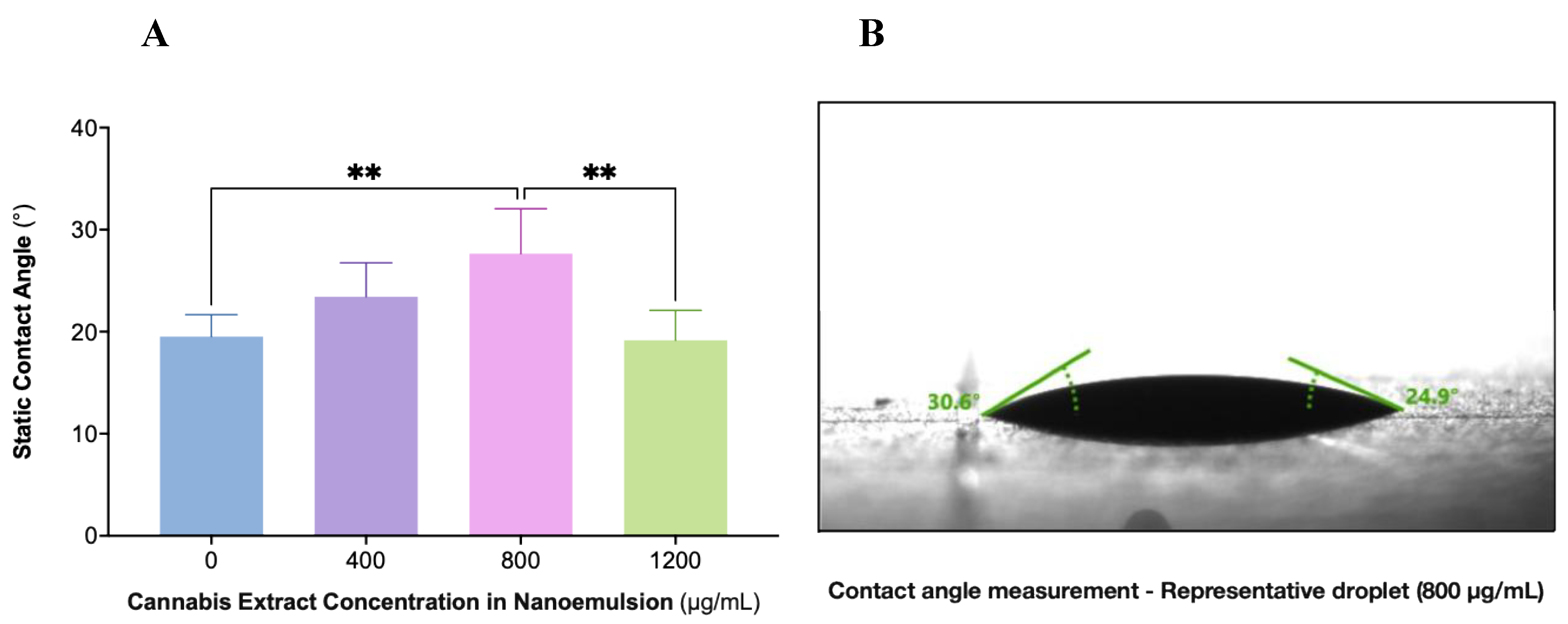
Figure 6.
Largest detected droplet size (nm) of CAN296 nanoemulsions after 30 days of storage at room temperature (25 °C; blue) and 4 °C (purple). DLS measured the largest detected droplet population for each formulation (400 and 800 µg/mL). Data are presented as mean ± SD (n = 3). Statistical significance was analyzed using two-way ANOVA with Šidák’s multiple comparisons test. Asterisks between grouped bars denote pairwise comparisons (* P = 0.0259,**** P < 0.0001), while asterisks above the concentration header denote the column-factor result (**** P < 0.0001). The dashed horizontal line marks 500 nm, serving as a visual reference for the nanoscale threshold comparison. Abbreviations: CAN296, CBD-rich cannabis extract (Type III strain); DLS, dynamic light scattering.
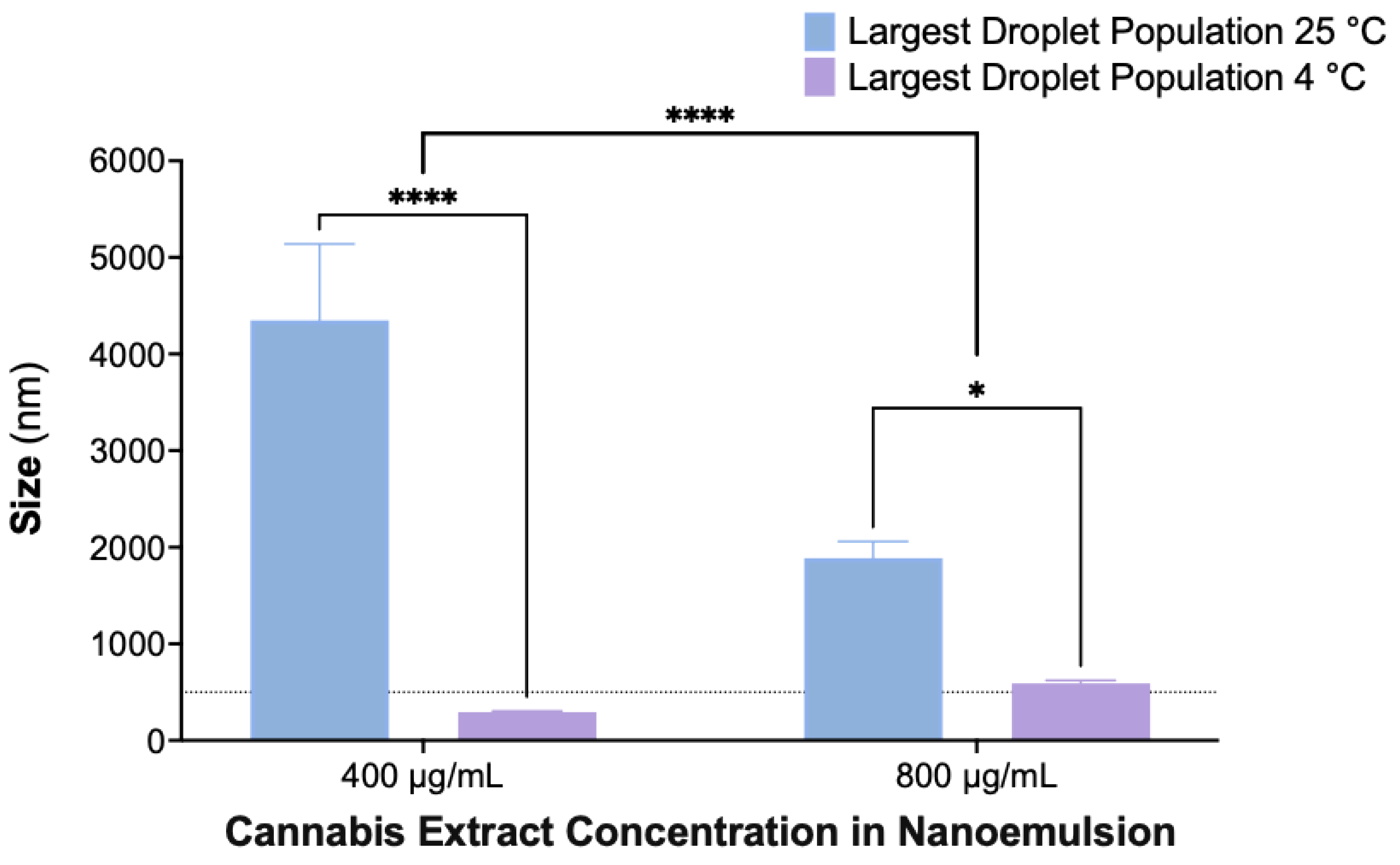
Figure 7.
Distribution of CAN296 across compartments during in vitro release from a nanoemulsion formulation. A CAN296-loaded nanoemulsion (800 µg/mL) was placed in the donor compartment of a dialysis device (Pur-A-Lyzer™ Mini 6000, 1 kDa pore size), and samples were collected at predefined time points (0–168 h). Bars represent the extract distribution as a percentage of the total loaded amount across the donor compartment (blue), receiver compartment (pink), and membrane-retained fraction (purple; calculated as the difference between donor loss and receiver gain). Statistical analysis was performed using one-way ANOVA (F(2, 27) = 21.27, P < 0.0001, R² = 0.6118). Abbreviations: CAN296, CBD-rich cannabis extract (Type III strain).
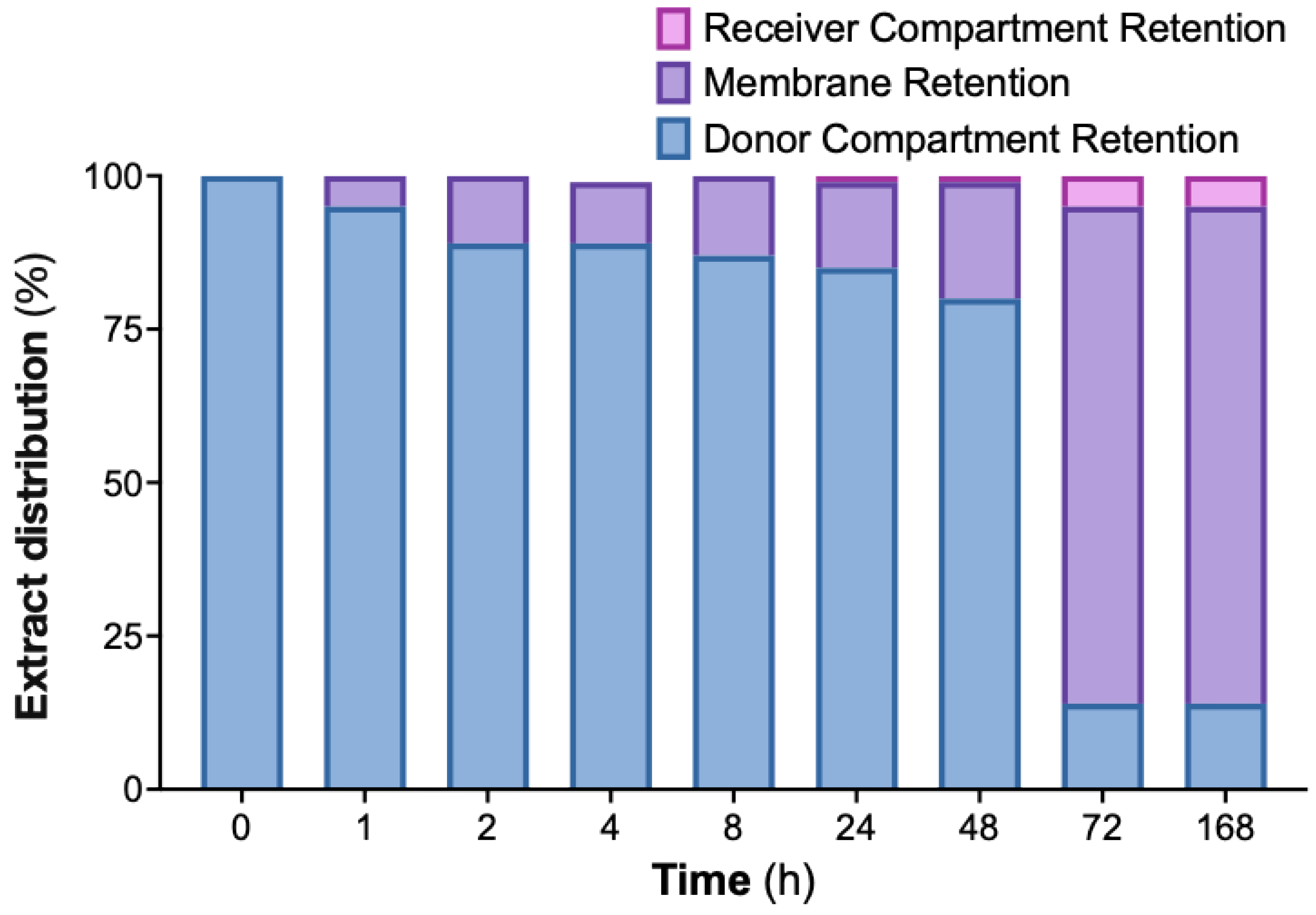
Figure 8.
SEM imaging of CAN296 nanoemulsion aggregates on the dialysis membrane surface. (A) Lower-magnification micrograph (white scale bar = 2 µm) showing scattered spherical droplets and small clusters distributed across the membrane. (B) Higher-magnification micrograph (white scale bar = 200 nm) revealing densely packed sub-micron aggregates with irregular morphology. Abbreviations: CAN296, CBD-rich cannabis extract (Type III strain); SEM, scanning electron microscopy.
Disclaimer/Publisher’s Note: The statements, opinions and data contained in all publications are solely those of the individual author(s) and contributor(s) and not of MDPI and/or the editor(s). MDPI and/or the editor(s) disclaim responsibility for any injury to people or property resulting from any ideas, methods, instructions or products referred to in the content. |
© 2025 by the authors. Licensee MDPI, Basel, Switzerland. This article is an open access article distributed under the terms and conditions of the Creative Commons Attribution (CC BY) license (http://creativecommons.org/licenses/by/4.0/).
Copyright: This open access article is published under a Creative Commons CC BY 4.0 license, which permit the free download, distribution, and reuse, provided that the author and preprint are cited in any reuse.


