Preprint
Article

This version is not peer-reviewed.

Osmolytes vs. Anabolic Reserves: Contrasting Gonadal Metabolomes in Two Sympatric Mediterranean Sea Urchins

A peer-reviewed article of this preprint also exists.

Submitted:

03 November 2025

Posted:

03 November 2025

You are already at the latest version

Abstract

The Mediterranean sea urchins Paracentrotus lividus and Arbacia lixula are ecologically coexisting grazers that exert strong control on benthic algal communities but display strikingly different physiological and ecological traits. To explore the biochemical basis of these differences, we applied high-resolution magic angle spinning nuclear magnetic resonance (HR-MAS NMR) spectroscopy combined with multivariate chemometric analysis to characterize the gonadal metabolomes of both species. Distinct species-specific metabolic fingerprints were observed. A. lixula exhibited an osmolyte- and redox-oriented profile dominated by betaine, taurine, sarcosine, TMA, TMAO, carnitine, and creatine, reflecting enhanced homeostatic regulation, antioxidant protection, and mitochondrial flexibility. In contrast, P. lividus showed an amino-acid- and lipid-enriched anabolic metabolism characterized by high levels of lysine, glycine, glutamine, and proline, consistent with a reproductive strategy focused on energy storage and protein biosynthesis. Additional metabolites—including malonate, methylmalonate, uridine, xanthine, formaldehyde, methanol, and 3-carboxypropyl-trimethyl-ammonium—highlighted microbial mediation of methylated substrates and host–microbiota metabolic interactions. These results reveal two complementary adaptive strategies: a resilience-oriented osmolyte metabolism in A. lixula and an efficiency-oriented anabolic metabolism in P. lividus. The findings demonstrate that metabolomic divergence, coupled with distinct microbial associations, underpins the ecological and physiological differentiation of these sympatric echinoids. HR-MAS NMR metabolomics thus provides a powerful tool for elucidating adaptive biochemical pathways in marine invertebrate holobionts.

Keywords: 
;  ;  ;  ;  ;  ;  ;  ;  

1. Introduction

The Mediterranean Sea represents a unique natural laboratory where temperate and subtropical biota coexist under accelerating environmental change. Among the most influential benthic grazers shaping these coastal ecosystems are the sea urchins Paracentrotus lividus (the purple sea urchin) and Arbacia lixula (the black sea urchin). Both species are abundant on rocky reefs and play pivotal ecological roles as herbivores and bioengineers, modulating algal assemblages and promoting shifts between macroalgal forests and barren grounds [1,2]. Despite their spatial overlap, they exhibit contrasting ecological and physiological traits that underpin different adaptive strategies within the same environment.
P. lividus is a primarily herbivorous species with an optimal performance under moderate thermal regimes (15–20 °C), showing seasonal reproductive cycles synchronized with macroalgal availability [3,4,5]. Its metabolism is oriented toward the accumulation of amino acids and lipids, supporting a strategy of reproductive efficiency and energy storage [6]. In contrast, A. lixula is more thermotolerant and opportunistic, feeding on crustose algae, biofilms, and sessile invertebrates, and maintaining gonadal development even in nutrient-poor “barren” habitats [7,8,9]. Stable isotope and transcriptomic analyses have confirmed its omnivorous habits and enhanced capacity for oxidative stress resistance and immune plasticity [10,11]. These species therefore represent two alternative physiological solutions: one (in A. lixula) based on metabolic resilience and homeostatic control, and another (in P. lividus) based on anabolic efficiency and reproductive investment.
Understanding the biochemical mechanisms underlying these divergent strategies is critical for predicting how Mediterranean ecosystems will respond to future environmental changes such as ocean warming and acidification. While previous research has examined sea urchin ecology, physiology, and genetics, the metabolic dimension of these adaptations remains poorly explored. Metabolomics—the comprehensive study of small-molecule metabolites—offers a powerful systems-level approach to link phenotype with physiology and environmental drivers [12,13]. It allows the detection of key metabolites involved in osmotic regulation, energy metabolism, and redox homeostasis, thereby revealing how organisms adjust their biochemical networks to changing conditions.
Among the analytical techniques available for metabolomics, high-resolution magic angle spinning nuclear magnetic resonance (HR-MAS NMR) spectroscopy provides distinct advantages for the study of marine invertebrates. HR-MAS enables the direct, non-destructive analysis of intact tissues, preserving the native biochemical environment and avoiding solvent extraction or derivatization [14]. This approach allows simultaneous identification of hydrophilic and hydrophobic metabolites, including amino acids, osmolytes, nucleotides, and lipids, while minimizing sample manipulation [15,16,17]. Moreover, HR-MAS NMR is highly reproducible and quantitative, making it ideal for comparative studies across species or environmental conditions. Its application in fish muscle, bivalves, and other marine taxa has yielded insights into nutritional status, stress responses, and environmental adaptation [17,18]; however, it has rarely been applied to echinoid gonadal tissues.
In this context, the present study applies HR-MAS NMR-based metabolomics, combined with multivariate chemometric analyses (PLS-LDA and SPA), to compare the gonadal metabolic profiles of Paracentrotus lividus and Arbacia lixula collected from Mediterranean coastal habitats. Our objectives were to: (i) identify species-specific metabolic signatures reflecting physiological and ecological strategies; (ii) characterize the major classes of metabolites—amino acids, osmolytes, and secondary compounds—that differentiate the two species; and (iii) explore potential links between metabolomic traits, microbiota composition, and ecological adaptation.
We hypothesized that A. lixula and P. lividus exhibit distinct biochemical strategies reflecting their evolutionary divergence and trophic ecology: a resilience-oriented osmolyte metabolism in A. lixula versus an efficiency-oriented anabolic metabolism in P. lividus. By elucidating these metabolomic differences, this study contributes to a broader understanding of how closely related marine invertebrates achieve functional coexistence through divergent biochemical pathways.

2. Experimental Design and Procedure

2.1. Sample Collection

Adult individuals of Paracentrotus lividus and Arbacia lixula were collected in spring 2025 from shallow rocky reefs along the Western Mediterranean coast (38.3452° N, 0.4810° W). Sea urchins were hand-collected by free diving at depths ranging from 1 to 3 meters. Specimens were transported to the laboratory in insulated containers within two hours of collection to ensure tissue preservation.

2.2. Tissue Preparation

Upon arrival at the laboratory, gonads were carefully dissected under sterile conditions. The tissues were rinsed with isotonic saline solution to remove any adherent debris and weighed to record wet mass. Gonadal samples were immediately snap-frozen in liquid nitrogen and stored at –80°C until NMR analysis.

2.3. Chemicals

D2O (99.9%) and sodium (3-trimethylsilyl)-2,2,3,3-tetradeuteriopropionate (TSP) were purchased from Sigma–Aldrich (Sigma-Aldrich, St. Louis, MO, USA).

2.4. HR-MAS NMR Spectroscopy

Gonadal metabolomic profiling was performed using high-resolution magic angle spinning nuclear magnetic resonance (HR-MAS NMR) spectroscopy. Approximately 20 mg of frozen gonadal tissue was placed into a 4 mm zirconia rotor with 10 µL of phosphate buffer solution in D₂O (pH 7.4) containing 0.1% (w/v) TSP (trimethylsilylpropionate) as a chemical shift reference. Spectra were acquired at 500 MHz and 4°C using a Bruker Avance III NMR spectrometer (Bruker, Rheinstetten, Germany), equipped with a HR-MAS probe. A Carr–Purcell–Meiboom–Gill (CPMG) pulse sequence was employed to suppress broad signals from macromolecules and enhance metabolite resolution. Also, TOCSY-HRMAS, and HSQC-MAS were acquired at the facilities of the Complutense University of Madrid (Centro de Bioimagen Complutense BIOIMAC) following the protocol outlined in the referenced article [16], using identical parameters for all experiments. Two-dimensional (2D) NMR experiments were conducted on the gilthead seabream samples, and the resulting 2D spectra were utilized to aid in the assignment of signals in the ¹H-HRMAS NMR spectra.
The H-HRMAS NMR technique employed in this study primarily facilitates qualitative comparisons among samples. Although it would provide reliable relative quantifications of certain metabolites, precise absolute quantifications were not within the scope of this analysis.

2.5. Data Processing and Statistical Analysis

NMR spectral data were processed using TopSpin 4.2.0 (Bruker, Rheinstetten, Germany) for baseline correction, phase adjustment, and metabolite identification. Signal alignment and normalization were performed prior to multivariate analysis.
Partial Least Squares–Linear Discriminant Analysis (PLS-LDA) was applied to the HR-MAS ¹H-NMR spectral dataset to identify the resonances most responsible for discriminating between Arbacia lixula and Paracentrotus lividus gonads. This approach combines the dimensionality-reduction power of PLS with the classification capabilities of LDA, allowing robust modeling of high-dimensional, collinear metabolomic data. The libPLS toolbox provides integrated routines for pretreatment, cross-validation and PLS-LDA modeling; it is openly available at the project website https://www.libpls.net/ (accessed on 31 October 2025). The algorithm was implemented in MATLAB version 2024 (MathWorks, Natick, MA, USA), following the NIPALS procedure described by [19]. In this framework, the spectral data matrix X was decomposed as X = T·P′ and the class vector y as y = T·r′ = U·q′, where T and U represent latent variables summarizing the systematic variance relevant for class separation.
The model was built with three latent variables (A = 3) and mean centering as the pretreatment method (method = 'center'). Centering was selected instead of autoscaling or Pareto scaling to preserve the relative signal amplitudes while simplifying the interpretation of Variable Importance in Projection (VIP) scores and target-projection loadings (tpLoadings). Both the VIP and tpLoading profiles were used to identify the spectral bins most relevant to the classification, enabling a focused selection of peaks for subsequent quantitative integration. The class labels were encoded as +1 (one species) and –1 (the other), as required by the LDA routine. Model accuracy, sensitivity, specificity, and the area under the ROC curve (AUC) were computed to assess the discriminant performance.
To identify the spectral regions most relevant for discriminating between Arbacia lixula and Paracentrotus lividus, a Subwindow Permutation Analysis (SPA) was applied to the integrated HR-MAS ¹H-NMR data matrix [20,21]. SPA is a supervised Monte-Carlo–based variable-selection algorithm that repeatedly builds Partial Least Squares–Linear Discriminant Analysis (PLS-LDA) sub-models using randomly selected subsets of samples and variables. The method evaluates the statistical contribution of each variable by comparing prediction errors from the original and permuted datasets, providing a p-value and the corresponding Conditional Synergistic Score (COSS = –log₁₀ p) as a quantitative index of variable importance.
The analysis was performed in MATLAB using the original SPA implementation with the following parameters: Q = 15 (number of variables sampled per Monte-Carlo sub-model), K = 3 (folds for cross-validation), ratio = 0.7 (fraction of calibration samples), and N = 1000 (number of Monte-Carlo iterations). Pareto scaling was selected as pretreatment (“method = pareto”), as it balances normalization of variance while preserving metabolite intensity patterns. The maximum number of latent variables (A) was optimized automatically within each sub-model. Variables yielding COSS values greater than 2 (p < 0.01) were considered statistically significant contributors to class separation.

2.6. Ethical Considerations

The sea urchins were collected under the permission granted by the Dirección General de Pesca of the Generalitat Valenciana on 26 September 2024. All procedures involving animal collection and handling were conducted in compliance with institutional ethical guidelines approved by the University of Alicante (Exp. UA-2024-10-24_2). The study adhered to national and international regulations for the use of marine invertebrates in scientific research.

3. Results

3.1. HR-MAS Spectral Analysis
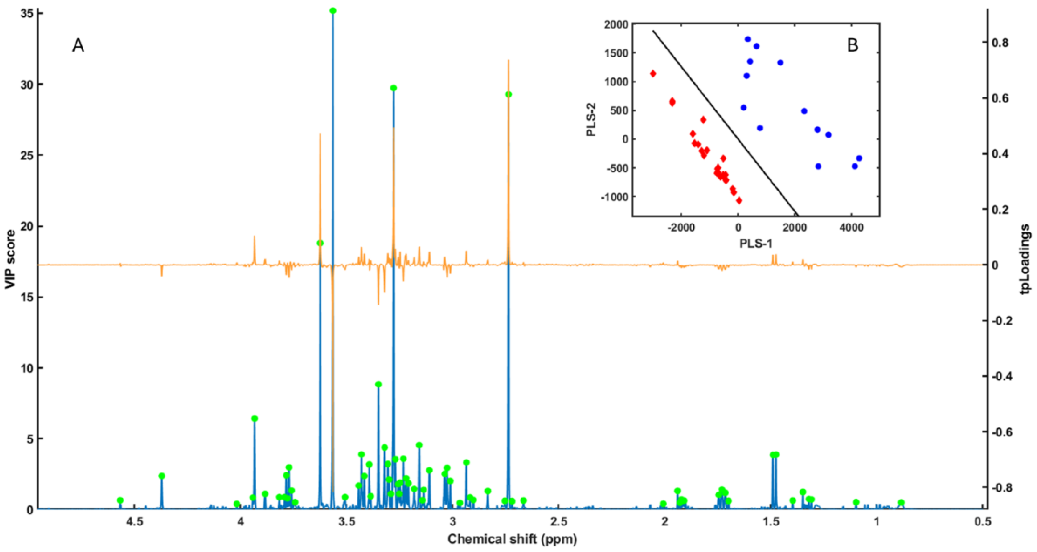
This study represents the first application of high-resolution magic angle spinning nuclear magnetic resonance (HR-MAS NMR) spectroscopy to the gonadal tissues of Mediterranean sea urchins. Representative 1H HR-MAS spectra obtained from Arbacia lixula and Paracentrotus lividus are shown in Figure 1. A wide range of metabolites was identified using combined one-dimensional (1D) and two-dimensional (2D) experiments, including COSY, TOCSY, and HSQC. Spectral assignments (Table 1) were based on direct signal identification, scalar correlations, heteronuclear couplings, and comparison with reference spectra and literature databases (HMDB; https://hmdb.ca/, accessed on 31 October 2025) [15,16,22,23,24]. The HR-MAS approach preserved the native biochemical composition of intact gonadal tissue without prior extraction or derivatization, enabling simultaneous detection of hydrophilic and hydrophobic compounds.
Distinct spectral patterns were observed between A. lixula and P. lividus, involving both the presence/absence of specific resonances and variation in signal intensity (Figure 1). In A. lixula, the spectrum was dominated by resonances of osmolytes and methylated amines (e.g., betaine, taurine, sarcosine, trimethylamine (TMA), and trimethylamine-N-oxide (TMAO)), as well as creatine and carnitine. In contrast, P. lividus spectra displayed higher intensities for amino acids (glycine, lysine, glutamine, glutamate, and proline) and stronger aliphatic signals from lipid methyl and methylene groups. These spectral trends suggested pronounced species-level differences in the chemical composition of gonadal tissues.

3.2. PLS-LDA Model

To identify the metabolites contributing most to the interspecific discrimination, a Partial Least Squares–Linear Discriminant Analysis (PLS-LDA) model was constructed using three latent variables. The model achieved a clear separation between A. lixula and P. lividus samples, as illustrated by the score plot (Figure 2B). The cumulative R²X = 0.93 and R²Y = 0.86 indicated that the selected components captured the majority of class-related variance while minimizing spectral noise.
Cross-validation and permutation testing confirmed the robustness of the classification model (AUC > 0.90), demonstrating strong predictive performance and reliable species-level separation. The Variable Importance in Projection (VIP) and target-projection loading (tpLoading) profiles highlighted consistent spectral regions with the highest discriminatory power (VIP > 1.0). These corresponded to signals associated with osmolytes, amino acids, and methylated metabolites (Figure 2A) [16].

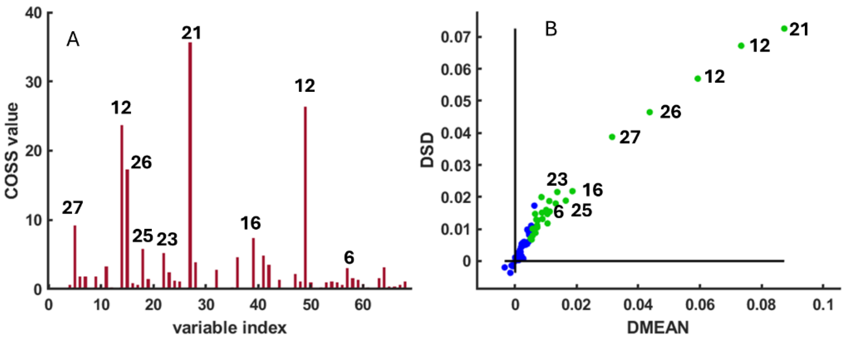
3.3. SPA Variable Selection

To further validate the discriminant variables and quantify their statistical significance, a Subwindow Permutation Analysis (SPA) was applied to the integrated HR-MAS dataset. SPA repeatedly constructed PLS-LDA sub-models using randomly selected subsets of samples and variables to assess each variable’s contribution to class separation [21].
The analysis was performed using 1000 Monte Carlo iterations (N = 1000), Q = 15 variables per sub-model, and K = 3 folds for cross-validation, with 70% of samples used for model calibration (ratio = 0.7). Variables with the highest Covariance Selection (COSS) scores (p < 0.01) were considered statistically significant discriminants and were highlighted as green circles in Figure 3A. The results of the SPA confirmed that several metabolites contributed most strongly to species differentiation, including betaine, sarcosine, glycine, malonate, taurine, formaldehyde, and lysine (Figure 3B). These variables were subsequently selected for quantitative comparison through spectral integration and visualized using boxplots of signal intensities (Figure 4). The parameter choices and statistical procedure followed the original implementation of SPA described by [20].

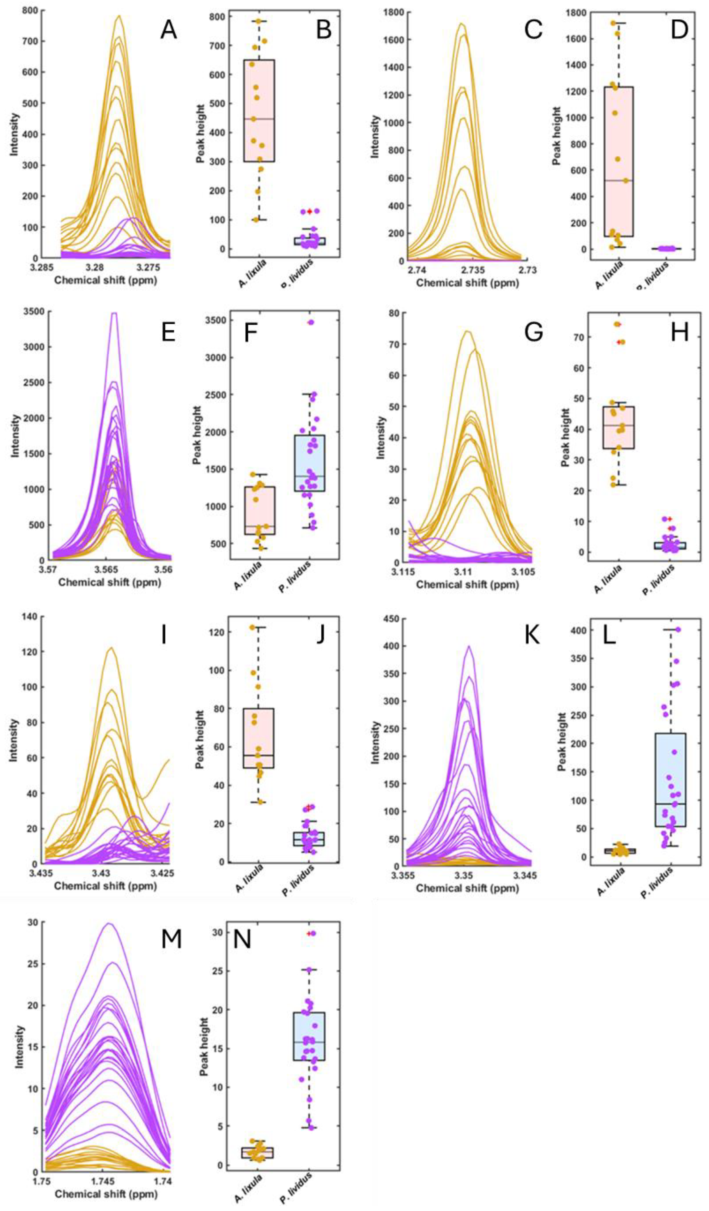
3.4. Selected Metabolites and Boxplots

The metabolites showing the highest COSS values in the SPA were integrated to quantify species-specific differences (Figure 4). Boxplots of normalized peak intensities confirmed statistically significant (p < 0.01) differences between A. lixula and P. lividus for all selected compounds.
In A. lixula, higher intensities were observed for osmolytes and methylated amines (betaine, sarcosine, TMA, TMAO), organic acids (malonate and methylmalonate), and energy-buffer molecules (carnitine and creatine). Additionally, nucleosides (uridine) and purine derivatives (xanthine) were detected exclusively or at higher levels in A. lixula.
Conversely, P. lividus samples showed elevated signals for amino acids (glycine and lysine) and for compounds likely linked to algal-derived or microbial methylotrophy, including formaldehyde, methanol, and 3-carboxypropyl-trimethyl-ammonium. These compounds were absent or near the detection limit in A. lixula.
In total, the analysis identified a consistent set of metabolites whose concentration differences account for the strong separation between the two sea urchin species. The full list of metabolites and their chemical shifts is provided in Table 1, while Figure 2, Figure 3 and Figure 4 summarize their statistical and graphical representation. Together, these results confirm substantial biochemical divergence in the gonadal metabolomes of A. lixula and P. lividus, providing the basis for the mechanistic interpretations developed in the Discussion.

4. Discussion

4.1. Core Metabolic Contrasts

Our comparative HR-MAS NMR metabolomic profiling of Arbacia lixula and Paracentrotus lividus reveals marked divergences in the biochemical composition of their gonadal tissues. These differences are not random but instead reflect species-specific physiological strategies shaped by ecology, diet, and evolutionary history. The metabolic contrasts can be organized into three main axes: (i) osmolyte-based cellular homeostasis, (ii) amino-acid and nitrogen-metabolism investment, and (iii) specific pathway activations linked to redox and immune balance.
In A. lixula, metabolites such as betaine (N-trimethylglycine), sarcosine (N-methylglycine), and taurine appear in markedly higher abundance than in P. lividus. These compounds are classic compatible osmolytes, maintaining intracellular osmotic equilibrium, stabilizing proteins, and protecting cellular structures against fluctuations in salinity, temperature, or oxidative stress. Betaine and taurine are dominant osmolytes in many marine invertebrates, where they help preserve enzyme functionality and membrane integrity under hyperosmotic or thermal stress [25,26]. The accumulation of these solutes in A. lixula thus likely represents an adaptive strategy toward homeostasis and resilience in environments characterized by greater variability in temperature and food availability.
Sarcosine, an intermediate in betaine and glycine metabolism, further indicates an active methyl-group turnover and one-carbon metabolic cycle. This enhanced methylation capacity might be related to the high metabolic flexibility of A. lixula, which occupies a broader trophic niche and frequently grazes on protein-rich biofilms or sessile invertebrates [2]. Taurine, beyond its osmotic role, acts as an antioxidant, cytoprotective and membrane-stabilizing molecule [27]. The combined accumulation of betaine, sarcosine, and taurine therefore suggests a metabolic orientation favouring stress tolerance and rapid cellular recovery — traits that align with A. lixula’s success in warm, oligotrophic, or “barren” Mediterranean habitats.
In contrast, P. lividus exhibited higher levels of amino acids such as glycine, glutamate, glutamine, arginine, proline, valine, and especially lysine, revealing a pronounced anabolic metabolism. These amino acids are central to nitrogen transport, protein synthesis, and energy storage and are often mobilized during gonadal maturation [3,4,28]. Elevated amino-acid pools have been associated with reproductive investment in echinoids, reflecting their role as precursors for gametogenic proteins and nucleotides. This pattern is consistent with previous biochemical analyses showing that P. lividus gonads exhibit a high proportion of free amino acids and lipids during active gametogenesis [8,29].
Among these, lysine deserves special attention due to its metabolic and developmental significance. Lysine is an essential amino acid that participates in multiple biosynthetic pathways, including the saccharopine pathway and carnitine biosynthesis, linking protein turnover to mitochondrial β-oxidation [30]. High lysine content in P. lividus may indicate both active anabolic processes and a readiness to synthesize carnitine for fatty-acid transport. This connection between lysine and lipid metabolism supports the interpretation of a reproductive-energy-storage strategy, where resources are channelled into gametogenesis and subsequent embryonic development. Historical studies already described elevated lysine levels during early developmental stages of sea urchins [28], supporting the view that this amino acid plays a crucial role in reproductive metabolism and growth.
Overall, P. lividus seems to prioritize anabolic efficiency—mobilizing amino acids and lipids as energy reserves—whereas A. lixula invests more in osmotic and antioxidant stability. These contrasting trends align with their ecological niches: P. lividus as a macroalgae-feeding species with seasonal reproductive peaks, and A. lixula as a thermotolerant omnivore sustaining reproduction even in nutrient-poor conditions [3,29].
Beyond the major amino acids and osmolytes, the presence of kynurenine in both species adds another layer of metabolic differentiation. Kynurenine is a central intermediate in the tryptophan-catabolic (kynurenine) pathway, which plays critical roles in redox regulation, immune modulation, and stress signalling. Recent studies have demonstrated the conservation of this pathway in a broad range of invertebrates, including molluscs, crustaceans, and annelids [31,32,33]. The detection of kynurenine in both A. lixula and P. lividus suggests that the pathway is also active in echinoderms, potentially contributing to immune regulation in gonadal tissues.
Kynurenine derivatives such as kynurenic acid and 3-hydroxykynurenine are known to balance oxidative processes and neuroactive functions, providing a biochemical interface between metabolism and cellular defence [34]. Its higher relative abundance in A. lixula may indicate a stronger oxidative-stress response, consistent with its osmolyte-enriched, stress-resilient phenotype. Moreover, marine natural products have been shown to modulate enzymes within this pathway [35], implying that environmental exposures or diet could influence its activity.
By contrast, P. lividus, with its lysine- and lipid-enriched anabolic metabolism, might rely less on kynurenine-mediated stress buffering and more on biosynthetic and energy pathways supporting reproductive output. This trade-off between stress-resilience (A. lixula) and anabolic efficiency (P. lividus) appears to define their respective metabolic strategies and may underpin their ecological partitioning within overlapping Mediterranean habitats.
Taken together, the observed patterns delineate two contrasting but complementary metabolic phenotypes. Arbacia lixula displays an osmolyte-dominated and antioxidant-fortified metabolism, emphasizing cellular homeostasis, redox balance, and stress adaptability. Paracentrotus lividus, by contrast, manifests an amino-acid- and lipid-driven anabolic metabolism, indicative of reproductive energy storage and biosynthetic investment.
These divergent strategies likely mirror long-term evolutionary adaptation to differing ecological niches: the first optimized for persistence and resilience in warmer, low-productivity habitats, the second tuned for rapid reproductive turnover in cooler, macroalgae-rich environments. The distinct metabolic fingerprints uncovered by HR-MAS NMR thus reflect not only biochemical variance but also adaptive physiological trade-offs that define each species’ ecological role.

4.2. Secondary Metabolites and Microbial Mediation

Beyond the primary osmolytes and amino acids that define the core metabolic contrast between Arbacia lixula and Paracentrotus lividus, several additional metabolites provide deeper insight into their divergent physiological strategies. These include methylated amines (TMA and TMAO), dicarboxylic acids (malonate and methylmalonate), energy-buffer molecules (carnitine and creatine), nucleosides and purine derivatives (uridine and xanthine), and a unique set of compounds restricted to P. lividus (formaldehyde, methanol, and 3-carboxypropyl-trimethyl-ammonium). The possible presence of the methylxanthine alkaloid theophylline or its derivatives in P. lividus also deserves consideration. Together, these metabolites reveal the interplay between host metabolism, diet, and microbial co-metabolism within the holobiont framework.
In A. lixula, elevated concentrations of trimethylamine (TMA) and trimethylamine N-oxide (TMAO) represent one of the most distinctive metabolic traits. TMAO is a well-known organic osmolyte that stabilizes proteins and counteracts the destabilizing effects of urea and hydrostatic pressure in marine organisms [27]. In fish and invertebrates, it also serves as a chemical indicator of tissue freshness because microbial catabolism of TMAO releases TMA during post-mortem decay [24]. In living organisms, however, high TMAO levels typically indicate osmotic and structural homeostasis.
In marine invertebrates, TMA is mainly derived from microbial oxidation or demethylation of quaternary ammonium compounds such as choline, carnitine, and betaine [36]. The presence of both TMA and TMAO in A. lixula thus reflects an active metabolic exchange between the host and its associated microbiota. Bacterial taxa capable of oxidizing TMA to TMAO or further demethylating it to formaldehyde and ammonium have been identified in several marine systems [37]. Consequently, A. lixula’s enrichment in these metabolites may signal a symbiotic metabolic loop involving microbial methylamine cycling and host osmolyte regulation — a relationship that would enhance resilience in fluctuating salinity and temperature regimes.
Two other compounds, malonate and methylmalonate, were notably more abundant in A. lixula. Malonate is a structural analogue of succinate and a competitive inhibitor of succinate dehydrogenase, thereby modulating mitochondrial electron transport and tricarboxylic-acid (TCA) cycle activity [38]. The co-accumulation of malonate and methylmalonate suggests the activation of propionate and odd-chain fatty-acid catabolism, pathways that feed into the TCA cycle via succinyl-CoA. In mammals and marine invertebrates alike, elevated methylmalonate concentrations often mark enhanced anaplerotic flux through propionyl-CoA metabolism [39]. In some molluscs, labelled propionate incorporation has confirmed methylmalonate-dependent biosynthesis of secondary metabolites [40].
The concurrent enrichment of carnitine and creatine in A. lixula further supports the idea of a dynamic and flexible energy metabolism. Carnitine facilitates the transport of long-chain fatty acids into mitochondria for β-oxidation, while creatine acts as a phosphagen buffer, maintaining ATP supply in tissues with variable energy demand [41,42,43]. The presence of both compounds suggests that A. lixula maintains an energetically responsive system capable of shifting between lipid oxidation and phosphocreatine buffering, consistent with an organism adapted to intermittent feeding and variable environmental stress.
The higher abundance of uridine and xanthine in A. lixula also provides clues to its cellular physiology. Uridine, a pyrimidine nucleoside, is essential for RNA synthesis, membrane phospholipid metabolism, and glycosylation reactions, whereas xanthine arises from purine catabolism via xanthine oxidase. Elevated levels of these compounds may indicate increased RNA turnover and oxidative metabolism in gonadal tissues. In other marine invertebrates, purine catabolites such as hypoxanthine and xanthine accumulate under oxidative or nutritional stress, acting as indicators of heightened metabolic activity [44]. The prominence of xanthine in A. lixula thus aligns with its stress-tolerant metabolic profile and may also reflect microbial purine degradation within its coelomic fluid [45].
In P. lividus, several metabolites — formaldehyde, methanol, and 3-carboxypropyl-trimethyl-ammonium — were detected at high levels but were absent or negligible in A. lixula. These compounds are unusual in animal metabolomes and likely originate from microbial or dietary methylotrophic processes. Macroalgae, the primary food source of P. lividus, release methanol and methylamines during cell-wall pectin and alginate degradation, particularly under light stress or senescence [46]. Methanol oxidation by associated microbiota yields formaldehyde as a transient intermediate, which can subsequently react with amino groups or enter the tetrahydrofolate-linked C1-metabolism pathway [47].
Formaldehyde and methanol therefore may not be toxic end products but rather transient metabolites within microbial-host co-metabolism. The detection of 3-carboxypropyl-trimethyl-ammonium (a quaternary ammonium compound structurally related to glycine betaine) supports this interpretation. It may represent an osmolyte derivative or intermediate of methylamine metabolism, arising either from algal ingestion or bacterial transformations of trimethylated substrates. The absence of these compounds in A. lixula strengthens the view that P. lividus harbours a distinct methylotrophic microbial community capable of processing algal-derived C1 compounds.
Although the canonical methylxanthine theophylline (1,3-dimethylxanthine) was not unequivocally identified in our spectra, its occurrence or that of structurally related derivatives in P. lividus remains plausible. Methylxanthines are secondary metabolites widespread among marine and terrestrial algae, where they play roles in chemical defence and allelopathy [48]. Herbivorous invertebrates grazing on these algae may ingest and partially metabolize such compounds, leading to the appearance of demethylated derivatives such as xanthine, which was indeed abundant in our dataset. Microbial communities associated with P. lividus could further oxidize theophylline to xanthine via xanthine oxidase or related pathways [49]. If confirmed, the occurrence of methylxanthine derivatives would suggest an additional level of xenobiotic or detoxification metabolism in P. lividus, possibly linked to its algal diet and microbiota activity.
Collectively, the presence of these secondary metabolites underscores the complex biochemical interplay between host metabolism and microbial mediation in both sea urchin species. In A. lixula, the constellation of TMA/TMAO, malonate, methylmalonate, carnitine, creatine, uridine, and xanthine reflects an osmolyte-reinforced, energetically versatile metabolism tightly coupled with microbial co-metabolism of methylated amines. In P. lividus, by contrast, the accumulation of formaldehyde, methanol, and 3-carboxypropyl-trimethyl-ammonium points to a methylotrophic metabolic network likely sustained by algal diet and associated microbiota.
These findings highlight that species-specific metabolomic profiles cannot be interpreted independently of their symbiotic microbial context. The following section therefore addresses how these biochemical differences correspond to variations in diet, microbiota composition, and evolutionary adaptation between A. lixula and P. lividus.

4.3. Integrative Ecological and Evolutionary Context

Although Arbacia lixula and Paracentrotus lividus share the same shallow rocky habitats and were sampled under identical environmental conditions, their gonadal metabolomes exhibit profound biochemical divergence. This divergence transcends simple interspecific variation, reflecting two distinct metabolic and ecological strategies shaped by diet, symbiotic microbiota, and evolutionary history. The data presented here support a dual adaptive framework: A. lixula adopts a resilience-oriented osmolyte strategy, whereas P. lividus follows an efficiency-oriented anabolic strategy.
A primary axis of metabolic differentiation between the two echinoids lies in their feeding ecology and nutrient assimilation. P. lividus is a strict herbivore that primarily consumes fleshy macroalgae rich in carbohydrates and nitrogen-poor biomass [3,50,51]. This diet promotes carbohydrate and amino-acid-oriented metabolism, reflected in the elevated levels of lysine, glycine, glutamine, and other amino acids, as well as higher lipid content in its gonads. These metabolites function as energy and structural reserves that sustain gametogenesis during seasonal reproductive peaks [3,29]. The observed anabolic profile of P. lividus aligns with its seasonally synchronized reproductive strategy, which depends on the availability of algal resources during cooler months [6].
Conversely, A. lixula exhibits omnivorous feeding behaviour, consuming filamentous and crustose algae, sessile invertebrates, and detrital biofilms [2]. Such dietary flexibility supplies a richer amino- and nitrogen-based substrate pool, fostering the synthesis of osmolytes such as betaine, taurine, and sarcosine. These metabolites enhance osmotic stability, oxidative protection, and protein maintenance, all of which are critical for surviving in nutrient-depleted, thermally variable “barren” habitats [7,52]. This metabolic orientation complements A. lixula’s capacity to maintain gonadal development under food-limited conditions, in contrast to the more resource-dependent P. lividus [6].
Emerging evidence indicates that the microbiota plays a pivotal role in modulating sea urchin metabolism, influencing both energy fluxes and chemical defence. The recent study by Arranz et al. (2025) revealed substantial differences in the gut and coelomic microbiota of A. lixula and P. lividus across Mediterranean locations [53]. Specifically, A. lixula harboured microbial consortia enriched in choline- and carnitine-degrading taxa, including methylotrophic and osmolyte-processing bacteria capable of producing or oxidizing TMA and TMAO. In contrast, P. lividus hosted communities enriched in formaldehyde- and methanol-utilizing bacteria, consistent with the presence of those metabolites in its gonadal tissues.
This microbiota–metabolite concordance provides a mechanistic explanation for the chemical profiles described in Section 4.1 and 4.2. The TMA/TMAO cycle in A. lixula suggests microbial mediation of quaternary amine metabolism that contributes to osmotic balance and stress tolerance, whereas the formaldehyde–methanol network in P. lividus points to algal-derived methylotrophy within its holobiont. Comparable relationships between microbial metabolism and host chemistry have been documented in other echinoderms, where gut bacteria contribute to amino acid biosynthesis, short-chain fatty acid production, and detoxification processes [54,55].
From an evolutionary standpoint, A. lixula and P. lividus belong to distinct phylogenetic lineages that diverged several million years ago, likely under differing selective pressures [56,57]. This evolutionary separation has produced divergent regulatory architectures for energy metabolism and stress response. A. lixula, as a warm-adapted species expanding in a “tropicalizing” Mediterranean Sea, displays traits associated with environmental plasticity—osmolyte regulation, antioxidant balance, and flexible energy metabolism. P. lividus, on the other hand, retains the biochemical hallmarks of a temperate species, emphasizing nutrient assimilation and reproductive efficiency within more stable habitats [13,58].
Such divergence exemplifies metabolic evolution as an adaptive continuum: one lineage optimized for stability and reproduction, the other for resilience and persistence under stress. Importantly, both strategies are ecologically successful within their niches

5. Conclusions

This study provides the first comprehensive comparative analysis of the gonadal metabolomes of the Mediterranean sea urchins Arbacia lixula and Paracentrotus lividus using high-resolution magic angle spinning nuclear magnetic resonance (HR-MAS NMR) spectroscopy. The results reveal profound species-specific metabolic differentiation that mirrors their ecological and physiological divergence.
A. lixula exhibits an osmolyte and redox-dominated metabolic phenotype, characterized by elevated concentrations of betaine, taurine, sarcosine, TMAO, carnitine, and creatine. These metabolites emphasize cellular homeostasis, antioxidant protection, and mitochondrial flexibility—traits consistent with this species’ tolerance to high temperature and low nutrient availability. In contrast, P. lividus displays an amino-acid- and lipid-oriented anabolic metabolism, with enhanced levels of lysine, glycine, glutamine, and fatty acids, reflecting a reproductive strategy centered on energy storage and protein biosynthesis.
Additional metabolites, including malonate, methylmalonate, uridine, xanthine, formaldehyde, methanol, and 3-carboxypropyl-trimethyl-ammonium, further illustrate the complexity of these biochemical strategies. Their occurrence points to microbial mediation of methylated compounds, establishing a functional link between host metabolism, dietary sources, and holobiont microbiota. The observed metabolite–microbiota associations—TMA/TMAO cycling in A. lixula and methylotrophic pathways in P. lividus—highlight that these echinoids function as metabolically integrated systems shaped by symbiotic interactions.
Taken together, the findings support a conceptual model in which A. lixula embodies a resilience-oriented strategy, prioritizing osmotic and oxidative stability, while P. lividus adopts an efficiency-oriented strategy based on reproductive and anabolic investment. These alternative physiological solutions enable the two sympatric species to coexist and partition ecological niches within the same Mediterranean habitats.
By linking metabolomic fingerprints with ecological function, feeding behaviour, and microbial mediation, this work demonstrates the power of HR-MAS NMR to unravel adaptive biochemical diversity in marine invertebrates. Future studies integrating metabolomics, microbiomics, and transcriptomics across seasons, sexes, and environmental gradients will be essential to understand how echinoid holobionts respond to ongoing ocean warming and acidification, and how metabolic plasticity contributes to their resilience in a rapidly changing sea.

Author Contributions

Funding obtained: F.C.M.-E. and P.S-J. Conceived and designed the experiments: R.I-C, E.C.-G., F.C.M.-E. and P.S-J. Performed the analysis and figures: , E.C.-G , R.I-C. and F.C.M.-E. Analyzed the data: , E.C.-G , R.I-C. and F.C.M.-E. Wrote the manuscript: F.C.M.-E. Reviewed the manuscript: , E.C.-G , R.I-C., F.C.M.-E. and P.S-J. Collected the urchin samples: E.C.-G. and P.S-J. All authors have read and agreed to the published version of the manuscript.

Funding

This research was funded by CONVOCATORIA DEL PROGRAMA PROPIO DEL CENTRO DE GASTRONOMÍA DEL MEDITERRÁNEO (Gasterra 2023–24) UA_DENIA PARA EL FOMENTO DE LA I+D+i EN El ÁMBITO DE LA GASTRONOMÍA (GASTERRA 2024).

Institutional Review Board Statement

This study was conducted under the framework of the GASTERRA projects (2024-2025) and received ethical approval from the Research Ethics Committee of the University of Alicante (Reference: UA-2024-10-24_2).

Informed Consent Statement

Not applicable.

Data Availability Statement

The data presented in this study are available on request from the corresponding author.

Acknowledgments

The authors would like to thank Palmira Villa-Valverde (Centro de Bioimagen Complutense BIOIMAC, Universidad Complutense de Madrid) for technical assistance with HR-MAS experiments.

Conflicts of Interest

The authors declare no conflicts of interest.

References

  1. Bonaviri, C. and et al. "Leading role of the sea urchin arbacia lixula in maintaining the barren state in southwestern mediterranean." Marine Biology 158 (2011): 2485–93. [CrossRef]
  2. Wangensteen, O. S., X. Turon, A. García-Cisneros, M. Recasens, J. Romero and C. Palacín. "A wolf in sheep’s clothing: Carnivory in dominant sea urchins in the mediterranean." Marine Ecology Progress Series 441 (2011): 117-28.
  3. Rocha, F., A. C. Rocha, L. F. Baião, J. Gadelha, C. Camacho, M. L. Carvalho, F. Arenas, A. Oliveira, M. R. G. Maia and A. R. Cabrita. "Seasonal effect in nutritional quality and safety of the wild sea urchin paracentrotus lividus harvested in the european atlantic shores." Food Chemistry 282 (2019): 84-94.
  4. Montero-Torreiro, M. F. and P. Garcia-Martinez. "Seasonal changes in the biochemical composition of body components of the sea urchin, paracentrotus lividus, in lorbé (galicia, north-western spain)." Journal of the Marine Biological Association of the United Kingdom 83 (2003): 575-81. [CrossRef]
  5. Arafa, S., M. Chouaibi, S. Sadok and A. El Abed. "The influence of season on the gonad index and biochemical composition of the sea urchin paracentrotus lividus from the golf of tunis." The Scientific World Journal 2012 (2012): 815935.
  6. Ouréns, R., L. Fernández and J. Freire. "Geographic, population, and seasonal patterns in the reproductive parameters of the sea urchin paracentrotus lividus." Marine Biology 158 (2011): 793-804.
  7. Foo, S. A., M. Munari, M. C. Gambi and M. Byrne. "Acclimation to low ph does not affect the thermal tolerance of arbacia lixula progeny." Biology Letters 18 (2022): 20220087. [CrossRef]
  8. Fernandez-Vilert, R., V. Arranz, M. Martín-Huete, J. C. Hernández, S. González-Delgado and R. Pérez-Portela. "Effect of ocean acidification on the oxygen consumption of the sea urchins paracentrotus lividus (lamarck, 1816) and arbacia lixula (linnaeus, 1758) living in co2 natural gradients." Frontiers in Marine Science 12 (2025): 1500646.
  9. Gianguzza, P., G. Visconti, F. Gianguzza, S. Vizzini, G. Sarà and S. Dupont. "Temperature modulates the response of the thermophilous sea urchin arbacia lixula early life stages to co2-driven acidification." Marine environmental research 93 (2014): 70-77.
  10. Pérez-Portela, R. and C. Leiva. "Sex-specific transcriptomic differences in the immune cells of a key atlantic-mediterranean sea urchin." Frontiers in Marine Science 9 (2022): 908387. [CrossRef]
  11. Luparello, C., D. Ragona, D. M. L. Asaro, V. Lazzara, F. Affranchi, V. Arizza and M. Vazzana. "Cell-free coelomic fluid extracts of the sea urchin arbacia lixula impair mitochondrial potential and cell cycle distribution and stimulate reactive oxygen species production and autophagic activity in triple-negative mda-mb231 breast cancer cells." Journal of Marine Science and Engineering 8 (2020): 261.
  12. Bundy, J. G., M. P. Davey and M. R. Viant. "Environmental metabolomics: A critical review and future perspectives." Metabolomics 5 (2009): 3-21.
  13. Bayona, L. M., N. J. de Voogd and Y. H. Choi. "Metabolomics on the study of marine organisms." Metabolomics 18 (2022): 17.
  14. Beckonert, O., M. Coen, H. C. Keun, Y. Wang, T. M. D. Ebbels, E. Holmes, J. C. Lindon and J. K. Nicholson. "High-resolution magic-angle-spinning nmr spectroscopy for metabolic profiling of intact tissues." Nature protocols 5 (2010): 1019-32.
  15. Villa, P., D. Castejon, M. Herraiz and A. Herrera. "H-1-hrmas nmr study of cold smoked atlantic salmon (salmo salar) treated with e-beam." Magnetic Resonance in Chemistry 51 (2013): 350-57. [CrossRef]
  16. Castejón, D., P. Villa, M. M. Calvo, G. Santa-María, M. Herraiz and A. Herrera. "1h-hrmas nmr study of smoked atlantic salmon (salmo salar)." Magnetic Resonance in Chemistry 48 (2010): 693-703. [CrossRef]
  17. Aru, V. "Application of nmr-based metabolomics techniques to biological systems: A case study on bivalves." (2016):.
  18. Lardon, I., M. Eyckmans, T. Vu, K. Laukens, G. De Boeck and R. Dommisse. "H-1-nmr study of the metabolome of a moderately hypoxia-tolerant fish, the common carp (cyprinus carpio)." Metabolomics 9 (2013): 1216-27. [CrossRef]
  19. Li, H., Y. Liang, Q. Xu and D. Cao. "Key wavelengths screening using competitive adaptive reweighted sampling method for multivariate calibration." Anal Chim Acta 648 (2009): 77-84. [CrossRef] [PubMed]
  20. Wang, Q., H.-D. Li, Q.-S. Xu and Y.-Z. Liang. "Noise incorporated subwindow permutation analysis for informative gene selection using support vector machines." Analyst 136 (2011): 1456-63.
  21. Yun, Y. H., F. Liang, B. C. Deng, G. B. Lai, C. M. V. Goncalves, H. M. Lu, J. Yan, X. Huang, L. Z. Yi and Y. Z. Liang. "Informative metabolites identification by variable importance analysis based on random variable combination." Metabolomics 11 (2015): 1539-51. <Go to ISI>://WOS:000363040600006. [CrossRef]
  22. Martinez, I., I. Standal, D. Axelson, B. Finstad and M. Aursand. "Identification of the farm origin of salmon by fatty acid and hr c-13 nmr profiling." Food Chemistry 116 (2009): 766-73. [CrossRef]
  23. Lindon, J., J. Nicholson and E. Holmes. "Hr-mas nmr spectroscopy for metabolomics and lipidomics: Applications in fisheries science." Molecules 26 (2021): 931. [CrossRef]
  24. Heude, C., E. Lemasson, K. Elbayed and M. Piotto. "Rapid assessment of fish freshness and quality by h-1 hr-mas nmr spectroscopy." Food Analytical Methods 8 (2015): 907-15. <Go to ISI>://WOS:000351289400011. [CrossRef]
  25. Huxtable, R. J. "Physiological actions of taurine." Physiological Reviews 72 (1992): 101–63.
  26. Podbielski, I., L. Schmittmann, T. Sanders and F. Melzner. "Acclimation of marine invertebrate osmolyte systems to low salinity: A systematic review & meta-analysis." Frontiers in Marine Science 9 (2022): 934378.
  27. Yancey, P. H. "Organic osmolytes as compatible, metabolic and counteracting cytoprotectants in high osmolarity and other stresses." Journal of Experimental Biology 208 (2005): 2819-30. [CrossRef]
  28. Gustafson, T. and M. B. Hjelte. "The amino acid metabolism of the developing sea urchin egg." Experimental Cell Research 2 (1951): 474–91.
  29. Martínez-Pita, I., F. J. García and M.-L. Pita. "The effect of seasonality on gonad fatty acids of the sea urchins paracentrotus lividus and arbacia lixula (echinodermata: Echinoidea)." Journal of Shellfish Research 29 (2010): 517-25.
  30. Benevenga, N. J. and K. P. Blemings. "Unique aspects of lysine nutrition and metabolism." The Journal of Nutrition 137 (2007): 1610S–15S. [CrossRef]
  31. Benatti, C., V. Rivi, S. Alboni, A. Grilli, S. Castellano, L. Pani, N. Brunello, J. M. C. Blom, S. Bicciato and F. Tascedda. "Identification and characterization of the kynurenine pathway in the pond snail lymnaea stagnalis." Scientific Reports 12 (2022): 15617.
  32. Huang, Z., J. J. Aweya, C. Zhu, N. T. Tran, Y. Hong, S. Li, D. Yao and Y. Zhang. "Modulation of crustacean innate immune response by amino acids and their metabolites: Inferences from other species." Frontiers in Immunology 11 (2020): 574721. [CrossRef]
  33. Jamshed, L., A. Debnath, S. Jamshed, J. V. Wish, J. C. Raine, G. T. Tomy, P. J. Thomas and A. C. Holloway. "An emerging cross-species marker for organismal health: Tryptophan-kynurenine pathway." International Journal of Molecular Sciences 23 (2022): 6300.
  34. Wang, Y., Y. Zhang, W. Wang, X. Dong and Y. Liu. "Diverse physiological roles of kynurenine pathway metabolites: Updated implications for health and disease." Metabolites 15 (2025): 210.
  35. Feng, Y., B. F. Bowden and V. Kapoor. "Screening marine natural products for selective inhibitors of key kynurenine pathway enzymes." Redox Report 5 (2000): 95–97.
  36. Sun, J., M. A. Mausz, Y. Chen and S. J. Giovannoni. "Microbial trimethylamine metabolism in marine environments." Environmental Microbiology 21 (2019): 513-20. [CrossRef]
  37. Mootapally, C., P. Sharma, S. Dash, M. Kumar, S. Sharma, R. Kothari and N. Nathani. "Microbial drivers of biogeochemical cycles in deep sediments of the kathiawar peninsula gulfs of india." Science of The Total Environment 965 (2025): 178609. https://www.sciencedirect.com/science/article/pii/S0048969725002438. [CrossRef]
  38. Kim, Y. S. "Malonate metabolism: Biochemistry, molecular biology, physiology, and industrial application." Journal of biochemistry and molecular biology 35 (2002): 443-51. [CrossRef]
  39. Forny, P., X. Bonilla, D. Lamparter, W. Shao, T. Plessl, C. Frei, A. Bingisser, S. Goetze, A. van Drogen and K. Harshman. "Integrated multi-omics reveals anaplerotic rewiring in methylmalonyl-coa mutase deficiency." Nature metabolism 5 (2023): 80-95.
  40. Torres, J. P., Z. Lin, J. M. Winter, P. J. Krug and E. W. Schmidt. "Animal biosynthesis of complex polyketides in a photosynthetic partnership." Nature Communications 11 (2020): 2882.
  41. Lee, M.-C., J. C. Park and J.-S. Lee. "Effects of environmental stressors on lipid metabolism in aquatic invertebrates." Aquatic toxicology 200 (2018): 83-92. [CrossRef]
  42. Ghonimy, A., L. S. L. Greco, J. Li and N. M. Wade. "An hypothesis on crustacean pigmentation metabolism: L-carnitine and nuclear hormone receptors as limiting factors." Crustaceana 96 (2023): 939-56.
  43. Ellington, W. R. "Evolution and physiological roles of phosphagen systems." Annual review of physiology 63 (2001): 289-325.
  44. Haider, F., H. I. Falfushynska, S. Timm and I. M. Sokolova. "Effects of hypoxia and reoxygenation on intermediary metabolite homeostasis of marine bivalves mytilus edulis and crassostrea gigas." Comparative Biochemistry and Physiology Part A: Molecular & Integrative Physiology 242 (2020): 110657. [CrossRef]
  45. Berg, G. M. and N. O. G. Jørgensen. "Purine and pyrimidine metabolism by estuarine bacteria." Aquatic Microbial Ecology 42 (2006): 215-26.
  46. Zhou, Z., G. C. Zhuang, S. H. Mao, J. Liu, X. J. Li, Q. Liu, G. D. Song, H. H. Zhang, Z. Chen and A. Montgomery. "Methanol concentrations and biological methanol consumption in the northwest pacific ocean." Geophysical Research Letters 50 (2023): e2022GL101605.
  47. Anthony, C. "The biochemistry of methylotrophic micro-organisms." Science Progress (1933-) (1975): 167-206.
  48. Esposito, R., S. Federico, F. Glaviano, E. Somma, V. Zupo and M. Costantini. "Bioactive compounds from marine sponges and algae: Effects on cancer cell metabolome and chemical structures." International journal of molecular sciences 23 (2022): 10680. [CrossRef]
  49. Lin, Z., J. Wei, Y. Hu, D. Pi, M. Jiang and T. Lang. "Caffeine synthesis and its mechanism and application by microbial degradation, a review." Foods 12 (2023): 2721.
  50. Rocha, F., H. Peres, P. Diogo, R. Ozório and L. M. P. Valente. "The effect of sex, season and gametogenic cycle on gonad yield, biochemical composition and quality traits of paracentrotus lividus along the north atlantic coast of portugal." PeerJ 7 (2019). [CrossRef]
  51. Pinna, S., A. Pais, P. Campus, N. Sechi and G. Ceccherelli. "Habitat preferences of the sea urchin paracentrotus lividus." Marine Ecology Progress Series 445 (2012): 173-80.
  52. Gianguzza, P. and et al. "Climate change and the expansion of arbacia lixula in the mediterranean: A potential shift in ecosystem balance." Global Change Biology 26 (2020): 3902–15.
  53. Arranz, V., L. Schmütsch-Molina, R. Fernandez Vilert, J. C. Hernández and R. Pérez-Portela. "Sea urchin holobionts: Microbiome variation across species, compartments and locations in paracentrotus lividus and arbacia lixula." Frontiers in Marine Science 12 (2025): 1615711. [CrossRef]
  54. Rodríguez-Barreras, R., A. Dominicci-Maura, E. L. Tosado-Rodríguez and F. Godoy-Vitorino. "The epibiotic microbiota of wild caribbean sea urchin spines is species specific." Microorganisms 11 (2023): 391.
  55. Baldassarre, L., H. Ying, A. Reitzel and S. Fraune. "Microbiota mediated plasticity promotes thermal adaptation in nematostella vectensis." bioRxiv (2021): 2021-10. [CrossRef]
  56. Lessios, H. A., S. Lockhart, R. Collin, G. Sotil, P. Sanchez-Jerez, K. S. Zigler, A. F. Perez, M. J. Garrido, L. B. Geyer and G. Bernardi. "Phylogeography and bindin evolution in arbacia, a sea urchin genus with an unusual distribution." Molecular Ecology 21 (2012): 130-44.
  57. De Giorgi, C., C. Lanave, M. D. Musci and C. Saccone. "Mitochondrial dna in the sea urchin arbacia lixula: Evolutionary inferences from nucleotide sequence analysis." Molecular biology and evolution 8 (1991): 515-29. [CrossRef]
  58. Fernández-Vilert, R. and et al. "Physiological response of paracentrotus lividus and arbacia lixula to ocean acidification: Implications for future mediterranean ecosystems." Marine Environmental Research 187 (2025): 106995.
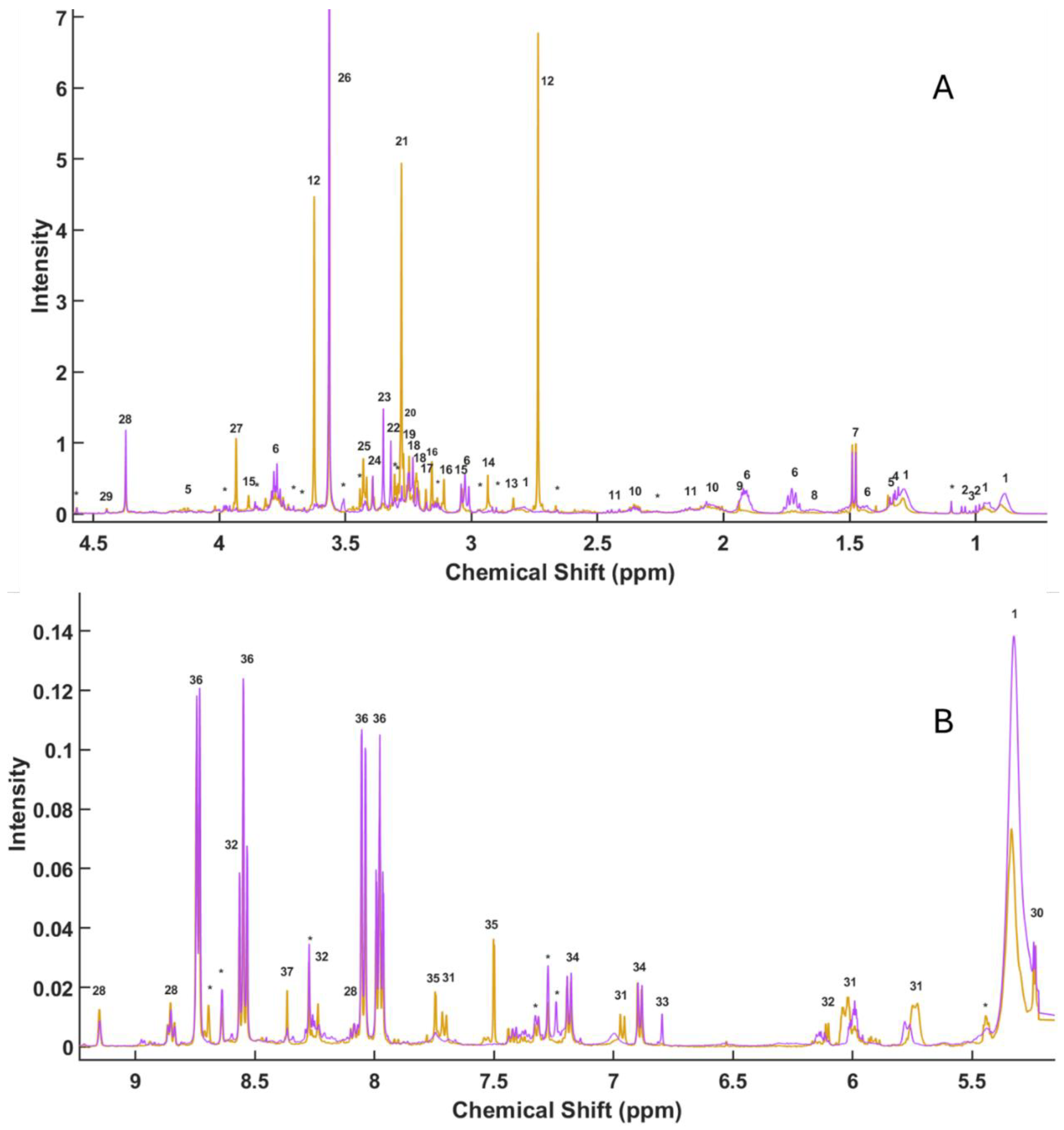
Figure 1. Representative 1D 1H CPMG HR-MAS NMR spectrum of gonadal tissues of Arbacia lixula (orange line) and Paracentrotus lividus (purple line).
Preprints 183494 g001
Figure 2. (A) VIP (blue line) and tpLoadings (orange line) values obtained from the PLS-LDA analysis of HR-MAS NMR spectra from Arbacia lixula and Paracentrotus lividus samples. Green dots indicate the spectral peaks with the highest VIP values, which were subsequently selected for the SPA (Successive Projections Algorithm) analysis. (B) PLS-LDA score plot showing sample clustering according to species: blue circles correspond to A. lixula and red diamonds to P. lividus.
Preprints 183494 g002
Figure 3. (A) COSS (Covariance Selection) values used to identify the most relevant spectral peaks differentiating Arbacia lixula and Paracentrotus lividus samples. (B) DMEAN versus DSD scatter plot. Variables with the highest discriminatory power are highlighted as green circles, whereas less relevant variables are shown as blue circles. The numbers appearing in the figures correspond to the compounds assigned in Table 1.
Preprints 183494 g003
Figure 4. HR-MAS NMR spectral regions and boxplots of selected metabolites differentiating Arbacia lixula and Paracentrotus lividus samples. For each metabolite, representative spectral peaks (left panels) and corresponding boxplots of signal intensities (right panels) are shown. Metabolites: (A–B) Betaine, (C–D) Sarcosine, (E–F) Glycine, (G–H) Malonate, (I–J) Taurine, (K–L) Formaldehyde, and (M–N) Lysine.
Preprints 183494 g004
Table 1. Assignment of 1D 1H CPMG HR-MAS NMR of gonadal tissues of Arbacia lixula and Paracentrotus lividus. The table lists the peak number used in Figure 1, the chemical shift (δ, ppm), the compound name, and the multiplicity observed in the spectra.
Peak δ (ppm) Compound Multiplicity
1 0.8846 F.A m
1 0.9657 F.A. m
2 0.9851 Valine d
3 1.0101 Isoleucine d
2 1.0416 Valine d
* 1.0962 Unassigned s
1 1.284 F.A. m
4 1.3074 Treonine d
5 1.3262 Lactate d
6 1.4307 Lysine m
7 1.4754 Alanine d
8 1.6562 Arginine m
6 1.7288 Lysine m
6 1.9209 Lysine m
9 1.9389 Acetate s
10 2.068 Glutamate m
11 2.1344 Glutamine m
* 2.2653 Unassigned m
10 2.3573 Glutamate m
11 2.4446 Glutamine m
* 2.6954 Unassigned s
12 2.7357 Sarcosine s
1 2.7893 F.A. m
13 2.8333 TMA s
* 2.9338 Unassigned d
14 2.9349 N-Methylhydantion s
* 2.9741 Unassigned d
6 3.0258 Lysine t
15 3.0375 Creatine s
16 3.1094 Malonate s
* 3.1351 Unassigned d
16 3.1556 Malonate s
17 3.1571 Methylmalonate s
18 3.2183 Choline s
18 3.2326 Choline s
19 3.2385 Carnitine s
19 3.2476 Carnitine s
20 3.2696 TMAO s
21 3.2781 Betaine s
* 3.3037 Unassigned s
* 3.3048 Unassigned s
22 3.3202 3-carboxypropyl-trimethyl-amonium s
23 3.35 Formaldehyde s
24 3.3918 Methanol s
25 3.4292 Taurine t
* 3.44 Unassigned s
* 3.50763 Unassigned s
26 3.56447 Glycine s
12 3.6242 Sarcosine s
* 3.6242 Unassigned s
* 3.662 Unassigned s
6 3.7717 Lysine/Alanine m
* 3.8589 Unassigned m
15 3.8846 Creatine s
27 3.9341 Betaine s
* 3.9741 Unassigned m
5 4.1255 Lactate q
28 4.3716 Trignelline s
29 4.446 Inosine s
30 5.2421 Glucose d
1 5.3261 F.A. s
* 5.4335 Unassigned m
31 5.7291 Uridine m
31 6.0202 Uridine m
32 6.1005 IMP d
33 6.7965 Timol s
34 6.8985 Tyrosine d
31 6.9718 Uridine d
34 7.1783 Tyrosine d
* 7.2399 Unassigned s
* 7.2751 Unassigned s
* 7.3286 Unassigned d
35 7.5021 Xantine s
31 7.6993 Uridine d
35 7.7477 Xantine s
36 7.978 Kinurenine t
36 8.0532 Kinurenine d
28 8.088 Trigonelline t
32 8.2373 IMP s
* 8.2736 Unassigned s
37 8.3667 Adenosine s
36 8.5501 Kinurenine t
32 8.6014 IMP s
* 8.6381 Unassigned s
* 8.6953 Unassigned s
36 8.7312 Kinurenine d
28 8.8508 Trigonelline t
28 9.1522 Trigonelline s
Abbreviations: s, singlet; d, doublet; t, triplet; q, quartet; m, multiplet.
Disclaimer/Publisher’s Note: The statements, opinions and data contained in all publications are solely those of the individual author(s) and contributor(s) and not of MDPI and/or the editor(s). MDPI and/or the editor(s) disclaim responsibility for any injury to people or property resulting from any ideas, methods, instructions or products referred to in the content.
Copyright: This open access article is published under a Creative Commons CC BY 4.0 license, which permit the free download, distribution, and reuse, provided that the author and preprint are cited in any reuse.
Prerpints.org logo

Preprints.org is a free preprint server supported by MDPI in Basel, Switzerland.

Subscribe

Disclaimer

Terms of Use

Privacy Policy

Privacy Settings

© 2026 MDPI (Basel, Switzerland) unless otherwise stated