
Submitted:
29 October 2025
Posted:
30 October 2025
You are already at the latest version
Abstract
Background: Supernumerary teeth (ST) are developmental anomalies that may interfere with eruption, alignment, and occlusal balance. Their etiopathogenesis and management remain controversial. This multicentric study aimed to evaluate the epidemiological, morphological, and radiographic features of ST in a Romanian population and identify impact predictors. Methods: Between 2020 and 2025, 153 patients (91 males, 62 females; mean age 14.8 ± 6.2 years) with clinically and radiographically confirmed ST were examined in three academic centers. Diagnostic assessment included orthopantomography and cone-beam computed tomography (CBCT). Teeth were classified by morphology, topography, and eruption status. Statistical analyses and binary logistic regression identified significant predictors (p < 0.05). Results: A total of 185 ST were recorded, most frequently conical (48.6%) and mesiodens (56.2%). Complications were observed in 40.5% of patients. Predictors of impaction included younger age (< 13 years) (OR = 3.12; p = 0.003), male gender (OR = 1.78; p = 0.046), tuberculate morphology (OR = 2.93; p = 0.021), and Class III malocclusion (OR = 1.89; p = 0.039). CBCT demonstrated superior diagnostic accuracy (κ = 0.89). Conclusions: Supernumerary teeth show morphology- and age-dependent impaction risk. CBCT provides enhanced diagnostic precision, supporting personalized, evidence-based management and future genetic investigations.
Keywords:
supernumerary teeth
; mesiodens
; impaction
; cone-beam computed tomography
; morphology
; malocclusion
; eruption disturbance
; dento-maxillary anomalies
; diagnostic accuracy
; epidemiology
1. Introduction
Supernumerary teeth (ST), defined as a developmental anomaly representing an excess of the normal dental arch count—32 in the permanent and 20 in the primary dentition—constitute a complex clinical condition that can significantly affect both the aesthetic and functional integrity of the dento-maxillary system [1]. These additional teeth, which display considerable variation in shape, size, position, and eruption pattern, are a subject of ongoing research focusing on their classification, epidemiological distribution, and multifactorial etiopathogenesis [2,3].
The presence of supernumerary teeth (Figure 1) is not a mere morphological curiosity but a potential source of diverse pathological complications [1,2,3,4].
The most frequently encountered issues include delayed eruption or impaction, displacement, and malformation of adjacent permanent teeth [4,5]. In more severe cases, ST may induce root resorption, cystic formations, or ectopic migrations into the nasal cavity or maxillary sinus [6,7,8,9,10]. Furthermore, these anomalies can interfere with alveolar bone grafting, orthodontic alignment, or implant placement, and in rare instances, have been associated with neurological disturbances such as neuralgia or paresthesia [11,12,13]. Therefore, early detection and accurate diagnosis are crucial to prevent or mitigate secondary complications [14,15,16,17].
Etiopathogeny, Epidemiology, and Clinical Relevance
The etiopathogenesis of supernumerary teeth remains incomplete, although most authors agree that their occurrence reflects a developmental disturbance during organogenesis and dental morphogenesis [2,18,19,20]. Several hypotheses have been proposed to elucidate this complex process. One of the earliest is the tooth bud division theory, which suggests that a single tooth germ divides partially or completely, resulting in two distinct dental structures [2,18,19]. Another explanation, known as the third dentition theory, proposes that supernumerary teeth arise from dental lamina remnants reactivation during a hypothetical third cycle of tooth development, following the primary and permanent dentitions [2,20,21].
A further perspective considers the proliferation of epithelial remnants, wherein residual elements of the dental lamina or Hertwig’s epithelial root sheath, persisting in the jaws after the normal tooth development process, may undergo abnormal proliferation and differentiation under the influence of local or systemic stimuli, giving rise to additional tooth structures [2,19].
The dental lamina hyperactivity theory has gained the widest acceptance in contemporary literature. This hypothesis attributes the formation of supernumerary teeth to localized and independent hyperactivity of the dental lamina, which leads to the development of supplementary epithelial buds during embryogenesis [19,20,22,23]. Such hyperactivity may be triggered by vascular, histochemical, or developing craniofacial region structural environment, affecting the delicate regulatory mechanisms that control tooth initiation and morphogenesis [21,22].
Overall, these theories indicate that the genesis of supernumerary teeth is multifactorial, arising from an interplay between genetic susceptibility, local developmental disturbances, and environmental influences that alter the normal morphogenetic potential of the dental lamina [3,24].
The increased frequency of ST in the maxillary incisor region (Figure 2) may be linked to intricate tissue rearrangements and morphogenetic activity in this area [2,17,20,25]. Nevertheless, no single theory fully accounts for the wide diversity of clinical presentations, suggesting that the etiology of ST is multifactorial, shaped by a complex interplay between genetic predisposition and environmental influences [3,24].
Genetic studies have revealed familial clustering of cases and associations with syndromic conditions such as Gardner’s syndrome and cleidocranial dysplasia [25,26]. Brook’s unified etiological model [2,26] postulates that supernumerary teeth result from genetically determined susceptibility, modulated by environmental and epigenetic factors. This framework reflects the modern understanding of dental developmental anomalies as polygenic and multifactorial disorders rather than isolated occurrences.
Epidemiological Overview
Epidemiological studies estimate the prevalence of supernumerary teeth at 0.1% to 3.8% in the general population, with a distinct male predominance (approximately 2:1) and a greater incidence in the permanent dentition [17,27]. The anterior maxilla represents the most common site of occurrence, particularly the mesiodens type. Approximately 75% of supernumerary teeth are impacted and asymptomatic, often discovered incidentally during routine radiographic assessments [2]. To provide a clearer understanding of the occurrence and clinical behavior of supernumerary teeth, Table 1 compiles essential epidemiological and clinical data reported in the literature.
The clinical impact of ST varies with location, morphology, and eruption status [29]. While some remain clinically silent, others contribute to malocclusion, delayed eruption of permanent successors, or diastemas, and may cause aesthetic and functional disturbances that require multidisciplinary management [31,32].
Diagnostic Essentials
Accurate diagnosis and precise localization are indispensable for successful treatment planning and prevention of secondary complications [2,18]. Traditional two-dimensional radiographic methods [33], such as panoramic (orthopantomogram) and periapical radiographs (Figure 3), remain valuable as initial diagnostic tools that often fail to provide sufficient information regarding the three-dimensional relationship between ST and adjacent anatomical structures [33,34].
Cone-Beam Computed Tomography (CBCT) addresses these limitations and has become a cornerstone in contemporary diagnostic protocols [35]. CBCT allows high-resolution, three-dimensional imaging from a single scan, offering detailed visualization of supernumerary tooth morphology, spatial orientation, and proximity to vital structures such as the nasal floor, maxillary sinus, and mandibular canal [35,36]. Moreover, CBCT has been shown to enhance the detection of associated pathologies, such as root resorption and cyst formation, with greater precision than conventional radiography [35,36]. Nevertheless, given the higher radiation exposure, its use should be carefully justified based on a risk-benefit assessment for each case [37,38].
AI systems that use CBCT scans can interpret the images precisely and suggest the most suitable surgical strategy for managing supernumerary teeth. This technology makes the operation easier and faster to perform [15].
Management Strategies and Current Controversies
The therapeutic management of supernumerary teeth generally follows two principal approaches, extraction or non-extraction (clinical monitoring), with the choice largely determined by the tooth’s position, its relationship to adjacent anatomical structures, and the presence or risk of associated pathology [39]. In cases where supernumerary teeth interfere with eruption, alignment, or occlusion, surgical removal is typically indicated. Asymptomatic and non-disruptive teeth may be observed periodically through clinical and radiographic follow-up [39,40].
A persistent point of debate within the literature concerns the optimal timing of surgical intervention [41,42,43]. The late approach advocates postponing extraction until after the apical closure of the permanent incisors, generally around the age of ten years. This strategy minimizes the risk of damaging developing roots and surrounding periodontal structures, thereby preserving long-term dental integrity. In contrast, the early approach supports immediate removal upon diagnosis, particularly when the supernumerary tooth exhibits an oblique orientation, distorted morphology, or a position that makes spontaneous eruption improbable [18,41,42,43].
Ultimately, the decision regarding timing must balance the risks of surgical trauma against the potential for eruption disturbances, the individual’s age, tooth development stage, and overall orthodontic and aesthetic prognosis [42,43].
Some clinicians advocate for non-intervention in asymptomatic cases, emphasizing the importance of long-term monitoring to avoid surgical complications [44,45]. This ongoing debate highlights the need for evidence-based, individualized management strategies grounded in comprehensive diagnostic assessment and interdisciplinary collaboration.
The primary objective of this multicentric study was to analyze the epidemiological, morphological, and radiographic characteristics of supernumerary teeth (ST) in a Romanian population, identify clinical and occlusal correlations, and determine predictors of impaction using multivariate analysis. Secondary objectives included assessing the diagnostic performance of conventional radiography versus CBCT and evaluating the relationship between supernumerary morphology, location, and associated pathologies.
2. Materials and Methods
This multicentric, observational, analytical study was conducted between January 2020 and March 2025 in two Romanian academic centers: the University Dental Clinics of Târgu Mureș and Sibiu. A retrospective review of clinical and radiographic records was performed to identify all eligible patients. One hundred fifty-three consecutive patients with clinically and radiographically confirmed supernumerary teeth (ST) who met the inclusion criteria were enrolled from the three academic centers. The use of consecutive sampling across the study period minimizes selection bias. The study adhered to the STROBE reporting guidelines for observational research. Written informed consent was obtained from all participants or legal guardians.
2.1. Inclusion and Exclusion Criteria
Inclusion criteria comprised patients presenting one or more supernumerary teeth in mixed or permanent dentition, with complete diagnostic records including orthopantomogram (OPG) and/or cone-beam computed tomography (CBCT) imaging.
Exclusion criteria included syndromic disorders (e.g., cleidocranial dysplasia, Gardner’s syndrome), craniofacial trauma, prior orthodontic or surgical interventions, or incomplete imaging documentation.
2.2. Clinical and Radiographic Examination
Clinical examination included assessment of eruption status, dental crowding, malposition, and associated cystic or resorptive changes. Radiological evaluation followed a standardized diagnostic protocol:
- OPG (Digital panoramic radiography) for screening and global assessment.
- Periapical and occlusal radiographs to refine localization.
- CBCT for complex or ambiguous cases, performed at 0.2–0.3 mm voxel resolution, 8 × 8 cm field of view, and 90 kVp/10 mA exposure parameters.
Two calibrated examiners (oral radiologists) independently analyzed the images, and inter-observer agreement was excellent (Cohen’s κ = 0.91). Discrepancies were resolved by consensus.
2.3. Classification Criteria
Supernumerary teeth were categorized according to:
- -
- Morphology: conical, tuberculate, supplemental, or odontomatous.
- -
- Topography: mesiodens, paramolar, distomolar, or supernumerary premolar.
- -
- Eruption status: erupted, impacted, or included.
Malocclusion was classified using Angle’s classification (Class I, II/1, II/2, III) to assess potential occlusal associations.
2.4. Bias Mitigation
To mitigate potential sources of bias inherent in this observational study design, several steps were taken:
- -
- Selection Bias: Sampling for all eligible patients seen between January 2020 and March 2025 aimed to minimize selection bias by preventing the deliberate exclusion or inclusion of patients based on their complication severity.
- -
- Information Bias (Measurement Bias): To ensure high reliability of radiological data, two calibrated examiners (oral radiologists) independently analyzed all images. The subsequent calculation and verification of excellent inter-observer agreement (kappa = 0.91) standardized the radiographic interpretation, limiting measurement bias.
- -
- Confounding Bias: The study controlled for several known potential confounders of impaction, including age, gender, and malocclusion, by including these variables in the Binary Logistic Regression model (as detailed in Section 2.5).
Given this study's retrospective, multicentric nature, a formal a priori power analysis was not performed. Instead, the final sample size of 153 consecutive patients represented the maximum available population meeting all inclusion criteria within the five-year study period (2020–2025). Based on the final logistic regression model, which identified four significant independent predictors of impaction using 153 cases, the sample size was deemed sufficient to achieve appropriate power to detect the reported effects (OR = 1.78 to 3.12) with a p < 0.05 and a confidence level of 95%.
2.5. Statistical Analysis
All data were entered into IBM SPSS v29.0 and RStudio v2024.3. Descriptive statistics summarized demographic, morphological, and radiographic findings. Chi-square or Fisher’s exact tests were used for categorical comparisons, and independent t-tests for continuous variables. Spearman’s correlation assessed relationships between continuous variables. Binary logistic regression was performed to identify independent predictors of impaction. Statistical significance was set at p < 0.05, and 95% confidence intervals (CIs) were reported.
3. Results
3.1. Demographic Characteristics
The study population comprised 153 patients (91 males, 62 females; mean age = 14.8 ± 6.2 years). Most participants (47%) were aged 6-12, and 67.3% resided in urban areas. A total of 185 supernumerary teeth were identified (mean 1.21 ± 0.56 per patient), with single occurrences in 83.0%, double in 13.1%, and multiple (>2) in 3.9% of cases.
Table 2 shows the distribution of patients by gender and dental class according to Angle’s classification.
3.2. Distribution by Gender and Tooth Position
Impaction patterns differed significantly by gender. Males (56%) exhibited more frequent impactions, particularly in anterior regions. As presented in Table 3, most erupted teeth occurred in the lateral zones, whereas combined frontal and lateral impactions were more common in males.
3.3. Age-Related Distribution
Younger patients (<13 years) exhibited significantly higher impaction rates (OR = 3.12, p = 0.003). The detailed age-group distribution is presented in Table 4.
3.4. Morphological and Topographical Features
Morphologically, conical teeth were most common (48.6%), followed by tuberculate (27.0%), supplemental (18.4%), and odontomatous (6.0%) forms. Topographically, mesiodens accounted for over half of the total (56.2%), followed by paramolar (16.2%), distomolar (14.1%), and supernumerary premolar (13.5%) variants (Table 5).
3.5. Clinical Complications
Clinical complications were observed in 40.5% of patients. The most frequent were delayed eruption (19.6%), followed by malposition or rotation (12.4%), cystic formation (4.6%), and root resorption (3.9%). Details are presented in Table 6.
3.6. Radiographic Findings
OPG successfully detected 92.4% of supernumerary teeth, whereas CBCT confirmed all cases and identified 11 root resorptions and 9 vestibulo-palatal displacements missed by the OPG. The diagnostic concordance between the two imaging modalities was high (κ = 0.89), supporting CBCT’s superior sensitivity for spatial localization and pathological assessment.
3.7. Predictors of Impaction
Binary logistic regression identified several significant predictors of impaction (Table 7). Younger age (<13 years), male gender, tuberculate morphology, and Class III malocclusion were independently associated with an increased risk of impaction.
3.8. Correlations
Spearman’s correlation revealed a moderate positive relationship between age and the number of supernumerary teeth (ρ = 0.33, p < 0.001), indicating a gradual increase in multiplicity with advancing age, likely due to progressive detection over time.
The study demonstrates a clear male predominance, high frequency of anterior mesiodens impactions, and strong age-dependent and morphology-dependent patterns of pathology. CBCT provided superior diagnostic accuracy, and impaction was significantly predicted by early age, male sex, tuberculate form, and Class III occlusal pattern.
4. Discussion
This multicentric investigation provides a detailed evaluation of the epidemiological, clinical, and radiological profiles of supernumerary teeth in a Romanian population, offering new insights into the interplay between morphology, occlusion, and impaction risk. The study’s findings reinforce and expand current understanding of ST behavior and support the importance of individualized, imaging-guided management strategies.
The male predominance (59.5%) observed aligns with the widely reported gender disparity, confirming previous data from European and Asian populations, where male-to-female ratios range from 1.4:1 to 3.2:1 [18,35,46,47,48]. This trend may reflect the influence of sex-linked genetic factors or differential expression of odontogenic regulatory genes such as WNT10A or RUNX2, known to participate in dental lamina signaling pathways [49]. The predominance of detection between 6 and 12 years further emphasizes the role of panoramic screening during early mixed dentition, when most supernumerary formations become radiographically visible and can be intercepted before causing eruption disturbances [50].
Morphologically, the conical form was the most prevalent (48.6%), consistent with earlier studies by Davidson et al. (2025) and Rajab and Hamdan (2002), which reported similar frequencies of 45–55% [2,51]. The second most common type, tuberculate teeth (27%), demonstrated a distinct pathological profile, showing stronger associations with delayed eruption and cystic transformation [2,15]. This is biologically plausible, as tuberculate teeth possess irregular morphology and multicuspid crowns that impede the normal eruption pathway, predisposing to impaction and pericoronal cyst development [52]. Supplemental and odontomatous forms in a smaller proportion support the theory that hyperactivity of the dental lamina leads to variable degrees of morphological differentiation [20,21,22,23].
Topographically, mesiodens accounted for 56.2% of all supernumerary teeth, confirming that the anterior maxilla is the predominant site of occurrence [1,2,28]. This localization correlates with the area of greatest embryonic epithelial complexity and supports the dental lamina hyperactivity theory as the prevailing etiopathogenic model [1,2,21,22,23,28]. The clustering of supernumerary teeth in this region may also be influenced by mechanical and spatial constraints during incisor eruption, explaining the higher frequency of impactions in anterior sectors [1,52,53,54].
From an orthodontic standpoint, the distribution of Angle classes provides a novel dimension to understanding the skeletal context of supernumerary teeth. The significant association between Class III malocclusion and anterior impactions (p = 0.008) represents an original finding. Similar tendencies have been noted in Korean and Turkish cohorts, where concave facial growth patterns and anterior cross-bites were correlated with restricted eruption spaces [55,56,57]. This relationship may be mediated by anteroposterior maxillary deficiency, altering the eruption trajectory of incisors and promoting mesiodens impaction.
The logistic regression model strengthened these associations, identifying younger age (<13 years), male gender, tuberculate morphology, and Class III malocclusion as independent predictors of impaction. Younger patients showed a 3.1-fold higher risk, reflecting developmental timing, since many permanent successors are still unerupted, and increased detectability in early radiographic surveys. Male gender nearly doubled the odds of impaction (OR = 1.78), paralleling reports from Ferrés-Padró et al. (2013) and Patil et al. (2020) [58,59], which attributed this tendency to sexual dimorphism in craniofacial growth and tooth bud spacing. The finding that tuberculate morphology increases cystic potential nearly three-fold (OR = 2.93) supports the notion that crown form directly influences pathological sequelae.
The positive correlation between age and the number of supernumerary teeth (ρ = 0.33, p < 0.001) can be interpreted as a detection bias but also indicates that multiple ST, especially distomolars and premolars, mineralize later and thus appear progressively in older patients. Comparable age-related trends were reported by Anthonappa et al. (2015), reinforcing the need for long-term radiographic follow-up in patients with known mesiodens, as additional teeth may develop subsequently [15].
Radiographically, this study confirmed the diagnostic superiority of CBCT over orthopantomography. While OPG detected 92.4% of ST, CBCT achieved full detection and revealed additional findings—11 unrecognized root resorptions and 9 vestibulo-palatal displacements. The high inter-modality concordance (κ = 0.89) supports the reliability of panoramic screening for preliminary assessment, validating CBCT as the gold standard for surgical planning, particularly for teeth adjacent to vital structures or in complex morphologies [60,61]. These observations echo the results of Primosch (1981) and recent analyses by Gündüz et al. (2022), emphasizing CBCT’s critical role in minimizing iatrogenic risks during extraction [62,63]. However, consistent with ALARA principles, CBCT should be reserved for cases where conventional imaging is inconclusive or where three-dimensional spatial orientation dictates treatment choice [64,65].
Clinically, 40.5% of patients presented complications, with delayed eruption (19.6%) as the most frequent, followed by malposition (12.4%), cyst formation (4.6%), and root resorption (3.9%). These frequencies are comparable to the 30–40% complication rates described by Fardi et al. (2011) and Kara et al. (2012) [66,67]. The link between tuberculate morphology and cystic change observed in our series mirrors the findings of He et al. (2023), who reported cystic transformation in 9–11% of cases, increasing with age [35]. Thus, early diagnosis prevents impaction and mitigates the risk of secondary pathologies that complicate orthodontic management.
Overall, the findings support early radiographic screening, ideally between 6 and 8 years, when eruption disturbances can be anticipated and minimally invasive extraction can be performed before root maturation of permanent successors. However, the debate regarding early versus delayed surgical removal remains relevant. Our data suggest early intervention for obliquely positioned or tuberculate forms, and delayed extraction after apical closure may be justified for conical mesiodens without associated complications. Therefore, a balanced, patient-specific approach remains fundamental.
Despite employing rigorous consecutive sampling and high inter-rater reliability, the study acknowledges several limitations inherent to its multicentric, observational design. First, the university clinic-based patient recruitment introduces referral bias, as our cohort may disproportionately represent symptomatic cases or those with complex impactions referred for specialized care, potentially overestimating the true prevalence of complications (40.5%) in the general population. Second, while CBCT protocols were standardized, slight variations in imaging equipment and voxel resolution across the participating centers may contribute to minimal information heterogeneity. Finally, the study’s cross-sectional nature precludes definitive causal inference; thus, the identified predictors (e.g., tuberculate morphology) define a strong risk association but not a direct cause-and-effect relationship.
Future research should aim for longitudinal follow-up to assess eruption outcomes after early versus delayed intervention and to evaluate recurrence or subsequent development of additional supernumerary teeth. Integrating genomic and epigenetic analyses, including candidate gene mapping for WNT10A, RUNX2, and AXIN2 polymorphisms, may clarify the molecular basis of dental lamina hyperactivity [49]. Expanding sample diversity across ethnic and age groups would enhance generalizability. Furthermore, AI-assisted radiographic evaluation could standardize detection and improve diagnostic reproducibility in large cohorts.
Supernumerary teeth represent a multifactorial developmental anomaly with diverse clinical presentations and potential complications. Their management demands a careful balance between timely intervention and conservative monitoring, guided by accurate imaging and individualized clinical judgment. Spanning genetic, environmental, and developmental factors, the complexity of their etiology underscores the importance of continued research to refine diagnostic methods and establish standardized treatment protocols. Ultimately, successful management of ST relies on an integrated approach that unites the expertise of orthodontists, oral surgeons, and radiologists to ensure optimal functional and aesthetic outcomes.
This study successfully characterizes the morphological and epidemiological patterns of supernumerary teeth, identifies risk factors for impaction, and evaluates diagnostic tools.
5. Conclusions
This study provides comprehensive evidence on the clinical and radiological behavior of supernumerary teeth within a Romanian population, emphasizing their multifactorial developmental nature. The results demonstrate that male gender, younger age, tuberculate morphology, and Class III malocclusion are significant predictors of impaction. CBCT remains the superior imaging method for precise localization and detection of associated complications. The morphological, radiological, and occlusal parameters offer a robust individualized diagnosis and management, setting the foundation for future genetic and developmental investigations.
References
- Lykousis, A.; Pouliezou, I.; Christoloukas, N.; Rontogianni, A.; Mitsea, A.; Angelopoulos, C. Supernumerary Teeth in the Anterior Maxilla of Non-Syndromic Children and Adolescents: A Retrospective Study Based on Cone-Beam Computed Tomography Scans. Pediatr. Rep. 2025, 17, 52. [Google Scholar] [CrossRef] [PubMed]
- Davidson, C.L.; Smit, C.; Nel, S. Supernumerary Teeth: A Pictorial Review and Revised Classification. J. Oral Biol. Craniofac. Res. 2025, 15, 454–462. [Google Scholar] [CrossRef]
- Khalaf, K.; Brook, A.H.; Smith, R.N. Genetic, Epigenetic and Environmental Factors Influence the Phenotype of Tooth Number, Size and Shape: Anterior Maxillary Supernumeraries and the Morphology of Mandibular Incisors. Genes 2022, 13, 2232. [Google Scholar] [CrossRef]
- Mallineni, S.K.; Aldhuwayhi, S.; Deeban, Y.; Almutairi, K.S.; Alhabrdi, S.N.; Almidaj, M.A.; Alrumi, B.A.; Assalman, A.S.; Joseph, A.M.; Thakare, A.A.; et al. Prevalence, Occurrence, and Characteristics of Supernumerary Teeth Among the Saudi Arabian Population Using Panoramic Radiographs. Diagnostics 2024, 14, 2542. [Google Scholar] [CrossRef] [PubMed]
- Rai, A.; Jain, A.; Agrawal, G.D.; Agrawal, S. Non-Syndromal Multiple Supernumerary and Permanent Impacted Teeth in Mother and Her One Child. BMJ Case Rep. 2020, 13, e236395. [Google Scholar] [CrossRef] [PubMed]
- Mathur, S.; Verma, B.; Dabholkar, Y.; Saberwal, A. Supernumerary Tooth in the Nasal Cavity. J. Oral Maxillofac. Pathol. 2021, 25, 373–374. [Google Scholar] [CrossRef]
- Touiheme, N.; Messary, A. Supernumerary Ectopic Tooth on the Maxillary Sinus. Pan Afr. Med. J. 2014, 18, 353. [Google Scholar] [CrossRef]
- Arora, P.; Nair, M.K.; Liang, H.; Patel, P.B.; Wright, J.M.; Tahmasbi-Arashlow, M. Ectopic Teeth with Disparate Migration: A Literature Review and New Case Series. Imaging Sci. Dent. 2023, 53, 229–238. [Google Scholar] [CrossRef]
- Kataoka, T.; Amemiya, K.; Goto, T.; Kina, H.; Tajima, E.; Okamoto, T. A Unique Case of Supernumerary Teeth Erupting Inside a Maxillary Sinus Osteoma. J. Clin. Med. 2024, 13, 4067. [Google Scholar] [CrossRef]
- Ainiwaer, A.; Yiminjiang, B.; Ling, W. Extraction of an Ectopic Supernumerary Tooth through Nasal Cavity with Piezosurgery under Local Anesthesia: A Case Report. Clin. Case Rep. 2024, 12, e9221. [Google Scholar] [CrossRef]
- Kaplansky, I.V.; Kurtzman, G.M. Implant Placement When an Impacted Tooth and Supernumerary Teeth Are Present in the Maxilla. Compend. Contin. Educ. Dent. 2024, 45, e1–e4. [Google Scholar]
- Parolia, A.; Kundabala, M.; Dahal, M.; Mohan, M.; Thomas, M.S. Management of Supernumerary Teeth. J. Conserv. Dent. 2011, 14, 221–224. [Google Scholar] [CrossRef]
- Ahmadi, S.; Liang, X.; Xu, F.; Wang, C.; Xie, L.; Yu, M.; Tu, J.; Na, S. Dynamic Navigation Systems in Dento-Alveolar Surgery: Advancements and Clinical Applications. Saudi Dent. J. 2025, 37, 60. [Google Scholar] [CrossRef]
- Makrygiannakis, M.A.; Giannakopoulos, K.; Kavadella, A.; Paraskevis, D.; Kaklamanos, E.G. Diagnostic Accuracy of an Artificial Intelligence-Based Software in Detecting Supernumerary and Congenitally Missing Teeth in Panoramic Radiographs. Eur. J. Orthod. 2025, 47, cjaf054. [Google Scholar] [CrossRef]
- Mladenovic, R.; Kalevski, K.; Davidovic, B.; Jankovic, S.; Todorovic, V.S.; Vasovic, M. The Role of Artificial Intelligence in the Accurate Diagnosis and Treatment Planning of Non-Syndromic Supernumerary Teeth: A Case Report in a Six-Year-Old Boy. Children 2023, 10, 839. [Google Scholar] [CrossRef] [PubMed]
- Aşar, E.M.; İpek, İ.; Bilge, K. Customized GPT-4V(ision) for Radiographic Diagnosis: Can Large Language Model Detect Supernumerary Teeth? BMC Oral Health 2025, 25, 756. [Google Scholar] [CrossRef] [PubMed]
- Katanaki, N.; Makrygiannakis, M.A.; Kaklamanos, E.G. The Prevalence of Supernumerary Teeth in a Sample of Non-Syndromic Young Patients from Greece. Dent. J. 2025, 13, 317. [Google Scholar] [CrossRef] [PubMed]
- Ata-Ali, F.; Ata-Ali, J.; Peñarrocha-Oltra, D.; Peñarrocha-Diago, M. Prevalence, Etiology, Diagnosis, Treatment and Complications of Supernumerary Teeth. J. Clin. Exp. Dent. 2014, 6, e414–e418. [Google Scholar] [CrossRef]
- Albu, Ş.D.; Pavlovici, R.C.; Imre, M.; Ion, G.; Ţâncu, A.M.C.; Albu, C.C. Phenotypic Heterogeneity of Non-Syndromic Supernumerary Teeth: Genetic Study. Rom. J. Morphol. Embryol. 2020, 61, 853–861. [Google Scholar] [CrossRef]
- Singh, A.K.; Soni, S.; Jaiswal, D.; Pani, P.; Sidhartha, R.; Nishant. Prevalence of Supernumerary Teeth and Its Associated Complications among School-Going Children between the Ages of 6 and 15 Years of Jamshedpur, Jharkhand, India. Int. J. Clin. Pediatr. Dent. 2022, 15, 504–508. [Google Scholar] [CrossRef]
- Kiso, H.; Takahashi, K.; Mishima, S.; et al. Third Dentition Is the Main Cause of Premolar Supernumerary Tooth Formation. J. Dent. Res. 2019, 98, 968–974. [Google Scholar] [CrossRef]
- Wang, X.P.; Fan, J. Molecular Genetics of Supernumerary Tooth Formation. Genesis 2011, 49, 261–277. [Google Scholar] [CrossRef]
- Ahtiainen, L.; Uski, I.; Thesleff, I.; Mikkola, M.L. Early Epithelial Signaling Center Governs Tooth Budding Morphogenesis. J. Cell Biol. 2016, 214, 753–767. [Google Scholar] [CrossRef] [PubMed]
- Lu, X.; Yu, F.; Liu, J.; Cai, W.; Zhao, Y.; Zhao, S.; Liu, S. The Epidemiology of Supernumerary Teeth and the Associated Molecular Mechanism. Organogenesis 2017, 13, 71–82. [Google Scholar] [CrossRef]
- Demiriz, L.; Durmuşlar, M.C.; Mısır, A.F. Prevalence and Characteristics of Supernumerary Teeth: A Survey on 7348 People. J. Int. Soc. Prev. Community Dent. 2015, 5 (Suppl. 1), S39–S43. [Google Scholar] [CrossRef]
- Subasioglu, A.; Savas, S.; Kucukyilmaz, E.; Kesim, S.; Yagci, A.; Dundar, M. Genetic Background of Supernumerary Teeth. Eur. J. Dent. 2015, 9, 153–158. [Google Scholar] [CrossRef] [PubMed]
- Syriac, G.; Joseph, E.; Rupesh, S.; Philip, J.; Cherian, S.A.; Mathew, J. Prevalence, Characteristics, and Complications of Supernumerary Teeth in Nonsyndromic Pediatric Population of South India: A Clinical and Radiographic Study. J. Pharm. Bioallied Sci. 2017, 9 (Suppl. 1), S231–S236. [Google Scholar] [CrossRef]
- Satish, V.; Panda, S.; Maganur, P.; Ahmed, A. Multiple Bilateral Unerupted Supplemental Premolars: An Unusual Presentation in a Nonsyndromic Patient. Int. J. Clin. Pediatr. Dent. 2017, 10, 217–222. [Google Scholar] [CrossRef] [PubMed]
- Henninger, E.; Friedli, L.; Makrygiannakis, M.A.; Zymperdikas, V.F.; Papadopoulos, M.A.; Kanavakis, G.; Gkantidis, N. Supernumerary Tooth Patterns in Non-Syndromic White European Subjects. Dent. J. 2023, 11, 230. [Google Scholar] [CrossRef]
- Impellizzeri A, Midulla G, Romeo U, La Monaca C, Barbato E, Galluccio G. Delayed Eruption of Permanent Dentition and Maxillary Contraction in Patients with Cleidocranial Dysplasia: Review and Report of a Family. Int J Dent. 2018 Jul 4;2018:6591414. [CrossRef]
- Mathias, M.F.; Lobo-Piller, R.G.; Corrêa, M.S. Treatment of Supernumerary Teeth. Eur. J. Paediatr. Dent. 2011, 12, 275–278. [Google Scholar]
- Kumar, A.; Namdev, R.; Bakshi, L.; Dutta, S. Supernumerary Teeth: Report of Four Unusual Cases. Contemp. Clin. Dent. 2012, 3 (Suppl. 1), S71–S77. [Google Scholar] [CrossRef]
- Anthonappa, R.P.; King, N.M.; Rabie, A.B. Diagnostic Tools Used to Predict the Prevalence of Supernumerary Teeth: A Meta-Analysis. Dentomaxillofac. Radiol. 2012, 41, 444–449. [Google Scholar] [CrossRef] [PubMed]
- Lin, Z.; Zhou, C.; Hu, Z.; Zhang, Z.; Cheng, Y.; Fang, B.; He, H.; Wang, H.; Li, G.; Guo, J.; Guo, W.; Li, X.; Zheng, G.; Li, Z.; Zeng, D.; Liu, Y.; Liu, Y.; Hu, M.; Xia, L.; Zhao, J.; Song, Y.; Li, H.; Ji, J.; Song, J.; Chen, L.; Wang, T. Expert Consensus on Imaging Diagnosis and Analysis of Early Correction of Childhood Malocclusion. Int. J. Oral Sci. 2025, 17, 21. [Google Scholar] [CrossRef] [PubMed]
- He, L.; Que, G.; Yang, X.; Yan, S.; Luo, S. Prevalence, Clinical Characteristics, and 3-Dimensional Radiographic Analysis of Supernumerary Teeth in Guangzhou, China: A Retrospective Study. BMC Oral Health 2023, 23, 351. [Google Scholar] [CrossRef] [PubMed]
- Gurgel, C.V.; Costa, A.L.; Kobayashi, T.Y.; et al. Cone Beam Computed Tomography for Diagnosis and Treatment Planning of Supernumerary Teeth. Gen. Dent. 2012, 60, e131–e135. [Google Scholar]
- Khader, A.; Jain, S.; Sarah; Mishra, S. ; Saleem, S.; Vijayan, A. Comparing Radiation Doses in CBCT and Medical CT Imaging for Dental Applications. J. Pharm. Bioallied Sci. 2024, 16 (Suppl. 2), S1795–S1797. [Google Scholar] [CrossRef]
- Hicks, D.; Melkers, M.; Barna, J.; Isett, K.R.; Gilbert, G.H. Comparison of the Accuracy of CBCT Effective Radiation Dose Information in Peer-Reviewed Journals and Dental Media. Gen. Dent. 2019, 67, 38–46. [Google Scholar]
- Belmehdi, A.; Bahbah, S.; El Harti, K.; El Wady, W. Non-Syndromic Supernumerary Teeth: Management of Two Clinical Cases. Pan Afr. Med. J. 2018, 29, 163. [Google Scholar] [CrossRef]
- Sobouti, F.; Ghadirian, H.; Dadgar, S.; Aryana, M.; Kamali, E. Radiographic Assessment and Management of Two Deeply and Horizontally Impacted Maxillary Central Incisors: A Clinical Case Report. Radiol. Case Rep. 2024, 19, 3089–3095. [Google Scholar] [CrossRef]
- Gupta, S.; Marwah, N. Impacted Supernumerary Teeth—Early or Delayed Intervention: Decision Making Dilemma? Int. J. Clin. Pediatr. Dent. 2012, 5, 226–230. [Google Scholar] [CrossRef]
- Jang, D.H.; Chae, Y.K.; Lee, K.E.; et al. Determination of the Range of Intervention Timing for Supernumerary Teeth Using the Korean Health Insurance Review and Assessment Service Database. J. Clin. Pediatr. Dent. 2023, 47, 67–73. [Google Scholar] [CrossRef] [PubMed]
- Barham, M.; Okada, S.; Hisatomi, M.; Khasawneh, A.; Tekiki, N.; Takeshita, Y.; Kawazu, T.; Fujita, M.; Yanagi, Y.; Asaumi, J. Influence of Mesiodens on Adjacent Teeth and the Timing of Its Safe Removal. Imaging Sci. Dent. 2022, 52, 67–74. [Google Scholar] [CrossRef]
- Permana, H.; Yusuf, A.S.H.; Alkaabi, S.A.; et al. Post-Surgical Complications of Supernumerary Teeth in the Mandibular Premolar Area: A Systematic Review. Heliyon 2024, 10, e35386. [Google Scholar] [CrossRef]
- Adhikari, M.; Jha, K.; Shah, A.; Kc, S.; Rayamajhi, M.; Koirala, R. Impacted Supernumerary Tooth in the Horizontal Plate of Palatine Bone: A Rare Case Report. BMC Oral Health 2024, 24, 1493. [Google Scholar] [CrossRef] [PubMed]
- McBeain, M.; Miloro, M. Characteristics of Supernumerary Teeth in Nonsyndromic Population in an Urban Dental School Setting. J. Oral Maxillofac. Surg. 2018, 76, 933–938. [Google Scholar] [CrossRef]
- Cassetta, M.; Altieri, F.; Giansanti, M.; Di-Giorgio, R.; Calasso, S. Morphological and Topographical Characteristics of Posterior Supernumerary Molar Teeth: An Epidemiological Study on 25,186 Subjects. Med. Oral Patol. Oral Cir. Bucal 2014, 19, e545–e549. [Google Scholar] [CrossRef] [PubMed]
- Shen, Z.; Wei, J.; Zhang, J.; Zhang, Y.; Yao, J. The Prevalence of Dental Agenesis, Supernumerary Teeth and Odontoma in a Chinese Paediatric Population: An Epidemiological Study. BMC Oral Health 2025, 25, 458. [Google Scholar] [CrossRef]
- Živković, M.; Stefanović, N.; Glišić, B.; et al. WNT10A and RUNX2 Mutations Associated with Non-Syndromic Tooth Agenesis. Eur. J. Oral Sci. 2022, 130, e12896. [Google Scholar] [CrossRef]
- Mine, Y.; Iwamoto, Y.; Okazaki, S.; et al. Detecting the Presence of Supernumerary Teeth during the Early Mixed Dentition Stage Using Deep Learning Algorithms: A Pilot Study. Int. J. Paediatr. Dent. 2022, 32, 678–685. [Google Scholar] [CrossRef]
- Rajab LD, Hamdan MA. Supernumerary teeth: review of the literature and a survey of 152 cases. Int J Paediatr Dent. 2002, 12, 244–254. [Google Scholar] [CrossRef]
- Yeluri, R.; Hegde, M.; Baliga, S.; Munshi, A.K. Multiple Supernumerary Teeth Associated with an Impacted Maxillary Central Incisor: Surgical and Orthodontic Management. Contemp. Clin. Dent. 2012, 3, 219–222. [Google Scholar] [CrossRef] [PubMed]
- Mukhtar, U.; Jaiswal, M.K.; Tamchos, R.; Goyal, A.; Singh, S.P.; Kapur, A. Effect of Age on the Treatment Duration and Proposed “Impacted Incisor Severity Score” of Impacted Maxillary Incisor: A Retrospective Study. J. Oral Biol. Craniofac. Res. 2025, 15, 297–304. [Google Scholar] [CrossRef]
- Siotou, K.; Kouskouki, M.P.; Christopoulou, I.; Tsolakis, A.I.; Tsolakis, I.A. Frequency and Local Etiological Factors of Impaction of Permanent Teeth among 1400 Patients in a Greek Population. Dent. J. 2022, 10, 150. [Google Scholar] [CrossRef]
- Ku, J.H.; Han, B.; Kim, J.; Oh, J.; Kook, Y.A.; Kim, Y. Common Dental Anomalies in Korean Orthodontic Patients: An Update. Korean J. Orthod. 2022, 52, 324–333. [Google Scholar] [CrossRef]
- Bereket, C.; Çakır-Özkan, N.; Şener, İ.; Bulut, E.; Baştan, A.İ. Analyses of 1100 Supernumerary Teeth in a Nonsyndromic Turkish Population: A Retrospective Multicenter Study. Niger J. Clin. Pract. 2015, 18, 731–738. [Google Scholar] [CrossRef]
- Park, S.Y.; Jang, H.J.; Hwang, D.S.; et al. Complications Associated with Specific Characteristics of Supernumerary Teeth. Oral Surg. Oral Med. Oral Pathol. Oral Radiol. 2020, 130, 150–155. [Google Scholar] [CrossRef]
- Ferrés-Padró, E.; Prats-Armengol, J.; Ferrés-Amat, E. A Descriptive Study of 113 Unerupted Supernumerary Teeth in 79 Pediatric Patients in Barcelona. Med. Oral Patol. Oral Cir. Bucal 2009, 14, E146–E152. [Google Scholar] [PubMed]
- Patil, S.R.; Gudipaneni, R.K.; AlZoubi, I.; Araki, K.; Rao, K.A.; Alam, M.K. Frequency and Characteristics of Mesiodens in Indian School Children: A Retrospective Radiographic Study. Pesqui. Bras. Odontopediatria Clín. Integr. 2020, 20, e092. [Google Scholar] [CrossRef]
- Weiss, R., II; Read-Fuller, A. Cone Beam Computed Tomography in Oral and Maxillofacial Surgery: An Evidence-Based Review. Dent. J. 2019, 7, 52. [Google Scholar] [CrossRef]
- Merrett, S.J.; Drage, N.; Siphahi, S.D. The Use of Cone Beam Computed Tomography in Planning Supernumerary Cases. J. Orthod. 2013, 40, 38–46. [Google Scholar] [CrossRef]
- Primosch, R.E. Anterior Supernumerary Teeth—Assessment and Surgical Intervention in Children. Pediatr. Dent. 1981, 3, 204–215. [Google Scholar] [PubMed]
- Gündüz, H.; Özlek, E. Evaluation of Root Morphology and Root Canal Configuration of Mandibular and Maxillary Premolar Teeth in a Turkish Subpopulation by Using Cone Beam Computed Tomography. East J. Med. 2022, 27, 465–471. [Google Scholar] [CrossRef]
- Mendonça, R.P.d.; Estrela, C.; Bueno, M.R.; Carvalho, T.C.A.S.G.; Estrela, L.R.d.A.; Chilvarquer, I. Principles of Radiological Protection and Application of ALARA, ALADA, and ALADAIP: A Critical Review. Braz. Oral Res. 2025, 39, e014. [Google Scholar] [CrossRef]
- Jaju, P.P.; Jaju, S.P. Cone-Beam Computed Tomography: Time to Move from ALARA to ALADA. Imaging Sci. Dent. 2015, 45, 263–265. [Google Scholar] [CrossRef]
- Fardi, A.; Kondylidou-Sidira, A.; Bachour, Z.; Parisis, N.; Tsirlis, A. Incidence of Impacted and Supernumerary Teeth—A Radiographic Study in a North Greek Population. Med. Oral Patol. Oral Cir. Bucal 2011, 16, e56–e61. [Google Scholar] [CrossRef]
- Kara, M.İ.; Aktan, A.M.; Ay, S.; Bereket, C.; Şener, İ.; Bülbül, M.; Ezirganlı, Ş.; Polat, H.B. Characteristics of 351 Supernumerary Molar Teeth in a Turkish Population. Med. Oral Patol. Oral Cir. Bucal 2012, 17, e395–e400. [Google Scholar] [CrossRef] [PubMed]
Figure 1.
Supernumerary teeth in the upper left quadrant.
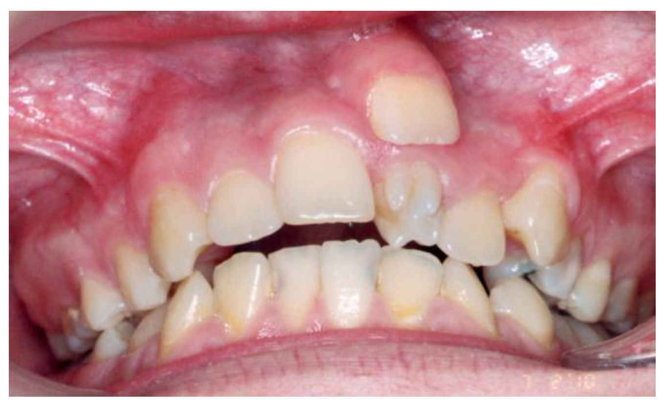
Figure 2.
Supernumerary teeth located in the maxillary incisor area.
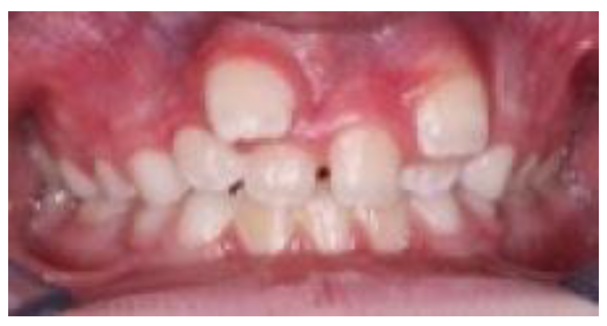
Figure 3.
Orthopantomogram showing a supernumerary tooth located between the left mandibular canine (tooth 3.3) and the left mandibular first premolar (tooth 3.4).
Figure 3.
Orthopantomogram showing a supernumerary tooth located between the left mandibular canine (tooth 3.3) and the left mandibular first premolar (tooth 3.4).
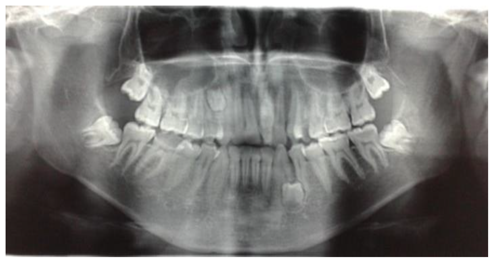

Table 1.
Epidemiological and Clinical Profile of Supernumerary Teeth.
| Key Epidemiological and Clinical Data | Value / Observation | Source |
|---|---|---|
| Prevalence in the General Population | 0.1% – 3.8% | [27] |
| Gender Predominance | Male (approx. 2:1 ratio) | [27] |
| Most Frequent Location | Anterior Maxilla (Mesiodens) | [2,28] |
| Impaction / Asymptomatic Rate | ~75% | [2,28] |
| Reported Complication Frequency | 21.6% – 30% | [20] |
| Cystic Formation Frequency | 9% – 11% | [20,25] |
Table 2.
Distribution of Patients by Gender and Dental Class (Angle).
| Gender | Class I | Class II/1 | Class II/2 | Class III | Total |
|---|---|---|---|---|---|
| Male | 28 | 21 | 10 | 32 | 91 |
| Female | 14 | 12 | 8 | 28 | 62 |
| Total (n = 153) | 42 (27.5%) | 33 (21.6%) | 18 (11.8%) | 60 (39.2%) | 100% |
Note: A significant association was observed between Angle class and supernumerary tooth location (χ² = 11.78, p = 0.008).
Table 3.
Distribution of Patients by Gender and Supernumerary Tooth Position.
| Gender | Erupted | Erupted & Impacted (Lateral Zone) | Impacted (Frontal Zone) | Impacted (Frontal & Lateral) | Impacted (Lateral) | Unspecified | Total |
|---|---|---|---|---|---|---|---|
| Male | 38 | 12 | 6 | 19 | 11 | 5 | 91 |
| Female | 25 | 10 | 3 | 12 | 8 | 4 | 62 |
| Total (n = 153) | 63 (41.2%) | 22 (14.4%) | 9 (5.9%) | 31 (20.3%) | 19 (12.4%) | 9 (5.9%) | 100% |
Table 4.
Distribution of Patients by Age Group and Supernumerary Tooth Position.
| Age Group (years) | Erupted | Erupted & Impacted (Lateral) | Impacted (Frontal) | Impacted (Frontal & Lateral) | Impacted (Lateral) | Unspecified | Total |
|---|---|---|---|---|---|---|---|
| 6–12 | 30 | 14 | 4 | 12 | 8 | 4 | 72 |
| 13–18 | 20 | 6 | 3 | 10 | 6 | 3 | 48 |
| 19–45 | 13 | 2 | 2 | 9 | 5 | 2 | 33 |
| Total (n= 153) | 63 (41.2%) | 22 (14.4%) | 9 (5.9%) | 31 (20.3%) | 19 (12.4%) | 9 (5.9%) | 100% |
Table 5.
Morphological and Topographical Distribution of Supernumerary Teeth (n = 185).
| Variable | Category | Frequency (n) | Percentage (%) |
|---|---|---|---|
| Morphology | Conical | 90 | 48.6 |
| Tuberculate | 50 | 27.0 | |
| Supplemental | 34 | 18.4 | |
| Odontomatous | 11 | 6.0 | |
| Location | Mesiodens | 104 | 56.2 |
| Paramolar | 30 | 16.2 | |
| Distomolar | 26 | 14.1 | |
| Supernumerary Premolar | 25 | 13.5 |
Table 6.
Complications Associated with Supernumerary Teeth.
| Complication | Number of Patients (n) | Percentage (%) |
|---|---|---|
| Delayed eruption of adjacent teeth | 30 | 19.6 |
| Malposition/rotation | 19 | 12.4 |
| Cystic formation | 7 | 4.6 |
| Root resorption | 6 | 3.9 |
| No complications | 91 | 59.5 |
Table 7.
Logistic Regression Predictors of Supernumerary Tooth Impaction.
| Variable | Odds Ratio (OR) | 95% CI | p-value |
|---|---|---|---|
| Age < 13 years | 3.12 | 1.47 – 6.64 | 0.003 |
| Male gender | 1.78 | 1.01 – 3.15 | 0.046 |
| Tuberculate morphology | 2.93 | 1.17 – 5.72 | 0.021 |
| Class III malocclusion | 1.89 | 1.03 – 3.02 | 0.039 |
Disclaimer/Publisher’s Note: The statements, opinions and data contained in all publications are solely those of the individual author(s) and contributor(s) and not of MDPI and/or the editor(s). MDPI and/or the editor(s) disclaim responsibility for any injury to people or property resulting from any ideas, methods, instructions or products referred to in the content. |
© 2025 by the authors. Licensee MDPI, Basel, Switzerland. This article is an open access article distributed under the terms and conditions of the Creative Commons Attribution (CC BY) license (http://creativecommons.org/licenses/by/4.0/).
Copyright: This open access article is published under a Creative Commons CC BY 4.0 license, which permit the free download, distribution, and reuse, provided that the author and preprint are cited in any reuse.


