Submitted:
24 October 2025
Posted:
29 October 2025
You are already at the latest version
Abstract
A catalog of values of predicted energies of the K-XRF escape-peaks from compounds, used for radiation detectors, is presented for given incident gamma-rays suited for nuclear molecular medicine and low-energy spectrometry. The information relating to the compounds adds to that relating to the individual natural elements recently posted [RSC2025a]. The results of powerfunctions best-fit of XRF energy values vs. Z are listed in Table 1 for Kα2 orbital and Kedge. These expressions, allow simple calculations, avoiding the development of complex software. An extensive literature survey has been carried out mainly considering text-books, review articles and research reports. A final number compounds (Nc) equal to 729 has been obtained, whose distribution per number of constituting elements (Ne) is reported in Table 2. The predicted energy peaks are arranged in Tables 3 to 7 per Ne in the range from 2 to 8, showing the average value of 3.92 elements per compound. The catalog is conceived for nuclear molecular medicine and gamma-ray spectrometry where such peaks need to be recognized and their underlying area known in order to allow quantitative information.
Keywords:
radiation detector
; nuclear molecular medicine
; gamma-ray spectrometry
Introduction
Since the XRF photons are typical of the element making-up the detector, the escape-process changes the detector response function by moving away events from the full-energy peak towards an additional peak, sometimes completely separate from the photopeak, located at Egamma-EXRF, for Egamma > EXRF. Due to this reason, the evaluations based on the value of the integral of the counts underlying the full-energy peak are systematically underestimated if the escape photons corrections are not taken into account.
In particular assemblies (like thin and/or discretized detectors) the number of events whose energy falls under the escape peak may even equal or even exceed that of photons under the full-energy peak making necessary to apply corrections [PAX1954].
It must be remarked that this is due to unavoidable edge-effect since a real detector has limited dimensions and fluorescence photons undergo to non-zero probability to escape from the sensitive volume, not being revealed. Only the ideal case of a detector with infinite dimensions is free from this effect, as schematized in [JBR1954].
It is to be noted that the emissions from the L and the other lower-energy orbitals are discarded in the present evaluation since the involved values are negligible with respect to the K-ones, and so also occur for the respective fluorescence yields.
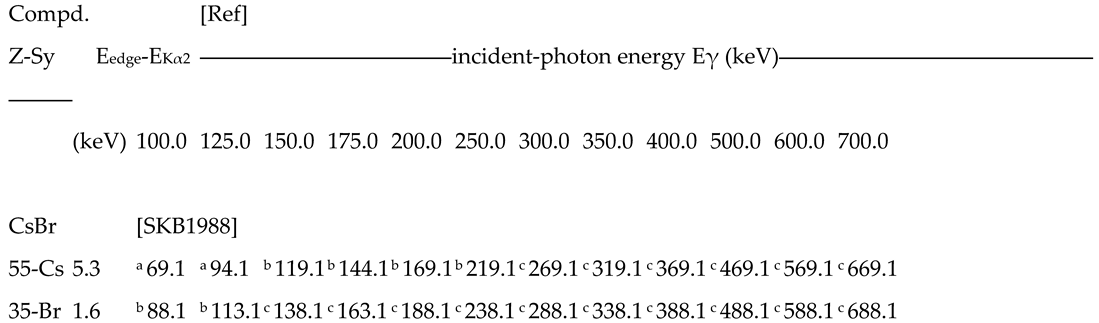
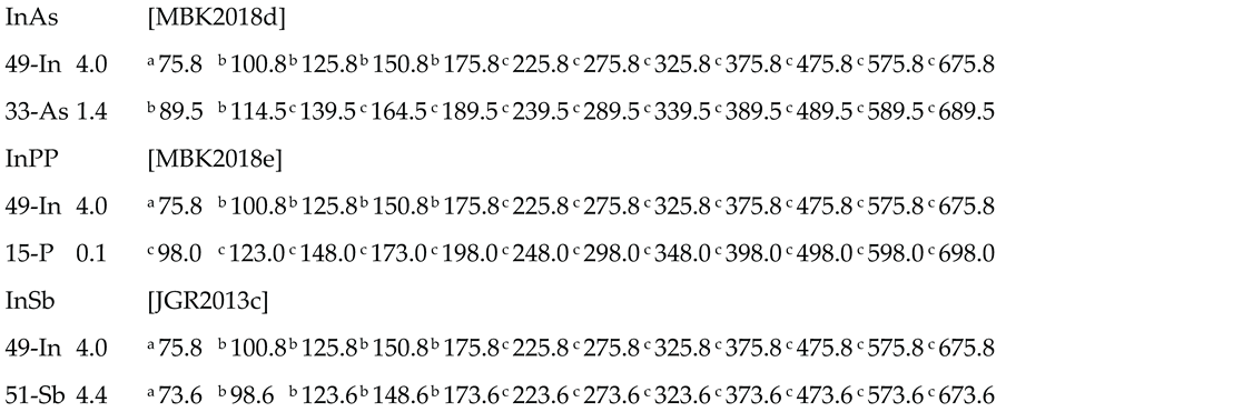
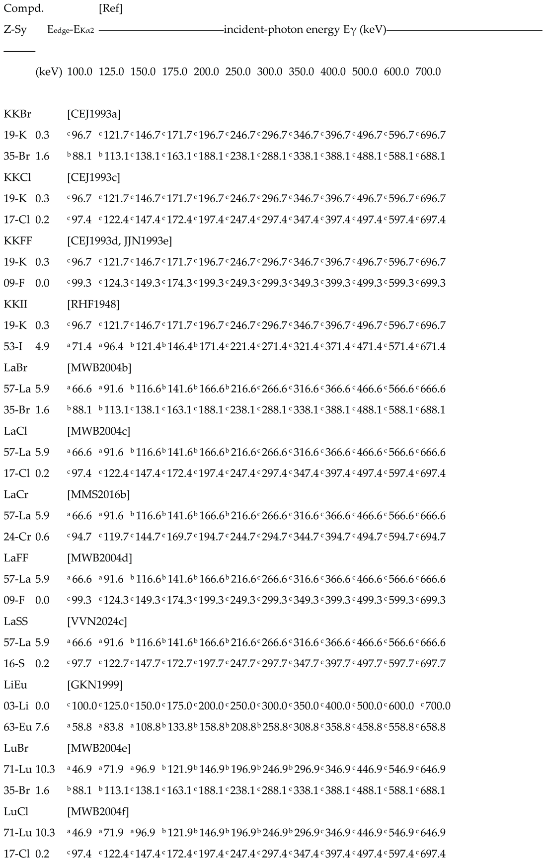
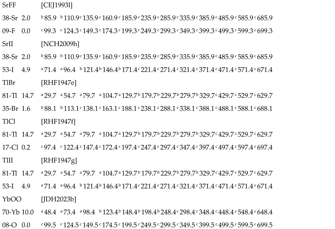
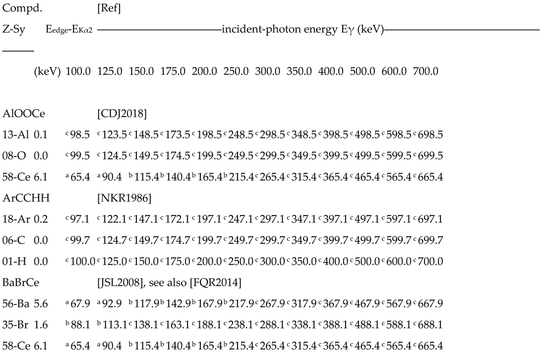
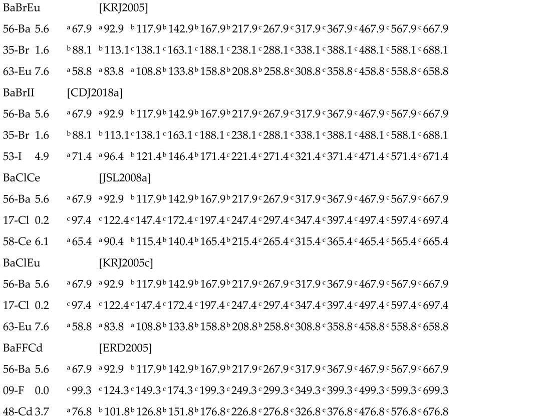
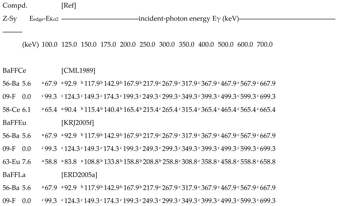
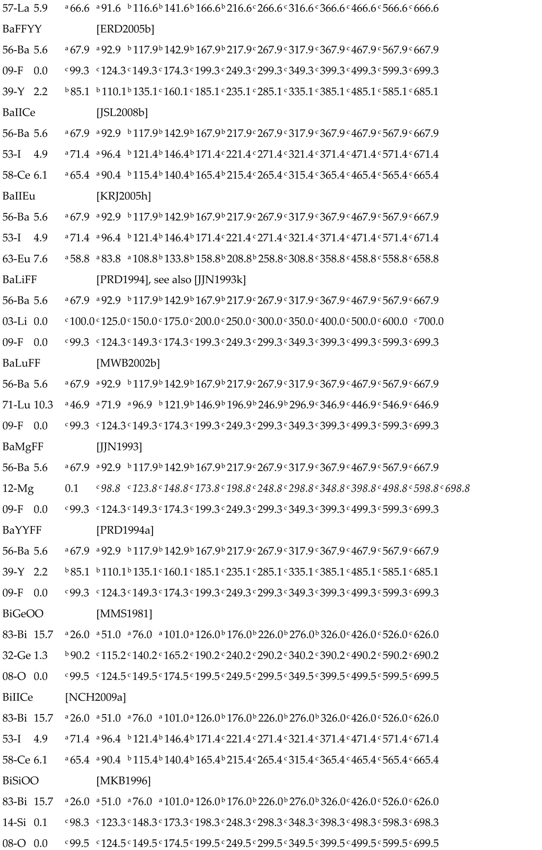
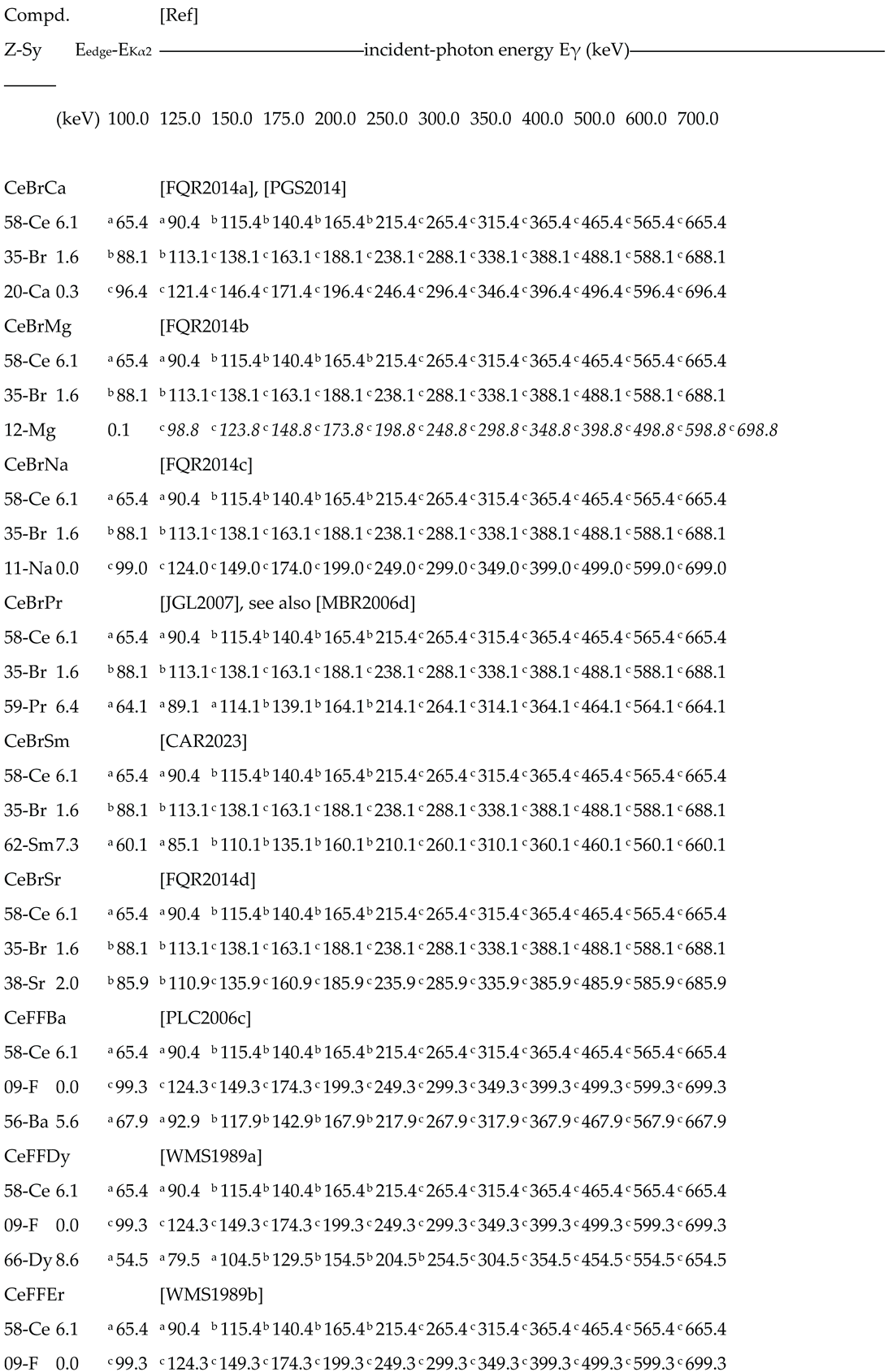
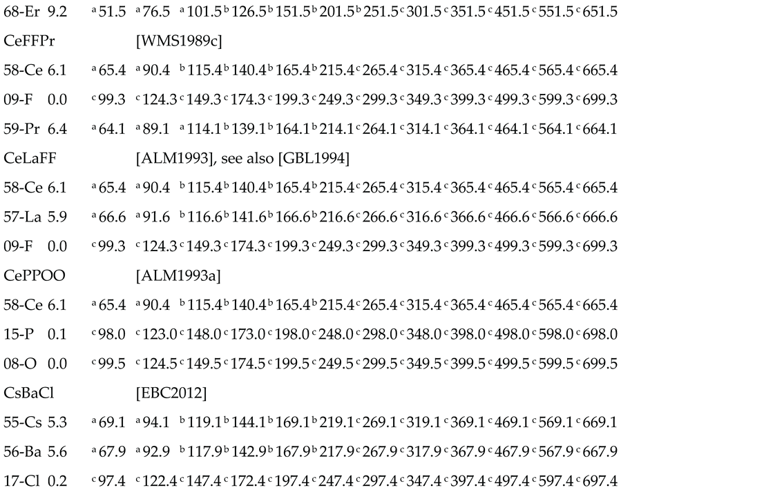
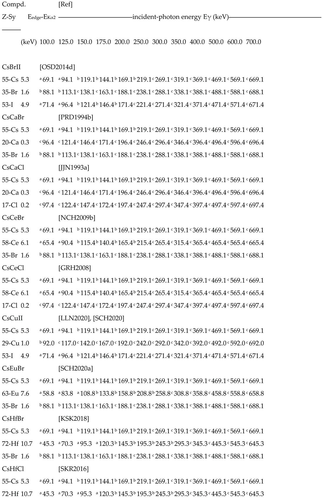
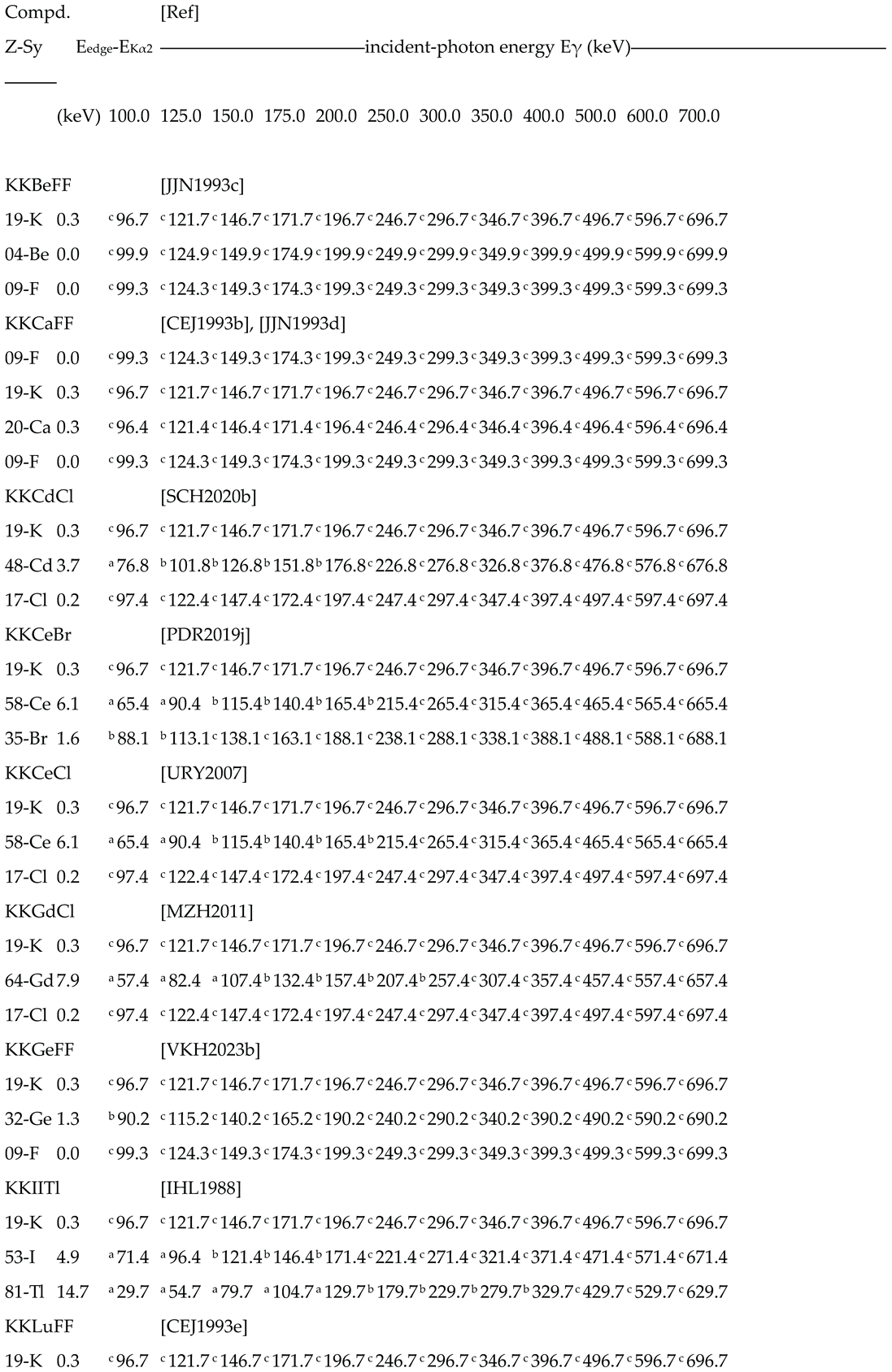
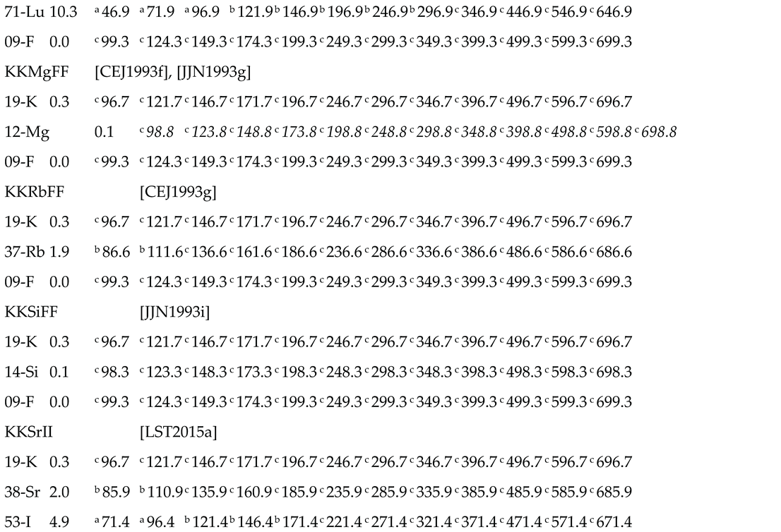
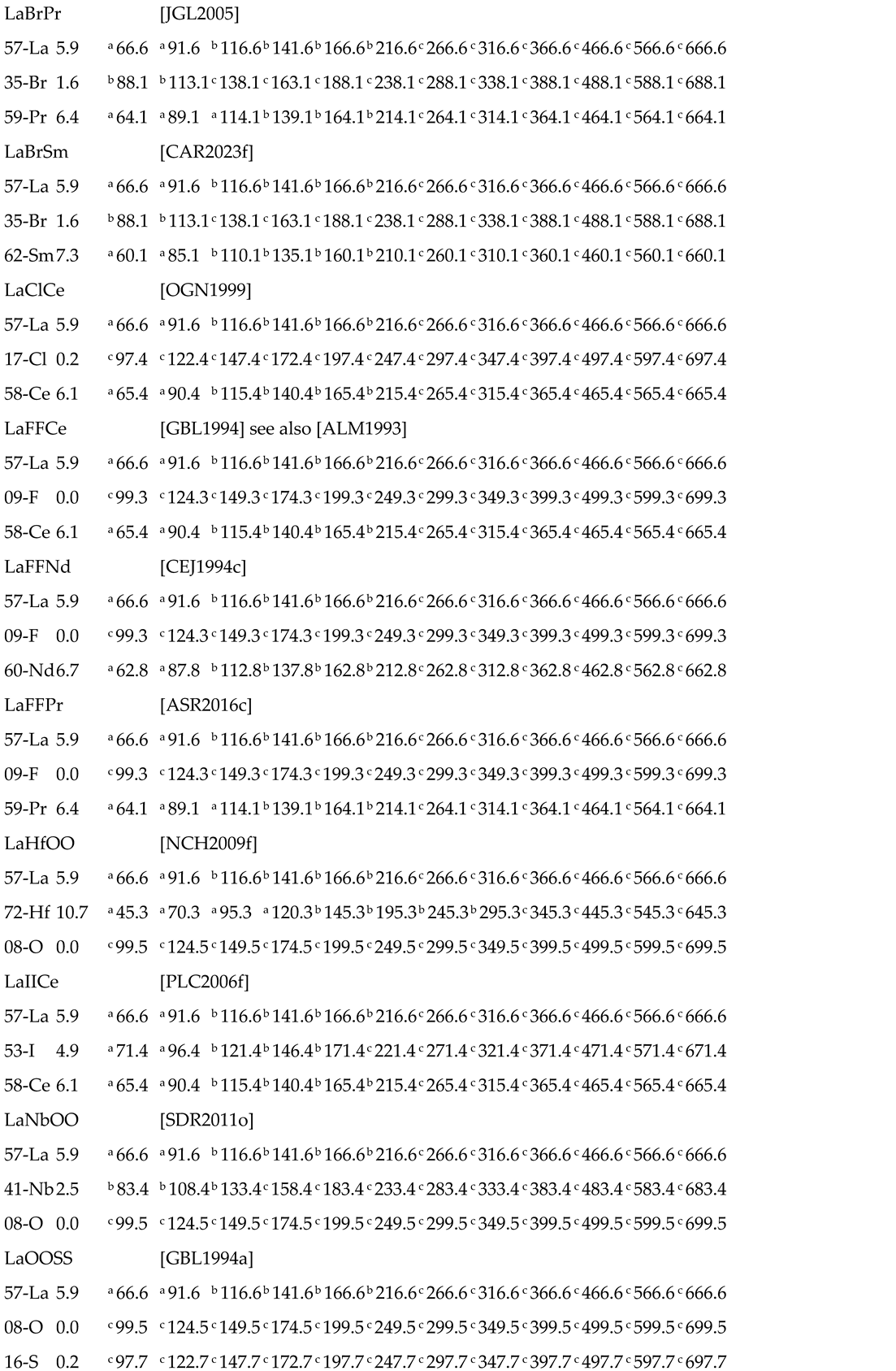
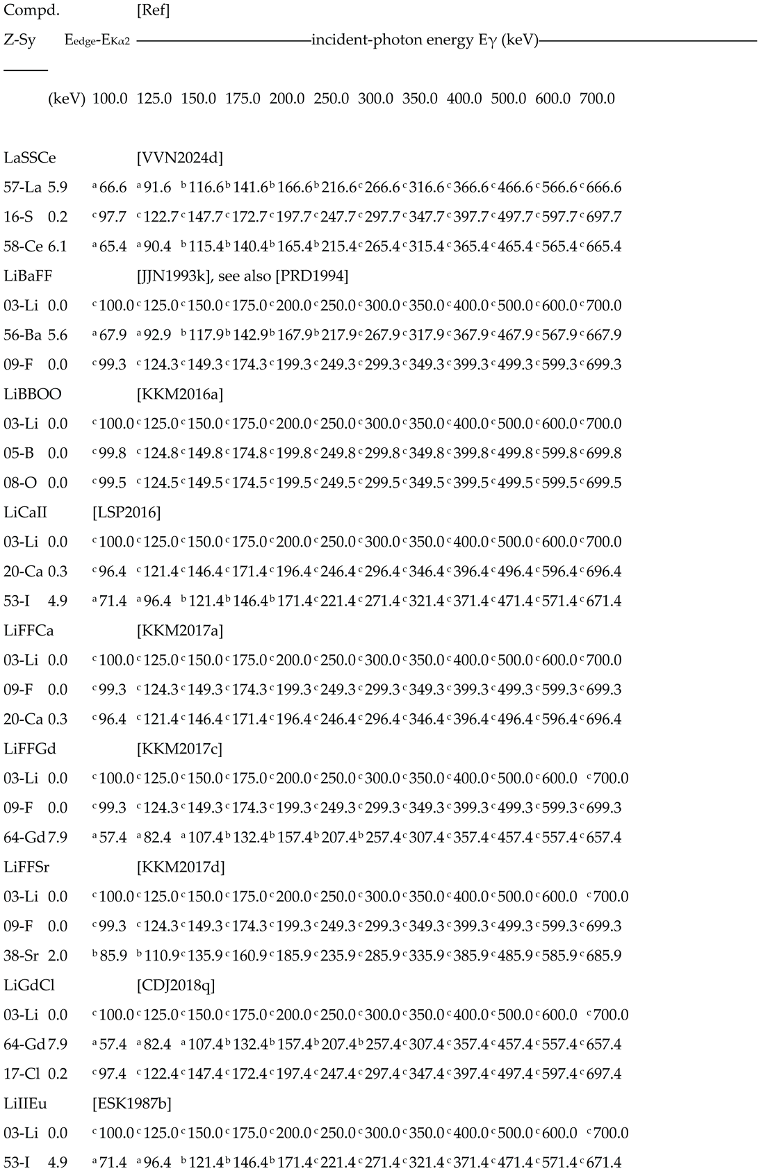
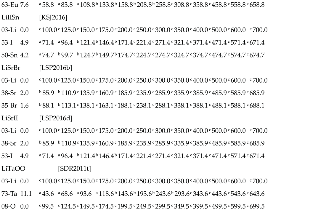
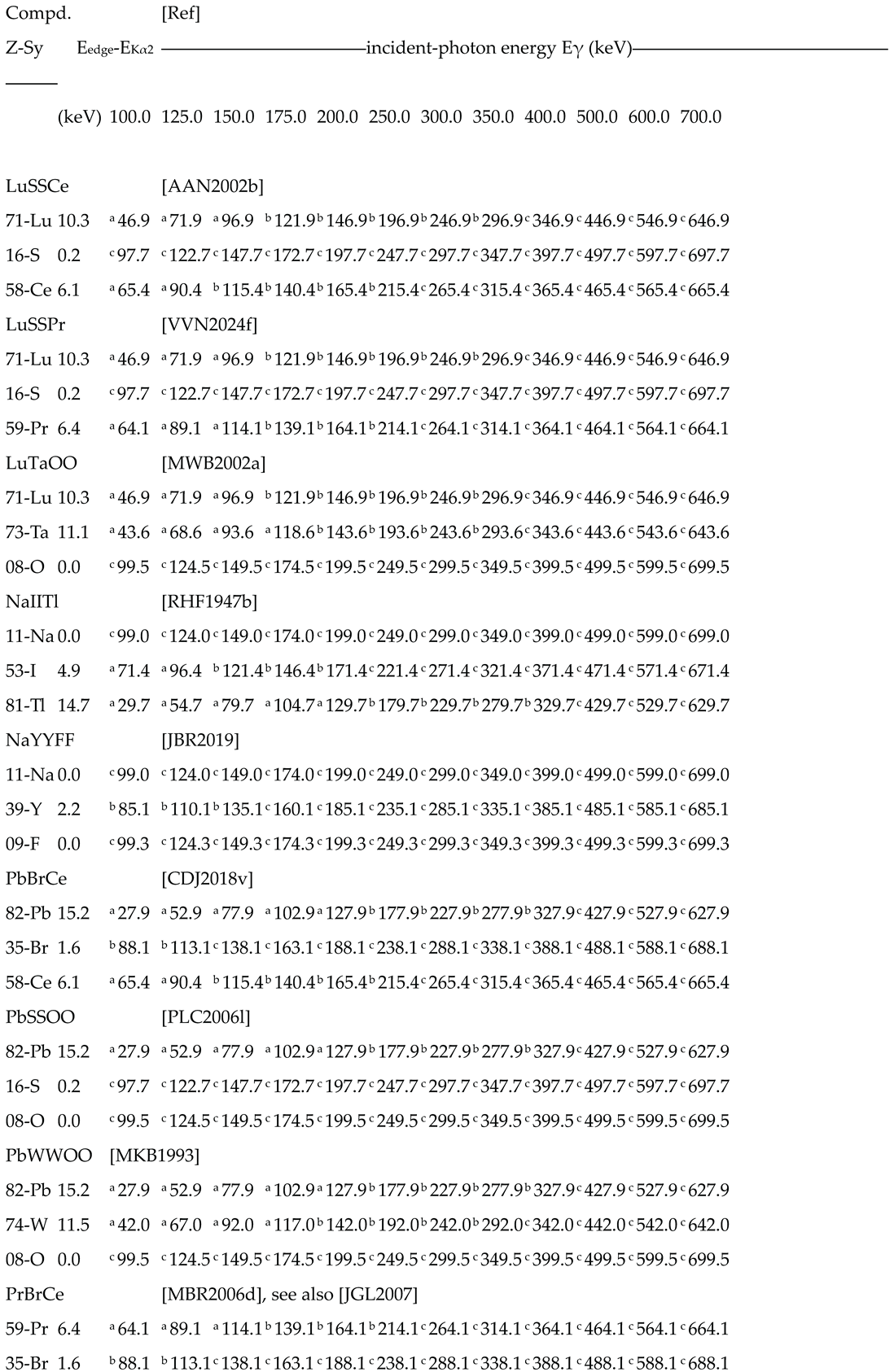
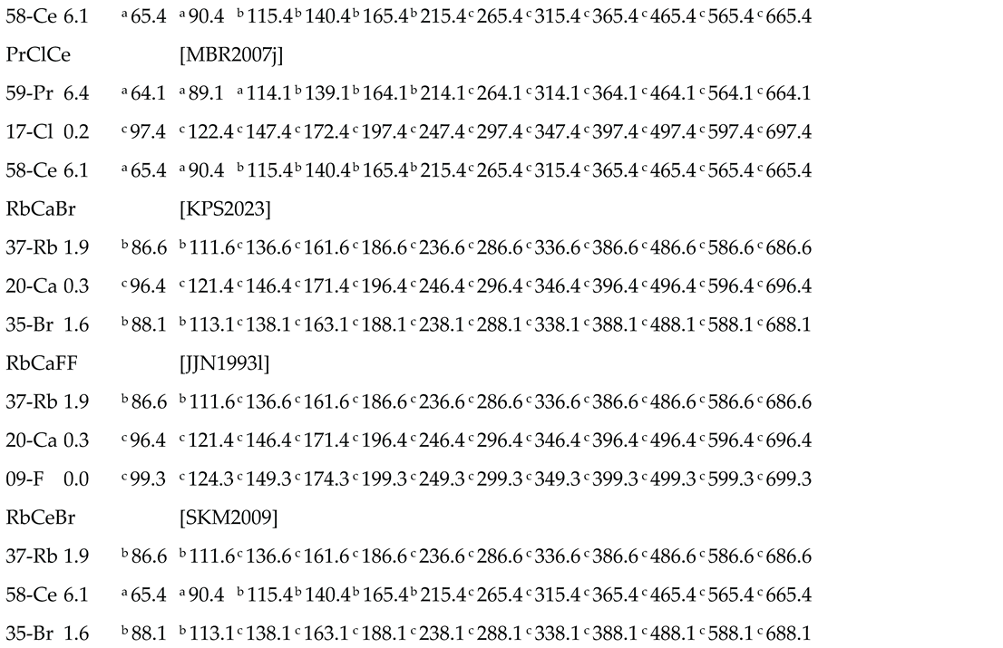
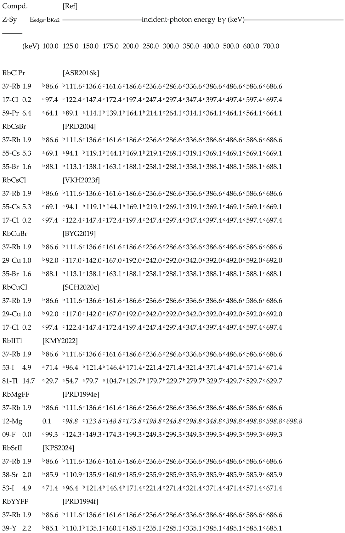
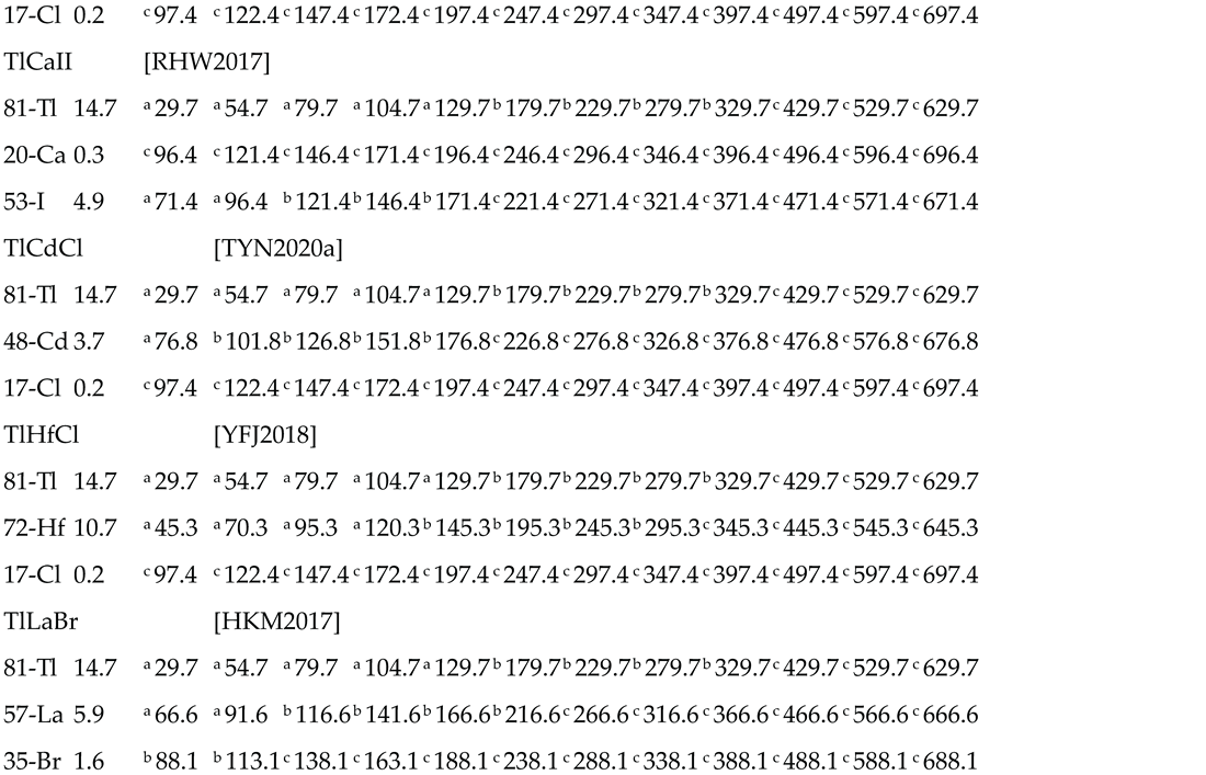
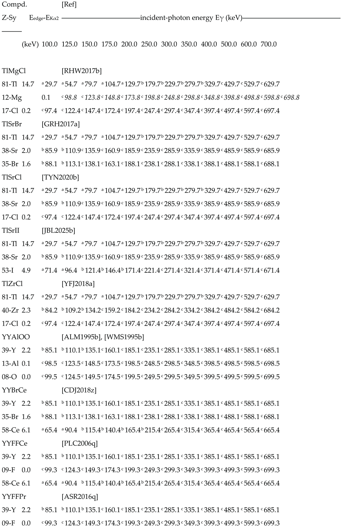
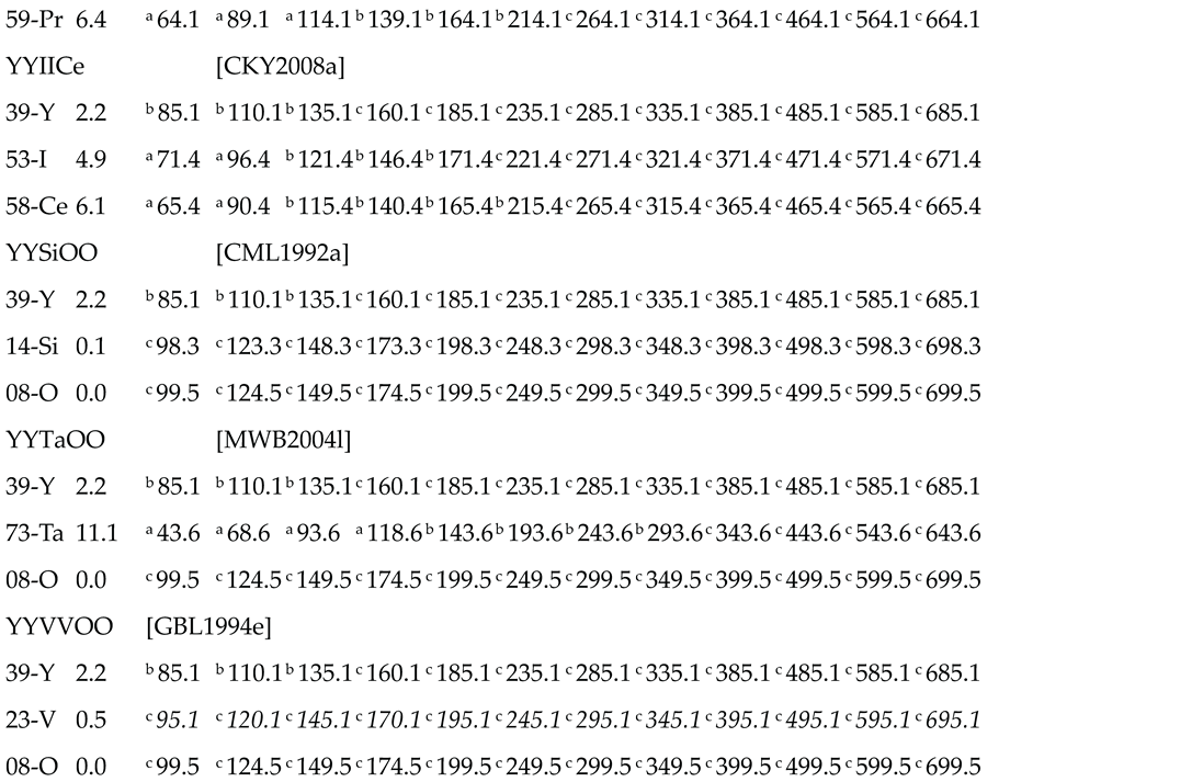
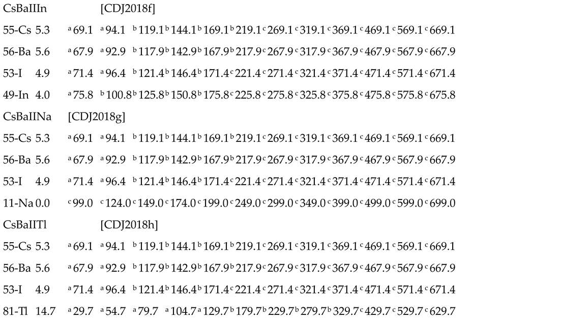
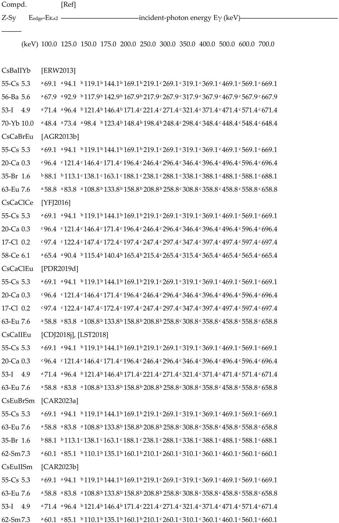
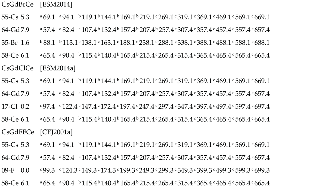
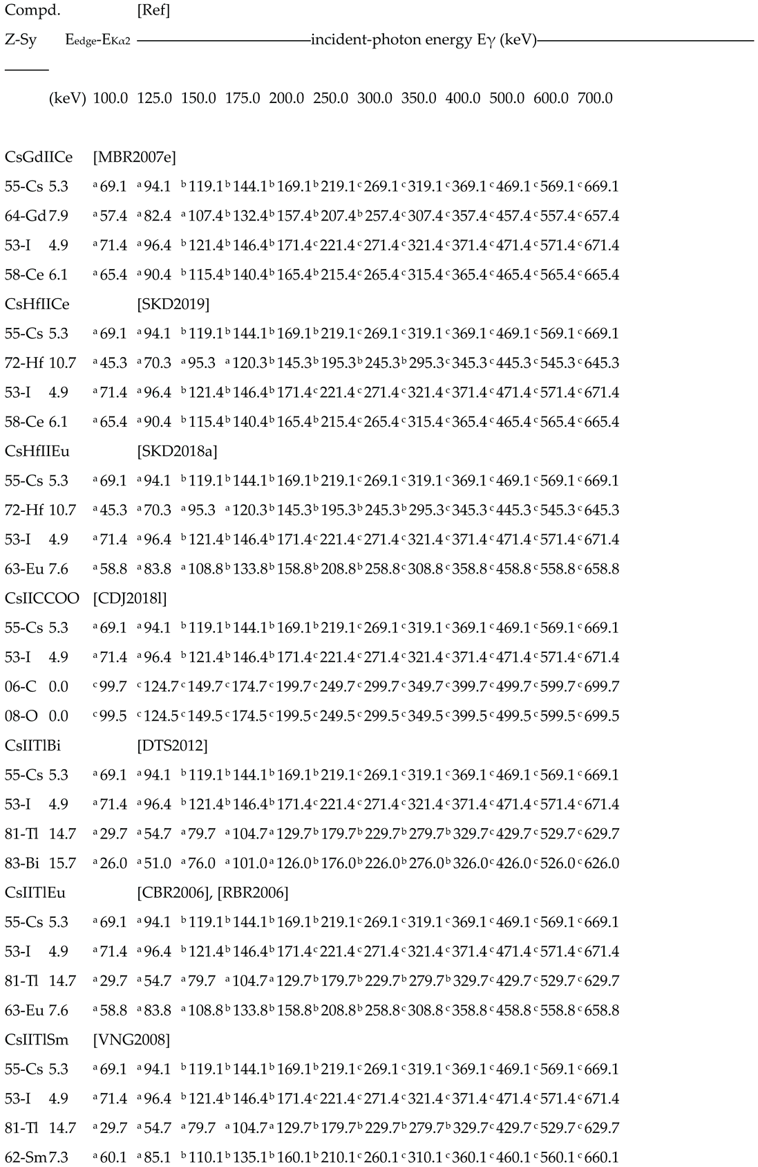
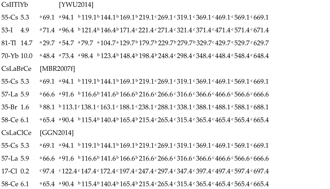
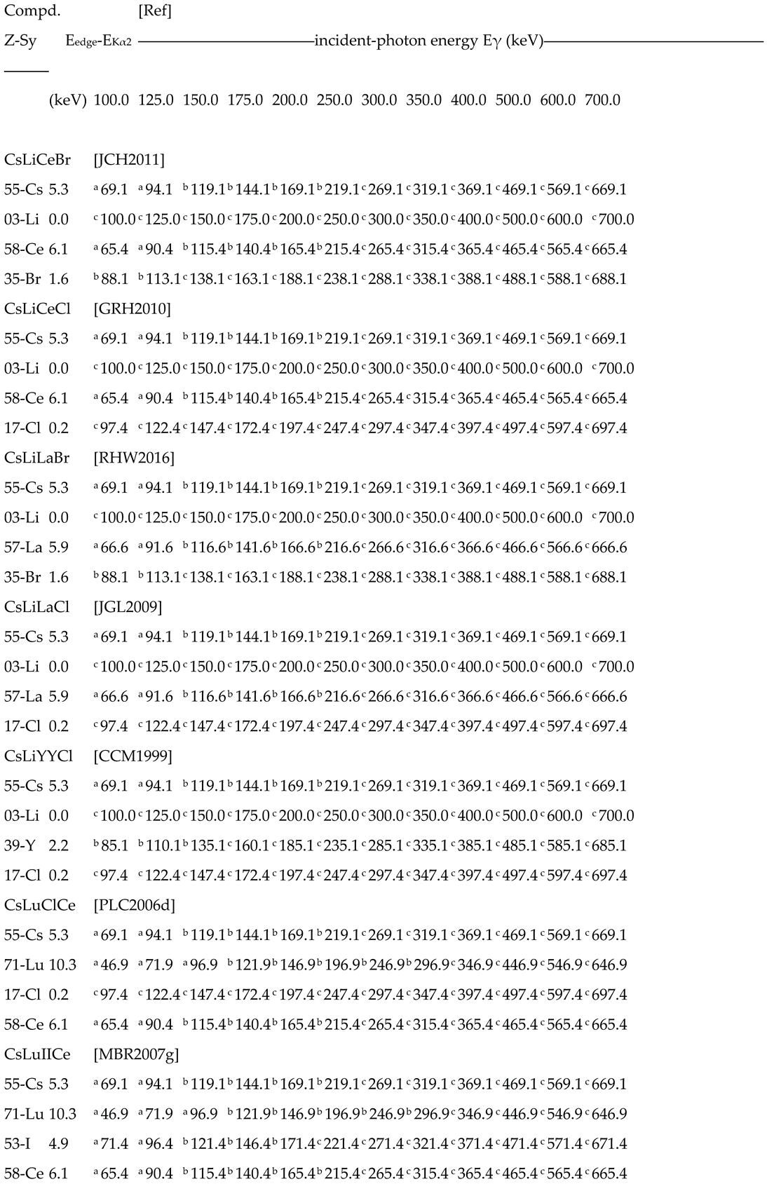
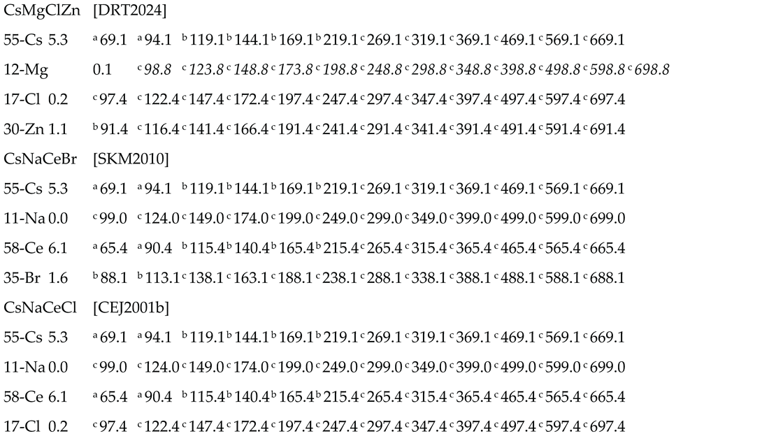
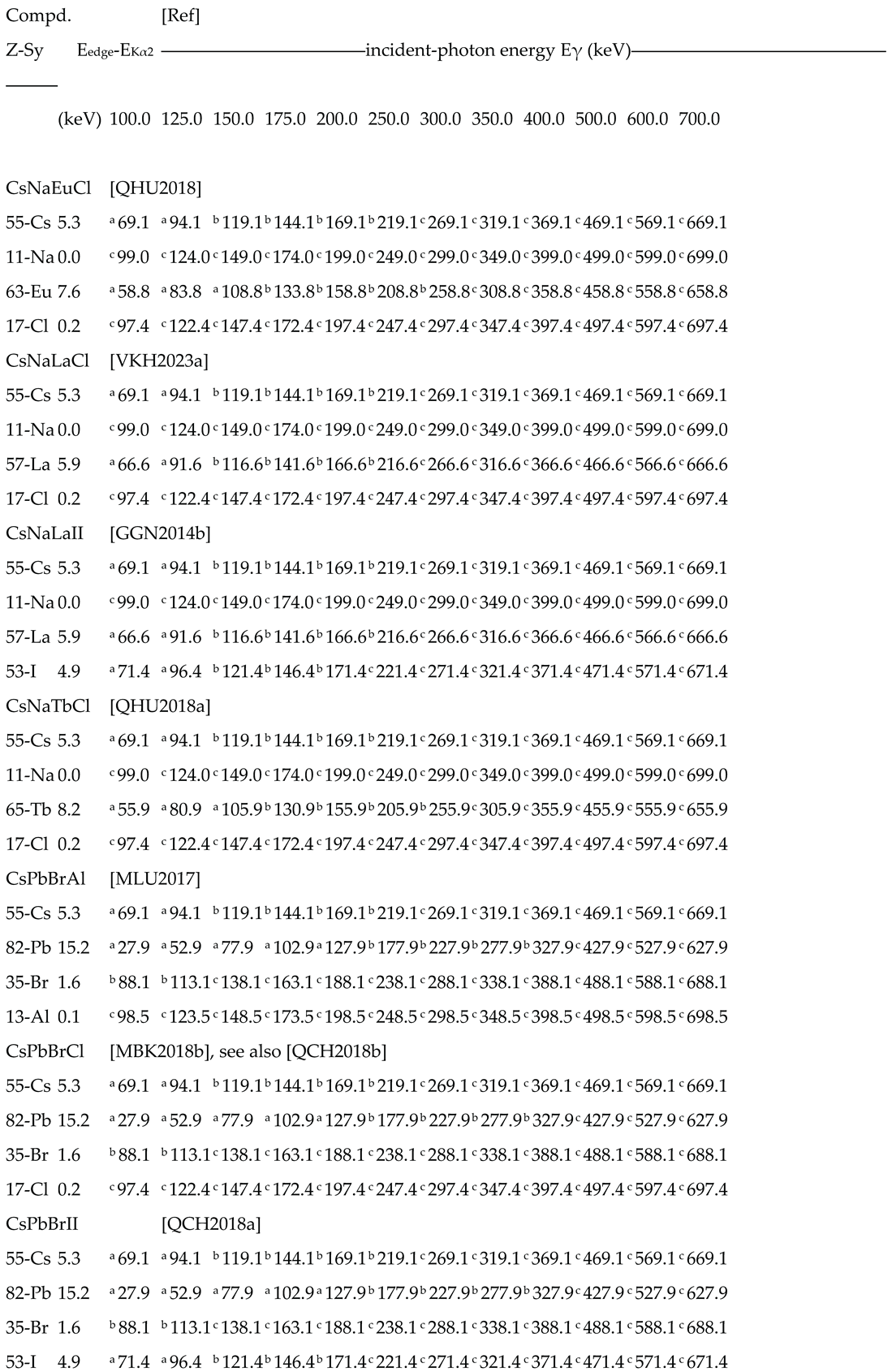
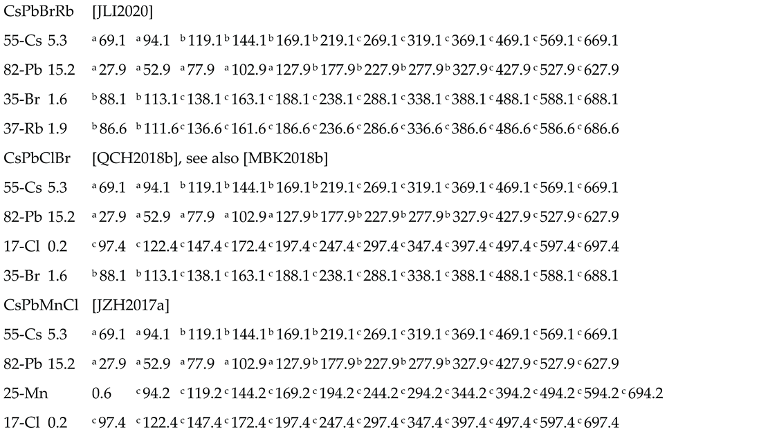
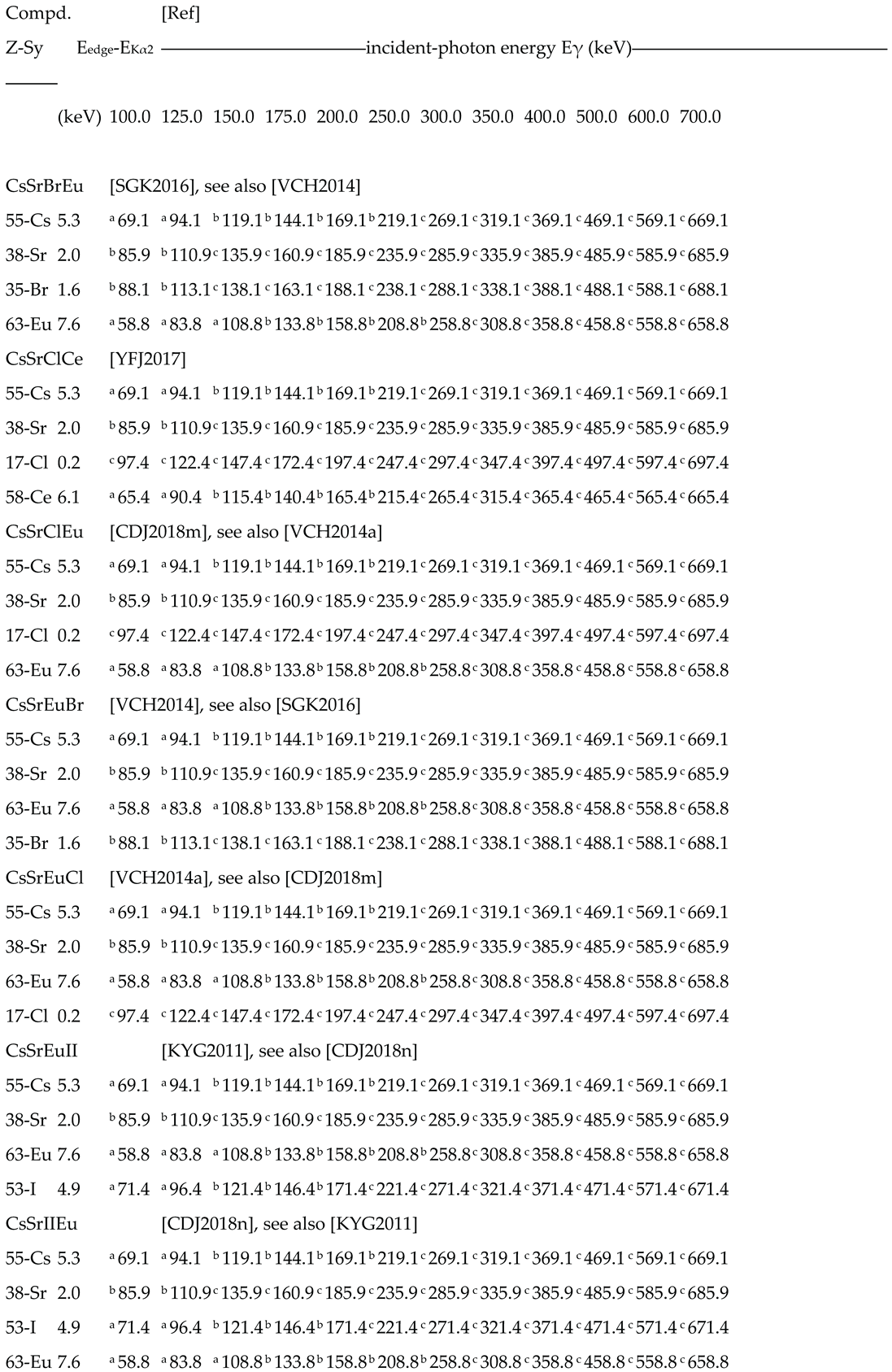
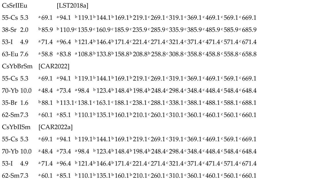
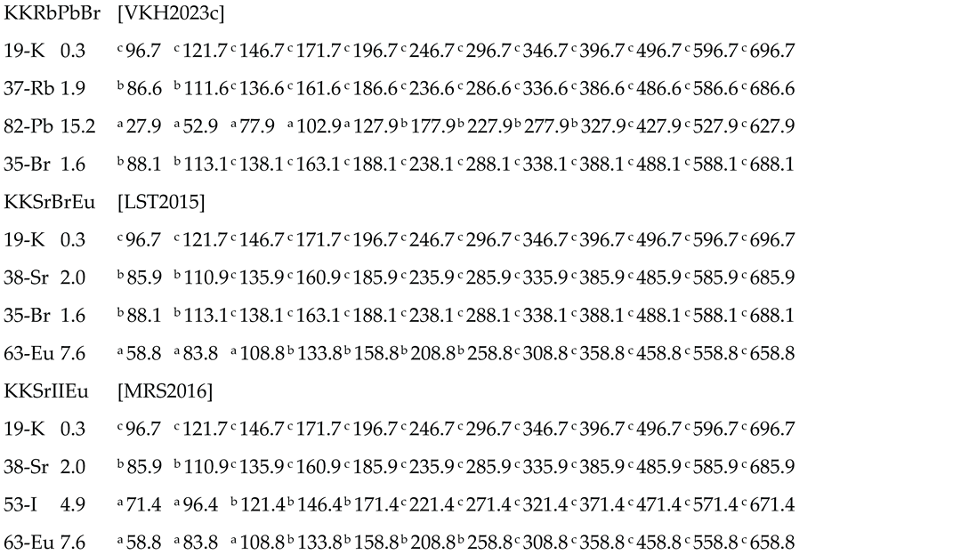
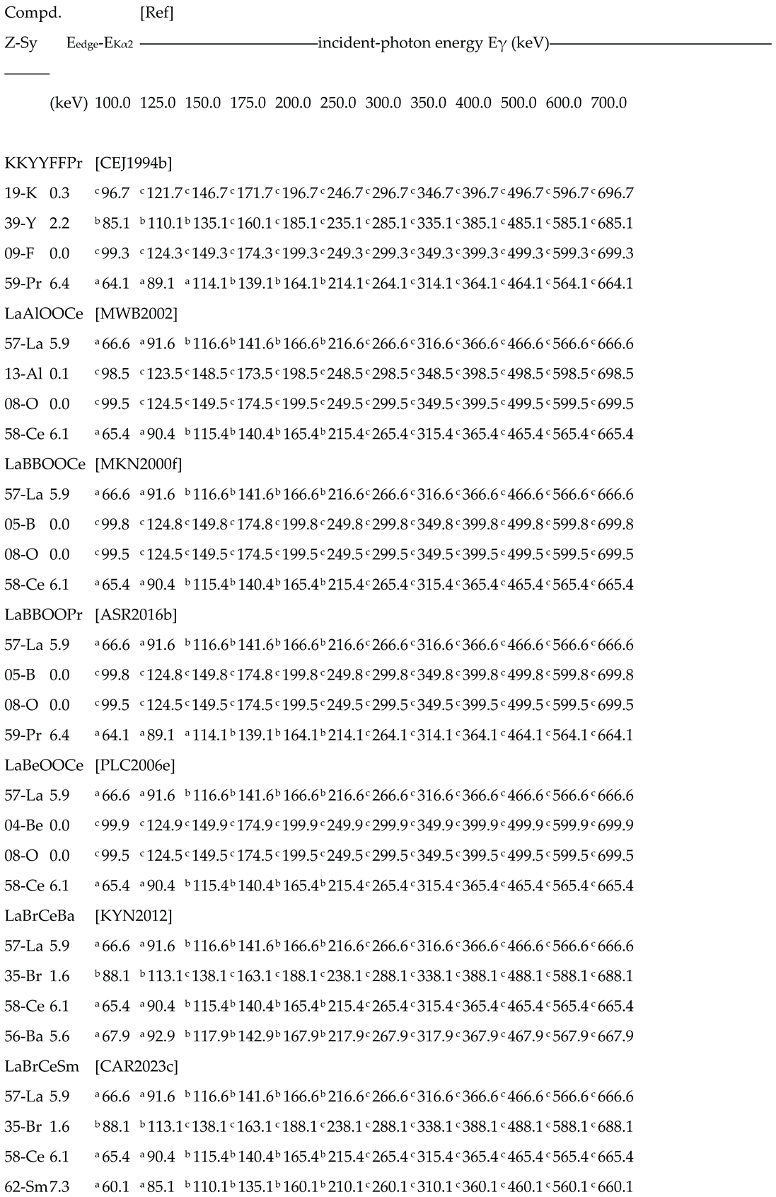
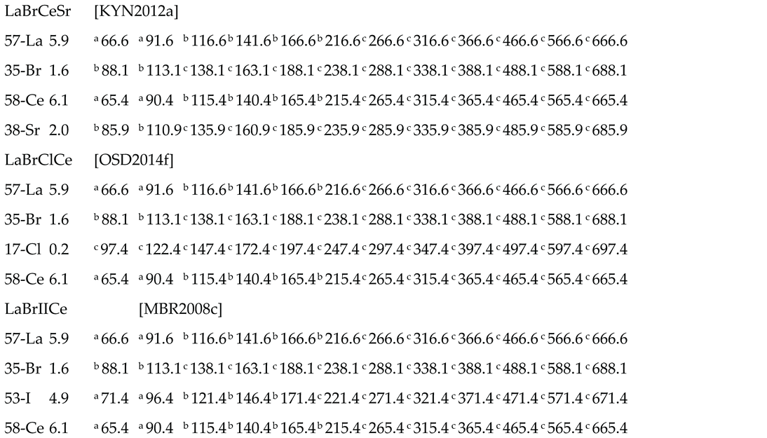
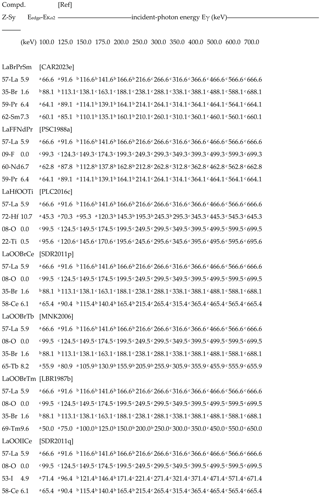
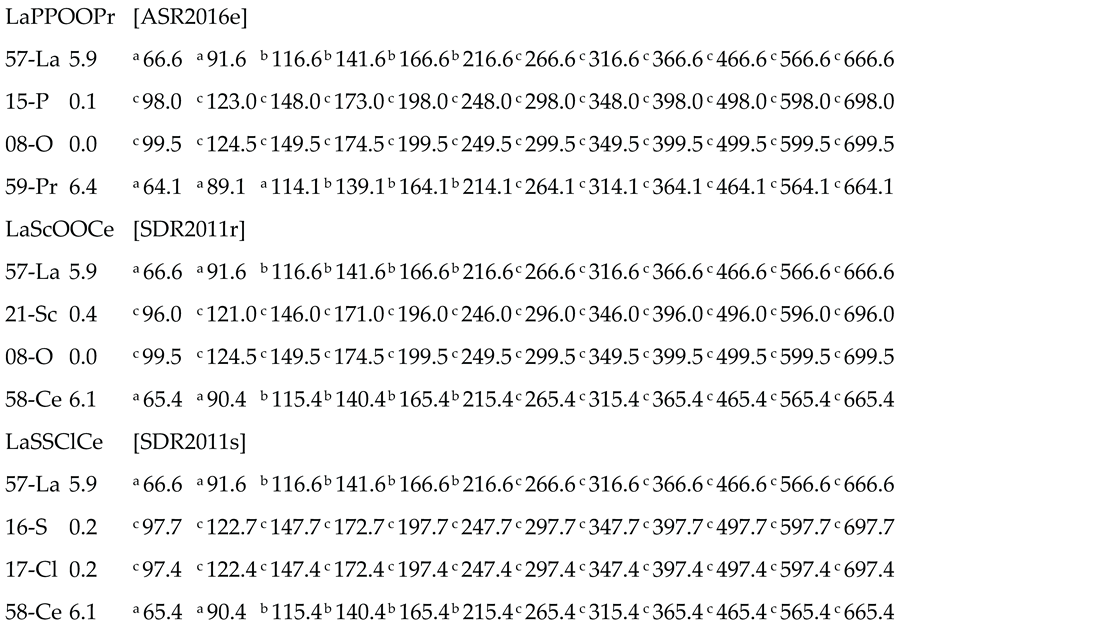
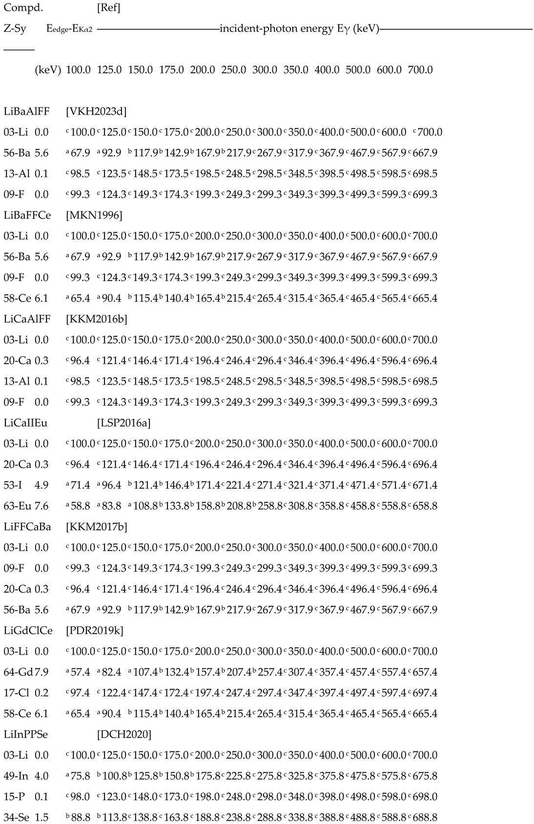
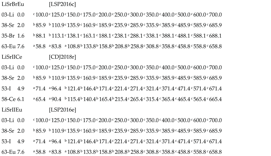
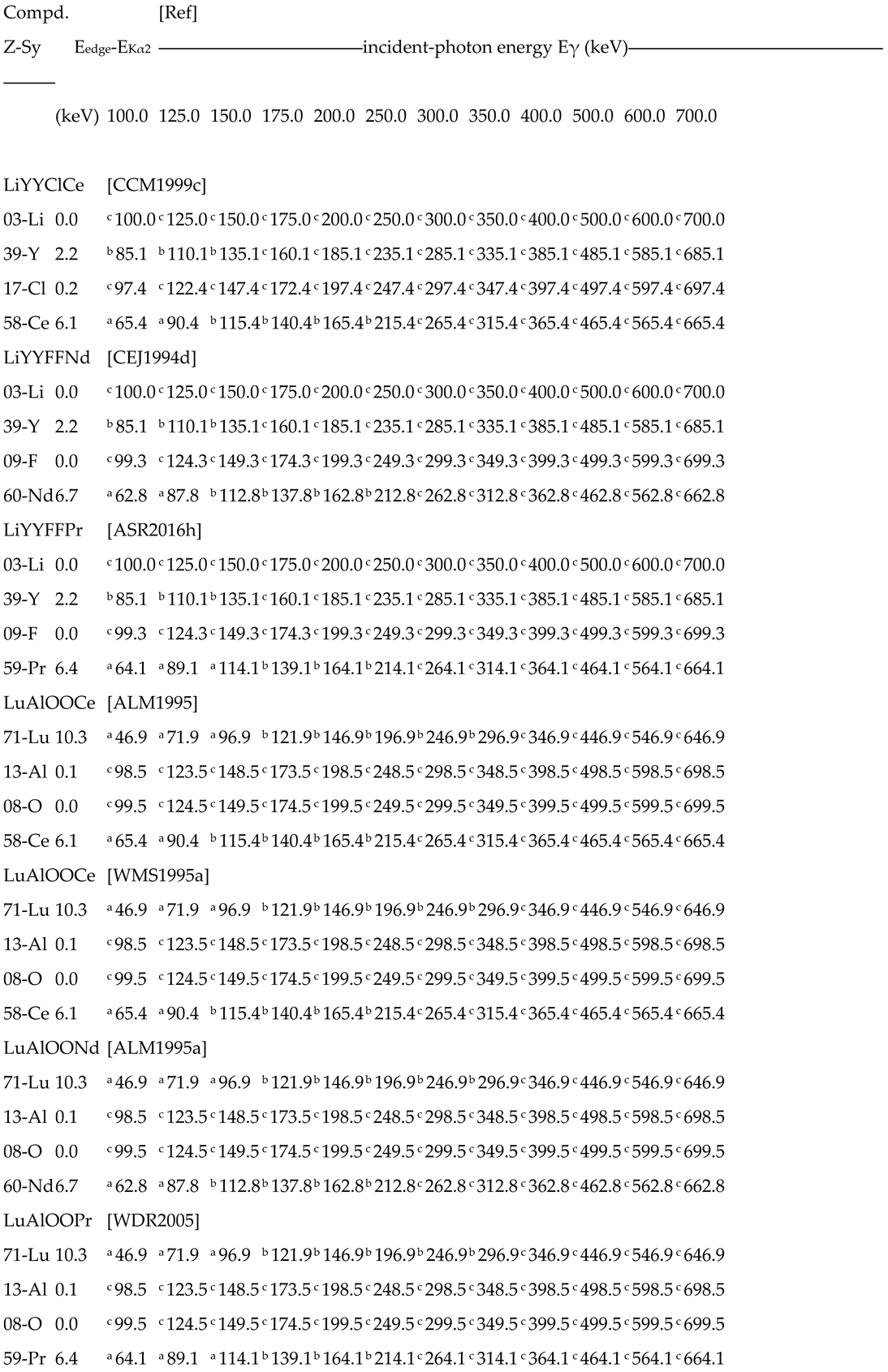
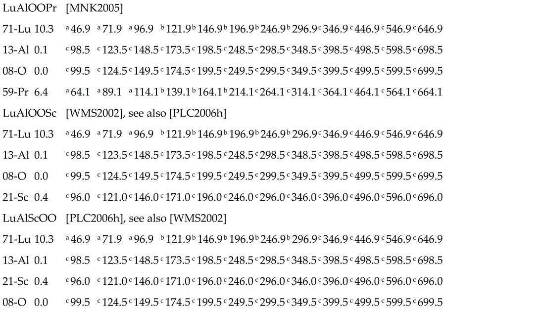
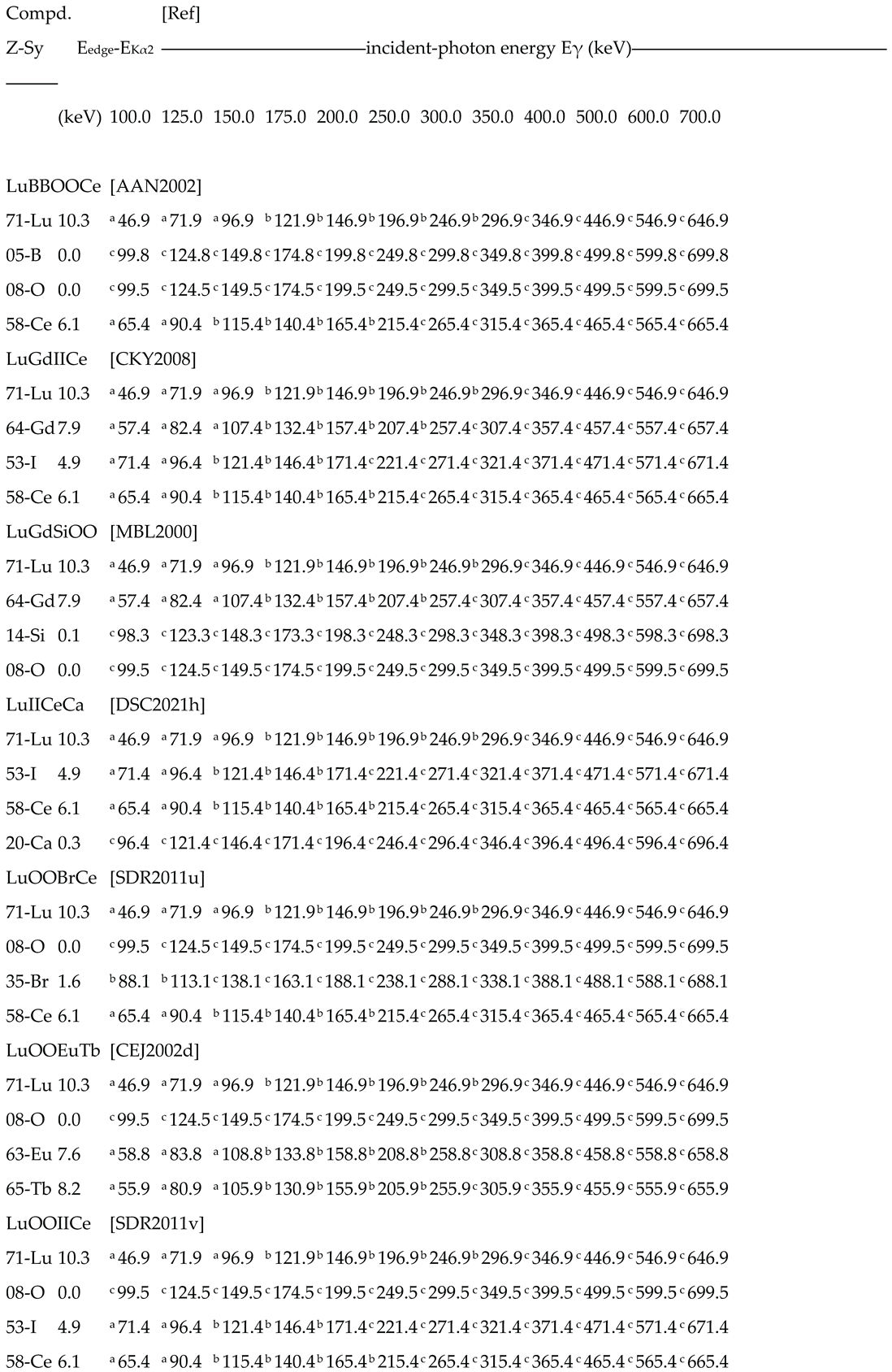
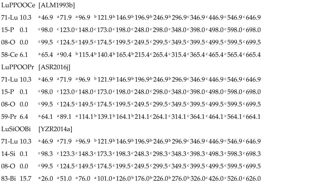
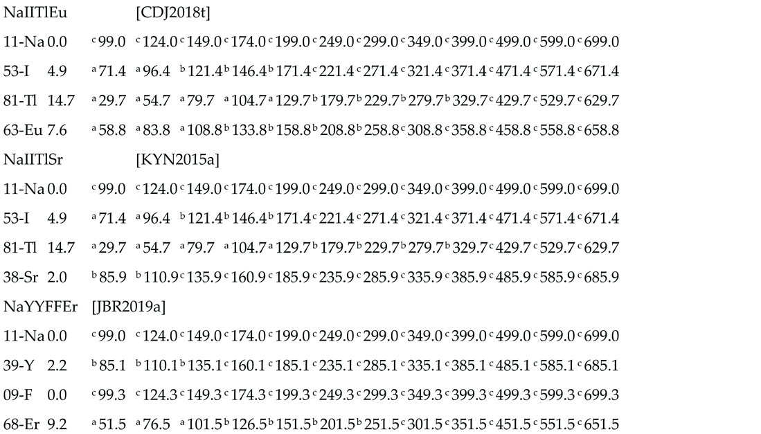
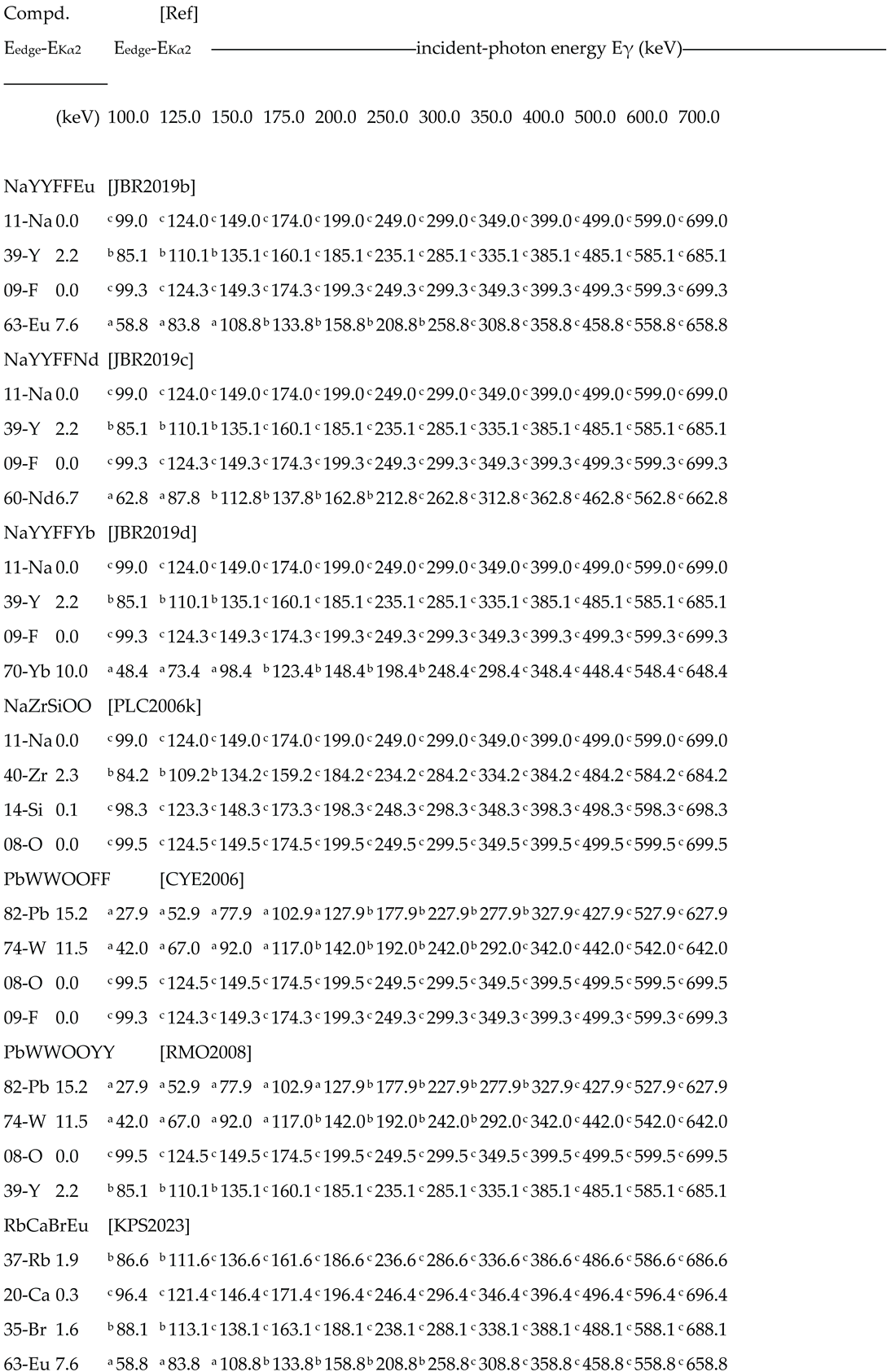
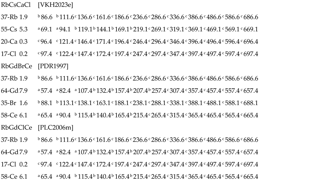
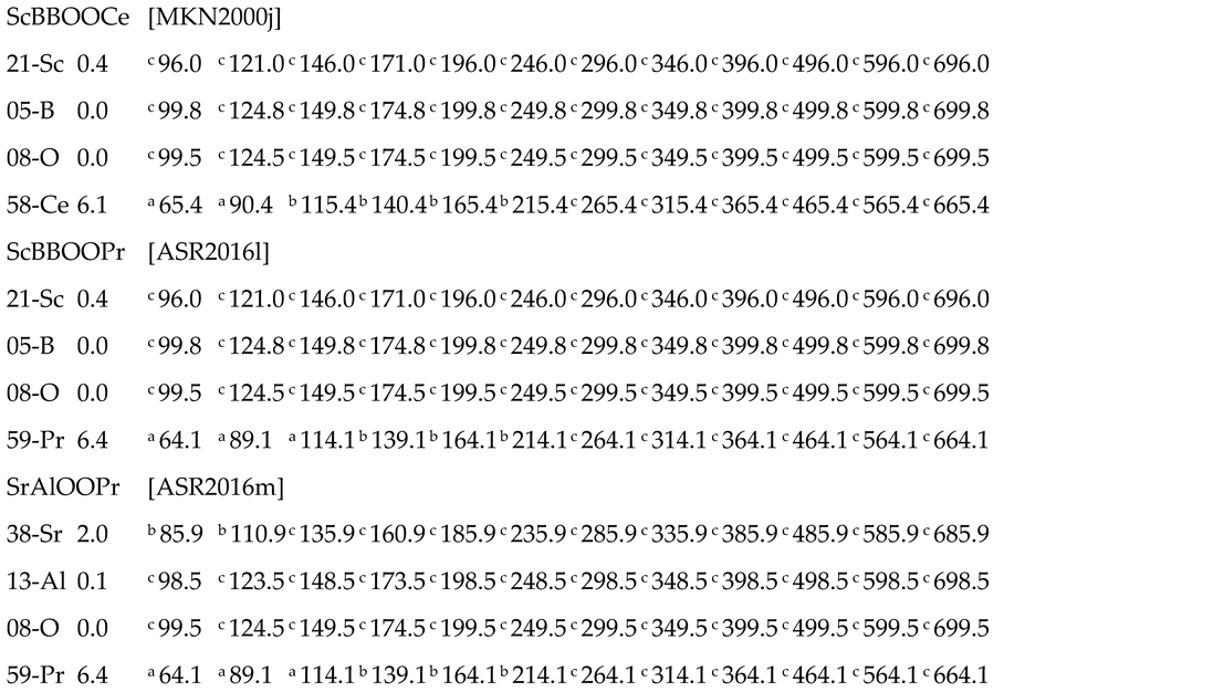
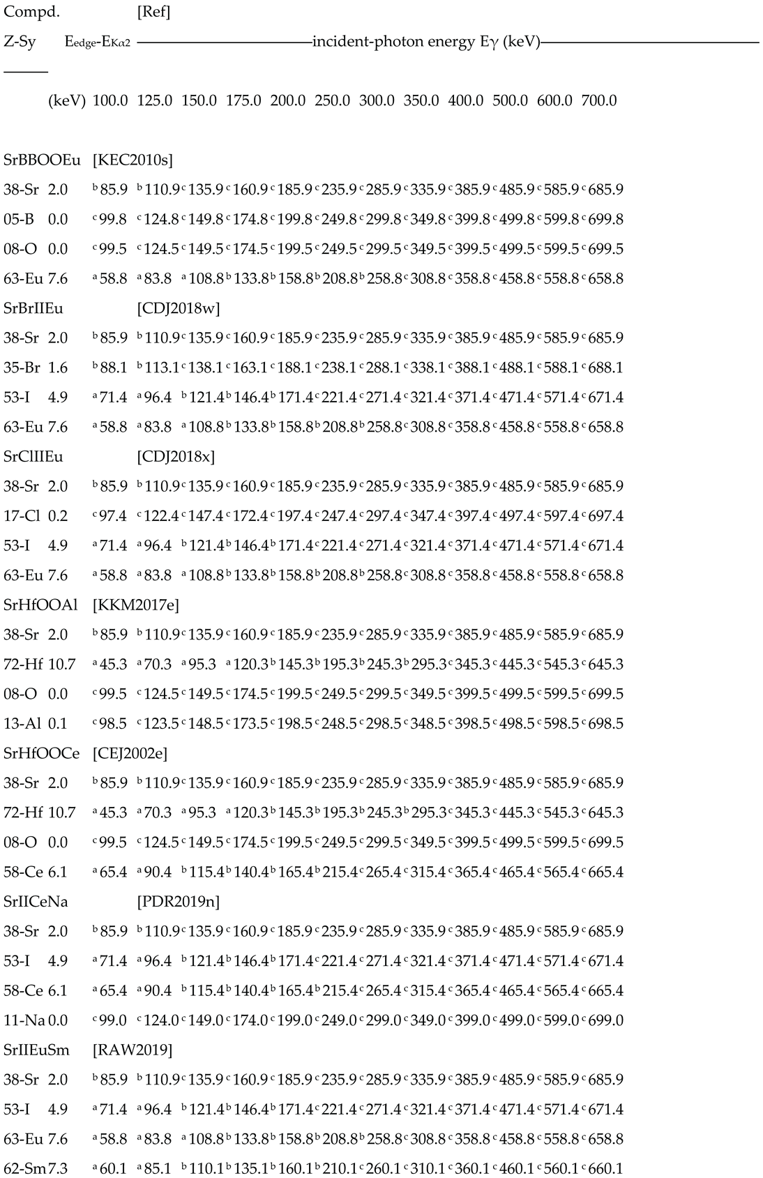
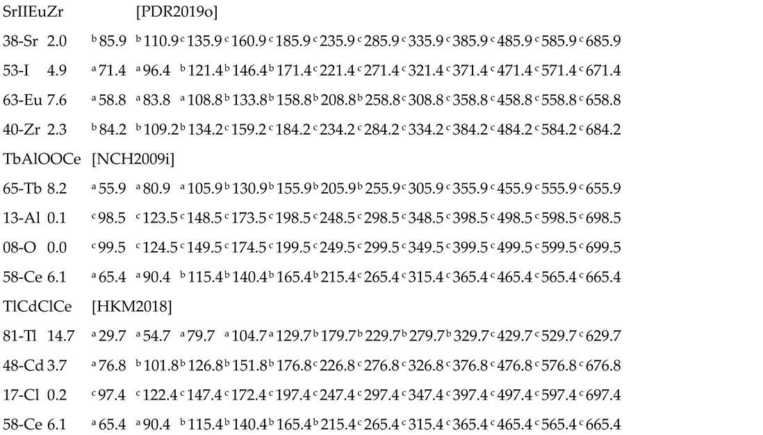
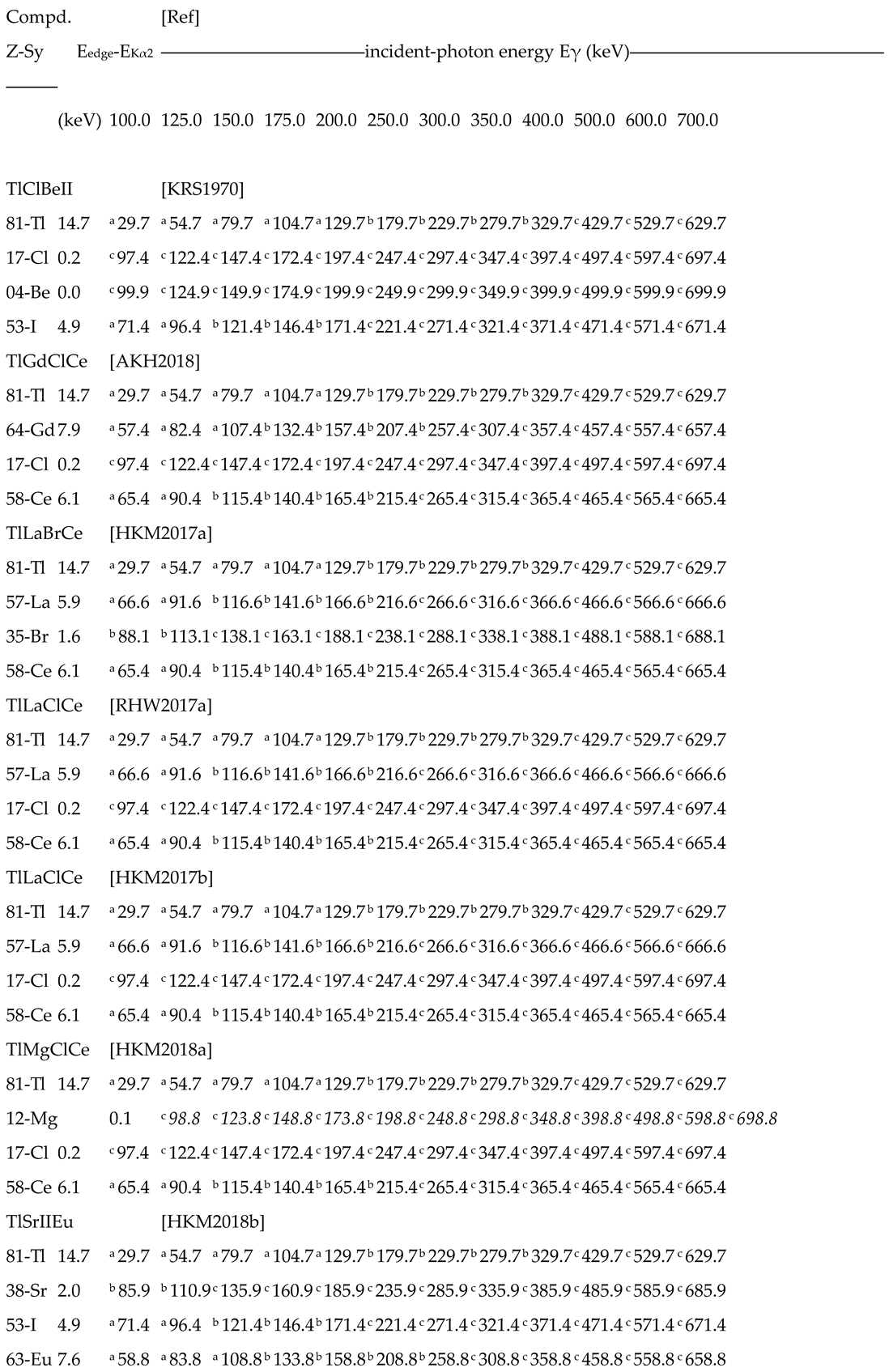
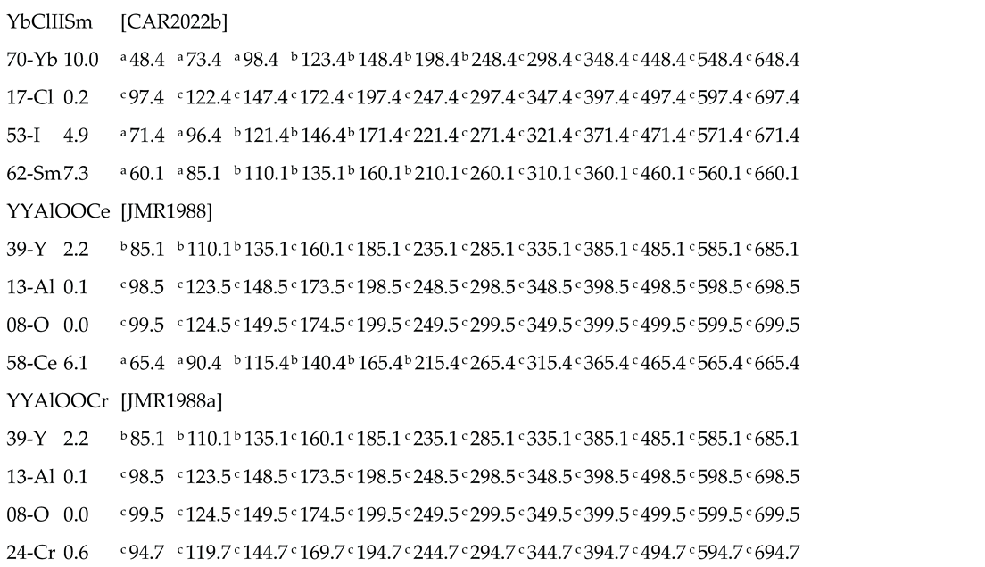
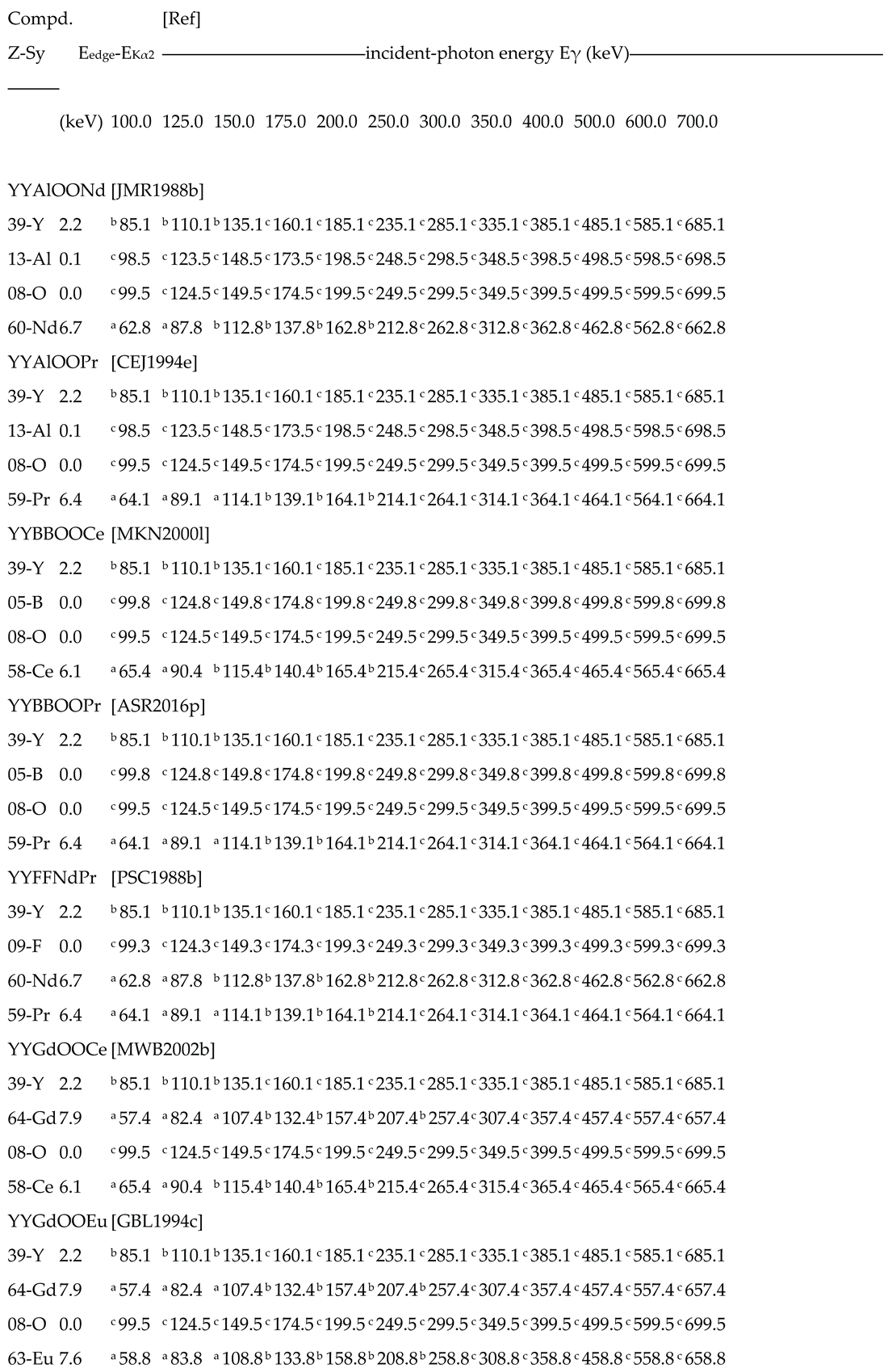
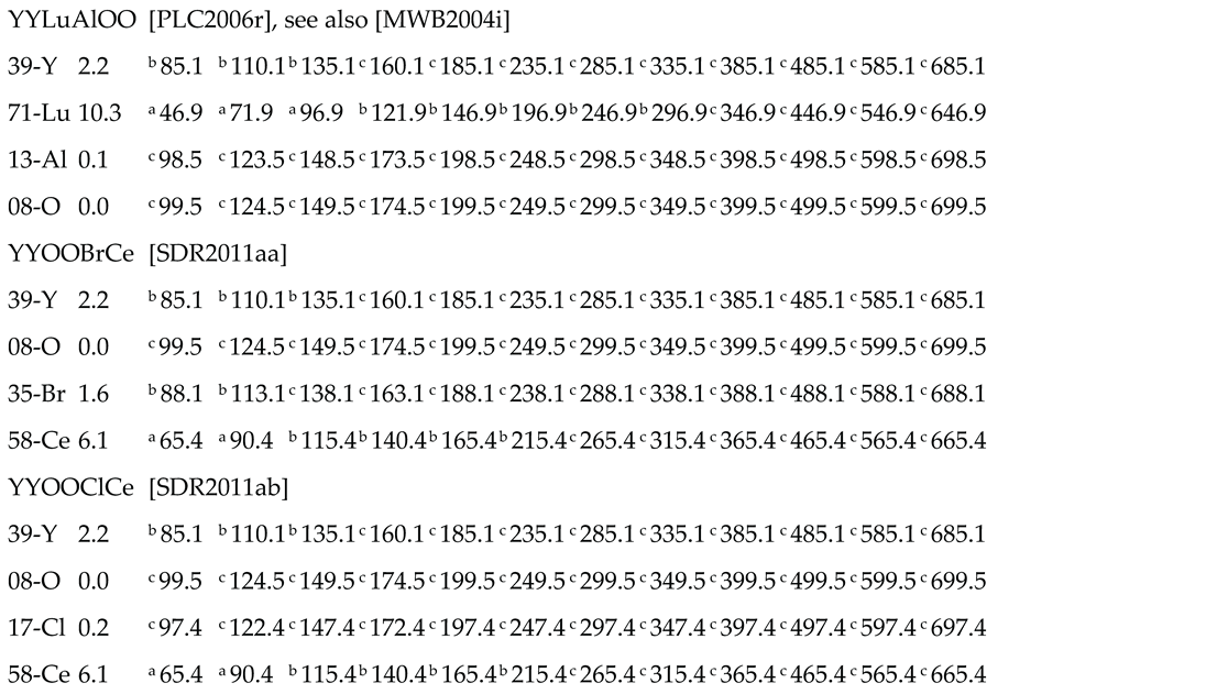
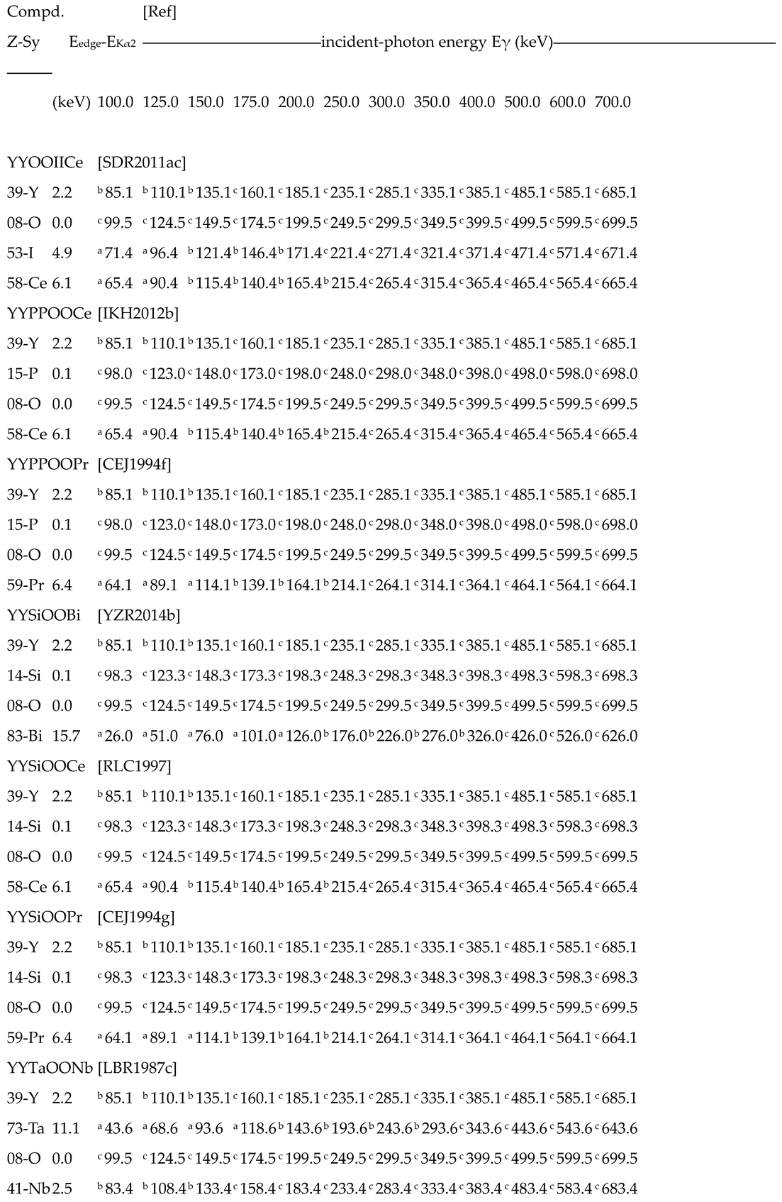
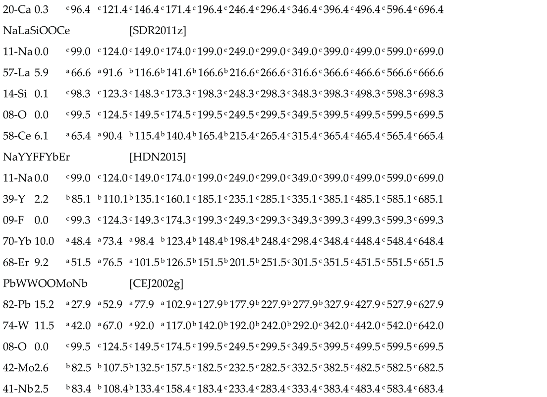
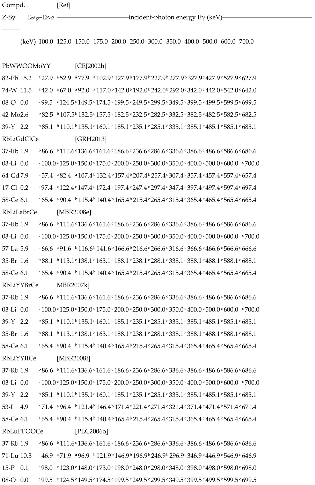
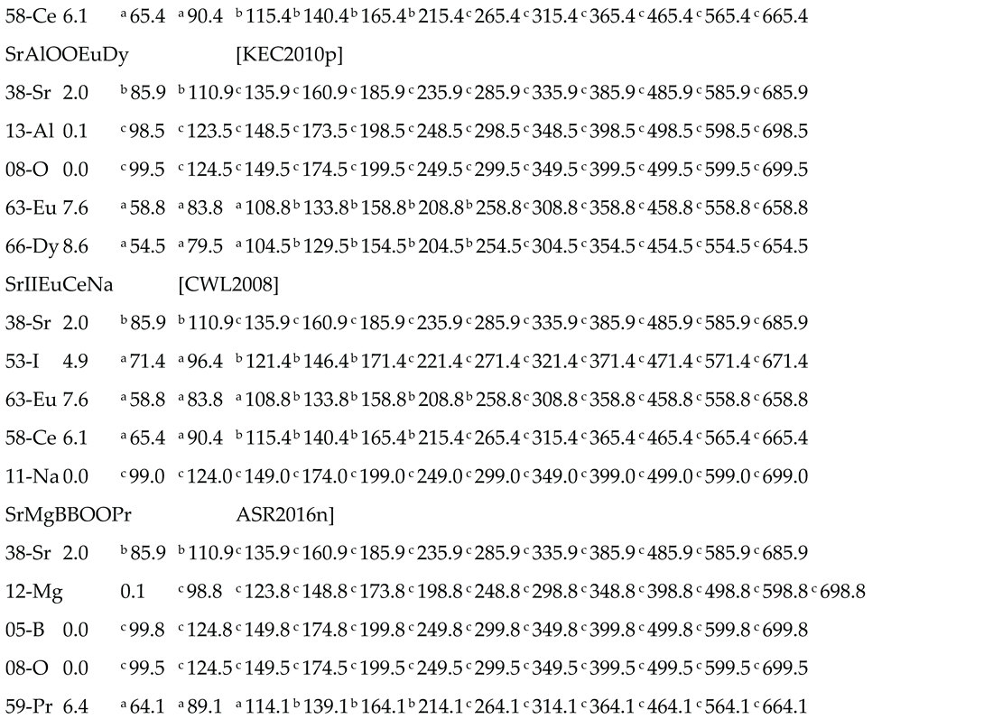
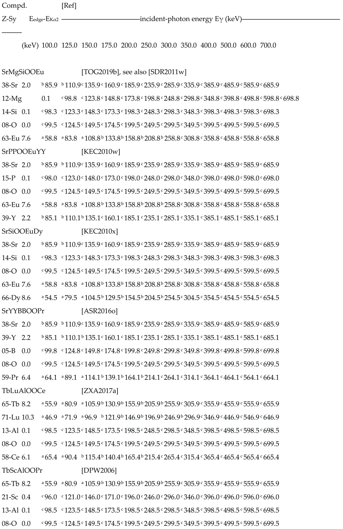
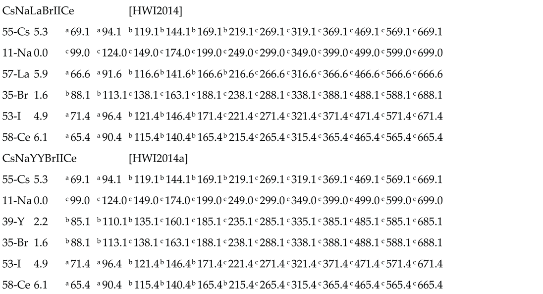
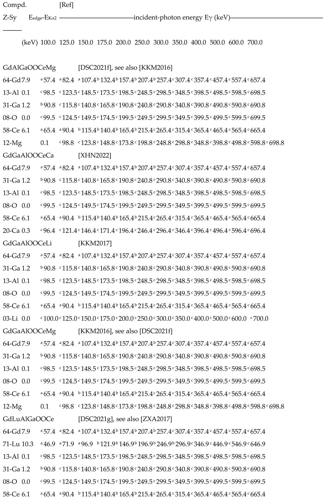
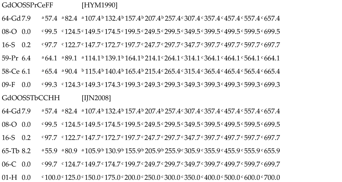
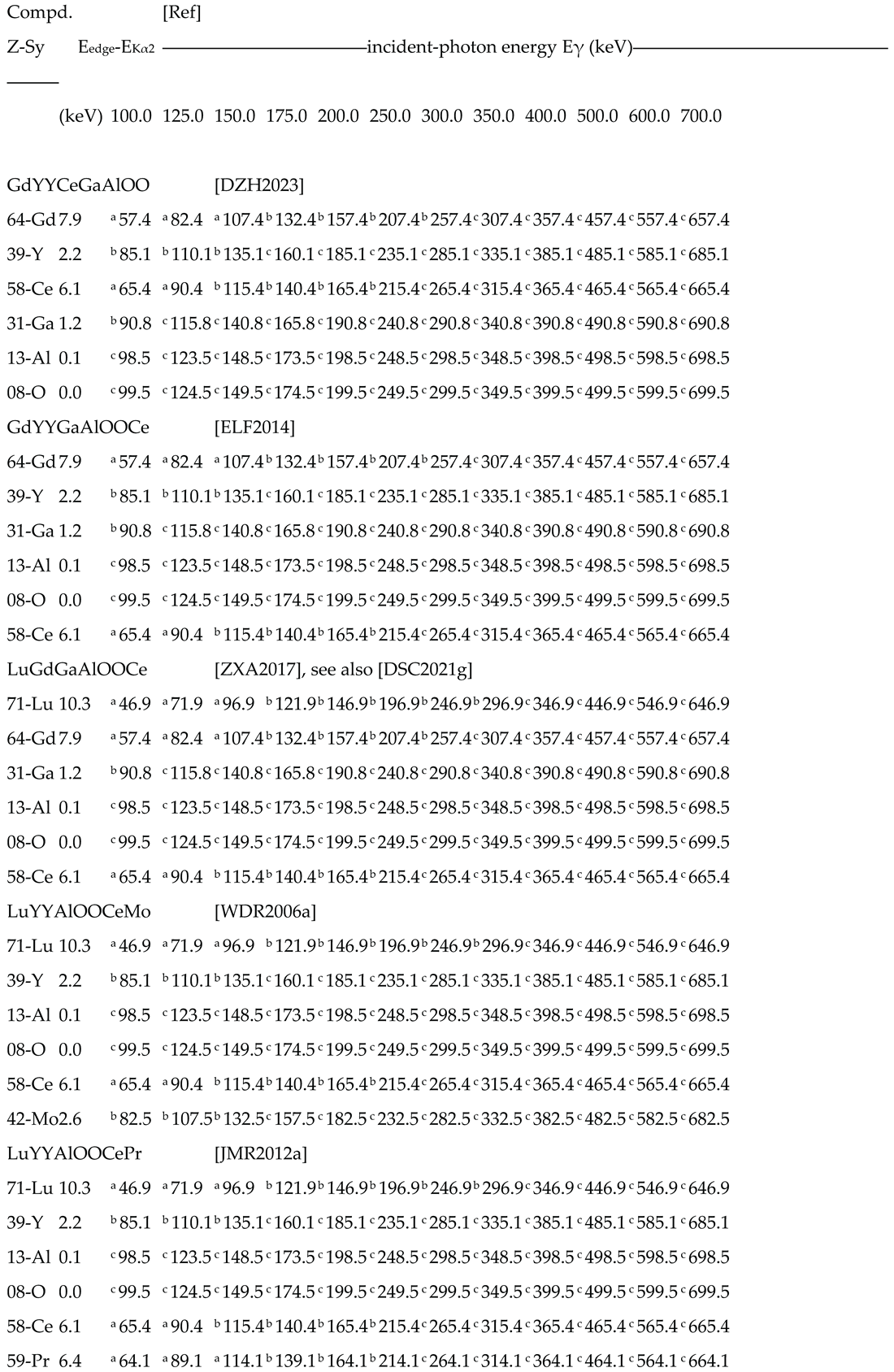
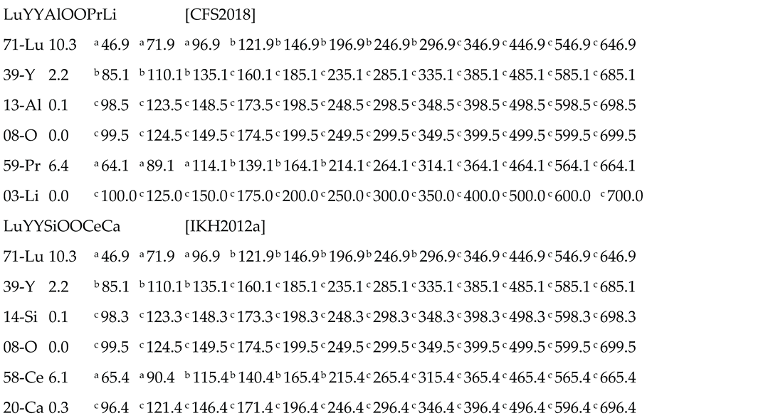
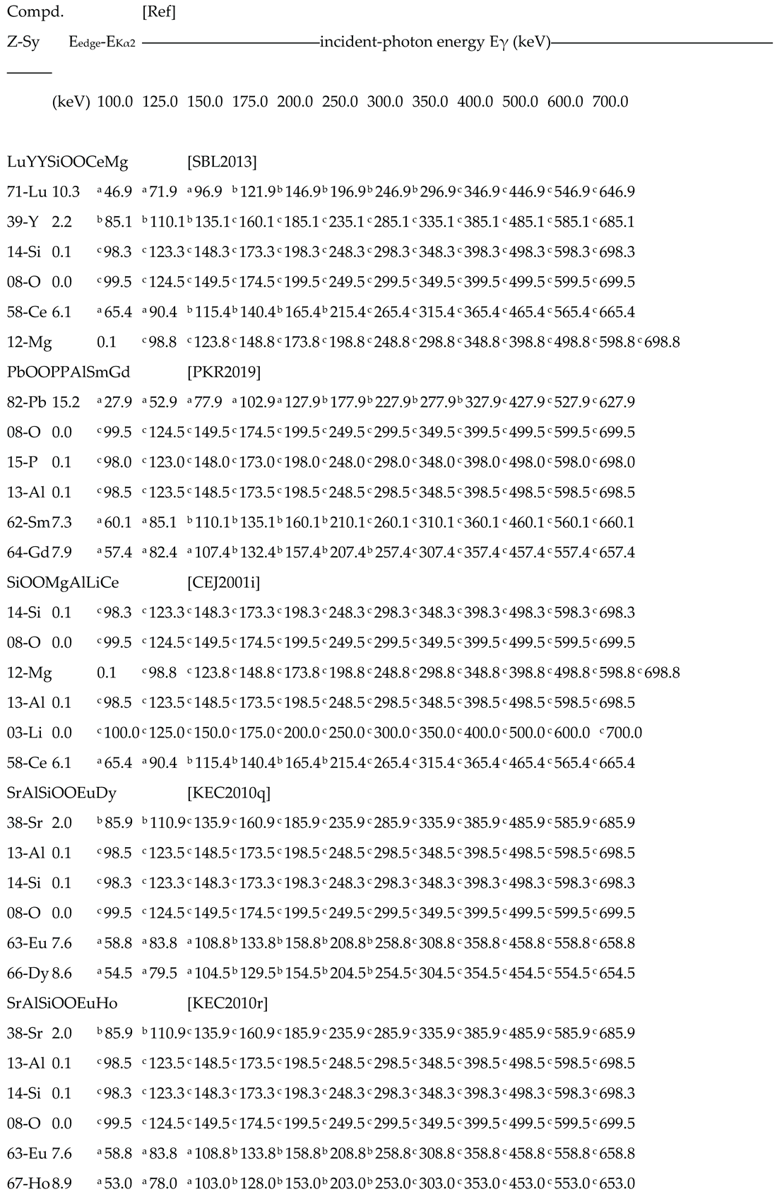
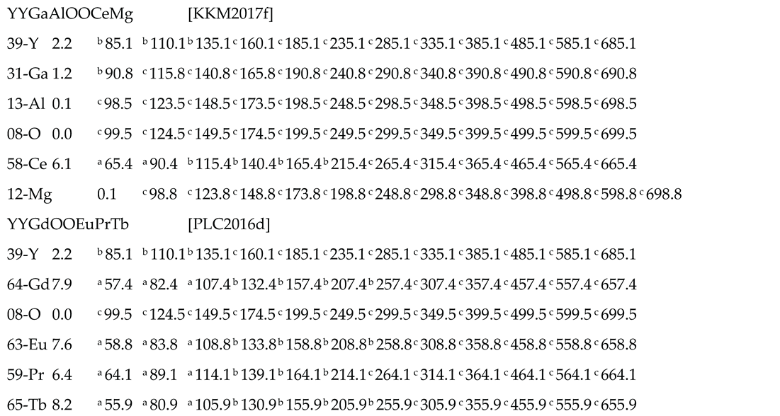
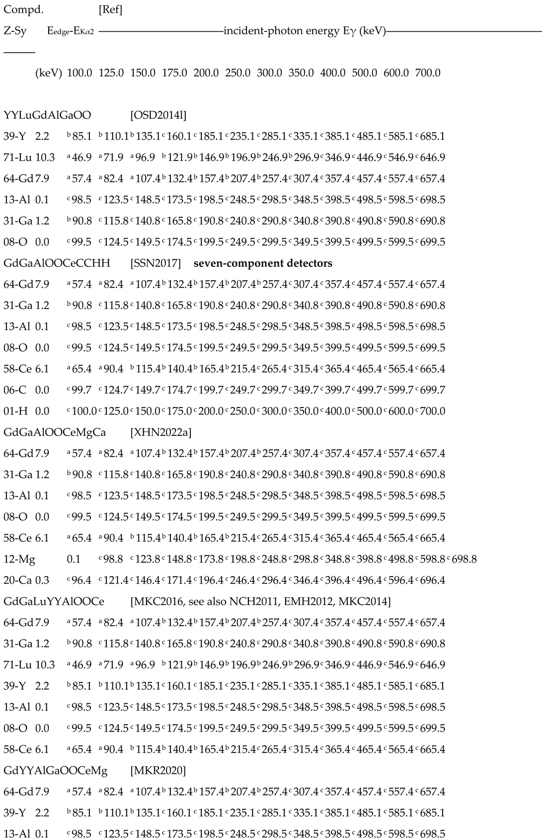
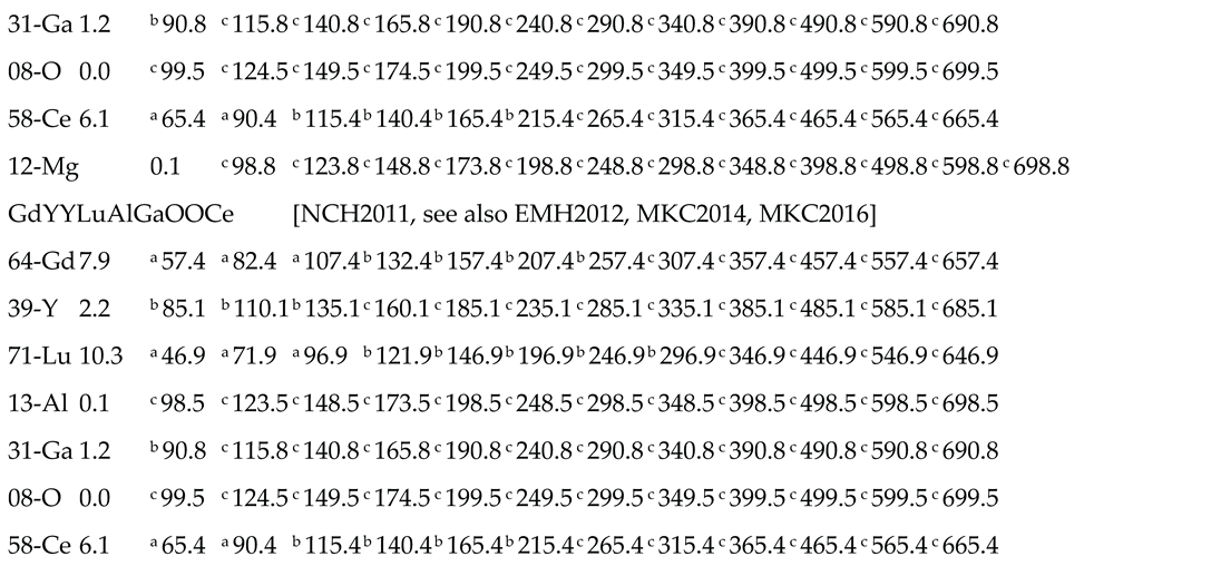
The values of energy of K-XRF-escape gamma-ray are predicted per 12 incident photons-energy values from 100 to 700 keV. Results are presented in Tables 3 to 7, arranged per Ne = 2, … , 8 and per compound id. This will confirm as the detector transfer function is perturbed more and more as Ne increases, due to the multiple presence of escape energies in the pulse-height spectrum.
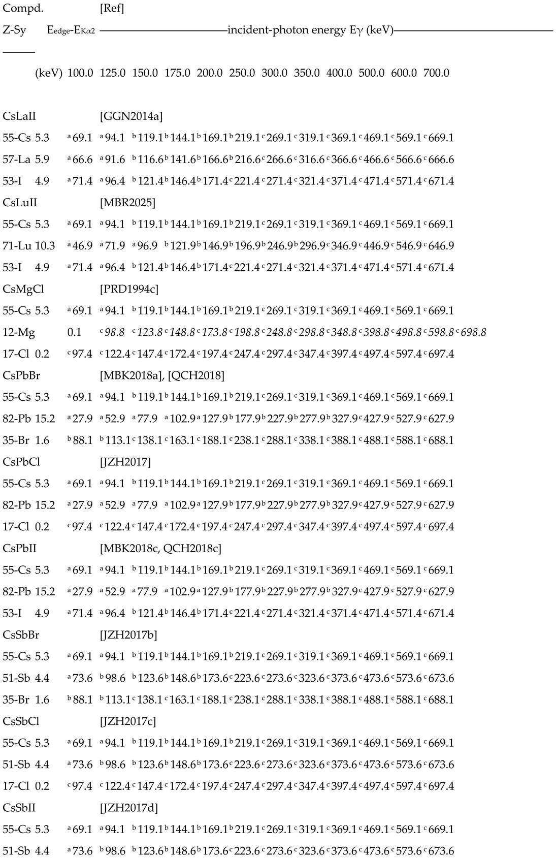
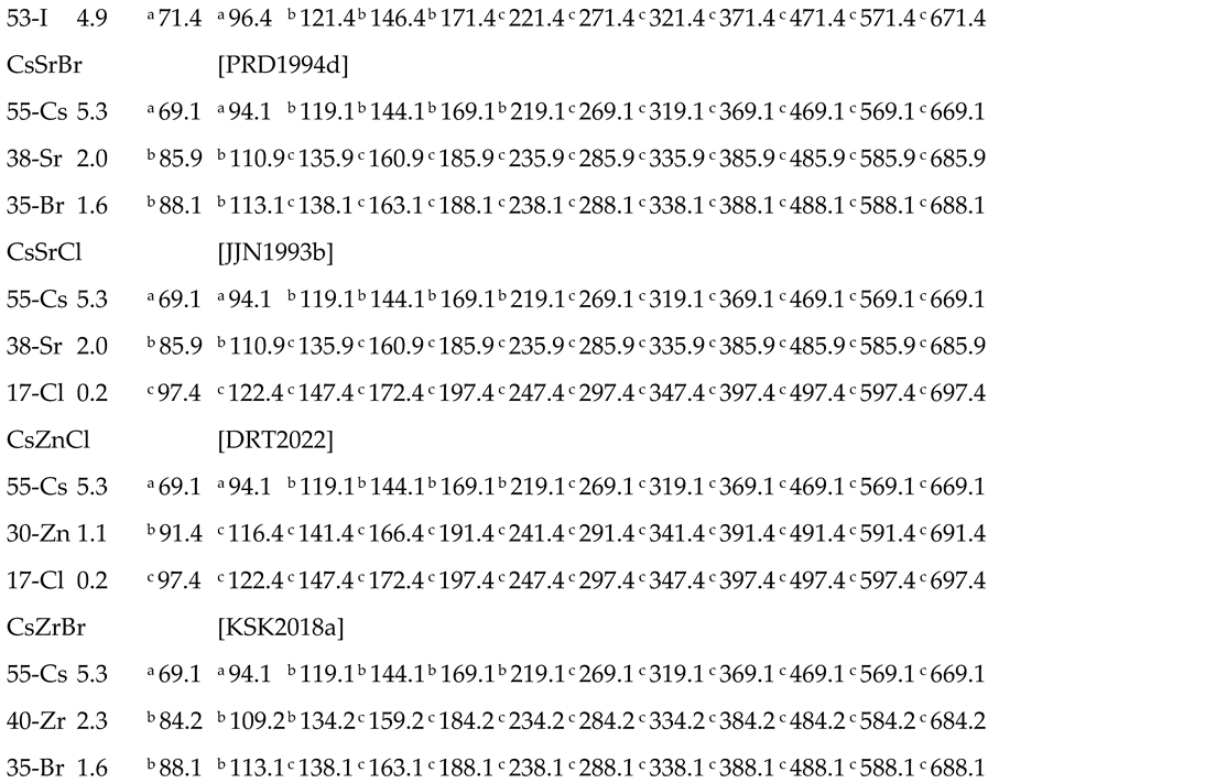
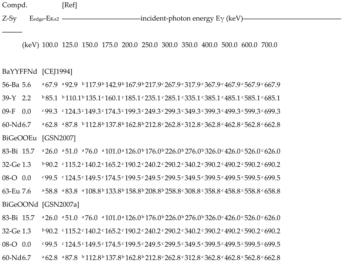
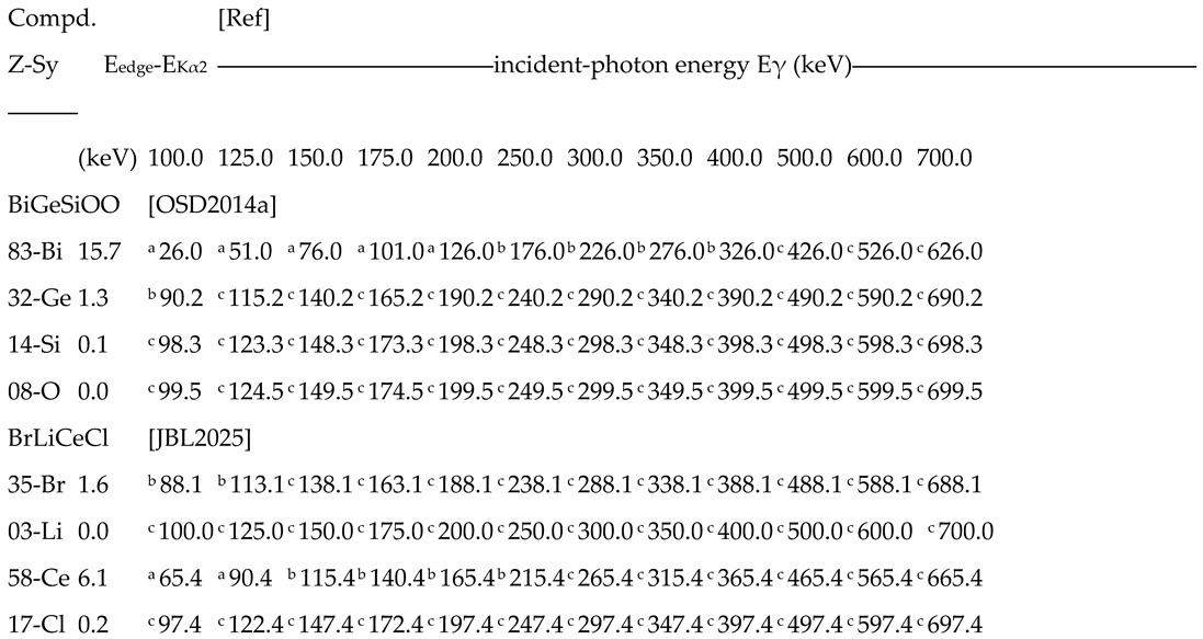
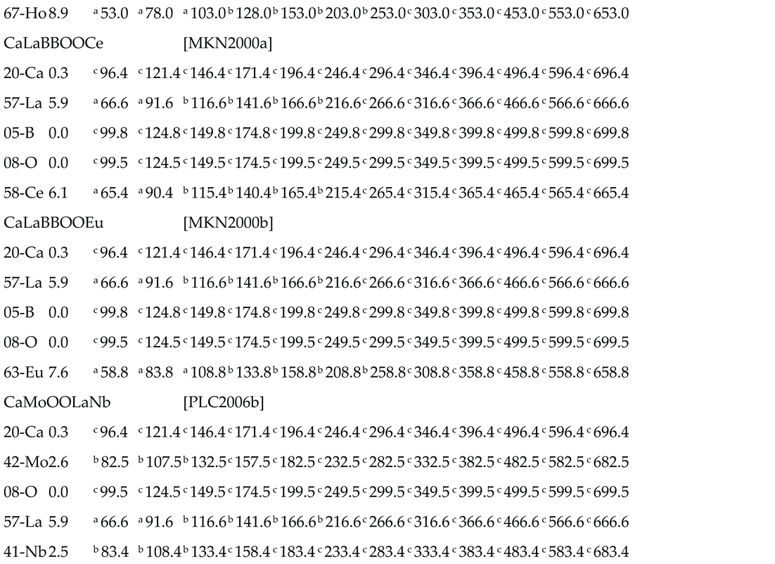
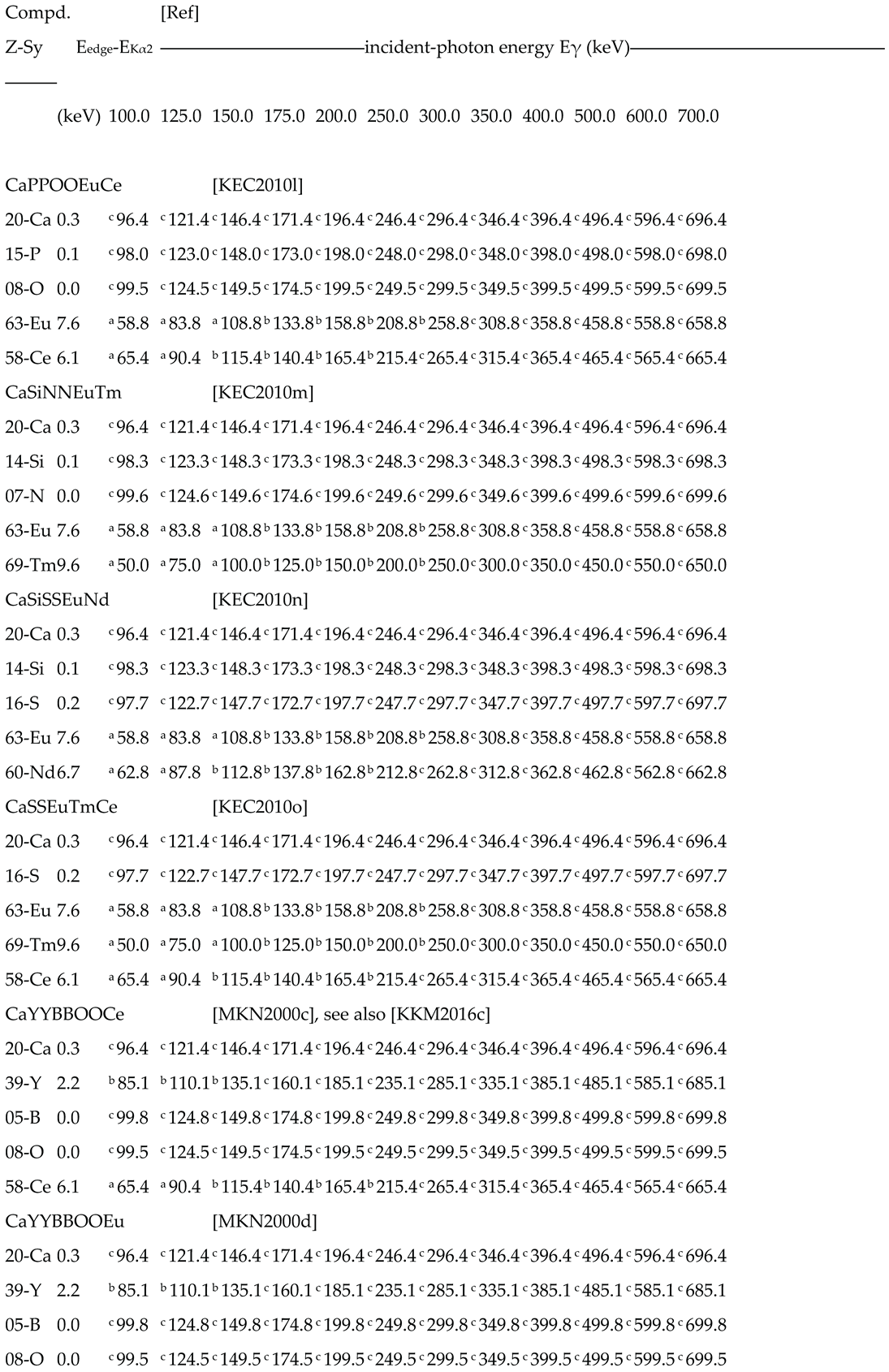
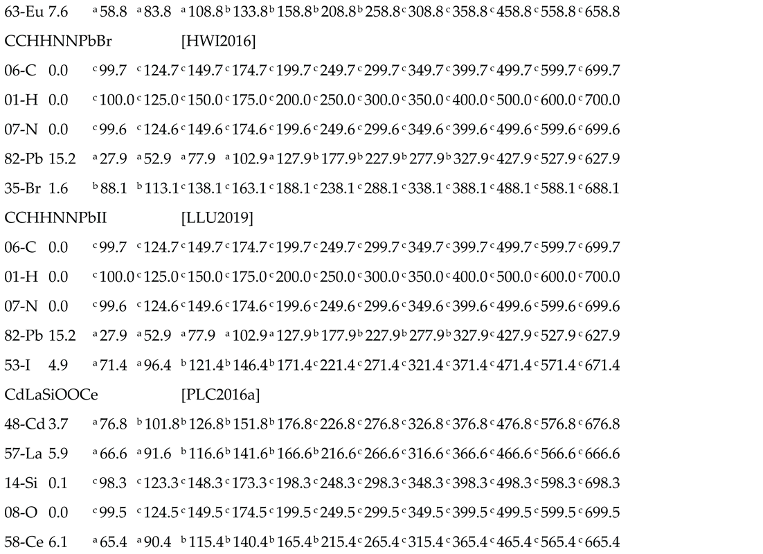
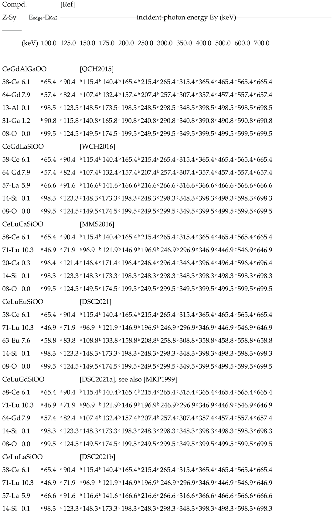
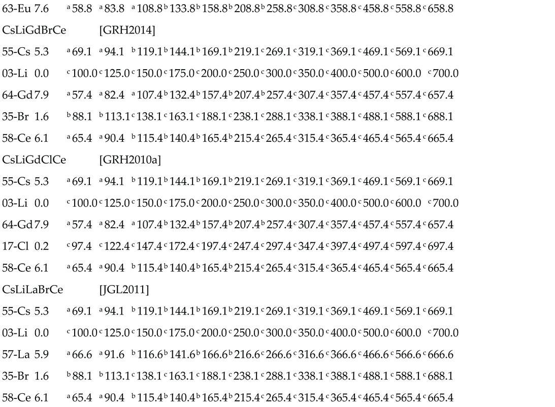
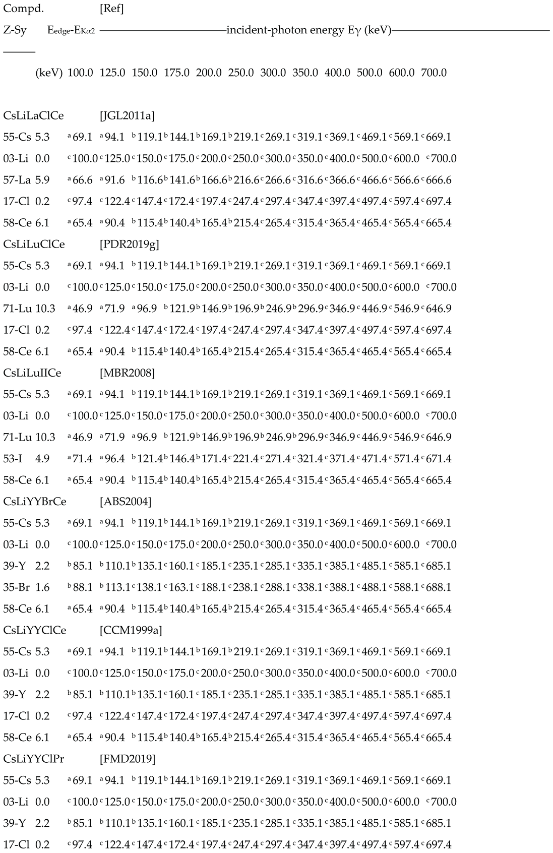
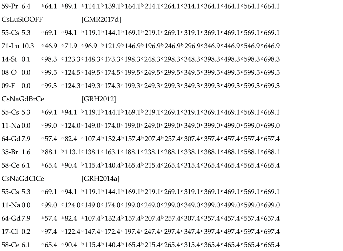
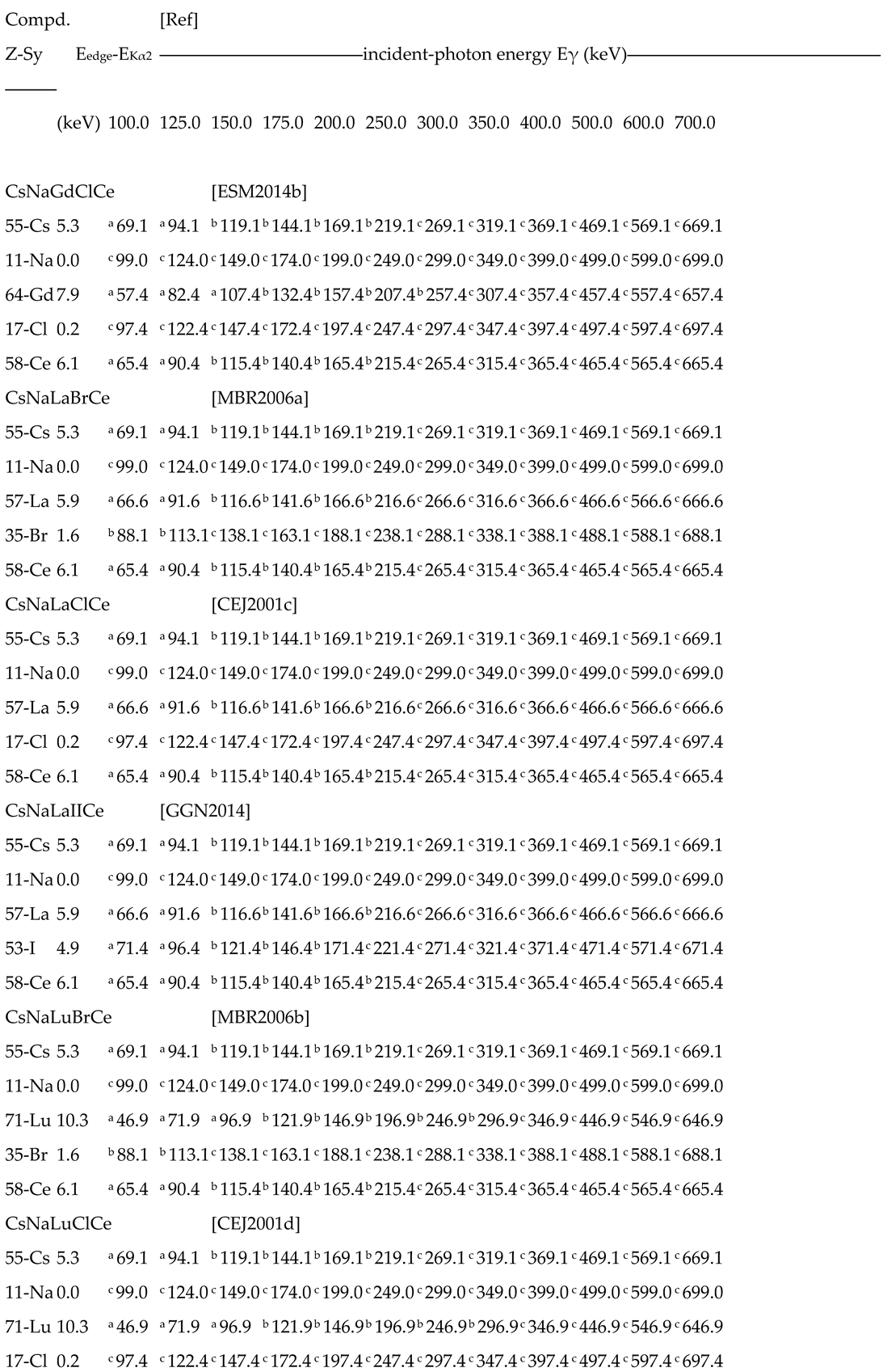
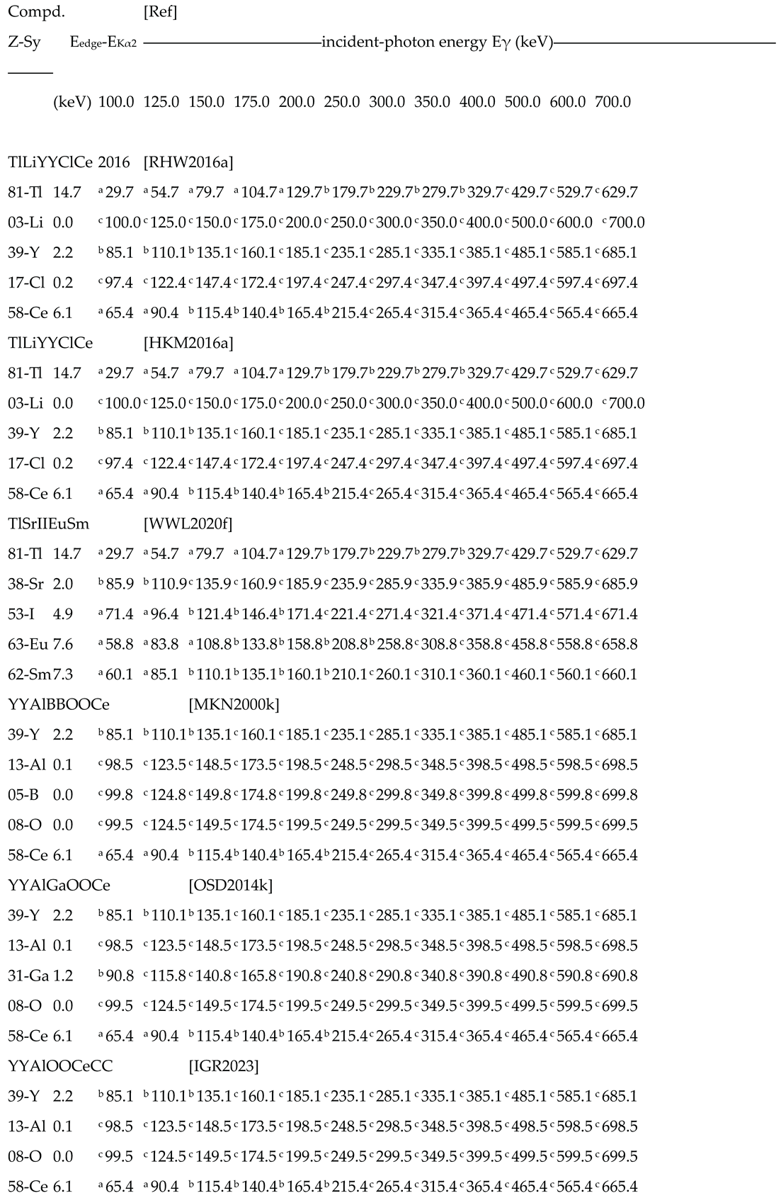
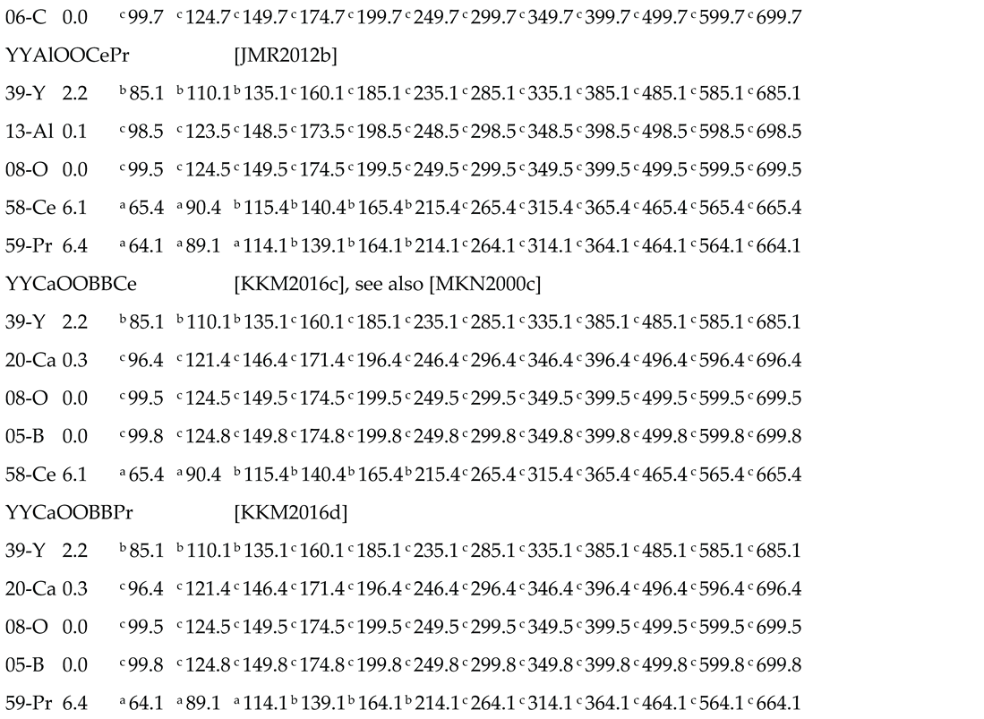
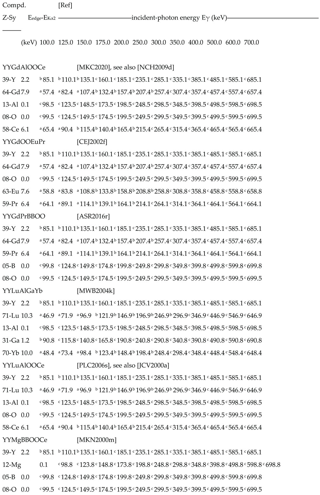
The elements sequence utilized in the chemical formula, from left to right, is the same identifying the lines from top to bottom.
The first line of the compound shows, from left to right, the compound-id and the citing reference.
The format of the compound id. takes into account the different symbol-length by doubling the single-character ones (so, as an example, BB stays for B – boron and so on).
The format of reference code (AAAYYYYa) is made by sub-strings related to (a) first-Author, (b) year of first publication, and (c) a code of multiple publication in the year.
The following Ne rows of the compound show, from left to right.
- the atomic number Z of the element and their symbol;
- the value of the energy-spread (in keV) between Kedge and Kα2 lines, representing the gap of the rest of K-orbital emissions, i.e. Kβ2, Kβ1, Kβ3, and Kα1; and
- a dozen of numbers representing the escape-peak energy predicted for the correspondent mono-energetic incident photons.
Policies
Literature Coverage
Fundamentals of radiation detection including the concepts herein exposed are deeply and clearly given in Ref.[GKN1999]. The basic X-rays data are from Kaye & Laby, UK National Physical Laboratory [K&L2013], whose completeness and accuracy fulfill the requirements of the Catalog. The other literature citations are from text-books, review articles, research reports, conference papers, etc… whose titles are reported in the Reference list.
Data Selected
Data for this catalog are taken from [K&L2013], limited to those of XRF energy in the columns Kα2 and Kedge (for 4 ≤ Z ≤ 92) that are used for power-function best fit performing whose parameters are needed for calculations.
Incident Gamma-Rays
Listed in each result Table is the header specifying the dozen of values of mono-energetic incident gamma-rays considered for the calculations of escape-peaks of different elements.
The values considered in calculations are: 100, 125, 150, 175, 200, 250, 300, 350, 400, 500, 600 and 700 keV. The range includes the energies from all the radio-isotopes used for tracing pharmaceutical administered in SPECT and PET protocols. The last interval is extended up to 700 keV for considering also the 662 keV photons from Cs-137 sources, widely used for energy-calibration.
Escape-Energy Uncertainty
The uncertainty of 0.2-0.3 keV can be assumed, despite submission to the fitting process. On the other hand the tabulated X-rays can be considered affected by uncertainties in the order of a few eV because they are listed in keV with 3 decimal places.
Escape Gamma-Ray Intensity
The intensity of escape-gamma-ray cannot be easily estimated like their energy. Appropriate Monte Carlo simulation studies of the detector-source systems must be performed aimed to predict intensity values.
Element and Compound Identification
Both the atomic number Z and the symbol of chemical element are given in the results Tables 3 to 7.
The format of the compound id. takes into account the different symbol-length by doubling the single-character ones (so, as an example, BB stays for B – boron and so on).
Photo-Electric Peak
The response of a detector to mono-energetic gamma-rays Eγ is mainly made, in an ideal pulse-height spectrum, of a vertical segment, representing the photo-electric interactions, whose abscissa is proportional to Eγ and whose length is proportional to the number of incident gammas. In the reality, the segment becomes a Gaussian curve, due to the enlargement produced by the detector-energy-resolution.
Compton-Effect
Another process affecting the detector response is the Compton scattering, that produces a continuum ranging from zero to the so-called Compton-edge, arising at the end of this continuum. The energy-distance between the Compton edge and the centroid of photo-electric peak can be calculated as (see [GKN1999], p.310):
EC = Eγ / ( 1 + 2 ( Eγ / m0 c2 ) ) , that is < Eγ .
Detector Response-Function (DRF)
If Eγ < 2 m0 c2 the response function shows only the two items above described. Based on these values it is reasonable, in a first approximation, to localize the energy at which a valley occurs between the photoelectric-peak and the Compton-edge, as: Evalley ≈ ½ ( Eγ + EC ).
Escape Peaks
Energies of gamma-ray peaks which result from escape of XRF photons from the detector are calculated, according to [GKN1999], as: EEscape = Eγ - EXRF , for Eγ > EXRF . Based on the energy of a given escape photon and on the above considerations regarding the DRF, these photons can be classified according to their position on the DRF. The three following styles are used in Tables 3 to 7 to show the predicted values of escape photon energy: (a) for values overlapping the Compton continuum; (b) for those falling in the region between Compton edge and photo-electric peak; (c) for those overlapped to the photo-peak left-tail.

Table 1.
Results of power-function best fit of XRF energy vs. Z for Kα2 orbital and Kedge. See Policies and Explanation
Table 1.
Results of power-function best fit of XRF energy vs. Z for Kα2 orbital and Kedge. See Policies and Explanation
| K Orbital | a (keV) | b | Rsquare |
| α2 | 0.00560144430106232 | 2.19086250995425 | 0.99995 |
| edge | 0.00628705594993865 | 2.12213694400228 | 0.99990 |
XRF characteristic energy Vs. atomic number, for a given orbital:
EXRF = a * (Z ^ b), where a and b characterize the orbitals for Z = 4 to 92.
Table 2.
Statistics of compounds per number of elements Ne = 2, …, 8, See Policies and Explanation.
| N° of elements Ne |
N° of compounds Nc |
Table |
| 2 | 77 | III |
| 3 | 198 | IV |
| 4 | 222 | V |
| 5 | 179 | VI |
| 6 | 43 | VII |
| 7 | 9 | VII |
| 8 | 1 | VII |
| Total | 729 |
The distribution shows the average value of 3.92 elements per compound.
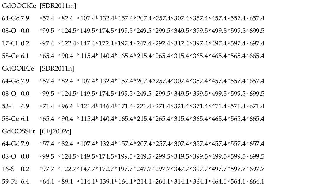
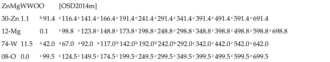
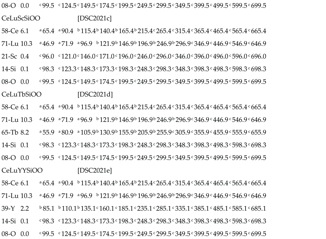
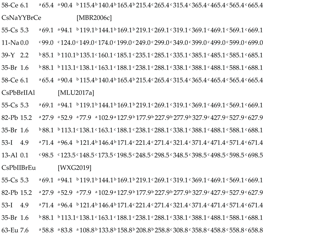
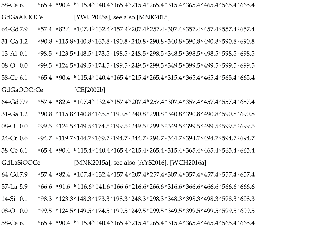
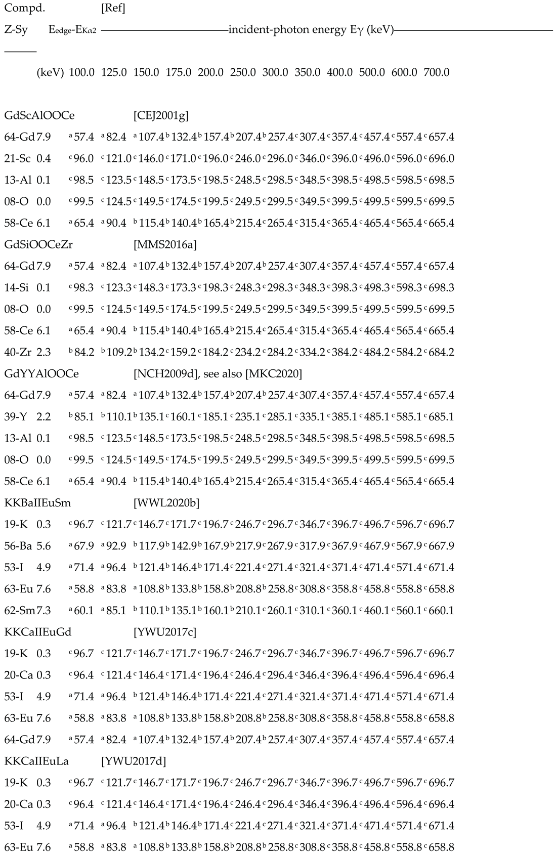
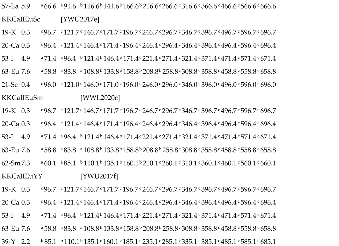
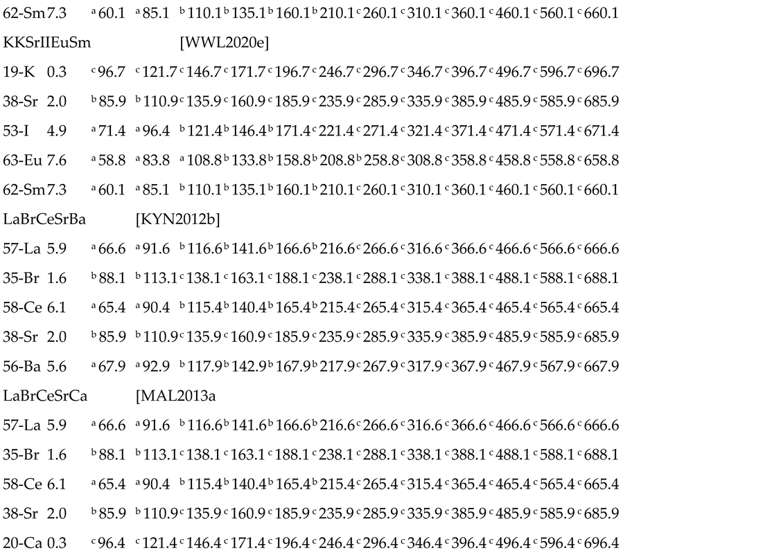
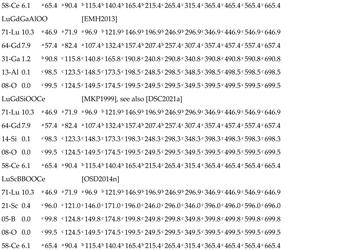
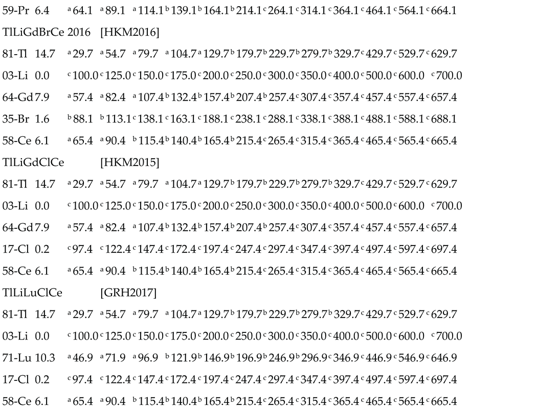
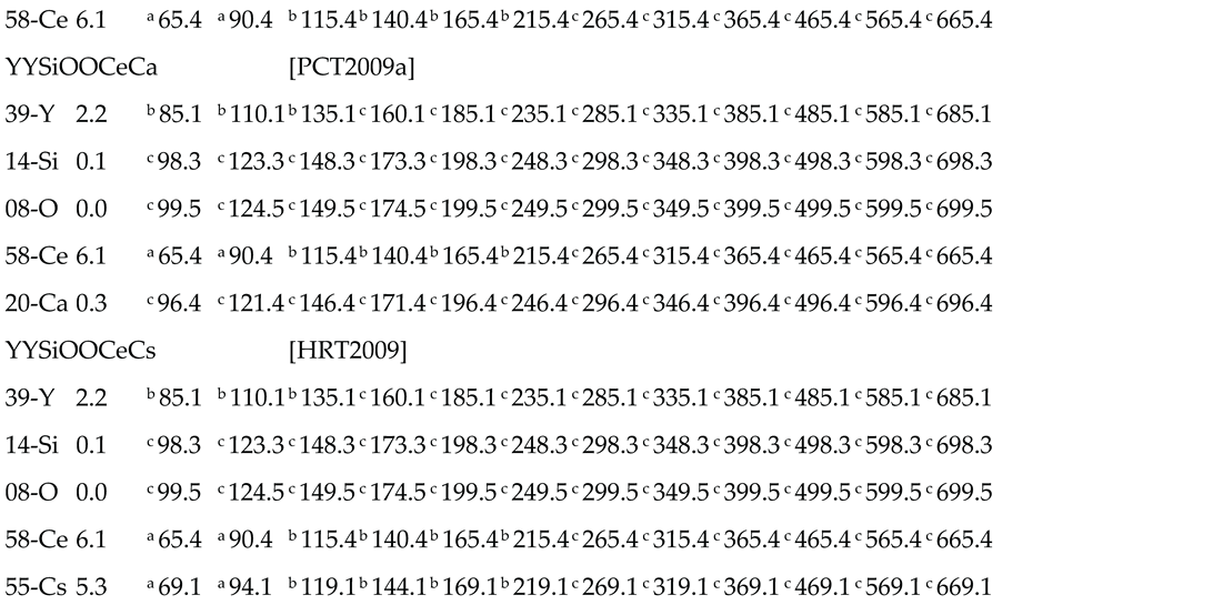
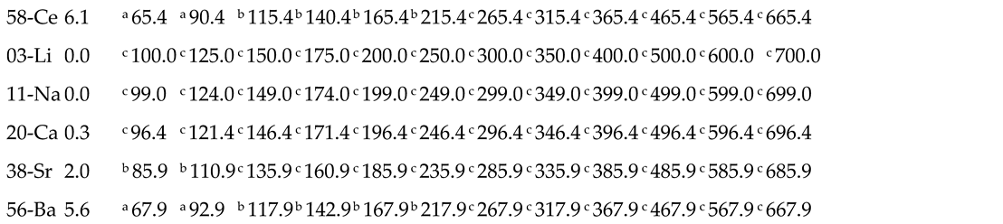
Table 3.
part (1/5) – Predicted values of XRF-escape energy Eγ-EKα2 for two-component detectors.
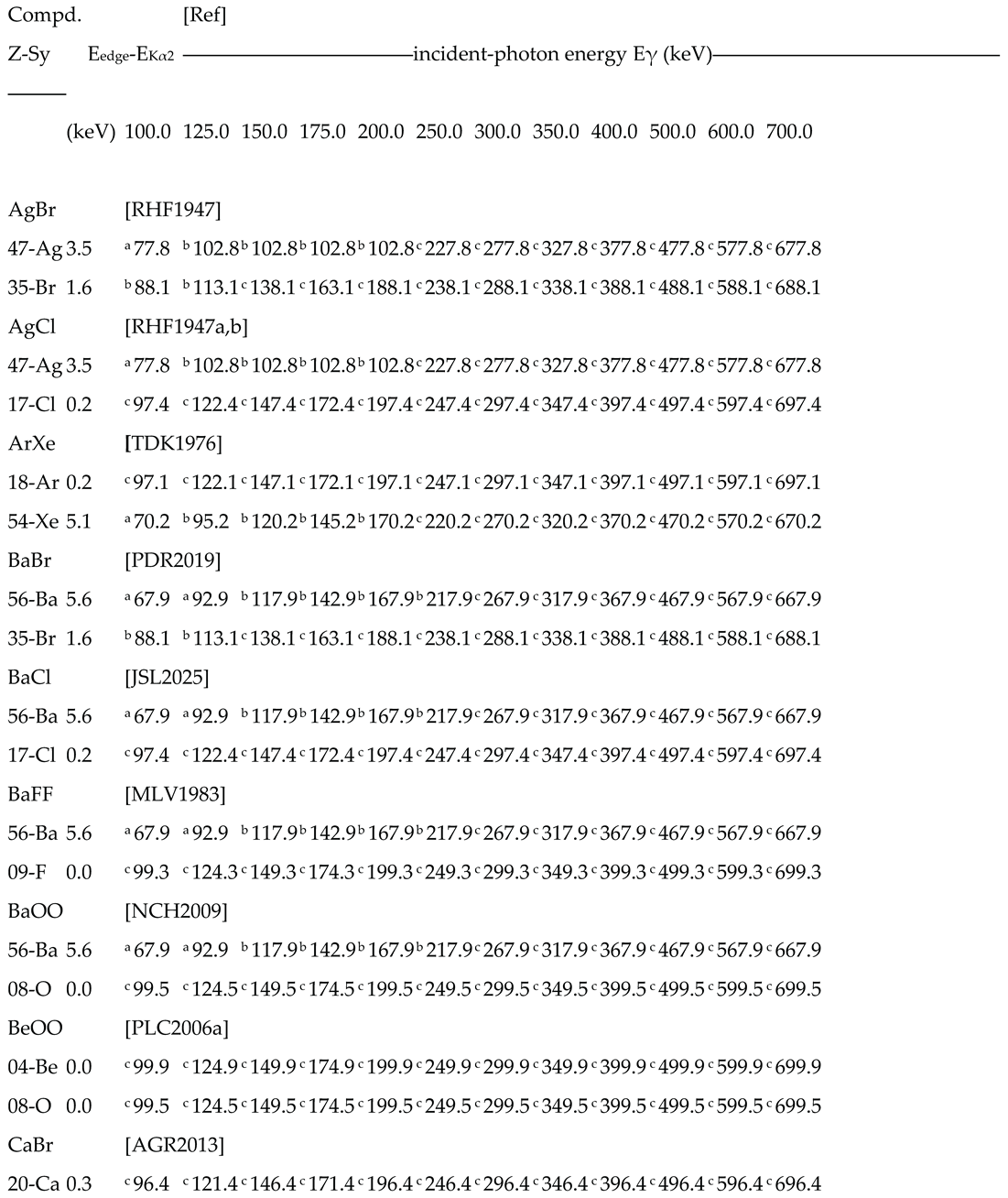 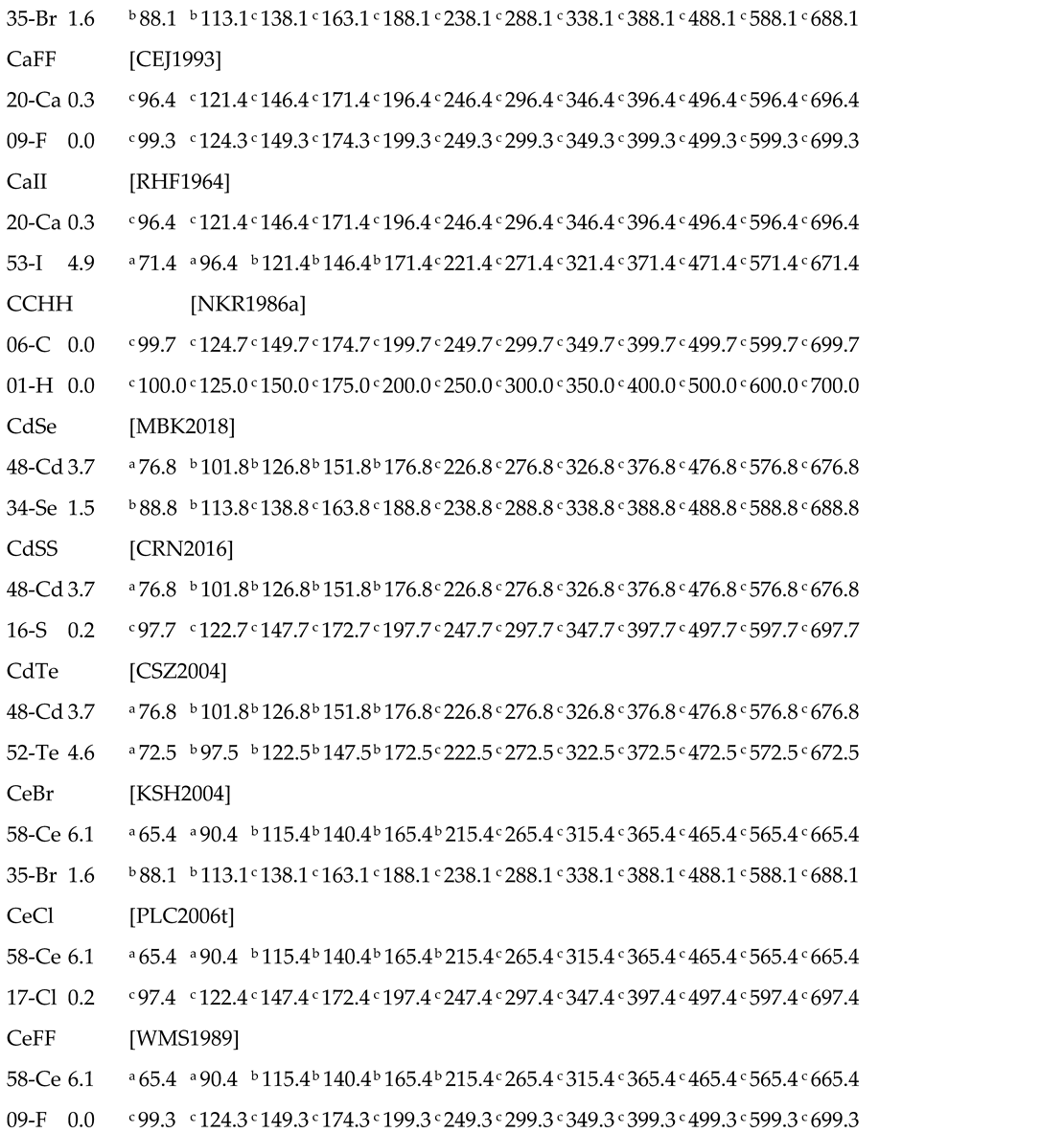 |
Notes for estimated escape peak position in the detector response function: a overlapped to the Compton continuum; b in the region between Compton edge and photo-electric peak; c overlapped to the photo-peak left-tail.
Table 3.
part (2/5) – Predicted values of XRF-escape energy Eγ-EKα2 for two-component detectors.
   |
Notes for estimated escape peak position in the detector response function: a overlapped to the Compton continuum; b in the region between Compton edge and photo-electric peak; c overlapped to the photo-peak left-tail.
Table 3.
part (3/5) – Predicted values of XRF-escape energy Eγ-E Kα2 for two-component detectors
  |
Notes for estimated escape peak position in the detector response function: a overlapped to the Compton continuum; b in the region between Compton edge and photo-electric peak; c overlapped to the photo-peak left-tail.
Table 3.
part (4/5) – Predicted values of XRF-escape energy Eγ-E Kα2 for two-component detectors
  |
Notes for estimated escape peak position in the detector response function: a overlapped to the Compton continuum; b in the region between Compton edge and photo-electric peak; c overlapped to the photo-peak left-tail.
Table 3.
part (5/5) – Predicted values of XRF-escape energy Eγ-E Kα2 for two-component detectors
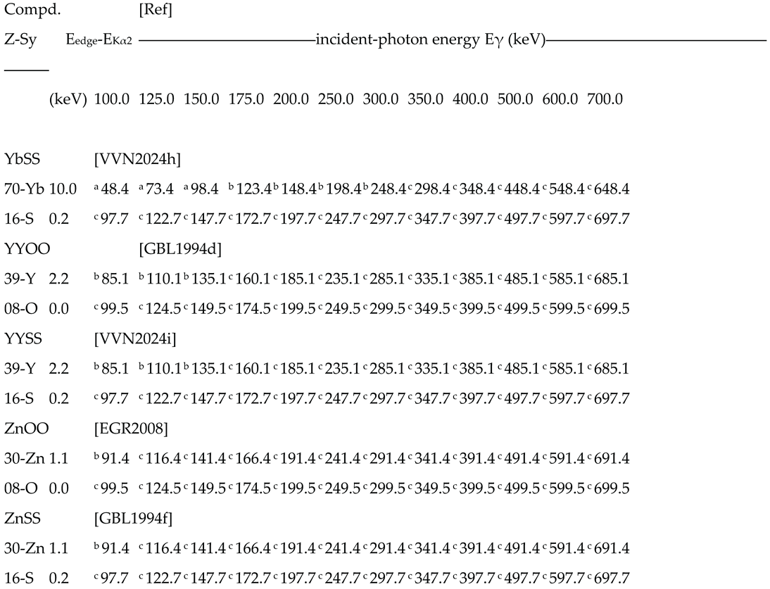 |
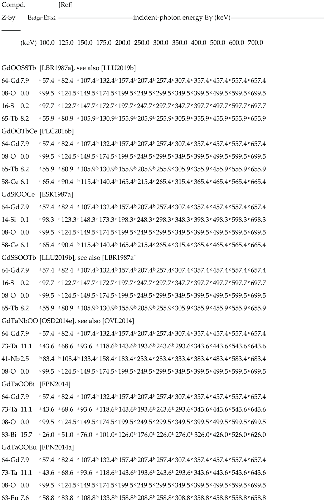
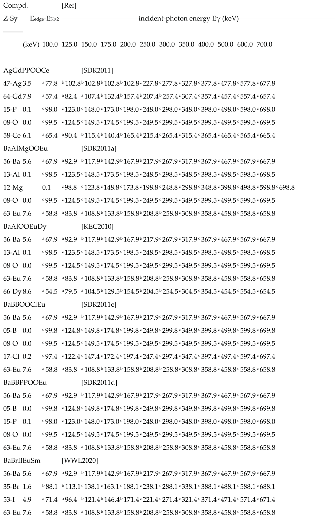
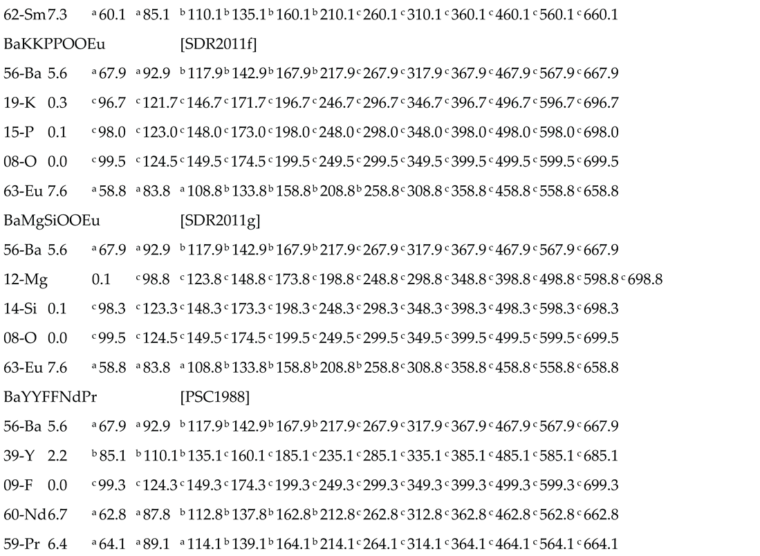
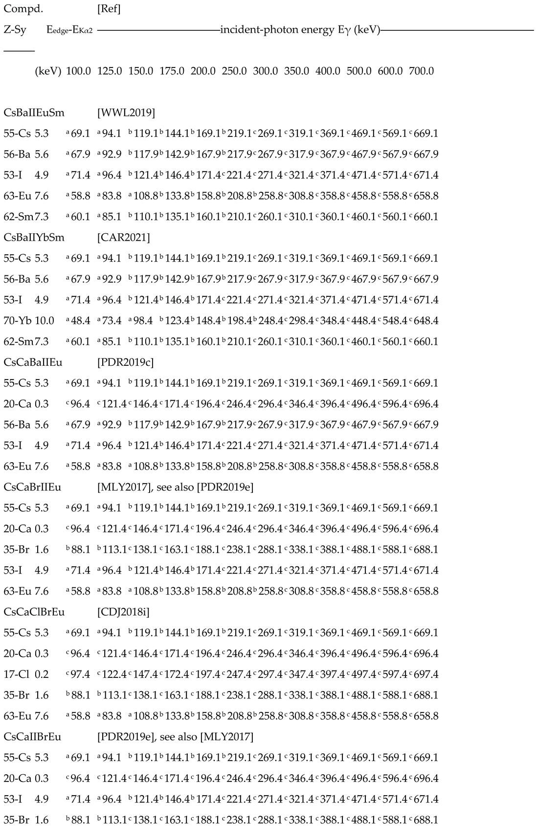
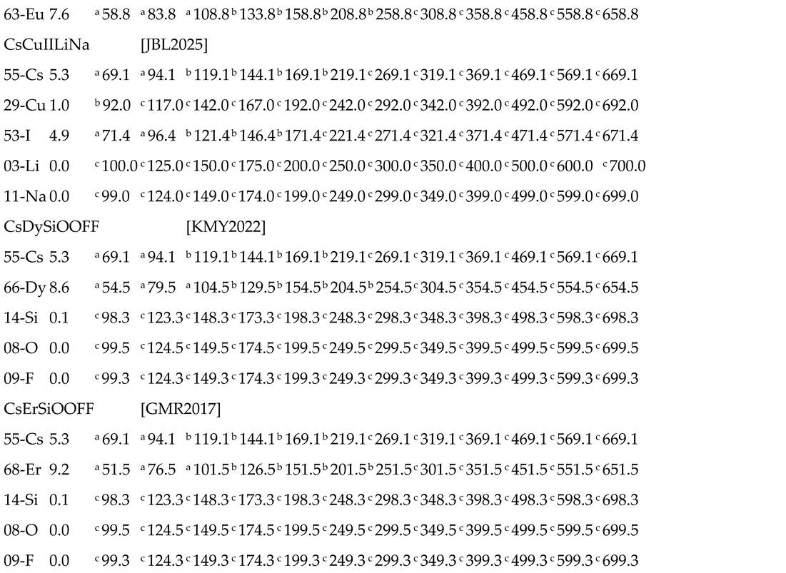
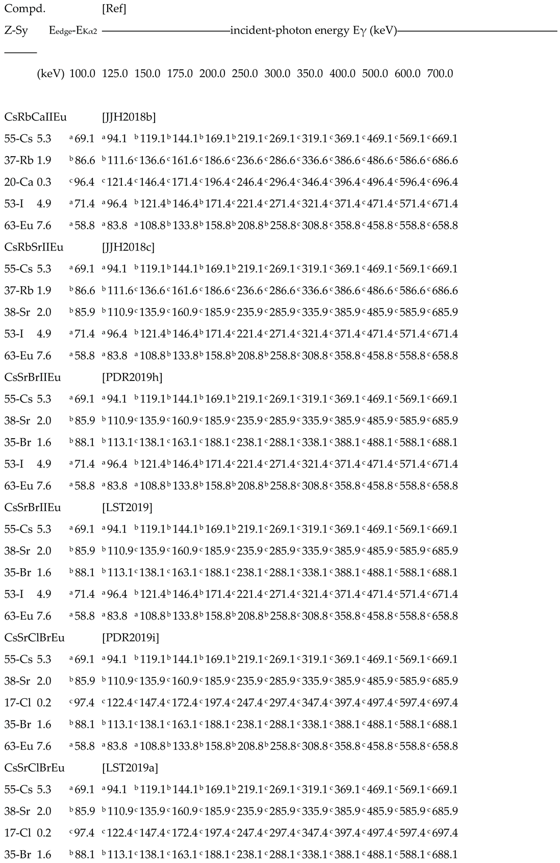
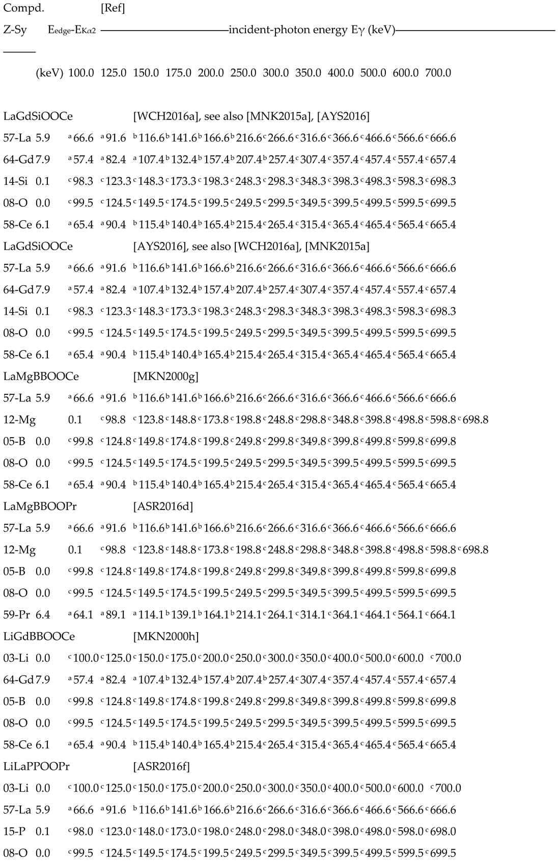
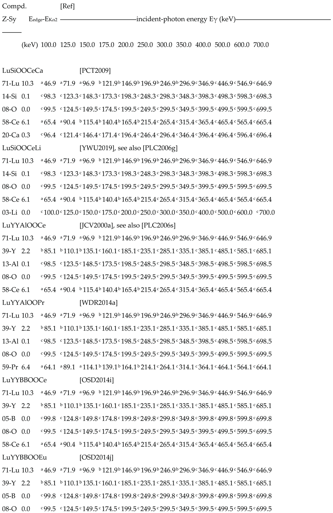
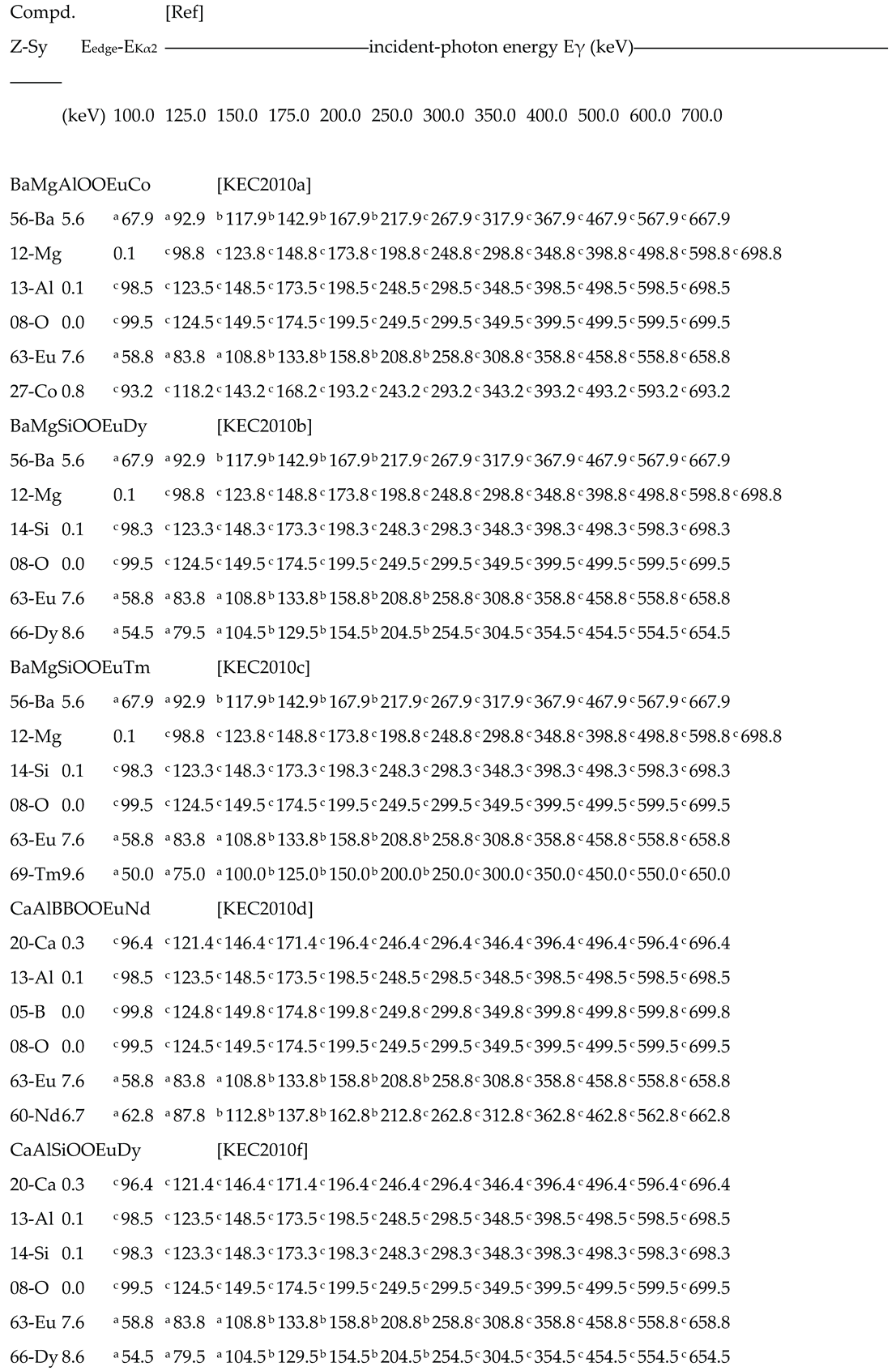
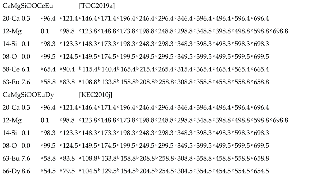
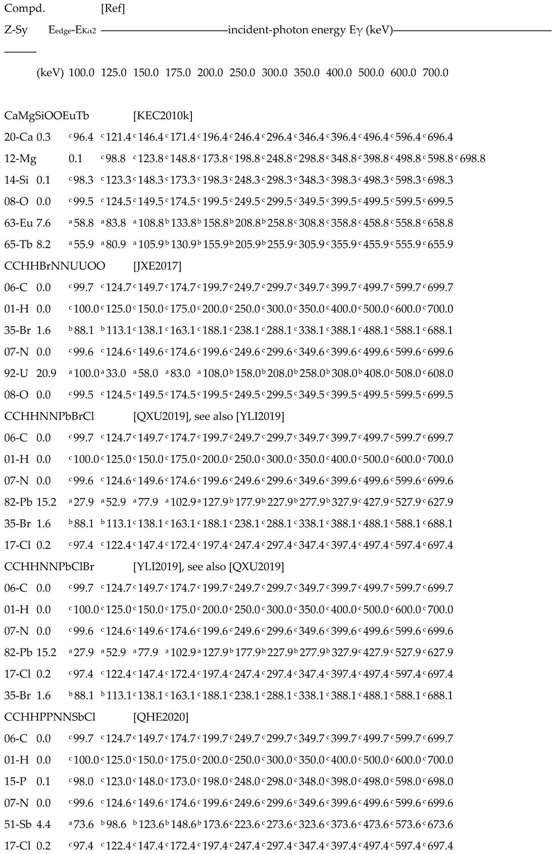
Table 4.
part (1/16) – Predicted values of XRF-escape energy Eγ-E Kα2 for three-component detectors
  |
Notes for estimated escape peak position in the detector response function: a overlapped to the Compton continuum; b in the region between Compton edge and photo-electric peak; c overlapped to the photo-peak left-tail.
Table 4.
part (2/16) – Predicted values of XRF-escape energy Eγ-E Kα2 for three-component detectors
  |
Notes for estimated escape peak position in the detector response function: a overlapped to the Compton continuum; b in the region between Compton edge and photo-electric peak; c overlapped to the photo-peak left-tail.
Table 4.
part (3/16) – Predicted values of XRF-escape energy Eγ-E Kα2 for three-component detectors
  |
Notes for estimated escape peak position in the detector response function: a overlapped to the Compton continuum; b in the region between Compton edge and photo-electric peak; c overlapped to the photo-peak left-tail.
Table 4.
part (4/16) – Predicted values of XRF-escape energy Eγ-E Kα2 for three-component detectors
  |
Notes for estimated escape peak position in the detector response function: a overlapped to the Compton continuum; b in the region between Compton edge and photo-electric peak; c overlapped to the photo-peak left-tail.
Table 4.
part (5/16) – Predicted values of XRF-escape energy Eγ-E Kα2 for three-component detectors
  |
Notes for estimated escape peak position in the detector response function: a overlapped to the Compton continuum; b in the region between Compton edge and photo-electric peak; c overlapped to the photo-peak left-tail.
Table 4.
part (6/16) – Predicted values of XRF-escape energy Eγ-E Kα2 for three-component detectors
  |
Notes for estimated escape peak position in the detector response function: a overlapped to the Compton continuum; b in the region between Compton edge and photo-electric peak; c overlapped to the photo-peak left-tail.
Table 4.
part (7/16) – Predicted values of XRF-escape energy Eγ-E Kα2 for three-component detectors
  |
Notes for estimated escape peak position in the detector response function: a overlapped to the Compton continuum; b in the region between Compton edge and photo-electric peak; c overlapped to the photo-peak left-tail.
Table 4.
part (8/16) – Predicted values of XRF-escape energy Eγ-E Kα2 for three-component detectors
  |
Notes for estimated escape peak position in the detector response function: a overlapped to the Compton continuum; b in the region between Compton edge and photo-electric peak; c overlapped to the photo-peak left-tail.
Table 4.
part (9/16) – Predicted values of XRF-escape energy Eγ-E Kα2 for three-component detectors
  |
Notes for estimated escape peak position in the detector response function: a overlapped to the Compton continuum; b in the region between Compton edge and photo-electric peak; c overlapped to the photo-peak left-tail.
Table 4.
part (10/16) – Predicted values of XRF-escape energy Eγ-E Kα2 for three-component detectors
Table 4.
part (10/16) – Predicted values of XRF-escape energy Eγ-E Kα2 for three-component detectors
  |
Notes for estimated escape peak position in the detector response function: a overlapped to the Compton continuum; b in the region between Compton edge and photo-electric peak; c overlapped to the photo-peak left-tail.
Table 4.
part (11/16) – Predicted values of XRF-escape energy Eγ-E Kα2 for three-component detectors
Table 4.
part (11/16) – Predicted values of XRF-escape energy Eγ-E Kα2 for three-component detectors
  |
Notes for estimated escape peak position in the detector response function: a overlapped to the Compton continuum; b in the region between Compton edge and photo-electric peak; c overlapped to the photo-peak left-tail.
Table 4.
part (12/16) – Predicted values of XRF-escape energy Eγ-E Kα2 for three-component detectors
Table 4.
part (12/16) – Predicted values of XRF-escape energy Eγ-E Kα2 for three-component detectors
  |
Notes for estimated escape peak position in the detector response function: a overlapped to the Compton continuum; b in the region between Compton edge and photo-electric peak; c overlapped to the photo-peak left-tail.
Table 4.
part (13/16) – Predicted values of XRF-escape energy Eγ-E Kα2 for three-component detectors
Table 4.
part (13/16) – Predicted values of XRF-escape energy Eγ-E Kα2 for three-component detectors
  |
Notes for estimated escape peak position in the detector response function: a overlapped to the Compton continuum; b in the region between Compton edge and photo-electric peak; c overlapped to the photo-peak left-tail.
Table 4.
part (14/16) – Predicted values of XRF-escape energy Eγ-E Kα2 for three-component detectors
Table 4.
part (14/16) – Predicted values of XRF-escape energy Eγ-E Kα2 for three-component detectors
  |
Notes for estimated escape peak position in the detector response function: a overlapped to the Compton continuum; b in the region between Compton edge and photo-electric peak; c overlapped to the photo-peak left-tail.
Table 4.
part (15/16) – Predicted values of XRF-escape energy Eγ-E Kα2 for three-component detectors
Table 4.
part (15/16) – Predicted values of XRF-escape energy Eγ-E Kα2 for three-component detectors
  |
Notes for estimated escape peak position in the detector response function: a overlapped to the Compton continuum; b in the region between Compton edge and photo-electric peak; c overlapped to the photo-peak left-tail.
Table 4.
part (16/16) – Predicted values of XRF-escape energy Eγ-E Kα2 for three-component detectors
Table 4.
part (16/16) – Predicted values of XRF-escape energy Eγ-E Kα2 for three-component detectors
 |
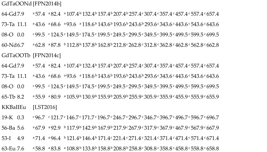
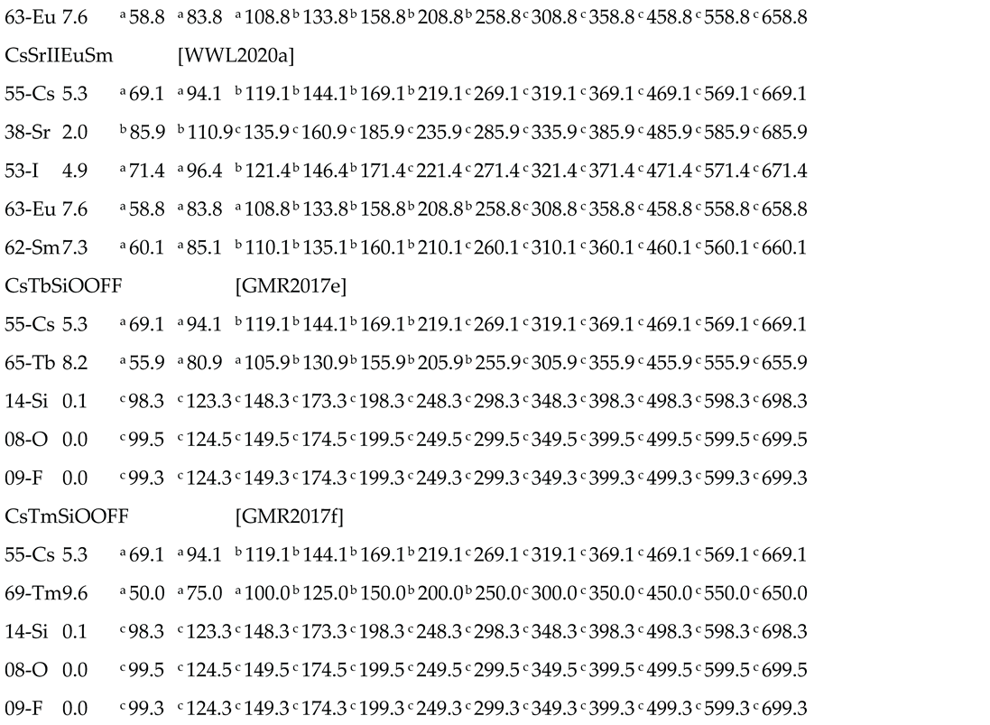
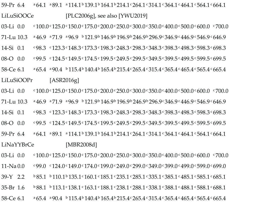
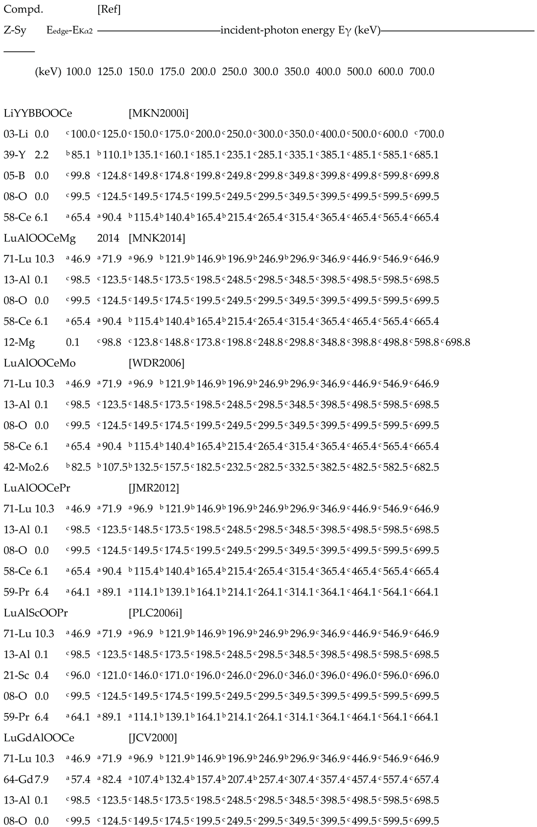
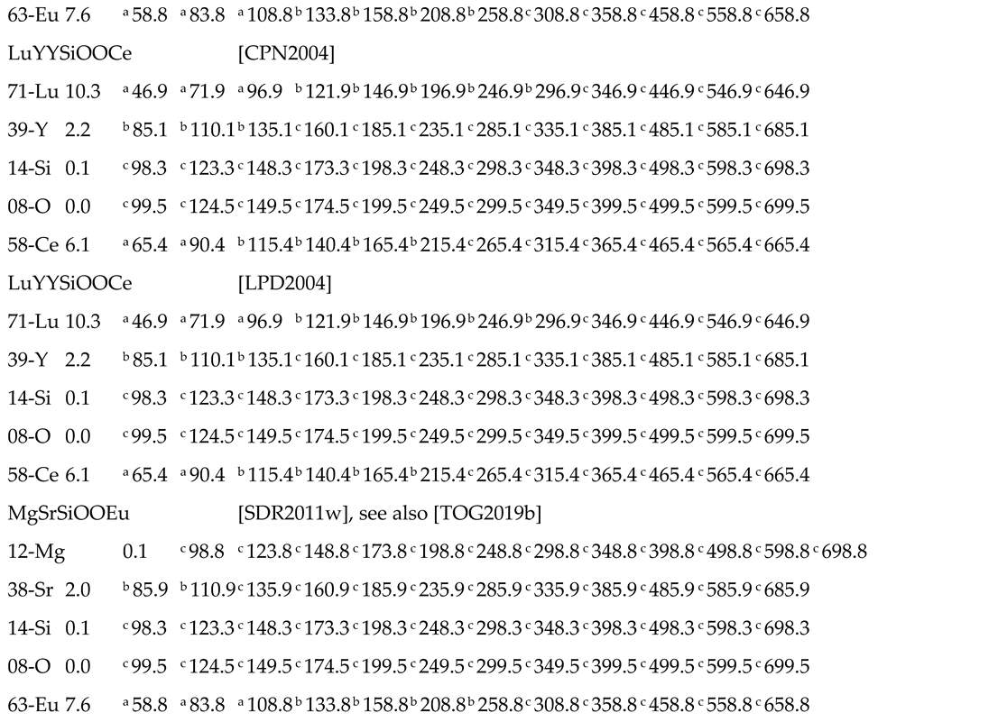
Table 5.
part (1/23) – Predicted values of XRF-escape energy Eγ-E Kα2 for four-component detectors
 |
Notes for estimated escape peak position in the detector response function: a overlapped to the Compton continuum; b in the region between Compton edge and photo-electric peak; c overlapped to the photo-peak left-tail.
Table 5.
part (2/23) – Predicted values of XRF-escape energy Eγ-E Kα2 for four-component detectors
   |
Notes for estimated escape peak position in the detector response function: a overlapped to the Compton continuum; b in the region between Compton edge and photo-electric peak; c overlapped to the photo-peak left-tail.
Table 5.
part (3/23) – Predicted values of XRF-escape energy Eγ-E Kα2 for four-component detectors
  |
Notes for estimated escape peak position in the detector response function: a overlapped to the Compton continuum; b in the region between Compton edge and photo-electric peak; c overlapped to the photo-peak left-tail.
Table 5.
part (4/23) – Predicted values of XRF-escape energy Eγ-E Kα2 for four-component detectors
  |
Notes for estimated escape peak position in the detector response function: a overlapped to the Compton continuum; b in the region between Compton edge and photo-electric peak; c overlapped to the photo-peak left-tail.
Table 5.
part (5/23) – Predicted values of XRF-escape energy Eγ-E Kα2 for four-component detectors
  |
Notes for estimated escape peak position in the detector response function: a overlapped to the Compton continuum; b in the region between Compton edge and photo-electric peak; c overlapped to the photo-peak left-tail.
Table 5.
part (6/23) – Predicted values of XRF-escape energy Eγ-E Kα2 for four-component detectors
  |
Notes for estimated escape peak position in the detector response function: a overlapped to the Compton continuum; b in the region between Compton edge and photo-electric peak; c overlapped to the photo-peak left-tail.
Table 5.
part (7/23) – Predicted values of XRF-escape energy Eγ-E Kα2 for four-component detectors
  |
Notes for estimated escape peak position in the detector response function: a overlapped to the Compton continuum; b in the region between Compton edge and photo-electric peak; c overlapped to the photo-peak left-tail.
Table 5.
part (8/23) – Predicted values of XRF-escape energy Eγ-E Kα2 for four-component detectors
  |
Notes for estimated escape peak position in the detector response function: a overlapped to the Compton continuum; b in the region between Compton edge and photo-electric peak; c overlapped to the photo-peak left-tail.
Table 5.
part (9/23) – Predicted values of XRF-escape energy Eγ-E Kα2 for four-component detectors
  |
Notes for estimated escape peak position in the detector response function: a overlapped to the Compton continuum; b in the region between Compton edge and photo-electric peak; c overlapped to the photo-peak left-tail.
Table 5.
part (10/23) – Predicted values of XRF-escape energy Eγ-E Kα2 for four-component detectors
  |
Notes for estimated escape peak position in the detector response function: a overlapped to the Compton continuum; b in the region between Compton edge and photo-electric peak; c overlapped to the photo-peak left-tail.
Table 5.
part (11/23) – Predicted values of XRF-escape energy Eγ-E Kα2 for four-component detectors
  |
Notes for estimated escape peak position in the detector response function: a overlapped to the Compton continuum; b in the region between Compton edge and photo-electric peak; c overlapped to the photo-peak left-tail.
Table 5.
part (12/23) – Predicted values of XRF-escape energy Eγ-E Kα2 for four-component detectors
  |
Notes for estimated escape peak position in the detector response function: a overlapped to the Compton continuum; b in the region between Compton edge and photo-electric peak; c overlapped to the photo-peak left-tail.
Table 5.
part (13/23) – Predicted values of XRF-escape energy Eγ-E Kα2 for four-component detectors
  |
Notes for estimated escape peak position in the detector response function: a overlapped to the Compton continuum; b in the region between Compton edge and photo-electric peak; c overlapped to the photo-peak left-tail.
Table 5.
part (14/23) – Predicted values of XRF-escape energy Eγ-E Kα2 for four-component detectors
  |
Notes for estimated escape peak position in the detector response function: a overlapped to the Compton continuum; b in the region between Compton edge and photo-electric peak; c overlapped to the photo-peak left-tail.
Table 5.
part (15/23) – Predicted values of XRF-escape energy Eγ-E Kα2 for four-component detectors
  |
Notes for estimated escape peak position in the detector response function: a overlapped to the Compton continuum; b in the region between Compton edge and photo-electric peak; c overlapped to the photo-peak left-tail.
Table 5.
part (16/23) – Predicted values of XRF-escape energy Eγ-E Kα2 for four-component detectors
  |
Notes for estimated escape peak position in the detector response function: a overlapped to the Compton continuum; b in the region between Compton edge and photo-electric peak; c overlapped to the photo-peak left-tail.
Table 5.
part (17/23) – Predicted values of XRF-escape energy Eγ-E Kα2 for four-component detectors
  |
Notes for estimated escape peak position in the detector response function: a overlapped to the Compton continuum; b in the region between Compton edge and photo-electric peak; c overlapped to the photo-peak left-tail.
Table 5.
part (18/23) – Predicted values of XRF-escape energy Eγ-E Kα2 for four-component detectors
  |
Notes for estimated escape peak position in the detector response function: a overlapped to the Compton continuum; b in the region between Compton edge and photo-electric peak; c overlapped to the photo-peak left-tail.
Table 5.
part (19/23) – Predicted values of XRF-escape energy Eγ-E Kα2 for four-component detectors
  |
Notes for estimated escape peak position in the detector response function: a overlapped to the Compton continuum; b in the region between Compton edge and photo-electric peak; c overlapped to the photo-peak left-tail.
Table 5.
part (20/23) – Predicted values of XRF-escape energy Eγ-E Kα2 for four-component detectors
  |
Notes for estimated escape peak position in the detector response function: a overlapped to the Compton continuum; b in the region between Compton edge and photo-electric peak; c overlapped to the photo-peak left-tail.
Table 5.
part (21/23) – Predicted values of XRF-escape energy Eγ-E Kα2 for four-component detectors
  |
Notes for estimated escape peak position in the detector response function: a overlapped to the Compton continuum; b in the region between Compton edge and photo-electric peak; c overlapped to the photo-peak left-tail.
Table 5.
part (22/23) – Predicted values of XRF-escape energy Eγ-E Kα2 for four-component detectors
  |
Notes for estimated escape peak position in the detector response function: a overlapped to the Compton continuum; b in the region between Compton edge and photo-electric peak; c overlapped to the photo-peak left-tail.
Table 5.
part (23/23) – Predicted values of XRF-escape energy Eγ-E Kα2 for four-component detectors
  |
Notes for estimated escape peak position in the detector response function: a overlapped to the Compton continuum; b in the region between Compton edge and photo-electric peak; c overlapped to the photo-peak left-tail.
Table 6.
part (1/20) – Predicted values of XRF-escape energy Eγ-E Kα2 for five-component detectors
  |
Notes for estimated escape peak position in the detector response function: a overlapped to the Compton continuum; b in the region between Compton edge and photo-electric peak; c overlapped to the photo-peak left-tail.
Table 6.
part (2/20) – Predicted values of XRF-escape energy Eγ-E Kα2 for five-component detectors
  |
Notes for estimated escape peak position in the detector response function: a overlapped to the Compton continuum; b in the region between Compton edge and photo-electric peak; c overlapped to the photo-peak left-tail.
Table 6.
part (3/20) – Predicted values of XRF-escape energy Eγ-E Kα2 for five-component detectors
  |
Notes for estimated escape peak position in the detector response function: a overlapped to the Compton continuum; b in the region between Compton edge and photo-electric peak; c overlapped to the photo-peak left-tail.
Table 6.
part (4/20) – Predicted values of XRF-escape energy Eγ-E Kα2 for five-component detectors
  |
Notes for estimated escape peak position in the detector response function: a overlapped to the Compton continuum; b in the region between Compton edge and photo-electric peak; c overlapped to the photo-peak left-tail.
Table 6.
part (5/20) – Predicted values of XRF-escape energy Eγ-E Kα2 for five-component detectors
  |
Notes for estimated escape peak position in the detector response function: a overlapped to the Compton continuum; b in the region between Compton edge and photo-electric peak; c overlapped to the photo-peak left-tail.
Table 6.
part (6/20) – Predicted values of XRF-escape energy Eγ-E Kα2 for five-component detectors
  |
Notes for estimated escape peak position in the detector response function: a overlapped to the Compton continuum; b in the region between Compton edge and photo-electric peak; c overlapped to the photo-peak left-tail.
Table 6.
part (7/20) – Predicted values of XRF-escape energy Eγ-E Kα2 for five-component detectors
  |
Notes for estimated escape peak position in the detector response function: a overlapped to the Compton continuum; b in the region between Compton edge and photo-electric peak; c overlapped to the photo-peak left-tail.
Table 6.
part (8/20) – Predicted values of XRF-escape energy Eγ-E Kα2 for five-component detectors
  |
Notes for estimated escape peak position in the detector response function: a overlapped to the Compton continuum; b in the region between Compton edge and photo-electric peak; c overlapped to the photo-peak left-tail.
Table 6.
part (9/20) – Predicted values of XRF-escape energy Eγ-E Kα2 for five-component detectors
  |
Notes for estimated escape peak position in the detector response function: a overlapped to the Compton continuum; b in the region between Compton edge and photo-electric peak; c overlapped to the photo-peak left-tail.
Table 6.
part (10/20) – Predicted values of XRF-escape energy Eγ-E Kα2 for five-component detectors
  |
Notes for estimated escape peak position in the detector response function: a overlapped to the Compton continuum; b in the region between Compton edge and photo-electric peak; c overlapped to the photo-peak left-tail.
Table 6.
part (11/20) – Predicted values of XRF-escape energy Eγ-E Kα2 for five-component detectors
  |
Notes for estimated escape peak position in the detector response function: a overlapped to the Compton continuum; b in the region between Compton edge and photo-electric peak; c overlapped to the photo-peak left-tail.
Table 6.
part (12/20) – Predicted values of XRF-escape energy Eγ-E Kα2 for five-component detectors
  |
Notes for estimated escape peak position in the detector response function: a overlapped to the Compton continuum; b in the region between Compton edge and photo-electric peak; c overlapped to the photo-peak left-tail.
Table 6.
part (13/20) – Predicted values of XRF-escape energy Eγ-E Kα2 for five-component detectors
  |
Notes for estimated escape peak position in the detector response function: a overlapped to the Compton continuum; b in the region between Compton edge and photo-electric peak; c overlapped to the photo-peak left-tail.
Table 6.
part (14/20) – Predicted values of XRF-escape energy Eγ-E Kα2 for five-component detectors
  |
Notes for estimated escape peak position in the detector response function: a overlapped to the Compton continuum; b in the region between Compton edge and photo-electric peak; c overlapped to the photo-peak left-tail.
Table 6.
part (15/20) – Predicted values of XRF-escape energy Eγ-E Kα2 for five-component detectors
  |
Notes for estimated escape peak position in the detector response function: a overlapped to the Compton continuum; b in the region between Compton edge and photo-electric peak; c overlapped to the photo-peak left-tail.
Table 6.
part (16/20) – Predicted values of XRF-escape energy Eγ-E Kα2 for five-component detectors
  |
Notes for estimated escape peak position in the detector response function: a overlapped to the Compton continuum; b in the region between Compton edge and photo-electric peak; c overlapped to the photo-peak left-tail.
Table 6.
part (17/20) – Predicted values of XRF-escape energy Eγ-E Kα2 for five-component detectors
  |
Notes for estimated escape peak position in the detector response function: a overlapped to the Compton continuum; b in the region between Compton edge and photo-electric peak; c overlapped to the photo-peak left-tail.
Table 6.
part (18/20) – Predicted values of XRF-escape energy Eγ-E Kα2 for five-component detectors
  |
Notes for estimated escape peak position in the detector response function: a overlapped to the Compton continuum; b in the region between Compton edge and photo-electric peak; c overlapped to the photo-peak left-tail.
Table 6.
part (19/20) – Predicted values of XRF-escape energy Eγ-E Kα2 for five-component detectors
  |
Notes for estimated escape peak position in the detector response function: a overlapped to the Compton continuum; b in the region between Compton edge and photo-electric peak; c overlapped to the photo-peak left-tail.
Table 6.
part (20/20) – Predicted values of XRF-escape energy Eγ-E Kα2 for five-component detectors
  |
Notes for estimated escape peak position in the detector response function: a overlapped to the Compton continuum; b in the region between Compton edge and photo-electric peak; c overlapped to the photo-peak left-tail.
Table 7.
part (1/8) – Predicted values of XRF-escape energy Eγ-E Kα2 for six and more-component detectors
Table 7.
part (1/8) – Predicted values of XRF-escape energy Eγ-E Kα2 for six and more-component detectors
  |
Notes for estimated escape peak position in the detector response function: a overlapped to the Compton continuum; b in the region between Compton edge and photo-electric peak; c overlapped to the photo-peak left-tail.
Table 7.
part (2/8) – Predicted values of XRF-escape energy Eγ-E Kα2 for six and more-component detectors
Table 7.
part (2/8) – Predicted values of XRF-escape energy Eγ-E Kα2 for six and more-component detectors
  |
Notes for estimated escape peak position in the detector response function: a overlapped to the Compton continuum; b in the region between Compton edge and photo-electric peak; c overlapped to the photo-peak left-tail.
Table 7.
part (3/8) – Predicted values of XRF-escape energy Eγ-E Kα2 for six and more-component detectors
Table 7.
part (3/8) – Predicted values of XRF-escape energy Eγ-E Kα2 for six and more-component detectors
  |
Notes for estimated escape peak position in the detector response function: a overlapped to the Compton continuum; b in the region between Compton edge and photo-electric peak; c overlapped to the photo-peak left-tail.
Table 7.
part (4/8) – Predicted values of XRF-escape energy Eγ-E Kα2 for six and more-component detectors
Table 7.
part (4/8) – Predicted values of XRF-escape energy Eγ-E Kα2 for six and more-component detectors
  |
Notes for estimated escape peak position in the detector response function: a overlapped to the Compton continuum; b in the region between Compton edge and photo-electric peak; c overlapped to the photo-peak left-tail.
Table 7.
part (5/8) – Predicted values of XRF-escape energy Eγ-E Kα2 for six and more-component detectors
Table 7.
part (5/8) – Predicted values of XRF-escape energy Eγ-E Kα2 for six and more-component detectors
  |
Notes for estimated escape peak position in the detector response function: a overlapped to the Compton continuum; b in the region between Compton edge and photo-electric peak; c overlapped to the photo-peak left-tail.
Table 7.
part (6/8) – Predicted values of XRF-escape energy Eγ-E Kα2 for six and more-component detectors
Table 7.
part (6/8) – Predicted values of XRF-escape energy Eγ-E Kα2 for six and more-component detectors
  |
Notes for estimated escape peak position in the detector response function: a overlapped to the Compton continuum; b in the region between Compton edge and photo-electric peak; c overlapped to the photo-peak left-tail.
Table 7.
part (7/8) – Predicted values of XRF-escape energy Eγ-E Kα2 for six and more-component detectors
Table 7.
part (7/8) – Predicted values of XRF-escape energy Eγ-E Kα2 for six and more-component detectors
  |
Notes for estimated escape peak position in the detector response function: a overlapped to the Compton continuum; b in the region between Compton edge and photo-electric peak; c overlapped to the photo-peak left-tail.
Table 7.
part (8/8) – Predicted values of XRF-escape energy Eγ-E Kα2 for six and more-component detectors
Table 7.
part (8/8) – Predicted values of XRF-escape energy Eγ-E Kα2 for six and more-component detectors
  |
Notes for estimated escape peak position in the detector response function: a overlapped to the Compton continuum; b in the region between Compton edge and photo-electric peak; c overlapped to the photo-peak left-tail.
References
- Annenkov, A.; Korzhik, M.; Lecoq, P. Lead tungstate scintillation material. Nucl. Instruments Methods Phys. Res. Sect. A: Accel. Spectrometers, Detect. Assoc. Equip. 2002, 490, 30–50. [Google Scholar] [CrossRef]
- ABS2004] Bessiere, A.; et al. New thermal neutron scintillators: Cs2LiYCl6:Ce3+ and Cs2LiYBr6:Ce3+. IEEE Trans. Nucl. Sci. 2004 51 2970.
- AFK2012] Fukabori, A.; et al. , Scintillation Characteristics of Undoped Sc2O3 Single Crystals and Ceramics. IEEE Trans. Nucl. Sci. 2012 59 2594.
- Grippa, A.; Rebrova, N.; Gorbacheva, T.; Pedash, V.; Kosinov, N.; Cherginets, V.; Tarasov, V.; Tarasenko, O. Scintillation properties of CaBr2 crystals doped with Eu2+ ions. Nucl. Instruments Methods Phys. Res. Sect. A: Accel. Spectrometers, Detect. Assoc. Equip. 2013, 729, 356–359. [Google Scholar] [CrossRef]
- AGR2013b] Grippa, A.Y.; et al. Crystal growth and scintillation properties of CsCaBr3:Eu2+ (CsCa1-xEuxBr, 0≤x≤0.08). J.Cryst. 2013. [Google Scholar]
- Khan, A.; Rooh, G.; Kim, H.; Park, H.; Kim, S. Intrinsically activated TlCaCl3: A new halide scintillator for radiation detection. Radiat. Meas. 2017, 107, 115–118. [Google Scholar] [CrossRef]
- Khan, A.; Rooh, G.; Kim, H.; Kim, S. Ce3+-activated Tl2GdCl5: Novel halide scintillator for X-ray and γ-ray detection. J. Alloy. Compd. 2018, 741, 878–882. [Google Scholar] [CrossRef]
- Lempicki, A.; Wojtowicz, A.; Berman, E. Fundamental limits of scintillator performance. Nucl. Instruments Methods Phys. Res. Sect. A: Accel. Spectrometers, Detect. Assoc. Equip. 1993, 333, 304–311. [Google Scholar] [CrossRef]
- ALM1995,a,b] Lempicki, A. et Al. LuAlO3:Ce and other Aluminate Scintillators; IEEE Trans. Nucl. Sci. 1995.
- Lempicki, A.; Brecher, C.; Szupryczynski, P.; Lingertat, H.; Nagarkar, V.; Tipnis, S.; Miller, S. A new lutetia-based ceramic scintillator for X-ray imaging. Nucl. Instruments Methods Phys. Res. Sect. A: Accel. Spectrometers, Detect. Assoc. Equip. 2002, 488, 579–590. [Google Scholar] [CrossRef]
- Lindsey, A.C.; Zhuravleva, M.; Stand, L.; Wu, Y.; Melcher, C.L. Crystal growth and characterization of europium doped KCaI3, a high light yield scintillator. Opt. Mater. 2015, 48, 1–6. [Google Scholar] [CrossRef]
- Srivastava, A. Aspects of Pr3+ luminescence in solids. J. Lumin- 2016, 169, 445–449. [Google Scholar] [CrossRef]
- Yoshikawa, A.; Kamada, K.; Kurosawa, S.; Shoji, Y.; Yokota, Y.; Chani, V.; Nikl, M. Crystal growth and scintillation properties of multi-component oxide single crystals: Ce:GGAG and Ce:La-GPS. J. Lumin- 2016, 169, 387–393. [Google Scholar] [CrossRef]
- BGR1984,a] Grabmaier, B.C. Crystal Scintillators. IEEE Trans. Nucl. Sci. 1984.
- BYG2019] Yang, B.; et al. Lead-Free Halide Rb2CuBr3 as Sensitive X-Ray Scintillator. Adv. Mater. 2019 31 44 e1904711. (Ref.
- CAR2021] van Aarle, C.; et al. The role of Yb2+ as a scintillation sensitiser in the near-infrared scintillator CsBa2I5:Sm2+. J. Lumin. 2021 238 11 8257.
- CAR2022,a,b] van Aarle, C. et Al. Characterisation of Sm2+ -doped CsYbBr3,CsYbI3 and YbCl2 for near-infrared scintillator application. J. Lumin. 2022; 25.
- CAR2023] van Aarle, C.; et al. Light yield and thermal quenching of Ce3+ and Pr3+ co-doped LaBr3:Sm2+ near-infrared scintillators. Opt. Mater. 2023 145 11 4375.
- CAR2023a,b] van Aarle, C. et Al. Avoiding concentration quenching and self-absorption in Cs4EuX6(X=Br,I) by Sm2+ doping. J. Mat. Chem. 2023.
- CAR2023c,d,e,f] van Aarle, C. et Al. Light yield and thermal quenching of Ce3+ and Pr3+ co-doped LaBr3:Sm2+ near-infrared scintillators. Opt. Mater. 2023.
- Brecher, C.; Lempicki, A.; Miller, S.; Glodo, J.; Ovechkina, E.; Gaysinskiy, V.; Nagarkar, V.; Bartram, R. Suppression of afterglow in CsI:Tl by codoping with Eu2+—I: Experimental. Nucl. Instruments Methods Phys. Res. Sect. A: Accel. Spectrometers, Detect. Assoc. Equip. 2006, 558, 450–457. [Google Scholar] [CrossRef]
- Combes, C.; Dorenbos, P.; van Eijk, C.; Krämer, K.; Güdel, H. Optical and scintillation properties of pure and Ce3+-doped Cs2LiYCl6 and Li3YCl6:Ce3+ crystals. J. Lumin- 1999, 82, 299–305. [Google Scholar] [CrossRef]
- Dujardin, C.; Auffray, E.; Bourret-Courchesne, E.; Dorenbos, P.; Lecoq, P.; Nikl, M.; Vasil'EV, A.N.; Yoshikawa, A.; Zhu, R.-Y. Needs, Trends, and Advances in Inorganic Scintillators. IEEE Trans. Nucl. Sci. 2018, 65, 1977–1997. [Google Scholar] [CrossRef]
- CEJ1993,a,...l] van Eijk, C.W.E. Fast Scintillators and Their Applications. Nucl. Tracks Radiat. Meas. 1993.
- CEJ1994,a,…,g] van Eijk, C.W.E. et Al. Nd3+ and Pr3+ Doped Inorganic Scintillators. IEEE Trans. Nucl. Sci. 1994.
- CEJ2001,a,…,i] van Eijk, C.W.E. Inorganic-scintillator development, Nucl. Instr. Meth. Phys. Res. 2001.
- CEJ2002,a,…,f] van Eijk, C.W.E. Inorganic Scintillators in Medical Imaging. Phys. Med. Biol. 2002.
- CEJ2002g,h] van Eijk, C.W.E. Inorganic scintillators in medical imaging detectors. Nucl. Instr. Meth. Phys. Res. 2003.
- CFS2018] Foster, C.; et al. Improvements in Light Yield and Energy Resolution by Li+ Codoping (Lu0.75Y0.25)3Al5O12:Pr3+ Single Crystal Scintillators. Phys. Stat. 2018. [Google Scholar]
- Gundiah, G.; Gascón, M.; Bizarri, G.; Derenzo, S.E.; Bourret-Courchesne, E.D. Structure and scintillation of Eu2+-activated calcium bromide iodide. J. Lumin- 2015, 159, 274–279. [Google Scholar] [CrossRef]
- Kyba, C.C.M.; Glodo, J.; van Loef, E.V.D.; Karp, J.S.; Shah, K.S. Energy and Timing Response of Six Prototype Scintillators for TOF-PET. IEEE Trans. Nucl. Sci. 2008, 55, 1404–1408. [Google Scholar] [CrossRef]
- Melcher, C.; Manente, R.; Schweitzer, J. Applicability of barium fluoride and cadmium tungstate scintillators for well logging. IEEE Trans. Nucl. Sci. 1989, 36, 1188–1192. [Google Scholar] [CrossRef]
- Melcher, C.L.; Schweitzer, J.S. A promising new scintillator: cerium-doped lutetium oxyorthosilicate. Nucl. Instruments Methods Phys. Res. Sect. A Accel. Spectrometers Detect. Assoc. Equip. 1992, 314, 212–214. [Google Scholar] [CrossRef]
- Melcher, C.; Schweitzer, J. Cerium-doped lutetium oxyorthosilicate: a fast, efficient new scintillator. IEEE Trans. Nucl. Sci. 1992, 39, 502–505. [Google Scholar] [CrossRef]
- CPN2004] Pepin, C.M.; et al. Properties of LYSO and recent LSO scintillators for phoswich PET detectors. IEEE Trans. Nucl. Sci. 2004. [Google Scholar]
- Ronda, C.; Wieczorek, H.; Khanin, V.; Rodnyi, P. Review—Scintillators for Medical Imaging: A Tutorial Overview. ECS J. Solid State Sci. Technol. 2015, 5, R3121–R3125. [Google Scholar] [CrossRef]
- Szeles, C. CdZnTe and CdTe materials for X-ray and gamma ray radiation detector applications. Phys. Status solidi (b) 2004, 241, 783–790. [Google Scholar] [CrossRef]
- Wilson, C.M.; van Loef, E.V.; Glodo, J.; Cherepy, N.; Hull, G.; Payne, S.; Choong, W.-S.; Moses, W.; Shah, K.S. Strontium iodide scintillators for high energy resolution gamma ray spectroscopy. Optical Engineering + Applications. LOCATION OF CONFERENCE, United StatesDATE OF CONFERENCE; p. 707917.
- Ye, C.; Liao, J.; Shao, P.; Xie, J. Growth and scintillation properties of F-doped PWO crystals. Nucl. Instruments Methods Phys. Res. Sect. A: Accel. Spectrometers, Detect. Assoc. Equip. 2006, 566, 757–761. [Google Scholar] [CrossRef]
- Aitken, D.W.; Beron, B.L.; Yenicay, G.; Zulliger, H.R. The Fluorescent Response of NaI(Tl), CsI(Tl), CsI(Na) and CaF2(Eu) to X-Rays and Low Energy Gamma Rays. IEEE Trans. Nucl. Sci. 1967, 14, 468–477. [Google Scholar] [CrossRef]
- DCH2020] Chica, D.G.; et al. Direct Thermal Neutron Detection by the 2d Semiconductor 6LiInP2Se6. 2020. [Google Scholar]
- Nakauchi, D.; Okada, G.; Kawaguchi, N.; Yanagida, T. Scintillation properties of RE 2Hf2O7 (RE = La, Gd, Lu) single crystals prepared by xenon arc floating zone furnace. Jpn. J. Appl. Phys. 2018, 57. [Google Scholar] [CrossRef]
- DPW2006] Pawlak, D.A.; et al. Self-Organized, Rodlike, Micrometer-Scale Microstructure of Tb3Sc2Al3O12-TbScO3:Pr Eutectic. Chem. Mater. 2006 18 9 2450–7.
- DRB2014] Rodriguez Burbano, D.C.; et al. ; The near-Ir Photo-Stimulated Luminescence of CaS:Eu2+/Dy3+ Nanophosphors. J. Mat. Chem. 2014. [Google Scholar]
- Rutstrom, D.; Stand, L.; Delzer, C.; Kapusta, M.; Glodo, J.; van Loef, E.; Shah, K.; Koschan, M.; Melcher, C.L.; Zhuravleva, M. Improved light yield and growth of large-volume ultrafast single crystal scintillators Cs2ZnCl4 and Cs3ZnCl5. Opt. Mater. 2022, 133. [Google Scholar] [CrossRef]
- Rutstrom, D.; Stand, L.; Kapusta, M.; Windsor, D.; Xu, H.; Melcher, C.L.; Zhuravleva, M. Impurity-enhanced core valence luminescence via Zn-doping in cesium magnesium chlorides. Opt. Mater. X 2024, 24. [Google Scholar] [CrossRef]
- Schaart, D.R. Physics and technology of time-of-flight PET detectors. Phys. Med. Biol. 2021, 66, 09TR01. [Google Scholar] [CrossRef]
- DTS2012] Totsuka, D.; et al. Afterglow suppression by codoping with Bi in CsI:TI crystal scintillator. Appl. Phys. Express 2012 5 5 05 2601.
- Wisniewski, D.; Wojtowicz, A.; Drozdowski, W.; Farmer, J.; Boatner, L. Scintillation and luminescence properties of Ce-activated K3Lu(PO4)2. J. Alloy. Compd. 2004, 380, 191–195. [Google Scholar] [CrossRef]
- Zhu, D.; Wu, L.; Beitlerova, A.; Kucerkova, R.; Chewpraditkul, W.; Nikl, M.; Li, J. Compositional regulation of multi-component GYGAG:Ce scintillation ceramics: Self-sintering-aid effect and afterglow suppression. J. Adv. Ceram. 2023, 12, 1919–1929. [Google Scholar] [CrossRef]
- Bourret-Courchesne, E.; Bizarri, G.; Borade, R.; Gundiah, G.; Samulon, E.; Yan, Z.; Derenzo, S. Crystal growth and characterization of alkali-earth halide scintillators. J. Cryst. Growth 2012, 352, 78–83. [Google Scholar] [CrossRef]
- Gorokhova, E.I.; Anan'Eva, G.V.; Demidenko, V.A.; Rodnyĭ, P.A.; Khodyuk, I.V.; Bourret-Courchesne, E.D. Optical, luminescence, and scintillation properties of ZnO and ZnO:Ga ceramics. J. Opt. Technol. 2008, 75, 741–746. [Google Scholar] [CrossRef]
- ELF2001] van Loef, E.V.D.; et al. Optical and scintillation properties of pure and Ce3+ doped GdBr3. Opt. Commun. 2001. [Google Scholar]
- ELF2001a] van Loef, E.V.D.; et al. High-energy-resolution scintillator: Ce 3+ activated LaBr3. Appl. Phys. Lett. 2001 79 1573–5.
- ELF2005,a] van Loef, E.V.D. et Al. Scintillation properties of K2LaX5:Ce3+ (X=Cl,Br,I). Nucl. Instr. Meth. Phys. Res. 2005.
- ELF2008] van Loef, E.V.; et al. Crystal growth and characterization of rare earth iodides for scintillation detection. J. Cryst. Growth 2008 310 2090.
- ELF2014] van Loef, E.V.; Shah, K.S. Advances in scintillators for medical imaging applications. Proc. 9214 Medical Applications of Radiation Detectors IV 2014 9 2140A.
- ELF2023] van Loef, E.; et al. Crystal Growth, Density Functional Theory, and Scintillation Properties of TlCaX3(X=Cl,Br,I). IEEE Trans. Nucl. Sci. 2023 70 1378.
- Mihóková, E.; Vávrů, K.; Kamada, K.; Babin, V.; Yoshikawa, A.; Nikl, M. Deep trapping states in cerium doped (Lu,Y,Gd)3(Ga,Al)5O12 single crystal scintillators. Radiat. Meas. 2013, 56, 98–101. [Google Scholar] [CrossRef]
- Mihóková, E.; Vávrů, K.; Kamada, K.; Babin, V.; Yoshikawa, A.; Nikl, M. Deep trapping states in cerium doped (Lu,Y,Gd)3(Ga,Al)5O12 single crystal scintillators. Radiat. Meas. 2013, 56, 98–101. [Google Scholar] [CrossRef]
- Radzhabov, E.; Istomin, A.; Nepomnyashikh, A.; Egranov, A.; Ivashechkin, V. Exciton interaction with impurity in barium fluoride crystals. Nucl. Instruments Methods Phys. Res. Sect. A: Accel. Spectrometers, Detect. Assoc. Equip. 2005, 537, 71–75. [Google Scholar] [CrossRef]
- ERW2013,a] Rowe, E. et Al. A New Lanthanide Activator for Iodide Based Scintillators: Yb2+. IEEE Trans. Nucl. Sci. 2013.
- Sakai, E. Recent Measurements on Scintillator-Photodetector Systems. IEEE Trans. Nucl. Sci. 1987, 34, 418–422. [Google Scholar] [CrossRef]
- Samulon, E.; Gundiah, G.; Gascón, M.; Khodyuk, I.; Derenzo, S.; Bizarri, G.; Bourret-Courchesne, E. Luminescence and scintillation properties of Ce3+-activated Cs2NaGdCl6, Cs3GdCl6, Cs2NaGdBr6 and Cs3GdBr6. J. Lumin- 2014, 153, 64–72. [Google Scholar] [CrossRef]
- Maddalena, F.; Tjahjana, L.; Xie, A.; Arramel; Zeng, S. ; Wang, H.; Coquet, P.; Drozdowski, W.; Dujardin, C.; Dang, C.; et al. Inorganic, Organic, and Perovskite Halides with Nanotechnology for High–Light Yield X- and γ-ray Scintillators. Crystals 2019, 9, 88. [Google Scholar] [CrossRef]
- Peng, F.; Liu, W.; Zhang, Q.; Yang, H.; Shi, C.; Mao, R.; Sun, D.; Luo, J.; Sun, G. Crystal growth, optical and scintillation properties of Nd3+ doped GdTaO4 single crystal. J. Cryst. Growth 2014, 406, 31–35. [Google Scholar] [CrossRef]
- Quarati, F.; Alekhin, M.; Krämer, K.; Dorenbos, P. Co-doping of CeBr3 scintillator detectors for energy resolution enhancement. Nucl. Instruments Methods Phys. Res. Sect. A: Accel. Spectrometers, Detect. Assoc. Equip. 2014, 735, 655–658. [Google Scholar] [CrossRef]
- GBL1994,a,…,f] Blasse, G. Scintillator Materials. Chem. Mater. 1994 6 1465–75.
- GBZ2011] Bizarri, G.; et al. Scintillation and Optical Properties of BaBrI:Eu2+ and CsBa2I5:Eu2+. IEEE Trans. Nucl. Sci. 2011 58 3403.
- Gundiah, G.; Brennan, K.; Yan, Z.; Samulon, E.; Wu, G.; Bizarri, G.; Derenzo, S.; Bourret-Courchesne, E. Structure and scintillation properties of Ce3+-activated Cs2NaLaCl6, Cs3LaCl6, Cs2NaLaBr6, Cs3LaBr6, Cs2NaLaI6 and Cs3LaI6. J. Lumin- 2014, 149, 374–384. [Google Scholar] [CrossRef]
- Kim, G.B.; Choi, J.H.; Jo, H.S.; Kang, C.S.; Kim, H.J.; Kim, H.L.; Kim, I.W.; Kim, S.R.; Kim, Y.D.; Kim, Y.H.; et al. Heat and Light Measurement of a Crystal for the AMoRE Double Beta Decay Experiment. IEEE Trans. Nucl. Sci. 2016, 63, 539–542. [Google Scholar] [CrossRef]
- GKN1999,a] Knoll, G.F. Radiation Detection and Measurement, 3rd Edition Wiley New York 1999 p.
- Morrison, G.; Latshaw, A.M.; Spagnuolo, N.R.; Loye, H.-C.Z. Observation of Intense X-ray Scintillation in a Family of Mixed Anion Silicates, Cs3RESi4O10F2 (RE = Y, Eu–Lu), Obtained via an Enhanced Flux Crystal Growth Technique. J. Am. Chem. Soc. 2017, 139, 14743–14748. [Google Scholar] [CrossRef]
- Rooh, G.; Kang, H.; Kim, H.; Park, H.; Doh, S.-H.; Kim, S. The growth and scintillation properties of CsCe2Cl7 crystal. J. Cryst. Growth 2008, 311, 128–131. [Google Scholar] [CrossRef]
- Rooh, G.; Kim, H.J.; Kim, S. Study on Crystal Growth and Scintillation Characteristics of Cs$_{2}$ LiCeCl$_{6}$. IEEE Trans. Nucl. Sci. 2010, 57, 1255–1259. [Google Scholar] [CrossRef]
- Rooh, G.; Kim, H.; Kim, S. Scintillation properties of Cs2LiGdCl6: Ce3+. Radiat. Meas. 2010, 45, 412–414. [Google Scholar] [CrossRef]
- Rooh, G.; Kim, H.; Park, H.; Kim, S. Luminescence and Scintillation Characterization of Cs2NaGdBr6: Ce3+ Single Crystal. J. Lumin- 2012, 132, 713–716. [Google Scholar] [CrossRef]
- Rooh, G.; Kim, H.J.; Park, H.; Kim, S. Scintillation Characterization of Rb$_{2}$LiCeCl$_{6}$ Single Crystal. IEEE Trans. Nucl. Sci. 2012, 59, 2248–2251. [Google Scholar] [CrossRef]
- Rooh, G.; Kim, H.; Park, H.; Kim, S. Investigation of scintillation and luminescence properties of cerium doped Rb2LiGdCl6 single crystals. J. Cryst. Growth 2013, 377, 28–31. [Google Scholar] [CrossRef]
- Rooh, G.; Kim, H.; Park, H.; Kim, S. Luminescence and scintillation characterizations of cerium doped Cs2LiGdBr6 single crystal. J. Lumin- 2014, 146, 404–407. [Google Scholar] [CrossRef]
- GRH2014a] Rooh, G.; et al. Cerium-Doped Cs2NaGdCl6 Scintillator for X-Ray and y-Ray Detection. IEEE Trans. Nucl. Sci. 2014. [Google Scholar]
- Rooh, G.; Kim, H.; Jang, J.; Kim, S. Scintillation characterizations of Tl 2 LiLuCl 6 : Ce 3+ single crystal. J. Lumin- 2017, 187, 347–351. [Google Scholar] [CrossRef]
- Rooh, G.; Khan, A.; Kim, H.; Park, H.; Kim, S. TlSr2Br5: New intrinsic scintillator for X-ray and γ-ray detection. Opt. Mater. 2017, 73, 523–526. [Google Scholar] [CrossRef]
- GSN2007,a] Santana G. C. et Al. Scintillating Properties of Pure and Doped BGO Ceramics. J. Mater. Sci. 2007 42 7 2231–5.
- HDN2015] Dong, H.; et al. Efficient Tailoring of Upconversion Selectivity by Engineering Local Structure of Lanthanides in NaXREF3+X Nanocrystals. J. Am. Chem. Soc. 2015 137 20 6569–76.
- Kim, H.; Rooh, G.; Park, H.; Kim, S. Luminescence and scintillation properties of the new Ce-doped Tl2LiGdCl6 single crystals. J. Lumin- 2015, 164, 86–89. [Google Scholar] [CrossRef]
- Kim, H.; Rooh, G.; Park, H.; Kim, S. Investigations of scintillation characterization of Ce-activated Tl2LiGdBr6 single crystal. Radiat. Meas. 2016, 90, 279–281. [Google Scholar] [CrossRef]
- HKM2016a] Kim, H.J.; et al. Tl2LiYCl6(Ce3+): New Tl-based Elpasolite Scintillation Material. IEEE Trans. Nucl. Sci. 2016. [Google Scholar]
- Kim, H.; Rooh, G.; Khan, A.; Kim, S. New Tl2LaBr5: Ce3+ crystal scintillator for γ-rays detection. Nucl. Instruments Methods Phys. Res. Sect. A: Accel. Spectrometers, Detect. Assoc. Equip. 2017, 849, 72–75. [Google Scholar] [CrossRef]
- Kim, H.; Rooh, G.; Kim, S. Tl 2 LaCl 5 (Ce 3+ ): New fast and efficient scintillator for X- and γ-ray detection. J. Lumin- 2017, 186, 219–222. [Google Scholar] [CrossRef]
- Kim, H.; Rooh, G.; Khan, A.; Park, H.; Kim, S. Scintillation performance of the TlSr2I5 (Eu2+) single crystal. Opt. Mater. 2018, 82, 7–10. [Google Scholar] [CrossRef]
- HLO2016] Luo, H.; et al. Controlled Electron–Hole Trapping and Detrapping Process in GdAlO3 by Valence Band Engineering. J. Phys. Chem. 2016. [Google Scholar]
- HRT2009] Rothfuss, H.E.; et al. The effect of Cs2+ Codoping on Shallow Traps in YSO:Ce Scintillators. IEEE Trans. Nucl. Sci. 2009. [Google Scholar]
- Wei, H.; Stand, L.; Zhuravleva, M.; Meng, F.; Martin, V.; Melcher, C.L. Two new cerium-doped mixed-anion elpasolite scintillators: Cs2NaYBr3I3 and Cs2NaLaBr3I3. Opt. Mater. 2014, 38, 154–160. [Google Scholar] [CrossRef]
- HWI2016 ] Wei, H.; et al. Sensitive X-Ray Detectors Made of Methylammonium Lead Tribromide Perovskite Single Crystals. 2016. [Google Scholar]
- HYM1990] Yamada, H.; et al. A Scintillator Gd2O2S:Pr,Ce,F for X-Ray Computed Tomography. J. Electrochem. Soc. 1990 136 9 2713–6.
- Gerasymov, I.; Witkiewicz-Lukaszek, S.; Zorenko, T.; Bartosiewicz, K.; Zorenko, Y.; Winiecki, J.; Kofanov, D.; Boyaryntseva, Y.; Tkachenko, S.; Arhipov, P.; et al. Effects of Codoping With Divalent Cations on Performance of YAG:Ce,C Scintillator. IEEE Trans. Nucl. Sci. 2023, 70, 1362–1369. [Google Scholar] [CrossRef]
- Holl, I.; Lorenz, E.; Mageras, G. A measurement of the light yield of common inorganic scintillators. IEEE Trans. Nucl. Sci. 1988, 35, 105–109. [Google Scholar] [CrossRef]
- Jung, I.D.; Cho, M.K.; Lee, S.M.; Bae, K.M.; Jung, P.G.; Lee, C.H.; Lee, J.M.; Yun, S.; Kim, H.K.; Kim, S.S.; et al. Flexible Gd2O2S:Tb scintillators pixelated with polyethylene microstructures for digital x-ray image sensors. J. Micromechanics Microengineering 2008, 19. [Google Scholar] [CrossRef]
- Khodyuk, I.V.; Dorenbos, P. Trends and Patterns of Scintillator Nonproportionality. IEEE Trans. Nucl. Sci. 2012, 59, 3320–3331. [Google Scholar] [CrossRef]
- Khodyuk, I.V.; Dorenbos, P. Trends and Patterns of Scintillator Nonproportionality. IEEE Trans. Nucl. Sci. 2012, 59, 3320–3331. [Google Scholar] [CrossRef]
- JBL2025,a,b] van Blaaderen, J.J. et Al. Guidelines for the Selection of Scintillators for Indirect Photon-Counting X-ray Detectors. Chem. Mater. 2025.
- Birks, J.B. Scintillation Counters. 1954. [Google Scholar]
- Barranco, J.; Méndez-Blas, A.; Calixto, M.E. Structural, morphology and optical properties of NaYF4 thin films doped with trivalent lanthanide ions. J. Mater. Sci. Mater. Electron. 2019, 30, 4855–4866. [Google Scholar] [CrossRef]
- Cheon, J.K.; Kim, S.; Rooh, G.; So, J.; Kim, H.; Park, H. Scintillation characteristics of Cs2LiCeBr6 crystal. Nucl. Instruments Methods Phys. Res. Sect. A: Accel. Spectrometers, Detect. Assoc. Equip. 2011, 652, 205–208. [Google Scholar] [CrossRef]
- Chval, J.; Clément, D.; Giba, J.; Hybler, J.; Loude, J.-F.; Mares, J.; Mihokova, E.; Morel, C.; Nejezchleb, K.; Nikl, M.; et al. Development of new mixed Lux(RE3+)1−xAP:Ce scintillators (RE3+=Y3+ or Gd3+):comparison with other Ce-doped or intrinsic scintillating crystals. Nucl. Instruments Methods Phys. Res. Sect. A: Accel. Spectrometers, Detect. Assoc. Equip. 2000, 443, 331–341. [Google Scholar] [CrossRef]
- Dhillon, J.S.; Vermani, Y.K. Evaluation of Selected Rare-Earth Scintillators for Gamma-Ray Sensing Applications.CONFERENCE NAME, LOCATION OF CONFERENCE, COUNTRYDATE OF CONFERENCE;
- Eberth, J.; Simpson, J. From Ge(Li) detectors to gamma-ray tracking arrays–50 years of gamma spectroscopy with germanium detectors. Prog. Part. Nucl. Phys. 2008, 60, 283–337. [Google Scholar] [CrossRef]
- JGL2005] Glodo, J.; et al. LaBr3:Pr3+ - a new red-emitting scintillator, IEEE Nucl. Sci. Symp. Conf. Rec. 2005.
- Glodo, J.; van Loef, E.V.D.; Kyba, C.; Karp, J.S.; Shah, K.S. CeBr3-PrBr3 scintillators. 2007 IEEE Nuclear Science Symposium Conference Record. LOCATION OF CONFERENCE, United StatesDATE OF CONFERENCE; pp. 2178–2181.
- JGL2009] Glodo, J.; et al. Dual gamma neutron detection with Cs2LiLaCl6. SPIE Proc. Vol. 7449. [Google Scholar]
- JGL2011,a] Glodo, J. et Al. Selected Properties of Cs2LiYCl6, Cs2LiLaCl6, and Cs2LiLaBr6 Scintillators. IEEE Trans. Nucl. Sci. 2011.
- Grim, J.Q.; Ucer, K.B.; Burger, A.; Bhattacharya, P.; Tupitsyn, E.; Rowe, E.; Buliga, V.M.; Trefilova, L.; Gektin, A.; Bizarri, G.A.; et al. Nonlinear quenching of densely excited states in wide-gap solids. Phys. Rev. B 2013, 87, 125117. [Google Scholar] [CrossRef]
- Johnson, J.A.; Zhuravleva, M.; Stand, L.; Chakoumakos, B.C.; Wu, Y.; Greeley, I.; Rutstrom, D.J.; Koschan, M.; Melcher, C.L. Discovery of New Compounds and Scintillators of the A4BX6 Family: Crystal Structure, Thermal, Optical, and Scintillation Properties. Cryst. Growth Des. 2018, 18, 5220–5230. [Google Scholar] [CrossRef]
- Jansons, J.; Rachko, Z.; Valbis, J.; Andriessen, J.; Dorenbos, P.; E van Eijk, C.W.; Khaidukov, N.M. Cross-luminescence of complex halide crystals. J. Physics: Condens. Matter 1993, 5, 1589–1596. [Google Scholar] [CrossRef]
- Li, J.; Du, X.; Niu, G.; Xie, H.; Chen, Y.; Yuan, Y.; Gao, Y.; Xiao, H.; Tang, J.; Pan, A.; et al. Rubidium Doping to Enhance Carrier Transport in CsPbBr3 Single Crystals for High-Performance X-Ray Detection. ACS Appl. Mater. Interfaces 2019, 12, 989–996. [Google Scholar] [CrossRef]
- Menefee, J.; Swinehart, C.F.; O'Dell, E.W. Calcium Fluoride as an X-Ray and Charged Particle Detector. IEEE Trans. Nucl. Sci. 1966, 13, 720–724. [Google Scholar] [CrossRef]
- Mareš, J.A.; Jacquier, B.; Pédrini, C.; Boulon, G. Fluorescence decays and lifetimes of Nd3+, Ce3+ and Cr3+ in YAG. Czechoslov. J. Phys. 1988, 38, 802–816. [Google Scholar] [CrossRef]
- JMR2012,a,b] Mares, J.A. et Al. Scintillation properties of Ce3+ - and Pr3+ -doped LuAG, YAG and mixed LuxY1-xAG garnet crystals. IEEE Trans. Nucl. Sci. 2012.
- JSL2008,a,b] Selling, J. et Al. Eu- or Ce-Doped Barium Halide Scintillators for X-Ray and y-Ray Detections. IEEE Trans. Nucl. Sci. 2008.
- Selling, J.; Birowosuto, M.D.; Dorenbos, P.; Schweizer, S. Europium-doped barium halide scintillators for x-ray and γ-ray detections. J. Appl. Phys. 2007, 101. [Google Scholar] [CrossRef]
- JXE2017] Xie, J.; et al. Highly Sensitive Detection of Ionizing Radiations by a Photoluminescent Uranyl Organic Framework. Angewandte Chemie International Ed. 2017 56 26 7500–4.
- Zhang, J.; Yang, Y.; Deng, H.; Farooq, U.; Yang, X.; Khan, J.; Tang, J.; Song, H. High Quantum Yield Blue Emission from Lead-Free Inorganic Antimony Halide Perovskite Colloidal Quantum Dots. ACS Nano 2017, 11, 9294–9302. [Google Scholar] [CrossRef]
- K&L2013] Kaye & Laby. Tables of Physical & Chemical Constants, X-Ray Absorption Edges, Characteristic X-Ray Lines and Fluorescence Yields. 2013.
- Eeckhout, K.V.D.; Smet, P.F.; Poelman, D. Persistent Luminescence in Eu2+-Doped Compounds: A Review. Materials 2010, 3, 2536–2566. [Google Scholar] [CrossRef]
- KKM2016] Kamada, K.; et al. Large Size Czochralski Growth and Scintillation Properties of Mg2+ Co-doped Ce:Gd3Ga3Al2O12. IEEE Trans. Nucl. Sci. 2016. [Google Scholar]
- KKM2016a,b,c,d] Kamada, K. et Al. Single Crystal Growth of Cerium and Praseodymium Doped YCa4O(BO3)3 Scintillator by Micro-Pulling Down Method. IEEE Trans. Nucl. Sci. 2016.
- Kamada, K.; Shoji, Y.; Kochurikhin, V.V.; Yoshino, M.; Okumura, S.; Yamamoto, S.; Yeom, J.Y.; Kurosawa, S.; Yokota, Y.; Ohashi, Y.; et al. 2 inch size Czochralski growth and scintillation properties of Li + co-doped Ce:Gd 3 Ga 3 Al 2 O 12. Opt. Mater. 2017, 65, 52–55. [Google Scholar] [CrossRef]
- Kamada, K.; Chiba, H.; Yoshino, M.; Yamaji, A.; Shoji, Y.; Kurosawa, S.; Yokota, Y.; Ohashi, Y.; Yoshikawa, A. Growth and scintillation properties of Eu doped LiSrI3/LiI eutectics. Opt. Mater. 2017, 68, 70–74. [Google Scholar] [CrossRef]
- KKM2017f] Kamada, K.; et al. Mg co-doping effects on Ce doped Y3(Ga,Al)5O12 scintillator. IOP Conf. Series Mat. Sci. Eng. 2017 169 01 2013.
- Miyazaki, K.; Nakauchi, D.; Kato, T.; Kawaguchi, N.; Yanagida, T. Tl-concentration dependence of photoluminescence and scintillation properties in Tl-doped RbI single crystals. J. Mater. Sci. Mater. Electron. 2022, 33, 1–7. [Google Scholar] [CrossRef]
- Pestovich, K.S.; Stand, L.; Van Loef, E.; Melcher, C.L.; Zhuravleva, M. Crystal Growth and Characterization of Europium-Doped Rubidium Calcium Bromide Scintillators. IEEE Trans. Nucl. Sci. 2023, 70, 1370–1377. [Google Scholar] [CrossRef]
- Pestovich, K.S.; Stand, L.; Melcher, C.L.; Van Loef, E.; Zhuravleva, M. Crystal growth of new high light yield halide perovskite scintillator RbSrI3. J. Cryst. Growth 2023, 627. [Google Scholar] [CrossRef]
- Rajan, K.G.; Lenus, A.J. X-ray excited optical luminescence studies on the system BaXY (X,Y=F, Cl, Br, I). Pramana 2005, 65, 323–338. [Google Scholar] [CrossRef]
- Rosette, K.H.; Farukhi, M.R.; Kramer, G.R.; Swinehart, C.; Hofstadter, R. A High Z Scintillator. IEEE Trans. Nucl. Sci. 1970, 17, 89–94. [Google Scholar] [CrossRef]
- KSH2004 ] Shah, K.S.; et al. CeBr3 Scintillators for Gamma-Ray Spectroscopy. 2004 IEEE Nucl. Sci. Symp. Conf. Rec. 4278–81.
- Shah, K.; Glodo, J.; Klugerman, M.; Higgins, W.; Gupta, T.; Wong, P.; Moses, W.; Derenzo, S.; Weber, M.; Dorenbos, P. LuI/sub 3/:Ce-a new scintillator for gamma ray spectroscopy. IEEE Trans. Nucl. Sci. 2004, 51, 2302–2305. [Google Scholar] [CrossRef]
- Khan, S.; Kim, H.J.; Kim, Y.D.; Lee, M.H. Scintillation Characterizations of Tin Doped Lithium Iodide Crystals at Room and Low Temperature. IEEE Trans. Nucl. Sci. 2016, 63, 448–452. [Google Scholar] [CrossRef]
- KSK2018,a] Saeki, K. et Al. Luminescence and scintillation properties of Cs2HfBr6 and Cs2ZrBr6 crystals. Jpn. J. Appl. Phys. 2018 57 03 0310.
- KYG2011] Yang, K.; et al. Crystal growth and characterization of CsSr1-xEuxI3 high light yield scintillators. Phys. Stat. Solidi Rapid Res. Lett. 2011. [Google Scholar]
- KYN2012,a,b] Yang, K. et Al. Performance Improvement of Large Sr2+ and Ba2+ Co-Doped LaBr3:Ce3+ Scintillation Crystals. 2012 IEEE Nucl. Sci. Symp. Med. Imag. Conf. Rec. 2012.
- Yang, K.; Menge, P.R. Improving γ-ray energy resolution, non-proportionality, and decay time of NaI:Tl+ with Sr2+ and Ca2+ co-doping. J. Appl. Phys. 2015, 118, 213106. [Google Scholar] [CrossRef]
- Brixner, L. New X-ray phosphors. Mater. Chem. Phys. 1987, 16, 253–281. [Google Scholar] [CrossRef]
- LLN2020] Lian. L. et Al. Efficient and Reabsorption-Free Radioluminescence in Cs3cu2i5 Nanocrystals with Self-Trapped Excitons. Adv. Sci. 2020.
- Lu, L.; Sun, M.; Lu, Q.; Wu, T.; Huang, B. High energy X-ray radiation sensitive scintillating materials for medical imaging, cancer diagnosis and therapy. Nano Energy 2021, 79. [Google Scholar] [CrossRef]
- Pidol, L.; Kahn-Harari, A.; Viana, B.; Virey, E.; Ferrand, B.; Dorenbos, P.; de Haas, J.; van Eijk, C. High efficiency of lutetium silicate scintillators, Ce-doped LPS, and LYSO crystals. IEEE Trans. Nucl. Sci. 2004, 51, 1084–1087. [Google Scholar] [CrossRef]
- LSP2016,a…,e] Soundara-Pandian, L. et Al. Lithium Alkaline Halides-Next Generation of Dual Mode Scintillators. IEEE Trans. Nucl. Sci. 2016.
- Stand, L.; Zhuravleva, M.; Wei, H.; Melcher, C. Crystal growth and scintillation properties of potassium strontium bromide. Opt. Mater. 2015, 46, 59–63. [Google Scholar] [CrossRef]
- Stand, L.; Zhuravleva, M.; Lindsey, A.; Melcher, C. Growth and characterization of potassium strontium iodide: A new high light yield scintillator with 2.4% energy resolution. Nucl. Instruments Methods Phys. Res. Sect. A: Accel. Spectrometers, Detect. Assoc. Equip. 2015, 780, 40–44. [Google Scholar] [CrossRef]
- Stand, L.; Zhuravleva, M.; Chakoumakos, B.; Johnson, J.; Lindsey, A.; Melcher, C. Scintillation properties of Eu2+-doped KBa2I5 and K2BaI4. J. Lumin- 2016, 169, 301–307. [Google Scholar] [CrossRef]
- Stand, L.; Zhuravleva, M.; Johnson, J.; Koschan, M.; Lukosi, E.; Melcher, C. New high performing scintillators: RbSr2Br5:Eu and RbSr2I5:Eu. Opt. Mater. 2017, 73, 408–414. [Google Scholar] [CrossRef]
- Stand, L.; Zhuravleva, M.; Chakoumakos, B.; Johnson, J.; Loyd, M.; Wu, Y.; Koschan, M.; Melcher, C. Crystal Growth and Scintillation Properties of Eu2+ doped Cs4CaI6 and Cs4SrI6. J. Cryst. Growth 2018, 486, 162–168. [Google Scholar] [CrossRef]
- Stand, L.; Zhuravleva, M.; Chakoumakos, B.; Wei, H.; Johnson, J.; Martin, V.; Loyd, M.; Rutstrom, D.; McAlexander, W.; Wu, Y.; et al. Characterization of mixed halide scintillators: CsSrBrI2:Eu, CsCaBrI2:Eu and CsSrClBr2:Eu. J. Lumin- 2019, 207, 70–77. [Google Scholar] [CrossRef]
- Wollesen, L.; Douissard, P.-A.; Pauwels, K.; Baillard, A.; Loiko, P.; Brasse, G.; Mathieu, J.; Simeth, S.J.; Kratz, M.; Dujardin, C.; et al. Microstructured growth by liquid phase epitaxy of scintillating Gd3Ga5O12 (GGG) doped with Eu3+. J. Alloy. Compd. 2024, 1010. [Google Scholar] [CrossRef]
- Alekhin, M.S.; Biner, D.A.; Krämer, K.W.; Dorenbos, P. Improvement of LaBr3:5%Ce scintillation properties by Li+, Na+, Mg2+, Ca2+, Sr2+, and Ba2+ co-doping. J. Appl. Phys. 2013, 113. [Google Scholar] [CrossRef]
- Alekhin, M.S.; de Haas, J.T.M.; Khodyuk, I.V.; Krämer, K.W.; Menge, P.R.; Ouspenski, V.; Dorenbos, P. Improvement of γ-ray energy resolution of LaBr3:Ce3+ scintillation detectors by Sr2+ and Ca2+ co-doping. Appl. Phys. Lett. 2013, 102. [Google Scholar] [CrossRef]
- Becker, M.A.; Vaxenburg, R.; Nedelcu, G.; Sercel, P.C.; Shabaev, A.; Mehl, M.J.; Michopoulos, J.G.; Lambrakos, S.G.; Bernstein, N.; Lyons, J.L.; et al. Bright triplet excitons in caesium lead halide perovskites. Nature 2018, 553, 189–193. [Google Scholar] [CrossRef]
- MBL2000] Balcerzyk, M.; et al. YSO, LSO, GSO and LGSO. A study of energy resolution and nonproportionality. IEEE Trans. Nucl. Sci. 2000 47 1319.
- MBR2006a,b,c] Birowosuto, M.D. et Al. Scintillation properties and anomalous Ce3+ emission of Cs2NaREBr6:Ce3+ (RE=La,Y,Lu). J. Phys. Condens. 2006.
- Birowosuto, M.D.; Dorenbos, P.; van Eijk, C.; Kramer, K.; Gudel, H. PrBr$_3$: Ce$^3+$: A New Fast Lanthanide Trihalide Scintillator. IEEE Trans. Nucl. Sci. 2006, 53, 3028–3030. [Google Scholar] [CrossRef]
- Birowosuto, M.D.; Dorenbos, P.; van Eijk, C.W.E.; Krämer, K.W.; Güdel, H.U. Scintillation and luminescence properties of Ce3+ doped ternary cesium rare-earth halides. Phys. Status solidi (a) 2007, 204, 850–860. [Google Scholar] [CrossRef]
- MBR2007h,i,j] Birowosuto, M.D. et Al. Thermal quenching of Ce3+ emission in PrX3 (X=Cl,Br) by intervalence charge transfer. J. Phys. Condens. 2007.
- Birowosuto, M.D.; Dorenbos, P.; de Haas, J.T.M.; van Eijk, C.W.E.; Krämer, K.W.; Güdel, H.U. Thermal-neutron scintillator: Ce3+ activated Rb2LiYBr6. J. Appl. Phys. 2007, 101, 066107. [Google Scholar] [CrossRef]
- Birowosuto, M.D.; Dorenbos, P.; Bizarri, G.; van Eijk, C.W.E.; Krämer, K.W.; Güdel, H.U. Temperature Dependent Scintillation and Luminescence Characteristics of GdI$_{3}$: Ce$^{3+}$. IEEE Trans. Nucl. Sci. 2008, 55, 1164–1169. [Google Scholar] [CrossRef]
- Birowosuto, M.D.; Dorenbos, P.; Krämer, K.W.; Güdel, H.U. Ce 3 + activated LaBr3−xIx: High-light-yield and fast-response mixed halide scintillators. J. Appl. Phys. 2008, 103, 103517. [Google Scholar] [CrossRef]
- MBR2008b,…,f] Birowosuto, M.D. et Al. Li-Based Thermal Neutron Scintillator Research; Rb2LiYBr6:Ce3+ and Other Elpasolites. IEEE Trans. Nucl. Sci. 2008.
- Birowosuto, M.D.; Dorenbos, P.; van Eijk, C.W.E.; Krämer, K.W.; Güdel, H.U. Scintillation and luminescence properties of Ce3+ doped ternary cesium rare-earth halides. Phys. Status solidi (a) 2007, 204, 850–860. [Google Scholar] [CrossRef]
- Kobayashi, M.; Ishii, M.; Usuki, Y.; Yahagi, H. Scintillation characteristics of PbWO4 single crystals at room temperature. Nucl. Instruments Methods Phys. Res. Sect. A: Accel. Spectrometers, Detect. Assoc. Equip. 1993, 333, 429–433. [Google Scholar] [CrossRef]
- Kobayashi, M.; Ishii, M.; Harada, K.; Yamaga, I. Bismuth silicate Bi4Si3O12, a faster scintillator than bismuth germanate Bi4Ge3O12. Nucl. Instruments Methods Phys. Res. Sect. A: Accel. Spectrometers, Detect. Assoc. Equip. 1996, 372, 45–50. [Google Scholar] [CrossRef]
- MKC2014] Kucera, M.; et al. Energy Transfer and Scintillation Properties of Ce Doped (LuYGd)(AlGa)O Multicomponent Garnets. IEEE Trans. Nucl. Sci. 61 1 2014.
- Kucera, M.; Onderisinova, Z.; Bok, J.; Hanus, M.; Schauer, P.; Nikl, M. Scintillation response of Ce3+ doped GdGa-LuAG multicomponent garnet films under e-beam excitation. J. Lumin- 2016, 169, 674–677. [Google Scholar] [CrossRef]
- Kucera, M.; Rathaiah, M.; Beitlerova, A.; Kucerkova, R.; Nikl, M. Scintillation Properties and Energy Transfer in (GdY)AlO₃:Ce³⁺ Perovskites With High Gd Content. IEEE Trans. Nucl. Sci. 2020, 67, 1049–1054. [Google Scholar] [CrossRef]
- Knitel, M.; Dorenbos, P.; de Haas, J.; van Eijk, C. LiBaF3, a thermal neutron scintillator with optimal n-γ discrimination. Nucl. Instruments Methods Phys. Res. Sect. A: Accel. Spectrometers, Detect. Assoc. Equip. 1996, 374, 197–201. [Google Scholar] [CrossRef]
- Knitel, M.; Dorenbos, P.; van Eijk, C.; Plasteig, B.; Viana, B.; Kahn-Harari, A.; Vivien, D. Photoluminescence, and scintillation/thermoluminescence yields of several Ce3+ and Eu2+ activated borates. Nucl. Instruments Methods Phys. Res. Sect. A: Accel. Spectrometers, Detect. Assoc. Equip. 2000, 443, 364–374. [Google Scholar] [CrossRef]
- MKP1999] Kapusta, M.; et al. Comparison of the scintillation properties of LSO:Ce manufactured by different laboratories and of LGSO:Ce. IEEE Nucl. Sci. Symp. 1999. [Google Scholar]
- MKR2020] Korzhik, M.; et al. Engineering of a new single-crystal multi-ionic fast and high-light-yield scintillation material (Gd0.5–Y0.5)3Al2Ga3 O12:Ce,Mg. Cryst. Eng. Comm. 2020. [Google Scholar]
- Liu, M.; Zhong, G.; Yin, Y.; Miao, J.; Li, K.; Wang, C.; Xu, X.; Shen, C.; Meng, H. Aluminum-Doped Cesium Lead Bromide Perovskite Nanocrystals with Stable Blue Photoluminescence Used for Display Backlight. Adv. Sci. 2017, 4, 1700335. [Google Scholar] [CrossRef]
- Laval, M.; Moszyński, M.; Allemand, R.; Cormoreche, E.; Guinet, P.; Odru, R.; Vacher, J. Barium fluoride — Inorganic scintillator for subnanosecond timing. Nucl. Instruments Methods Phys. Res. 1983, 206, 169–176. [Google Scholar] [CrossRef]
- Loyd, M.; Lindsey, A.; Stand, L.; Zhuravleva, M.; Melcher, C.; Koschan, M. Tuning the structure of CsCaI3:Eu via substitution of bromine for iodine. Opt. Mater. 2017, 68, 47–52. [Google Scholar] [CrossRef]
- Moszyński, M.; Gresset, C.; Vacher, J.; Odru, R. Timing properties of BGO scintillator. Nucl. Instruments Methods Phys. Res. 1981, 188, 403–409. [Google Scholar] [CrossRef]
- MMS2016,a,b,c] Moszyński, M. et Al. Energy Resolution of Scintillation Detectors. Nucl. Instr. Meth. Phys. Res. 2016.
- MNK2005] Nikl, M.; et al. Photo- and radioluminescence of Pr-Doped Lu3Al5O12 single crystal, Phys. Stat. Solidi a. Appl. Mater. Sci. 2005. [Google Scholar]
- MNK2006,a] Nikl, M. Scintillation detectors for x-rays. Meas. Sci. Technol. 2006.
- Nikl, M.; Kamada, K.; Babin, V.; Pejchal, J.; Pilarova, K.; Mihokova, E.; Beitlerova, A.; Bartosiewicz, K.; Kurosawa, S.; Yoshikawa, A. Defect Engineering in Ce-Doped Aluminum Garnet Single Crystal Scintillators. Cryst. Growth Des. 2014, 14, 4827–4833. [Google Scholar] [CrossRef]
- Nikl, M.; Yoshikawa, A. Recent R&D Trends in Inorganic Single-Crystal Scintillator Materials for Radiation Detection. Adv. Opt. Mater. 2015, 3, 463–481. [Google Scholar] [CrossRef]
- MRS2016] Rust, M.; et al. Intrinsic radioactivity of KSr2I5:Eu2+. Nucl. Instr. Meth. Phys. Res. 2016. [Google Scholar]
- Weber, M.J. Inorganic scintillators: today and tomorrow. J. Lumin- 2002, 100, 35–45. [Google Scholar] [CrossRef]
- Weber, M. Scintillation: mechanisms and new crystals. Nucl. Instruments Methods Phys. Res. Sect. A: Accel. Spectrometers, Detect. Assoc. Equip. 2004, 527, 9–14. [Google Scholar] [CrossRef]
- Zhuravleva, M.; Yang, K.; Green, A.; Melcher, C. Crystal growth and scintillation properties of Ce3+-doped KGd2Cl7. J. Cryst. Growth 2011, 318, 796–799. [Google Scholar] [CrossRef]
- Cherepy, N.J.; Hull, G.; Drobshoff, A.D.; Payne, S.A.; van Loef, E.; Wilson, C.M.; Shah, K.S.; Roy, U.N.; Burger, A.; Boatner, L.A.; et al. Strontium and barium iodide high light yield scintillators. Appl. Phys. Lett. 2008, 92, 083508. [Google Scholar] [CrossRef]
- Cherepy, N.J.; Payne, S.A.; Asztalos, S.J.; Hull, G.; Kuntz, J.D.; Niedermayr, T.; Pimputkar, S.; Roberts, J.J.; Sanner, R.D.; Tillotson, T.M.; et al. Scintillators With Potential to Supersede Lanthanum Bromide. IEEE Trans. Nucl. Sci. 2009, 56, 873–880. [Google Scholar] [CrossRef]
- NCH2011] Cherepy, N.J.; et al. Scintillator Materials. 4685. [Google Scholar]
- Koori, N.; Ueda, T.; Ogawa, K.; Sakae, T.; Kametani, H.; Matoba, M.; Kumabe, I. Observation of Self-Quenching Streamers in Quenching Gases of Hydrocarbons and Carbon Dioxide. IEEE Trans. Nucl. Sci. 1986, 33, 395–398. [Google Scholar] [CrossRef]
- Shiran, N.V.; Gektin, A.V.; Boyarintseva, Y.; Vasyukov, S.; Boyarintsev, A.; Pedash, V.; Tkachenko, S.; Zelenskaya, O.; Kosinov, N.; Kisil, O.; et al. Eu Doped and Eu, Tl Co-Doped NaI Scintillators. IEEE Trans. Nucl. Sci. 2010, 57, 1233–1235. [Google Scholar] [CrossRef]
- OGN1999,a,b] Guillot-Noël, O. et Al. Optical and scintillation properties of cerium-doped LaCl3, LuBr3 and LuCl3; J. Lumin.
- Sidletskiy, O.; Gektin, A.; Belsky, A. Light-yield improvement trends in mixed scintillation crystals. Phys. Status solidi (a) 2014, 211, 2384–2387. [Google Scholar] [CrossRef]
- Voloshyna, O.; Boiaryntseva, I.; Baumer, V.; Ivanov, A.; Korjik, M.; Sidletskiy, O. New, dense, and fast scintillators based on rare-earth tantalo-niobates. Nucl. Instruments Methods Phys. Res. Sect. A: Accel. Spectrometers, Detect. Assoc. Equip. 2014, 764, 227–231. [Google Scholar] [CrossRef]
- Axel, P. Intensity Corrections for Iodine X-Rays Escaping from Sodium Iodide Scintillation Crystals. Rev. Sci. Instruments 1954, 25, 391–391. [Google Scholar] [CrossRef]
- Cutler, P.A.; Melcher, C.L.; Spurrier, M.A.; Szupryczynski, P.; Eriksson, L.A. Scintillation Non-Proportionality of Lutetium- and Yttrium-Based Silicates and Aluminates. IEEE Trans. Nucl. Sci. 2009, 56, 915–919. [Google Scholar] [CrossRef]
- Dorenbos, P.; Spijker, J.V.; Frijns, O.; van Eijk, C.; Krämer, K.; Güdel, H.; Ellens, A. Scintillation properties of RbGd2Br7 : Ce3+ crystals; fast, efficient, and high density scintillators. Nucl. Instruments Methods Phys. Res. Sect. B: Beam Interactions Mater. Atoms 1997, 132, 728–731. [Google Scholar] [CrossRef]
- PDR2019,a,…,p] Dorenbos, P. The quest for high resolution y-ray scintillators. Opt. Mater. 2019.
- Guss, P.; Foster, M.E.; Wong, B.M.; Doty, F.P.; Shah, K.; Squillante, M.R.; Shirwadkar, U.; Hawrami, R.; Tower, J.; Yuan, D. Results for aliovalent doping of CeBr3 with Ca2+. J. Appl. Phys. 2014, 115, 034908. [Google Scholar] [CrossRef]
- Kaur, P.; Singh, D.; Singh, T. Sm3+ and Gd3+ Co-doped lead phosphate glasses for γ-rays shielding and sensing. J. Lumin- 2019, 209, 74–88. [Google Scholar] [CrossRef]
- PLC2006,a,…,t] Lecoq. P. et Al. Inorganic Scintillators for Detector Systems: Physical Principles and Crystal Engineering. 2006.
- PLC2016a,b,c,d] Lecoq, P. Development of new scintillators for medical applications. Nucl. Instr. Meth. Phys. Res. 2016.
- Rodnyi, P.A. Choice of Compounds with Fast Core-Valence Transitions. MRS Proc. 1994, 348. [Google Scholar] [CrossRef]
- Rodnyi, P.A. Core–valence luminescence in scintillators. Radiat. Meas. 2004, 38, 343–352. [Google Scholar] [CrossRef]
- Rodnyi, P.A.; Chernenko, K.A.; Gorokhova, E.I.; Kozlovskii, S.S.; Khanin, V.M.; Khodyuk, I.V. Novel Scintillation Material—ZnO Transparent Ceramics. IEEE Trans. Nucl. Sci. 2012, 59, 2152–2155. [Google Scholar] [CrossRef]
- PSC1988,a,b] Schotanus P. et Al. Detection of LaF3:Nd3+ Scintillation Light in a Photosensitive Multiwire Chamber. Nucl. Instr. Meth. Phys. Res. 1988.
- Schauer, P.; Lalinský, O.; Kučera, M.; Lučeničová, Z.; Hanuš, M. Effect of Mg co-doping on cathodoluminescence properties of LuGAGG:Ce single crystalline garnet films. Opt. Mater. 2017, 72, 359–366. [Google Scholar] [CrossRef]
- Chen, X.; Qin, H.; Zhang, Y.; Luo, Z.; Jiang, J.; Jiang, H. Preparation and Optical Properties of Transparent (Ce,Gd)3Al3Ga2O12 Ceramics. J. Am. Ceram. Soc. 2015, 98, 2352–2356. [Google Scholar] [CrossRef]
- Chen, Q.; Wu, J.; Ou, X.; Huang, B.; Almutlaq, J.; Zhumekenov, A.A.; Guan, X.; Han, S.; Liang, L.; Yi, Z.; et al. All-inorganic perovskite nanocrystal scintillators. Nature 2018, 561, 88–93. [Google Scholar] [CrossRef]
- He, Q.; Zhou, C.; Xu, L.; Lee, S.; Lin, X.; Neu, J.; Worku, M.; Chaaban, M.; Ma, B. Highly Stable Organic Antimony Halide Crystals for X-ray Scintillation. ACS Mater. Lett. 2020, 2, 633–638. [Google Scholar] [CrossRef]
- Hu, Q.; Deng, Z.; Hu, M.; Zhao, A.; Zhang, Y.; Tan, Z.; Niu, G.; Wu, H.; Tang, J. X-ray scintillation in lead-free double perovskite crystals. Sci. China Chem. 2018, 61, 1581–1586. [Google Scholar] [CrossRef]
- Xu, Q.; Shao, W.; Liu, J.; Zhu, Z.; Ouyang, X.; Cai, J.; Liu, B.; Liang, B.; Wu, Z.-Y.; Ouyang, X. Bulk Organic–Inorganic Methylammonium Lead Halide Perovskite Single Crystals for Indirect Gamma Ray Detection. ACS Appl. Mater. Interfaces 2019, 11, 47485–47490. [Google Scholar] [CrossRef]
- Awater, R.; Alekhin, M.; Biner, D.; Krämer, K.; Dorenbos, P. Converting SrI2:Eu2+ into a near infrared scintillator by Sm2+ co-doping. J. Lumin- 2019, 212, 1–4. [Google Scholar] [CrossRef]
- RBR2006] Bartram, R.H.; et al. Suppression of afterglow in CsI:Tl by co-doping with Eu2+ II: Theoretical model. Nucl. Instr. Meth. Phys. Res. 2006. [Google Scholar]
- Hofstadter, R.; Milton, J.C.D.; Ridgway, S.L. Behavior of Silver Chloride Crystal Counters. Phys. Rev. B 1947, 72, 977–978. [Google Scholar] [CrossRef]
- Hofstadter, R. Thallium Halide Crystal Counter. Phys. Rev. B 1947, 72, 1120–1121. [Google Scholar] [CrossRef]
- Hofstadter, R. Alkali Halide Scintillation Counters. Phys. Rev. B 1948, 74, 100–101. [Google Scholar] [CrossRef]
- Hofstadter, R.; O'Dell, E.W.; Schmidt, C.T. CaI2 and CaI2(Eu) Scintillation Crystals. Rev. Sci. Instruments 1964, 35, 246–247. [Google Scholar] [CrossRef]
- Hawrami, R.; Pandian, L.S.; Ariesanti, E.; Glodo, J.; Finkelstein, J.; Tower, J.; Shah, K. Crystals for Nuclear Security Applications. IEEE Trans. Nucl. Sci. 2016, 63, 509–512. [Google Scholar] [CrossRef]
- RHW2016a] Hawrami, R.; et al. Tl2LiYCl6:Ce: A New Elpasolite Scintillator. IEEE Trans. Nucl. Sci. 2016 63 2838.
- Hawrami, R.; Ariesanti, E.; Wei, H.; Finkelstein, J.; Glodo, J.; Shah, K. Intrinsic scintillators: TlMgCl3 and TlCaI3. J. Cryst. Growth 2017, 475, 216–219. [Google Scholar] [CrossRef]
- Hawrami, R.; Ariesanti, E.; Wei, H.; Finkelstein, J.; Glodo, J.; Shah, K. Tl2LaCl5:Ce, high performance scintillator for gamma-ray detectors. Nucl. Instruments Methods Phys. Res. Sect. A: Accel. Spectrometers, Detect. Assoc. Equip. 2017, 869, 107–109. [Google Scholar] [CrossRef]
- RLC1997] Lecomte, R.; et al. Investigation of GSO, LSO and YSO Scintillators using Reverse Avalanche Photodiodes. IEEE Nucl. Sci.Symp. 1997. [Google Scholar]
- Mao, R.; Zhang, L.; Zhu, R.-Y. Optical and Scintillation Properties of Inorganic Scintillators in High Energy Physics. IEEE Trans. Nucl. Sci. 2008, 55, 2425–2431. [Google Scholar] [CrossRef]
- Zhu, R.-Y. Crystal calorimeters in the next decade.CONFERENCE NAME, LOCATION OF CONFERENCE, COUNTRYDATE OF CONFERENCE;
- Blahuta, S.; Bessiere, A.; Viana, B.; Dorenbos, P.; Ouspenski, V. Evidence and Consequences of Ce$^{4+}$ in LYSO:Ce,Ca and LYSO:Ce,Mg Single Crystals for Medical Imaging Applications. IEEE Trans. Nucl. Sci. 2013, 60, 3134–3141. [Google Scholar] [CrossRef]
- Cheng, S.; Beitlerova, A.; Kucerkova, R.; Mihokova, E.; Nikl, M.; Zhou, Z.; Ren, G.; Wu, Y. Non-Hygroscopic, Self-Absorption Free, and Efficient 1D CsCu2I3 Perovskite Single Crystal for Radiation Detection. ACS Appl. Mater. Interfaces 2021, 13, 12198–12202. [Google Scholar] [CrossRef]
- SCH2020a,b,c] Cheng, S. et Al. Zero-Dimensional Cs3Cu2I5 Perovskite Single Crystal as Sensitive X-Ray and γ-Ray Scintillator. Phys. Stat. Sol. 2020.
- SDR2000,a] Derenzo, S.E. et Al. Measurements of the intrinsic rise times of common inorganic scintillators. IEEE Trans. Nucl. Sci. 2000.
- SDR2001,a,b,c] Derenzo, S.E. et Al. Temperature dependence of the fast, near-band-edge scintillation from CuI, HgI2, PbI2, ZnO:Ga, and CdS:In; Nucl. Instr. Meth. 2001.
- Derenzo, S.; Weber, M.; Bourret-Courchesne, E.; Klintenberg, M. The quest for the ideal inorganic scintillator. Nucl. Instruments Methods Phys. Res. Sect. A: Accel. Spectrometers, Detect. Assoc. Equip. 2003, 505, 111–117. [Google Scholar] [CrossRef]
- Derenzo, S.; Bizarri, G.; Borade, R.; Bourret-Courchesne, E.; Boutchko, R.; Canning, A.; Chaudhry, A.; Eagleman, Y.; Gundiah, G.; Hanrahan, S.; et al. New scintillators discovered by high-throughput screening. Nucl. Instruments Methods Phys. Res. Sect. A: Accel. Spectrometers, Detect. Assoc. Equip. 2011, 652, 247–250. [Google Scholar] [CrossRef]
- Gokhale, S.S.; Stand, L.; Lindsey, A.; Koschan, M.; Zhuravleva, M.; Melcher, C.L. Improvement in the optical quality and energy resolution of CsSrBr3: Eu scintillator crystals. J. Cryst. Growth 2016, 445, 1–8. [Google Scholar] [CrossRef]
- Kubota, S.; Itoh, M.; (Gen), J.-Z.R.; Sakuragi, S.; Hashimoto, S. Observation of Interatomic Radiative Transition of Valence Electrons to Outermost-Core-Hole States in Alkali Halides. Phys. Rev. Lett. 1988, 60, 2319–2322. [Google Scholar] [CrossRef]
- Kodama, S.; Kurosawa, S.; Yamaji, A.; Pejchal, J.; Král, R.; Ohashi, Y.; Kamada, K.; Yokota, Y.; Nikl, M.; Yoshikawa, A. Growth and luminescent properties of Ce and Eu doped Cesium Hafnium Iodide single crystalline scintillators. J. Cryst. Growth 2018, 492, 1–5. [Google Scholar] [CrossRef]
- Kodama, S.; Kurosawa, S.; Ohno, M.; Yamaji, A.; Yoshino, M.; Pejchal, J.; Král, R.; Ohashi, Y.; Kamada, K.; Yokota, Y.; et al. Development of a novel red-emitting cesium hafnium iodide scintillator. Radiat. Meas. 2019, 124, 54–58. [Google Scholar] [CrossRef]
- Khalfin, S.; Bekenstein, Y. Advances in lead-free double perovskite nanocrystals, engineering band-gaps and enhancing stability through composition tunability. Nanoscale 2019, 11, 8665–8679. [Google Scholar] [CrossRef]
- SKM2009] Kim, S.; et al. Crystal Growth and Scintillation Properties of Rb2CeBr5. IEEE Trans. Nucl. Sci. 2009, 56, 982. [Google Scholar]
- Cheon, J.-K.; Kim, S.-H.; Kim, H. Crystal growth and scintillation properties of CsI:Na. J. Sens. Sci. Technol. 2010, 19, 443–448. [Google Scholar] [CrossRef]
- Kurosawa, S.; Kodama, S.; Yokota, Y.; Horiai, T.; Yamaji, A.; Shoji, Y.; Král, R.; Pejchal, J.; Ohashi, Y.; Kamada, K.; et al. Cesium hafnium chloride scintillator coupled with an avalanche photodiode photodetector. J. Instrum. 2017, 12, C02042–C02042. [Google Scholar] [CrossRef]
- RSC2025a] Scafè, R.; et al. 1766.
- SSN2017] Sen, S.; et al. Organic–Inorganic Composite Films Based on Gd3Ga3Al2O12:Ce Scintillator Nanoparticles for X-Ray Imaging Applications. ACS Appl. Mater. Interfaces 2017 9 42 3 7310–20.
- TDK1976 ] Doke, T.; et al. Estimation of Fano factors in liquid argon, krypton, xenon and xenon-doped liquid argon. Nucl. Instr. Meth. 1976. [Google Scholar]
- Ludziejewski, T.; Moszynska, K.; Moszynski, M.; Wolski, D.; Klamra, W.; Norlin, L.; Devitsin, E.; Kozlov, V. Advantages and limitations of LSO scintillator in nuclear physics experiments. IEEE Trans. Nucl. Sci. 1995, 42, 328–336. [Google Scholar] [CrossRef]
- Ogawa, T.; Nakauchi, D.; Okada, G.; Kawaguchi, N.; Yanagida, T. Scintillation properties of Ce- and Eu-doped Ca2MgSi2O7 crystals. Opt. Mater. 2019, 89, 63–67. [Google Scholar] [CrossRef]
- Yanagida, T.; Fujimoto, Y.; Arai, M.; Koshimizu, M.; Kato, T.; Nakauchi, D.; Kawaguchi, N. Comparative Studies of Scintillation Properties of Tl-based Crystals. Sensors Mater. 2020, 32, 1351–1356. [Google Scholar] [CrossRef]
- URY2007 ] Roy, U.N.; et al. K2CeCl5: A new scintillator material. Nucl. Instr. Meth. in Phys. Res. 2007. [Google Scholar]
- Roy, U.N.; Camarda, G.S.; Cui, Y.; Gul, R.; Hossain, A.; Yang, G.; Zazvorka, J.; Dedic, V.; Franc, J.; James, R.B. Role of selenium addition to CdZnTe matrix for room-temperature radiation detector applications. Sci. Rep. 2019, 9, 1–7. [Google Scholar] [CrossRef]
- VCH2014,a] Cherginets, V.L. et Al. Scintillation properties of CsSrX3:Eu2+ (CsSr1-yEuyX3, X=Cl,Br;0≤y≤0.05) single crystal grown by the Bridgman method. Mater. Chem. Phys. 2014.
- Khanin, V.; Venevtsev, I.; Rodnyi, P. Recent advances in the study of core-valence luminescence (cross luminescence). Review. Opt. Mater. 2022, 136. [Google Scholar] [CrossRef]
- Nagarkar, V.V.; Brecher, C.; Ovechkina, E.E.; Gaysinskiy, V.; Miller, S.R.; Thacker, S.; Lempicki, A.; Bartram, R.H. Scintillation Properties of CsI:Tl Crystals Codoped With ${\rm Sm}^{2+}$. IEEE Trans. Nucl. Sci. 2008, 55, 1270–1274. [Google Scholar] [CrossRef]
- VVN2024,a,..,i] Vanecek, V. et Al. Growth and Spectroscopic Properties of Pr3+ Doped Lu2S3 Single Crystals. Cryst.GrowthDes. 2024.
- Chewpraditkul, W.; Sakthong, O.; Yawai, N.; Szczesniak, T.; Swiderski, L.; Moszynski, M.; Kurosawa, S.; Murakami, R.; Horiai, T.; Yoshikawa, A.; et al. Scintillation timing characteristics of (La,Gd)2Si2O7:Ce and Gd2SiO5:Ce single crystal scintillators: A comparative study. Radiat. Meas. 2016, 92, 49–53. [Google Scholar] [CrossRef]
- Drozdowski, W.; Lukasiewicz, T.; Wojtowicz, A.J.; Wisniewski, D.; Kisielewski, J. Thermoluminescence and scintillation of praseodymium-activated Y3Al5O12 and LuAlO3 crystals. J. Cryst. Growth 2005, 275, e709–e714. [Google Scholar] [CrossRef]
- Drozdowski, W.; Wojtowicz, A.J.; Łukasiewicz, T.; Kisielewski, J. Scintillation properties of LuAP and LuYAP crystals activated with Cerium and Molybdenum. Nucl. Instruments Methods Phys. Res. Sect. A Accel. Spectrom. Detect. Assoc. Equip. 2006, 562, 254–261. [Google Scholar] [CrossRef]
- Drozdowski, W.; Brylew, K.; Witkowski, M.; Wojtowicz, A.; Solarz, P.; Kamada, K.; Yoshikawa, A. Studies of light yield as a function of temperature and low temperature thermoluminescence of Gd3Al2Ga3O12:Ce scintillator crystals. Opt. Mater. 2014, 36, 1665–1669. [Google Scholar] [CrossRef]
- Drozdowski, W.; Brylew, K.; Wojtowicz, A.J.; Kisielewski, J.; Świrkowicz, M.; Łukasiewicz, T.; de Haas, J.T.; Dorenbos, P. 33000 photons per MeV from mixed (Lu_075Y_025)_3Al_5O_12:Pr scintillator crystals. Opt. Mater. Express 2014, 4, 1207–1212. [Google Scholar] [CrossRef]
- Moses, W.; Derenzo, S. Cerium fluoride, a new fast, heavy scintillator. IEEE Trans. Nucl. Sci. 1989, 36, 173–176. [Google Scholar] [CrossRef]
- Moses, W.; Derenzo, S.; Fyodorov, A.; Korzhik, M.; Gektin, A.; Minkov, B.; Aslanov, V. LuAlO/sub 3/:Ce-a high density, high speed scintillator for gamma detection. IEEE Trans. Nucl. Sci. 1995, 42, 275–279. [Google Scholar] [CrossRef]
- Moses, W.W. Current trends in scintillator detectors and materials. Nucl. Instruments Methods Phys. Res. Sect. A: Accel. Spectrometers, Detect. Assoc. Equip. 2002, 487, 123–128. [Google Scholar] [CrossRef]
- WSC1951,a] van Sciver, W.; Hofstadter, R. Scintillations in Thallium-Activated CaI2 and CsI, Phys. Rev. 1951.
- Van Sciver, W.; Hofstadter, R. Gamma- and Alpha-Produced Scintillations in Cesium Fluoride. Phys. Rev. B 1952, 87, 522–522. [Google Scholar] [CrossRef]
- WWL2019] Wolszczak, W.; et al. CsBa2I5:Eu2+,Sm2+ The First High-Energy Resolution Black Scintillator for y-Ray Spectroscopy. Phys. Stat. Sol. 2019. [Google Scholar]
- Wolszczak, W.; Krämer, K.; Dorenbos, P. Engineering near-infrared emitting scintillators with efficient Eu2+ → Sm2+ energy transfer. J. Lumin- 2020, 222. [Google Scholar] [CrossRef]
- Xiang, W.; Wang, Z.; Kubicki, D.J.; Tress, W.; Luo, J.; Prochowicz, D.; Akin, S.; Emsley, L.; Zhou, J.; Dietler, G.; et al. Europium-Doped CsPbI2Br for Stable and Highly Efficient Inorganic Perovskite Solar Cells. Joule 2019, 3, 205–214. [Google Scholar] [CrossRef]
- Huang, X.; He, J.; Jiang, Y.; Chewpraditkul, W.; Zhang, L. Ultrafast GGAG:Ce X-ray scintillation ceramics with Ca2+ and Mg2+ co-dopants. Ceram. Int. 2022, 48, 23571–23577. [Google Scholar] [CrossRef]
- Zhong, X.; Wang, X.; Zhan, G.; Tang, Y.; Yao, Y.; Dong, Z.; Hou, L.; Zhao, H.; Zeng, S.; Hu, J.; et al. NaCeF4:Gd,Tb Scintillator as an X-ray Responsive Photosensitizer for Multimodal Imaging-Guided Synchronous Radio/Radiodynamic Therapy. Nano Lett. 2019, 19, 8234–8244. [Google Scholar] [CrossRef]
- YFJ2016] Fujimoto, Y.; et al. Photoluminescence and radiation response properties of Ce3+ -doped CsCaCl3 crystalline scintillator. Phys Scri. 91 9 2016.
- YFJ2017] Fujimoto, Y.; et al. Characterisations of CsSrCl3:Ce crystalline scintillator. Sens. Mater. 2017 29 1425.
- Fujimoto, Y.; Saeki, K.; Nakauchi, D.; Yanagida, T.; Koshimizu, M.; Asai, K. New Intrinsic Scintillator with Large Effective Atomic Number: Tl2HfCl6 and Tl2ZrCl6 Crystals for X-ray and Gamma-ray Detections. Sensors Mater. 2018, 30, 1577–1583. [Google Scholar] [CrossRef]
- Li, Y.; Shao, W.; Ouyang, X.; Zhu, Z.; Zhang, H.; Ouyang, X.; Liu, B.; Xu, Q. Scintillation Properties of Perovskite Single Crystals. J. Phys. Chem. C 2019, 123, 17449–17453. [Google Scholar] [CrossRef]
- Usui, Y.; Nakauchi, D.; Kawano, N.; Okada, G.; Kawaguchi, N.; Yanagida, T. Scintillation and optical properties of Sn-doped Ga2O3 single crystals. J. Phys. Chem. Solids 2018, 117, 36–41. [Google Scholar] [CrossRef]
- YWU2014] Wu, Y.; et al. CsI:Tl+,Yb2+: ultra-high light yield scintillator with reduced afterglow. Cryst. Eng. Comm. 2014 16 3312–7.
- Wu, Y.; Ren, G.; Meng, F.; Chen, X.; Ding, D.; Li, H.; Pan, S.; Melcher, C.L. Scintillation Characteristics of Indium Doped Cesium Iodide Single Crystal. IEEE Trans. Nucl. Sci. 2015, 62, 571–576. [Google Scholar] [CrossRef]
- Wu, Y.; Zhuravleva, M.; Lindsey, A.C.; Koschan, M.; Melcher, C.L. Eu2+ concentration effects in KCa0.8Sr0.2I3:Eu2+: A novel high-performance scintillator. Nucl. Instruments Methods Phys. Res. Sect. A: Accel. Spectrometers, Detect. Assoc. Equip. 2016, 820, 132–140. [Google Scholar] [CrossRef]
- YWU2017,a,...f] Wu, Y. et Al. Defect Engineering by Codoping in KCaI3:Eu2+ Single-Crystalline Scintillators. Phys. Rev. Appl. 2017.
- Wu, Y.; Li, Q.; Rutstrom, D.J.; Zhuravleva, M.; Loyd, M.; Stand, L.; Koschan, M.; Melcher, C.L. Tailoring the Properties of Europium-Doped Potassium Calcium Iodide Scintillators Through Defect Engineering. Phys. Status solidi (RRL) – Rapid Res. Lett. 2017, 12. [Google Scholar] [CrossRef]
- YWU2019] Wu, Y.; et al. Unraveling the Critical Role of Site Occupancy of Lithium Codopants in Lu2SiO5:Ce3+ Single-Crystalline Scintillators. ACS Appl. Mater. Interfaces 2019 11 8 8194–201.
- Zorenko, Y.; Gorbenko, V.; Zorenko, T.; Savchyn, V.; Voloshinovskii, A. Luminescent and scintillation properties of CaWO4 and CaWO4:Bi single crystalline films. In Proceedings of the International Conference on Oxide Materials for Electronic Engineering—Fabrication, Properties and Applications, Lviv, Ukraine, 26–30 May 2014; pp. 253–254. [Google Scholar] [CrossRef]
- Zorenko, Y.; Gorbenko, V.; Zorenko, T.; Malinowski, P.; Jary, V.; Kucerkova, R.; Beitlerova, A.; Mares, J.; Nikl, M.; Fedorov, A. Luminescent and scintillation properties of Bi3+ doped Y2SiO5 and Lu2SiO5 single crystalline films. J. Lumin- 2014, 154, 525–530. [Google Scholar] [CrossRef]
- Lei, Z.; Zhu, C.; Xu, C.; Yao, B.; Yang, C. Growth of crack-free ZnGeP2 large single crystals for high-power mid-infrared OPO applications. J. Cryst. Growth 2014, 389, 23–29. [Google Scholar] [CrossRef]
- Xia, Z.; Meijerink, A. Ce3+-Doped garnet phosphors: composition modification, luminescence properties and applications. Chem. Soc. Rev. 2017, 46, 275–299. [Google Scholar] [CrossRef] [PubMed]
Disclaimer/Publisher’s Note: The statements, opinions and data contained in all publications are solely those of the individual author(s) and contributor(s) and not of MDPI and/or the editor(s). MDPI and/or the editor(s) disclaim responsibility for any injury to people or property resulting from any ideas, methods, instructions or products referred to in the content. |
© 2025 by the authors. Licensee MDPI, Basel, Switzerland. This article is an open access article distributed under the terms and conditions of the Creative Commons Attribution (CC BY) license (http://creativecommons.org/licenses/by/4.0/).
Copyright: This open access article is published under a Creative Commons CC BY 4.0 license, which permit the free download, distribution, and reuse, provided that the author and preprint are cited in any reuse.


