Submitted:
14 October 2025
Posted:
16 October 2025
You are already at the latest version
Abstract
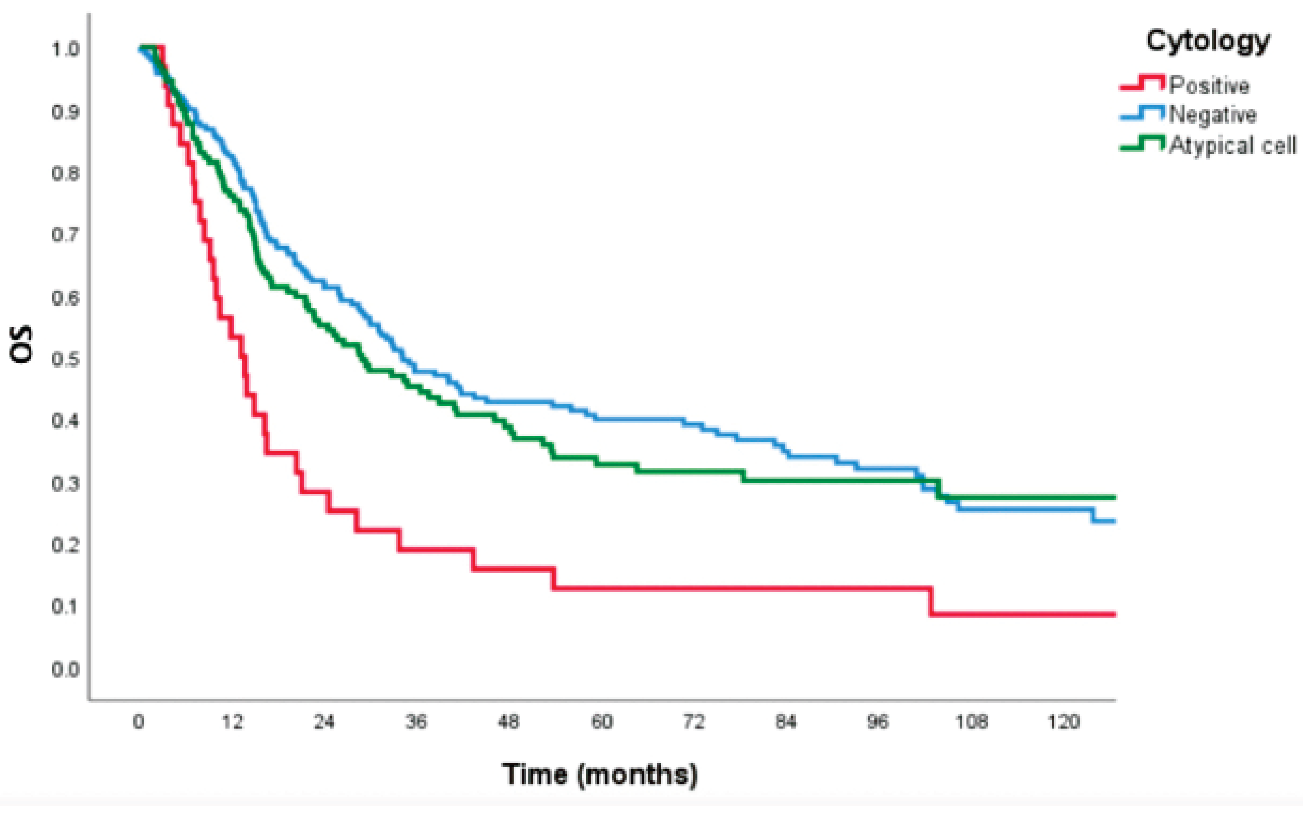
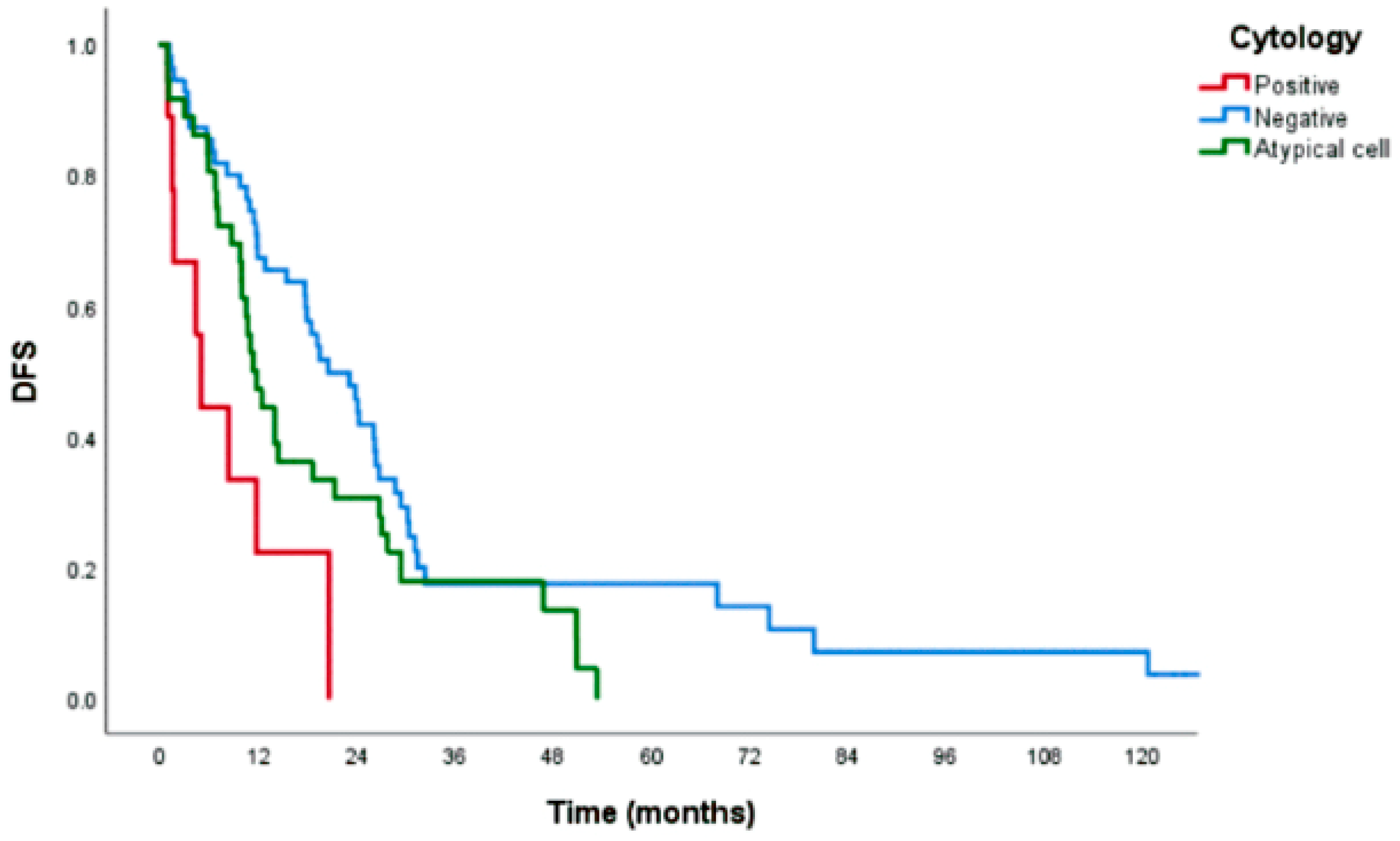
Background/Objectives: To identify independent predictors of free peritoneal cancer cells (FPCC), and to investigate survival outcomes relative to peritoneal cytology status among patients underwent intended curative gastrectomy for adenocarcinoma of stomach or esophagogastric junction. Methods: Medical record of patients underwent radical surgery during January 2005-December 2020 were retrospectively reviewed. Clinical data and cytology results were evaluated. Multivariate Cox regression analysis was used to identify independent predictors of FPCC. Kaplan-Meier survival analysis was used to estimate disease recurrence and survival outcomes. Results: Of the 349 enrolled patients, 188 (53.8%) had negative cytology, 32 (9.2%) had positive cytology, and 129 (36.9%) had atypical cells in peritoneal cytology. Multivariate analysis revealed poor differentiation (adjusted odds ratio [aOR]: 2.63, 95% confidence interval [95%CI]: 1.04-6.82; p=0.015), pT4 (aOR: 4.62, 95%CI: 1.28-14.34; p=0.018), pN3 (aOR: 4.13, 95%CI: 1.14-15.03; p=0.031), and metastatic lymph node ratio >0.40 (aOR: 6.49, 95%CI: 1.44-29.14; p=0.015) as independent predictors of FPCC. Median survival duration of patients with negative, positive, and atypical cell cytology was 34.1, 13.1, and 28.7 months, respectively (p<0.001). 5-year OS was 27.2%, 8.3%, and 25.3%, respectively (p<0.001). 3-year DFS was 17.8%, 0.0%, and 17.4%, respectively (p<0.001). Median time to disease recurrence was 20.5, 4.9, and 11.3 months, respectively (p<0.001). Survival outcome and disease recurrence were comparable between atypical cell and negative peritoneal cytology patients. Conclusions: Poorly differentiated histology, pT4, pN3, and metastatic lymph node ratio >0.40 are independent predictors of FPCC. The presence of FPCC was significantly associated with poor survival and disease recurrence outcomes.
Keywords:
Introduction
Materials and Methods
Results
Discussion
Conclusions
Author Contributions
Funding
Institutional Review Board Statement
Informed Consent Statement
Data Availability Statement
Acknowledgments
Conflicts of Interest
References
- Tustumi F, Bernardo WM, Dias AR, Ramos MF, Cecconello I, Zilberstein B, et al. Detection value of free cancer cells in peritoneal washing in gastric cancer: a systematic review and meta-analysis. Clinics (Sao Paulo). 2016;71(12):733-45. [CrossRef]
- Smyth EC, Verheij M, Allum W, Cunningham D, Cervantes A, Arnold D, et al. Gastric cancer: ESMO Clinical Practice Guidelines for diagnosis, treatment and follow-up. Ann Oncol. 2016;27(suppl 5):v38-v49. [CrossRef]
- Japanese Gastric Cancer A. Japanese Gastric Cancer Treatment Guidelines 2021 (6th edition). Gastric Cancer. 2023;26(1):1-25.
- Kim IH, Kang SJ, Choi W, Seo AN, Eom BW, Kang B, et al. Korean Practice Guidelines for Gastric Cancer 2024: An Evidence-based, Multidisciplinary Approach (Update of 2022 Guideline). J Gastric Cancer. 2025;25(1):5-114. [CrossRef]
- Koganti SB, Boddepalli S, Nambada M, Thumma VM, Nagari B, Sastry RA. Positive Peritoneal Lavage Cytology -Implications for Staging and Management of Gastric Cancer. Indian J Surg Oncol. 2016;7(4):430-5. [CrossRef]
- Pecqueux M, Fritzmann J, Adamu M, Thorlund K, Kahlert C, Reissfelder C, et al. Free intraperitoneal tumor cells and outcome in gastric cancer patients: a systematic review and meta-analysis. Oncotarget. 2015;6(34):35564-78. [CrossRef]
- Allen CJ, Newhook TE, Vreeland TJ, Das P, Minsky BD, Blum M, et al. Yield of peritoneal cytology in staging patients with gastric and gastroesophageal cancer. J Surg Oncol. 2019;120(8):1350-7. [CrossRef]
- Yoshida K, Yamaguchi K, Okumura N, Tanahashi T, Kodera Y. Is conversion therapy possible in stage IV gastric cancer: the proposal of new biological categories of classification. Gastric Cancer. 2016;19(2):329-38. [CrossRef]
- Yoshida K, Yasufuku I, Terashima M, Young Rha S, Moon Bae J, Li G, et al. International Retrospective Cohort Study of Conversion Therapy for Stage IV Gastric Cancer 1 (CONVO-GC-1). Ann Gastroenterol Surg. 2022;6(2):227-40. [CrossRef]
- Valletti M, Eshmuminov D, Gnecco N, Gutschow CA, Schneider PM, Lehmann K. Gastric cancer with positive peritoneal cytology: survival benefit after induction chemotherapy and conversion to negative peritoneal cytology. World J Surg Oncol. 2021;19(1):245. [CrossRef]
- Jamel S, Markar SR, Malietzis G, Acharya A, Athanasiou T, Hanna GB. Prognostic significance of peritoneal lavage cytology in staging gastric cancer: systematic review and meta-analysis. Gastric Cancer. 2018;21(1):10-8.
- Bausys A, Ümarik T, Dobrzhanskyi O, Luksta M, Kondratskyi Y, Reinsoo A, et al. Neoadjuvant Chemotherapy Followed by Gastrectomy for Cytology-Positive Gastric Cancer without Any Other Non-Curative Factors in a Western Setting: An International Eastern European Cohort Study. Cancers. 2023;15(24):5794. [CrossRef]
- Leake PA, Cardoso R, Seevaratnam R, Lourenco L, Helyer L, Mahar A, et al. A systematic review of the accuracy and utility of peritoneal cytology in patients with gastric cancer. Gastric Cancer. 2012;15 Suppl 1:S27-37. [CrossRef]
- Gęca K, Skórzewska M, Rawicz-Pruszyński K, Mlak R, Sędłak K, Pelc Z, et al. Prognostic value of molecular cytology by one-step nucleic acid amplification (OSNA) assay of peritoneal washings in advanced gastric cancer patients. Scientific Reports. 2022;12(1):12477. [CrossRef]
- Hayes N, Wayman J, Wadehra V, Scott DJ, Raimes SA, Griffin SM. Peritoneal cytology in the surgical evaluation of gastric carcinoma. Br J Cancer. 1999;79(3-4):520-4. [CrossRef]
- Lisiecki R, Kruszwicka M, Spychała A, Murawa D. Prognostic significance, diagnosis and treatment in patients with gastric cancer and positive peritoneal washings. A review of the literature. Rep Pract Oncol Radiother. 2017;22(6):434-40. [CrossRef]
- Sakata T, Takahata T, Kimura T, Yasuhara I, Kojima T, Akazai Y, et al. A single-institution retrospective analysis of gastric carcinoma with positive peritoneal lavage cytology and without serosal invasion: A case series. Ann Med Surg (Lond). 2019;39:10-5. [CrossRef]
- Kano Y, Kosugi S, Ishikawa T, Otani T, Muneoka Y, Sato Y, et al. Prognostic significance of peritoneal lavage cytology at three cavities in patients with gastric cancer. Surgery. 2015;158(6):1581-9. [CrossRef]
- La Torre M, Ferri M, Giovagnoli MR, Sforza N, Cosenza G, Giarnieri E, et al. Peritoneal wash cytology in gastric carcinoma. Prognostic significance and therapeutic consequences. Eur J Surg Oncol. 2010;36(10):982-6. [CrossRef]
- Higaki E, Yanagi S, Gotohda N, Kinoshita T, Kuwata T, Nagino M, et al. Intraoperative peritoneal lavage cytology offers prognostic significance for gastric cancer patients with curative resection. Cancer Sci. 2017;108(5):978-86. [CrossRef]
- Kobayashi H, Honda M, Kawamura H, Takiguchi K, Muto A, Yamazaki S, et al. Clinical impact of gastrectomy for gastric cancer patients with positive lavage cytology without gross peritoneal dissemination. J Surg Oncol. 2022;125(4):615-20. [CrossRef]
- Jiang CG, Xu Y, Wang ZN, Sun Z, Liu FN, Yu M, et al. Clinicopathological analysis and prognostic significance of peritoneal cytology in Chinese patients with advanced gastric cancer. ANZ J Surg. 2011;81(9):608-13. [CrossRef]
- Mezhir JJ, Shah MA, Jacks LM, Brennan MF, Coit DG, Strong VE. Positive peritoneal cytology in patients with gastric cancer: natural history and outcome of 291 patients. Ann Surg Oncol. 2010;17(12):3173-80. [CrossRef]
- Parakonthun T, Parichardsombat N, Salomon H, Paredes R, Phalanusittheph C, Taweerutchana V, et al. Significance of Microscopic Residual Tumor in Adenocarcinoma of Stomach and Esophagogastric Junction after Gastrectomy with D2 Lymphadenectomy. Siriraj Med J. 2018;70(2):95-102.
- Graversen M, Rouvelas I, Ainsworth AP, Bjarnesen AP, Detlefsen S, Ellebaek SB, et al. Feasibility and Safety of Laparoscopic D2 Gastrectomy in Combination with Pressurized Intraperitoneal Aerosol Chemotherapy (PIPAC) in Patients with Gastric Cancer at High Risk of Recurrence-The PIPAC-OPC4 Study. Ann Surg Oncol. 2023;30(7):4433-41.
- Imano M, Imamoto H, Itoh T, Satou T, Peng YF, Yasuda A, et al. Impact of intraperitoneal chemotherapy after gastrectomy with positive cytological findings in peritoneal washings. Eur Surg Res. 2011;47(4):254-9. [CrossRef]

| Characteristics |
Positive (n=32) |
Negative (n=188) |
Atypical (n=129) |
p-value |
|
Age (years) Male gender Operation - Distal gastrectomy - Total gastrectomy - Others * Maximal tumor diameter (cm) Tumor location - Esophagogastric junction - Upper 1/3 stomach - Middle 1/3 stomach - Lower 1/3 stomach - Diffuse - Anastomosis Borrmann type - I - II - III - IV Lauren classification - Intestinal - Diffuse Tumor differentiation - Well - Moderately - Poorly Angiolymphatic invasion Perineural invasion Pathological T-stage - 1 to 2 - 3 to 4 Pathological N-stage - 0 to 1 - 2 to 3 |
58.6±14.9 17 (53.1%) 10 (31.3%) 16 (50.0%) 6 (18.7%) 5 (3.9, 8.5) 8 (25.0%) 1 (3.1%) 8 (25.0%) 12 (37.5%) 2 (6.3%) 1 (3.1%) 4 (12.5%) 10 (31.3%) 17 (53.1%) 1 (3.1%) 4 (12.5%) 9 (28.1%) 0 (0.0%) 6 (19.4%) 25 (80.6%) 21 (65.6%) 26 (83.9%) 8 (26.7%) 22 (73.3%) 13 (40.6%) 19 (59.4%) |
65.1±13.4 107 (56.9%) 80 (42.8%) 95 (50.8%) 13 (6.4%) 4.5 (3.0, 7.0) 24 (13%) 12 (6.5%) 45 (24.5%) 90 (48.9%) 12 (6.5%) 1 (0.5%) 28 (15.4%) 61 (33.5%) 78 (42.9%) 15 (8.2%) 28 (14.9%) 36 (19.1%) 8 (4.4%) 63 (34.6%) 111 (61.0%) 92 (50.3%) 125 (69.1%) 38 (20.9%) 144 (79.1%) 73 (40.1%) 109 (59.9%) |
58.7±12.9 66 (51.2%) 35 (27.1%) 63 (48.8%) 31 (24.1%) 5 (3.5, 8) 36 (28.1%) 5 (3.9%) 31 (24.2%) 45 (35.2%) 10 (7.8%) 1 (0.8%) 19 (16.8%) 26 (23.0%) 59 (52.2%) 9 (8.0%) 22 (18.6%) 27 (22.9%) 5 (4.2%) 32 (26.9%) 82 (68.9%) 82 (66.1%) 92 (73.6%) 30 (23.6%) 97 (76.4%) 52 (41.3%) 74 (58.7%) |
<0.001 0.593 <0.001 0.070 0.090 0.486 0.792 0.200 0.014 0.212 0.717 0.979 |
| Factors | Univariate analysis | Multivariate analysis | ||
| OR (95%CI) | p-value | aOR (95%CI) | p-value | |
|
Tumor differentiation - Well and moderate - Poor Borrmann type - I to II - III to IV Tumor size - 5 to 10 cm - >10 cm Angiolymphatic invasion Perineural invasion Pathological T-stage - T3 - T4 Pathological N-stage - N2 - N3 Metastatic lymph node ratio - <0.20 - 0.21 to 0.40 - >0.40 |
0.47 (0.05-4.56) 2.67 (1.04-6.82) 1.15 (0.33-3.98) 1.53 (0.47-4.92) 1.01 (0.43-2.41) 2.61 (0.74-9.27) 1.89 (0.86-4.14) 2.33 (0.85-6.38) 1.41 (0.39-5.10) 3.82 (1.24-11.82) 0.81 (0.20-3.26) 2.70 (1.12-6.47) 2.92 (0.74-11.61) 2.32 (0.52-10.25) 8.22 (2.24-30.17) |
0.512 0.041 0.828 0.480 0.976 0.137 0.112 0.090 0.595 0.020 0.771 0.026 0.127 0.268 0.002 |
2.63 (1.04-6.82) 4.62 (1.28-14.34) 4.13 (1.14-15.03) 6.49 (1.44-29.14) |
0.015 0.018 0.031 0.015 |
Disclaimer/Publisher’s Note: The statements, opinions and data contained in all publications are solely those of the individual author(s) and contributor(s) and not of MDPI and/or the editor(s). MDPI and/or the editor(s) disclaim responsibility for any injury to people or property resulting from any ideas, methods, instructions or products referred to in the content. |
© 2025 by the authors. Licensee MDPI, Basel, Switzerland. This article is an open access article distributed under the terms and conditions of the Creative Commons Attribution (CC BY) license (http://creativecommons.org/licenses/by/4.0/).


