Preprint
Article

This version is not peer-reviewed.

Silent Persistence: Molecular Evidence of Clonal Transmission in Fluconazole-Resistant Candida parapsilosis Hospital Outbreaks over Decades

A peer-reviewed article of this preprint also exists.

Submitted:

06 October 2025

Posted:

22 October 2025

You are already at the latest version

Abstract

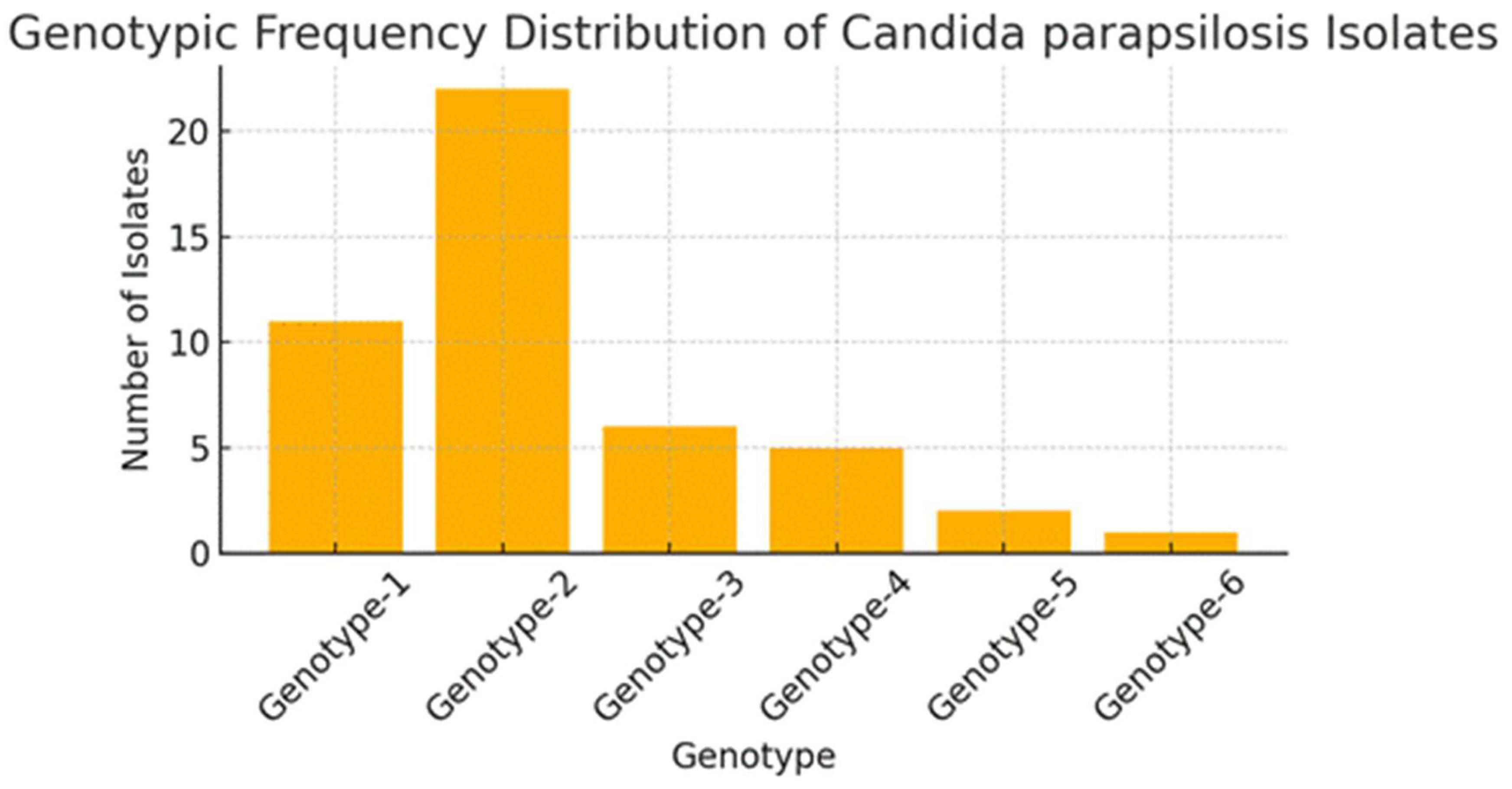
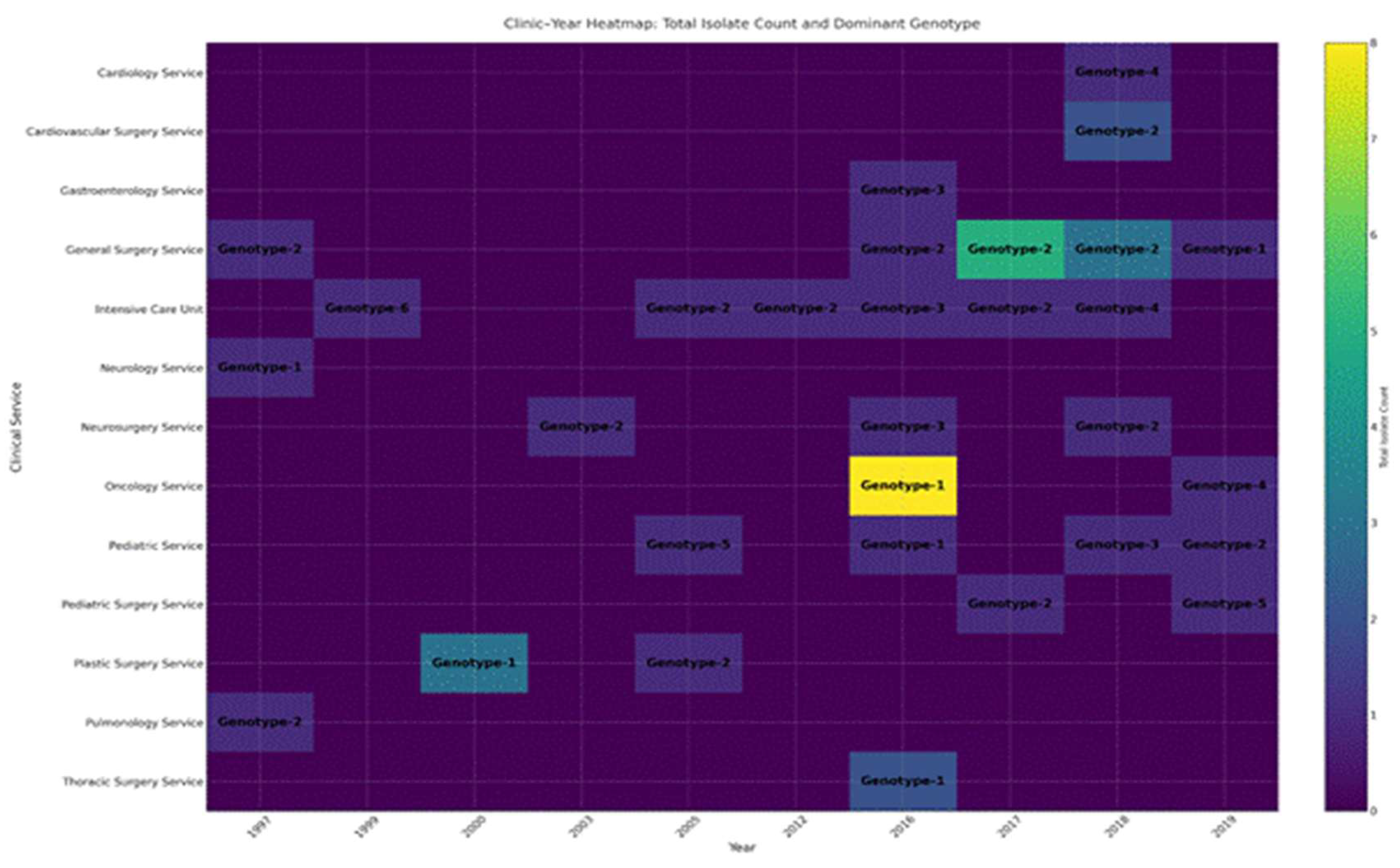
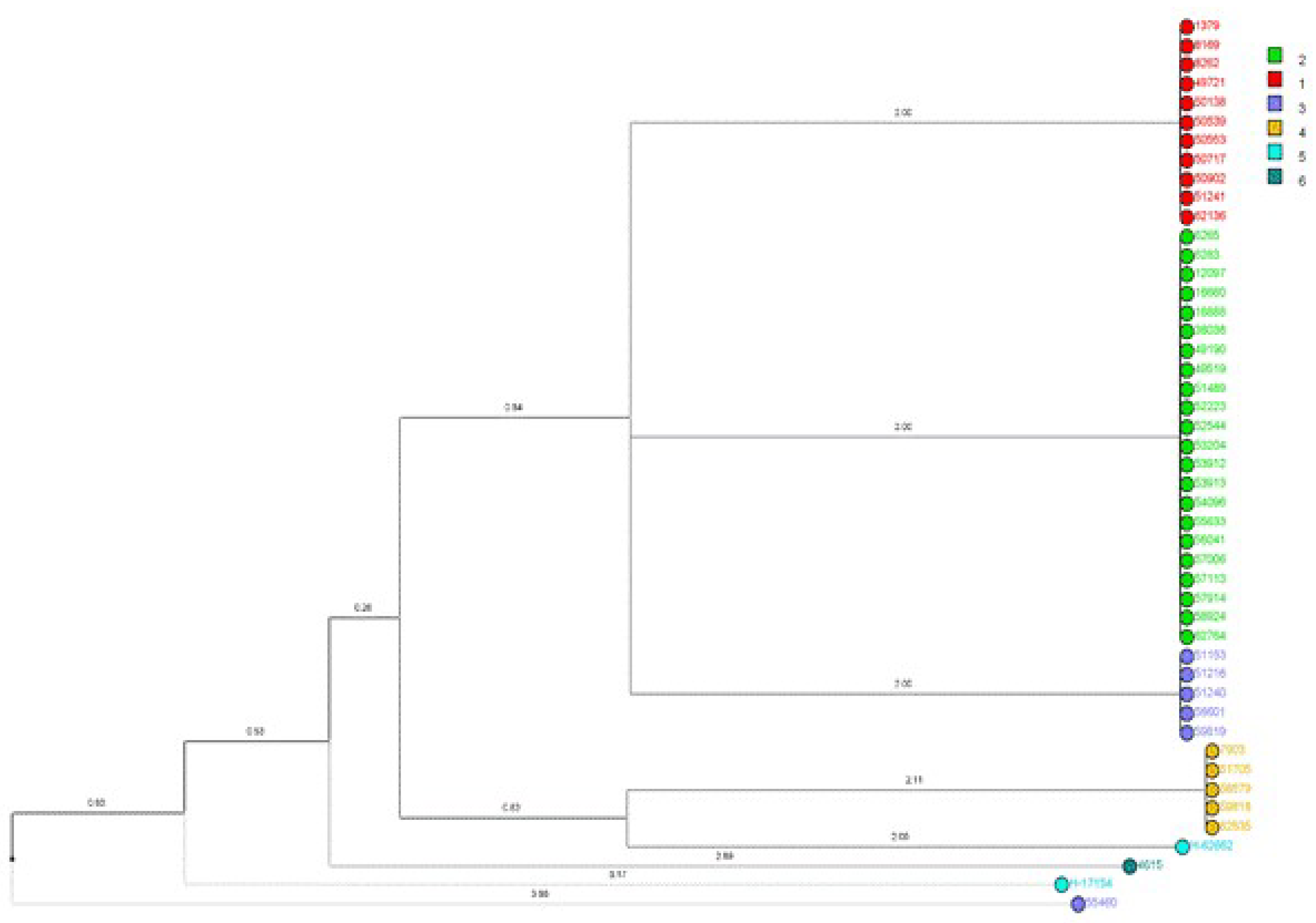
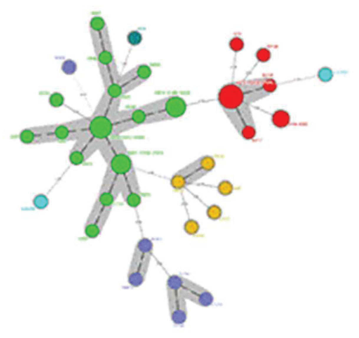
Fluconazole-resistant Candida parapsilosis has emerged as a significant nosocomial pathogen, contributing to extensive outbreaks with severe clinical implications. Despite increasing evidence of clonal transmission, the genetic mechanisms that facilitate the persistence of hospital reservoirs remain inadequately characterized. We aimed to determine the extent of clonal spread and persistence patterns of fluconazole-resistant Candida parapsilosis strains across a 22-year period in a tertiary care hospital, using high-resolution microsatellite genotyping. Forty-seven fluconazole-resistant Candida parapsilosis isolates from candidemia patients (1997-2019) underwent microsatellite analysis using three polymorphic markers (CP1, CP4, B5). Genetic diversity, temporal distribution, and clonal relationships were assessed through phylogenetic analysis and discriminatory power calculations. Microsatellite analysis revealed minimal genetic diversity (combined discriminatory power: 0.7114), with only six distinct genotypes identified. Two dominant clones (Genotype-1: 23.4%, Genotype-2: 46.8%) persisted throughout the study, showing apparent spatiotemporal clustering in surgical and intensive care units. Phylogenetic analysis demonstrated tight genetic clustering, confirming prolonged clonal persistence spanning multiple years and clinical departments. Our findings provide compelling molecular evidence for persistent, multi-year clonal transmission of fluconazole-resistant Candida parapsilosis within hospital environments. These results challenge current infection control paradigms and highlight the urgent need for enhanced surveillance strategies and targeted interventions to interrupt these persistent transmission chains.

Keywords: 
;  ;  

1. Introduction

Candida albicans, traditionally considered to be the most common species among the causative agents of candidemia, is increasingly being replaced by non-albicans Candida (NAC) species such as C. glabrata, C. tropicalis and C. parapsilosis [1, 2]. The increase in NAC species is thought to be due to multiple factors. The widespread use of broad-spectrum antibiotics disrupts the normal microbiota and creates a favorable environment for the development of opportunistic fungal infections [3]. C. parapsilosis, which is common in the skin flora and can settle on foreign surfaces such as catheters, has started to be seen frequently among the healthcare-associated candidemia agents among NAC species with the effect of increasing invasive procedures [4]. Fluconazole is recommended in the treatment guidelines [5]. However, the incidence of C. parapsilosis strains showing fluconazole resistance has increased significantly worldwide in recent years [6]. This resistance limits clinical treatment options and negatively affects the clinical prognosis of patients [7]. Fluconazole-resistant C. parapsilosis strains cause an increase in morbidity and mortality rates by decreasing treatment success, especially in intensive care units and patients with invasive medical devices [8]. Therefore, monitoring fluconazole resistance and preventing the spread of resistant strains in the hospital environment is of great importance.
C. parapsilosis has been associated with fungemia outbreaks in the hospital environment and has been frequently isolated from the skin flora of healthcare workers and surfaces in the hospital environment [9-11]. The fact that it has the capacity to form biofilms by colonizing on hospital surfaces and invasive medical devices significantly increases the risk of cross-infection by increasing the virulence of the pathogen [12].
In various studies, it has been found that fluconazole-resistant C. parapsilosis strains are genetically similar and this supports inter-patient transmission [13]. It has been reported that resistant strains isolated from the hands of hospital staff and environmental surfaces are genetically similar to strains isolated from patients [14,15]. In addition, genetic analyses have shown that certain resistant genotypes remain persistent in the hospital environment for years and are transmitted to different patients [16]. This situation has become particularly evident in intensive care units and neonatal units [17,13].
Microsatellite genotyping method stands out as an effective method to determine the genetic relationship of fluconazole-resistant C. parapsilosis strains. Analyses performed with this method show that resistant strains found in the hospital environment and on the hands of healthcare workers are genetically similar and indicate transmission routes [18]. In this study, we aimed to genotype fluconazole-resistant C. parapsilosis strains isolated from patients diagnosed with candidemia in our hospital over a 22-year period by microsatellite analysis method and to evaluate the genetic similarity between them.

2. Materials and Methods

Ethics committee approval was obtained from Bursa Uludag University, Non-Interventional Clinical Research Ethics Committee (Decision No: No. 2020-10/15). This study was conducted in a tertiary level reference hospital with intensive care units and clinical services.

2.1. DNA Isolation and Polymerase Chain Reaction (PCR)

In our study, 47 blood isolates previously identified as C. parapsilosis sensu stricto, which were isolated from patients diagnosed with candidemia between 1997 and 2019 and found to be fluconazole-resistant, were used [19]. DNA isolation from the isolates was performed using UltraClean Microbial DNA Isolation Kit (Qiagen, USA) according to the manufacturer's recommendations. The concentration and purity of the DNA samples obtained were evaluated with a Beckman Coulter DU-640 spectrophotometer using absorbance measurement at 260/280 nm wavelengths.

2.2. Antifungal Susceptibility Test

C. parapsilosis species complex isolates grown on blood cultures (BACTEC-FX: Becton-Dickinson, Sparks, MD, USA) were identified using germ tube test, morphology and biochemical profile (API ID 32C; BioMérieux, France) on corn flour tween 80 and chromogenic culture media. Antifungal susceptibility testing was performed using the microdilution method according to Clinical and Laboratory Standards Institute (CLSI) guidelines. According to the CLSIM27M44S document, strains with a minimum inhibitory concentration MIC ≥4 µg/ml were considered fluconazole-insensitive (MIC=4 µg/ml sensitive dose-dependent; MIC≥8 µg/ml resistant) [20, 21]. Although CLSI specifies 4 µg/ml as dose-dependent susceptible, studies have shown that resistance starts at this MIC value and resistance genes are present. Therefore, in this study, all isolates with ≥4 µg/ml were expressed as fluconazole resistant [22, 23].

2.3. Microsatellite Instability Analysis

For the Microsatellite Instability (MSI) test, PCR was performed with primers of fluorescently labeled microsatellite markers (CP1, CP4a and B) using the genomic DNA obtained. The reaction mixture in a total volume of 10 μl contained 50-100 ng genomic DNA, 1.25 units of Taq Polymerase, 0.8 mM of each dNTP, 1.5 mM MgCl₂ and 0.5 μM of each primer.
High-quality PCR products were processed using the “Sample Loading Solution” and “DNA Size Standard Kit” (Beckman Coulter, USA) and fragment analysis was performed with the “CEQ 8000 Genetic Analysis System” (Beckman Coulter Inc., Fullerton, CA, USA). After analysis, the data of each sample were comparatively evaluated using CEQ 8000 software, the integrated analysis software of the device.

2.4. Phylogenetic Tree Construction

The phylogenetic tree for microsatellite analysis was constructed using “BioNumerics” version 6.6 (Applied Maths, NV) software. Allele numbers, repeat numbers and allele frequencies were calculated with “CONVERT” software version 1.31. Hardy-Weinberg equilibrium and Chi-square test results were analyzed using “Genepop” software version 4.2. Discrimination power (DP) was calculated according to the formula proposed by Hunter et al. [24].

3. Results

Forty-seven fluconazole-resistant C. parapsilosis strains isolated from patients with candidemia were analyzed at the molecular level using CP1, CP4 and B5 microsatellite markers; the genetic diversity and possible epidemiological relationships of the isolates were comprehensively evaluated by microsatellite typing method.
The discrimination levels of three microsatellite markers differed. The CP1 marker revealed three different alleles ranging from 224 to 302 base pairs; the number of repeats ranged from 1 to 40 and allele frequencies ranged from 0.0213 to 0.9574. The DP of this marker was calculated as 0.0842; although heterozygosity was not observed, it provided basic information on genetic variation. The CP4 marker showed a higher level of diversity; four alleles ranging between 249-286 base pairs were detected, the number of repeats was between 1 and 19, and the allele frequencies were between 0.0213 and 0.8511. In CP4 marker, 4.25% heterozygosity was observed and the DP was determined as 0.2683. The B5 marker stood out as the marker with the highest discrimination power (DP: 0.5495); three alleles ranging between 140-148 base pairs were identified, repeat numbers ranged between 1 and 5, and allele frequencies ranged between 0.1277 and 0.6170. Heterozygosity was not detected in this marker. When all markers were considered, the total DP was calculated as 0.7114 (Table 1).
A total of 47 fluconazole-resistant C. parapsilosis isolates were genotyped and six different genotypes were identified. When the genotype distribution was analyzed, it was found that the most common genotype was Genotype-2 and it was the most represented group with a total of 22 isolates (46.8%). Genotype-2 was followed by Genotype-1 with 11 isolates (23.4%). The distribution of other genotypes was as follows: Genotype-3 (6 isolates), Genotype-4 (4 isolates), Genotype-5 and Genotype-6 (2 isolates each). The average number of isolates per genotype was 12.7% (Figure 1.) (Supplementary Table A1).
The distribution of genotypes in clinics and years is shown in Figure 3. Accordingly, Genotype-2 was the most frequently isolated type in General Surgery and Reanimation wards in 2017 and 2018, while Genotype-1 was the predominant type in internal clinics, especially in Oncology and Neurology, in the 2000-2016 period. Genotypes-3, -4, -5 and -6 were rarely isolated in different clinical services and in variable years (Figure 2.) (Supplementary Table A2).
Phylogenetic relationships and genetic similarities among isolates were visualized through a dendrogram (Figure 3.) and phylogenetic tree (Figure 4.). Dendrogram analysis revealed clear clustering patterns, with Genotype-2 in particular exhibiting a highly compact and homogeneous structure. In contrast, Genotype-1 showed a more disorganized genetic distribution, while Genotypes-3, -4, -5 and -6, which were observed less frequently, showed more genetic heterogeneity. Phylogenetic tree analysis also supported these findings; genotypes were identified with different colors and genetically close isolates were highlighted with gray shading with a genetic distance of ≤1.

4. Discussion

In this study, we genotyped 47 fluconazole-resistant C. parapsilosis isolates using the CP1, CP4, and B5 markers and observed a limited diversity among them. The CP1 marker was nearly monomorphic, exhibiting a DP of 0.0842, indicating 100% homozygosity. The CP4 marker showed moderate polymorphism, with a DP of 0.2683 and 4.25% heterozygosity. Although the B5 marker demonstrated the highest DP at 0.5495, it also displayed complete homozygosity. The combined DP of 0.7114 is notably lower than the approximately 0.99 reported for four-locus panels [25].
In our comparison of data with international multilocus schemes, we noted significantly lower DP. Sabino et al. utilized a four-locus panel (CP1, CP4, CP6, and B5) on 236 C. parapsilosis isolates, reporting DP values of approximately 0.85 for CP1, 0.90 for CP4, and 0.86 for B5, resulting in a combined DP of 0.99 and 192 distinct genotypes. In contrast, our findings revealed a DP of only 0.084 for CP1, 0.2683 for CP4, and 0.55 for B5—all loci being homozygous—culminating in a combined DP of 0.7114 [25]. Similarly, Desnos-Ollivier et al. identified around 30 multilocus genotypes and reported a panel DP close to 0.97 in isolates from France and Uruguay [26]. These elevated DP values suggest a significant degree of allelic diversity. In contrast, the lower metrics observed in our study imply that many of our fluconazole-resistant isolates possess identical or closely related genotypes, indicative of a predominantly clonal population. These results emphasize that three loci may not be adequate to capture the full genetic heterogeneity of persistent C. parapsilosis strains in our hospital environment.
Previous research has shown that fluconazole-resistant C. parapsilosis clones may emerge and, under selective pressures, become predominant in clinical settings [27]. Some of these resistant strains can persist for several years; for instance, a study from Canada documented a single resistant strain that circulated for 5.5 years, causing ongoing infections [28]. This clonal expansion significantly contributes to the low diversity observed in our collection. Previous genomic studies have highlighted a predominantly clonal population structure, as indicated by significant deviations from the Hardy-Weinberg equilibrium and heterozygosity levels 25 to 70 times lower than those found in other diploid Candida species [25,29]. Consequently, slight genetic variation accumulates during replication, allowing identical clones to infect multiple patients. Our cohort's lack of variation at loci CP1 and B5, alongside only minimal polymorphism at CP4, suggests that our 47 isolates originate from several ancestral clones, with CP1 as a "clonal trace". While three-locus microsatellite panels are commonly used and deemed sufficiently discriminatory, our findings indicate they may underestimate the true genetic diversity of long-standing resistant populations [26, 30].
We note that C. parapsilosis easily colonizes hospital environments through the hands of healthcare workers and invasive medical devices, resulting in infection clusters dominated by specific clones [31]. Intensive care units for neonates and adults are especially susceptible to such outbreaks [32-34]. For instance, a Turkish hospital reported an outbreak involving a single genotype, while a Chinese study encompassing ten hospitals identified 122 different genotypes, with 32 of those forming clonal clusters—one specific MT42 clone was responsible for infections in 22 neonates within a single NICU [35,31]. These findings highlight how swiftly a single clone can spread under favorable conditions. Additionally, the high genotypic similarity observed among our 47 isolates suggests a likely occurrence of nosocomial clonal transmission, although direct epidemiological confirmation is still pending.
As a diploid organism, C. parapsilosis has the potential to carry two different alleles per locus, which would reflect sexual recombination or admixture [36]. However, its predominantly clonal, asexual lifecycle results in very low rates of heterozygous SNPs and microsatellite loci [29]. In our collection, CP1 and B5 were completely homozygous (0% heterozygosity), while CP4 exhibited only a single heterozygous isolate (4.25%). This evidence confirms that these 47 isolates originate from a largely monotypic, clonal population with minimal signs of sexual recombination.
In contrast to the 5–7% average genotype representation reported by Sabino et al. [25], our isolates exhibited an average of 12.7%, indicating moderate genetic similarity and limited diversity. Genotype 2 accounted for 46.8% of our cohort, suggesting significant clonal expansion. Such outbreaks predominantly involving a single genotype are well-documented in intensive and neonatal care settings [32]. The pronounced presence of Genotype 2 in our hospital implies dissemination from a common source or transmission chain, highlighting the urgent need to strengthen infection control measures, especially hand hygiene, catheter care, and protocols for invasive procedures [36].
Fluconazole-resistant C. parapsilosis infections are primarily found in high-risk environments—specifically, adult and neonatal ICUs, transplant units, and COVID-19 wards—where vulnerable patients are concentrated [37]. In our study, Genotype 2 was particularly prevalent in general surgery and reanimation wards during 2017–2018, aligning with findings that indicate how ICUs and surgical services—through invasive interventions such as central venous catheters and parenteral nutrition—facilitate clonal dissemination [38-40]. Resistant strains also formed clusters in the Chest Diseases and Pediatric Surgery units during the Ege University candidemia outbreak of 2019–2020 [41]. In contrast, Genotype 1 was detected in oncology and neurology wards between 2000 and 2016—areas characterized by immunosuppression, prolonged stays, and invasive procedures—underscoring distinct epidemiological patterns specific to different units [35, 42]. The occasional detection of genotypes 3–6, which are typically associated with isolated or exogenous introductions, further suggests multiple entry points and heterogeneous transmission routes [18, 31].
Our dendrogram and phylogenetic analyses revealed that Genotype 2 isolates form a compact, homogeneous cluster, which indicates significant nosocomial clonal transmission. Similar clustering patterns have been observed in outbreaks of C. parapsilosis and are recognized as key indicators of in-hospital transmission, particularly in ICU and surgical ward environments [43,44]. In the phylogenetic tree, distinct genotypes were color-coded, and clusters with genetic distances of ≤ 1 were highlighted, further confirming the close relatedness of the dominant clones.
We recognize several limitations in our study. Its retrospective, single-center design may limit the generalizability of our findings regarding clonal spread. The use of only three microsatellite loci (CP1, CP4, and B5) may have underestimated the overall genetic diversity. In addition, the use of archival isolates raises the possibility of DNA degradation, which could have affected some molecular results.
In conclusion our data reveal limited genetic diversity among fluconazole-resistant C. parapsilosis isolates, with Genotype 2 and Genotype 1 dominating throughout the hospital over two decades—findings consistent with nosocomial clonal spread. These results highlight the urgent need to strengthen infection control measures, particularly in surgical and intensive care units. We advocate for future multicenter studies incorporating larger collections of isolates, expanded multilocus panels, and comprehensive patient clinical metadata to enhance DP and better elucidate the transmission dynamics of resistant C. parapsilosis.

Funding

This research received no external funding.

Author Contributions

C.S.: methodology, formal analysis, investigation, writing original draft. E.K.: methodology, formal analysis, investigation, editing & review. S.A.: conceptualization, methodology. H.A.: conceptualization, methodology. B.E.: editing & review, supervision.

Institutional Review Board Statement

Not applicable.

Informed Consent Statement

Not applicable.

Data Availability Statement

Not applicable.

Acknowledgments

None

Conflicts of Interest

The author declares no conflict of interest.

References

  1. Sida H, Shah P, Pethani J, Patel L, Shah H. Study of biofilm formation as a virulence marker in Candida species isolated from various clinical specimens. International Journal of Medical Science and Public Health 2015;5:842. [CrossRef]
  2. Mukul, P. Characterization of Candida Species isolated from samples taken from patients with known Immunocompromised state presenting with Oral Thrush. Journal of Medical Science and Clinical Research 2018;6. [CrossRef]
  3. Cortegiani A, Misseri G, Fasciana T, Giammanco A, Giarratano A, Chowdhary A. Epidemiology, clinical characteristics, resistance, and treatment of infections by Candida auris. Journal of Intensive Care 2018;6. [CrossRef]
  4. Asogan, M. , Kim, H., Kidd, S., Alastruey-Izquierdo, A., Govender, N., Dao, A., Shin, J., Heim, J., Ford, N., Gigante, V., Sati, H., Morrissey, C., Alffenaar, J., & Beardsley, J. Candida parapsilosis: A systematic review to inform the World Health Organization fungal priority pathogens list. Medical Mycology, 2024;62. [CrossRef]
  5. Pappas PG, Kauffman CA, Andes DR, Clancy CJ, Marr KA, Ostrosky-Zeichner L, et al. Clinical Practice Guideline for the Management of Candidiasis: 2016 update by the Infectious Diseases Society of America. 2015. [CrossRef]
  6. Díaz-García J, Machado M, Alcalá L, Reigadas E, Pérez-Ayala A, De La Pedrosa EG-G, et al. Trends in antifungal resistance in Candida from a multicenter study conducted in Madrid (CANDIMAD study): fluconazole-resistant C. parapsilosis spreading has gained traction in 2022. Antimicrobial Agents and Chemotherapy 2023;67. [CrossRef]
  7. Sharma V, Paul RA, Kaur H, Das S, Choudhary H, Rudramurthy SM, et al. P019 A 5-year study on prevalence and molecular determinants of fluconazole -resistance in C. parapsilosis spp. complex. Medical Mycology 2022;60. [CrossRef]
  8. Ünal N, Spruijtenburg B, Arastehfar A, Gümral R, De Groot T, Meijer EFJ, et al. Multicentre Study of Candida parapsilosis Blood Isolates in Türkiye Highlights an Increasing Rate of Fluconazole Resistance and Emergence of Echinocandin and Multidrug Resistance. Mycoses 2024;67. [CrossRef]
  9. Prigitano A, Blasi E, Calabrò M, Cavanna C, Cornetta M, Farina C, et al. Yeast bloodstream infections in the COVID-19 patient: a multicenter Italian study (FICOV study). Journal of Fungi 2023;9:277. [CrossRef]
  10. Levin AS, Costa SF, Mussi NS, Basso M, Sinto SI, Machado C, et al. Candida parapsilosis Fungemia Associated with Implantable and Semi-Implantable Central Venous Catheters and the Hands of Healthcare Workers. Diagnostic Microbiology and Infectious Disease 1998;30:243–9. [CrossRef]
  11. Qi L, Fan W, Xia X, Yao L, Liu L, Zhao H, et al. Nosocomial outbreak of Candida parapsilosis sensu stricto fungaemia in a neonatal intensive care unit in China. Journal of Hospital Infection 2018;100:e246–52. [CrossRef]
  12. Cacaci M, Squitieri D, Palmieri V, Torelli R, Perini G, Campolo M, et al. Curcumin-Functionalized Graphene Oxide Strongly Prevents Candida parapsilosis Adhesion and Biofilm Formation. Pharmaceuticals 2023;16:275. [CrossRef]
  13. Fekkar A, Blaize M, Bouglé A, Normand A-C, Raoelina A, Kornblum D, et al. Hospital Outbreak of Fluconazole-Resistant Candida parapsilosis: Arguments for Clonal Transmission and Long-Term Persistence. Antimicrobial Agents and Chemotherapy 2021;65. [CrossRef]
  14. Sanchez V, Vazquez JA, Barth-Jones D, Dembry L, Sobel JD, Zervos MJ. Nosocomial acquisition of Candida parapsilosis: An epidemiologic study. The American Journal of Medicine 1993;94:577–82. [CrossRef]
  15. Lupetti A, Tavanti A, Davini P, Ghelardi E, Corsini V, Merusi I, et al. Horizontal Transmission of Candida parapsilosis Candidemia in a Neonatal Intensive Care Unit. Journal of Clinical Microbiology 2002;40:2363–9. [CrossRef]
  16. Asadzadeh M, Ahmad S, Al-Sweih N, Hagen F, Meis JF, Khan Z. High-resolution fingerprinting of Candida parapsilosis isolates suggests persistence and transmission of infections among neonatal intensive care unit patients in Kuwait. Scientific Reports 2019a;9. [CrossRef]
  17. Thomaz DY, Del Negro GMB, Ribeiro LB, Da Silva M, Carvalho GOMH, Camargo CH, et al. A Brazilian Inter-Hospital Candidemia Outbreak Caused by Fluconazole-Resistant Candida parapsilosis in the COVID-19 Era. Journal of Fungi 2022;8:100. [CrossRef]
  18. Sabino R, Sampaio P, Rosado L, Videira Z, Grenouillet F, Pais C. Analysis of clinical and environmental Candida parapsilosis isolates by microsatellite genotyping—a tool for hospital infection surveillance. Clinical Microbiology and Infection 2015;21:954.e1-954.e8. [CrossRef]
  19. Cilo BD, Agca H, Ener B. Identification of Candida parapsilosis Complex Strains Isolated from Blood Samples at Species Level and Determination of Their Antifungal Susceptibilities. Türk Mikrobiyoloji Cemiyeti Dergisi 2019. [CrossRef]
  20. Reference method for broth dilution antifungal susceptibility of yeasts. 4th ed. CLSI Standard M27. C: Wayne, PA, 2017.
  21. CLSI. Performance standards for antifungal susceptibility of yeasts. 3rd ed. CLSI supplement M27M44S. 2022.
  22. Neji S, Hadrich I, Ilahi A, Trabelsi H, Chelly H, Mahfoudh N, et al. Molecular Genotyping of Candida parapsilosis Species Complex. Mycopathologia 2018;183:765–75. [CrossRef]
  23. Arastehfar A, Daneshnia F, Hilmioğlu-Polat S, Fang W, Yaşar M, Polat F, et al. First Report of Candidemia Clonal Outbreak Caused by Emerging Fluconazole-Resistant Candida parapsilosis Isolates Harboring Y132F and/or Y132F+K143R in Turkey. Antimicrobial Agents and Chemotherapy 2020;64. [CrossRef]
  24. Hunter, PR. Reproducibility and indices of discriminatory power of microbial typing methods. Journal of Clinical Microbiology 1990;28:1903–5. [CrossRef]
  25. Sabino R, Sampaio P, Rosado L, Stevens DA, Clemons KV, Pais C. New Polymorphic Microsatellite Markers Able To Distinguish among Candida parapsilosis Sensu Stricto Isolates. Journal of Clinical Microbiology 2010;48:1677–82. [CrossRef]
  26. Desnos-Ollivier M, Bórmida V, Poirier P, Nourrisson C, Pan D, Bretagne S, et al. Population Structure of Candida parapsilosis: No Genetic Difference Between French and Uruguayan Isolates Using Microsatellite Length Polymorphism. Mycopathologia 2017;183:381–90. [CrossRef]
  27. Escribano P, Guinea J. Fluconazole-resistant Candida parapsilosis: A new emerging threat in the fungi arena. Frontiers in Fungal Biology 2022;3. [CrossRef]
  28. McTaggart LR, Eshaghi A, Hota S, Poutanen SM, Johnstone J, De Luca DG, et al. First Canadian report of transmission of fluconazole-resistant Candida parapsilosis within two hospital networks confirmed by genomic analysis. Journal of Clinical Microbiology 2023;62. [CrossRef]
  29. Tóth R, Nosek J, Mora-Montes HM, Gabaldon T, Bliss JM, Nosanchuk JD, et al. Candida parapsilosis: from Genes to the Bedside. Clinical Microbiology Reviews 2019;32. [CrossRef]
  30. Reiss E, Lasker BA, Lott TJ, Bendel CM, Kaufman DA, Hazen KC, et al. Genotyping of Candida parapsilosis from three neonatal intensive care units (NICUs) using a panel of five multilocus microsatellite markers: Broad genetic diversity and a cluster of related strains in one NICU. Infection Genetics and Evolution 2012;12:1654–60. [CrossRef]
  31. Zhang L, Yu S-Y, Chen SC -a., Xiao M, Kong F, Wang H, et al. Molecular Characterization of Candida parapsilosis by Microsatellite Typing and Emergence of Clonal Antifungal Drug Resistant Strains in a Multicenter Surveillance in China. Frontiers in Microbiology 2020;11. [CrossRef]
  32. Romeo O, Delfino D, Cascio A, Lo Passo C, Amorini M, Romeo D, et al. Microsatellite-based genotyping of Candida parapsilosis sensu stricto isolates reveals dominance and persistence of a particular epidemiological clone among neonatal intensive care unit patients. Infection Genetics and Evolution 2012;13:105–8. [CrossRef]
  33. Hernández-Castro R, Arroyo-Escalante S, Carrillo-Casas EM, Moncada-Barrón D, Álvarez-Verona E, Hernández-Delgado L, et al. Outbreak of Candida parapsilosis in a neonatal intensive care unit: a health care workers source. European Journal of Pediatrics 2009;169:783–7. [CrossRef]
  34. Miranda LDN, Rodrigues ECA, Costa SF, Van Der Heijden IM, Dantas KC, Lobo RD, et al. Candida parapsilosiscandidaemia in a neonatal unit over 7 years: a case series study. BMJ Open 2012;2:e000992. [CrossRef]
  35. Dizbay M, Kalkanci A, Sezer BE, Aktas F, Aydogan S, Fidan I, et al. Molecular investigation of a fungemia outbreak due to Candida parapsilosis in an intensive care unit. The Brazilian Journal of Infectious Diseases 2008;12:395–9. [CrossRef]
  36. Chen Y-C, Lin Y-H, Chen K-W, Lii J, Teng H-J, Li S-Y. Molecular epidemiology and antifungal susceptibility of Candida parapsilosis sensu stricto, Candida orthopsilosis, and Candida metapsilosis in Taiwan. Diagnostic Microbiology and Infectious Disease 2010;68:284–92. [CrossRef]
  37. Daneshnia F, De Almeida Júnior JN, Ilkit M, Lombardi L, Perry AM, Gao M, et al. Worldwide emergence of fluconazole-resistant Candida parapsilosis: current framework and future research roadmap. The Lancet Microbe 2023;4:e470–80. [CrossRef]
  38. Caggiano G, Fioriti S, Morroni G, Apollonio F, Triggiano F, D’Achille G, et al. Genotypic and phenotypic characteristics of Candida parapsilosis bloodstream isolates: Health Care Associated Infections in a teaching Hospital in Italy. Journal of Infection and Public Health 2024;17:967–74. [CrossRef]
  39. Almirante B, RodríGuez D, Cuenca-Estrella M, Almela M, Sanchez F, Ayats J, et al. Epidemiology, Risk Factors, and Prognosis of Candida parapsilosis Bloodstream Infections: Case-Control Population-Based Surveillance Study of Patients in Barcelona, Spain, from 2002 to 2003. Journal of Clinical Microbiology 2006;44:1681–5. [CrossRef]
  40. Miyake A, Gotoh K, Iwahashi J, Togo A, Horita R, Miura M, et al. Characteristics of Biofilms Formed by C. parapsilosis Causing an Outbreak in a Neonatal Intensive Care Unit. Journal of Fungi 2022;8:700. [CrossRef]
  41. Arastehfar A, Hilmioğlu-Polat S, Daneshnia F, Pan W, Hafez A, Fang W, et al. Clonal Candidemia Outbreak by Candida parapsilosis Carrying Y132F in Turkey: Evolution of a Persisting Challenge. Frontiers in Cellular and Infection Microbiology 2021;11. [CrossRef]
  42. Lortholary O, Renaudat C, Sitbon K, Desnos-Ollivier M, Bretagne S, Dromer F. The risk and clinical outcome of candidemia depending on underlying malignancy. Intensive Care Medicine 2017;43:652–62. [CrossRef]
  43. Pulcrano G, Roscetto E, Iula VD, Panellis D, Rossano F, Catania MR. MALDI-TOF mass spectrometry and microsatellite markers to evaluate Candida parapsilosis transmission in neonatal intensive care units. European Journal of Clinical Microbiology & Infectious Diseases 2012;31:2919–28. [CrossRef]
  44. Magobo RE, Naicker SD, Wadula J, Nchabeleng M, Coovadia Y, Hoosen A, et al. Detection of neonatal unit clusters of Candida parapsilosis fungaemia by microsatellite genotyping: Results from laboratory-based sentinel surveillance, South Africa, 2009-2010. Mycoses 2017;60:320–7. [CrossRef]
Figure 1. Genotype frequency distribution of Candida parapsilosis isolates.
Preprints 179693 g001
Figure 2. Clinical-year heatmap: total number of isolates and dominant genotype.
Preprints 179693 g002
Figure 3. Dendrogram showing phylogenetic relationships and clustering of genotypes among isolates.
Preprints 179693 g003
Figure 4. Phylogenetic tree showing genetic similarities and relationships among isolates, with genotypes represented by different colors and closely related isolates shaded and highlighted in gray.
Preprints 179693 g004
Table 1. Characteristics of Microsatellite Markers.
Marker Number of alleles Allele sizes Number of repeats Allele frequencies Number of genotypes Genotype frequencies Heterozygosity rate (%) DP*
CP1 3 224-302 1-40 0.0213 - 0.9574 3 0.0110 - 0.8123 0 0.0842
CP4 4 249-286 1-19 0.0213 - 0.8511 3 0.0222 - 0.6691 0.0425 0.2683
B5 3 140-148 1-5 0.1277 - 0.6170 3 0.0676 - 0.3901 0 0.5495
Total 0.7114
*DP: discriminatory power.
Disclaimer/Publisher’s Note: The statements, opinions and data contained in all publications are solely those of the individual author(s) and contributor(s) and not of MDPI and/or the editor(s). MDPI and/or the editor(s) disclaim responsibility for any injury to people or property resulting from any ideas, methods, instructions or products referred to in the content.
Copyright: This open access article is published under a Creative Commons CC BY 4.0 license, which permit the free download, distribution, and reuse, provided that the author and preprint are cited in any reuse.
Prerpints.org logo

Preprints.org is a free preprint server supported by MDPI in Basel, Switzerland.

Subscribe

Disclaimer

Terms of Use

Privacy Policy

Privacy Settings

© 2025 MDPI (Basel, Switzerland) unless otherwise stated