Preprint
Article

This version is not peer-reviewed.

Tamaricicola fenicis sp. nov. (Pleosporaceae, Pleosporales), a New Marine Fungus with Significant Antiviral Activity

A peer-reviewed article of this preprint also exists.

Submitted:

13 October 2025

Posted:

14 October 2025

You are already at the latest version

Abstract

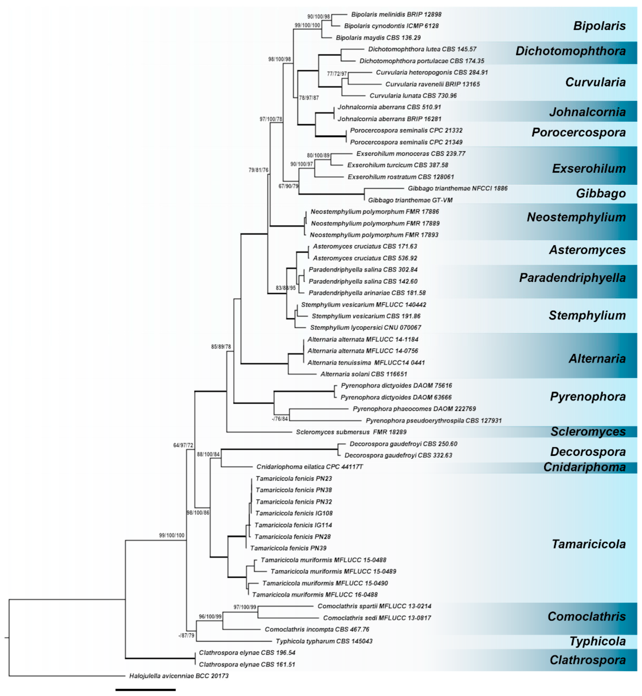
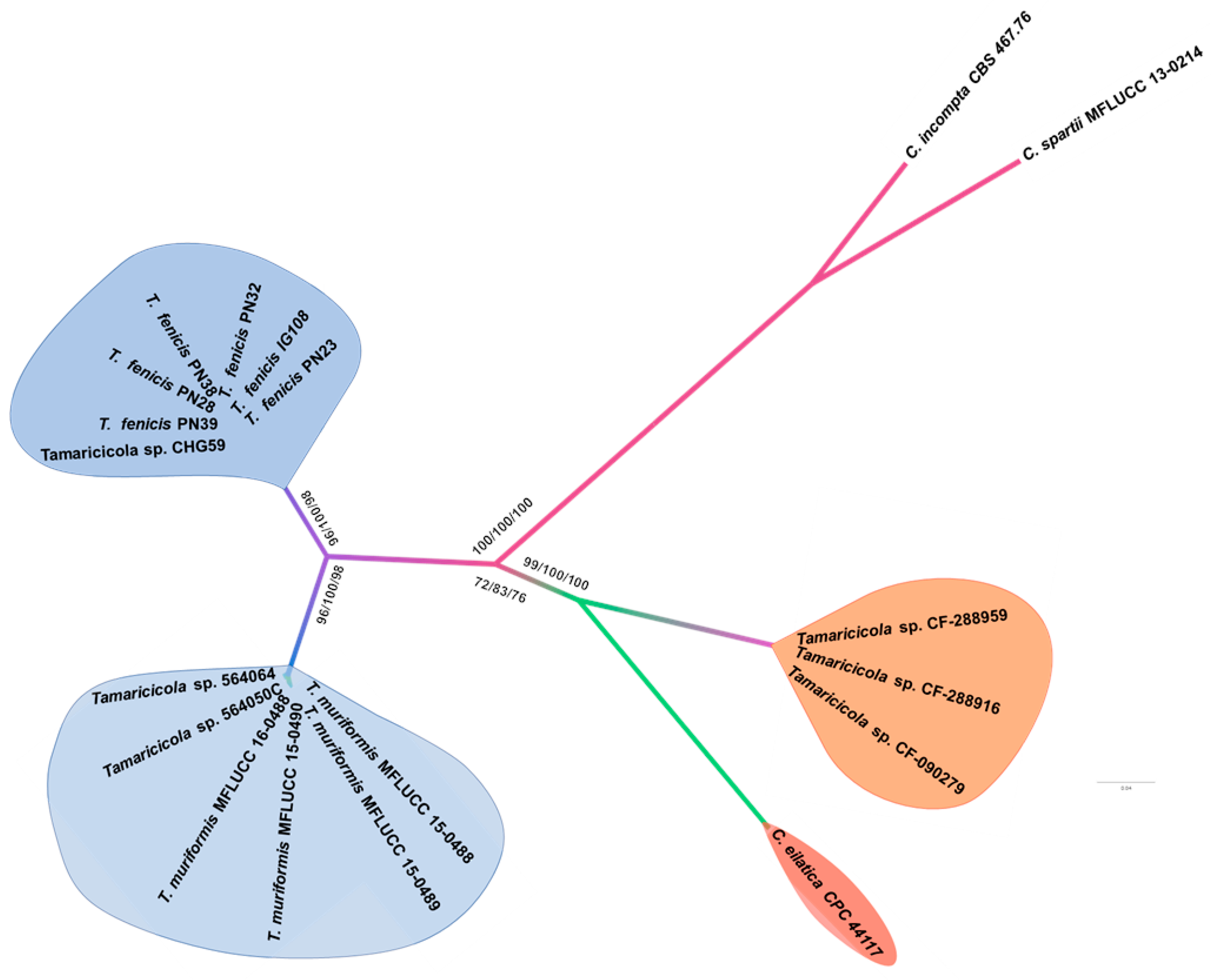
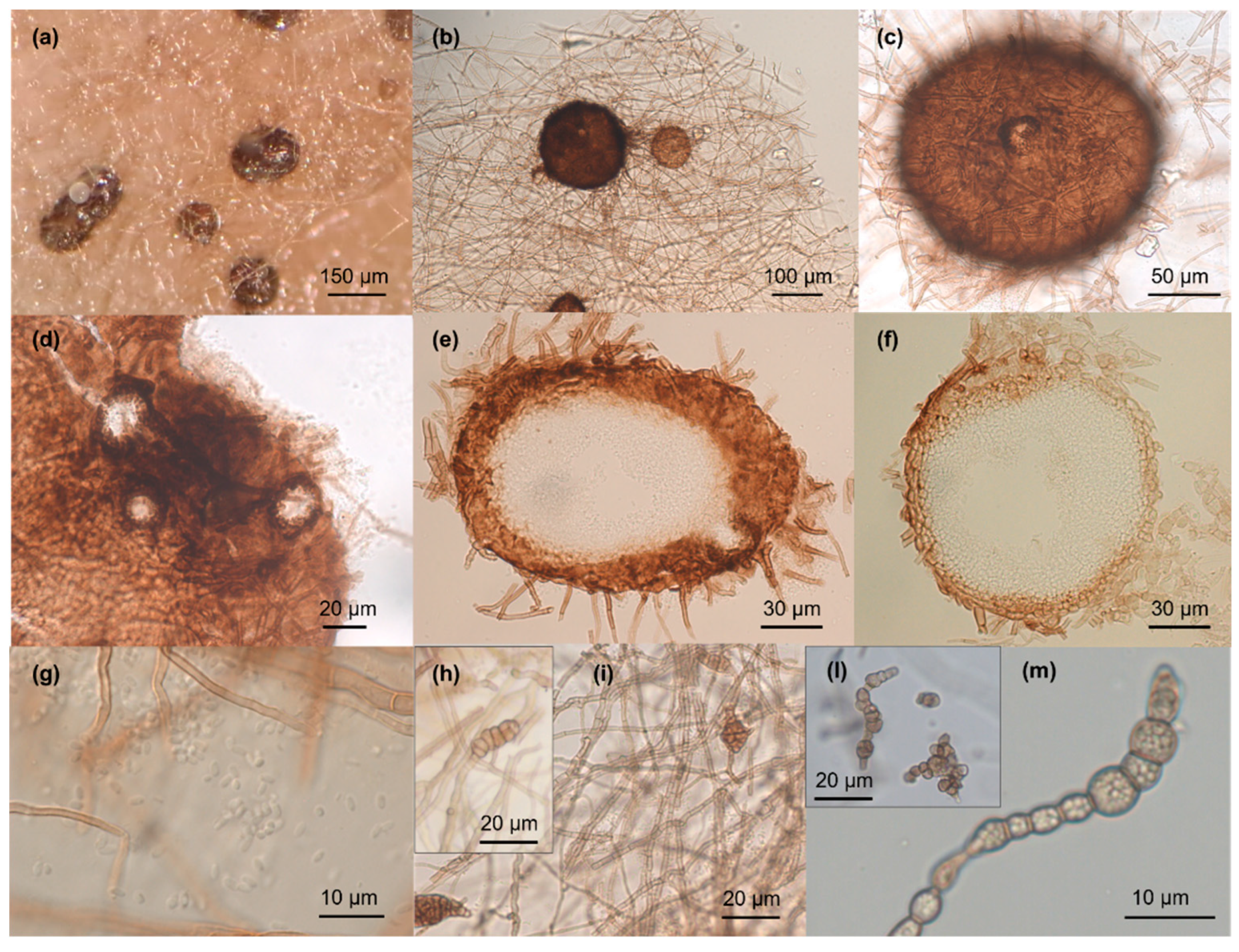
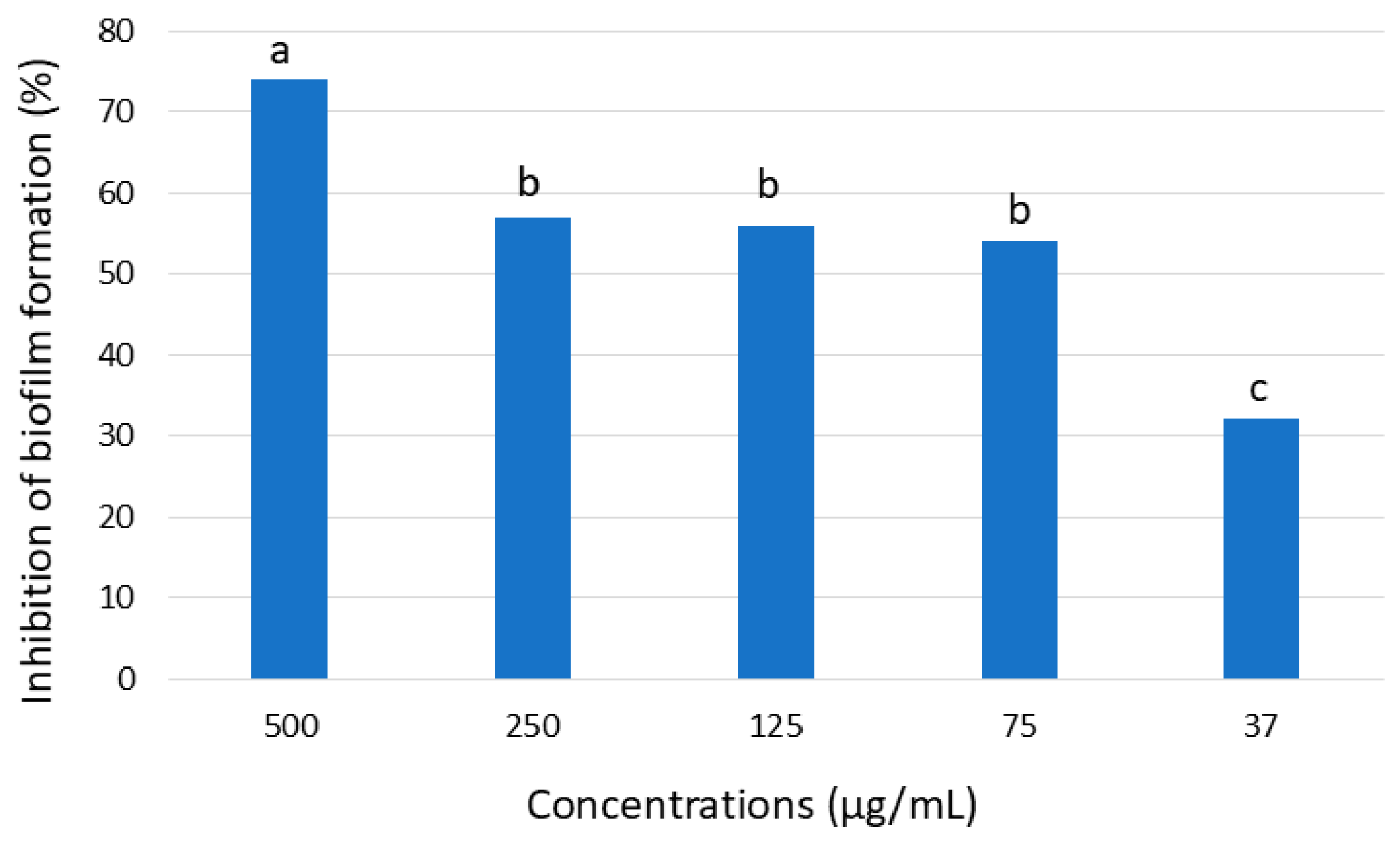
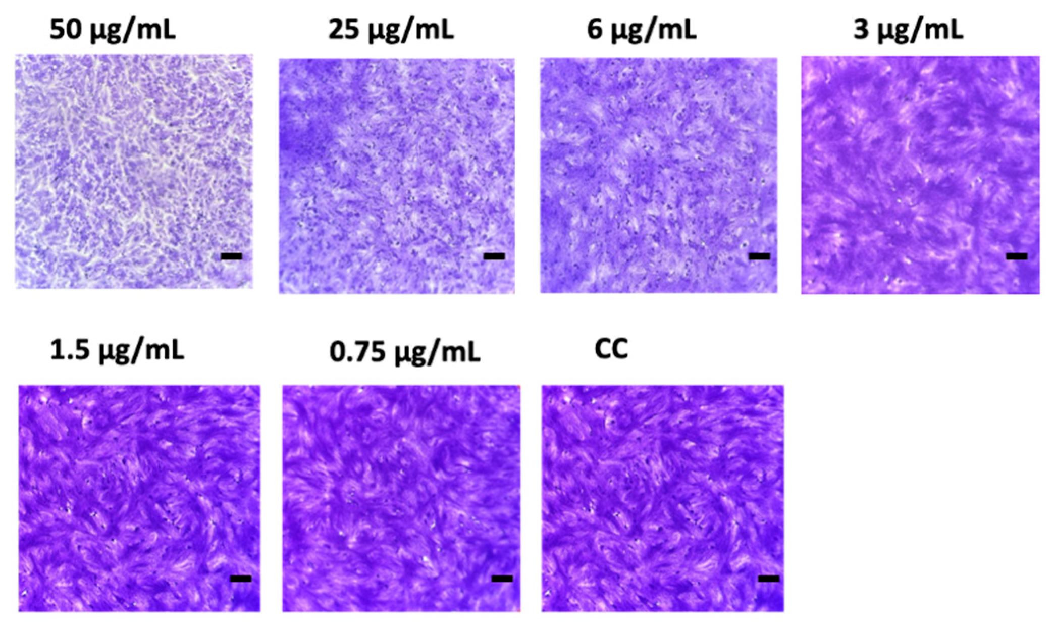
In this study, seven Pleosporaceae strains isolated from the seagrass Posidonia oceanica and the jellyfish Pelagia noctiluca in the central Tyrrhenian Sea were characterized using a polyphasic approach (morpho-physiological, molecular, and phylogenetic analyses). Based on multi-locus phylogenetic inference and morphological characters, a new species, Tamaricicola fenicis was proposed. Multi-locus phylogenetic analyses, using the nuclear ribosomal regions of DNA (nrITS1-nr5.8S-nrITS2, nrLSU, and nrSSU) as well as the rpb-2 and tef-1α gene sequences, strongly supported the new taxon. The phylogenetic inference, estimated using Maximum Likelihood and Bayesian Inference, clearly indicates that Tamaricicola fenicis sp. nov. forms a distinct clade within the monospecific genus Tamaricicola. The antimicrobial activity of the chloroformic and butanolic extracts from malt agar cultures of the new species exhibited interesting antiviral and antibiofilm properties. In particular, a MIC of 3.0 µg/mL was observed against the Echovirus E11 in Vero-76 cells; moreover, a biofilm BIC50 reduction at 53 µg/mL was observed against Staphylococcus aureus ATCC25923.

Keywords: 
;  ;  ;  ;  

1. Introduction

Marine environments represent a huge reservoir of biological and biochemical diversity, which is yet mostly unexplored. The oceans cover more than 70% of the surface of our planet and host a substantial proportion of Earth’s biodiversity. Up today 245,000 species have been reported [1]. Several researchers have recently emphasized the need to expand our knowledge of marine fungal biodiversity, which is estimated to exceed 12,500 species, although only about 2,000 of them have been formally recorded to date [2,3]. Understanding the biodiversity and species distribution of marine fungi is crucial for gaining deeper insights into their ecological roles. The vast and largely unexplored diversity of marine fungi, shaped by unique environmental conditions, offers a rich source of novel enzymes and bioactive metabolites with promising biotechnological and pharmaceutical applications, including the development of new drugs against multi-drug-resistant infections [4,5,6,7,8,9,10,11].
The majority of known marine fungi belongs to the phylum Ascomycota (82%), followed by Basidiomycota (8.3%) and Microsporidia (6.7%); while phyla such as Chytridiomycota and Mucoromycota are less common, each representing less than 1.5% of recorded species [12]. The class Dothideomycetes (Ascomycota) includes numerous marine species, particularly in the orders Pleosporales and Dothideales. The family Pleosporaceae is one of the largest within the order Pleosporales, comprising 23 genera and more than 200 species [13]. The members of the family are morphologically characterized by the presence of globose ascomata with thick walled peridium, bitunicate cylindrical asci producing eight, septate, sometimes muriforms, ascospores [14,15]. For a long time, the genera of the family were primarily distinguished by their ascospore features. However, phylogenetic molecular investigations have recently led to include in the Pleosporaceae family new taxa that produce only asexual structures, such as the two novel genera Neostemphylium and Scleromyces, isolated from freshwater sediments in Spain [16]. The species of Pleosporaceae generally exhibit dematiaceous hyphomycetous anamorphs that produce phragmo- or dyctioconidia from blastic conidiogenous cells on macronematous conidiophores, even if coelomycetous anamorphs with phialidic or anellydic conidia have also been reported [15].
The pleosporalen species exhibit a wide range of behaviors; they can be saprophytic, endo-/epiphytic, or parasitic on a variety of hosts in both terrestrial and marine environments. The family includes common ubiquitous species distributed world-wide (e.g., Alternaria spp.) and others with a narrower distribution such as the species of the genus Tamaricicola [15,17].
The genus Tamaricicola was introduced to accommodate a new taxon T. muriformis isolated from Tamarix gallica [17]. Up to date the genus is monospecific even if some new strains, possibly representing new lineages, have been isolated from Limonium majus and L. insigne (CF-288959, CF288916, CF090279), from lungs of Antigone canadensis tabida (Greater sandhill crane) and from the ascidian Ciona intestinalis (CHG59) [18,19,20]. It is worth noting that Tamaricicola sp. strain CF-288959 showed an interesting antifungal activity against Candida albicans [18]. In general, several marine pleosporalen strains produced new and interesting bioactive compounds [21,22,23,24].
During an extensive investigation aimed at expanding our knowledge of marine fungal biodiversity in the Tyrrhenian Sea, seven strains belonging to the genus Tamaricicola have been isolated from the seagrass Posidonia oceanica (IG108, IG114) and the jellyfish Pelagia noctiluca (PN23, PN28, PN32, PN38, PN39) [3,25]. Preliminary analyses suggested that these strains could represent a novel lineage within the genus [3,25]. This study aimed to characterize this novel species based on morphological, physiological, and molecular analyses. In addition, considering the biotechnological potential of pleosporalen marine fungi, the antibacterial, antifungal, and antiviral activities of different extracts were assessed.

2. Materials and Methods

2.1. Fungal Isolates

The fungal strains analysed in this study were isolated from different substrata collected in May from the “Cala Cupa” cove (42°22008.0300N,10°55004.0900E), Giglio Island (Tuscan Archipelago, North Tyrrhenian Sea), at 10-15 m depth by scuba divers. Strains IG108 and IG114 were isolated from the leaves of the seagrass P. oceanica (15 m depth - May 2015), while strains PN23, PN28, PN32, PN38, and PN39 were isolated from the inner tissues of the jellyfish P. noctiluca (10 m depth -May 2019), [3,25]. All strains were cryogenically maintained at -40 °C in the culture collection of microorganisms of the “Laboratory of Ecology of Marine Fungi” (Department of Ecological and Biological Sciences – DEB, University of Tuscia). Strain PN38 (MUT6838) is also preserved in the Mycotheca Universitatis Taurinensis (MUT) culture collection, Italy.

2.2. Morphological Analysis on Different Media

Strains had been revitalized and sub-cultured on Potato Dextrose Agar seawater (PDAs; 39 g PDA dissolved in 1 L of filtered seawater) at 23 °C.
Morphological analyses were carried out on plates utilizing different cultural media: PDAs, Malt Extract Agar seawater (MEAs; 30 g malt extract, 5 g peptone, 15 g agar, dissolved in 1 L of seawater), Malt Extract Broth seawater, Corn Meal Agar seawater (CMAs; 17 g CMA dissolved in 1 L of seawater) and Oatmeal Agar seawater (OAs; 30 g oatmeal powder, 20 g agar dissolved in 1 L of seawater). All media and components were purchased from Sigma-Aldrich, Saint Louis, USA.
The plates (5 cm diameter Ø) were inoculated with a single agar disc (2 mm2) cut from the actively growing margin of 15 days old PDAs cultures and incubated at 23 °C in sealed plastic boxes.
Growth was monitored for 28 days, and the macroscopic and microscopic features were reported.
To promote sexual reproduction, fungal strains were inoculated on different natural substrata such as barks (Quercus robur, Pinus halepensis), pine needles (P. halepensis), twigs (Tamarix gallica, T. africana) and Posidonia leaves (substrate of isolation) [26].
All substrata were cut into small pieces (3 × 1 cm), sterilized, and placed on the surface of well-developed colonies. The plates were incubated for 1 month at 23 °C to allow substrata colonization. Following this period, some of the inoculated fragments were transferred into tubes containing 20 mL of sterile seawater to simulate natural conditions, while others into moist chambers and further incubated for 18 months at 23 °C. All inoculated fragments were checked regularly. All experiments were carried out in triplicate.
Microscopic characterization of somatic and reproductive structures was carried out on slides with lactic acid. Conidioma sections (10 – 15 μm) were obtained using Leica cryostat (reproductive structures were collected, embedded in OCT -Optimal Cutting Temperature- mounting media (VWR), frozen in liquid N2, and maintained at -20 °C before sectioning). All samples were observed using a Zeiss AxioPhot microscope (Jena, Germany), and micrographs were taken with a Jenoptik ProgRes® camera (JenOptik AG, Jena, Germany).
Size ranges of relevant structures in species descriptions were derived from at least 35 measurements.

2.3. Molecular Analysis

Genomic DNA was extracted from fresh mycelium using the ZR Fungal/Bacterial DNA MiniPrep Kit (Zymo Research,USA) according to the manufacturer's directions. The extracted DNA was spectrophotometrically quantified (Qubit, Thermo Fisher Scientific,Waltham, MA, USA) and stored at −20 °C. For each strain several loci were amplified and sequenced: ITSrDNA, LSUrDNA, SSU rDNA, tef-1α (translation elongation factor) and rpb-2 (RNA polymerase II subunit). Primers for the amplifications were reported in Table 1.
Amplifications were performed in a 25 µL reaction volume containing: 2 µL of genomic DNA, 0.5 µL of each primer (10 µM), 2.5 µL of MgCl2 (25 mM), 1.5 µL of 5× buffer, 0.5 µL of dNTPs (10 mM), 0.2 µL of Go-Taq Polymerase (Promega, Madison, WI, USA); the final volume was reached by adding ultrapure water. Amplifications were run in a 2720 Thermal Cycler (Applied Biosystem, Waltham, MA, USA) using different PCR conditions (Table 1).
The PCR products were purified (E.Z.N.A. Cycle Pure kit -Omega Bio-tek, Norcross, GA, USA) and sequenced (Eurofins Genomics, Ebersberg, Germany). The sequences obtained were checked and trimmed with the Chromas Lite 2.1 program. Newly generated sequences were deposited in GenBank NCBI (National Center for Biotechnology Information) (Table 2).

2.4. Sequence Alignment and Phylogenetic Analyses

For the phylogenetic analyses a concatenated dataset of nrSSU, nrITS, nrLSU, rpb-2, and tef-1α sequences (Table 2) including the most representative species of the family Pleosporaceae was used [16,17]. The single gene sequence datasets were aligned with the Clustal X 2.1 software [31]. Alignments were checked and edited with BioEdit Alignment Editor 7.2.5 [32] and manually adjusted in MEGA 11 [33], when necessary. For the multi-locus phylogenetic analysis, alignments of different markers were concatenated with MEGA 11. Phylogenetic inference was estimated using Maximum Likelihood (ML) and Bayesian Inference (BI) as previously reported by Pasqualetti et al. [3]. The best-scoring trees were visualized using FigTree v.1.4 (http://tree.bio.ed.ac.uk/software/figtree/).

2.5. Antimicrobial and Antiviral Activity

2.5.1. Cultivation and Extraction

The fungal strain PN38 was cultivated on two different media: MEAs and Glucose Peptone Yeast seawater (GPYs; 1 g glucose, 0.5 g peptone special, 0.1 g yeast extract, 15 g agar dissolved in 1 L of filtered seawater). For each medium fifty plates (9 cm Ø), were inoculated as previously reported, and incubated at 23 °C for 30 and 60 days for MEAs and GPYs, respectively. Subsequently, the fungal cultures were cut into small pieces and extracted overnight with MeOH (1 L x 2 times). The MeOH extract solution was concentrated to dryness by a rotary evaporator. Subsequently the nBuOH, and CHCl3 fractions of the extract were obtained.

2.5.2. Antimicrobial Activity Assay

The minimum inhibitory concentrations (MICs) of extracts were determined using a microdilution method against reference strains -Staphylococcus aureus ATCC 25923, Pseudomonas aeruginosa ATCC 15442, Escherichia coli ATCC 25922, Enterococcus faecalis ATCC 29212 and the yeast Candida albicans ATCC 10231- as recommended by the Clinical and Laboratory Standards Institute. Each sample was tested at concentrations ranging from 2.5 to 0.3 mg/mL using two-fold serial dilution in Mueller–Hinton broth (Sigma Aldrich) for bacteria and Sabouraud broth (Sigma Aldrich) for yeast. The assay was performed in a 96-wells plate, starting from a stock solution of 50 mg/mL of the nBuOH, and CHCl3 extract fractions diluted in Dimethyl Sulfoxide (DMSO).
The microbial inoculum was obtained from cultures grown at 37 °C for 24 hours on Tryptic Soy Agar (TSA- Sigma Aldrich) for bacteria and Sabouraud agar for the yeast. The concentration was adjusted to 1.5 × 10⁸ CFU/mL (McFarland standard 0.5) using 0.9% NaCl saline solution. Ten microliters of each microbial suspension were used as inoculum. A positive control (microorganism and medium without PN38 extract), a negative control (medium without inoculum), and a substance control (medium with extract without microbial inoculum, to evaluate the absorbance of extracts), were also included in the 96-wells plate. The 96-well plates were incubated at 37 °C for 24 h and MICs were determined by a microplate spectrophotometer (GloMax®-Multi Detection System, Promega) as the lowest concentration of extract whose OD, read at 570 nm, was comparable with the negative control wells. Each assay was performed in triplicate. The biofilm inhibition formation was tested on S. aureus ATCC 25923. The strain was incubated in Tryptic Soy Broth (TSB) containing 2% (w/v) glucose at 37 °C for 24 h. After incubation, 2.5 μL of microbial suspension was placed into each well of a flat-bottom 96-well loaded with 200 μL of TSB with 2% glucose. Aliquots at sub-MIC concentration of each extract, ranging from 500 to 1.5 μg/mL, were directly added to the wells, positive, negative and substrate control were also included.
The microplates were incubated at 37 °C for 24 h. After biofilm growth, wells were washed twice with sterile NaCl 0.9% solution, and sessile biomass stained with 100 μL of 0.1% crystal violet solution for 30 min at 37 °C. After incubation, the plate was washed twice, and 200 μL of ethanol was added into each well. The plate was then incubated for 10 min at room temperature. OD was read at a wavelength of 540 nm using a plate reader (Glomax Multidetection System TM297). The percentage of biofilm inhibition was determined through the following formula:
I n h i b i t i o n   % =   O D   g r o w t h   c o n t r o l     O D   s a m p l e   O D   g r o w t h   c o n t r o l   x   100
BIC50 (concentration at which the percentage of inhibition of biofilm formation is equal to 50%), was obtained by comparing the ODs of control wells with that of the sample wells at different concentrations, and the value was calculated using AAT Bioquest, Inc. Quest Graph™ IC50 Calculator (v.1) retrieved from https://www.aatbio.com/ tools/ic50-calculator-v1. Each assay was performed in triplicate.

2.5.3. Antiviral Activity Assay

Virus - Epithelial monkey kidney Vero-76 cells [ATCC CRL 1587, Cercopithecus aethiops] were maintained in Dulbecco’s modified Eagle’s medium (DMEM; Sigma-Aldrich) supplemented with 1% penicillin-streptomycin, and 2 mM L-glutamine (Sigma-Aldrich), and 10% fetal bovine serum (FBS; Sigma-Aldrich), at 37 °C and 5 % CO2. Echovirus 11 lineage 1, was maintained and propagated in Vero-76 cells. The virus was stored in small aliquots at −80 °C until use. All experimental work involving viruses was performed in an appropriate biosafety level containment laboratory.
Cytotoxicity evaluation by optical microscopy - Cell morphology alterations of Vero-76 cells induced by the cytotoxic effects of PN38 extract were evaluated by optical microscopy observation. Vero-76 cells were seeded in 96-wells plate at an initial density of 2 × 10⁴ cell/mL, in DMEM medium. After 24 h, the cell monolayers were treated with different dilutions (25-0.19 µg/mL) of PN38 extract for 24 h. Absorbance was measured at 570 nm. The percentage of cell viability was determined using the following formula:
C e l l   v i a b i l i t y % =   S a m p l e   a b s o r b a n c e   t r e a t e d   s a m p l e s C o n t r o l   a b s o r b a n c e   C o n t r o l   a b s o r b a n c e   x   100
The experiment was conducted in triplicate.
Cytopathic effect inhibition assay - The potential antiviral activity of PN38 extract against E11 was evaluated by the cytopathic effect (CPE) inhibition assay in a co-treatment antiviral assay: Vero-76 cells were seeded into 96-wells plate (2x10⁴ cell/mL) and incubated overnight at 37 °C a humidified CO2 (5%) atmosphere. Different concentrations of PN38 extract (25-0.19 μg/mL) and E11 at a multiplicity of infection (MOI) of 0.01 PFU/cell were added to the cell monolayer and incubated for 48 h at 37 °C in DMEM supplemented with 5% FBS. CPE reduction was evaluated by optical microscopy observation. Also, cells were fixed with 4% formaldehyde for 15 min at room temperature and then stained with 0.1% (w/v) crystal violet for 30 min at room temperature. The intensity of the crystal violet stain was evaluated by spectrophotometry at 620 nm. The minimal inhibition concentration (MIC) was also determined. All experiments were carried out in triplicate.
E11 genome quantification in Vero-76 infected cells – The E11 viral genome copies in the supernatants of infected cells treated with different concentrations of PN38 (25-0.19 μg/mL) were quantified. After 48 h post-infection, viral RNA was extracted from the supernatant of treated and control cells and quantified by qRT-PCR using the automated Elite InGenius one step RNA Enterovirus ELITe MGB® Kit, according to manufacturer’s protocols. Each experiment was performed in triplicate, and RNA yields are reported as the mean values of three independent assays.

3. Results

3.1. Phylogenetic Inference

A preliminary phylogenetic analysis was carried out individually for nrLSU, nrITS, nrSSU, rpb-2, and tef-1α. Since no incongruences were observed among the single-loci phylogenetic trees, a multi-locus analysis was performed. The final dataset included 58 strains, representing 37 species, and 21 genera belonging to the family Pleosporaceae. In total, 33 sequences were newly generated, whereas 176 were obtained from GenBank (Table 2). The aligned concatenated dataset has 4,534 characters (861 for nrLSU, 636 for nrITS, 1020 for nrSSU, 851 for rpb-2, and 1166 tef-1α) including gaps. Among them, 1,337 distinct patterns, 882 parsimony-informative sites, 304 singleton sites, and 3,348 constant sites, were observed. ML analysis yielded a best-scoring three with a final optimization likelihood value of -22,563.004. The ML and BI analyses resulted in generally congruent topologies, thus, only the ML tree with BS and BYPP values was reported (Figure 1).
The phylogenetic analysis performed on the concatenated dataset showed that the seven isolates under investigation formed a well-supported clade (BS=100%; BYPP=100%) inside the genus Tamaricicola clearly distinguished by T. muriformis.
Although T. muriformis remains the only species formally described within the genus to date, several other Tamaricicola strains, beyond those investigated in this study, have been reported from marine and terrestrial substrates. Notably, two strains were isolated from avian lungs [20], three from Limonium spp. [18], and one from the ascidian Ciona intestinalis [19]. These strains were not included in the global phylogenetic analyses carried out for the Pleosporaceae family, as only nrITS sequences are currently available for them. For these reasons, a supplementary ITS-based phylogenetic analysis, incorporating all known Tamaricicola strains, was performed (Figure 2). The sixteen strains attributed to the genus were resolved into three distinct and well-supported clades. The strains Tamaricicola sp. 56406A and Tamaricicola sp. 56405C clustered with T. muriformis, while Tamaricicola sp. CHG59 grouped with the strains of the newly proposed species T. fenicis. The remaining three strains—Tamaricicola sp. CF-288959, CF-288916, and CF-090279—appear to represent a separate lineage. Interestingly, this latter lineage shows phylogenetic affinity with the newly established genus and species Cnidariophoma eilatica, described from a coral-associated strain [34]. Based on ITS sequence data alone, the three strains (Tamaricicola sp. CF-288959, CF-288916, and CF-090279) appear not belonging to Tamaricicola but, instead, represent a distinct lineage within or closely related to Cnidariophoma.
The phylogenetic analyses strongly indicated that the seven investigated strains belong to a new lineage within the genus Tamaricicola: the new species Tamaricicola fenicis is herein proposed.

3.2. Taxonomy

Tamaricicola fenicis Pasqualetti & Braconcini sp. nov. (Figure 3)
MycoBank.
Etimology. In honour of the Italian Microbiologist Massimiliano Fenice.
Type. Italy, Tuscany, Mediterranean Sea, Giglio Island (Grosseto), Cala Cupa, 4222008.0300N, 1055004.0900E, 10 m depth. Isolated from the jellyfish Pelagia noctiluca, May 2019, Marcella Pasqualetti. Holotype MUT 6838 (strain PN38), living culture permanently preserved in a metabolically inactive state at MUT.
Diagnosis. T. fenicis is introduced to accommodate seven novel strains retrieved on two different substrata in the Tyrrhenian Sea. T. fenicis is a biotic marine fungus associated with P. noctiluca and P. oceanica. Multi-locus phylogenetic analysis showed that T. fenicis clustered into a distinct clade in the monospecific genus Tamaricicola and differs from its closest phylogenetic neighbour T. muriformis by genetic characters in nrITS, nrLSU, nrSSU, tef-1α, and rpb-2 sequences and in conidia dimensions as well as in the production of large, thick-walled, and muriform resting spore (chlamydospore). Morphologically, T. fenicis resembles the species T. muriformis in having similar asexual reproductive structures [17].
Description. Growing on Posidonia oceanica leaves, internal tissues of Pelagia noctiluca, Quercus robur and Pinus halepensis barks, P. halepensis pine needle and twigs of Tamarix gallica and T. africana.
Hyphae 2.2 – 4.5 µm wide, irregular, septate, sometimes toruloid containing large amount of lipid droplets, sub-hyaline to slightly pigmented. Sexual morph: not observed. Asexual morph: coelomycetous. Conidiomata 80-190 μm diam, pycnidial, superficial or partially immersed, dark brown to black, spheroidal to sub-spheroidal, ostiolate (1-3). Conidiomatal wall 10-15 μm wide, comprising few layers of dark brown to hyaline cells of textura angularis. Conidiophores micronematous, reduced to conidiogenous cells. Conidiogenous cells 3-4.5 × 2-4 μm, phialidic, hyaline, smooth, ampulliform. Conidia 1.8 - 4 x 1.5-2.3 μm, ellipsoidal to cylindrical, hyaline, rounded at both ends, 1-celled, smooth-walled, slightly falcate. Chlamydospores 18 - 25 x 7.5-10 μm, muriform, irregular, with several transverse, longitudinal, and oblique septa.
Colony description (Figure 4). Colonies on MEAs-PDAs, reaching 48-50 mm diameter after 21 days at 23 °C, plane, umbonate, surface velutinous centrally feltrose, grey, with a light brown marginal area; aerial mycelium sparse, whitish to light brown, mainly in the central area; margins regular moderately deep, reverse dark grey. Soluble pigment and exudates absent. Conidiomata produced on PDAs, not observed on MEAs. Colonies on OAs, reaching 50 mm diameter after 21 days at 23 °C, plane, surface slightly feltrose, whitish, margins regular, reverse white-grey darker in the centre. Soluble pigment and exudates absent. Conidiomata produced in very large amount. Colonies on CMAs, reaching 48 mm diameter after 21 days at 23 °C, surface slightly feltrose, greyish, mycelium prevalently immersed, margins regular, reverse hyaline to greyish. Soluble pigment and exudates absent. Conidiomata present.
Notes. No Sexual form (ascomata) was detected in artificial and natural substrata. In liquid media the mycelium, prevalently toruloid, shows characteristic rounded cells full of a large amount of small lipidic droplets (Figure 3).
Based on a Megablast search on NCBI nucleotide database, the closest hits of nrITS sequences are Ascomycota sp. Di283-2 (GenBank accession no. OR367423; identities 554/554 – 100%); Ascomycota sp. San Juan 55-1 (GenBank accession no. KF638538; identities533/533 – 100%) and Tamaricicola sp. strain CHG59 (GenBank accession no. MW064152; identities 494/494 – 100%).
The closest hits using the nrLSU sequences are Pleosporaceae sp. M306 (GenBank accession no. KJ443126; identities 1276/1306 – 98%, 7 gaps), Comoclathris typhicola MUT<ITA>:4379 (GenBank accession no. KF636774; identities 1275/1306 – 98%, 10 gaps) and Comoclathris typhicola strain CBS 132.69 (GenBank accession no. JF740325; identities 1275/1306 – 98%, 10 gaps). The closest hit using the nrSSU sequences is T. muriformis isolate IT_9172 (GenBank accession no. KU870908; identities 1043/1049 – 99%, 6 gaps). The closest hit using the tef-1α sequences is T. muriformis isolate IT_9175 (GenBank accession no. KU600014; 922/947 – 97%, 0 gaps). The closest hits using the rpb-2 sequence (ON887328) are Cnidariophoma eilatica CPC 44117 (GenBank accession no. OQ627943; identities 610/656 – 93%, 0 gaps) and T. muriformis isolate IT_9173 (GenBank accession no. KU820870; identities 551/579 – 95%, 0 gaps).
Additional material examined. Italy, Tuscany, Mediterranean Sea, Giglio Island (Grosseto), Cala Cupa, 4222008.0300N, 1055004.0900E, 10 m depth. Isolated from the jellyfish P. noctiluca, May 2019, Marcella Pasqualetti, living culture PN23, PN28, PN32, PN39. Italy, Tuscany, Mediterranean Sea, Giglio Island (Grosseto), Cala Cupa, 4222008.0300N, 1055004.0900E, 15 m depth. Isolated from P. oceanica, May 2015, Marcella Pasqualetti, living culture IG108, IG114.

3.3. Screening for Antimicrobial and Antiviral Activity

The extracts (nBuOH, and CHCl3) of the strain PN38 cultivated in MEAs and GYPs were assessed to evaluate antimicrobial and antiviral activity. The extracts were tested against several pathogenic organisms (S. aureus ATCC 25923, P. aeruginosa ATCC 15442, E. coli ATCC 25922, E. faecalis ATCC 29212 and C. albicans) using the microdilution method. No activity was observed against bacterial and fungal strains at the screening concentration of 2.5 mg/mL. However, an interesting anti-biofilm activity against S. aureus ATCC 25923 was observed at sub-MIC concentration of MEAs nBuOH extract with a BIC50 value of 53 μg/mL (Figure 5).
The antiviral activity was assessed only on the CHCl3 extract due to the cell toxicity of the nBuOH extract. The optical microscopy observation revealed a dose-dependent cytotoxic effect of MEAs CHCl3 extract on Vero-76 cells. A marked viability decrease was observed at concentration higher than 25 µg/mL, while 6 μg/mL was the highest non-cytotoxic concentration observed (Figure 6).
A co-treatment assay with MEAs CHCl3 extract (25-0.19 μg/mL) and E11 (MOI of 0.01 PFU/cell), showed a strong antiviral effect, with a 90% CPE reduction at 3 and 6 μg/mL, identifying 3 μg/mL as the MIC (Figure 7). This activity was confirmed by the reduction of viral RNA levels in infected Vero-76 supernatants, 86% and 94% at 3 and 6 μg/mL, respectively, compared to the untreated cells (CV) (Figure 7C).

4. Discussion

Marine fungi have long been considered an “exotic” group of microorganisms with low species richness and abundance [35]. However, it has become increasingly evident that they represent a substantial proportion of marine microbial diversity and contribute to various key ecological processes in the marine environments (e.g., aquatic carbon pump efficiency and regulation of phytoplankton composition). Despite recent progress in marine mycology, our knowledge of marine fungi remains limited. In this context, research focused on underexplored habitats and substrates can significantly enhance our understanding of these neglected organisms, particularly regarding their distribution, ecology, and contributions to marine ecosystem services. It is worth noting that in recent years, numerous new taxa have been established from marine substrata that have never been studied from a mycological point of view [26,36].
The seven strains investigated in this study were isolated from the leaves of the seagrass P. oceanica (IG108, IG114) and the inner tissues of the jellyfish P. noctiluca (PN23, PN28, PN32, PN38, PN39) [3,25]. P. oceanica is an endemic seagrass of the Mediterranean Sea; the seagrass meadows rank amongst the most valuable coastal ecosystems on Earth in terms of benefits and services they provide. P. oceanica is one of the most studied substrata from marine mycologists [25,37,38,39,40,41,42,43,44,45]. In contrast, P. noctiluca mycobiota was only studied by Pasqualetti and co-authors [3]. The isolated strains do not produce reproductive structures on MEAs, utilized for their maintenance. The experiments to promote the sexual or asexual reproduction [46,47,48] lead only to the development of coelomycetous conidiomata (asexual structures) in PDAs, CMAs and OAs and natural inoculated substrata (barks of Q. robur, P. halepensis, pine needles of P. halepensis, twigs of T. gallica, T. africana and Posidonia leaves) while ascomata were not detected.
A preliminary molecular characterization based on nrITS, nrLSU, and nrSSU allowed to place the strains IG108, IG114, PN23, PN28, PN32, PN38, PN39 in the family Pleosporaceae [3]. The analysis suggested that the strains were strictly related to the monospecific genus Tamaricicola and also indicated that they could represent a new lineage inside the genus [3,25]. To confirm these preliminary observations, a multi-locus phylogenetic analysis, based on molecular markers usually utilized to study the taxonomy of the Pleosporaceae family, was performed [16,17]. The phylogenetic tree (Figure 1), including the genera of the family Pleosporaceae, showed that the strains formed a well-supported clade within the genus Tamaricicola, separated from the species T. muriformis. Thus, the new species T. fenicis was proposed and established. T. fenicis is the second species ascribed to the genus Tamaricicola. Nevertheless, in literature some other strains are attributed to the genus Tamaricicola: three terrestrial strains isolated from L. majus and L. insigne, two isolated from avian lung and a marine strain (CHG59) isolated from the gut of the ascidian C. intestinalis sampled in the German North Sea [18,19,20].
The nrITS phylogenetic analysis, including all strains attributed to Tamaricicola, clearly reveals the presence of three distinct evolutionary lineages: one corresponding to the terrestrial strains of T. muriformis, another to the marine strains of T. fenicis, and a third comprising the strains isolated from L. majus and L. insigne. González-Menéndez and collaborators [18] suggested that the Limonium-associated strains might represent a novel species within Tamaricicola. However, the phylogenetic analysis conducted in this study (Figure 2) indicates that these strains may instead constitute a distinct lineage within the recently established genus Cnidariophoma [34], rather than a new lineage within Tamaricicola.
Considering the ecology of T. fenicis, it is worth noting that strains IG108, IG114, and PN23, PN28, PN32, PN38, and PN39 were isolated from different types of substrates (plant and animal) during two sampling campaigns in the same area (Cala Cupa – Giglio Island) in the Central Tyrrhenian Sea. Thus, it seems reasonable that this species could have a not-specialized habitus [49] even considering the similarity, and probably identity, with the strain Tamaricicola sp. CHG59 isolated from C. intestinalis. Nevertheless, up to date the new species seems strictly marine with a biontic behavior. The identification of novel taxa significantly contributes to the knowledge advancement on marine fungi, confirming that the marine ecosystem constitutes an extensive repository of biodiversity and chemo-diversity, which are largely unexplored, in particular for its microbial components. It is worth noting that new lineages could be extremely interesting from a blue-biotechnological perspective considering the biological activity of their extract.
T. fenicis shows significant antiviral activity against Echovirus 11 (E11), a member of the Enterovirus genus within the Picornaviridae family. This virus has recently gained renewed clinical relevance following the emergence of a novel variant associated with severe neonatal hepatitis and liver failure in several European countries [50]. Enteroviruses are well-recognized for their capacity to cause a wide spectrum of human diseases, and the lack of effective antiviral therapies underscores the urgent need for new therapeutic agents. Despite extensive research efforts, many compounds, showing promising in vitro anti-Enterovirus activity, have failed to demonstrate comparable efficacy in vivo [51,52]. In view of this the potential in vitro antiviral activity of the PN38 extract against the newly identified E11 variant represents a valuable assumption for the future development of new antiviral leads.
In addition, the ability of nBuOH extract of PN38 to inhibit biofilm formation represents a noteworthy biological activity. The control of infectious diseases associated with biofilms remains a significant challenge, and there is a pressing need for new, effective molecules. Although further studies are necessary to identify the specific bioactive compounds of the texted extract and to elucidate their mechanisms of action, the results suggest that the T. fenicis PN38 strain is a promising producer of compounds with both antiviral and anti-biofilm activities.

5. Conclusions

The present study provides a morphological and phylogenetic study of seven strains obtained from the P. oceanica leaves and the inner tissues of P. noctiluca collected in the central Tyrrhenian Sea; the jellyfish has never been studied for its mycobiota. The strains form a new lineage within the genus Tamaricicola, family Pleosporaceae. In light of this, the new species Tamaricicola fenicis has been established. The identification of new species contributes to the advancement of knowledge about this genus that until now included only a terrestrial species. It is worth noting that all strains attributed to Tamaricicola fenicis derived from marine substrata. Finally, the CHCl₃ extract of T. fenicis PN38 exhibits significant antiviral activity against Echovirus 11, while the n-BuOH extract markedly reduces biofilm formation by S. aureus suggesting that this strain may have high biotechnological potential for drug discovery.
The emergence of drug-resistant pathogens and novel infections has moved toward the sea the search for new pharmaceuticals, and undoubtedly, the potential of marine fungi, particularly of novel taxa, in this context is very promising.

Author Contributions

Conceptualization, M.P. and M.B.; methodology, M.P., M.B., D.F. and D.S..; validation and formal analysis, M.P., M.B., E.P., D.F. and D.S.; investigation, M.P., M.B., S.G, P.B., E.P., D.F. and D.S; resources, M.P., D.F and D.S.; data curation and visualization, M.B., S.G., P.B., E.P.; supervision, M.P., D.F. and D.S.; project administration, M.P.; funding acquisition, M.P., D.S. All authors have read and agreed to the published version of the manuscript.

Funding

This research was funded by “Progetto di Ricerca di Interesse Nazionale” (PRIN)-Production and characterization of new bioactive molecules against emerging and/or multidrug-resistant pathogens by neglected polyextremophilic marine fungi (MYCOSEAS, no. 2022MPTT35).

Institutional Review Board Statement

Not applicable.

Informed Consent Statement

Not applicable.

Data Availability Statement

All data generated or analyzed during this study are included in this published article files. All sequences were deposited in GenBank (https://www.ncbi.nlm.nih.gov/nuccore, accessed on 06 October 2025).

Acknowledgments

The Research project implemented under the National Recovery and Resilience Plan (NRRP), Mission 4 Component 2 Investment 1.4—Call for tender No. 3138 of 16 December 2021, rectified by Decree n.3175 of 18 December 2021 of Italian Ministry of University and Research funded by the European Union—Next Generation EU. Project code CN_00000033, Concession Decree No. 1034 of 17 June 2022 adopted by the Italian Ministry of University and Research, CUP J83C22000860007, Project title “National Biodiversity Future Center—NBFC”.

Conflicts of Interest

The authors declare no conflict of interest.

References

  1. Available online: www.marinespecies.org (accessed on 7 October 2025).
  2. Jones, E.G.; Pang, K.L.; Abdel-Wahab, M.A.; Scholz, B.; Hyde, K.D.; Boekhout, T.; Ebel, R.; Rateb, M.E.; Henderson, L.; Sakayaroj, J.; et al. An online resource for marine fungi. Fungal Divers. 2019, 96, 347–433. [Google Scholar] [CrossRef]
  3. Pasqualetti, M.; Braconcini, M.; Barghini, P.; Gorrasi, S.; Schillaci, D.; Ferraro, D.; et al. From marine neglected substrata new fungal taxa of potential biotechnological interest: the case of Pelagia noctiluca. Front. Microbiol. 2024, 15, 1473269. [Google Scholar] [CrossRef] [PubMed]
  4. Giovannini, V.; Barghini, P.; Gorrasi, S.; Massimiliano, F.; Pasqualetti, M. Marine fungi: A potential source of novel enzymes for environmental and biotechnological applications. J. Environ. Prot. Ecol. 2019, 20, 1214–1222. [Google Scholar]
  5. Pasqualetti, M.; Barghini, P.; Giovannini, V.; Fenice, M. High production of chitinolytic activity in halophilic conditions by a new marine strain of Clonostachys rosea. Molecules 2019, 24, 101880. [Google Scholar] [CrossRef] [PubMed]
  6. Hou, X.M.; Hai, Y.; Gu, Y.C.; Wang, C.Y.; Shao, C.L. Chemical and bioactive marine natural products of coral-derived microorganisms (2015–2017). Curr. Med. Chem. 2019, 26, 6930–6941. [Google Scholar] [CrossRef]
  7. Botta, L.; Saladino, R.; Barghini, P.; Fenice, M.; Pasqualetti, M. Production and identification of two antifungal terpenoids from the Posidonia oceanica epiphytic Ascomycota Mariannaea humicola IG100. Microb. Cell Fact. 2020, 19, 184. [Google Scholar] [CrossRef]
  8. Teng, Y.F.; Xu, L.; Wei, M.Y.; Wang, C.Y.; Gu, Y.C.; Shao, C.L. Recent progresses in marine microbial-derived antiviral natural products. Arch. Pharm. Res. 2020, 43, 1215–1229. [Google Scholar] [CrossRef]
  9. Ameen, F.; AlNadhari, S.; Al-Homaidan, A.A. Marine microorganisms as an untapped source of bioactive compounds. Saudi J. Biol. Sci. 2021, 28, 224–231. [Google Scholar] [CrossRef]
  10. Pasqualetti, M.; Gorrasi, S.; Giovannini, V.; Braconcini, M.; Fenice, M. Polyextremophilic chitinolytic activity by a marine strain (IG119) of Clonostachys rosea. Molecules 2022, 27, 688. [Google Scholar] [CrossRef]
  11. Pang, K.L.; Jones, E.G.; Abdel-Wahab, M.A.; Adams, S.J.; Alves, A.; Azevedo, E.; et al. Recent progress in marine mycological research in different countries, and prospects for future developments worldwide. Bot. Mar. 2023, 66, 239–269. [Google Scholar] [CrossRef]
  12. Calabon, M.S.; Jones, E.G.; Pang, K.L.; Abdel-Wahab, M.A.; Jin, J.; Devadatha, B.; et al. Updates on the classification and numbers of marine fungi. Bot. Mar. 2023, 66, 213–238. [Google Scholar] [CrossRef]
  13. Available online: www.indexfungorum.org (accessed on 7 October 2025).
  14. Dong, J.; Chen, W.; Crane, J.L. Phylogenetic studies of the Leptosphaeriaceae, Pleosporaceae and some other Loculoascomycetes based on nuclear ribosomal DNA sequences. Mycol. Res. 1998, 102, 151–156. [Google Scholar] [CrossRef]
  15. Ariyawansa, H.A.; Thambugala, K.M.; Manamgoda, D.S.; Jayawardena, R.; Camporesi, E.; Boonmee, S.; et al. Towards a natural classification and backbone tree for Pleosporaceae. Fungal Divers. 2015, 71, 85–139. [Google Scholar] [CrossRef]
  16. Torres-Garcia, D.; García, D.; Cano-Lira, J.F.; Gené, J. Two novel genera, Neostemphylium and Scleromyces (Pleosporaceae) from freshwater sediments and their global biogeography. J. Fungi 2022, 8, 868. [Google Scholar] [CrossRef] [PubMed]
  17. Thambugala, K.M.; Daranagama, D.A.; Phillips, A.J.; Bulgakov, T.S.; Bhat, D.J.; Camporesi, E.; et al. Microfungi on Tamarix. Fungal Divers. 2017, 82, 239–306. [Google Scholar] [CrossRef]
  18. González-Menéndez, V.; Crespo, G.; De Pedro, N.; Diaz, C.; Martín, J.; Serrano, R.; et al. Fungal endophytes from arid areas of Andalusia: high potential sources for antifungal and antitumoral agents. Sci. Rep. 2018, 8, 9729. [Google Scholar] [CrossRef] [PubMed]
  19. Utermann, C.; Echelmeyer, V.A.; Oppong-Danquah, E.; Blümel, M.; Tasdemir, D. Diversity, bioactivity profiling and untargeted metabolomics of the cultivable gut microbiota of Ciona intestinalis. Mar. Drugs 2020, 19, 6. [Google Scholar] [CrossRef]
  20. Salazar-Hamm, P.S.; Gadek, C.R.; Mann, M.A.; Steinberg, M.; Montoya, K.N.; Behnia, M.; et al. Phylogenetic and ecological drivers of the avian lung mycobiome and its potentially pathogenic component. Commun. Biol. 2025, 8. [Google Scholar] [CrossRef]
  21. Chen, C.J.; Zhou, Y.Q.; Liu, X.X.; Zhang, W.J.; Hu, S.S.; Lin, L.P.; et al. Antimicrobial and anti-inflammatory compounds from a marine fungus Pleosporales sp. Tetrahedron Lett. 2015, 56, 6183–6189. [Google Scholar] [CrossRef]
  22. Yang, J.; Jiao, R.H.; Yao, L.Y.; Han, W.B.; Lu, Y.H.; Tan, R.X. Control of fungal morphology for improved production of a novel antimicrobial alkaloid by marine-derived fungus Curvularia sp. IFB-Z10 under submerged fermentation. Process Biochem. 2016, 51, 185–194. [Google Scholar] [CrossRef]
  23. Cao, F.; Meng, Z.H.; Mu, X.; Yue, Y.F.; Zhu, H.J. Absolute configuration of bioactive azaphilones from the marine-derived fungus Pleosporales sp. CF09-1. J. Nat. Prod. 2019, 82, 386–392. [Google Scholar] [CrossRef]
  24. Han, Y.; Sun, C.; Li, C.; Zhang, G.; Zhu, T.; Li, D.; Che, Q. Antibacterial phenalenone derivatives from marine-derived fungus Pleosporales sp. HDN1811400. Tetrahedron Lett. 2021, 68, 152938. [Google Scholar] [CrossRef]
  25. Pasqualetti, M.; Giovannini, V.; Barghini, P.; Gorrasi, S.; Fenice, M. Diversity and ecology of culturable marine fungi associated with Posidonia oceanica leaves and their epiphytic algae Dictyota dichotoma and Sphaerococcus coronopifolius. Fungal Ecol. 2020, 44, 100906. [Google Scholar] [CrossRef]
  26. Braconcini, M.; Gorrasi, S.; Fenice, M.; Barghini, P.; Pasqualetti, M. Rambellisea gigliensis and Rambellisea halocynthiae gen. et spp. nov. (Lulworthiaceae) from the marine tunicate Halocynthia papillosa. J. Fungi 2024, 10, 127. [Google Scholar] [CrossRef]
  27. White, T.J.; Bruns, T.; Lee, S.; Taylor, J. Amplification and direct sequencing of fungal ribosomal RNA genes for phylogenetics. In PCR Protocols; Elsevier: Amsterdam, The Netherlands; Academic Press: New York, NY, USA, 1990; pp. 315–322. [Google Scholar] [CrossRef]
  28. Vilgalys, R.; Hester, M. Rapid genetic identification and mapping of enzymatically amplified ribosomal DNA from several Cryptococcus species. J. Bacteriol. 1990, 172, 4238–4246. [Google Scholar] [CrossRef] [PubMed]
  29. Rehner, S.A.; Buckley, E. A Beauveria phylogeny inferred from nuclear ITS and EF1-α sequences: evidence for cryptic diversification and links to Cordyceps teleomorphs. Mycologia 2005, 97, 84–98. [Google Scholar] [CrossRef]
  30. Liu, Y.J.; Whelen, S.; Hall, B.D. Phylogenetic relationships among ascomycetes: evidence from an RNA polymerase II subunit. Mol. Biol. Evol. 1999, 16, 1799–1808. [Google Scholar] [CrossRef]
  31. Thompson, J.D.; Gibson, T.J.; Plewniak, F.; Jeanmougin, F.; Higgins, D.G. The CLUSTAL_X windows interface: flexible strategies for multiple sequence alignment aided by quality analysis tools. Nucleic Acids Res. 1997, 25, 4876–4882. [Google Scholar] [CrossRef]
  32. Hall, T.A. BioEdit: A user-friendly biological sequence alignment editor and analysis program for Windows 95/98/NT. Nucleic Acids Symp. 1990, 41, 95–98. [Google Scholar]
  33. Tamura, K.; Stecher, G.; Kumar, S. MEGA11: molecular evolutionary genetics analysis version 11. Mol. Biol. Evol. 2021, 38, 3022–3037. [Google Scholar] [CrossRef]
  34. Crous, P.W.; Osieck, E.R.; Shivas, R.G.; Tan, Y.P.; Bishop-Hurley, S.L.; Esteve-Raventós, F.; et al. Fungal Planet description sheets: 1478–1549. Persoonia 2023, 50, 158–310. [Google Scholar] [CrossRef] [PubMed]
  35. Gonçalves, M.F.; Esteves, A.C.; Alves, A. Marine fungi: opportunities and challenges. Encyclopedia 2022, 2, 559–577. [Google Scholar] [CrossRef]
  36. Bovio, E.; Garzoli, L.; Poli, A.; Prigione, V.; Firsova, D.; McCormack, G.; et al. The culturable mycobiota associated with three Atlantic sponges, including two new species: Thelebolus balaustiformis and T. spongiae. Fungal Syst. Evol. 2018, 1, 141–167. [Google Scholar] [CrossRef] [PubMed]
  37. Cuomo, V.; Vanzanella, F.; Fresi, E.; Cinelli, F.; Mazzella, L. Fungal flora of Posidonia oceanica and its ecological significance. Trans. Br. Mycol. Soc. 1985, 84, 35–40. [Google Scholar] [CrossRef]
  38. Cuomo, V.; Jones, E.B.G.; Grasso, S. Occurrence and distribution of marine fungi along the coast of the Mediterranean Sea. Prog. Oceanogr. 1988, 21, 189–200. [Google Scholar] [CrossRef]
  39. Panno, L.; Bruno, M.; Voyron, S.; Anastasi, A.; Gnavi, G.; Miserere, L.; Varese, G.C. Diversity, ecological role and potential biotechnological applications of marine fungi associated to the seagrass Posidonia oceanica. New Biotechnol. 2013, 30, 685–694. [Google Scholar] [CrossRef]
  40. Gnavi, G.; Ercole, E.; Panno, L.; Vizzini, A.; Varese, G.C. Dothideomycetes and Leotiomycetes sterile mycelia isolated from the Italian seagrass Posidonia oceanica based on rDNA data. SpringerPlus 2014, 3, 1–11. [Google Scholar] [CrossRef]
  41. Vohník, M.; Borovec, O.; Župan, I.; Vondrášek, D.; Petrtýl, M.; Sudová, R. Anatomically and morphologically unique dark septate endophytic association in the roots of the Mediterranean endemic seagrass Posidonia oceanica. Mycorrhiza 2015, 25, 663–672. [Google Scholar] [CrossRef]
  42. Vohník, M.; Borovec, O.; Kolařík, M.; Sudová, R. Fungal root symbionts of the seagrass Posidonia oceanica in the central Adriatic Sea revealed by microscopy, culturing and 454-pyrosequencing. Mar. Ecol. Prog. Ser. 2017, 583, 107–120. [Google Scholar] [CrossRef]
  43. Vohník, M.; Borovec, O.; Kolaříková, Z.; Sudová, R.; Réblová, M. Extensive sampling and high-throughput sequencing reveal Posidoniomyces atricolor gen. et sp. nov. (Aigialaceae, Pleosporales) as the dominant root mycobiont of the Mediterranean seagrass Posidonia oceanica. MycoKeys 2019, 55, 59. [Google Scholar] [CrossRef]
  44. Poli, A.; Bovio, E.; Ranieri, L.; Varese, G.C.; Prigione, V. Fungal diversity in the Neptune forest: Comparison of the mycobiota of Posidonia oceanica, Flabellia petiolata, and Padina pavonica. Front. Microbiol. 2020, 11, 933. [Google Scholar] [CrossRef]
  45. Torta, L.; Burruano, S.; Giambra, S.; Conigliaro, G.; Piazza, G.; Mirabile, G.; et al. Cultivable fungal endophytes in roots, rhizomes and leaves of Posidonia oceanica (L.) Delile along the coast of Sicily, Italy. Plants 2022, 11, 1139. [Google Scholar] [CrossRef] [PubMed]
  46. Panebianco, C.; Tam, W.Y.; Jones, E.B.G. The effect of pre-inoculation of balsa wood by selected marine fungi and their effect on subsequent colonisation in the sea. Fungal Divers. 2002, 10, 77–88. [Google Scholar]
  47. Su, Y.Y.; Qi, Y.L.; Cai, L. Induction of sporulation in plant pathogenic fungi. Mycology 2012, 3, 195–200. [Google Scholar] [CrossRef]
  48. Senanayake, I.C.; Rathnayaka, A.R.; Marasinghe, D.S.; Calabon, M.S.; Gentekaki, E.; Lee, H.B.; et al. Morphological approaches in studying fungi: collection, examination, isolation, sporulation and preservation. Mycosphere 2020, 11, 2678–2754. [Google Scholar] [CrossRef]
  49. Pasqualetti, M.; Mulas, B.; Rambelli, A.; Tempesta, S. Saprotrophic litter fungi in a Mediterranean ecosystem: Behaviour on different substrata. Plant Biosystems, 2014, 148, 342–356. [Google Scholar] [CrossRef]
  50. Ying L, Qiang S, Jinbo X, Binzhi R, Hua Z, Yong S et al. Genetic variation and evolutionary characteristics of Echovirus 11: new variant within genotype D5 associated with neonatal death found in China. Emerg Microbes Infect, 2024; 13, 2361814. [CrossRef]
  51. Thibaut, H.J.; Leyssen, P.; Puerstinger, G.; Muigg, A.; Neyts, J.; De Palma, A.M. Towards the design of combination therapy for the treatment of enterovirus infections. Antivir. Res. 2011, 90, 213–217. [Google Scholar] [CrossRef]
  52. Wang, J.; Hu, Y.; Zheng, M. Enterovirus A71 antivirals: past, present, and future. Acta Pharm. Sin. B 2022, 12, 1542–1566. [Google Scholar] [CrossRef]
Figure 1. Phylogenetic inference based on a combined dataset of nrLSU, nrITS, nrSSU, rpb-2, and tef-1α inferred using the Maximum likelihood method. The tree is rooted in the species Halojulella avicenniae. Branch numbers indicate SH-aLRT/BYPP/BS values. Bold branches indicate SH-aLRT/BYPP/BS values of 100/100/100. Bar = expected changes per site (0.06). Evolution models used:part1-TNe+G4; part2 JC+I+G4; part3 TIM2e+G4; part4 TIM2e+I+G4; part5 TNe+I+G4; part6 K2P+I; part7 TNe+G4; part8 TN+F+I+G4.
Preprints 180690 g001
Figure 2. Phylogenetic inference based on nrITS inferred using the Maximum likelihood method. The tree is rooted in the species Comoclathris incompta CBS 467.76 and Comoclathris spartii MFLUCC 13-0214. Branch numbers indicate SH-aLRT/BYPP/BS values. Bar = expected changes per site (0.04).
Preprints 180690 g002
Figure 3. Tamaricicola fenicis sp. nov. (a)- Conidioma on Corn Meal Agar, white drops of released conidia are present; (b)- Conidioma in different stage of maturation; (c,d) conidioma with single and multiple ostiole; (e,f)- Conidiomata section; (g) – Conidia; (h,i)- mycelium and muriform chlamydospores; (l,m) – Toruloid mycelium with abundant lipids globules.
Preprints 180690 g003
Figure 4. Tamaricicola fenicis 21-day-old colonies on different media (Ø 5 cm): (a, b) CMAs; (c,d) MEAs; (e,f) PDAs; (g,h) OMAs. For each substratum front and reverse pictures were reported.
Preprints 180690 g004
Figure 5. Inhibition of biofilm formation of the strain PN38 (MEAs nBuOH extract) against S. aureus ATCC 25923. Different letters represent significant differences (P<0.005, Post Hoc test).
Preprints 180690 g005
Figure 6. Evaluation of cytotoxic effects of PN38 extracts on Vero-76 cells. Representative microscopy images of crystal violet stained Vero-76 cells exposed for 24 h to increasing concentrations of PN38, or DMSO (CC). Scale bar: 100 µm.
Preprints 180690 g006
Figure 7. (a) Cytopathic effect inhibition assay. Crystal violet staining of Vero-76 cells treated with PN38 extract at different concentrations (25-0.75 µg/mL) in antiviral co-treatment assays. Infected (CV) and untreated (CC) cells were used as positive and negative controls respectively. Scale bar: 100 µm. (b) Protective activity of PN38 extract against E11 infection. The antiviral activity was evaluated by the increase of cell viability assessed by crystal violet assay in Vero-76 cells co-treated with PN38 and E11. Different letters represent significant differences (P<0.005, Post Hoc test). (c) Quantification of viral RNA detected in the supernatant of Vero-76 cells infected with E11 and treated with PN38 extracts.
Preprints 180690 g007
Table 1. PCR amplification protocols used for the different markers.
Molecular Marker
(Primer F/ Primer R)
Thermocycler conditions References
nrITS
(ITS5/ITS4)
94 °C x 2 min, (94 °C x 40 s, 55 °C: 30 s, 72 °C: 45 s) × 35 cycles; 72 °C: 10 min [27]
nrLSU
(LR0R/LR7)
95 °C: 10 min, (95 °C: 60 s, 50 °C: 30 s, 72 °C: 90 s) × 40 cycles; 72 °C: 10 min [28]
nrSSU
(NS1/NS4)
95 °C: 10 min, (95 ◦C: 60 s, 50 °C: 30 s, 72 °C: 90 s) × 40 cycles; 72 °C: 10 min [27]
tef-1α
(EF1-983F/EF1-2218R)
95 °C: 10 min, (95 °C: 30 s, 55 °C: 30 s, 72 °C: 60 s) × 40 cycles; 72 °C: 10 min [29]
rpb-2
(fRPB2-5F/fRPB2-7CR)
94 °C: 5 min, (94 °C: 45 s, 60 °C: 45 s, 72 °C: 120 s) × 5 cycles; (94 °C: 45 s, 58 °C: 45 s, 72 °C: 120 s) × 5 cycles; (94 °C: 45 s, 54 °C: 45 s, 72 °C: 120 s) × 30 cycles; 72 °C: 8 min [30]
nrITS= nuclear ribosomal Internal Transcribed Spacer, nrLSU= nuclear ribosomal Large SubUnit; nrSSU = nuclear ribosomal Small SubUnit; tef-1α = translation elongation factor; rpb-2 = RNA polymerase II subunit.
Table 2. Taxa used for the phylogenetic analyses and GenBank accession number. Newly generated sequences are indicated in bold.
Species Strain GenBank Accesion Number
nrITS nrLSU nrSSU rpb-2 tef-1α
Alternaria alternata MFLUCC 14-1184 KP334711 KP334701 KP334721 KP334737 KP334735
A. alternata MFLUCC 14-0756 KP334712 KP334702 KP334722 KP334738 KP334736
Alternaria solani CBS 116651 KC584217 KC584306 KC584562 KC584430 KC584688
Alternaria tenuissima MFLUCC140441 KU752186 KU561876 KU870907 - KU577440
Alternaria cruciatus CBS 171.63 MH858254 MH869856 - ON703247 ON542234
A. cruciatus CBS 536.92 ON773141 ON773155 - ON703248 ON542235
Bipolaris cynodontis ICMP 6128 JX256412 JX256380 - - JX266581
Bipolaris maydis CBS 136.29 MH855024 MH866491 - HF934828 -
Bipolaris melinidis BRIP 12898 JN192382 JN600994 - - KM093771
Clathrospora elynae CBS 196.54 MH857290 KC584371 KC584629 KC584496 -
CBS 161.51 KC584370 KC584628 KC584495 - -
Cnidariophoma eilatica CPC 44117 OQ628480 OQ629062 - OQ627943 -
Comoclathris incompta CBS 467.76 KY940770 GU238087 GU238220 KC584504 -
Comoclathris sedi MFLUCC13-0817 KP334715 KP334705 KP334725 - -
Comoclathris spartii MFLUCC13-0214 KM577159 KM577160 KM577161 - -
Curvularia heteropogonis CBS 284.91 MH862253 LT631396 - HF934821 -
Curvularia lunata CBS 730.96 MG722981 LT631416 - HF934813 -
Curvularia ravenelii BRIP 13165 JN192386 JN601001 - - JN601024
Decorospora gaudefroyi CBS 250.60 MH857974 MH869526 - - -
Decorospora gaudefroyi CBS 332.63 MH858305 MH869915 - - -
Dichotomophthora lutea CBS 145.57 MH857676 NG069497 - LT990634 -
D. portulacae CBS 174.35 NR158421 MH867137 - LT990638 LT990668
Exserohilum monoceras CBS 239.77 LT837474 LT883405 - LT852506 -
Exserohilum rostratum CBS 128061 KT265240 MH877986 - LT715752 -
Exserohilum turcicum CBS 387.58 MH857820 LT883412 - LT852514 -
Gibbago trianthemae NFCCI 1886 HM448998 - - - -
Gibbago trianthemae GT-VM KJ825852 - - - -
Halojulella avicenniae BCC 20173 - GU371822 GU371830 GU371786 GU371815
Johnalcornia aberrans CBS 510.91 MH862272 KM243286 - LT715737 -
Johnalcornia aberrans BRIP 16281 KJ415522 KJ415475 - - KJ415473
Neostemphylium polymorphum FMR 17886 OU195609 OU195892 - OU196009 ON368192
FMR 17889 OU195610 OU195914 - OU196957 ON368193
FMR 17893 OU195631 OU195915 - OU197255 ON368194
Paradendriphyella arinariae CBS 181.58 MH857747 KC793338 NG062992 DQ435065 -
Paradendriphyella salina CBS 302.84 MH873443 KC584325 KC584583 KC584450 KC584709
Paradendriphyella salina CBS 142.60 MH857928 MH869472 KF156098 DQ435066 DQ414251
Porocercospora seminalis CPC 21332 HF934941 HF934862 - HF934843 -
Porocercospora seminalis CPC 21349 HF934945 HF934861 - HF934845 -
Pyrenophora dictyoides DAOM 75616 JN943654 JN940079 JN940962 JN993617 -
Pyrenophora dictyoides DAOM 63666 JN943653 JN940080 - - -
Pyrenophora phaeocomes DAOM 222769 JN943649 JN940093 JN940960 DQ497614 DQ497607
P. pseudoerythrospila CBS 127931 NR164465 NG066344 - - -
Tamaricicola fenicis IG108 MG977425 MG976984 MG976980 PX411237 PX431678
Tamaricicola tyrrhenica IG114 - PX091581 PX412374 - PX431679
Tamaricicola tyrrhenica PN23 OP793911 PX412372 PX412375 PX431674 PX431680
Tamaricicola tyrrhenica PN28 OP793912 PX412373 PX412376 PX431675 PX431681
Tamaricicola tyrrhenica PN32 OP793995 PX412371 PX412377 PX431676 PX431682
Tamaricicola tyrrhenica PN38 ON807355 PX091580 PX412378 ON887328 ON952522
Tamaricicola tyrrhenica PN39 OP794025 PX412370 PX412379 PX431677 PX431683
Tamaricicola muriformis MFLUCC150488 KU752187 KU561879 KU870909 KU820870 KU577441
Tamaricicola muriformis MFLUCC150489 KU752188 KU729857 KU870910 - KU600013
Tamaricicola muriformis MFLUCC150490 KU752189 KU729856 KU870911 - KU600014
Tamaricicola muriformis MFLUCC160488 KU900317 KU900293 KU870908 - -
Tamaricicola sp. CF-288959 MG065814 - - - -
CF-288916 MG065815 - - - -
CF-090279 MG065816 - - - -
CHG59 MW064152 - - - -
56405C PP804371 - - - -
56406A PP804313 - - - -
Scleromyces submersus FMR 18289 OU195893 OU195959 - OU197244 OU196982
Stemphylium lycopersici CNU 070067 JF417683 - - JF417698 JX213347
Stemphylium vesicarium CBS 191.86 MH861935 JX681120 - KC584471 KC584731
Stemphylium vesicarium MFLUCC 140442 KU752185 KU561878 KU870906 - -
Typhicola typharum CBS 145043 MK442590 MK442530 - MK442666 MK442696
nrITS= nuclear ribosomal Internal Transcribed Spacer, nrLSU= nuclear ribosomal Large SubUnit; nrSSU = nuclear ribosomal Small SubUnit; tef-1α = translation elongation factor; rpb-2 = RNA polymerase II subunit.
Disclaimer/Publisher’s Note: The statements, opinions and data contained in all publications are solely those of the individual author(s) and contributor(s) and not of MDPI and/or the editor(s). MDPI and/or the editor(s) disclaim responsibility for any injury to people or property resulting from any ideas, methods, instructions or products referred to in the content.
Copyright: This open access article is published under a Creative Commons CC BY 4.0 license, which permit the free download, distribution, and reuse, provided that the author and preprint are cited in any reuse.
Prerpints.org logo

Preprints.org is a free preprint server supported by MDPI in Basel, Switzerland.

Subscribe

Disclaimer

Terms of Use

Privacy Policy

Privacy Settings

© 2025 MDPI (Basel, Switzerland) unless otherwise stated