Preprint
Article

This version is not peer-reviewed.

Association of SLC11A1 3′UTR (GT)n Microsatellite Polymorphisms with Resistance to Paratuberculosis in Sheep

A peer-reviewed article of this preprint also exists.

Submitted:

13 October 2025

Posted:

14 October 2025

You are already at the latest version

Abstract

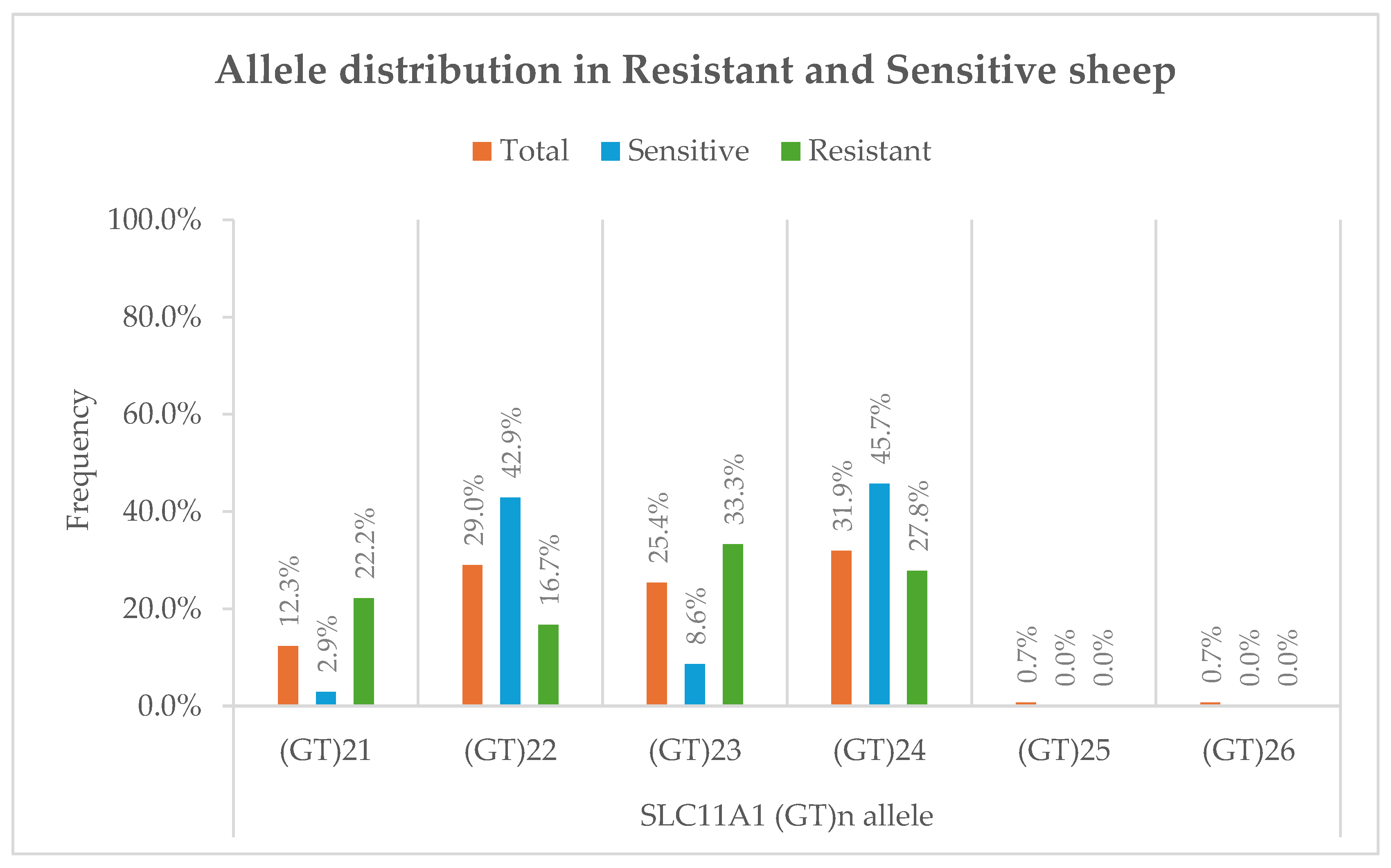
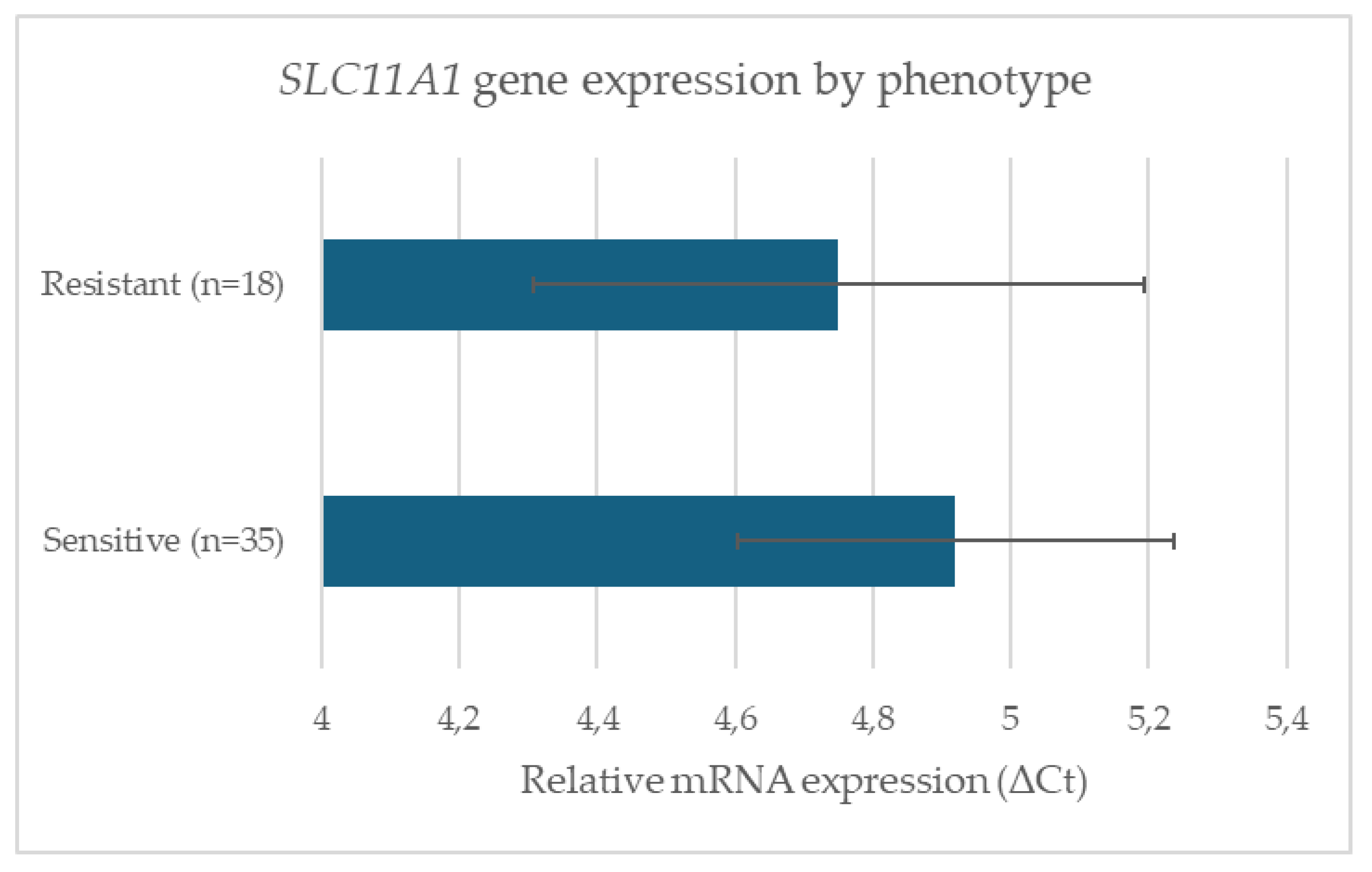
Paratuberculosis (Johne’s disease), caused by Mycobacterium avium subspecies paratuberculosis (MAP), is a chronic enteric infection that significantly impacts small ruminant health and productivity. Genetic variation in host immune genes, particularly SLC11A1, has been implicated in resistance to intracellular pathogens. The aim of this study was to investigate whether polymorphisms in the 3′UTR (GT)n microsatellite of SLC11A1 are associated with resistance or susceptibility to paratuberculosis in sheep, complementing existing SNP-based genome-wide association studies (GWAS) in cattle and goats. A total of 138 animals were genotyped, and a subset of 53 was analyzed for SLC11A1 expression. Six alleles were identified, with (GT)21 and (GT)23 significantly enriched in resistant sheep (p < 0.05), while (GT)22 and (GT)24 were more common in sensitive animals. Overall allele distribution showed a significant genotype–phenotype association (χ2 = 12.4, p = 0.006, Cramér’s V = 0.38). In contrast, no significant differences were observed in basal SLC11A1 mRNA expression between groups or across genotypes. Our findings extend previous GWAS results in sheep by providing preliminary allele-level resolution of a functional microsatellite locus. Identification of resistance-associated alleles provides a foundation for genetic selection strategies that complement vaccination and management, supporting sustainable control of paratuberculosis in sheep.

Keywords: 
;  ;  ;  ;  ;  

1. Introduction

Paratuberculosis, also known as Johne’s disease, is a chronic enteric infection caused by Mycobacterium avium subspecies paratuberculosis (MAP) that significantly affects ruminant livestock, particularly cattle, sheep, and goats [1,2]. The disease results in progressive weight loss, diarrhea, and eventual death, producing substantial economic losses for the livestock industry [3,4,5]. Prevalence studies have demonstrated wide distribution across Europe and worldwide, with evidence of endemicity in several countries [6,7]. The economic and animal welfare consequences of MAP infection highlight the urgent need for effective control strategies [8,9].
The pathogenesis of paratuberculosis is characterized by a prolonged subclinical phase during which MAP survives within host macrophages, evading immune detection and establishing chronic infection [10,11]. Pathological studies reveal a spectrum of granulomatous lesions associated with disease progression [12,13]. Experimental models in sheep and cattle have further delineated the immune response, confirming that both innate and adaptive immunity shape disease outcomes [14,15]. Vaccination has been used as a control measure, with variable success in reducing clinical signs and fecal shedding, though not completely eliminating infection [16,17,18,19]. However, vaccine-based strategies remain controversial due to diagnostic interference and incomplete protection [9,16].
Host genetics is a critical factor in determining susceptibility and resistance to MAP, as evidenced by variability among breeds and individuals [20,21,22]. Genome-wide association studies (GWAS) in cattle and sheep have identified quantitative trait loci (QTLs) associated with disease outcomes, many implicating genes involved in innate immunity and macrophage function [23,24,25,26,27,28,29]. Likewise, candidate-gene studies have associated polymorphisms in immune regulatory genes, including SLC11A1, TLR2, CARD15/NOD2 and IFNG, with paratuberculosis risk [30,31,32]. Among these, the solute carrier family 11 member 1 (SLC11A1) gene, a key regulator of macrophage antimicrobial activity, has emerged as a leading candidate across species [33,34,35,36]. SLC11A1 encodes a divalent metal transporter expressed in phagosomal membranes, controlling the intracellular availability of iron and other cations essential for bacterial replication [36,37]. Functional studies have consistently linked polymorphisms in SLC11A1 to altered susceptibility to intracellular pathogens, including M. tuberculosis, Brucella, and Salmonella [30,31,38,39,40].
In ruminants, a polymorphic microsatellite in the 3′ untranslated region (UTR) of SLC11A1 has been a major focus of research due to its potential role in post-transcriptional regulation. In goats, specific (GT)n alleles are strongly associated with resistance to MAP, and functional assays confirm these variants modulate inducible gene expression upon pathogen challenge [41,42,43,44]. Similarly, associations between 3′UTR polymorphisms and disease susceptibility have been reported in cattle and buffalo [45,46]. These findings indicate the functional significance of this locus and its potential utility as a genetic marker for resistance.
Recent sheep GWAS have identified genomic regions associated with MAP resistance and have implicated the SLC11A1 gene region among other candidates [28,29,47], however, SNP-based approaches are limited in their ability to detect microsatellite variation [48]. Therefore, although SLC11A1 is known to be important in other species, the allelic variation of its 3′UTR (GT)n microsatellite and its association with paratuberculosis outcomes remain uninvestigated in sheep, leaving a defined knowledge gap.
This preliminary study aimed to determine whether specific, functionally relevant allelic variants of the (GT)n microsatellite in the ovine SLC11A1 3′UTR are associated with resistance or susceptibility to paratuberculosis, rather than to discover novel alleles. By genotyping this locus in a well-characterized sheep population with defined infection status, we sought to provide the first preliminary allele-level evidence for its role in sheep, thereby complementing the findings from previous GWAS. This work aims to identify possible genetic markers to support breeding programs for improved disease resistance.

2. Materials and Methods

2.1. Study population and sample collection

The study population comprised 138 adult sheep (Karagouniki, Boutsika, and Chios breeds) maintained at the Agricultural University of Athens, Greece. The flock consisted predominantly of Karagouniki sheep (n = 124), with smaller numbers of Boutsika (n = 5) and Chios (n = 9) animals. The flock has been monitored for paratuberculosis since 2014 using fecal real-time qPCR and blood ELISA [49] and has not been vaccinated against the disease. The animals are housed in an area isolated from other domestic and free-ranging species. Although qPCR testing regularly detects MAP infection in the flock, only a small number of clinical cases occur annually, which are promptly removed [49].
Sample testing was conducted in compliance with ISO17025 requirements (ISO/IEC 17025:2017) in connection with ELISA, DNA isolation, and qPCR. The analysis incorporated the recommended measures of quality assurance, including use of positive and negative controls, validation of the DNA quality, detection of PCR inhibitors, and confirmation of the specificity of the amplification product using sequence analysis.
Whole blood samples were collected from the 138 adult animals of the flock and were processed for DNA isolation and sequence analysis of the 3’ UTR of the SLC11A1 gene (n=138, 1 sample/animal). Two groups consisting of total 53 individuals were formed from these animals, based on the results of the qPCR and ELISA tests conducted during the 2 years that preceded the investigation (2020-2022). These groups, referred to as resistant (R) and sensitive (S), consisted of the individuals that reacted negatively to all the ELISA and qPCR tests that were conducted (group R, n=18), and of those that reacted positively to either of these tests at least 3 times (group S, n=35). All animals classified as S group and R group belonged exclusively to the Karagouniki breed. Individuals from the Boutsika and Chios breeds did not meet the inclusion criteria for either phenotypic group, as none showed consistent diagnostic reactivity over the monitoring period.
Samples of whole blood were collected in heparinized sterile blood collection tubes from the animals of the R and S group and were transported to the laboratory within 30min after collection, where they were stored at -80oC. These samples (n=53, 1 sample/animal) were processed for RNA isolation and RT-qPCR designed for the assessment of expression variations of the SLC11A1 gene.

2.2. DNA and RNA isolation

DNA and RNA isolation was performed on whole blood (n=138) and buffy coat (n=53), respectively. In brief, isolation of DNA was conducted with Nucleospin Tissue DNA kit (Macheray-Nagel GmbH & Co. KG, Germany), whereas, with regards to the isolation of RNA, 5ml of whole blood samples were centrifuged at 600×g for 15min, within 30 min after collection. The sediment (buffy coat) was diluted in an equal volume of PBS-citrate (Sigma-Aldrich, USA) and then layered over Ficoll-Paque (Amersham Bioscienses, Sweden) solution. After centrifugation at 500×g for 40min, the mononuclear cells were collected and washed three times in PBS (Sigma-Aldrich, USA). RNA isolation was performed with the Nucleospin RNA Plus XS kit (Macheray-Nagel GmbH & Co. KG, Germany), whereas purification, with the RNA-Zol Direct Clean-Up kit (Fisher Molecular Biology, USA). All procedures were conducted according to the instructions of the respective manufacturers.
The quality and quantity of the DNA and RNA isolated was assessed for purity and integrity by submerged gel electrophoresis followed by image analysis using a Bio-Rad ChemiDoc XRS+ Molecular Imager (Bio-Rad Laboratories Inc., U.S.), and by optical density count at 260/280 nm, using a NanoDropTM 8000 spectrophotometer (Thermo Fisher Scientific Inc., U.S.). The presence of inhibitors in the DNA samples was assessed by a PCR assay targeting the housekeeping gene β-actin [50].

2.3. Sequence analysis of the ovine SLC11A1 gene

The sequence analysis of the 3′UTR of the ovine SLC11A1 gene (GeneBank: U70255) was performed on the DNA isolated from the whole blood samples (n=138). The PCR assay incorporated into the amplification of the target region was performed as previously described (Table 1). Reactions were prepared in 20 µl containing 1× KAPA SYBR Fast qPCR Master Mix (Kapa Biosystems, USA), 200 nM of each primer, 10 ng bovine serum albumin (Thermo Fisher Scientific, USA), 2 µl template DNA, and nuclease-free water. Amplicons were sequenced on both strands using the BigDye® Terminator Cycle Sequencing Kit and PRISM® 377 DNA Sequencer (Applied Biosystems, USA).

2.4. Gene expression analysis

Relative expression of SLC11A1 was quantified using the One Step SYBR® PrimeScript™ RT-qPCR Kit II (Takara Bio Inc., Japan) as previously described (Table 1). Reactions contained 1× Takara buffer, 400 nM of each primer, 0.8 µl enzyme mix, 10 ng bovine serum albumin, 2 µl RNA, and RNase-free PCR grade water to a final volume of 20μl.
The relative quantification of the SLC11A1 gene expression was performed using as reference the GAPDH gene whose amplification was conducted as previously described (Table 1). Relative expression was calculated using the 2−ΔΔCt method [53].

2.5. Statistical analysis

Statistical analyses were conducted to assess (i) differences in allele frequency distributions of the (GT)n microsatellite region of the SLC11A1 gene between resistant and sensitive phenotypic groups, and (ii) differences in relative gene expression levels of SLC11A1 mRNA.
Allele frequencies were calculated as the proportion of individuals carrying each (GT)n repeat allele in the study population and in each subgroup. Associations between allele frequencies and phenotypic group (resistant vs. sensitive) were evaluated using Fisher’s exact test for individual alleles and a chi-square test of independence for overall allele distribution. Effect sizes for contingency tests were estimated using Cramér’s V.
Gene expression data were analyzed using the 2–ΔΔCt method, with GAPDH as the reference gene. Expression data were log-transformed for normality where appropriate. A two-tailed independent samples t-test was used to compare gene expression levels between groups. To assess the effect of genotype on gene expression, a one-way analysis of variance (ANOVA) followed by Tukey’s HSD post hoc tests was applied.
Assumptions of normality and homogeneity of variances for parametric tests were tested using the Shapiro–Wilk test and Levene’s test, respectively. All analyses were performed using GraphPad Prism (v10) and IBM SPSS Statistics (v29.0). A p-value of <0.05 was considered statistically significant.

3. Results

3.1.(. GT)n repeat polymorphism frequencies

A total of 138 sheep were successfully genotyped for the (GT)n microsatellite in the 3′UTR of ovine SLC11A1. In the overall population (n=138), the (GT)24 allele was the most frequent, observed in 44 individuals (31.9%), followed by (GT)22 in 40 individuals (29.0%), (GT)23 in 35 individuals (25.4%), and (GT)21 in 17 individuals (12.3%). The rare alleles (GT)25 and (GT)26 were each found in only 1 animal each (0.7% each), (Figure 1).
For the resistant subgroup (n=18; animals repeatedly negative in diagnostic tests), (GT)23 was most common with 33.3%, followed by (GT)24 with 27.8%, (GT)21 with 22.2% and (GT)22 with 16.7%. In the sensitive subgroup (n=35; animals repeatedly positive in diagnostic tests), (GT)24 was most frequent with 45.7%, followed by (GT)22 with 42.9%, (GT)23 with 8.6%, and (GT)21 with 2.9%. The study population consisted predominantly of Karagouniki sheep (n = 124), with smaller numbers of Boutsika (n = 5) and Chios (n = 9). Diagnostic testing indicated that all animals classified as resistant (n = 18) and sensitive (n = 35) belonged exclusively to the Karagouniki breed. The Boutsika and Chios individuals did not show consistent positive or negative reactivity during the two-year monitoring period. Consequently, statistical comparisons among breeds were not possible, and all subsequent genotype–phenotype analyses were based on the Karagouniki subset. This composition reflects the structure of the monitored flock and supports the representativeness of the dataset for preliminary association testing [21,22]. Moreover, the number of animals classified as resistant (n = 18) was considerably smaller than that of sensitive (n = 35) individuals. This imbalance reflects the natural epidemiology of paratuberculosis, in which persistently negative animals are rare even in well-managed flocks due to the chronic, subclinical progression of infection [7,11,15,22]. Inclusion in the resistant group required consecutive negative results in both ELISA and qPCR testing over a two-year surveillance period, a stringent criterion that limited the number of qualifying animals. Although the small size of the resistant group reduces statistical power, the analysis still revealed significant genotype–phenotype associations (χ2 = 12.4, p = 0.006) and strong effect sizes for certain alleles, suggesting that the observed relationships are biologically meaningful.

3.2. Genotype–Phenotype association

Despite the limited number of resistant animals (n = 18) described above, statistical analyses were performed to assess whether the observed microsatellite allelic variation in the SLC11A1 3′UTR was associated with MAP-resistance phenotype. Fisher’s exact test revealed significant enrichment of the (GT)21 allele in resistant sheep (22.2%) compared to sensitive sheep (2.9%) (p = 0.040, OR = 9.5, 95% CI: 1.00–89.5). Similarly, (GT)23 was more frequent in resistant animals (33.3%) than in sensitive ones (8.6%) (p = 0.048, OR = 5.3, 95% CI: 1.00–28.7). Conversely, the (GT)24 and (GT)22 alleles were more prevalent among sensitive sheep (45.7% and 42.9%, respectively) than resistant sheep (27.8% and 16.7%, respectively), though these differences did not reach statistical significance (p = 0.217 and p = 0.063, respectively).
When allele distributions were analyzed overall, a significant association was observed between genotype and phenotype (χ2 = 12.4, df = 3, p = 0.006), with Cramér’s V = 0.38, indicating a moderate effect size.

3.3. SLC11A1 gene expression

Relative mRNA levels of SLC11A1 were measured in 53 sheep of Karagouniki breed (18 resistant, 35 sensitive). The 2-ΔΔCt method was used, normalized to GAPDH. A t-test comparing sensitive and resistant groups revealed no significant difference [mean ΔCt_S = 4.92, mean ΔCt_R = 4.75; t(51) = 0.63, p = 0.531; 95% CI: –0.39 to 0.73; Cohen’s d = 0.17], (Figure 2). One-way ANOVA of expression levels across all genotypes also showed no statistically significant variation [F(2,50) = 1.42, p = 0.25]. Post hoc pairwise comparisons using Tukey’s HSD confirmed the absence of significant differences.
In summary, analysis of the SLC11A1 3′UTR (GT)n microsatellite in 138 sheep revealed a significant association between specific alleles and the MAP-resistance phenotype. The alleles (GT)21 and (GT)23 were enriched among resistant animals, whereas (GT)22 and (GT)24 were more common in sensitive individuals. Despite the small number of resistant animals and the absence of breed-level comparisons, the statistical strength of the association (χ2 = 12.4, p = 0.006; Cramér’s V = 0.38) suggests biological relevance. No significant differences were observed in basal SLC11A1 mRNA expression between phenotypic groups, indicating that microsatellite variation may influence inducible rather than constitutive gene regulation. Collectively, these results provide preliminary allele-level evidence supporting a role for SLC11A1 microsatellite polymorphisms in resistance to MAP infection in sheep.

4. Discussion

Paratuberculosis imposes significant production losses in sheep flocks, including reduced productivity and increased mortality [3,22]. Serological surveys continue to highlight widespread infection [7], and economic reviews emphasize its financial impact [4]. Genetic selection for resistance, as suggested by our results, could reduce prevalence and losses, complementing vaccination and management measures [8,9].
Advances in biomarker discovery and molecular diagnostics may improve early detection and monitoring of MAP infection [54,55]. Our genetic findings contribute to these approaches by providing early evidence that specific SLC11A1 alleles are associated with disease resistance in sheep. The present study provides additional insights into the role of SLC11A1 3′UTR (GT)n microsatellite polymorphisms in resistance to paratuberculosis in sheep. We identified significant enrichment of the (GT)21 and (GT)23 alleles in resistant animals, and higher prevalence of (GT)22 and (GT)24 in sensitive animals, though not always reaching statistical significance. These findings contribute to a growing body of evidence implicating SLC11A1 as a candidate gene modulating susceptibility to MAP and other intracellular pathogens in livestock and humans [33,36,38]. To place our findings in context, Table 2 summarizes reported associations between SLC11A1 polymorphisms and resistance/susceptibility across species.
Evidence from goats has consistently demonstrated an association between SLC11A1 polymorphisms and resistance to MAP. Korou et al. (2010) reported that shorter alleles at the 3′UTR microsatellite were enriched in resistant goats [43], findings later supported by functional analyses showing altered inducible gene expression [41,42]. Similarly, Abraham et al. (2017) identified specific SLC11A1 alleles associated with reduced incidence of paratuberculosis in goats [44]. Our observation that the alleles (GT)21 and (GT)23 are linked to resistance in sheep aligns with earlier exploratory evidence of genetic influences on Johne’s disease in sheep [21].
In cattle, SLC11A1 polymorphisms have been associated with MAP susceptibility, though results vary across populations. Pinedo et al. (2009) found associations between SLC11A1, TLR4, and IFNG variants with MAP infection [56], while Ruiz-Larrañaga et al. (2010) identified SNPs in SLC11A1 correlated with infection risk [46]. More recent GWAS studies also highlight SLC11A1 and other innate immunity genes as candidates [25,26,27,57]. Functional evidence further supports the role of SLC11A1 in macrophage control of MAP, as macrophages with certain genotypes show differential ability to limit bacterial replication [62]. Our results in sheep are consistent with these observations, however the absence of significant difference in SLC11A1 expression between groups suggesting that the microsatellite may influence gene inducibility or protein function rather than baseline transcription.
Other livestock species also reinforce the role of SLC11A1. In buffalo, 3′UTR polymorphisms influenced MCP1 mRNA expression in response to Brucella challenge [45]. In pigs, SLC11A1 polymorphisms were linked to immune traits [40,60] and disease susceptibility [31]. In goats, additional work showed effects of 3′UTR alleles on gene function during MAP infection [41]. These cross-species findings highlight that, although SLC11A1 mechanisms are generally conserved, the specific alleles conferring resistance may vary, necessitating species-specific investigations.
Our findings should also be interpreted considering previous GWAS in sheep, which identified genomic regions associated with MAP resistance or antibody response and highlighted the contribution of SLC11A1 among other innate immune genes [28,29,47]. Although GWAS approaches are powerful for detecting broad genomic regions, they often fail to capture functional variation at microsatellite loci [48]. By directly characterizing allelic diversity at the (GT)n microsatellite in the 3′UTR of SLC11A1, our study complements GWAS findings and provides preliminary evidence for the role of this functional polymorphism in shaping resistance to MAP in sheep.
Our results must be considered in the broader framework of host genetics in MAP infection. GWAS studies in cattle and sheep have consistently identified loci associated with paratuberculosis, involving genes related to DNA repair, stress response, and innate immunity [23,24,25,26,28,29]. In cattle, associations with CARD15/NOD2 [63,64], TLR2 [65], and other innate receptors further emphasize polygenic influences [59,66]. Our findings add SLC11A1 3′UTR polymorphisms to this complex genetic landscape in sheep, consistent with reviews emphasizing multifactorial control of MAP resistance [67,68]. Notably, while we found significant genotype–phenotype associations, effect sizes were moderate, suggesting that SLC11A1 contributes to but does not fully explain resistance.
One of the important aspects of our findings was the absence of significant differences in SLC11A1 mRNA levels between resistant and sensitive groups. This observation contrasts with some functional studies in goats and buffalo, where 3′UTR variants affected gene expression [41,45]. However, our results are consistent with the notion that basal expression in blood cells may not capture functional differences that manifest only upon pathogen challenge [11,42]. Indeed, macrophage assays in cattle demonstrate that genetic differences may influence intracellular control of MAP rather than constitutive transcription [62]. This limitation suggests that the SLC11A1 microsatellite in sheep may influence induced expression or protein function rather than constitutive transcription, therefore future research is important to include functional assays under MAP challenge and protein-level expression studies.
It should also be noted that our study population study population comprised three sheep breeds (Karagouniki, n=124; Boutsika, n=5; Chios, n=9), as breed-related genetic variation has been reported to influence MAP susceptibility in sheep [20,21,22]. However, as only Karagouniki sheep met the strict diagnostic criteria for inclusion in the sensitive or resistant phenotypic groups, we could not perform a breed-specific analysis. The low numbers of Boutsika and Chios sheep simply reflect their limited presence in the monitored flock. Nevertheless, this information has been retained for completeness, and future investigations including balanced multi-breed cohorts will be essential to determine whether the associations observed in Karagouniki sheep are consistent across other genetic backgrounds.
Another limitation was the small cohort of resistant animals (n=18). This is characteristic of paratuberculosis, as completely resistant individuals are uncommon even in infected flocks [7,11,15,22]. Our classification required consistent negative results over two years, making this a stringent and naturally small group. Similar challenges in identifying persistently negative animals have been reported in longitudinal and experimental infection studies [14,15,21]. Although the small sample size limits statistical power, the strong magnitude of certain associations (e.g., OR = 9.5 for (GT)21) implies a real biological effect. Future research with larger populations is needed to validate these findings [20,21,22,67].
Finally, while this study focused on SLC11A1, other genes such as TLR2, CARD15/NOD2, and IFNG have also been associated with MAP resistance in cattle and sheep [56,59,63,65,66,69,70]. Integrating SLC11A1 with other candidate genes such as TLR2, CARD15/NOD2, and IFNG will be essential to capture the full genetic landscape of resistance [59,66,69,70]. A more holistic understanding of genetic resistance will require integrating multiple loci into genomic selection strategies.
Evidence from tuberculosis research provides strong parallels. Human studies consistently link SLC11A1 polymorphisms to tuberculosis risk [38,39], and similar findings exist in cattle [32,71,72,73] and goats [30]. Experimental infection models also demonstrate that SLC11A1 influences macrophage activation and pathogen control [33,34,35].
The evolutionary conservation of SLC11A1 highlights its importance in resistance to intracellular pathogens [33,74]. Comparative studies across species demonstrate both shared mechanisms and population-specific effects [75,76]. For example, in goats, cattle, pigs, and humans, SLC11A1 variants are associated with disease outcomes, though the specific alleles differ [31,38,60,72]. Our identification of (GT)21 and (GT)23 as resistance alleles in sheep fits within this comparative framework, suggesting conserved mechanisms with species-specific variations.
Taken together, our findings underscore the potential of integrating genetic selection into paratuberculosis control. While vaccination provides partial protection [16,17], and management can reduce spread [8], genetic resistance offers a sustainable long-term strategy. The identification of resistant alleles in sheep aligns with broader efforts in cattle and goats to leverage genetic variation for disease control [28,29,77]. However, practical application requires further validation and consideration of breed-specific differences, diagnostic challenges, and integration with other control measures [9,11,70].

5. Conclusions

In conclusion, this study provides early evidence that polymorphisms in the 3′UTR (GT)n microsatellite of SLC11A1 are associated with resistance and susceptibility to paratuberculosis in sheep. Specifically, the (GT)21 and (GT)23 alleles were enriched in resistant animals, while (GT)22 and (GT)24 were more common in sensitive animals. Our findings extend previous GWAS in sheep [28,29,47] by offering preliminary allele-level resolution at a functional microsatellite locus that is not captured by SNP arrays. They are also consistent with studies in cattle, goats, and buffalo [41,42,43,44,45,46,56,57] supporting the conserved role of SLC11A1 in host defense across ruminants.
We acknowledge the limitations of our study, including the relatively small number of resistant sheep and the inability to perform breed-stratified analyses. Nevertheless, the strength of the observed associations suggests biological relevance. Future studies involving larger and more balanced cohorts, functional assays under pathogen challenge, and integration of additional candidate genes including TLR2, CARD15/NOD2, and IFNG [56,59,63,65,66,69,70] will be essential to confirm and extend these findings. Altogether, our results highlight that SLC11A1 microsatellite polymorphisms may contribute to the genetic architecture of resistance to paratuberculosis in sheep and could eventually serve as markers to support breeding programs aimed at sustainable disease control.

Author Contributions

Conceptualization, J.I.; methodology, A.M. and A.K.P.; software, A.M.; validation, A.M.; formal analysis, A.M. and A.K.P.; investigation, A.M.; resources, J.I.; data curation, A.M.; writing—original draft preparation, A.M., A.K.P. and J.I.; writing—review and editing, A.M. and J.I.; visualization, A.M.; supervision, J.I.; project administration, J.I. All authors have read and agreed to the published version of the manuscript.

Funding

This research received no external funding.

Institutional Review Board Statement

Protocol No. 77/03.09.2025 of the Research Ethics Committee (REC) of the Agricultural University of Athens.

Informed Consent Statement

Not applicable.

Data Availability Statement

The original contributions presented in this study are included in the article. Further inquiries can be directed to the corresponding author(s).

Conflicts of Interest

The authors declare no conflicts of interest.

References

  1. Harris, N. B., & Barletta, R. G. (2001). Mycobacterium avium subsp. paratuberculosis in Veterinary Medicine. Clinical Microbiology Reviews, 14(3), 489–512.
  2. Clarke, C. J. (1997). The pathology and pathogenesis of paratuberculosis in ruminants and other species. Journal of Comparative Pathology, 116(3), 217–261. [CrossRef]
  3. Bush, R. D., Windsor, P. A., & Toribio, J. A. L. M. L. (2006). Losses of adult sheep due to ovine Johne’s disease in 12 infected flocks over a 3-year period. Australian Veterinary Journal, 84(7), 246–253. [CrossRef]
  4. Hasonova, L., & Pavlik, I. (2006). Economic impact of paratuberculosis in dairy cattle herds: A review. Veterinarni Medicina, 51(5), 193–211. [CrossRef]
  5. Raizman, E. A., Fetrow, J., Wells, S. J., Godden, S. M., Oakes, M. J., & Vazquez, G. (2007). The association between Mycobacterium avium subsp. paratuberculosis fecal shedding or clinical Johne’s disease and lactation performance on two Minnesota, USA dairy farms. Preventive Veterinary Medicine, 78(3–4), 179–195. [CrossRef]
  6. Nielsen, S. S., & Toft, N. (2009). A review of prevalences of paratuberculosis in farmed animals in Europe. Preventive Veterinary Medicine, 88(1), 1–14. [CrossRef]
  7. di Marco Lo Presti, V., Ippolito, D., Migliore, S., Tolone, M., Mignacca, S. A., Marino, A. M. F., Amato, B., Calogero, R., Vitale, M., Vicari, D., Ciarello, F. P., & Fiasconaro, M. (2024). Large-scale serological survey on Mycobacterium avium subsp. paratuberculosis infection in sheep and goat herds in Sicily, Southern Italy. Frontiers in Veterinary Science, 11, 1334036. [CrossRef]
  8. Gautam, M., Ridler, A., Wilson, P. R., & Heuer, C. (2018). Control of clinical paratuberculosis in New Zealand pastoral livestock. New Zealand Veterinary Journal, 66(1), 1–8. [CrossRef]
  9. Windsor, P., & Whittington, R. (2020). Ovine Paratuberculosis Control in Australia Revisited. Animals : An Open Access Journal from MDPI, 10(9), 1–8. [CrossRef]
  10. Arsenault, R. J., Maattanen, P., Daigle, J., Potter, A., Griebel, P., & Napper, S. (2014). From mouth to macrophage: Mechanisms of innate immune subversion by Mycobacterium avium subsp. Paratuberculosis. Veterinary Research, 45(1). [CrossRef]
  11. Whittington, R. J., Begg, D. J., de Silva, K., Plain, K. M., & Purdie, A. C. (2012). Comparative immunological and microbiological aspects of paratuberculosis as a model mycobacterial infection. Veterinary Immunology and Immunopathology, 148(1–2), 29–47. [CrossRef]
  12. González, J., Geijo, M. v., García-Pariente, C., Verna, A., Corpa, J. M., Reyes, L. E., Ferreras, M. C., Juste, R. A., García Marín, J. F., & Pérez, V. (2005). Histopathological classification of lesions associated with natural paratuberculosis infection in cattle. Journal of Comparative Pathology, 133(2–3), 184–196. [CrossRef]
  13. Smith, S. L., West, D. M., Wilson, P. R., de Lisle, G. W., Collett, M. G., Heuer, C., & Chambers, J. P. (2013). The prevalence of disseminated Mycobacterium avium subsp. paratuberculosis infection in tissues of healthy ewes from a New Zealand farm with Johne’s disease present. New Zealand Veterinary Journal, 61(1), 41–44. [CrossRef]
  14. Stewart, D. J., Vaughan, J. A., Stiles, P. L., Noske, P. J., Tizard, M. L. V., Prowse, S. J., Michalski, W. P., Butler, K. L., & Jones, S. L. (2004). A long-term study in Merino sheep experimentally infected with Mycobacterium avium subsp. paratuberculosis: Clinical disease, faecal culture and immunological studies. Veterinary Microbiology, 104(3–4), 165–178. [CrossRef]
  15. Begg, D. J., & Whittington, R. J. (2008). Experimental animal infection models for Johne’s disease, an infectious enteropathy caused by Mycobacterium avium subsp. paratuberculosis. Veterinary Journal, 176(2), 129–145. [CrossRef]
  16. Begg, D. J., & Griffin, J. F. T. (2005). Vaccination of sheep against M. paratuberculosis: Immune parameters and protective efficacy. Vaccine, 23(42), 4999–5008. [CrossRef]
  17. Gwozdz, J. M., Thompson, K. G., Manktelow, B. W., Murray, A., & West, D. M. (2000). Vaccination against paratuberculosis of lambs already infected experimentally with Mycobacterium avium subspecies paratuberculosis. Australian Veterinary Journal, 78(8), 560–566. [CrossRef]
  18. Singh, S. v., Singh, P. K., Singh, A. v., Sohal, J. S., Gupta, V. K., & Vihan, V. S. (2007). Comparative efficacy of an indigenous “inactivated vaccine” using highly pathogenic field strain of Mycobacterium avium subspecies paratuberculosis “Bison type” with a commercial vaccine for the control of Capri-paratuberculosis in India. Vaccine, 25(41), 7102–7110. [CrossRef]
  19. Dhand, N. K., Johnson, W. O., Eppleston, J., Whittington, R. J., & Windsor, P. A. (2013). Comparison of pre- and post-vaccination ovine Johne’s disease prevalence using a Bayesian approach. Preventive Veterinary Medicine, 111(1–2), 81–91. [CrossRef]
  20. Koets, A. P., Adugna, G., Janss, L. L. G., van Weering, H. J., Kalis, C. H. J., Wentink, G. H., Rutten, V. P. M. G., & Schukken, Y. H. (2000). Genetic variation of susceptibility to Mycobacterium avium subsp. paratuberculosis infection in dairy cattle. Journal of Dairy Science, 83(11), 2702–2708. [CrossRef]
  21. Reddacliff, L. A., Beh, K., McGregor, H., & Whittington, R. J. (2005). A preliminary study of possible genetic influences on the susceptibility of sheep to Johne’s disease. Australian Veterinary Journal, 83(7), 435–441. [CrossRef]
  22. Lugton, I. W. (2004). Cross-sectional study of risk factors for the clinical expression of ovine Johne’s disease on New South Wales farms. Australian Veterinary Journal, 82(6), 355–365. [CrossRef]
  23. Bermingham, M. L., Bishop, S. C., Woolliams, J. A., Pong-Wong, R., Allen, A. R., McBride, S. H., Ryder, J. J., Wright, D. M., Skuce, R. A., McDowell, S. W., & Glass, E. J. (2014). Genome-wide association study identifies novel loci associated with resistance to bovine tuberculosis. Heredity, 112(5), 543–551. [CrossRef]
  24. Gao, Y., Jiang, J., Yang, S., Cao, J., Han, B., Wang, Y., Zhang, Y., Yu, Y., Zhang, S., Zhang, Q., Fang, L., Cantrell, B., & Sun, D. (2018). Genome-wide association study of Mycobacterium avium subspecies Paratuberculosis infection in Chinese Holstein. BMC Genomics, 19(1). [CrossRef]
  25. Canive, M., González-Recio, O., Fernández, A., Vázquez, P., Badia-Bringué, G., Lavín, J. L., Garrido, J. M., Juste, R. A., & Alonso-Hearn, M. (2021). Identification of loci associated with susceptibility to Mycobacterium avium subsp. paratuberculosis infection in Holstein cattle using combinations of diagnostic tests and imputed whole-genome sequence data. PLoS ONE, 16(8 August). [CrossRef]
  26. Canive, M., Badia-Bringué, G., Vázquez, P., Garrido, J. M., Juste, R. A., Fernandez, A., González-Recio, O., & Alonso-Hearn, M. (2022). A Genome-Wide Association Study for Tolerance to Paratuberculosis Identifies Candidate Genes Involved in DNA Packaging, DNA Damage Repair, Innate Immunity, and Pathogen Persistence. Frontiers in Immunology, 13. [CrossRef]
  27. Alonso-Hearn, M., Badia-Bringué, G., & Canive, M. (2022). Genome-wide association studies for the identification of cattle susceptible and resilient to paratuberculosis. Frontiers in Veterinary Science, 9, 935133. [CrossRef]
  28. Usai, M. G., Casu, S., Sechi, T., Salaris, S. L., Miari, S., Mulas, G., Cancedda, M. G., Ligios, C., & Carta, A. (2024). Advances in understanding the genetic architecture of antibody response to paratuberculosis in sheep by heritability estimate and LDLA mapping analyses and investigation of candidate regions using sequence-based data. Genetics Selection Evolution, 56(1). [CrossRef]
  29. Moioli, B., D’Andrea, S., de Grossi, L., Sezzi, E., de Sanctis, B., Catillo, G., Steri, R., Valentini, A., & Pilla, F. (2015). Genomic scan for identifying candidate genes for paratuberculosis resistance in sheep. Animal Production Science, 56(7), 1046–1055. [CrossRef]
  30. Iacoboni, P. A., Hasenauer, F. C., Caffaro, M. E., Gaido, A., Rossetto, C., Neumann, R. D., Salatin, A., Bertoni, E., Poli, M. A., & Rossetti, C. A. (2014). Polymorphisms at the 3′ untranslated region of SLC11A1 gene are associated with protection to Brucella infection in goats. Veterinary Immunology and Immunopathology, 160(3–4), 230–234. [CrossRef]
  31. Suwannawong, N., Thumarat, U., & Phongphanich, P. (2022). Association of natural resistance-associated macrophage protein 1 polymorphisms with Salmonella fecal shedding and hematological traits in pigs. Veterinary World, 15(11), 2738–2743. [CrossRef]
  32. Allen, A. R., Minozzi, G., Glass, E. J., Skuce, R. A., McDowell, S. W. J., Woolliams, J. A., & Bishop, S. C. (2010). Bovine tuberculosis: The genetic basis of host susceptibility. Proceedings of the Royal Society B: Biological Sciences, 277(1695), 2737–2745. [CrossRef]
  33. Blackwell, J. M., & Searle, S. (1999). Genetic regulation of macrophage activation: Understanding the function of Nramp1 (=Ity/Lsh/Bcg). Immunology Letters, 65(1–2), 73–80. [CrossRef]
  34. Buschman, E., Vidal, S., & Skamene, E. (1997). Nonspecific resistance to Mycobacteria: the role of the Nramp1 gene. Behring Institute Mitteilungen, 99, 51–57.
  35. Vidal, S., Gros, P., & Skamene, E. (1995). Natural resistance to infection with intracellular parasites: Molecular genetics identifies Nramp1 as the Bcg/Ity/Lsh locus. Journal of Leukocyte Biology, 58(4), 382–390. [CrossRef]
  36. Wyllie, S., Seu, P., & Goss, J. A. (2002). The natural resistance-associated macrophage protein 1 Slc11a1 (formerly Nramp1) and iron metabolism in macrophages. Microbes and Infection, 4(3), 351–359. [CrossRef]
  37. Thomas, N., & Joseph, S. (2012). Role of SLC11A1 gene in disease resistance. Biotechnology in Animal Husbandry, 28(1), 99–106. [CrossRef]
  38. Awomoyi, A. A., Marchant, A., Howson, J. M. M., McAdam, K. P. W. J., Blackwell, J. M., & Newport, M. J. (2002). Interleukin-10, polymorphism in SLC11A1 (formerly NRAMP1), and susceptibility to tuberculosis. Journal of Infectious Diseases, 186(12), 1808–1814. [CrossRef]
  39. Meilang, Q., Zhang, Y., Zhang, J., Zhao, Y., Tian, C., Huang, J., & Fan, H. (2012). Polymorphisms in the SLC11A1 gene and tuberculosis risk: A meta-analysis update. International Journal of Tuberculosis and Lung Disease, 16(4), 437–446. [CrossRef]
  40. Ding, X., Zhang, X., Yang, Y., Ding, Y., Xue, W., Meng, Y., Zhu, W., & Yin, Z. (2014). Polymorphism, Expression of Natural Resistance-associated Macrophage Protein 1 Encoding Gene (NRAMP1) and Its Association with Immune Traits in Pigs. Asian-Australasian Journal of Animal Sciences, 27(8), 1189–1195. [CrossRef]
  41. Taka, S., Gazouli, M., Sotirakoglou, K., Liandris, E., Andreadou, M., Triantaphyllopoulos, K., & Ikonomopoulos, J. (2015). Functional analysis of 3’UTR polymorphisms in the caprine SLC11A1 gene and its association with the Mycobacterium avium subsp. paratuberculosis infection. Veterinary Immunology and Immunopathology, 167(1–2), 75–79. [CrossRef]
  42. Taka, S., Liandris, E., Gazouli, M., Sotirakoglou, K., Theodoropoulos, G., Bountouri, M., Andreadou, M., & Ikonomopoulos, J. (2013). In vitro expression of the SLC11A1 gene in goat monocyte-derived macrophages challenged with Mycobacterium avium subsp paratuberculosis. Infection, Genetics and Evolution, 17, 8–15. [CrossRef]
  43. Korou, L. M., Liandris, E., Gazouli, M., & Ikonomopoulos, J. (2010). Investigation of the association of the SLC11A1 gene with resistance/sensitivity of goats (Capra hircus) to paratuberculosis. Veterinary Microbiology, 144(3–4), 353–358. [CrossRef]
  44. Abraham, A., Naicy, T., Raghavan, K. C., Siju, J., & Aravindakshan, T. (2017). Evaluation of the association of SLC11A1 gene polymorphism with incidence of paratuberculosis in goats. Journal of Genetics, 96(4), 641–646. [CrossRef]
  45. Balasubramaniam, S., Kumar, S., Sharma, A., & Mitra, A. (2013). Microsatellite (GT)n polymorphism at 3′UTR of SLC11A1 influences the expression of brucella LPS induced MCP1 mRNA in buffalo peripheral blood mononuclear cells. Veterinary Immunology and Immunopathology, 152(3–4), 295–302. [CrossRef]
  46. Ruiz-Larrañaga, O., Garrido, J. M., Manzano, C., Iriondo, M., Molina, E., Gil, A., Koets, A. P., Rutten, V. P. M. G., Juste, R. A., & Estonba, A. (2010). Identification of single nucleotide polymorphisms in the bovine solute carrier family 11 member 1 (SLC11A1) gene and their association with infection by Mycobacterium avium subspecies paratuberculosis. Journal of Dairy Science, 93(4), 1713–1721. [CrossRef]
  47. Kravitz, A., Liao, M., Morota, G., Tyler, R., Cockrum, R., Manohar, B. M., Ronald, B. S. M., Collins, M. T., & Sriranganathan, N. (2024). Retrospective Single Nucleotide Polymorphism Analysis of Host Resistance and Susceptibility to Ovine Johne’s Disease Using Restored FFPE DNA. International Journal of Molecular Sciences, 25(14). [CrossRef]
  48. Tam, V., Patel, N., Turcotte, M., Bossé, Y., Paré, G., & Meyre, D. (2019). Benefits and limitations of genome-wide association studies. Nature Reviews Genetics, 20(8), 467–484. [CrossRef]
  49. Mataragka, A., Sotirakoglou, K., Gazouli, M., Triantaphyllopoulos, K. A., & Ikonomopoulos, J. (2019). Parturition affects test-positivity in sheep with subclinical paratuberculosis; investigation following a preliminary analysis. Journal of King Saud University – Science, 31(4), 1399–1403. [CrossRef]
  50. Dowling, R. J. O., & Bienzle, D. (2005). Gene-expression changes induced by Feline immunodeficiency virus infection differ in epithelial cells and lymphocytes. Journal of General Virology, 86(8), 2239–2248. [CrossRef]
  51. Kim, S. G., Kim, E. H., Lafferty, C. J., Miller, L. J., Koo, H. J., Stehman, S. M., & Shin, S. J. (2004). Use of conventional and real-time polymerase chain reaction for confirmation of Mycobacterium avium subsp. paratuberculosis in a broth-based culture system ESP II. Journal of Veterinary Diagnostic Investigation, 16(5), 448–453. [CrossRef]
  52. Taylor, D. L., Zhong, L., Begg, D. J., de Silva, K., & Whittington, R. J. (2008). Toll-like receptor genes are differentially expressed at the sites of infection during the progression of Johne’s disease in outbred sheep. Veterinary Immunology and Immunopathology, 124(1–2), 132–151. [CrossRef]
  53. Livak, K. J., & Schmittgen, T. D. (2001). Analysis of Relative Gene Expression Data Using Real-Time Quantitative PCR and the 2−ΔΔCT Method. Methods, 25(4), 402–408. [CrossRef]
  54. Corneli, S., di Paolo, A., Vitale, N., Torricelli, M., Petrucci, L., Sebastiani, C., Ciullo, M., Curcio, L., Biagetti, M., Papa, P., Costarelli, S., Cagiola, M., Dondo, A., & Mazzone, P. (2021). Early Detection of Mycobacterium avium subsp. paratuberculosis Infected Cattle: Use of Experimental Johnins and Innovative Interferon-Gamma Test Interpretative Criteria. Frontiers in Veterinary Science, 8, 638890. [CrossRef]
  55. Vázquez, C. B., Alonso-Hearn, M., Juste, R. A., Canive, M., Iglesias, T., Iglesias, N., Amado, J., Vicente, F., Balseiro, A., & Casais, R. (2020). Detection of latent forms of Mycobacterium avium subsp. paratuberculosis infection using host biomarker-based ELISAs greatly improves paratuberculosis diagnostic sensitivity. PLoS ONE, 15(9 September 2020). [CrossRef]
  56. Pinedo, P. J., Buergelt, C. D., Donovan, G. A., Melendez, P., Morel, L., Wu, R., Langaee, T. Y., & Rae, D. O. (2009). Candidate gene polymorphisms (BoIFNG, TLR4, SLC11A1) as risk factors for paratuberculosis infection in cattle. Preventive Veterinary Medicine, 91(2–4), 189–196. [CrossRef]
  57. Canive, M., Casais, R., Jimenez, J. A., Blanco-Vazquez, C., Amado, J., Garrido, J. M., Juste, R. A., & Alonso-Hearn, M. (2020). Correlations between single nucleotide polymorphisms in bovine CD209, SLC11A1, SP110 and TLR2 genes and estimated breeding values for several traits in Spanish Holstein cattle. Heliyon, 6(6), e04254. [CrossRef]
  58. Okuni, J. B., Afayoa, M., & Ojok, L. (2021). Survey of Candidate Single-Nucleotide Polymorphisms in SLC11A1, TLR4, NOD2, PGLYRP1, and IFNγ in Ankole Longhorn Cattle in Central Region of Uganda to Determine Their Role in Mycobacterium avium Subspecies paratuberculosis Infection Outcome. Frontiers in Veterinary Science, 8, 614518. [CrossRef]
  59. Gopi, B., Vir Singh, R., Kumar, S., Kumar, S., Chauhan, A., Sonwane, A., Kumar, A., Bharati, J., & Vir Singh, S. (2022). Effect of selected single nucleotide polymorphisms in SLC11A1, ANKRA2, IFNG and PGLYRP1 genes on host susceptibility to Mycobacterium avium subspecies paratuberculosis infection in Indian cattle. Veterinary Research Communications, 46(1), 209–221. [CrossRef]
  60. Wu, H., Cheng, D., & Wang, L. (2008). Association of polymorphisms of Nramp1 gene with immune function and production performance of large white pig. Journal of Genetics and Genomics, 35(2), 91–95. [CrossRef]
  61. Sechi, L. A., & Dow, C. T. (2015). Mycobacterium avium ss. paratuberculosis Zoonosis - The Hundred Year War - Beyond Crohn’s Disease. Frontiers in Immunology, 6, 96. [CrossRef]
  62. Badia-Bringué, G., Canive, M., & Alonso-Hearn, M. (2023). Control of Mycobacterium avium subsp. paratuberculosis load within infected bovine monocyte-derived macrophages is associated with host genetics. Frontiers in Immunology, 14. [CrossRef]
  63. Pinedo, P. J., Buergelt, C. D., Donovan, G. A., Melendez, P., Morel, L., Wu, R., Langaee, T. Y., & Rae, D. O. (2009). Association between CARD15/NOD2 gene polymorphisms and paratuberculosis infection in cattle. Veterinary Microbiology, 134(3–4), 346–352. [CrossRef]
  64. Wang, Y., Wang, S., Liu, T., Tu, W., Li, W., Dong, G., Xu, C., Qin, B., Liu, K., Yang, J., Chai, J., Shi, X., & Zhang, Y. (2015). CARD15 gene polymorphisms are associated with tuberculosis susceptibility in Chinese Holstein cows. PLoS ONE, 10(8). [CrossRef]
  65. Yaman, Y., Aymaz, R., Keleş, M., Bay, V., Ün, C., & Heaton, M. P. (2021). Association of TLR2 haplotypes encoding Q650 with reduced susceptibility to ovine Johne’s disease in Turkish sheep. Scientific Reports, 11(1), 1–10. [CrossRef]
  66. Juste, R. A., Vazquez, P., Ruiz-Larrañaga, O., Iriondo, M., Manzano, C., Agirre, M., Estonba, A., Geijo, M. v, Molina, E., Sevilla, I. A., Alonso-Hearn, M., Gomez, N., Perez, V., Cortes, A., & Garrido, J. M. (2018). Association between combinations of genetic polymorphisms and epidemiopathogenic forms of bovine paratuberculosis. Heliyon, 4(2), e00535. [CrossRef]
  67. Purdie, A. C., Plain, K. M., Begg, D. J., de Silva, K., & Whittington, R. J. (2011). Candidate gene and genome-wide association studies of Mycobacterium avium subsp. paratuberculosis infection in cattle and sheep: A review. Comparative Immunology, Microbiology and Infectious Diseases, 34(3), 197–208. [CrossRef]
  68. Kravitz, A., Pelzer, K., & Sriranganathan, N. (2021). The Paratuberculosis Paradigm Examined: A Review of Host Genetic Resistance and Innate Immune Fitness in Mycobacterium avium subsp. Paratuberculosis Infection. Frontiers in Veterinary Science, 8. [CrossRef]
  69. Sadana, T., Vir Singh, R., Vir Singh, S., Kumar Saxena, V., Sharma, D., Kumar Singh, P., Kumar, N., Gupta, S., Kumar Chaubey, K., Jayaraman, S., Tiwari, R., Dhama, K., Kumar Bhatia, A., Singh Sohal, J., & Pradesh Pandit Deen Dayal Upadhayay Pashu Chikitsa Vigyan Vishwa Vidyalaya Evam Go-Anusandhan Sansthan, U. (2015). Single nucleotide polymorphism of SLC11A1, CARD15, IFNG and TLR2 genes and their association with Mycobacterium avium subspecies paratuberculosis infection in native Indian cattle population. Indian Journal of Biotechnology, 14(4), 469–475.
  70. Dukkipati, V. S. R., Blair, H. T., Garrick, D. J., Lopez-Villalobos, N., Whittington, R. J., Reddacliff, L. A., Eppleston, J., Windsor, P., & Murray, A. (2010). Association of microsatellite polymorphisms with immune responses to a killed Mycobacterium avium subsp. Paratuberculosis vaccine in Merino sheep. New Zealand Veterinary Journal, 58(5), 237–245. [CrossRef]
  71. Kadarmideen, H. N., Ali, A. A., Thomson, P. C., Müller, B., & Zinsstag, J. (2011). Polymorphisms of the SLC11A1 gene and resistance to bovine tuberculosis in African Zebu cattle. Animal Genetics, 42(6), 656–658. [CrossRef]
  72. Liu, K., Zhang, B., Teng, Z., Wang, Y., Dong, G., Xu, C., Qin, B., Song, C., Chai, J., Li, Y., Shi, X., Shu, X., & Zhang, Y. (2017). Association between SLC11A1 (NRAMP1) polymorphisms and susceptibility to tuberculosis in Chinese Holstein cattle. Tuberculosis, 103, 10–15. [CrossRef]
  73. Holder, A., Garty, R., Elder, C., Mesnard, P., Laquerbe, C., Bartens, M. C., Salavati, M., Shabbir, M. Z., Tzelos, T., Connelly, T., Villarreal-Ramos, B., & Werling, D. (2020). Analysis of Genetic Variation in the Bovine SLC11A1 Gene, Its Influence on the Expression of NRAMP1 and Potential Association With Resistance to Bovine Tuberculosis. Frontiers in Microbiology, 11. [CrossRef]
  74. Malo, D., Vogan, K., Vidal, S., Hu, J., Cellier, M., Schurr, E., Fuks, A., Bumstead, N., Morgan, K., & Gros, P. (1994). Haplotype mapping and sequence analysis of the mouse nramp gene predict susceptibility to infection with intracellular parasites. Genomics, 23(1), 51–61. [CrossRef]
  75. Paixão, T. A., Ferreira, C., Borges, Á. M., Oliveira, D. A. A., Lage, A. P., & Santos, R. L. (2006). Frequency of bovine Nramp1 (Slc11a1) alleles in Holstein and Zebu breeds. Veterinary Immunology and Immunopathology, 109(1–2), 37–42. [CrossRef]
  76. Ruiz-Larrañaga, O., Langa, J., Rendo, F., Manzano, C., Iriondo, M., & Estonba, A. (2018). Genomic selection signatures in sheep from the Western Pyrenees. Genetics Selection Evolution, 50(1). [CrossRef]
  77. Prajapati, B. M., Gupta, J. P., Pandey, D. P., Parmar, G. A., & Chaudhari, J. D. (2017). Molecular markers for resistance against infectious diseases of economic importance. Veterinary World, 10(1), 112–120. [CrossRef]
Figure 1. Distribution of ovine SLC11A1 (GT)n alleles in the study population and in MAP-resistant (R) and MAP-sensitive (S) phenotypic groups.
Preprints 180615 g001
Figure 2. Comparison of SLC11A1 gene expression levels between MAP-resistant and MAP-sensitive sheep. Relative mRNA expression was quantified by RT-qPCR and reported as ΔCt values (normalized to GAPDH). Bars show the mean ΔCt for each group (Sensitive, n = 35; Resistant, n = 18). Error bars indicate the 95% confidence interval of the mean (Resistant: 4.29–5.21; Sensitive: 4.60–5.24). An independent samples t-test found no statistically significant difference between groups (t(51) = 0.63, p = 0.531, Cohen’s d = 0.17).
Preprints 180615 g002
Table 1. The primer sequences, product sizes, cycling conditions and relevant references for the assays targeting IS900, SLC11A1 3′UTR, GAPDH, SLC11A1 mRNA, and β-actin used in this study.
Target Primers (5’-3’) Size Thermal profile Reference
IS900 F1: AATGACGGTTACGGAGGTGGT
R2: GCAGTAATGGTCGGCCTTACC
Pr3: TCCACGCCCGCCCAGACAGG
76 bp 95°C for 3min; 40 cycles of 95°C for 3sec, 60°C for 20sec, 72°C for 1sec; 43°C for 30sec [51]
3’UTR SLC11A1 F: ACCTGGTCTGGACCTGTCTCATCA
R: CATTGCAAGGTAGGTGTCCCCAT
346 bp 95°C for 3min; 35 cycles of 95°C for 10sec, 59°C for 20sec, 72°C for 1sec; 43°C for 30sec [43]
GAPDH F: TTCCAGTATGATTCCACCCATG
R: GCCTTTCCATTGATGACGAG
80 bp 42°C for 5min; 95°C for 15sec; 40 cycles of 95°C for 5sec, 52°C for 20sec, 72°C for 1sec; 43°C for 30sec [52]
SLC11A1 mRNA F: GGCTGTGGCTGGATTCAAAC
R: ATGGTCAGCCAGAGGAGAATG
168 bp 42°C for 5min; 95°C for 15sec; 40 cycles of 95°C for 5sec, 57°C for 20sec, 72°C for 1sec; 43°C for 30sec [43]
β-actin F: TGTCTCTGTACGCTTCTGG
R: GTGGTGGTGAAACTGTAGC
190 bp 95°C for 3min; 40 cycles of 95°C for 30sec, 55°C for 30sec, 72°C for 30sec; 72°C for 3min [50]
1 Forward; 2 Reverse; 3 Probe.
Table 2. Comparative summary of reported associations between SLC11A1 polymorphisms and disease-related phenotypes in livestock and humans.
Species Variant/Region Analyzed Association with Resistance/Susceptibility Notes References
Sheep Genetic influences (preliminary, candidate-based) Suggested possible genetic effect on Johne’s disease susceptibility Early evidence, not locus-specific [21]
Sheep GWAS (SNPs across genome) Regions associated with MAP resistance; included SLC11A1 SNP-based, no microsatellite resolution [29]
Sheep GWAS (antibody response to MAP) Regions linked to immune response; SLC11A1 implicated High-resolution genomic mapping [28]
Sheep Retrospective SNP analysis Identified associations near SLC11A1 with MAP resistance Based on FFPE DNA, SNP focus [47]
Sheep 3'UTR (GT)n microsatellite (GT)21 and (GT)23 associated with resistance; (GT)22 and (GT)24 with susceptibility Association found despite no difference in basal expression This study
Goats 3′UTR (GT)n microsatellite Shorter alleles enriched in resistant goats Consistent with ovine findings [43]
Goats Functional analysis, 3′UTR microsatellite Variants affected inducible expression under MAP challenge Demonstrated functional mechanism [41]
Goats 3′UTR microsatellite Specific alleles associated with reduced paratuberculosis incidence Validated earlier results [44]
Cattle Candidate gene SNPs (SLC11A1, TLR4, IFNG) Associations with MAP susceptibility Population-specific variation [56]
Cattle SNPs in SLC11A1 Associated with MAP infection risk Consistent across populations [46]
Cattle SNPs in SLC11A1 and others Linked with breeding values for MAP traits Large-scale genomic approach [57]
Cattle SNPs in SLC11A1 No association with MAP infection SNPs polymorphic variants showed no allele/genotype differences between cattle [58]
Cattle SLC11A1 SNP rs109453173 Associated with resistance (GG genotype/G allele protective; CC/CG linked to susceptibility) Case–control study; suggests potential resistance marker [59]
Buffalo 3′UTR microsatellite Allelic variation influenced MCP1 mRNA after Brucella challenge Functional immune effects [45]
Pigs SLC11A1 polymorphisms Associated with immune traits Cross-species evidence of functional role [60]
Humans SLC11A1 SNPs and promoter variants Associated with tuberculosis susceptibility Strong parallels with livestock [38,61]
Disclaimer/Publisher’s Note: The statements, opinions and data contained in all publications are solely those of the individual author(s) and contributor(s) and not of MDPI and/or the editor(s). MDPI and/or the editor(s) disclaim responsibility for any injury to people or property resulting from any ideas, methods, instructions or products referred to in the content.
Copyright: This open access article is published under a Creative Commons CC BY 4.0 license, which permit the free download, distribution, and reuse, provided that the author and preprint are cited in any reuse.
Prerpints.org logo

Preprints.org is a free preprint server supported by MDPI in Basel, Switzerland.

Subscribe

Disclaimer

Terms of Use

Privacy Policy

Privacy Settings

© 2025 MDPI (Basel, Switzerland) unless otherwise stated