Preprint
Review

This version is not peer-reviewed.

Rebalancing the Skin: The Microbiome, Acne Pathogenesis, and the Future of Natural and Synthetic Therapies

Submitted:

11 October 2025

Posted:

13 October 2025

You are already at the latest version

Abstract

The skin serves as the primary interface between the human body and the external environment, functioning both as a protective barrier and a habitat for a diverse array of microorganisms. The skin's varying conditions—dry, moist, and sebaceous—foster the growth of different microbial communities. While these microorganisms typically exist in a beneficial symbiosis with the host, some bacteria, such as Propionibacterium acnes, can lead to skin disorders like acne. Acne is a chronic inflammatory disease of the pilosebaceous units, predominantly affecting high-density pilosebaceous regions such as the face, back, and neck. This condition not only results in physical scarring but also has significant psychological impacts due to societal appearance standards. This review explores the skin and its microbiome, examining their interactions in detail. Additionally, it delves into the pathogenesis of acne, discussing its underlying mechanisms and potential treatments.

Keywords: 
;  ;  ;  ;  ;  ;  ;  ;  

1. Introduction

The human skin is a multifunctional organ serving as both a physical barrier and a dynamic biological ecosystem where host cells and microorganisms coexist in equilibrium. The skin microbiome refers to all microorganisms present on the skin and their collective genetic material, while the microbiota encompasses only the microorganisms themselves. This distinction is fundamental, as microbial genes contribute metabolic and immunological capacities that complement the host genome, conferring adaptive advantages the human genome alone has not evolved to provide [1,2,3,4,5].
Normal skin harbours an extensive and diverse microbiota comprising bacteria, fungi, viruses, and microscopic arthropods. These microbial communities form a structured ecosystem, with each organism occupying a niche defined by topography, humidity, temperature, sebum content, and pH. The skin stands out among epithelial surfaces for its complex ecological interactions with the external environment. Gram-positive species dominate, particularly Cutibacterium, Staphylococcus, and Corynebacterium, because their thick peptidoglycan walls provide high structural stability, enabling survival under desiccation, osmotic stress, and UV exposure [1,2,3,4].
Cutaneous microorganisms also perform crucial defensive and metabolic roles. They produce inhibitory substances—such as bacteriocins, enzymes, and low-molecular-weight antimicrobials—that prevent colonisation by pathogenic species. This ecological competition, both intra- and interspecific, contributes to maintaining a stable and resilient microbiome. The overall stability of the skin’s microbial community, even under environmental fluctuation, suggests long-term coevolution between host and microflora [4,5,6,7,8].
In healthy adults, pilosebaceous follicles and sebum-rich areas support abundant populations of Cutibacterium acnes (formerly Propionibacterium acnes) and related propionibacteria, which occupy ecological niches that might otherwise be colonised by pathogens. Under physiological conditions these bacteria are benign or beneficial, but they may acquire pathogenic potential when trauma, immune dysregulation, or barrier disruption occurs. Their gram-positive cell wall confers structural robustness, supporting survival in drier environments and under mechanical and osmotic stress—features well suited to the fluctuating conditions of the skin [1,7,8].
The cutaneous ecosystem must maintain homeostasis between microbiota and host. Disruptions—whether endogenous (e.g., genetic variation) or exogenous (e.g., excessive washing, antibiotic exposure, or altered hygiene practices)—can precipitate dysbiosis and increase susceptibility to inflammatory or infectious dermatoses. Because individuals differ in skin chemistry, site physiology, and product use, establishing universal correlations between specific organisms and skin function is challenging [4,9,10,11].
Host–microbe relationships on the skin can be categorised as mutualistic, commensal, or parasitic. In mutualism, both partners benefit (for example, Staphylococcus epidermidis can stimulate host antimicrobial peptide production); in commensalism, one benefits without harming the other; and in parasitism, one benefits at the host’s expense. Microbes gain nutrients and a stable niche, the host benefits from microbial metabolic flexibility and rapid evolutionary adaptability. These dynamics are central to the skin’s immune equilibrium [4,12].
Physiologically, the skin is generally cool, acidic (average pH ≈ 5.0), and relatively dry, yet it contains multiple micro-habitats that vary in temperature, humidity, lipid composition, and antimicrobial milieu. Structural appendages such as hair follicles, sebaceous, eccrine, and apocrine glands generate distinct environments that shape microbial diversity. In healthy skin, nutrients such as amino acids, vitamins, lactate, and lipids derive from sweat and sebum. Components of the innate immune system—including β-defensins (HBD-1, -2, -3) and dermcidin—exert antimicrobial activity and modulate microbial population density. Dermcidin, secreted in sweat, functions optimally under native saline and acidic conditions and inhibits both Gram-positive and Gram-negative bacteria [1,13,14,15,16].
From an ecological standpoint, the skin can be divided into three principal microenvironments—sebaceous, moist, and dry—each with characteristic taxa. Sebaceous zones (e.g., glabella, alar crease, external auditory canal, occiput, upper chest, and back) favour lipid-tolerant species such as Cutibacterium and Malassezia; moist regions (e.g., nares, axilla, antecubital fossa, interdigital spaces, inguinal crease, umbilicus, popliteal fossa, plantar heel) support Staphylococcus and Corynebacterium; dry sites (e.g., volar forearm, hypothenar palm, buttocks) exhibit greater diversity, including β-Proteobacteria and Flavobacteriales [9].
Local physicochemistry further shapes colonisation. Fatty acids in sweat contribute to the acid mantle (pH ≈ 5), which inhibits Staphylococcus aureus and Streptococcus pyogenes, while occlusion raises surface pH and can favour their growth. Warmer, more humid regions support Gram-negative bacilli, S. aureus, and coryneforms. Sites with high sebaceous gland density (e.g., the face) selectively encourage lipophilic Cutibacterium and Malassezia [5,17,18,19,20,21,22].
The skin microbiome also depends on host factors such as age, sex, and anatomical location. Delivery mode shapes early colonisation: infants born by Caesarean section acquire predominantly skin-associated taxa, whereas vaginally delivered infants acquire maternal vaginal communities. Puberty markedly alters the microbiome as sebaceous activity increases; prepubescent skin is enriched for Streptococcaceae, Firmicutes, Proteobacteria, and Bacteroidetes with a more diverse mycobiome, while postpubescent skin favours lipophilic Cutibacterium, Corynebacterium, and Malassezia. With ageing, community structure continues to evolve and aligns with age-specific dermatoses: staphylococcal atopic dermatitis in childhood, C. acnes–associated acne in adolescence, andMalassezia-associated tinea versicolor in adulthood. Sex-linked anatomical and physiological differences (e.g., sebum output, barrier features) and environmental exposures (clothing, medications, occupation) add further variance [1,5,23,24,25,26,27,28,29,30,31,32,33].
Figure 1. Sebaceous, Dry and Moist body sites. Created with BioRender.com.
Preprints 180409 g001
High-resolution surveys underscore this topographical and temporal diversity. In a 16S rRNA gene phylotyping study, 51.8% of sequences were assigned to Actinobacteria, 24.4% to Firmicutes, 16.5% to Proteobacteria, and 6.3% to Bacteroidetes. Among 205 genera, Corynebacterium, Cutibacterium (historically “Propionibacteria”), and Staphylococcustogether accounted for over 62% of sequences. Sebaceous sites were dominated by Cutibacterium and Staphylococcus, moist sites by Corynebacterium and Staphylococcus, and dry sites by a broader set including β-Proteobacteria and Flavobacteriales [9].
Cutaneous propionibacteria (now classified within Cutibacterium) are commensals of keratinised epithelia and include C. acnes (formerly P. acnes), C. granulosum, C. avidum, C. lymphophilum, and C. propionicum. They are non-motile, Gram-positive, and exhibit coryneform morphology. Over time these taxa have been variously grouped with Bacillus, anaerobic diphtheroids, and Corynebacterium. C. acnes and C. granulosum are typically isolated from sebaceous skin (face, back, upper chest), whereas C. avidum is enriched in the axilla [1,34,35].
Although bacteria predominate numerically, the cutaneous mycobiome—notably Malassezia spp.—is consistently detected. Smaller studies using 18S rRNA and other markers highlight Malassezia restricta, M. globosa, and M. sympodialis as prevalent isolates, especially in lipid-rich, sebum-dense sites, reflecting their lipophilic metabolism [36,37,38].
Taken together, these observations depict the skin as a site-stratified, physicochemically diverse ecosystem. Its resident communities are co-adapted with host structures and defences and vary predictably with life stage, sex, and environment—principles that are essential to understanding dysbiosis and the pathogenesis of acne.

Acne

Even though host and microorganisms typically coexist in balance, shifts in host physiology or the cutaneous environment can drive normally beneficial or benign microbes toward pathogenic behaviour. Many common dermatoses are associated with dysbiosis, i.e., alterations in the composition or function of commensal communities. In acne, dysbiosis reflects quantitative and qualitative changes in commensals within the pilosebaceous niche, and accumulating evidence suggests that both individual taxa and the broader microbial community contribute to disease expression [39,40].
  • Definition and clinical spectrum.
Acne is a chronic inflammatory dermatosis of the pilosebaceous unit, historically associated with Propionibacterium acnes (now Cutibacterium acnes), and characterised by non-inflammatory lesions such as open comedones, more commonly called blackheads, and closed comedones or whiteheads and inflammatory lesions like papules, pustules, nodules, and cysts [41,42]. The condition involves altered bacterial colonisation within follicles, but pathogenesis is multifactorial. Notably, C. acnes is abundant in the microbiota of most adults; however, only a subset develop acne, indicating that strain-level differences, host factors, and micro environmental context modulate pathogenicity. Transcriptional profiles of C. acnes also differ between healthy and acne skin, supporting a functional rather than purely abundance-based shift [1,40,43,44,45,46].
Figure 2. Acne lesions. Created with BioRender.com, with additional AI-assisted graphic design.
Preprints 180409 g002
  • Distribution and onset.
Lesions predominate at sites with a high density of pilosebaceous units—the face, back, neck, upper chest, and shoulders [47]. Acne typically begins in early puberty with increasing sebum output, mid-facial comedones, and subsequent inflammatory lesions; it is commonly seen in girls around 12 years and in boys somewhat later, often from ~15 years. Prepubertal acne can occur and is usually comedonal, reflecting limited sebaceous activity at younger ages [48,49,50]. Family history and early-onset comedones can help predict severity [48,51].
  • Modifiers and triggers.
Several endogenous and exogenous factors influence onset and flares. Emotional stress, cyclical hormonal fluctuations (e.g., perimenstrual), and excoriation/skin picking can exacerbate disease. Occlusion and comedogenic products, friction and sweat (e.g., sports gear), and certain medications (notably some antiepileptics) can provoke monomorphic eruptions; anabolic-androgenic steroids may induce severe forms. Evidence for sunlight, diet, and hygiene is mixed; counselling should be individualised [52,53,54,55,56,57,58].
  • Burden and psychosocial impact.
Beyond pruritus, soreness, and pain, acne carries a substantial psychosocial burden. Visible lesions and scarring can cause embarrassment, anxiety, and reduced well-being. Because acne peaks during adolescence—a critical period for identity and self-esteem—minimising scarring and controlling inflammation are central to care; clinicians should avoid trivialising acne as merely self-limited [47,59,60,61].

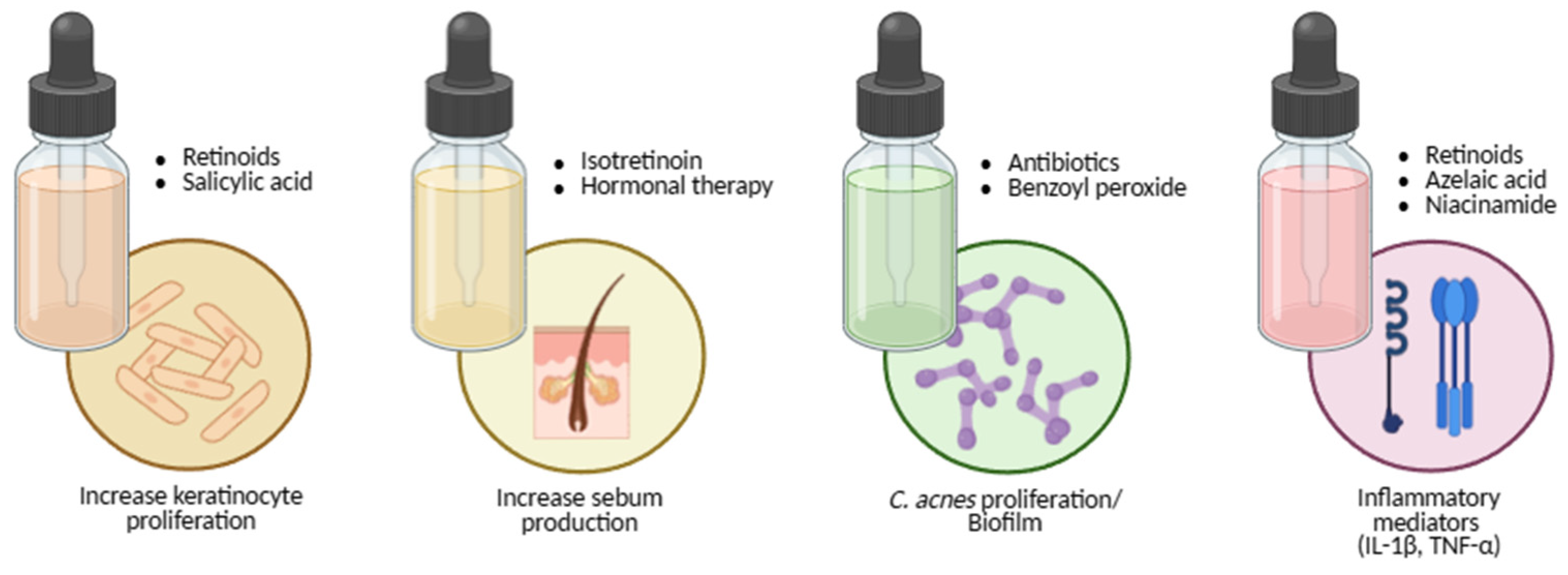
Pathogenesis: Four Interlocking Processes

Acne arises from the convergence of four tightly connected biological processes within the pilosebaceous unit:
  • Follicular hyperkeratinisation produces microcomedones through abnormal keratinocyte proliferation and desquamation, narrowing the infundibulum and obstructing outflow [62,63,64,65,66,67].
  • Sebum overproduction and altered composition, driven by androgens and metabolic signals such as IGF-1, creates a lipid-rich, anaerobic niche; sebum output correlates with acne severity [65,68,69].
  • Microbial factors—especially C. acnes colonisation—promote disease via lipases, hyaluronidases, and proteases that liberate free fatty acids, compromise the barrier, and foster comedogenesis; biofilm formation may shield bacteria from host defences and antibiotics [65,70,71,72].
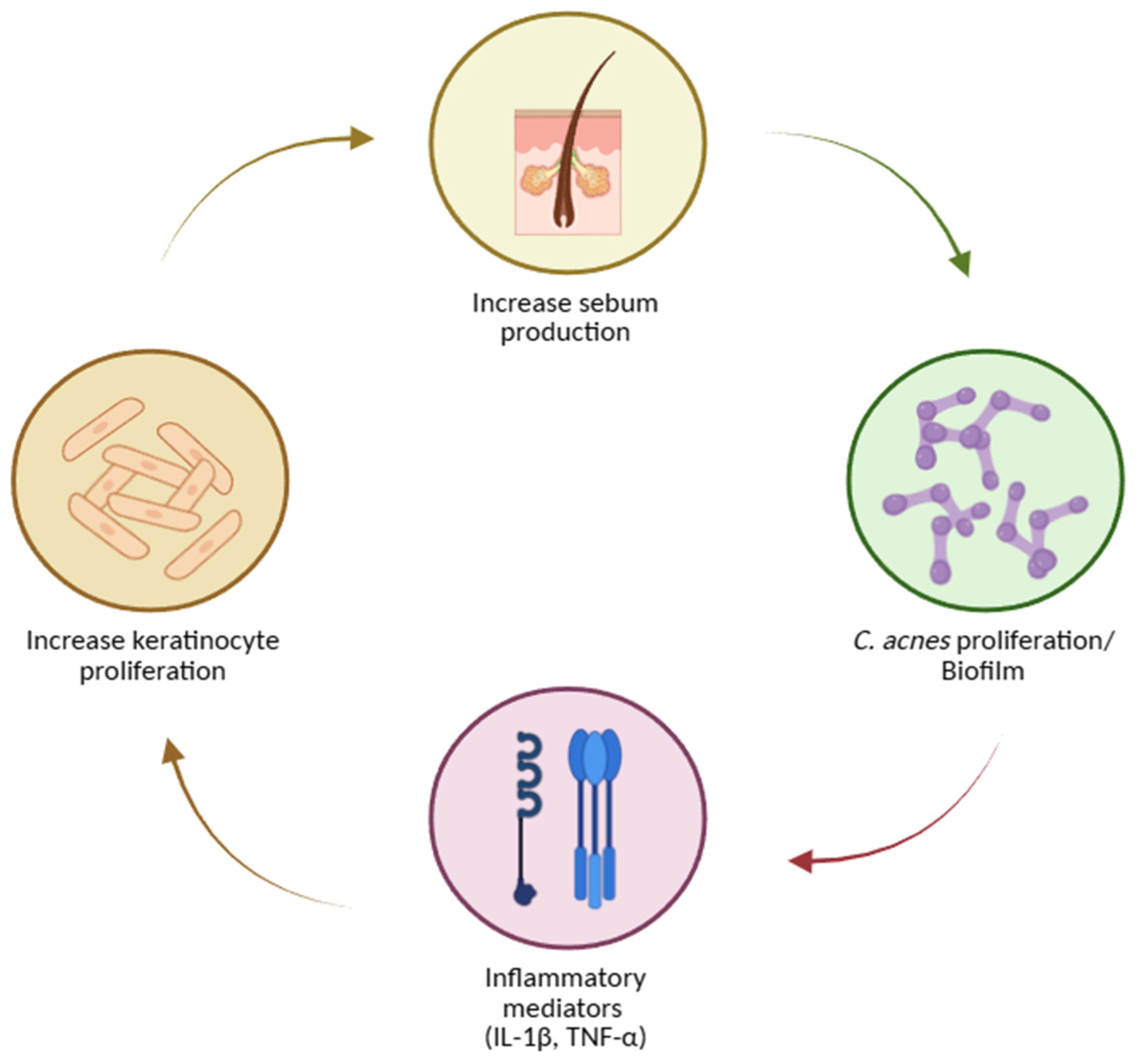
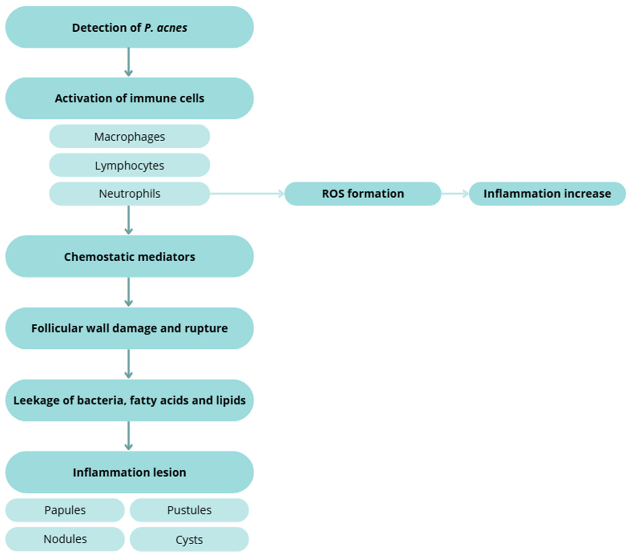
  • Inflammation and innate–adaptive crosstalk amplifies and sustain lesions: IL-1 signalling, neutrophil recruitment and ROS generation, and leukotriene B4–mediated cascades are implicated. Sebocytes synthesize neuropeptides, antimicrobial peptides, and antibacterial lipids, linking stress (CRH axis) and vitamin D signalling to sebaceous activity [62,63,64,68,69,73].
These processes can be envisioned as a self-reinforcing loop: hyperkeratinisation and altered sebum favour C. acnes growth and biofilms; bacterial products intensify inflammation; inflammatory mediators (including IL-1) further dysregulate keratinisation and sebaceous function [62,63,64,68,69,70,71,73,74].
Figure 3. Acne pathogenesis. Created with BioRender.com.
Preprints 180409 g003
Sebum, comedogenesis, and lesion evolution. Excess sebum—stimulated by androgens, particularly testosterone—correlates with disease severity [65]. Hyperproliferation and dysregulated shedding of keratinocytes lead to accumulation of corneocytes, lipids, and filamentous material within the follicle, forming microcomedones that progress to visible open or closed comedones [65,66,67]. In both normal follicles and comedones, microflora typically include Staphylococcus epidermidis (coagulase-negative), C. acnes and C. granulosum (anaerobic diphtheroids), and Pityrosporum spp. (lipophilic yeasts). S. epidermidis—an aerobic surface commensal—appears less implicated in inflammation than C. acnes, consistent with antibody response patterns. In contrast, C. acnes thrive in sebaceous follicles and hydrolyses triglycerides to free fatty acids and glycerol, promoting comedogenesis and inflammation [65,72,75].
Inflammatory cascades and tissue injury. Host detection of C. acnes triggers macrophage, lymphocyte, and neutrophil activation. Chemotactic factors and ROS generation contribute to follicular wall damage and rupture with extrusion of keratin, lipids, and bacteria into the dermis, producing papules, pustules, nodules, and cysts [60,65,66,73,76].
Strain-level insights. Culture-independent studies show that while overall C. acnes abundance may not differ dramatically between acne and controls, specific phylotypes/lineages associate with disease, whereas others are enriched in health. Follicular biopsies from acne patients reveal higher frequencies of disease-linked C. acnes strains and a greater proportion of colonised follicles, supporting a strain-selective colonisation model rather than a simple overgrowth model [45,77].
These data demonstrate the multifactorially, dysbiosis-linked inflammatory disorder of the pilosebaceous unit in which keratinisation dynamics, sebum quantity/quality, microbial traits (including biofilms and strain diversity), and immune networks intersect to drive lesion initiation, progression, and scarring.
Figure 4. Inflammatory response to C. acnes.
Preprints 180409 g004

Acne Treatments

Management of acne aims to control active lesions, reduce inflammation, prevent and treat scarring, and minimise relapses. Each treatment must be tailored to the patient’s clinical history and specific needs, considering lesion morphology, severity, distribution, age, sex, psychosocial impact, and prior response [78]. Several therapeutic approaches are available—topical, systemic, and natural/non-drug treatments—and combining agents that target different pathogenic mechanisms generally yields faster and more durable outcomes, as it can be seen in Table 1 [79].

Topical Treatments

Topical therapy remains the foundation for mild-to-moderate acne and is available in creams, gels, lotions, and cleansers. While these agents act directly on the affected area, local irritation and dryness may occur [80].
Retinoids are first-line agents because they normalise follicular desquamation, prevent microcomedone formation, and exert anti-inflammatory activity [66,81]. Tretinoin regulates epithelial desquamation, preventing blockage of the pilosebaceous unit and reducing inflammation [59,78]. Adapalene, a synthetic lipophilic analogue, penetrates the follicle efficiently, normalises keratinocyte differentiation, and is generally best tolerated for maintenance [59,60,82]. Tazarotene, a pro-drug converted to tazarotenic acid in keratinocytes, modulates proliferation and differentiation and has strong anti-inflammatory properties [59,65]. Retinoids can be used alone in predominantly comedonal acne or combined with antimicrobial agents for inflammatory forms. Gradual titration and attention to vehicle and formulation improve adherence and tolerability. Innovative micro- or nano-delivery systems are being explored to increase potency at the target while minimising irritation [59,60,61,65,66,67,81,82].
Topical antibiotics reduce Cutibacterium acnes and inflammatory lesions. Erythromycin (a macrolide) and clindamycin (a lincosamide) act by binding the bacterial 50S ribosomal subunit, inhibiting protein synthesis [59,78,81]. Resistance to erythromycin has reached approximately 60 % in C. acnes [78,80], so both agents should be prescribed only short-term (≈ 12 weeks) and never as monotherapy. Combining antibiotics with benzoyl peroxide (BPO) or retinoids enhances efficacy and mitigates resistance [83,84,85].
Benzoyl peroxide is a potent oxidising and comedolytic agent that kills C. acnes independently of resistance mechanisms. It reduces comedones but may cause peeling, dryness, erythema, or fabric bleaching [59,78,86]. Azelaic acid offers comedolytic, antibacterial, and anti-tyrosinase effects, making it valuable for post-inflammatory hyperpigmentation and safe in pregnancy [59,60,78,86].
Niacinamide (vitamin B₃ amide) decreases sebocyte lipid output and inflammation and strengthens the epidermal barrier [87,88,89,90]. Salicylic acid, a keratolytic agent, dissolves intercellular cement, enhances penetration of co-applied actives, and exerts mild bacteriostatic and fungistatic effects [59,78].

Systemic Treatments

Systemic therapy is indicated for moderate-to-severe, nodular, truncal, or scarring acne, or when topical management fails.
Oral isotretinoin remains the only drug capable of long-term remission. It induces sebaceous-gland involution, markedly suppresses sebum production, normalises keratinisation, and alters the follicular microenvironment [60,82,91,92,93]. Because of teratogenic risk, strict pregnancy-prevention protocols and laboratory monitoring are mandatory. Counselling should also address mucocutaneous dryness, lipid changes, and mood symptoms.
Oral antibiotics are reserved for moderate-to-severe inflammatory acne or widespread disease [86,91]. Tetracyclines (e.g., doxycycline) combine antimicrobial and anti-inflammatory activity with relatively low resistance rates [81,86]. Macrolides (erythromycin, clindamycin) serve as alternatives when tetracyclines are contraindicated but show higher resistance potential. Fluoroquinolones (e.g., levofloxacin) are discouraged under stewardship principles [66,81,92,94]. To maximise efficacy and reduce resistance, oral antibiotics should always be combined with topical retinoids or BPO and prescribed for the shortest effective duration [66,81,91].
Hormonal therapy targets androgen-driven sebogenesis. Combined oral contraceptives and anti-androgens such as spironolactone reduce free testosterone and suppress sebaceous activity [66,81,91]. They are suitable for adolescent and adult women when hormonal influence is evident, provided contraindications (e.g., thromboembolic risk) are assessed individually.
Figure 5. Therapeutic acne options and its targets. Created with BioRender.com.
Preprints 180409 g005

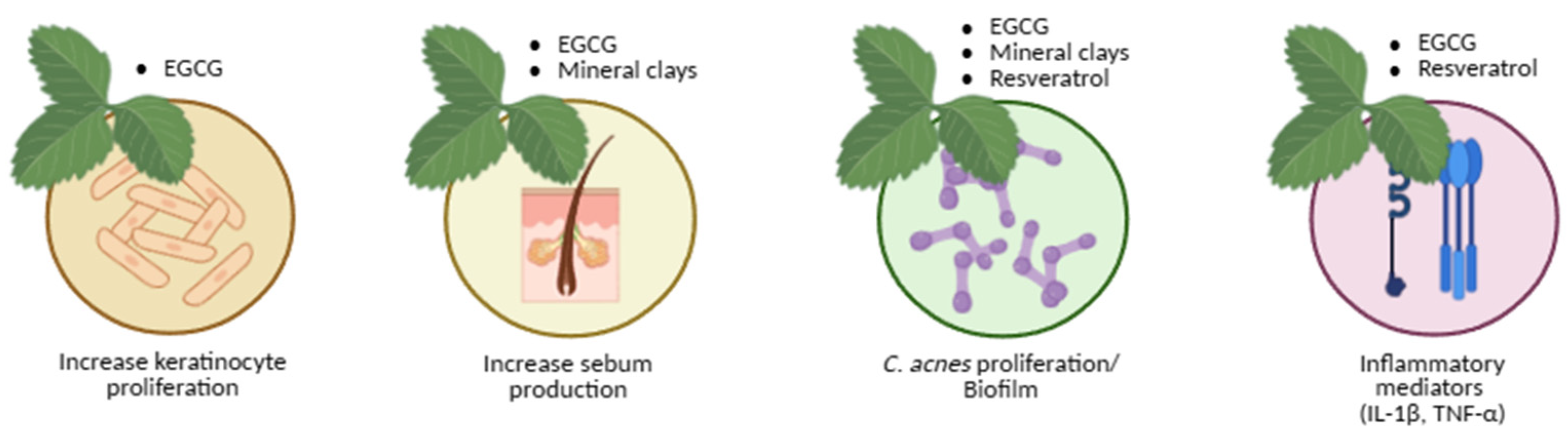
Natural and Adjunct Approaches

Natural Products Treatment

Given the side-effects and resistance issues associated with conventional drugs, natural bioactives have emerged as safe, multi-target alternatives. Therefore, researchers are focusing on natural bioactive options. It has been that these compounds may act differently disrupting distinct metabolic pathways [95]. It is important to mention that natural approaches can influence the hormonal characteristics of the disease, sebum production, inflammation, infection and hyperkeratinization [96].
  • Green tea
Green tea polyphenols, particularly epigallocatechin-3-gallate (EGCG), display anti-inflammatory, antioxidant, antimicrobial, and sebo-suppressive properties [81,97]. EGCG targets key acne pathways—reducing C. acnes growth, inflammation, and aberrant keratinisation—and clinical studies have shown decreased inflammatory and non-inflammatory lesions after eight weeks of topical use [98].
  • Mineral clays
Mineral clays (halloysite, talc, sericite, kaolin) have been used since antiquity for acne and blackheads. Applied as masks, they open pilosebaceous orifices, absorb sebum, and promote perspiration [99]. In vitro data show inhibition of C. acnes and S. epidermidis, though clinical outcomes depend on composition and formulation [100].
  • Resveratrol
Resveratrol, a natural phytoalexin, possesses anti-inflammatory and anti-proliferative activity and inhibits C. acnes [101,102,103,104,105]. In vitro, it acts bacteriostatically at 50–100 mg/L and bactericidally at 200 mg/L [102]; in pilot clinical trials, topical resveratrol gels used for 60 days reduced pustules, macro- and micro-comedones, and epidermal hyperproliferation [104].
Figure 6. Natural therapeutic acne option and its targets. Created with BioRender.com.
Preprints 180409 g006

Clinical Integration and Outlook

In practice, effective regimens combine a topical retinoid to normalise desquamation with BPO or azelaic acid to reduce bacterial load and inflammation, adding short antibiotic courses or isotretinoin for severe or refractory disease. In women with hormonal influence, anti-androgen therapy provides a targeted adjunct. Maintenance (low-irritancy retinoid ± azelaic acid), gentle skincare, and photoprotection reduce relapse.
Across all modalities, the guiding objective is to increase therapeutic potency at the target site while preserving tolerability, microbial balance, and long-term safety, in alignment with principles of antimicrobial stewardship [59,60,61,65,66,67,78,79,80,81,82,83,84,85,86,91,92,93,94].

Critical Perspective

Traditional, Synthetic, and Natural Approaches

Current acne management reflects a dual paradigm. On one side, traditional synthetic therapies—retinoids, antibiotics, hormonal agents—remain the cornerstone of evidence-based practice. They are supported by extensive clinical data, predictable pharmacodynamics, and regulatory familiarity. However, their limitations are increasingly evident: antibiotic resistance, irritation, photosensitivity, teratogenicity, systemic toxicity, and microbiome disruption. The overuse of broad-spectrum antibiotics has particularly raised concerns regarding antimicrobial stewardship, as Cutibacterium acnes resistance threatens both dermatological and systemic infection control [78,80,82,83,84,85,91,92,93,94]. Furthermore, patient adherence often declines with chronic irritation, dryness, or psychological burden linked to treatment complexity.
In contrast, natural and bioactive-derived therapies—including polyphenols (e.g., EGCG, resveratrol), minerals, and microbial metabolites—offer multi-target modulation of inflammation, sebum regulation, and oxidative stress with potentially fewer adverse effects. Their mechanisms often mimic endogenous pathways, supporting cutaneous homeostasis and preserving the skin microbiome. Yet, they face critical challenges: variability in extraction and formulation, lack of standardisation, limited pharmacokinetic data, and insufficient large-scale randomised clinical trials. As a result, despite promising preclinical findings, natural compounds still struggle to reach the same potency, reproducibility, and regulatory maturity as synthetic drugs.
The most rational future trajectory lies not in opposition but in integration. Combining synthetic pharmacophores with natural bioactives—or engineering hybrid formulations that couple retinoid-like efficacy with antioxidant and anti-inflammatory natural scaffolds—may yield synergistic effects that maximise potency and minimise irritation.
Advances in nanotechnology, controlled-release systems, and green chemistry now enable precise delivery of both synthetic and natural molecules directly to the pilosebaceous target, enhancing bioavailability and patient comfort.
In this evolving landscape, potency, precision, and preservation—of efficacy, safety, and microbiome equilibrium—should become the new therapeutic triad guiding acne innovation.

2. Conclusions and Future Perspectives

Acne vulgaris exemplifies the complex interface between host biology, microbial ecology, and environmental influences. Despite decades of progress, challenges persist growing antibiotic resistance, treatment intolerance, relapse, and psychological impact. Modern research increasingly recognises acne as a disorder of microbial and immune dysregulation, rather than simply a bacterial infection. This paradigm shift demands equally adaptive treatments.
Future acne management will likely move toward personalised, multimodal, and microbiome-conscious therapy. Artificial intelligence and multi-omics profiling could soon allow individualised treatment algorithms based on genetic, hormonal, and microbial signatures. At the same time, the emergence of bioinspired and sustainable materials—polyphenol-enriched hydrogels, peptide-functionalised carriers, piezoelectric or photothermal nanoscaffolds—offers exciting prospects for precise, stimuli-responsive drug delivery.
Regulatory harmonisation and rigorous clinical validation remain crucial to translate natural actives into mainstream dermatology. Interdisciplinary collaboration between chemists, dermatologists, microbiologists, and bioengineers will be essential to bridge efficacy with sustainability, pharmacology with ecology, and innovation with patient trust.
Ultimately, the convergence of traditional pharmacotherapy and bioactive natural science marks not a replacement but an evolution—toward high-potency, low-toxicity, ecologically balanced acne treatments that respect both the skin and its microbiome.

Author Contributions

Conceptualization: C.B.; Investigation and Data Curation: C.B. and M.B.O.; Writing—Original Draft Preparation: M.B.O.; Writing—Review and Editing: C.B., ACM, ANB; Visualization and Figure Preparation: M.B.O.; Supervision and Project Administration: C.B. All authors have read and agreed to the published version of the manuscript.

Funding

This research was supported by the Foundation for Science and Technology (FCT) through LA/P/0059/2020, UIDB/00211/2020 and UID/04033/2025.

Acknowledgments

The authors gratefully acknowledge institutional and technical support from Centre for Studies in Animal Science (CECA) from Institute of Sciences, Technologies and Agroenvironment (ICETA), University of Porto, Centre for the Research and Technology of Agro-Environmental and Biological Sciences (CITAB) and Department of Veterinary Clinics, School of Medicine and Biomedical Sciences (ICBAS), University of Porto (UP), and for the funding and availability of all technical, structural, and human resources necessary for the development of this work. This research was supported by the Foundation for Science and Technology (FCT) through LA/P/0059/2020, UIDB/00211/2020 and UID/04033/2025. The authors acknowledge the use of artificial intelligence tools for language refinement and editing assistance. All intellectual content, data interpretation, and conclusions were developed and verified by the authors.

Conflicts of Interest

The authors declare no conflict of interest. The funders had no role in the design of the study; in the collection, analyses, or interpretation of data; in the writing of the manuscript; or in the decision to publish the results.

References

  1. Bojar, R.A.; Holland, K.T. Acne and Propionibacterium Acnes. Clinics in Dermatology 2004, 22, 375–379. [CrossRef]
  2. Segre, J.A. Epidermal Barrier Formation and Recovery in Skin Disorders. J Clin Invest 2006, 116, 1150–1158. [CrossRef]
  3. Marples, M.J. The Ecology of the Human Skin; Ch. C. Thomas: Springfield (Illinois), 1965;
  4. Schommer, N.N.; Gallo, R.L. Structure and Function of the Human Skin Microbiome. Trends in Microbiology 2013, 21, 660–668. [CrossRef]
  5. Grice, E.A.; Segre, J.A. The Skin Microbiome. Nat Rev Microbiol 2011, 9, 244–253. [CrossRef]
  6. Holland, K.T.; Cunliffe, W.J.; Roberts, C.D. The Role of Bacteria in Acne Vulgaris: A New Approach. Clin Exp Dermatol 1978, 3, 253–257. [CrossRef]
  7. Holland, K.T.; Ingham, E.; Cunliffe, W.J. A Review, the Microbiology of Acne. J Appl Bacteriol 1981, 51, 195–215. [CrossRef]
  8. Eady, E.A.; Ingham, E. Propionibacterium Acnes - Friend or Foe?: Reviews in Medical Microbiology 1994, 5, 163–173. [CrossRef]
  9. Grice, E.A.; Kong, H.H.; Conlan, S.; Deming, C.B.; Davis, J.; Young, A.C.; NISC COMPARATIVE SEQUENCING PROGRAM; Bouffard, G.G.; Blakesley, R.W.; Murray, P.R.; et al. Topographical and Temporal Diversity of the Human Skin Microbiome. Science 2009, 324, 1190–1192. [CrossRef]
  10. Structure, Function and Diversity of the Healthy Human Microbiome. Nature 2012, 486, 207–214. [CrossRef]
  11. Costello, E.K.; Lauber, C.L.; Hamady, M.; Fierer, N.; Gordon, J.I.; Knight, R. Bacterial Community Variation in Human Body Habitats Across Space and Time. Science 2009, 326, 1694–1697. [CrossRef]
  12. Faust, K.; Raes, J. Microbial Interactions: From Networks to Models. Nat Rev Microbiol 2012, 10, 538–550. [CrossRef]
  13. Grice, E.A.; Kong, H.H.; Renaud, G.; Young, A.C.; Bouffard, G.G.; Blakesley, R.W.; Wolfsberg, T.G.; Turner, M.L.; Segre, J.A. A Diversity Profile of the Human Skin Microbiota. Genome Res. 2008, 18, 1043–1050. [CrossRef]
  14. Tagami, H. Location-Related Differences in Structure and Function of the Stratum Corneum with Special Emphasis on Those of the Facial Skin. Int J Cosmet Sci 2008, 30, 413–434. [CrossRef]
  15. Fulton, C.; Anderson, G.M.; Zasloff, M.; Bull, R.; Quinn, A.G. Expression of Natural Peptide Antibiotics in Human Skin. Lancet 1997, 350, 1750–1751. [CrossRef]
  16. Schittek, B.; Hipfel, R.; Sauer, B.; Bauer, J.; Kalbacher, H.; Stevanovic, S.; Schirle, M.; Schroeder, K.; Blin, N.; Meier, F.; et al. Dermcidin: A Novel Human Antibiotic Peptide Secreted by Sweat Glands. Nat Immunol 2001, 2, 1133–1137. [CrossRef]
  17. Gribbon, E.M.; Cunliffe, W.J.; Holland, K.T. Interaction of Propionibacterium Acnes with Skin Lipids in Vitro. J Gen Microbiol 1993, 139, 1745–1751. [CrossRef]
  18. Gallo, R.L.; Hooper, L.V. Epithelial Antimicrobial Defence of the Skin and Intestine. Nat Rev Immunol 2012, 12, 503–516. [CrossRef]
  19. Roth, R.R.; James, W.D. Microbial Ecology of the Skin. Annu Rev Microbiol 1988, 42, 441–464. [CrossRef]
  20. Elias, P.M. The Skin Barrier as an Innate Immune Element. Semin Immunopathol 2007, 29, 3–14. [CrossRef]
  21. Aly, R.; Shirley, C.; Cunico, B.; Maibach, H.I. Effect of Prolonged Occlusion on the Microbial Flora, pH, Carbon Dioxide and Transepidermal Water Loss on Human Skin. J Invest Dermatol 1978, 71, 378–381. [CrossRef]
  22. Korting, H.C.; Hübner, K.; Greiner, K.; Hamm, G.; Braun-Falco, O. Differences in the Skin Surface pH and Bacterial Microflora Due to the Long-Term Application of Synthetic Detergent Preparations of pH 5.5 and pH 7.0. Results of a Crossover Trial in Healthy Volunteers. Acta Derm Venereol 1990, 70, 429–431.
  23. Dominguez-Bello, M.G.; Costello, E.K.; Contreras, M.; Magris, M.; Hidalgo, G.; Fierer, N.; Knight, R. Delivery Mode Shapes the Acquisition and Structure of the Initial Microbiota across Multiple Body Habitats in Newborns. Proc Natl Acad Sci U S A 2010, 107, 11971–11975. [CrossRef]
  24. Mueller, N.T.; Bakacs, E.; Combellick, J.; Grigoryan, Z.; Dominguez-Bello, M.G. The Infant Microbiome Development: Mom Matters. Trends Mol Med 2015, 21, 109–117. [CrossRef]
  25. Somerville, D.A. The Normal Flora of the Skin in Different Age Groups. Br J Dermatol 1969, 81, 248–258. [CrossRef]
  26. Oh, J.; Conlan, S.; Polley, E.C.; Segre, J.A.; Kong, H.H. Shifts in Human Skin and Nares Microbiota of Healthy Children and Adults. Genome Med 2012, 4, 77. [CrossRef]
  27. Jo, J.-H.; Deming, C.; Kennedy, E.A.; Conlan, S.; Polley, E.C.; Ng, W.-I.; NISC Comparative Sequencing Program; Segre, J.A.; Kong, H.H. Diverse Human Skin Fungal Communities in Children Converge in Adulthood. J Invest Dermatol 2016, 136, 2356–2363. [CrossRef]
  28. Jo, J.-H.; Kennedy, E.A.; Kong, H.H. Topographical and Physiological Differences of the Skin Mycobiome in Health and Disease. Virulence 2017, 8, 324–333. [CrossRef]
  29. Havlickova, B.; Czaika, V.A.; Friedrich, M. Epidemiological Trends in Skin Mycoses Worldwide. Mycoses 2008, 51 Suppl 4, 2–15. [CrossRef]
  30. Seebacher, C.; Bouchara, J.-P.; Mignon, B. Updates on the Epidemiology of Dermatophyte Infections. Mycopathologia 2008, 166, 335–352. [CrossRef]
  31. Kyriakis, K.P.; Terzoudi, S.; Palamaras, I.; Pagana, G.; Michailides, C.; Emmanuelides, S. Pityriasis Versicolor Prevalence by Age and Gender. Mycoses 2006, 49, 517–518. [CrossRef]
  32. Marples, R.R. Sex, Constancy, and Skin Bacteria. Arch Dermatol Res 1982, 272, 317–320. [CrossRef]
  33. Giacomoni, P.U.; Mammone, T.; Teri, M. Gender-Linked Differences in Human Skin. J Dermatol Sci 2009, 55, 144–149. [CrossRef]
  34. Leeming, J.P.; Holland, K.T.; Cunliffe, W.J. The Microbial Ecology of Pilosebaceous Units Isolated from Human Skin. J Gen Microbiol 1984, 130, 803–807. [CrossRef]
  35. McGinley, K.J.; Webster, G.F.; Leyden, J.J. Regional Variations of Cutaneous Propionibacteria. Appl Environ Microbiol 1978, 35, 62–66.
  36. Findley, K.; Oh, J.; Yang, J.; Conlan, S.; Deming, C.; Meyer, J.A.; Schoenfeld, D.; Nomicos, E.; Park, M.; Kong, H.H.; et al. Topographic Diversity of Fungal and Bacterial Communities in Human Skin. Nature 2013, 498, 367–370. [CrossRef]
  37. Paulino, L.C.; Tseng, C.-H.; Blaser, M.J. Analysis of Malassezia Microbiota in Healthy Superficial Human Skin and in Psoriatic Lesions by Multiplex Real-Time PCR. FEMS Yeast Research 2008, 8, 460–471. [CrossRef]
  38. Xu, J.; Saunders, C.W.; Hu, P.; Grant, R.A.; Boekhout, T.; Kuramae, E.E.; Kronstad, J.W.; DeAngelis, Y.M.; Reeder, N.L.; Johnstone, K.R.; et al. Dandruff-Associated Malassezia Genomes Reveal Convergent and Divergent Virulence Traits Shared with Plant and Human Fungal Pathogens. Proceedings of the National Academy of Sciences 2007, 104, 18730–18735. [CrossRef]
  39. Iebba, V.; Totino, V.; Gagliardi, A.; Santangelo, F.; Cacciotti, F.; Trancassini, M.; Mancini, C.; Cicerone, C.; Corazziari, E.; Pantanella, F.; et al. Eubiosis and Dysbiosis: The Two Sides of the Microbiota. New Microbiol 2016, 39, 1–12.
  40. Byrd, A.L.; Belkaid, Y.; Segre, J.A. The Human Skin Microbiome. Nat Rev Microbiol 2018, 16, 143–155. [CrossRef]
  41. Leyden, J.J.; McGinley, K.J.; Mills, O.H.; Kligman, A.M. Propionibacterium Levels in Patients with and without Acne Vulgaris. J Invest Dermatol 1975, 65, 382–384. [CrossRef]
  42. Strauss, J.S.; Krowchuk, D.P.; Leyden, J.J.; Lucky, A.W.; Shalita, A.R.; Siegfried, E.C.; Thiboutot, D.M.; Van Voorhees, A.S.; Beutner, K.A.; Sieck, C.K.; et al. Guidelines of Care for Acne Vulgaris Management. J Am Acad Dermatol 2007, 56, 651–663. [CrossRef]
  43. Degitz, K.; Placzek, M.; Borelli, C.; Plewig, G. Pathophysiology of Acne. J Dtsch Dermatol Ges 2007, 5, 316–323. [CrossRef]
  44. Tomida, S.; Nguyen, L.; Chiu, B.-H.; Liu, J.; Sodergren, E.; Weinstock, G.M.; Li, H. Pan-Genome and Comparative Genome Analyses of Propionibacterium Acnes Reveal Its Genomic Diversity in the Healthy and Diseased Human Skin Microbiome. mBio 2013, 4, e00003-00013. [CrossRef]
  45. Fitz-Gibbon, S.; Tomida, S.; Chiu, B.-H.; Nguyen, L.; Du, C.; Liu, M.; Elashoff, D.; Erfe, M.C.; Loncaric, A.; Kim, J.; et al. Propionibacterium Acnes Strain Populations in the Human Skin Microbiome Associated with Acne. Journal of Investigative Dermatology 2013, 133, 2152–2160. [CrossRef]
  46. Kang, D.; Shi, B.; Erfe, M.C.; Craft, N.; Li, H. Vitamin B12 Modulates the Transcriptome of the Skin Microbiota in Acne Pathogenesis. Sci Transl Med 2015, 7, 293ra103. [CrossRef]
  47. Williams, H.C.; Dellavalle, R.P.; Garner, S. Acne Vulgaris. The Lancet 2012, 379, 361–372. [CrossRef]
  48. Lucky, A.W. A Review of Infantile and Pediatric Acne. Dermatology 1998, 196, 95–97. [CrossRef]
  49. Adebamowo, C.A.; Spiegelman, D.; Berkey, C.S.; Danby, F.W.; Rockett, H.H.; Colditz, G.A.; Willett, W.C.; Holmes, M.D. Milk Consumption and Acne in Teenaged Boys. J Am Acad Dermatol 2008, 58, 787–793. [CrossRef]
  50. Friedlander, S.F.; Eichenfield, L.F.; Fowler, J.F.; Fried, R.G.; Levy, M.L.; Webster, G.F. Acne Epidemiology and Pathophysiology. Semin Cutan Med Surg 2010, 29, 2–4. [CrossRef]
  51. Ghodsi, S.Z.; Orawa, H.; Zouboulis, C.C. Prevalence, Severity, and Severity Risk Factors of Acne in High School Pupils: A Community-Based Study. J Invest Dermatol 2009, 129, 2136–2141. [CrossRef]
  52. Stoll, S.; Shalita, A.R.; Webster, G.F.; Kaplan, R.; Danesh, S.; Penstein, A. The Effect of the Menstrual Cycle on Acne. J Am Acad Dermatol 2001, 45, 957–960. [CrossRef]
  53. Yosipovitch, G.; Tang, M.; Dawn, A.G.; Chen, M.; Goh, C.L.; Huak, Y.; Seng, L.F. Study of Psychological Stress, Sebum Production and Acne Vulgaris in Adolescents. Acta Derm Venereol 2007, 87, 135–139. [CrossRef]
  54. Plewig, G.; Fulton, J.E.; Kligman, A.M. Pomade Acne. Arch Dermatol 1970, 101, 580–584.
  55. Valeyrie-Allanore, L.; Sassolas, B.; Roujeau, J.-C. Drug-Induced Skin, Nail and Hair Disorders. Drug Saf 2007, 30, 1011–1030. [CrossRef]
  56. Melnik, B.; Jansen, T.; Grabbe, S. Abuse of Anabolic-Androgenic Steroids and Bodybuilding Acne: An Underestimated Health Problem. J Dtsch Dermatol Ges 2007, 5, 110–117. [CrossRef]
  57. Green, J.; Sinclair, R.D. Perceptions of Acne Vulgaris in Final Year Medical Student Written Examination Answers. Australas J Dermatol 2001, 42, 98–101. [CrossRef]
  58. Magin, P.; Pond, D.; Smith, W.; Watson, A. A Systematic Review of the Evidence for “myths and Misconceptions” in Acne Management: Diet, Face-Washing and Sunlight. Fam Pract 2005, 22, 62–70. [CrossRef]
  59. Akhavan, A.; Bershad, S. Topical Acne Drugs: Review of Clinical Properties, Systemic Exposure, and Safety. Am J Clin Dermatol 2003, 4, 473–492. [CrossRef]
  60. Feldman, S.; Careccia, R.E.; Barham, K.L.; Hancox, J. Diagnosis and Treatment of Acne. Am Fam Physician 2004, 69, 2123–2130.
  61. Ayer, J.; Burrows, N. Acne: More than Skin Deep. Postgrad Med J 2006, 82, 500–506. [CrossRef]
  62. Jeremy, A.H.T.; Holland, D.B.; Roberts, S.G.; Thomson, K.F.; Cunliffe, W.J. Inflammatory Events Are Involved in Acne Lesion Initiation. J Invest Dermatol 2003, 121, 20–27. [CrossRef]
  63. Kurokawa, I.; Danby, F.W.; Ju, Q.; Wang, X.; Xiang, L.F.; Xia, L.; Chen, W.; Nagy, I.; Picardo, M.; Suh, D.H.; et al. New Developments in Our Understanding of Acne Pathogenesis and Treatment. Exp Dermatol 2009, 18, 821–832. [CrossRef]
  64. Alestas, T.; Ganceviciene, R.; Fimmel, S.; Müller-Decker, K.; Zouboulis, C.C. Enzymes Involved in the Biosynthesis of Leukotriene B4 and Prostaglandin E2 Are Active in Sebaceous Glands. J Mol Med (Berl) 2006, 84, 75–87. [CrossRef]
  65. Gollnick, H. Current Concepts of the Pathogenesis of Acne: Implications for Drug Treatment. Drugs 2003, 63, 1579–1596. [CrossRef]
  66. Gollnick, H.; Cunliffe, W.; Berson, D.; Dreno, B.; Finlay, A.; Leyden, J.J.; Shalita, A.R.; Thiboutot, D. Management of Acne: A Report From a Global Alliance to Improve Outcomes in Acne. Journal of the American Academy of Dermatology 2003, 49, S1–S37. [CrossRef]
  67. Webster, G.F. Acne. Current Problems in Dermatology 1996, 8, 237–268. [CrossRef]
  68. Zouboulis, C.C.; Böhm, M. Neuroendocrine Regulation of Sebocytes -- a Pathogenetic Link between Stress and Acne. Exp Dermatol 2004, 13 Suppl 4, 31–35. [CrossRef]
  69. Zouboulis, C.C.; Baron, J.M.; Böhm, M.; Kippenberger, S.; Kurzen, H.; Reichrath, J.; Thielitz, A. Frontiers in Sebaceous Gland Biology and Pathology. Exp Dermatol 2008, 17, 542–551. [CrossRef]
  70. Iinuma, K.; Sato, T.; Akimoto, N.; Noguchi, N.; Sasatsu, M.; Nishijima, S.; Kurokawa, I.; Ito, A. Involvement of Propionibacterium Acnes in the Augmentation of Lipogenesis in Hamster Sebaceous Glands In Vivo and In Vitro. Journal of Investigative Dermatology 2009, 129, 2113–2119. [CrossRef]
  71. McDowell, A.; Gao, A.; Barnard, E.; Fink, C.; Murray, P.I.; Dowson, C.G.; Nagy, I.; Lambert, P.A.; Patrick, S. A Novel Multilocus Sequence Typing Scheme for the Opportunistic Pathogen Propionibacterium Acnes and Characterization of Type I Cell Surface-Associated Antigens. Microbiology 2011, 157, 1990–2003. [CrossRef]
  72. Burkhart, C.G.; Burkhart, C.N.; Lehmann, P.F. Acne: A Review of Immunologic and Microbiologic Factors. Postgrad Med J 1999, 75, 328–331. [CrossRef]
  73. Akamatsu, H.; Horio, T.; Hattori, K. Increased Hydrogen Peroxide Generation by Neutrophils from Patients with Acne Inflammation. International Journal of Dermatology 2003, 42, 366–369. [CrossRef]
  74. Thiboutot, D.; Gollnick, H.; Bettoli, V.; Dréno, B.; Kang, S.; Leyden, J.J.; Shalita, A.R.; Lozada, V.T.; Berson, D.; Finlay, A.; et al. New Insights into the Management of Acne: An Update from the Global Alliance to Improve Outcomes in Acne Group. J Am Acad Dermatol 2009, 60, S1-50. [CrossRef]
  75. Białecka, A.; Mak, M.; Biedroń, R.; Bobek, M.; Kasprowicz, A.; Marcinkiewicz, J. Different Pro-Inflammatory and Immunogenic Potentials of Propionibacterium Acnes and Staphylococcus Epidermidis: Implications for Chronic Inflammatory Acne. Arch Immunol Ther Exp (Warsz) 2005, 53, 79–85.
  76. Webster, G.F.; Poyner, T.; Cunliffe, B. Clinical reviewAcne vulgarisCommentary: A UK Primary Care Perspective on Treating Acne. BMJ 2002, 325, 475–479. [CrossRef]
  77. Jahns, A.C.; Lundskog, B.; Ganceviciene, R.; Palmer, R.H.; Golovleva, I.; Zouboulis, C.C.; McDowell, A.; Patrick, S.; Alexeyev, O.A. An Increased Incidence of Propionibacterium Acnes Biofilms in Acne Vulgaris: A Case–Control Study. British Journal of Dermatology 2012, 167, 50–58. [CrossRef]
  78. Krautheim, A.; Gollnick, H. Transdermal Penetration of Topical Drugs Used in the Treatment of Acne. Clin Pharmacokinet 2003, 42, 1287–1304. [CrossRef]
  79. Olutunmbi, Y.; Paley, K.; English, J.C. Adolescent Female Acne: Etiology and Management. J Pediatr Adolesc Gynecol 2008, 21, 171–176. [CrossRef]
  80. Bowe, W.P.; Shalita, A.R. Effective Over-the-Counter Acne Treatments. Semin Cutan Med Surg 2008, 27, 170–176. [CrossRef]
  81. Fox, L.; Csongradi, C.; Aucamp, M.; Du Plessis, J.; Gerber, M. Treatment Modalities for Acne. Molecules 2016, 21, 1063. [CrossRef]
  82. Bershad, S.V. The Modern Age of Acne Therapy: A Review of Current Treatment Options. Mt Sinai J Med 2001, 68, 279–286.
  83. Shaw, L.; Kennedy, C. The Treatment of Acne. Paediatrics and Child Health 2007, 17, 385–389. [CrossRef]
  84. Gollnick, H.P.M.; Krautheim, A. Topical Treatment in Acne: Current Status and Future Aspects. Dermatology 2003, 206, 29–36. [CrossRef]
  85. Lavers, I. Diagnosis and Management of Acne Vulgaris. Nurse Prescribing 2014, 12, 330–336. [CrossRef]
  86. Webster, G.F.; Graber, E.M. Antibiotic Treatment for Acne Vulgaris. Semin Cutan Med Surg 2008, 27, 183–187. [CrossRef]
  87. Namazi, M.R. Nicotinamide in Dermatology: A Capsule Summary. Int J Dermatol 2007, 46, 1229–1231. [CrossRef]
  88. Draelos, Z.D.; Matsubara, A.; Smiles, K. The Effect of 2% Niacinamide on Facial Sebum Production. J Cosmet Laser Ther 2006, 8, 96–101. [CrossRef]
  89. Draelos, Z.D. Novel Topical Therapies in Cosmetic Dermatology. Current Problems in Dermatology 2000, 12, 235–239. [CrossRef]
  90. Gehring, W. Nicotinic Acid/Niacinamide and the Skin. J Cosmet Dermatol 2004, 3, 88–93. [CrossRef]
  91. Katsambas, A.; Papakonstantinou, A. Acne: Systemic Treatment. Clin Dermatol 2004, 22, 412–418. [CrossRef]
  92. Zouboulis, C.C.; Piquero-Martin, J. Update and Future of Systemic Acne Treatment. Dermatology 2003, 206, 37–53. [CrossRef]
  93. Leyden, J.J.; McGinley, K.J.; Foglia, A.N. Qualitative and Quantitative Changes in Cutaneous Bacteria Associated with Systemic Isotretinoin Therapy for Acne Conglobata. J Invest Dermatol 1986, 86, 390–393. [CrossRef]
  94. Sinclair, W.; Jordaan, H.F.; Global Alliance to Improve Outcomes in Acne Acne Guideline 2005 Update. S Afr Med J 2005, 95, 881–892.
  95. Fisk, W.A.; Lev-Tov, H.A.; Sivamani, R.K. Botanical and Phytochemical Therapy of Acne: A Systematic Review. Phytother Res 2014, 28, 1137–1152. [CrossRef]
  96. Magin, P.J.; Adams, J.; Pond, C.D.; Smith, W. Topical and Oral CAM in Acne: A Review of the Empirical Evidence and a Consideration of Its Context. Complement Ther Med 2006, 14, 62–76. [CrossRef]
  97. Gaur, S.; Agnihotri, R. Green Tea: A Novel Functional Food for the Oral Health of Older Adults. Geriatr Gerontol Int 2014, 14, 238–250. [CrossRef]
  98. Yoon, J.Y.; Kwon, H.H.; Min, S.U.; Thiboutot, D.M.; Suh, D.H. Epigallocatechin-3-Gallate Improves Acne in Humans by Modulating Intracellular Molecular Targets and Inhibiting P. Acnes. J Invest Dermatol 2013, 133, 429–440. [CrossRef]
  99. Carretero, M.I. Clay Minerals and Their Beneficial Effects upon Human Health. A Review. Applied Clay Science 2002, 21, 155–163. [CrossRef]
  100. Park, S.K.; Lee, C.W.; Lee, M.Y. Antibacterial Effects of Minerals from Ores Indigenous to Korea. J Environ Biol 2009, 30, 151–154.
  101. Simonart, T. Newer Approaches to the Treatment of Acne Vulgaris. Am J Clin Dermatol 2012, 13, 357–364. [CrossRef]
  102. Docherty, J.J.; McEwen, H.A.; Sweet, T.J.; Bailey, E.; Booth, T.D. Resveratrol Inhibition of Propionibacterium Acnes. J Antimicrob Chemother 2007, 59, 1182–1184. [CrossRef]
  103. Donnelly, L.E.; Newton, R.; Kennedy, G.E.; Fenwick, P.S.; Leung, R.H.F.; Ito, K.; Russell, R.E.K.; Barnes, P.J. Anti-Inflammatory Effects of Resveratrol in Lung Epithelial Cells: Molecular Mechanisms. Am J Physiol Lung Cell Mol Physiol 2004, 287, L774-783. [CrossRef]
  104. Fabbrocini, G.; Staibano, S.; De Rosa, G.; Battimiello, V.; Fardella, N.; Ilardi, G.; La Rotonda, M.I.; Longobardi, A.; Mazzella, M.; Siano, M.; et al. Resveratrol-Containing Gel for the Treatment of Acne Vulgaris: A Single-Blind, Vehicle-Controlled, Pilot Study. Am J Clin Dermatol 2011, 12, 133–141. [CrossRef]
  105. Frémont, L. Biological Effects of Resveratrol. Life Sciences 2000, 66, 663–673. [CrossRef]
Table 1. Acne treatments by class and exemplar agents.
Treatment Method Products
Topical Retinoids: tretinoin, adapalene, tazarotene • Antibiotics: erythromycin, clindamycin • Diverse: benzoyl peroxide, azelaic acid, niacinamide, salicylic acid
Systemic Retinoids: isotretinoin • Antibiotics: erythromycin, clindamycin, levofloxacin, doxycycline • Hormonal: combined contraceptives, spironolactone
Natural/Adjunct Green tea polyphenols (EGCG) • Minerals (clay-based) • Resveratrol
Disclaimer/Publisher’s Note: The statements, opinions and data contained in all publications are solely those of the individual author(s) and contributor(s) and not of MDPI and/or the editor(s). MDPI and/or the editor(s) disclaim responsibility for any injury to people or property resulting from any ideas, methods, instructions or products referred to in the content.
Copyright: This open access article is published under a Creative Commons CC BY 4.0 license, which permit the free download, distribution, and reuse, provided that the author and preprint are cited in any reuse.
Prerpints.org logo

Preprints.org is a free preprint server supported by MDPI in Basel, Switzerland.

Subscribe

Disclaimer

Terms of Use

Privacy Policy

Privacy Settings

© 2025 MDPI (Basel, Switzerland) unless otherwise stated