Submitted:
03 October 2025
Posted:
11 October 2025
You are already at the latest version
Abstract
Oleuropein and hydroxytyrosol, two major polyphenolic compounds extracted from the olive tree (Olea europaea), have involved significant consideration scientific interest due to their strong pharmacological properties, including anticancer, antioxidant, an-ti-inflammatory, and antidiabetic effects. These compounds play a crucial role in modu-lating oxidative stress, neutralizing free radicals, and protecting against cellular damage, thereby contributing to neuroprotection, cardiovascular health, and metabolic regula-tion. Their anticancer potential has been widely demonstrated through their ability to inhibit tumor progression, induce apoptosis, and in various tumor models they boost the efficiency of chemotherapy, including breast and hepatocellular carcinoma. Addi-tionally, their anti-inflammatory activity is attributed to the regulation of cytokines, suppression of NF-κB signaling, and modulation of immune responses, which could be beneficial in managing chronic inflammatory and autoimmune diseases. Moreover, their role in diabetes management has been extensively studied, showing their ability to im-prove insulin sensitivity, regulate glucose metabolism, and reduce lipid accumulation in hepatic and adipose tissues, thus preventing metabolic complications. Despite extensive preclinical evidence supporting the therapeutic benefits of these compounds, their clin-ical translation remains challenging due to issues related to bioavailability, stability, and optimal dosage. Future research should focus on improving their pharmacokinetic properties, developing advanced drug delivery systems, and carrying out extensive clinical trials to confirm their safety and effectiveness in humans. The integration of these compounds into functional foods, nutraceuticals, and pharmaceutical formula-tions holds immense promise for preventive and therapeutic applications, potentially offering natural alternatives to conventional therapeutic agents for managing metabolic, inflammatory, and degenerative diseases.
Keywords:
Olea europaea
; oleuropein
; hydroxytyrosol
; antioxidant
; anticancer
; anti-inflammatory
; antidiabetic
; bioactive compounds
1. Introduction
There are 30 species of the genus Olea in the world, and there are about 80 distinct names for it. The most well-known species is Olea europaea, is found in the Mediterranean areas, is the only one with global commercial significance. In the coastal parts of the eastern Mediterranean basin, the olive plants are mainly found, the neighboring seaside regions of South Europe, North Iran, North Africa and Western Asia [1]. The hierarchy is thought to have originated in southwest Asia and higher Mesopotamia [2]. Some refer to it as Hatay, Kahramamaras, and Gaziantep. Three distributions are alleged to have occurred: Spain, Italy, Greece, and the Islands. China and Pakistan come in third place after Iran [3].
Typically, Olea europaea grows to a height of 10 m and is a short, dense tree or shrub. The blooms might be functionally unisexual, bisexual, or many. In t this tree, the most important part are its fruit, an oval-shaped that is before ripening it is green in color and then turns blackish-violet later it does. In wild plants, it is usually smaller than in cultivated ones, measuring between 1 and 2.5 cm in length [1]. These trees have a thousand-year lifespan. Despite the fact that fruit is produced when the trees are three to four years old, plants that are twelve to twenty years old yield the most. It can continue to produce fruit for up to 100 years after it is pruned, and it can continue to do so for many more [2]. These trees' extremely sturdy wood is typically used to make long-lasting furniture and various handicrafts, but in some locations it is also valued as firewood because when it became wet it burns [3]. A monoecious plant is the olive tree that depends on the wind for cross-pollination or self-pollination. It has both perfect and imperfect blossoms. Typically, nearby cultivars or the wild type, such as oleaster, pollinate cultivated plants [1]. The stomatal density varies among cultivars, and the leaves exhibit hypostomatic characteristics [4].
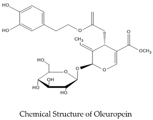
The most prevalent bioactive ingredients in goods made from olive trees are oleuropein. The principal bioactive ingredient in olive trees, this secoiridoid, gives them their distinctively bitter flavor and protects them from becoming rotten [5]. According to reports, in olive leaves the dry source concentration of oleuropein ranges from 6 to 9% (60 to 90 mg/g in olive plants) [6]. But, based on the cultivar and planting circumstances, this could change [7]. In the olive leaves, the oleuropein is a secoiridoid glycoside that is a member of the coumarin component class found. It is distinguished by an ester bond between 2-(3′,4′-dihydroxyphenyl) ethanol (hydroxytyrosol) and the elenolic acid glucoside [8,9]. An ortho-diphenolic group in oleuropein's chemical structure has the ability to scavenge ROS by the donation of hydrogen and to stabilize the ROS by the intramolecular hydrogen bonding. The tyrosol and supplementary single hydroxyl substitutions don't have any antioxidant qualities, whereas o-diOH substitutions ensure [10] and the antioxidant activity of oleuropein is increased due to its capacity which binds the metal ions like iron [11]. The primary health benefits of oleuropein are typically linked to this antioxidant capability. According to a current study, the oleuropein has antimicrobial, cardioprotective, antiviral, antioxidative, anti-inflammatory and antihypertensive properties. It can also act as a natural prooxidant and anticancer agent and has hypocholesterolemic and hypoglycemic effects, as well as the ability to enhance lipid metabolism [12]. Although individual components show certain effects, consuming olive leaf extracts in their whole form, without isolating the main element, may provide greater health benefits, because all of the extracts bioactive chemicals work in concert, which probably influences how well they are absorbed and how bioavailable they are [13].
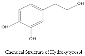
Leaf extract has been discovered to contain a phenolic alcohol known as hydroxytyrosol (3,4-dihydroxyphenylethanol) at a concentration around 2.28 mg per 100 g. This strong antioxidant is produced when oleuropein is hydrolyzed [14]. Because of its straightforward chemical structure, hydroxytyrosol is highly bioavailable and easy for the body to absorb. It doesn't accumulate or cause toxicity because in 15 or 20 minutes, it reaches blood plasma and removed by the digestive or renal systems 6–8 hours later. Since it contains both a lipophilic and a hydrophilic end, it can cross cellular membranes more readily, this amphipathi molecule that is soluble in both water and fats may move molecules throughout the body. Consuming hydroxytyrosol has several advantages for the body due to its structural and molecular characteristics [15]. Because of its great antioxidant potential, hydroxytyrosol shows promise in protecting against chronic diseases. The o-dihydroxyphenyl moiety is principally responsible for this efficiency. Its primary functions include breaking chains, giving hydrogen atoms to peroxyl radicals (ROO*), scavenging free radicals, and chelating metals. Additionally, it strengthens the body's defenses against activates specific cellular signaling pathways and oxidative stress to improve antioxidant protection [16].
2. Anticancer Properties of the Olive Leaves Compounds
According to published research, oleuropein and hydroxytyrosol, the two primary phenolic compounds present in olive leaves, have anticancer effects when taken alone and as pharmacological adjuvants. The anti-tumor properties of these substances have been studied in vitro by using different varieties of cellular models that include hepatocellular carcinoma and breast cancer.

Table 1.
An overview of research using cell lines to examine the anticancer properties of the primary chemicals inside the olive plants.
Table 1.
An overview of research using cell lines to examine the anticancer properties of the primary chemicals inside the olive plants.
| Complex | Range of Concentration | Types of Cancer | Cell based Models | Examined | References |
| Oleuropein | 1, 10, 100 μM | Breast | MCF-7 and T-47D | Decline in cell viability with cell cycle arrest at G2/M phase. | [8] |
| Oleuropein | 100, 200 μM | Breast | MCF-7 | Bax Gene Upregulation and activation of p53-dependent apoptotic pathways through increased expression of p53 gene. | [17] |
| Oleuropein | 0 to 100 μM | Breast | MDA-MB-231 and MCF-7 | Viability of cell and migration were decreased, with cell cycle arrest at the sub-G1 phase was observed. Apoptosis was increased, indicated by elevated PARP and caspase-3/7 cleavage, along with reduced the activation of NF-κB. | [18] |
| Oleuropein | 0 to 700 μM | Breast | MDA-MB-231 and MDA-MB-468 | Reduced the viability of cell, with apoptosis primarily driven by downregulation of the anti-apoptotic genes TNFRSF11B, BIRC5, and CASP4. | [19] |
| Oleuropein | 20 to 100 μM | HCC | HepG2 | Cells showed morphological changes and reduced proliferation, with increased caspase activity and the involvement of family Bcl-2, reduced signaling of PI3K/AKT, but was no change in feasibility. | [20] |
| Oleuropein | 10 to 100 μmol/L | HCC | HepG2 | Cell viability was maintained, with lowered caspase-3 activation. | [21] |
| Oleuropein | 200 μM and 50 μM | HCC | HepG2 | Modulation of the Pro-NGF/NGF ratio through regulation of MMP-7 activity. | [22] |
| Hydroxytyrosol | 10 to 40 μM | HCC | HepG2 | No variations in cell integrity or antioxidant levels were observed. | [23] |
| Hydroxytyrosol | 0.5, 1.0, 5.0 and 10.0 μM | HCC | HepG2 | Increased appearance of antioxidant enzymes, enhanced the activation of ERK and AKT pathways, and promoted nuclear translocation of Nrf2 transcription aspect. | [24] |
| Hydroxytyrosol | 30 to 200 μM | HCC | Hep3B e HepG2 | Cytostatic effects through increased FAS expression, enhancement of the endogenous antioxidant system, increased the IL-6 reduction. | [25] |
| Hydroxytyrosol | 1 μM and 5 μM | HCC | HepG2 | Reduced ER stress. | [26] |
| Hydroxytyrosol | 100 to 400 μM | HCC | HepG2, Hep3B, SK-HEP-1 and Huh-7 | Reduced cell proliferation with G2-M arrest, increased cleavage of PARP, suppression of the NF-κB and PI3K/AKT pathways | [27] |
| Hydroxytyrosol | 0 to 100 μM | HCC | HepG2 | Reduced cell viability accompanied by increased cytosolic calcium. | [28] |
2.1. In Vitro Antitumor Effects of Oleuropein
Previous research conducted a follow-up study to investigate the properties of different doses of the oleuropein compound and its derivative on the survival of MCF-7 cells. The peracetylated aglycone had the strongest effect, and at 100 μM, the only derivatives of oleuropein showed the growth inhibition. Cell viability was extensively decreased as a result of the peracetylated chemical noticeably greater cytotoxic effect compared to the control. These results showed during the G2/M phase, the peracetylated substances (peracetylated hydroxytyrosol and peracetylated aglycone) stop the development of MCF-7 cells, hence causing an antiproliferative impact [8]. When the tumor cells of breast cancer (MCF-7) were treated with the concentration of 200 μM oleuropein, the Bax-expression, Bcl-2-expression, and TP53 gene expression were subsequently examined. According to the results, the Bax gene was significantly upregulated by 0.6 times when compared to cells that were left untreated and cells that were exposed to 100 μM oleuropein. Furthermore, it was discovered that oleuropein causes MCF-7 cells to undergo apoptosis via a p53-dependent mechanism, and that treatment significantly increases expression of the p53 gene (by 2.5 and 3.5 times in comparison to the cells that were not treated) [17].
Treatment of breast cancer cells with oleuropein, a concentration of 0–100 μM for a period of 24h, 48h, and 72h produced similar trends. The viability of breast tumor cells (ER-negative breast tumor) reduced by the oleuropein significantly in a quantity and time-dependent way, whereas the MCF-7 cells showed higher resistance against the treatment. According to the wound curing experiment, oleuropein therapy markedly reduced MDA-MB-231 cell migration. After 72 hours of intervention, the MDA-MB-231 cells exposed a marked enhanced in sub-G1 phase accumulation with increasing the concentration of oleuropein. Additionally, in a dose-related manner, the apoptotic percentage in MDA-MB-231 cells increased dramatically. Furthermore, by significantly the study demonstrated that oleuropein inhibited the signaling pathway of NF-κB by dropping NF-κB activity at 36 and 48 hours. [18].
The cell lines for triple-negative breast tumor, such as MDA-MB-231 and MDA-MB-468 have been shown to have a lower cell survival rate when exposed to Oleuropein. The antiproliferative properties of compound in both cell lines, were demonstrated to be quantity and time-dependent. The MDA-MB-231 cells lines were analyzed with a concentration of 100 to 700 μM, and the MDA-MB-468 cells lines with a concentration of 100 to 400 μM were shown to have cytotoxic effects that were statistically significant (p < 0.0001). According to data, the expected main mechanism of cell death in MDA-MB-468 cells lines treated to oleuropein was apoptosis. MDA-MB-231, on the other hand, tended to necrotize and showed more resistance to apoptosis. Additionally, oleuropein altered the expression of several genes implicated in apoptosis. In the MDA-MB-468 cells lines, the indication of pro-apoptotic proteins such as BNIP2, GADD45A, BNIP3, BCL10 and BID, caspase family adherents (CASP1 and CASP14), the TNF receptor superfamily members (FADD and TNFRSF21), as well as CYCS and CFLAR, was significantly increased. These proteins promote programmed cell death by starting intrinsic, extrinsic, or both pathways. The antiapoptotic genes TNFRSF11B and surviving (BIRC5) were downregulated in MDA-MB-231 cell lines, although CASP4 remained marginally enhanced, despite alterations in genes that inhibit apoptosis induction [19]. When used as a treatment alone, oleuropein has little effect on the viability of hepatocellular carcinoma cells. In HepG2 cell lines, oleuropein at doses between 20 and 100 μM caused morphological changes, decreased cell growth, and cell decease. These cells showed increased caspases and Bcl-2 expression intimate proteins, along with inhibition of the PI3K/AKT pathway, a crucial regulator of apoptosis [20].
For a period of 24 hours, the HepG2 cells was exposed with different quantities of isolated oleuropein present no change in MTT decrease, indicating that cell viability was unaffected. However, under the condition of oxidative stress, oleuropein partially restores the viability of HepG2 cells. The cell viability of HepG2 and cytoskeletal stability are partially restored when oleuropein is used in conjunction with paraquat, a poisonous weedkiller known to induce the oxidative stress and promote cell decease. The Oleuropein's protective effects on cellular viability include a marked decrease increased necrotic cell death and suppression of caspase-3 cleavage. In the multimodal therapy group such as (PQ + OP), PQ-induced necrosis seems to occur after an apoptotic event, suggesting a transition from the necrotic route to apoptosis. Instead of experiencing immediate necrosis as observed in cells treated with oxidative stress, oleuropein helps HepG2 cells showed improved resistance to PQ-induced damage, surviving longer before undergoing a more regulated cell death process [21].
Research has focused on concurrent use of phytochemicals and chemotherapeutic medications. It has been demonstrated that oleuropein (200 μM) enhances cisplatin's (50 μM) anti-tumor effect on HepG2 cells. The activation of gene for pattern metalloproteinase-7 and nerve growth factor (NGF) was measured. MMP-7 activity is necessary promoting the alteration of pro-NGF, that induces cell death, into NGF, that supports cell persistence and variation. Growth factors signaling dysregulation has a key effect on the onset and spread of tumor, including angiogenesis and tumor metastasis. Concurrent therapy the cisplatin-oleuropein synergistic action may offer a more effective chemotherapeutic strategy against HCC than either agent alone because oleuropein alters MMP-7 action without varying NGF gene regulation [22].
2.2. In Vitro Antitumor Effect of Hydroxytyrosol
Hepatocellular carcinoma (HCC) cells have also been used to study hydroxytyrosol anticancer effect. When hydroxytyrosol (10–40 μM) was first applied to the HepG2 cell line, no alterations in the antioxidant status or cell integrity were observed [23]. However, Martin (2010) showed that when the HepG2 cell lines were exposed with HT (0.5, 1.0, 5.0, and 10.0 μM) exhibited a significant rise in both the regulation and activity of glutathione-associated enzymes, including glutathione reductase, glutathione peroxidase and glutathione S-transferase), which are crucial for neutralizing ROS. Moreover, the Nrf2 transcription factor underwent nuclear translocation as a result of HT. The Nrf2 is essential because it supports the body’s defense against oxidative stress by regulating the appearance of many antioxidant and detoxifying enzymes. Two signaling proteins, extracellular regulated kinases (ERK) and protein kinase B (AKT), which belong to the ERK and PI3K/AKT pathways, respectively, were activated to promote this translocation [24].
The antiproliferative effect of hydroxytyrosol (30 to 200 μM) was observed in Hep3B and HepG2 cell lines through suppression of the lipogenic enzyme fatty acid synthase (FAS), activation of the cellular antioxidant structure, and decrease of IL-6 levels at dosage concentrations up to 80 μM. But, IL-6 levels increased with higher levels [25]. Furthermore, hydroxytyrosol with a concentration of (1μM and 5μM) treatment was demonstrated to lessen the endoplasmic reticulum stress that tunicamycin induced in HepG2 cells [26]. In various hepatocellular carcinoma cell models (HepG2, Hep3B, SK-HEP-1, and Huh-7), the hydroxytyrosol with a concentration of (100–400 μM) suppressed cell growth, a conclusion not detected in non-tumoral hepatic cells (HL-7702). This anti-proliferative activity is attributed to hydroxytyrosol pro-apoptotic mechanisms, including enhanced cleavage of PARP by procaspase-3, induction of cell cycle arrest at G2/M, and modulation of the PI3K/AKT and NF-κB signaling tracks [27].
Hydroxytyrosol-induced elevation of [Ca²⁺]i in HepG2 cell lines, cell viability is presently being investigated. It was discovered that hydroxytyrosol inhibited the viability of HepG2 hepatoma cells at doses ranging from 40 to 100 μM. Furthermore, it has been noted that hydroxytyrosol raises intracellular calcium levels ([Ca2+] i), suggesting that calcium signaling plays a part in the compound's cytotoxic actions. The effects of hydroxytyrosol are mediated by store-operated, calcium entrance that is sensitive to protein kinase C (PKC), suggesting that PKC regulates how cells respond to the material. The findings indicate that hydroxytyrosol activates release of calcium from thapsigargin-sensitive ER stores via a PLC-independent pathway, revealing additional ways that it influences cell death processes. Consequently, it proposed a different way whereby hydroxytyrosol might stop the development of hepatocellular carcinoma cells [28].
3. Olive-Derived Antioxidants: Anti-Inflammatory Properties in In Vitro Studies
3.1. Olive Polyphenols as Antioxidant and Anti-Inflammatory Agents in Animal Studies
Olive oil contains antioxidant compounds such as polyphenols are known to restore neuronal function by improving the redox status, and it is commonly acknowledged that inflammation and oxidative and nitrosative stress are the main abnormalities underlying neurodegeneration. Consuming EVOO polyphenols has been linked to several positive benefits of the MD, including anti-inflammatory, anti-aging, cardioprotective, antiviral, anticancer, antioxidant, hypoglycemic, and antibacterial properties [29]. EVOO polyphenols have been demonstrated to be neuroprotective functional decline related to aging and neurodegenerative disorders while preserving endogenous antioxidant stability because by scavenging free radicals, they can defend the DNA against oxidative stress, which stop impaired mitochondrial function, and reduce lipid from oxidation [30]. Additionally, they can prevent the aggregation and toxicity of amyloid β (Aβ) and τ proteins, which are the primary culprits of the neurodegenerative pathway in Alzheimer disease [31,32]. EVOO polyphenols contribute to the cell's redox stability by acting modest pro-oxidants and antioxidants, which causes the cell's antioxidant defenses to be upregulated. they may therefore be viewed as hormetic factors. For example, HT may go through a redox cycle that produces superoxide when peroxidases are present [30], Moreover, tyrosol lengthens the life of C. elegans by triggering the heat shock response [32]. By promoting Nrf2-dependent gene expression, In transgenic mice with AD, HT has been demonstrated to reduce neuroinflammation and brain mitochondrial oxidative stress [33]. By triggering the Nrf2 pathway, oleuropein with a concentration of (60 mg/kg/day) enhanced the activity of mitochondria and decreased the oxidative stress in SHR rats after eight weeks of treatment [34]. Additionally, it was discovered that tyrosol (240 mg/kg) protected exerts protective effects against LPS-induced acute lung injury over the restriction of NF-κB and activating the Nrf-2 and AP-1 pathways. Additionally, EVOO polyphenols improve hepatic Nrf-2 activation and the subsequent release of antioxidant enzymes [35]. When Nrf2, the primary regulator of redox homeostasis, is activated, pro-inflammatory mediators such cytokines, COX-2, and iNOS are inhibited [36]. By downregulating NF-κB and AP-1 expression and activity, EVOO polyphenols help suppress inflammation that reduce inflammation by inhibiting free radical activity, dissolving radical chains, and reducing the HT reduces the formation of reactive oxygen and nitrogen species (ROS and RNS). Moreover, HT stops the progression of the inflammatory cascade following LPS and carrageenan injection by reducing DNA damage and downregulating the stages of pro-inflammatory cytokines (TNF-α and IL-1β), COX2, iNOS, NO, PGE2, and NF-kB [37]. Co-injecting OLE (450 µM) and Aβ42 (50 µM) into adult rats nucleus basalis magnocellularis was found to inhibit Aβ aggregation, reduce astrocyte and microglia activation, and counteract Aβ-mediated damage to choline acetyltransferase-positive neurons within the NBM [38]. According to a different study, OLE prevents motor impairments and Aβ plaque load in transgenic C. elegans lines that continuously express Aβ3–42 [39]. Notably, the consumption of food with added oleuropein, hydroxytyrosol, or a combination of polyphenols from olive processing is of interest effluent was found to exhibit considerable with anti-aggregation and neuroprotective actions in the TgCRND8 mouse model of Aβ accumulation. When given the OLE-supplemented food for eight weeks, mice that were three and six months old (during the initial and intermediate stages of Aβ accumulation, respectively) demonstrated a significant improvement in cognitive function as well as a substantial decline in the quantity, compactness of Aβ plaques and size [40]. Despite treatment starting at 10 months, by which time Aβ and pE3-Aβ had become elevated in the mice deposition in the cortex and hippocampus, there was clear development in the synaptic transmission and decrease in both the level and aggregation density of Aβ42 and pE3-Aβ species. These findings suggest that oral OLE supplementation leads to a decrease in pE3-Aβ production, the disintegration of preformed plaques, and the avoidance of amyloid deposition [40]. By supplementing the diet with other polyphenols obtained from olive mill wastewater or HT administered at the same dosage as pure OLE, the dose-dependent impact of OLE on Aβ peptide aggregation can be reproduced [41,42]. It is interesting to note that OLE therapy (delivered through the diet at 50 mg/kg for a period of 8 weeks) promoted significant neuronal autophagy, even in TgCRND8 mice at late stages of disease. This was accompanied by increased histone H3 acetylation at lysine 9 (H3K9) and histone H4 acetylation at lysine 5 (H4K5) in the brain and hippocampus, along with a decrease in HDAC2 expression. Collectively, these findings suggest that HT and related olive polyphenols act as hormetic agents, mitigating oxidative stress, inflammation, and epigenetic dysregulation connected with aging and neurodegenerative disorders. [40].
3.2. Molecular Mechanism of Antioxidant Activity
Defense mechanisms against increased reactive oxygen species (ROS) are essential for cell survival, because ROS overproduction is associated to lipid, protein, or DNA damage that precedes degenerative diseases [43]. To adapt to oxidative stress, organisms maintain complex antioxidant systems, primarily centered on glutathione (GSH), because antioxidants inhibit oxidation. Sadly, only a small number of medications and biological substances like vitamins have been shown to have antioxidant properties, but they may also have negative side effects [44]. These days, scientists are concentrating on the antioxidant qualities of organic substances that don't have any negative side effects. Specifically, the significance of the hydrophilic and lipophilic phenols' antioxidant activity in EVOO has come to light [45]. Plants biologically create this proportion to respond to wounds caused by different diseases or insects [46]. The primary phenolic compounds of EVOO, such as OLE, exhibit potent antioxidant activity and HT is fundamental to applying their metabolic and pharmacokinetic properties, and it is connected with their comparative bioavailability with a considerable degree of consumption [47]. Because of their catecholic structures, OLE and HT exhibit distinct antioxidant behaviors at the molecular level: (i) Through neutralization of peroxyl radicals and interruption of peroxidative chain reactions, stable resonance structures are generated [48] and (ii) via chelating metal ions and protecting LDL from copper sulfate-induced oxidation [49]. OLE and HT may chelate metals through their hydroxyl groups, which donate electrons and form intramolecular H-bonds with free radicals [50]. In addition, the ROS-scavenging effects of OLE and HT were examined using non-metal oxidation models. Polyphenols effectively protect catalase (CAT) from inactivation by hypochlorous acid (HOCl), which inhibits atherosclerosis when HOCl oxidizes LDL by apoB-100 chlorination, according to in vitro data [11]. Furthermore, HT has been shown to raise GSH levels, which improves the cell's redox state [51]. It has recently been discovered that decreased stages of the transcriptional Nuclear factor erythroid 2 (NF-E2)-related aspect 2 (Nrf2) are the key source of oxidative damage in age-related disorders [52], and because of its role as a facilitator of the cell's general adaptive responses, such as inflammation and proteostasis, It has been identified as a promising target for treating metabolic diseases, including obesity [53,54]. Nevertheless, Nrf2 plays a critical function in changing oxidation resistance [55]. Resulting the Nrf2 initiation and subsequent the nucleus translocation, the Nrf2 binding to antioxidant response elements (ARE) influences the transcriptional regulation of a number of antioxidant enzymes, with superoxide dismutase (SOD), c-glutamylcysteine synthetase (c-GCS), glutathione S-transferase (GST), and NADPH quinone oxidoreductase-1 (NQO1) [56].
3.3. Molecular Mechanism of Anti-inflammatory of Olive Plant In Vitro
The immune system uses inflammation as a vital defense mechanism to identify and destroy pathogens and contaminated cells while also encouraging tissue healing and reestablishing equilibrium in the body. This process, which involves temporarily increased amounts of cytokines with the capacity to trigger both innate and adaptive immune responses systems, is incorporated into numerous coordinated processes. There are several negative effects on organismal homeostasis when an inflammatory response is not controlled. Chronic inflammation, which contains leukocytes collected by macrophages and lymphocytes, is a long-term, unresolved, and uncontrollable immune response that can result from an ongoing inflammatory response. This can cause injury to the mass or organs locally or systemically, as well as a breakdown in normal physiological function.
Chronic inflammation increases with age and is causally linked to the beginning or progression of disease. In actuality, cytokines, chemokines, and the regulation of proteins associated with irritation are all increased in the adults and autoimmune disease patients. These individuals also show higher rates of metabolic syndrome, cardiovascular disease, frailty, multimorbidity, and reduced physical and cognitive function. Therefore, treatments the interventions that modulate inflammation and correct dysregulated inflammatory responses can effectively halt disease progression. Regular consumption of foods high in polyphenol has been shown to help prevent chronic disorders like heart disease such as diabetes and obesity. The main reason of this advantageous effect is the poly phenolic molecule's anti-inflammatory qualities, which are demonstrated by a quantity of mechanisms, with antioxidant activity and the modification of signaling cascades and transcriptional processes.
4. In Vivo and In Vitro Studies of Antidiabetic Activities of Oleuropein and Hydroxytyrosol:
4.1. In Vitro Studies of Oleuropein's Impact on Skeletal Muscle Cells
In C2Cl2 muscle cells, OLE treatment led to higher stages of stimulated AMPK, ACC, and ERK proteins and promoted glucose utilization. Furthermore, OLE showed antioxidant qualities by dramatically reducing H2O2-induced (ROS) as compared to untreated cells [57]. When C2C12 myotubes were exposed to OLE, Fujiwara et al. observed a considerable rise in glucose uptake that was similar to what insulin had. In contrast to insulin, OLE raised the levels of protein of phosphorylated AMPK but not the content of protein of phosphorylated Akt, per the previously described Hadrich et al. study. Furthermore, the OLE pretreatment also enhanced the level of GLUT4 mRNA and rescued insulin-mediated uptake of glucose under palmitate exposure by reducing palmitate-induced decreases in phosphorylated/activated AMPK [58]. OLE treatment in cultured avian muscle cells promoted mitochondrial biogenesis, upregulated avUCP, PGC1-α, TFAM, NRF1, ATP5a1, and SIRT1 expression, increased cytochrome c oxidase activity, and reduced the superoxide levels of mitochondria, reflecting lower ROS formation (Table 2) [59].
1.1. Oleuropein's Effects on Hepatocytes (In Vitro)
OLE attenuated FFA-induced lipid accretion, suggesting hepatoprotective effects against steatosis, in both human hepatoma-derived (HepG2) cell lines and mouse hepatocyte (FL83B) cell lines. Additionally, the OLE (10 µM) dramatically reduced the size of lipid droplets in FL38B and HepG2 cells. FFA-induced elevations in phosphorylated/activated ERK protein levels were inhibited by the OLE treatment. Adipose differentiation-related protein (ADRP), which associates with lipid droplets, and tail interacting protein of 47 kDa (TIP47) mRNA levels were unaffected by OLE therapy. Additionally, OLE had no effect on JNK or phosphorylated/activated Akt protein levels (Table 3) [60] The cellular triglyceride content significantly decreased when Vergani et al. treated FaO rat hepatoma cells to OLE, suggesting that FFA-induced steatosis was attenuated. Furthermore, OLE decreased the rise in malondialdehyde (MDA) levels, a sign of lipid peroxidation, brought on by FFA (Table 3) [61]. According to Malliou et al.'s molecular docking simulations, OLE fits structurally within the PPARα binding cavity in its integral form. Similarly, a luciferase reporter gene experiment showed that the PPRE-luc plasmid activity had increased, suggesting that it might activate PPARα and cause heterodimerization with RXR. Additionally, mRNA and protein levels of PPARα, along with numerous downstream target proteins including acyl-CoA oxidase 1 (ACOX1), cytochrome P450 family 4 subfamily 3 polypeptide 14 (CYP4A14), lipin 1, and acyl-CoA thioesterase 4 (ACOT4), were elevated in HepG2 cells treated with OLE (Table 3) [62]. Using HepG2 cells, Santini et al. discovered that OLE administration significantly decreased palmitate-induced hepatic lipid buildup [63].
1.1. Effect of Oleuropein on Animal Models of Diet-Induced Diabetes (In Vivo)
Numerous research has looked into OLE's capacity to protect against the harmful consequences of a high-fat or "Western style" diet (HFD). Oi-Kano et al. discovered that OLE had anti-obesogenic qualities by protecting male Sprague-Dawley rats against weight gain brought on by an HFD. Administration of OLE resulted in a considerable decrease in the rises in plasma TG, FFA, TC, and leptin levels that were produced by HFD. Rats given OLE supplementation presented advanced levels of UCP-1, a protein implicated in adipocyte thermogenesis, in their interscapular brown adipose tissue (iBAT) (Table 4) [64]. In a different study, Jemai et al. observed that how oleuropein affected a male Wistar rats assumed a high-cholesterol diet (HCD) and revealed that it disallowed the liver/bodyweight relation from growing as a result of HCD. According to serum lipid study, the OLE significantly reduced the growth in TC, TG, and LDL-C plasma levels carried on by HCD. The qualities of the phenolic compound's hypolipidemic were further verified by its capability to increase HDL-C levels in HCD rats, that had earlier dropped. OLE therapy restored the action of hepatic antioxidant enzymes SOD and CAT impaired by a high-cholesterol diet. Secoiridoid supplementation in HCD-fed rats enhanced ABTS radical scavenging and reduced the lipid peroxidation across the heart, aorta, liver and kidneys demonstrated that the OLE supplementation enhanced antioxidant activity. OLE treatment stopped the development of aortic wall lesioning and cardiac muscle hypertrophy brought on by HCD. Likewise, OLE stopped the development of fatty cysts and the movement showing peripheral localization of hepatocyte nuclei (Table 4) [65]. Microarray research showed that OLE with a concentration of (0.03% w/w in diet) more than doubled the hepatic expression of over 90 genes in a mice fed with a high-fat diet. In specific, OLE treatment reduced the quantity of hepatic genes associated with inflammation and oxidative stress and also decrease the level of mRNA genes involved in the absorption pathway of hepatic fatty acid [66]. When mice were treated with OLE with a concentration of (0.03% w/w) ad libitum for a period of 10 weeks, a reduction in body and liver weight gain induced by a high-fat diet was observed. Elevated liver and plasma FFA, TC, and TG, as well as plasma AST and ALT, were attenuated after OLE therapy. Additionally, OLE markedly reduced expression levels of mRNAs encoding cyclin D (Cyc-D), E2F1, cathepsin S (CTSS), secreted frizzled-related protein 5 (SFRP5), dickkopf homolog 2 (DKK2), and lipid metabolism-related genes such as liver X receptor (LXR), fatty acid-binding protein 2 (aP2), lipoprotein lipase (LPL), and PPARγ2. Furthermore, HFD-induced increases in phosphorylated ERK decreases in β-catenin amount were mitigated by OLE treatment. Additionally, OLE exhibited anti-inflammatory effects by modulating TLR2/4, myeloid differentiation factor 88 (MyD88), IL-1β, IL-6, TNFα, interferon beta (IFNβ), TNF receptor superfamily member 6 (FAS), and TNF-related apoptosis-inducing ligand when OLE reduced the rise in mRNA levels brought on by HFD [67].
1.1. Hydroxytyrosol (HT) Effects on Hepatocytes (In Vitro)
Hydroxytyrosol with a concentration of (100 µM) maintenance of primary mouse hepatocytes, under ischemia/reperfusion (I/R)-like circumstances led to a dose-dependent reduction in apoptosis and an increase in cell feasibility (Table 5) [68]. Furthermore, HT considerably reduced the decline in antioxidant enzymes like SOD1, SOD2, and catalase (CAT) brought on by I/R. Overall, HT therapy shielded mouse hepatocytes against oxidative damage and I/R-induced injury. Rat hepatocytes treated with HT (25 µM) showed decreased de novo lipid production of cholesterol, triglycerides, and fatty acids without compromising cell viability. Furthermore, HT treatment markedly reduced the enzymatic activity that involved in cholesterogenesis (3-hydroxy-3-methyl-glutaryl-CoA reductase), fatty acid formation (ACC), and triglyceride formation (diacylglycerol acyltransferase) [69]. Additionally, HT enhanced AMPK and its downstream target ACC phosphorylation, indicating that AMPK mediates the process of HT's decreased lipid production [69]. These outcomes recommend that HT prevents the synthesis of lipids in hepatic tissue. According to another study, HT (0.05–2 mM) reduced the production of thiobarbituric acid reactive substances (TBARS), a biomarker of lipid peroxidation, in vitamin E-deficient rat liver microsomes, which in turn reduced lipid peroxidation [70]. Malonaldehyde, a byproduct of lipid oxidation, combines with thiobarbituric acid to generate TBARS, which damages cells. A current research found that when microsomal fractions from vitamin E-low rats were exposed to improved HT compounds (0.05–2 mM), the generation of TBARS and detrimental lipid peroxidation were more strongly and potently inhibited than when pure HT was used (Table 5) [71].
Overall, the aforementioned research has demonstrated that HT has hepatoprotective qualities by boosting antioxidant activity, hepatocyte survival, and reducing apoptosis [68,70,71]. More significantly, HT therapy dramatically decreased lipid production [69]. Lipid buildup in hepatocytes is frequently linked to hepatic steatosis, which in turn causes decreased glucose utilization, liver damage, and fibrosis, all of which exacerbate the onset of insulin resistance and type 2 diabetes. According to these investigations, HT may offer protection against the development of hepatic insulin resistance as well as hepatic steatosis.
1.1. Effect of Hydroxytyrosol (HT) on In Vivo Diabetes in Rodents Caused by Alloxan
Intraperitoneal injections of alloxan monohydrate (150 mg/kg) were administered to male Wistar rats to induce diabetes; animals with hyperglycemia (blood glucose levels of 2 g/L after two weeks) were kept for further study (Table 6) [72]. For two months, the treatment groups received daily intraperitoneal injections of pure hydroxytyrosol (F3), monomeric phenols (F1), or polymeric phenols (F2) from olive mill waste at a dose of 20 mg/kg. Blood glucose levels were markedly lowered by all three treatments, but pure HT (F3) was particularly effective. Animals treated with HT showed decreased levels of fatty cysts, bilirubin, and TBARS, markers of liver damage. HT enhanced circulating high-density lipoprotein (HDL), hepatic glycogen, and antioxidant enzymes (SOD, CAT, and GPX) in the kidney and liver. Furthermore, HT therapy reduced the harmful effects of alloxan on pancreatic β cells [72]. In 2009, Jemai et al. investigated the potential antioxidant and antidiabetic effects of administering HT and oleuropein to male Wistar rats. After four weeks of treatment with HT dissolved in drinking water to concentrations of 8 or 16 mg/kg, diabetes was caused by an intraperitoneal injection of alloxan (180 mg/kg) [73]. Hepatic glycogen and antioxidant enzymes (SOD, CAT) increased concurrently with reduction of hyperglycemia, hypercholesterolemia, and hepatic oxidative damage (TBARS) in all treatment groups. Dose-dependence was demonstrated by the higher effects of the 16 mg/kg dose compared to the 8 mg/kg dose [73]. In a diabetic rat model, this study shown that HT could maintain better lipid profiles, glucose levels, and antioxidant activity.
5. Conclusion
Oleuropein and hydroxytyrosol, the primary bioactive compounds of Olea europaea, exhibit remarkable antioxidant, anti-inflammatory, anticancer, and antidiabetic activities. These composites serve an important function in combating oxidative stress, regulating immune responses, and improving metabolic health, making them valuable in disease prevention and treatment. The antioxidant activity of these compounds protects against cellular damage and enhances mitochondrial function, which is essential for neuroprotection and cardiovascular health. Their anticancer effects, demonstrated in various In vitro and In vivo studies, highlight their potential to inhibit tumor progression, induce apoptosis, and enhance the efficacy of chemotherapy. The anti-inflammatory properties of olive-derived bioactives help modulate cytokine levels, reduce chronic inflammation, and protect against neurodegenerative and autoimmune diseases. Additionally, their role in diabetes management is evident through improved glucose metabolism, enhanced insulin sensitivity, and reduced lipid accumulation in hepatic and adipose tissues. These findings suggest that oleuropein and hydroxytyrosol could serve as natural alternatives to conventional therapeutic agents for managing metabolic and inflammatory disorders. Despite extensive research, challenges remain regarding the clinical translation of these compounds. Bioavailability, stability, and optimal dosage require further exploration to maximize their therapeutic efficacy. Future research should focus on enhancing their absorption through innovative drug delivery systems and conducting well-structured clinical trials to establish their safety and effectiveness in humans. The integration of these compounds into nutraceuticals, functional foods, and pharmaceuticals presents a promising strategy for natural and sustainable healthcare solutions. Given the increasing global interest in plant-based therapeutic agents, Olea europaea bioactives offer immense potential for future biomedical research and clinical applications. Collaborative efforts between researchers, healthcare professionals, and the pharmaceutical industry will be essential in unlocking their full therapeutic benefits and ensuring their successful application in modern medicine.
6. Future Perspectives
Future research on oleuropein and hydroxytyrosol should focus on optimizing their pharmacokinetic properties to enhance bioavailability and stability through novel drug delivery systems such as nano formulations, liposomes, and polymeric nanoparticles. Further investigations into their precise molecular mechanisms and interactions with key cellular pathways in cancer, inflammation, and metabolic disorders are necessary to establish their therapeutic potential. To validate their efficacy, safety, and long-term outcomes in humans, large randomized clinical trials are indispensable. Additionally, exploring their synergistic interactions with conventional chemotherapeutic and metabolic drugs could enhance their clinical relevance. The incorporation of these bioactive compounds into nutraceuticals and functional foods could offer a natural and sustainable strategy for disease prevention and health promotion. A personalized medicine approach, considering genetic and metabolic variations, could further optimize their therapeutic benefits. Multidisciplinary collaborations between researchers, clinicians, and the pharmaceutical industry will be critical in translating these natural compounds into effective clinical applications, paving the way for their use in modern medicine.
Author Contributions
Conceptualization and study design, F.S., A.U.K., M.I., and F.M.; Investigation, F.S. and S.S.; Writing original draft preparation, F.S. and M.I.; Writing review and editing, F.M., S.S., and C.L.; Supervision, A.U.K., C.L., and M.I. All authors have read and agreed to the published final version of the manuscript.
Funding
The author(s) received no financial support for the research, authorship, and/or publication of this review article.
Conflicts of Interest
This study has no conflict of interest to be declared by any author.
References
- Hashmi, M.A.; Khan, A.; Hanif, M.; Farooq, U.; Perveen, S. Traditional uses, phytochemistry, and pharmacology of Olea europaea (olive). Evidence-Based Complementary and Alternative Medicine 2015, 2015, 541591. [Google Scholar] [CrossRef] [PubMed]
- Ilgar, R. Çanakkale ilinde zeytin yetiştiriciliği ve yaşanan sorunlar. Coğrafya Dergisi 2016, 19–32. [Google Scholar]
- Breton, C.; Terral, J.-F.; Pinatel, C.; Médail, F.; Bonhomme, F.; Bervillé, A. The origins of the domestication of the olive tree. Comptes rendus biologies 2009, 332, 1059–1064. [Google Scholar] [CrossRef]
- Abdulrahman, H.A.; Al-Bamarny, S.F. Influence of light intensity and some chemical compounds on physiological responses in olive transplants (Olea europaea L.). Pak. J. Bot 2020, 52, 435–445. [Google Scholar] [CrossRef]
- Scicchitano, S.; Vecchio, E.; Battaglia, A.M.; Oliverio, M.; Nardi, M.; Procopio, A.; Costanzo, F.; Biamonte, F.; Faniello, M.C. The double-edged sword of oleuropein in ovarian cancer cells: From antioxidant functions to cytotoxic effects. International Journal of Molecular Sciences 2023, 24, 842. [Google Scholar] [CrossRef]
- Antunes, B.d.F.; Otero, D.M.; Bonemann, D.H.; Ribeiro, A.S.; Jacques, A.C.; Zambiazi, R.C. Evaluation of physicochemical, bioactive composition and profile of fatty acids in leaves of different olive cultivars. Revista Ceres 2021, 68, 511–520. [Google Scholar] [CrossRef]
- Martínez-Navarro, M.E.; Kaparakou, E.H.; Kanakis, C.D.; Cebrián-Tarancón, C.; Alonso, G.L.; Salinas, M.R.; Tarantilis, P.A. Quantitative determination of the main phenolic compounds, antioxidant activity, and toxicity of aqueous extracts of olive leaves of Greek and Spanish genotypes. Horticulturae 2023, 9, 55. [Google Scholar] [CrossRef]
- Bulotta, S.; Corradino, R.; Celano, M.; D’Agostino, M.; Maiuolo, J.; Oliverio, M.; Procopio, A.; Iannone, M.; Rotiroti, D.; Russo, D. Antiproliferative and antioxidant effects on breast cancer cells of oleuropein and its semisynthetic peracetylated derivatives. Food chemistry 2011, 127, 1609–1614. [Google Scholar] [CrossRef]
- Samara, P.; Christoforidou, N.; Lemus, C.; Argyropoulou, A.; Ioannou, K.; Vougogiannopoulou, K.; Aligiannis, N.; Paronis, E.; Gaboriaud-Kolar, N.; Tsitsilonis, O. New semi-synthetic analogs of oleuropein show improved anticancer activity in vitro and in vivo. European Journal of Medicinal Chemistry 2017, 137, 11–29. [Google Scholar] [CrossRef] [PubMed]
- Nediani, C.; Ruzzolini, J.; Romani, A.; Calorini, L. Oleuropein, a bioactive compound from Olea europaea L., as a potential preventive and therapeutic agent in non-communicable diseases. Antioxidants 2019, 8, 578. [Google Scholar] [CrossRef] [PubMed]
- Bulotta, S.; Oliverio, M.; Russo, D.; Procopio, A. Biological activity of oleuropein and its derivatives. In Handbook of Natural Products, KG Ramawat, JM Merillon: 2013; pp. 3605-3638.
- Ruzzolini, J.; Peppicelli, S.; Bianchini, F.; Andreucci, E.; Urciuoli, S.; Romani, A.; Tortora, K.; Caderni, G.; Nediani, C.; Calorini, L. Cancer glycolytic dependence as a new target of olive leaf extract. Cancers 2020, 12, 317. [Google Scholar] [CrossRef] [PubMed]
- García-Villalba, R.; Larrosa, M.; Possemiers, S.; Tomás-Barberán, F.; Espín, J. Bioavailability of phenolics from an oleuropein-rich olive (Olea europaea) leaf extract and its acute effect on plasma antioxidant status: Comparison between pre-and postmenopausal women. European journal of nutrition 2014, 53, 1015–1027. [Google Scholar] [CrossRef] [PubMed]
- Martín-García, B.; Verardo, V.; León, L.; De la Rosa, R.; Arráez-Román, D.; Segura-Carretero, A.; Gómez-Caravaca, A.M. GC-QTOF-MS as valuable tool to evaluate the influence of cultivar and sample time on olive leaves triterpenic components. Food Research International 2019, 115, 219–226. [Google Scholar] [CrossRef] [PubMed]
- González-Santiago, M.; Fonollá, J.; Lopez-Huertas, E. Human absorption of a supplement containing purified hydroxytyrosol, a natural antioxidant from olive oil, and evidence for its transient association with low-density lipoproteins. Pharmacological Research 2010, 61, 364–370. [Google Scholar] [CrossRef]
- Torić, J.; Barbarić, M.; Brala, J. Hydroxytyrosol, Tyrosol and Derivatives and Their Potential Effects on Human Health. Molecules (Basel, Switzerland) 2019, 24, E2001–E2001. [Google Scholar]
- Hassan, Z.K.; Elamin, M.H.; Omer, S.A.; Daghestani, M.H.; Al-Olayan, E.S.; Elobeid, M.A.; Virk, P. Oleuropein induces apoptosis via the p53 pathway in breast cancer cells. Asian Pacific Journal of Cancer Prevention 2013, 14, 6739–6742. [Google Scholar] [CrossRef]
- Liu, L.; Ahn, K.S.; Shanmugam, M.K.; Wang, H.; Shen, H.; Arfuso, F.; Chinnathambi, A.; Alharbi, S.A.; Chang, Y.; Sethi, G. Oleuropein induces apoptosis via abrogating NF-κB activation cascade in estrogen receptor–negative breast cancer cells. Journal of cellular biochemistry 2019, 120, 4504–4513. [Google Scholar] [CrossRef]
- Messeha, S.S.; Zarmouh, N.O.; Asiri, A.; Soliman, K.F. Gene expression alterations associated with oleuropein-induced antiproliferative effects and S-phase cell cycle arrest in triple-negative breast cancer cells. Nutrients 2020, 12, 3755. [Google Scholar] [CrossRef]
- Yan, C.M.; Chai, E.Q.; Cai, H.Y.; Miao, G.Y.; Ma, W. Oleuropein induces apoptosis via activation of caspases and suppression of phosphatidylinositol 3-kinase/protein kinase B pathway in HepG2 human hepatoma cell line. Molecular Medicine Reports 2015, 11, 4617–4624. [Google Scholar] [CrossRef]
- Katsoulieris, E.N. The olive leaf extract oleuropein exerts protective effects against oxidant-induced cell death, concurrently displaying pro-oxidant activity in human hepatocarcinoma cells. Redox report 2016, 21, 90–97. [Google Scholar] [CrossRef]
- Sherif, I.O.; Al-Gayyar, M.M. Oleuropein potentiates anti-tumor activity of cisplatin against HepG2 through affecting proNGF/NGF balance. Life Sciences 2018, 198, 87–93. [Google Scholar] [CrossRef]
- Goya, L.; Mateos, R.; Bravo, L. Effect of the olive oil phenol hydroxytyrosol on human hepatoma HepG2 cells: Protection against oxidative stress induced by tert-butylhydroperoxide. European Journal of Nutrition 2007, 46, 70–78. [Google Scholar] [CrossRef]
- Martín, M.A.; Ramos, S.; Granado-Serrano, A.B.; Rodríguez-Ramiro, I.; Trujillo, M.; Bravo, L.; Goya, L. Hydroxytyrosol induces antioxidant/detoxificant enzymes and Nrf2 translocation via extracellular regulated kinases and phosphatidylinositol-3-kinase/protein kinase B pathways in HepG2 cells. Molecular nutrition & food research 2010, 54, 956–966. [Google Scholar]
- Tutino, V.; Caruso, M.G.; Messa, C.; Perri, E.; Notarnicola, M. Antiproliferative, antioxidant and anti-inflammatory effects of hydroxytyrosol on human hepatoma HepG2 and Hep3B cell lines. Anticancer research 2012, 32, 5371–5377. [Google Scholar]
- Giordano, E.; Davalos, A.; Nicod, N.; Visioli, F. Hydroxytyrosol attenuates tunicamycin-induced endoplasmic reticulum stress in human hepatocarcinoma cells. Molecular nutrition & food research 2014, 58, 954–962. [Google Scholar]
- Zhao, B.; Ma, Y.; Xu, Z.; Wang, J.; Wang, F.; Wang, D.; Pan, S.; Wu, Y.; Pan, H.; Xu, D. Hydroxytyrosol, a natural molecule from olive oil, suppresses the growth of human hepatocellular carcinoma cells via inactivating AKT and nuclear factor-kappa B pathways. Cancer letters 2014, 347, 79–87. [Google Scholar] [CrossRef] [PubMed]
- Cheng, H.-H.; Liao, W.-C.; Lin, R.-A.; Chen, I.-S.; Wang, J.-L.; Chien, J.-M.; Kuo, C.-C.; Hao, L.-J.; Chou, C.-T.; Jan, C.-R. Hydroxytyrosol [2-(3, 4-dihydroxyphenyl)-ethanol], a natural phenolic compound found in the olive, alters Ca2+ signaling and viability in human HepG2 hepatoma cells. Journal of Physiology Investigation 2022, 65, 30–36. [Google Scholar] [CrossRef] [PubMed]
- Serreli, G.; Deiana, M. Extra virgin olive oil polyphenols: Modulation of cellular pathways related to oxidant species and inflammation in aging. Cells 2020, 9, 478. [Google Scholar] [CrossRef]
- Sarsour, E.H.; Kumar, M.G.; Kalen, A.L.; Goswami, M.; Buettner, G.R.; Goswami, P.C. MnSOD activity regulates hydroxytyrosol-induced extension of chronological lifespan. Age 2012, 34, 95–109. [Google Scholar] [CrossRef]
- Pandey, K.B.; Rizvi, S.I. Plant polyphenols as dietary antioxidants in human health and disease. Oxidative medicine and cellular longevity 2009, 2, 270–278. [Google Scholar] [CrossRef]
- Canuelo, A.; Gilbert-Lopez, B.; Pacheco-Linan, P.; Martínez-Lara, E.; Siles, E.; Miranda-Vizuete, A. Tyrosol, a main phenol present in extra virgin olive oil, increases lifespan and stress resistance in Caenorhabditis elegans. Mechanisms of ageing and development 2012, 133, 563–574. [Google Scholar] [CrossRef]
- Peng, Y.; Hou, C.; Yang, Z.; Li, C.; Jia, L.; Liu, J.; Tang, Y.; Shi, L.; Li, Y.; Long, J. Hydroxytyrosol mildly improve cognitive function independent of APP processing in APP/PS1 mice. Molecular nutrition & food research 2016, 60, 2331–2342. [Google Scholar]
- Sun, W.; Wang, X.; Hou, C.; Yang, L.; Li, H.; Guo, J.; Huo, C.; Wang, M.; Miao, Y.; Liu, J. Oleuropein improves mitochondrial function to attenuate oxidative stress by activating the Nrf2 pathway in the hypothalamic paraventricular nucleus of spontaneously hypertensive rats. Neuropharmacology 2017, 113, 556–566. [Google Scholar] [CrossRef] [PubMed]
- Soto-Alarcon, S.A.; Valenzuela, R.; Valenzuela, A.; Videla, L.A. Liver protective effects of extra virgin olive oil: Interaction between its chemical composition and the cell-signaling pathways involved in protection. Endocrine, Metabolic & Immune Disorders-Drug Targets (Formerly Current Drug Targets-Immune, Endocrine & Metabolic Disorders) 2018, 18, 75–84. [Google Scholar]
- Ahmed, S.M.U.; Luo, L.; Namani, A.; Wang, X.J.; Tang, X. Nrf2 signaling pathway: Pivotal roles in inflammation. Biochimica et Biophysica Acta (BBA)-Molecular basis of disease 2017, 1863, 585–597. [Google Scholar] [CrossRef]
- Fki, I.; Sayadi, S.; Mahmoudi, A.; Daoued, I.; Marrekchi, R.; Ghorbel, H. Comparative study on beneficial effects of hydroxytyrosol-and oleuropein-rich olive leaf extracts on high-fat diet-induced lipid metabolism disturbance and liver injury in rats. BioMed Research International 2020, 2020, 1315202. [Google Scholar] [CrossRef]
- Luccarini, I.; Dami, T.E.; Grossi, C.; Rigacci, S.; Stefani, M.; Casamenti, F. Oleuropein aglycone counteracts Aβ42 toxicity in the rat brain. Neuroscience Letters 2014, 558, 67–72. [Google Scholar] [CrossRef]
- Diomede, L.; Rigacci, S.; Romeo, M.; Stefani, M.; Salmona, M. Oleuropein aglycone protects transgenic C. elegans strains expressing Aβ42 by reducing plaque load and motor deficit. PLoS one 2013, 8, e58893. [Google Scholar] [CrossRef]
- Luccarini, I.; Grossi, C.; Rigacci, S.; Coppi, E.; Pugliese, A.M.; Pantano, D.; la Marca, G.; Dami, T.E.; Berti, A.; Stefani, M. Oleuropein aglycone protects against pyroglutamylated-3 amyloid-ß toxicity: biochemical, epigenetic and functional correlates. Neurobiology of aging 2015, 36, 648–663. [Google Scholar] [CrossRef]
- Pantano, D.; Luccarini, I.; Nardiello, P.; Servili, M.; Stefani, M.; Casamenti, F. Oleuropein aglycone and polyphenols from olive mill waste water ameliorate cognitive deficits and neuropathology. British Journal of Clinical Pharmacology 2017, 83, 54–62. [Google Scholar] [CrossRef]
- Nardiello, P.; Pantano, D.; Lapucci, A.; Stefani, M.; Casamenti, F. Diet supplementation with hydroxytyrosol ameliorates brain pathology and restores cognitive functions in a mouse model of amyloid-β deposition. Journal of Alzheimer's Disease 2018, 63, 1161–1172. [Google Scholar] [CrossRef] [PubMed]
- Halliwell, B. Oxidative stress and cancer: have we moved forward? Biochemical Journal 2007, 401, 1–11. [Google Scholar] [PubMed]
- Ďuračková, Z. Some current insights into oxidative stress. Physiological research 2010, 59. [Google Scholar] [CrossRef] [PubMed]
- Jiang, L.; Yang, K.-h.; Tian, J.-h.; Guan, Q.-l.; Yao, N.; Cao, N.; Mi, D.-h.; Wu, J.; Ma, B.; Yang, S.-h. Efficacy of antioxidant vitamins and selenium supplement in prostate cancer prevention: a meta-analysis of randomized controlled trials. Nutrition and cancer 2010, 62, 719–727. [Google Scholar]
- Bendini, A.; Cerretani, L.; Carrasco-Pancorbo, A.; Gómez-Caravaca, A.M.; Segura-Carretero, A.; Fernández-Gutiérrez, A.; Lercker, G. Phenolic molecules in virgin olive oils: a survey of their sensory properties, health effects, antioxidant activity and analytical methods. An overview of the last decade. Molecules 2007, 12, 1679–1719. [Google Scholar] [CrossRef]
- Servili, M.; Montedoro, G. Contribution of phenolic compounds to virgin olive oil quality. European Journal of Lipid Science and Technology 2002, 104, 602–613. [Google Scholar] [CrossRef]
- Carluccio, M.A.; Massaro, M.; Scoditti, E.; De Caterina, R. Vasculoprotective potential of olive oil components. Molecular nutrition & food research 2007, 51, 1225–1234. [Google Scholar]
- Cicerale, S.; Lucas, L.; Keast, R. Antimicrobial, antioxidant and anti-inflammatory phenolic activities in extra virgin olive oil. Current opinion in biotechnology 2012, 23, 129–135. [Google Scholar] [CrossRef]
- Cicerale, S.; Lucas, L.; Keast, R. Biological activities of phenolic compounds present in virgin olive oil. International journal of molecular sciences 2010, 11, 458–479. [Google Scholar]
- Visioli, F.; Poli, A.; Gall, C. Antioxidant and other biological activities of phenols from olives and olive oil. Medicinal research reviews 2002, 22, 65–75. [Google Scholar]
- Lucas, L.; Russell, A.; Keast, R. Molecular mechanisms of inflammation. Anti-inflammatory benefits of virgin olive oil and the phenolic compound oleocanthal. Current pharmaceutical design 2011, 17, 754–768. [Google Scholar] [CrossRef] [PubMed]
- Visioli, F.; Bellomo, G.; Galli, C. Free radical-scavenging properties of olive oil polyphenols. Biochemical and biophysical research communications 1998, 247, 60–64. [Google Scholar] [CrossRef] [PubMed]
- Kouka, P.; Priftis, A.; Stagos, D.; Angelis, A.; Stathopoulos, P.; Xinos, N.; Skaltsounis, A.-L.; Mamoulakis, C.; Tsatsakis, A.M.; Spandidos, D.A. Assessment of the antioxidant activity of an olive oil total polyphenolic fraction and hydroxytyrosol from a Greek Olea europea variety in endothelial cells and myoblasts. International journal of molecular medicine 2017, 40, 703–712. [Google Scholar] [CrossRef] [PubMed]
- Tan, B.L.; Norhaizan, M.E.; Liew, W.-P.-P.; Sulaiman Rahman, H. Antioxidant and oxidative stress: a mutual interplay in age-related diseases. Frontiers in pharmacology 2018, 9, 1162. [Google Scholar] [CrossRef]
- Hayes, J.D.; Dinkova-Kostova, A.T. The Nrf2 regulatory network provides an interface between redox and intermediary metabolism. Trends in biochemical sciences 2014, 39, 199–218. [Google Scholar] [CrossRef]
- Hadrich, F.; Garcia, M.; Maalej, A.; Moldes, M.; Isoda, H.; Feve, B.; Sayadi, S. Oleuropein activated AMPK and induced insulin sensitivity in C2C12 muscle cells. Life Sciences 2016, 151, 167–173. [Google Scholar] [CrossRef]
- Fujiwara, Y.; Tsukahara, C.; Ikeda, N.; Sone, Y.; Ishikawa, T.; Ichi, I.; Koike, T.; Aoki, Y. Oleuropein improves insulin resistance in skeletal muscle by promoting the translocation of GLUT4. Journal of Clinical Biochemistry and Nutrition 2017, 61, 196–202. [Google Scholar] [CrossRef]
- Kikusato, M.; Muroi, H.; Uwabe, Y.; Furukawa, K.; Toyomizu, M. Oleuropein induces mitochondrial biogenesis and decreases reactive oxygen species generation in cultured avian muscle cells, possibly via an up-regulation of peroxisome proliferator-activated receptor γ coactivator-1α. Animal Science Journal 2016, 87, 1371–1378. [Google Scholar]
- Hur, W.; Kim, S.W.; Lee, Y.K.; Choi, J.E.; Hong, S.W.; Song, M.J.; Bae, S.H.; Park, T.; Um, S.-J.; Yoon, S.K. Oleuropein reduces free fatty acid-induced lipogenesis via lowered extracellular signal-regulated kinase activation in hepatocytes. Nutrition Research 2012, 32, 778–786. [Google Scholar]
- Vergani, L.; Vecchione, G.; Baldini, F.; Grasselli, E.; Voci, A.; Portincasa, P.; Ferrari, P.F.; Aliakbarian, B.; Casazza, A.A.; Perego, P. Polyphenolic extract attenuates fatty acid-induced steatosis and oxidative stress in hepatic and endothelial cells. European journal of nutrition 2018, 57, 1793–1805. [Google Scholar] [CrossRef]
- Malliou, F.; Andreadou, I.; Gonzalez, F.J.; Lazou, A.; Xepapadaki, E.; Vallianou, I.; Lambrinidis, G.; Mikros, E.; Marselos, M.; Skaltsounis, A.-L. The olive constituent oleuropein, as a PPARα agonist, markedly reduces serum triglycerides. The Journal of nutritional biochemistry 2018, 59, 17–28. [Google Scholar] [CrossRef]
- Santini, S.J.; Porcu, C.; Tarantino, G.; Amicarelli, F.; Balsano, C. Oleuropein overrides liver damage in steatotic mice. Journal of Functional Foods 2020, 65, 103756. [Google Scholar] [CrossRef]
- Oi-Kano, Y.; Kawada, T.; Watanabe, T.; Koyama, F.; Watanabe, K.; Senbongi, R.; Iwai, K. Oleuropein, a phenolic compound in extra virgin olive oil, increases uncoupling protein 1 content in brown adipose tissue and enhances noradrenaline and adrenaline secretions in rats. Journal of nutritional science and vitaminology 2008, 54, 363–370. [Google Scholar] [CrossRef]
- Jemai, H.; Bouaziz, M.; Fki, I.; El Feki, A.; Sayadi, S. Hypolipidimic and antioxidant activities of oleuropein and its hydrolysis derivative-rich extracts from Chemlali olive leaves. Chemico-biological interactions 2008, 176, 88–98. [Google Scholar] [PubMed]
- Kim, Y.; Choi, Y.; Park, T. Hepatoprotective effect of oleuropein in mice: mechanisms uncovered by gene expression profiling. Biotechnology journal 2010, 5, 950–960. [Google Scholar]
- Park, S.; Choi, Y.; Um, S.-J.; Yoon, S.K.; Park, T. Oleuropein attenuates hepatic steatosis induced by high-fat diet in mice. Journal of hepatology 2011, 54, 984–993. [Google Scholar] [CrossRef] [PubMed]
- Pan, S.; Liu, L.; Pan, H.; Ma, Y.; Wang, D.; Kang, K.; Wang, J.; Sun, B.; Sun, X.; Jiang, H. Protective effects of hydroxytyrosol on liver ischemia/reperfusion injury in mice. Molecular Nutrition & Food Research 2013, 57, 1218–1227. [Google Scholar] [CrossRef] [PubMed]
- Priore, P.; Siculella, L.; Gnoni, G.V. Extra virgin olive oil phenols down-regulate lipid synthesis in primary-cultured rat-hepatocytes. The Journal of Nutritional Biochemistry 2014, 25, 683–691. [Google Scholar] [CrossRef]
- Rubio-Senent, F.; de Roos, B.; Duthie, G.; Fernández-Bolaños, J.; Rodríguez-Gutiérrez, G. Inhibitory and synergistic effects of natural olive phenols on human platelet aggregation and lipid peroxidation of microsomes from vitamin E-deficient rats. European Journal of Nutrition 2015, 54, 1287–1295. [Google Scholar] [CrossRef]
- Rodríguez-Gutiérrez, G.; Rubio-Senent, F.; Gómez-Carretero, A.; Maya, I.; Fernández-Bolaños, J.; Duthie, G.G.; de Roos, B. Selenium and sulphur derivatives of hydroxytyrosol: inhibition of lipid peroxidation in liver microsomes of vitamin E-deficient rats. European journal of nutrition 2019, 58, 1847–1851. [Google Scholar]
- Hamden, K.; Allouche, N.; Damak, M.; Elfeki, A. Hypoglycemic and antioxidant effects of phenolic extracts and purified hydroxytyrosol from olive mill waste in vitro and in rats. Chemico-biological interactions 2009, 180, 421–432. [Google Scholar] [CrossRef]
- Jemai, H.; El Feki, A.; Sayadi, S. Antidiabetic and antioxidant effects of hydroxytyrosol and oleuropein from olive leaves in alloxan-diabetic rats. Journal of agricultural and food chemistry 2009, 57, 8798–8804. [Google Scholar] [CrossRef]
Table 2.
Oleuropein's effects on In vitro skeletal muscle cells.
| Type of Cell | Treatment of Cells | Conclusions | References |
| C2C12 myotubes n = 3 | With OLE the cells were exposed at a concentration of 200 µM or 400 µM for 30 minutes, followed by exposure to 400 µM H₂O₂ for 24 h. | Increased glucose consumption and insulin sensitivity, reduced H₂O₂-induced ROS, and elevated levels of phosphorylated AMPK, ACC, and ERK proteins | [57] |
| C2C12 myotubes n = 3 | With OLE the cells were exposed at a concentration of 1 µM, 10 µM, or 100 µM for 30 minutes to 24 hours, followed by exposure to 250 µM palmitic acid for a period of 24 h. | Increased glucose uptake, elevated GLUT4 mRNA expression, and higher levels of phosphorylated AMPK (p-AMPK) protein. | [58] |
| Male chicks (Ross strain, Gallus Domesticus) n = 4–6 | OLE was administered orally at 5 mg/kg body weight per day for 15 days | Increased mRNA levels of avUCP, PGC1-α, TFAM, NRF1, ATP5a1, and SIRT1, elevated cytochrome c oxidase activity, and reduced mitochondrial superoxide activity, indicating decreased ROS production | [59] |
Table 3.
In Vitro Studies of Oleuropein’s Impact on Hepatocyte Function.
| Cell Types | Treatment of cells | Conclusions | References |
| HepG2 and FL83B hepatocytes n = 3 | With OLE the cells were exposed at a concentration of 10 and 50 µM for 24 hours, followed by exposure to 0.5 mmol/L of a 2:1 oleic acid–palmitic acid mixture. | Lipid growth and size of droplet were decreased, TIP47 and ADRP mRNA levels were reduced, FFA-induced p-ERK protein levels were lowered, and p-JNK and p-Akt protein levels were unchanged/modulated. | [60] |
| FaO cells n = 3 | With OLE the cells were exposed with at a concentration of 50 µg/mL for 24 hours, followed by exposure to 0.75 mM of a 2:1 oleate–palmitate mixture. | Triglyceride accumulation was decreased, and lipid peroxidation/oxidative stress was reduced. | [61] |
| HepG2 hepatocytes n = 6 | Cells were treated with OLE at 10 µM for 2 and 24 hours | OLE acts as a PPARα ligand, increasing PPARα mRNA and protein levels, as well as upregulating ACOX1, CYP4A14, Lipin 1, and ACOT4 mRNA levels. | [62] |
| HepG2 hepatocytes n= 5 | ells were treated with OLE at 10, 25, 50, 100, and 200 µM for 24 hours in a steatosis model using 0.5 mM palmitic acid/oleic acid (PA/OA). | Lipid accumulation was decreased at concentrations of 50, 100, and 200 µM. | [63] |
Table 4.
Oleuropein's Effects on Animal Models of Diabetes produced by a high-fat diet (in vivo).
| Cell Types | Treatment | Findings | References |
| Male Sprague–Dawley rats n = 6 | OLE was administered at 1, 2, or 4 mg/kg, or provided as a 0.1%, 0.2%, or 0.4% dietary supplement for 28 days in a high-fat diet containing 30% shortening. Additionally, cells were treated with OLE at 10–50 mmol/L for 10 minutes |
Body weight and weight gain, as well as epididymal and perirenal fat pad weights, were reduced. Plasma TG, TC, FFA, and leptin levels were lowered, while IBAT UCP-1 protein levels and urine and plasma norepinephrine and epinephrine levels were increased. | [64] |
| Male Wistar rats n = 10 | OLE was administered at 3 mg/kg body weight for a period of 16 weeks in a high-cholesterol diet containing 1% cholesterol and 0.25% bile salts. | Liver-to-body weight ratio was decreased, along with reduced plasma levels of total cholesterol (TC), triglycerides (TG), and LDL-C. Plasma HDL-C levels were increased, as were SOD and CAT activities and overall antioxidant capacity measured by the TEAC assay. Lipid peroxidation was reduced, and OLE prohibited cardiac muscle hypertrophy, aortic wall lesions, and hepatic steatosis. | [65] |
| Male C57BL/6N mice n = 8 |
Oleuropein was provided as a 0.03% (w/w) dietary supplement for a period of 10 weeks. | Oxidative stress- and pro-inflammatory–related hepatic genes were downregulated, hepatic genes involved in lipid peroxidation product detoxification were reduced, and hepatic mRNA levels of fatty acid absorbed and transporter of genes were decreased. | [66] |
| Male C57BL/6N mice n = 8 | OLE was administered ad libitum at 0.03% (w/w) for 10 weeks. | Body weight gain and liver weight were reduced, along with decreased plasma levels of AST and ALT. Plasma and liver levels of free fatty acids (FFA), total cholesterol (TC), and triglycerides (TG) were lowered. Hepatic mRNA levels of LXR, PPARγ2, LPL, aP2, Cyc-D, E2F1, CTSS, SFRP5, and DKK2 were decreased, and liver p-ERK protein levels were reduced, while β-catenin protein levels were increased. | [67] |
Table 5.
Hydroxytyrosol (HT) Effect on Hepatocytes (In Vitro).
| Cell Type | Hydroxytyrosol Concentration/Duration | Effect | References |
| Mouse hepatocytes | 100 µM for 4 hrs. (hypoxia); followed by reoxygenation | Cell apoptosis decreased, while hepatocyte viability and the activities of SOD1, SOD2, and CAT increased. | [68] |
| Rat hepatocytes | 25 µM for 2 h | Lipid synthesis, including fatty acids, cholesterol, and triglycerides was reduced, accompanied by decreased expression of ACC, diacylglycerol acyltransferase, and HMG-CoA reductase, while AMPK and ACC phosphorylation were increased. | [69] |
| Vit. E-deficient rat liver microsomes | 0.05–2 mM for 30 min | Lipid peroxidation and TBARS levels were decreased. | [70] |
| microsomes from vitamin E-deprived rats | 0.05–0.25 mM for 20 min | Lipid peroxidation, as indicated by TBARS levels, was reduced. | [71] |
Table 6.
Hydroxytyrosol anti-diabetic effects: in vivo experiments on animals with diabetes produced by streptozotocin, alloxan, and genetics.
Table 6.
Hydroxytyrosol anti-diabetic effects: in vivo experiments on animals with diabetes produced by streptozotocin, alloxan, and genetics.
| Model of Study |
Hydroxytyrosol Concentration/ Duration |
Effects | References |
| Alloxan-induced diabetic male Wistar rats | 20 mg/kg for 2 months; intraperitoneal injection | decreased blood glucose levels, reduced liver TBARS, bilirubin, and fatty cysts; increased hepatic glycogen and HDL, enhanced antioxidant enzyme activities such as SOD, CAT, and GPX in the liver and kidney; and diminished β-cell damage | [72] |
| Alloxan-induced diabetic male Wistar rats | 8 or 16 mg/kg orally for 4 weeks; | Reduced blood glucose levels, decrease TC and hepatic oxidative damage (TBARS), increased hepatic glycogen and antioxidant enzymes (SOD, CAT) | [73] |
Disclaimer/Publisher’s Note: The statements, opinions and data contained in all publications are solely those of the individual author(s) and contributor(s) and not of MDPI and/or the editor(s). MDPI and/or the editor(s) disclaim responsibility for any injury to people or property resulting from any ideas, methods, instructions or products referred to in the content. |
© 2025 by the authors. Licensee MDPI, Basel, Switzerland. This article is an open access article distributed under the terms and conditions of the Creative Commons Attribution (CC BY) license (http://creativecommons.org/licenses/by/4.0/).
Copyright: This open access article is published under a Creative Commons CC BY 4.0 license, which permit the free download, distribution, and reuse, provided that the author and preprint are cited in any reuse.


