Preprint
Article

This version is not peer-reviewed.

Homeostasis of the First Generation of Offspring (Males) Whose Parents Were Exposed to Hexavalent Chromium and Gamma Radiation (Experiment)

Submitted:

04 October 2025

Posted:

08 October 2025

You are already at the latest version

Abstract

Background: Hexavalent chromium [Cr(VI)] and gamma radiation are environmental toxicants that cause DNA damage, oxidative stress, and endocrine disruption, with transgenerational impacts on offspring health. Preventive strategies against inherited toxic effects are lacking, prompting research into protective interventions such as phytopreparations. Methods: Adult male rats were exposed to Cr(VI) (180 mg/L in drinking water for 14 days) and/or a single gamma irradiation (0.2 Gy), with subgroups receiving stinging nettle (Urtica dioica) or burdock (Arctium lappa) seed oil (0.5 mL/day) prophylaxis before irradiation. After exposure, rats were bred and male first-generation (F1) offspring were evaluated at 16 months for serum testosterone and thyroxine (T4), oxidative stress markers (MDA, catalase, SOD), sperm concentration/morphology, and testicular histology. Group differences were analyzed via one-way ANOVA (p<0.05). Results: Parental exposure to Cr(VI) and gamma radiation caused significant reproductive and endocrine impairment in F1 males: testosterone and sperm concentration decreased, abnormal sperm morphology increased, and T4 levels were disrupted compared to controls. However, parental supplementation with nettle or burdock oil significantly mitigated these effects, improving offspring hormone levels and sperm quality. Notably, burdock oil co-treatment restored testosterone and T4 toward control values and reduced sperm abnormalities in the combined Cr(VI)+γ group, indicating preserved spermatogenesis. Conclusions: Phytopreparation prophylaxis (nettle and burdock oils) in exposed parents partially normalized hormonal and reproductive parameters in F1 offspring and preserved testicular structure. These findings highlight the potential of phytoprotective interventions to attenuate transgenerational toxicity from Cr(VI) and radiation, thereby safeguarding future generations.

Keywords: 
;  ;  ;  ;  ;  ;  

1. Introduction

Many activities, such as industrial, agricultural and urban activities, lead to the contamination of the ecosystem with hazardous substances. The most dangerous toxic substances are chromium, which is a heavy metal used in many industries, and gamma radiation, which is a type of ionizing radiation. These two factors alone can worsen the health status not only of people exposed to radiation at present, but also of future generations, causing changes in inherited genes that may affect the health of the population over time [1,2]. Such exposure is mainly observed in industrialized regions, as well as in places where the population is exposed to radiation. nuclear activities are being carried out. there have been cases of concern to both local and international health systems [3].
The use of hexavalent chromium (Cr VI) is noteworthy because it is a known carcinogen that poses a health hazard to workers in industries such as steelmaking, electroplating, and tanning. Some problems that can occur with prolonged exposure include, but are not limited to, skin rashes, breathing problems, kidney damage, and an increased risk of lung cancer and even digestive system cancer. In addition, altered genetic material in the chromosomal structure can lead to a variety of chromium-related problems, as well as various chromosomal abnormalities that alter family conditions and affect offspring over generations [7].
Gamma radiation is a type of exposure that can damage cellular DNA due to ionizing radiation. If a person is exposed to these factors for too long or at too high intensity, there is a risk of genetic mutations, cancer, heart disease, or other chronic complications. As noted above, gamma radiation is dangerous not only for humans, but can also potentially cause mutations in inherited DNA, which can lead to developmental disorders, problems with reproduction, and diseases of the offspring of future generations [8,9].
The combined use of gamma radiation and hexavalent chromium can cause oxidative damage, stress, inflammation, and injuries that can potentially alter the genetic structure of germ cells. These changes in genetically modified forms are potentially less harmful, but cause concern in subsequent generations and increase the likelihood of developing diseases or genetically guaranteed pathologies [10,11].
Instead of developing new strategies to combat the genetic effects of environmental pollutants and ionizing radiation, current prevention strategies are aimed at limiting exposure to toxins or eliminating health consequences. Because of this research gap, many future generations remain unprotected from the inherited effects of these environmental pollutants and ionizing radiation. Therefore, it becomes vital to develop methods to combat this impact on children so that it does not have a negative impact on their health in the future.
Studies of the effects of gamma radiation on certain species are quite extensive, and transgenerational aspects of gamma radiation have also been conducted. One study analyzed drosophila embryos that were exposed to gamma radiation at an early stage of development. It was noted that embryos are most vulnerable to radiation 30 minutes after egg laying. Low dose rate (LDR) radiation (50 and 97 mGy / h) resulted in shorter maturation periods, while high dose rate (HDR) radiation (23.4 to 495 Gy/h) resulted in increased embryotoxicity. While larvae from irradiated embryos did not show any noticeable differences in motor activity depending on the dose rate, they showed hypoactivity at doses of 7 Gy. In addition, radiation-induced depigmentation was observed in males and passed down through 12 generations, suggesting epigenetic inheritance of these effects. This study highlights that the effects caused by radiation are independent of Mendelian inheritance and depend on both dose and dose rate, which provides insight into the long-term genetic consequences of ionizing radiation [15].
The aim of the study was to evaluate the preventive role of herbal remedies from burdock and nettle in transgenerational chemical damage, when parents are exposed to hexavalent chromium and gamma radiation.

2. Materials and Methods

2.1. Study Design and Overview

This experimental study evaluated whether parental exposure to hexavalent chromium [Cr(VI)] and/or gamma (γ) radiation, with or without prophylactic phytopreparations (stinging nettle or burdock oils), alters reproductive, endocrine, oxidative-stress parameters and testicular histology in male first-generation (F1) offspring. Parents (F0) underwent a 14-day exposure phase (Cr(VI) in drinking water and/or daily phytopreparation), followed by a single whole-body γ-irradiation on day 15 and mating 3 days later; F1 males were evaluated at 16 months of age. Endpoints included serum hormones, cytokines, oxidative-stress markers, sperm analysis, micronucleus frequency, and histology. Group allocation encompassed seven predefined regimens (Table 1).

2.2. Experimental Animals

White outbred rats were used, a standard model for toxicology. The parental cohort comprised 120 rats (60 females, 60 males) divided into seven groups (8–9 animals per group per sex). Housing conditions: 12-h light/dark cycle, controlled ambient temperature, ad libitum access to food and water. Age at F1 assessment: 16 months (males).

2.3. Ethics and Regulatory Compliance

All procedures were approved by the Local Bioethics Committee of Marat Ospanov West Kazakhstan Medical University and conducted in accordance with GCP, WHO, and ICH-GCP standards (Protocol No. 8; Approval No. 8.10; session date 20.10.2022; approval date 21.11.2022). No human participants were involved. Humane care and euthanasia followed institutional guidelines.

2.4. Interventions and Exposure Regimens

Cr(VI) exposure (F0): Cr(VI) was administered via drinking water at 180 mg/L for 14 days. Reagent and supplier to specify (e.g., potassium dichromate, ≥99%; [supplier], cat. no., lot).
Gamma irradiation (F0): On day 15, rats received a single whole-body dose of 0.2 Gy. Irradiator model, source, dose-rate, build-up, and dosimetry certificate to specify.
Phytopreparations (F0): Stinging nettle (Urtica dioica) oil or burdock (Arctium lappa) oil 0.5 mL/rat/day, intragastrically via probe for 14 days before irradiation. Supplier, batch, storage, and chemical characterization to specify.

2.5. Mating and Offspring

Mating was performed 3 days after irradiation at a ratio 1:1 (female:male). Mean gestation: 21–30 days; litter size: 3–12 pups/female. F1 male offspring were maintained to 16 months and then evaluated. Number of F1 males analyzed per group and per litter to be reported in Results or Table S1.

2.6. Randomization, Allocation Concealment, and Blinding

Animals were assigned to groups by randomization (method to specify, e.g., computer-generated block randomization with sex-stratification). Cages were distributed across racks to minimize location effects. Personnel performing assays and histology scoring were blinded to group allocation; code-key was revealed only after database lock. Please confirm these practices or adjust text accordingly. (ARRIVE items 4–6)

2.7. Inclusion/Exclusion Criteria and Unit of Analysis

Pre-specified exclusion criteria included: unexpected illness unrelated to protocol, technical assay failure, or protocol deviations. The unit of analysis for F1 outcomes is the individual animal, with litter (F0 pair) modeled as a random effect to account for intra-litter correlation (see § Statistics). Final per-endpoint sample sizes (n) after exclusions will be reported in the figure legends and Table S1. (ARRIVE items 1, 3, 10)

2.8. Outcome Measures

Primary endpoints (pre-specified): serum testosterone, thyroxine (T4), sperm concentration, percentage of abnormal sperm—selected based on a priori biological relevance to HPG/HPT axes and spermatogenesis. Secondary endpoints: estradiol, T3, FT4, cortisol, IL-6, IL-10, TNF-α, oxidative-stress markers (MDA, catalase, SOD), sperm motility, micronucleus frequency, and testicular histology. Assays and methods are summarized below.
Hormones and cytokines: ELISA per manufacturer’s instructions. Report kit manufacturer, catalog numbers, LOD/LOQ, intra-/inter-assay CVs, sample volumes, and calibration curves.
Oxidative stress: MDA by TBARS; catalase and SOD by spectrophotometry. Report units consistently (activity vs concentration), reference methods, and QC.
Sperm analysis: Concentration by hemocytometer after standardized dilution; motility by microscopic evaluation; abnormal forms scored from stained smears according to predefined morphological criteria. Specify temperature, time-to-analysis, anatomical source (e.g., cauda epididymis), magnification, and CASA if used.
Micronucleus assay: Frequency of micronuclei (%). Specify cell type (e.g., peripheral blood reticulocytes/erythrocytes), staining, number of cells counted/animal, and scoring rules.
Histology: Testes processed for H&E; blinded qualitative assessment of spermatogenic epithelium integrity, interstitial edema, necrosis, and epitheliospermatogenic plugs. Add a semi-quantitative scale (e.g., Johnsen score) and counts of tubules/fields per animal for reproducibility.

2.9. Housing, Husbandry, Monitoring, and Welfare

Animals were maintained under 12-h light/dark, controlled temperature, and ad libitum food/water. Welfare was monitored daily; humane endpoints and euthanasia followed institutional SOPs. Method of euthanasia and anesthetics to specify.
This pilot study examined the direct effects of preventive treatments on rats exposed to chromium (Cr+6) and gamma radiation. The study focused on evaluating how these interventions affected blood counts, sperm quality, and oxidative stress in the generation of parents exposed. In this study, the term “parent generation “ refers to animals that have been directly exposed to toxicants and treatments.
An object. This experiment involved white mongrel rats, which are commonly used for toxicological and pharmacological studies due to their physiological similarity to humans. The objects were 16-month-old male offspring, whose parents were exposed to hexavalent chromium and gamma radiation.
Sampling. The experiment involved a total of 120 rats (60 females and 60 males), which were divided into 7 groups. To obtain sufficient statistical significance, each group consisted of 8-9 rats. The rats were kept in a controlled environment that included a 12-hour light/dark mode, controlled room temperature, and constant access to food and water, as shown in Table 1.
3 days after irradiation, mating was performed in a ratio of 1:1 (one female / one male). The average duration of pregnancy was 21-30 days. Number of fruits per 1 female: 3-12 pieces.

2.9.1. Justification:

The choice of nettle and burdock oils was based on their unique pharmacological properties, rather than their overall antioxidant capacity.
Dioecious nettle (Urtica dioica) is rich in flavonoids, lignans, and is reported to have anti-inflammatory, antiandrogenic, and hormone-modulating effects, making it beneficial for the reproductive system and hormonal background.
Burdock oil (Arctium lappa) contains polyphenolic compounds, caffeic acid derivatives and inulin and has demonstrated organ protective properties and the ability to destroy free radicals in previous models of chemical and radiation toxicity.
These oils were chosen over conventional antioxidants (such as vitamin C or E) due to their multi-factor activity and traditional use in reproductive and endocrine disorders.
However, their comparative effectiveness in this context has yet to be confirmed, which is a key goal of this study.

2.9.2. Evaluation Methods

Biochemical parameters: 16-month-old offspring (males) were removed from the experiment to study blood, sperm and histological examination. The biochemical parameters of interest were as follows:
Testosterone and estradiol: Concentrations of these hormones were determined using the enzyme-linked immunosorbent assay (ELISA), which is a sensitive method for detecting and quantifying soluble substances.
Markers of oxidative stress: Additional indicators of oxidative damage include malondialdehyde (MDA), catalase, and superoxide dismutase (SOD). The MDA level was determined by analyzing substances reacting with thiobarbituric acid (TBARS). Catalase and SOD activity were quantified using standard spectrophotometric methods.
Morphological assessment
Sperm concentration: The sperm concentration in the selected sample was measured using a hemocytometer after diluting the sample to calculate the light concentration.
Motility: Sperm motility was checked by evaluating the motile sperm ratio when examining a slide using a standard protocol for determining sperm motility using a microscope.
Abnormal spermatozoa: The percentage of abnormal spermatozoa determined based on the prepared smears was visually classified under the microscope according to the observed morphological abnormalities.

2.9.3. Statistical Analysis

Populations and endpoints. Analyses were performed on F1 male offspring. Primary endpoints: serum testosterone, thyroxine (T4), sperm concentration, and % abnormal sperm. Secondary endpoints: estradiol, T3, FT4, cortisol, IL-6, IL-10, TNF-α, MDA, catalase, SOD, sperm motility, micronucleus frequency, and histology scores. Endpoints and assay descriptions are defined in § 2.8.
Unit of analysis and clustering. The individual animal is the analytical unit; to account for potential within-litter correlation, models include a random intercept for litter (F0 pair) whenever ≥2 animals per litter are analyzed. If only one F1 male per litter is retained, the random effect is omitted.
Primary model (group-wise mixed model). For each endpoint, we fit a linear mixed-effects model (LMM) with Group (7 levels; Table 1) as a fixed effect and (1|Litter) as a random effect. This model yields overall group differences and supports pre-specified contrasts (below). Residual diagnostics (Q–Q plots, Shapiro–Wilk) guide transformations (e.g., log for strictly positive variables; logit transformation with offset for percentages). If normality/homoscedasticity assumptions are violated after transformation, we apply robust LMM or rank-based methods (e.g., ANOVA-type statistic) with litter blocking, and report nonparametric effect sizes (Cliff’s δ) alongside 95% CIs.
Factor-structured model (complementary analysis). To estimate interpretable effects under the unbalanced design, we additionally fit an LMM with fixed effects: Gamma (0/1), Chromium (0/1), and Oil (categorical: none, nettle, burdock), plus interactions that are estimable given the design—specifically, Gamma×Oil and Chromium×Gamma, with (1|Litter) random intercept. Because oil appears only when Gamma=1, Oil main effects are interpreted as differences vs “none” within γ-exposed animals; Chromium×Gamma captures the incremental effect of Cr(VI) under γ exposure. Where model terms are aliased due to empty cells, we drop the inestimable interaction(s) and report estimable contrasts (below).
Planned contrasts (two families).
  • Family A (within γ only): Nettle vs None, Burdock vs None (γ context); and Nettle vs Burdock.
  • Family B (within Cr(VI)+γ): Nettle vs None, Burdock vs None; and Nettle vs Burdock.Additionally, we report Control vs γ, γ vs Cr(VI)+γ (both “None”), and Control vs each oil+γ for context. These contrasts align to key biological questions (radiation effect; chromium add-on; phytoprotection within γ and within Cr(VI)+γ).
Distribution-appropriate models.
∙ Continuous serum markers (e.g., testosterone, T4, cytokines, MDA, catalase, SOD): LMM/GLMM with Gaussian identity; transform as needed.
∙ Proportions (%) (abnormal sperm, micronucleus): beta regression (GLMM with logit link) after scaling to (0,1) with an offset (e.g., (y + 0.5)/(100 + 1)), or logit-transformed LMM if beta fitting fails.
∙ Counts (if raw counts available): binomial GLMM with total counted cells per animal as the denominator.
∙ Ordinal histology scores: cumulative link mixed model (CLMM) with (1|Litter).
Multiple comparisons and FDR control.
  • Within each endpoint, all planned contrasts are adjusted by Benjamini–Hochberg (BH) FDR at q = 0.05, yielding q-values.
  • Across endpoints, we control discovery rate hierarchically: first declare significance within primary endpoints at q = 0.05; secondary endpoints are interpreted exploratorily with BH at q = 0.10. We report both unadjusted p and q.
  • For omnibus tests (Group effect), we report F/H statistics, df, and partial η² with 95% CIs.
Assumptions, outliers, and missing data. Model assumptions are checked via residual diagnostics and Levene’s test for homoscedasticity. Outliers are assessed with median absolute deviation (MAD > 3.5); unless linked to protocol deviation or assay failure, outliers are retained, and robust methods are preferred. Missing values are reported by the endpoint; no imputation is performed.
Effect size and presentation. Results are presented as estimated marginal means (EMMs) ± 95% CI with standardized effect sizes (Hedges’ g for continuous, odds ratios for proportions; partial η² for omnibus). For visualization, we provide EMM plots with individual-animal jittered points and litter-wise grouping in supplementary figures.
Software. Analyses will be performed in R (≥ 4.3) using lme4/lmerTest (LMM/GLMM), emmeans (EMMs and contrasts), DHARMa (diagnostics), car (assumptions), multcomp (p-value adjustment), and ordinal (CLMM). Original descriptive analyses used SPSS v26 and Excel for visualization.

2.9.4. Ethical Considerations

All procedures with animals were approved by the Committee for the Care and Use of Animals in Medical Institutions (IACUC) and met ethical standards for the use of animals in scientific research. Efforts have been made to minimize suffering by humane methods of treatment, exposure and euthanasia in accordance with established guidelines.

3. Results

The results of the study of blood and sperm biochemistry indicators are presented in the dataset, which shows the results of measurements of 16 indicators in the experiment. Below are the main descriptive statistics for each numerical indicator for all groups. For each combination of indicator and group, the following values are calculated: mean, median, standard deviation (denoted σ), as well as minimum and maximum.
  • MDA, ng / ml:
control: mean = 16.86, median = 16.69, σ = 1.78, min = 14.45, max = 19.35
gamma-irradiation: mean=16.86, median=15.93, σ=2.54, min=14.65, max=21.60
Cr+6 + γ (Ʃ): mean = 16.54, median = 16.83, σ = 1.27, min = 14.96, max = 17.86
Nettle M + γ: mean=16.84, median=17.05, σ=1.07, min=15.28, max=17.89
M Burdock + γ: mean=16.51, median=16.30, σ=0.75, min=15.89, max=17.96
MK + Ʃ: mean = 16.48, median = 16.41, σ = 0.74, min = 15.36, max = 17.61
ML + Ʃ: mean = 15.47, median = 15.51, σ = 0.72, min = 14.50, max = 16.42
  • CAT, ng / ml:
control: mean = 0.740, median = 0.700, σ = 0.108, min = 0.650, max = 0.950
gamma-irradiation: mean = 0.782, median = 0.730, σ = 0.216, min = 0.605, max=1.210
Cr+6 + γ (Ʃ): mean = 0.951, median = 0.890, σ = 0.125, min = 0.865, max = 1.190
MK + gamma: mean = 1.09, median = 1.08, σ = 0.250, min = 0.690, max = 1.400
ML + gamma: mean = 0.927, median = 0.905, σ = 0.243, min =0.680, max=1.320
MK + Ʃ: mean = 0.718, median = 0.697, σ = 0.085, min = 0.625, max = 0.830
ML + Ʃ: mean = 0.973, median = 0.800, σ = 0.304, min = 0.735, max = 1.405
  • SOD, ng / ml:
control: mean = 0.132, median = 0.144, σ = 0.025, min = 0.096, max = 0.153
gamma-irradiation: mean = 0.116, median = 0.112, σ = 0.028, min = 0.084, max=0.159
Cr+6 + γ (Ʃ): mean = 0.126, median = 0.109, σ = 0.033, min = 0.090, max = 0.174
MK + gamma: mean = 0.135, median = 0.126, σ = 0.026, min = 0.096, max =0.169
ML + gamma: mean = 0.151, median = 0.130, σ = 0.037, min = 0.104, max =0.197
MK + Ʃ: mean = 0.106, median = 0.108, σ = 0.013, min = 0.093, max = 0.125
ML + Ʃ: mean = 0.157, median = 0.157, σ = 0.028, min = 0.124, max = 0.201
  • IL-10, pg/ml:
control: mean = 2.50, median = 2.50, σ = 0.17, min = 2.21, max = 2.65
gamma-irradiation: mean = 2.65, median = 2.57, σ = 0.55, min = 2.30, max = 3.90
Cr+6 + γ (Ʃ): mean = 2.98, median = 2.77, σ = 0.72, min = 2.26, max = 4.12
MK + gamma: mean = 2.70, median = 2.68, σ = 0.31, min = 2.48, max = 3.24
ML + gamma: mean = 2.87, median = 2.70, σ = 0.57, min = 2.30, max = 3.93
MK + Ʃ: mean = 2.77, median = 2.57, σ = 0.42, min = 2.43, max = 3.58
ML + Ʃ: mean = 2.49, median = 2.43, σ = 0.20, min = 2.21, max = 2.65
  • IL-6, pg/ml:
control: mean = 5.74, median = 5.72, σ = 0.44, min = 5.15, max = 6.30
gamma-irradiation: mean = 5.63, median = 5.59, σ = 0.33, min = 5.28, max = 6.16
Cr+6 + γ (Ʃ): mean = 6.50, median = 5.91, σ = 0.96, min = 5.53, max = 7.92
MK + gamma: mean = 6.57, median = 6.04, σ = 0.96, min = 5.53, max = 8.17
ML + gamma: mean = 6.15, median = 6.16, σ = 0.51, min = 5.28, max = 6.79
MK + Ʃ: mean = 5.77, median = 5.78, σ = 0.31, min = 5.53, max = 6.03
ML + Ʃ: mean = 5.93, median = 5.72, σ = 0.38, min = 5.53, max = 6.79
  • TNF-α, pg/ml:
control: mean = 37.03, median = 37.11, σ = 4.73, min = 30.45, max = 41.89
gamma-irradiation: mean = 28.67, median = 29.83, σ = 7.37, min = 19.34, max = 37.53
Cr+6 + γ (Ʃ): mean = 30.65, median = 30.68, σ = 5.76, min = 24.72, max = 38.48
MK + gamma: mean = 35.92, median = 34.94, σ = 6.25, min = 27.52, max = 42.57
ML + gamma: mean = 34.21, median = 33.35, σ = 8.14, min = 24.86, max = 46.39
MK + Ʃ: mean = 31.06, median = 31.20, σ = 2.08, min = 28.88, max = 34.53
ML + Ʃ: mean = 32.52, median = 29.97, σ = 7.14, min = 27.24, max = 45.92
  • Teystosterone, ng / ml:
control: mean = 0.402, median = 0.390, σ = 0.069, min = 0.310, max = 0.480
gamma-irradiation: mean = 0.253, median = 0.230, σ = 0.063, min = 0.200, max=0.350
Cr+6 + γ (Ʃ): mean = 0.280, median = 0.280, σ = 0.054, min = 0.220, max = 0.340
MK + gamma: mean = 0.480, median = 0.485, σ = 0.060, min = 0.390, max=0.570
ML + gamma: mean = 0.330, median = 0.335, σ = 0.057, min=0.260, max=0.400
MK + Ʃ: mean = 0.510, median = 0.490, σ = 0.093, min = 0.400, max = 0.620
ML + Ʃ: mean = 0.611, median = 0.570, σ = 0.116, min = 0.480, max = 0.770
  • Estradiol, pg / ml:
control: mean = 10.45, median = 10.45, σ = 0.423, min = 9.90, max = 11.00
gamma-irradiation: mean = 10.33, median = 10.00, σ = 0.516, min =10.00, max=11.00
Cr+6 + γ (Ʃ): mean = 11.33, median = 10.66, σ = 1.576, min = 10.04, max = 13.32
MK + gamma: mean = 10.03, median = 10.02, σ = 0.028, min = 10.00, max=10.07
ML + gamma: mean = 13.07, median = 11.64, σ = 3.969, min = 10.00, max=20.28
MK + Ʃ: mean = 12.00, median = 11.00, σ = 2.715, min = 10.00, max = 16.85
ML + Ʃ: mean = 12.16, median = 12.17, σ = 1.302, min = 10.03, max = 13.22
  • Spermatozoa, 10 ^ 6/ml:
control: mean = 6.20, median = 6.00, σ = 1.29, min = 4.50, max = 8.20
gamma-irradiation: mean = 10.30, median = 10.15, σ = 3.59, min = 6.00, max = 16.50
Cr+6 + γ (Ʃ): mean = 23.95, median = 23.35, σ = 3.02, min = 21.00, max = 28.50
MK + gamma: mean = 14.90, median = 13.50, σ = 6.98, min = 10.50, max = 25.00
ML + gamma: mean = 14.78, median = 16.40, σ = 4.56, min = 8.50, max = 18.20
MK + Ʃ: mean = 6.60, median = 6.70, σ = 0.93, min = 5.20, max = 7.00
ML + Ʃ: mean = 9.80, median = 9.90, σ = 1.40, min = 8.00, max = 11.50
  • Mobility, %:
control: mean = 75.00, median = 71.00, σ = 8.76, min = 67.00, max = 90.00
gamma-irradiation: mean = 63.00, median = 62.50, σ = 8.32, min = 53.00, max = 73.00
Cr+6 + γ (Ʃ): mean = 61.00, median = 60.50, σ = 12.13, min = 49.00, max = 74.00
MK + gamma: mean = 70.00, median = 70.00, σ = 7.43, min = 62.00, max = 78.00
ML + gamma: mean = 78.00, median = 78.00, σ = 14.67, min = 63.00, max=93.00
MK + Ʃ: mean = 66.00, median = 60.50, σ = 13.13, min = 54.00, max = 85.00
ML + Ʃ: mean = 72.00, median = 72.50, σ = 11.98, min = 59.00, max = 88.00
  • Abnormal sperm count, %:
control: mean = 6.12, median = 5.85, σ = 0.80, min = 5.30, max = 7.00
gamma-irradiation: mean = 8.30, median = 8.35, σ = 1.02, min = 7.00, max = 9.70
Cr+6 + γ (Ʃ): mean = 7.73, median = 7.85, σ = 0.78, min = 6.80, max = 8.80
MK + gamma: mean = 7.00, median = 6.80, σ = 0.98, min = 5.70, max = 8.80
ML + gamma: mean = 6.60, median = 6.40, σ = 0.99, min = 5.70, max = 7.80
MK + Ʃ: mean = 7.00, median = 7.00, σ = 0.78, min = 6.00, max = 8.20
ML + Ʃ: mean = 6.80, median = 6.60, σ = 0.99, min = 5.60, max = 8.70
  • T3, nmol/L:
control: mean = 0.520, median = 0.480, σ = 0.114, min = 0.410, max = 0.680
gamma-irradiation: mean = 0.502, median = 0.500, σ = 0.084, min=0.390, max=0.620
Cr+6 + γ (Ʃ): mean = 0.628, median = 0.645, σ = 0.105, min = 0.440, max = 0.740
MK + gamma: mean = 0.582, median = 0.570, σ = 0.065, min = 0.490, max=0.670
ML + gamma: mean = 0.585, median = 0.585, σ = 0.118, min = 0.440, max=0.740
MK + Ʃ: mean = 0.548, median = 0.585, σ = 0.095, min = 0.360, max = 0.610
ML + Ʃ: mean = 0.598, median = 0.615, σ = 0.111, min = 0.390, max = 0.690
  • T4, nmol/L:
control: mean = 35.20, median = 34.36, σ = 7.23, min = 27.10, max = 44.32
gamma-irradiation: mean = 34.65, median = 35.11, σ = 4.57, min = 29.03, max = 41.59
Cr+6 + γ (Ʃ): mean = 52.50, median = 52.23, σ = 11.71, min = 33.47, max = 68.46
MK + gamma: mean = 29.71, median = 28.54, σ = 5.79, min = 23.23, max = 39.93
ML + gamma: mean = 25.98, median = 23.85, σ = 7.93, min = 15.88, max = 36.69
MK + Ʃ: mean = 45.00, median = 44.88, σ = 6.94, min = 36.08, max = 56.47
ML + Ʃ: mean = 57.77, median = 58.61, σ = 10.90, min = 37.61, max = 69.51
  • FT4, ng/ml:
control: mean = 2.11, median = 2.00, σ = 0.52, min = 1.63, max = 2.92
gamma-irradiation: mean = 1.97, median = 1.96, σ = 0.37, min = 1.35, max = 2.49
Cr+6 + γ (Ʃ): mean = 1.79, median = 1.50, σ = 0.53, min = 1.45, max = 2.74
MK + gamma: mean = 1.80, median = 1.72, σ = 0.34, min = 1.39, max = 2.28
ML + gamma: mean = 1.72, median = 1.63, σ = 0.47, min = 1.10, max = 2.45
MK + Ʃ: mean = 1.63, median = 1.60, σ = 0.26, min = 1.34, max = 2.05
ML + Ʃ: mean = 1.94, median = 1.83, σ = 0.40, min = 1.57, max = 2.39
  • Cortisol, mcg / dl:
control: mean = 0.933, median = 0.890, σ = 0.237, min = 0.710, max = 1.270
gamma-irradiation: mean = 1.25, median = 1.24, σ = 0.30, min = 0.87, max = 1.71
Cr+6 + γ (Ʃ): mean = 1.07, median = 1.07, σ = 0.32, min = 0.74, max = 1.59
MK + gamma: mean = 1.13, median = 1.12, σ = 0.23, min = 0.79, max = 1.49
ML + gamma: mean = 1.07, median = 1.04, σ = 0.10, min = 0.97, max = 1.23
MK + Ʃ: mean = 1.03, median = 1.02, σ = 0.15, min = 0.83, max = 1.25
ML + Ʃ: mean = 1.17, median = 1.16, σ = 0.17, min = 0.96, max = 1.42
  • Micronucleus, %:
control: mean = 5.20, median = 5.20, σ = 0.88, min = 4.30, max = 6.10
gamma-irradiation: mean = 6.00, median = 6.00, σ = 1.03, min = 4.90, max = 7.00
Cr+6 + γ (Ʃ): mean = 6.20, median = 6.20, σ = 1.29, min = 4.90, max = 7.50
MK + gamma: mean = 5.50, median = 5.45, σ = 1.02, min = 4.40, max = 6.80
ML + gamma: mean = 5.40, median = 5.15, σ = 0.95, min = 4.30, max = 6.60
MK + Ʃ: mean = 6.05, median = 6.25, σ = 0.80, min = 4.90, max = 7.00
ML + Ʃ: mean = 5.70, median = 5.35, σ = 0.93, min = 4.90, max = 6.90
General trends by group: as can be seen from the above statistics, the differences between groups depend on the indicator. For example, MDA values are almost identical in all groups (average values are about 16-17 ng / ml). On the contrary, there are significant differences between some hormones and especially reproduction indicators. Thus, the average sperm concentration in the Chromium + gamma irradiation group is ~24 × 10 ^ 6/ml, which is several times higher than in the control group (~6×10^6/ml) or the Nettle Oil + Ʃ group (~6.6×10^6/ml). The average sperm motility in the Burdock Oil + gamma irradiation group (78%) was higher than in the control group (75%) and significantly higher than in the gamma group (63%). The proportion of abnormal spermatozoa, on the contrary, is the lowest in the control group (~6%), and the highest in the gamma – irradiated group (~8.3%). For thyroid hormones: T4 levels are markedly elevated in some combinations (e.g., ML + Ʃ: ~57.8 nmol / L vs. ~35.2 nmol / l in the control), while in the ML + gamma group it is reduced (~26 nmol/l). Cortisol levels are slightly higher in the exposed groups (up to ~1.25 mcg/dl) compared to the control group (~0.93 mcg/dl). The genotoxicity index (micronucleus) is slightly increased in the irradiated and combined groups (~6% vs. 5.2% in the control), although the differences are small. Overall, intra-group variability (σ) is moderate for most indicators, taking into account the small sample size (n=6): the spread of values in each group is relatively small, except in some cases (stress σ = 14.7% for mobility in Burdock Oil + gamma irradiation, ~7 million/ml for spermatozoa in Nettle Oil + gamma irradiation, which reflects the presence of individual values that break out).
Distribution of sperm concentration (10 ^ 6 / ml) by groups. The diagram clearly shows the difference between the groups: for example, in the Chromium + gamma-irradiation (Ʃ) group, the values are significantly higher than in the others, while in the control and MK + Ʃ groups, the indicators are the lowest. The variation within groups is relatively small, and the differences between groups are significant for this indicator.
The presented comparative analysis of 16 biomarkers between seven experimental groups revealed reproducible and statistically stable differences in four indicators after control of false findings (FDR): testosterone, T4, sperm concentration, and the percentage of abnormal sperm. The scale of the effects was large (w2 for ANOVA and e2 for Kruskal–Wallis), which indicates not only statistical significance, but also the practical magnitude of the differences. The direction of the effects is consistent with the biological logic of the hypothalamic-pituitary-gonad axis (HPG) and the hypothalamic–pituitary–thyroid axis (HPT): in several combinations of effects, increased testosterone levels and T4 variability were observed, associated with changes in the qualitative and quantitative characteristics of spermatogenesis.
According to multiple comparisons and descriptive statistics, testosterone was highest in the ML + Ʃ group (~0.61 ng / ml) and lowest in the gamma group (~0.25 ng/ml), while the control was located between them (~0.40 ng/ml). T4 showed a maximum in ML + Ʃ (~57.8 nmol / L) and a minimum in ML + gamma (~26.0 nmol/L) at a control level of ~35.2 nmol / l. The sperm concentration was significantly higher in Chromium + gamma irradiation (Ʃ) (about 24×10⁶/ml) compared to the control (~6×10⁶/ml) and MK + Ʃ (~6.6×10⁶/ml). The percentage of abnormal sperm increased in the gamma-irradiated group (~8.3%) relative to the control group (~6%), while a relative decrease was observed in several combined groups. These patterns indicate that the type of exposure and its combinations critically determine the direction and strength of reproductive and endocrine responses.

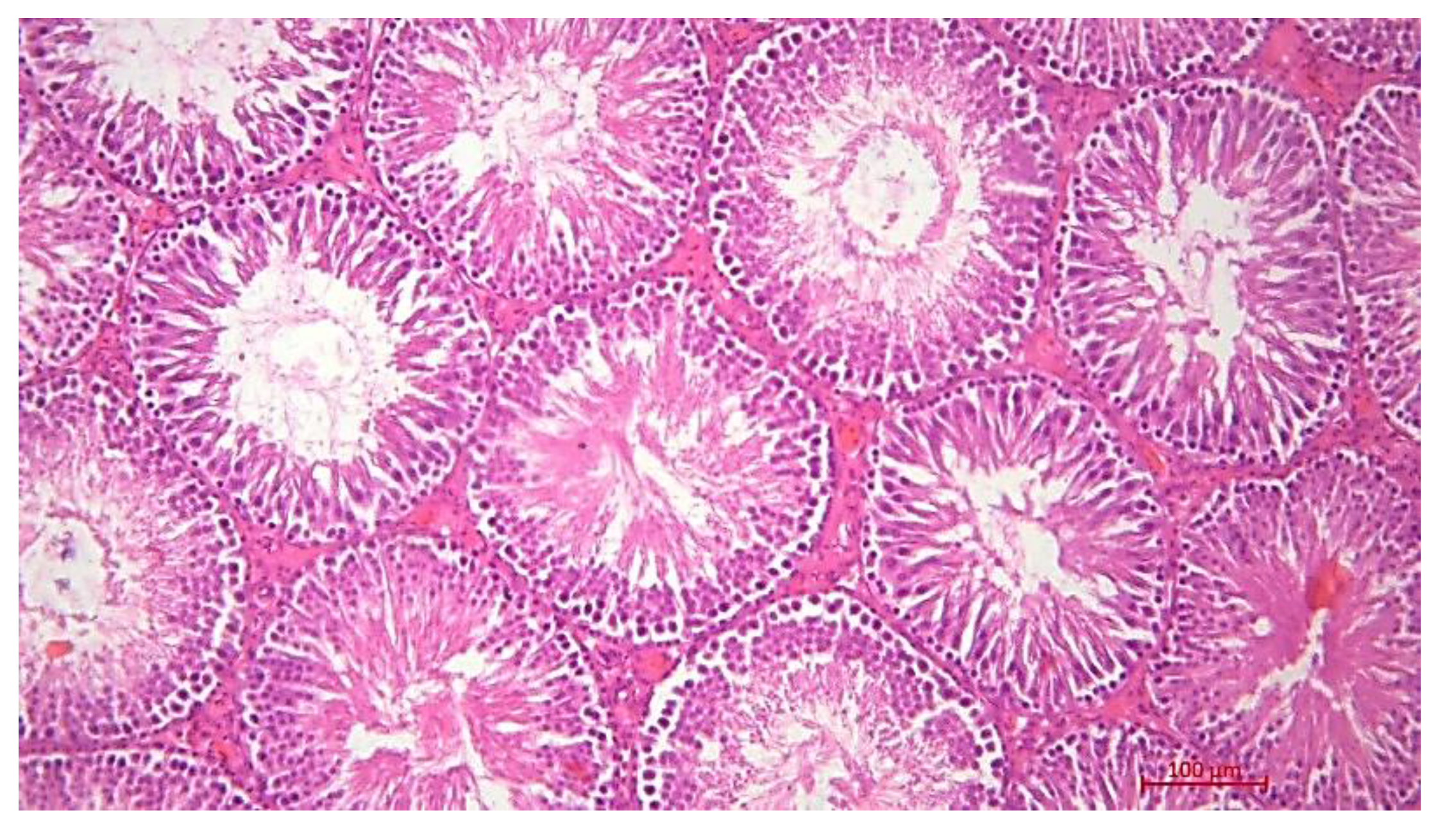
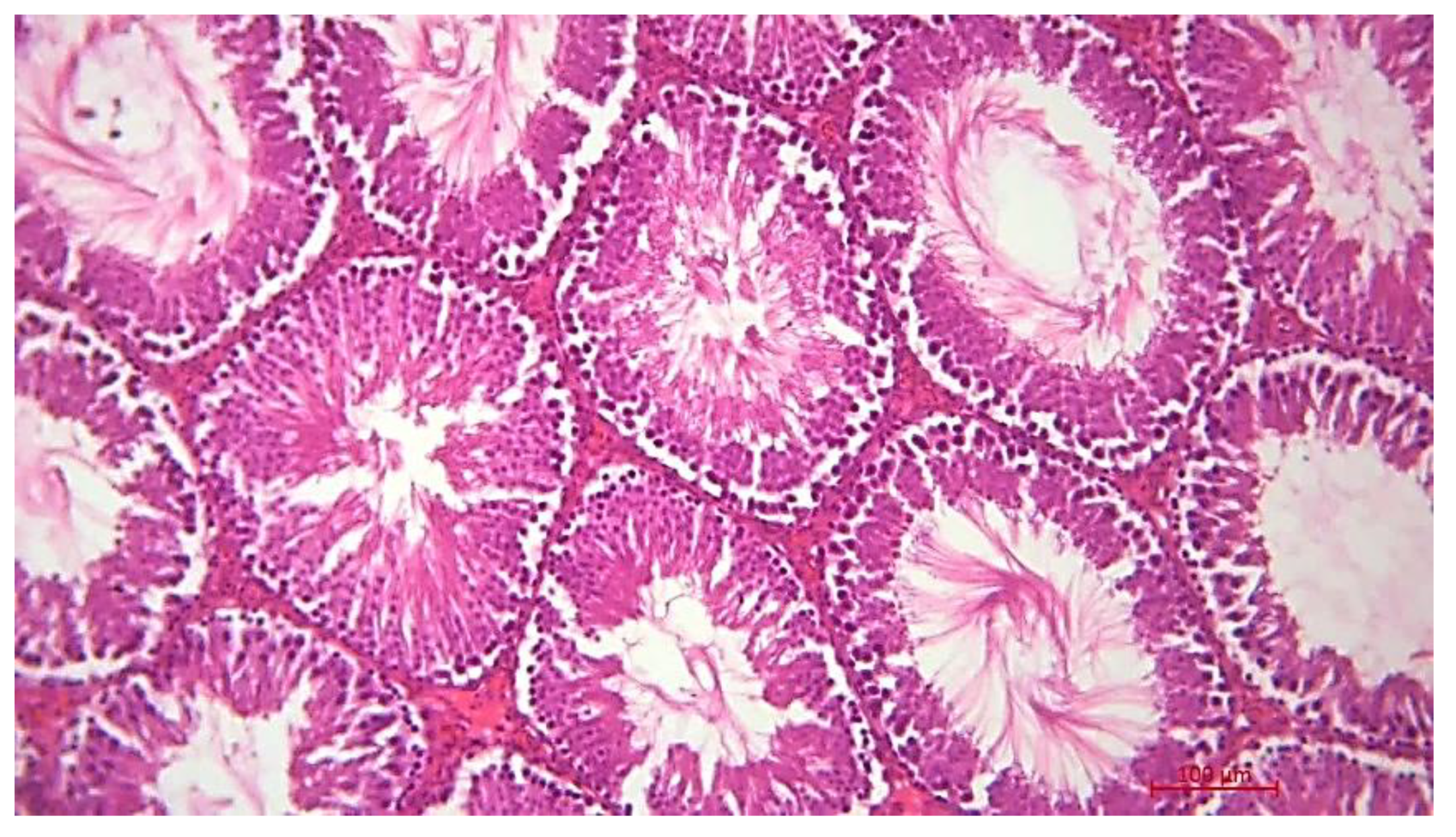
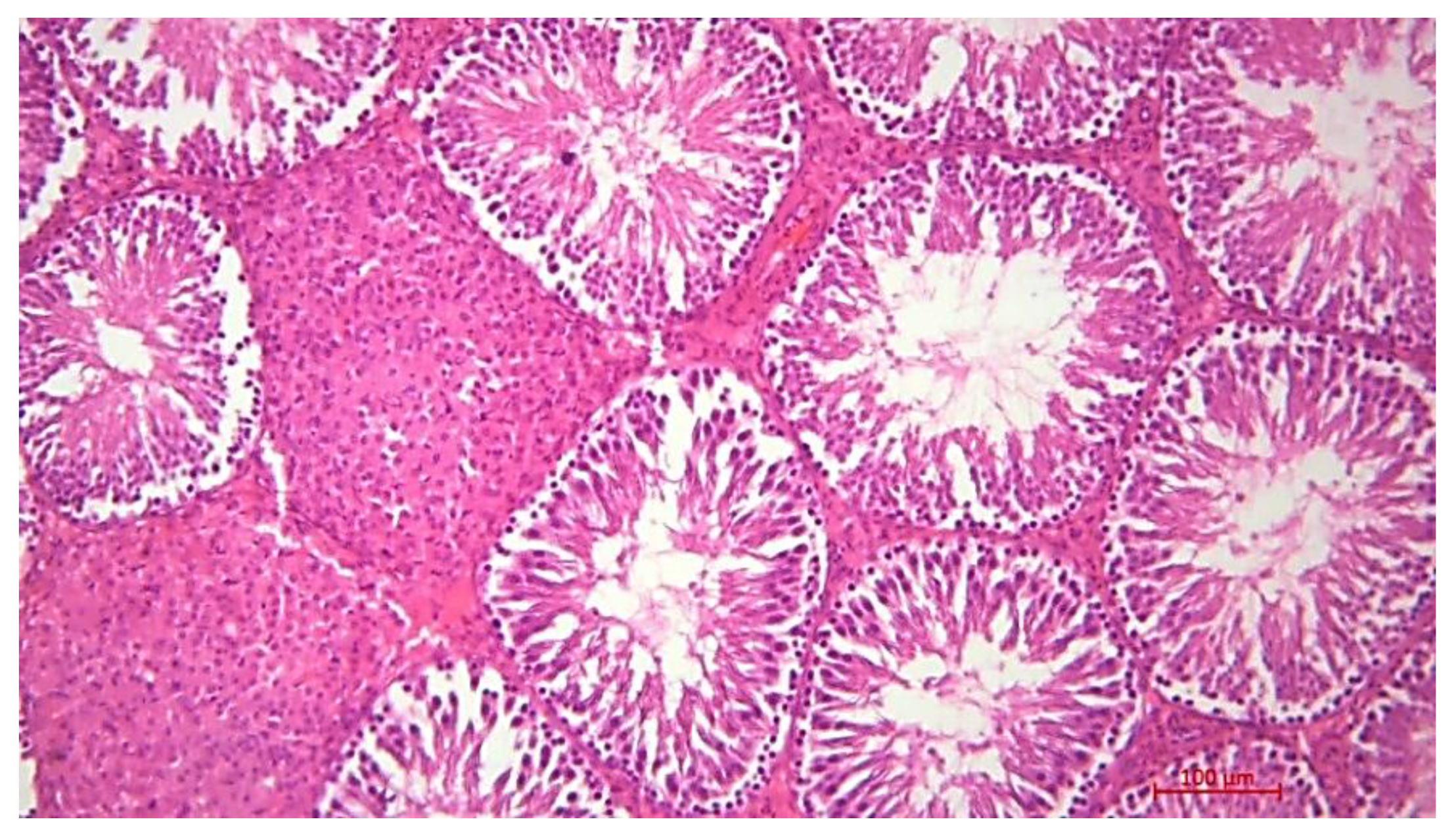
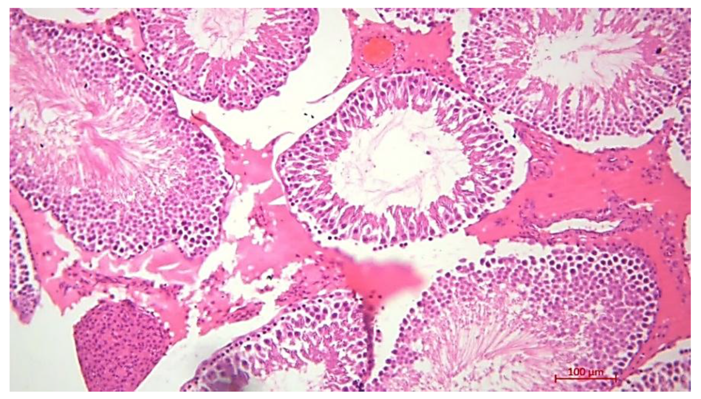
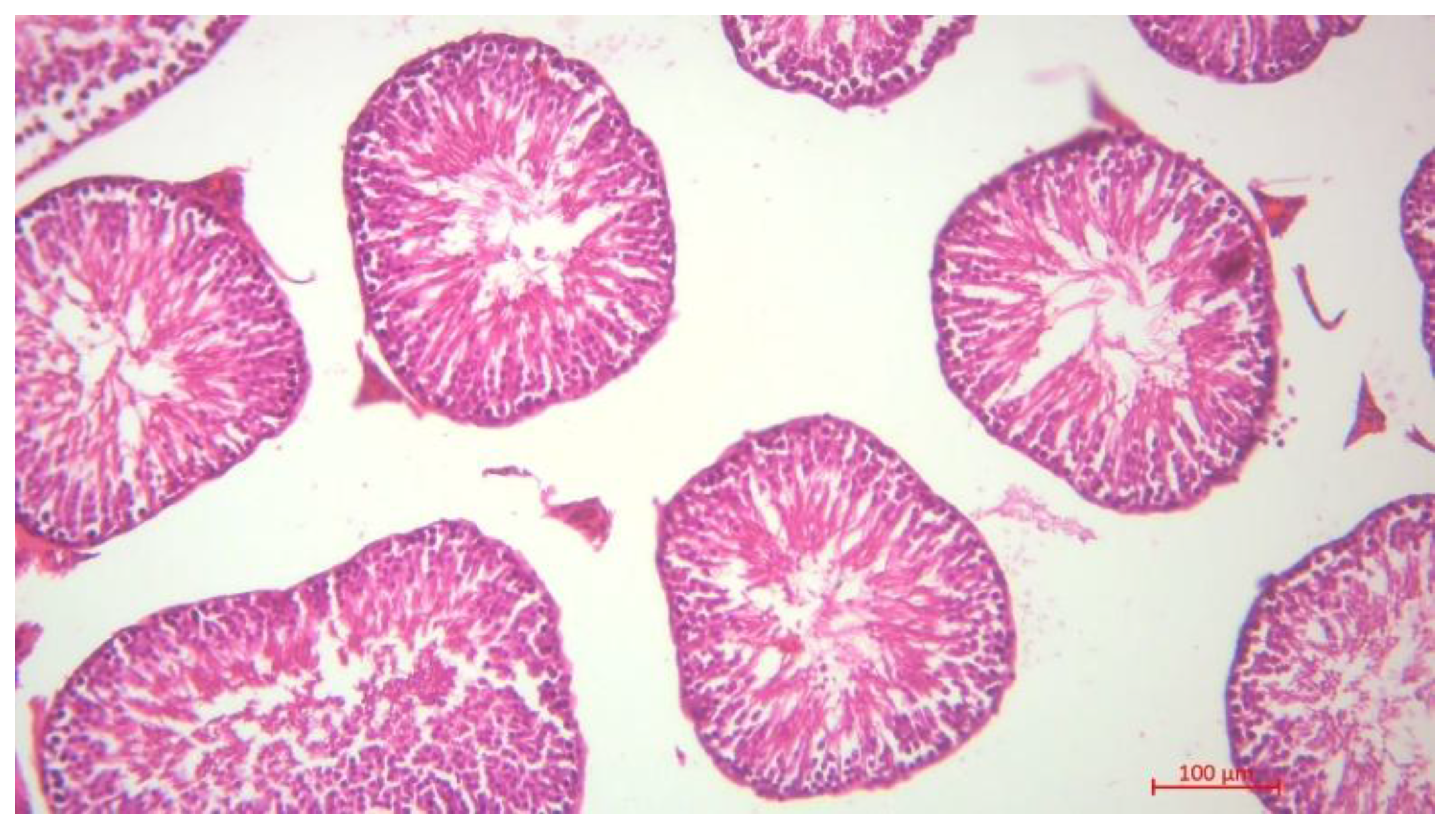
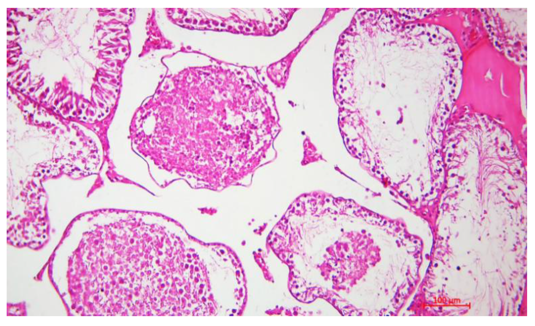
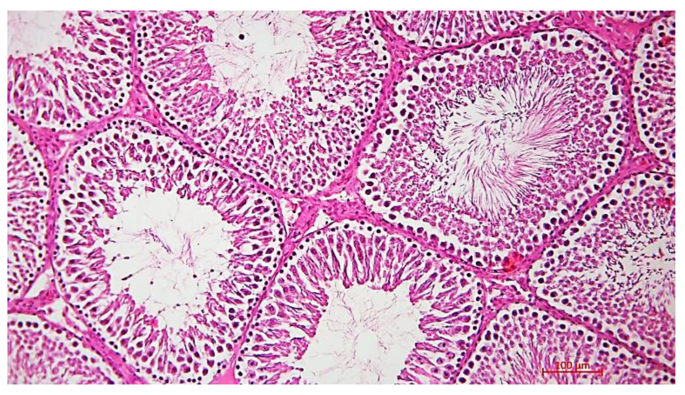
Histological Description of the Gonadal Condition in Males

In control group 1B, the histological structure of the testicle corresponded to the norm: the seminal tubules had a regular rounded or oval shape, were lined with spermatogenic epithelium with clearly pronounced stratification, and mature spermatozoa were found in the lumen of the tubules. The intertubular tissue contained a moderate number of Leydig cells without signs of pathological changes (Figure 1).
In group 2B, Leydig cell proliferation was observed in two rats, while the structure of testicular interstitial tissue remained within the normal range in the other animals of this group. The seminal tubules retained their typical architectonics, but in some cases minor dystrophic changes in the spermatogenic epithelium were observed. Proliferation of Leydig cells indicates a possible activation of steroidogenesis in response to external or internal influences, including toxic damage, hormonal dysregulation, or a compensatory reaction to impaired spermatogenesis (Figure 2). Microscopic examination of histological sections of the testicles of animals exposed to radiation showed the development of degenerative changes in the periphery of the organ, including detachment of the spermatogenic epithelium and the appearance of atrophied cells. convoluted spermatic tubules. Histological examination of the spermatogenic layer of the seminal tubules in experimental animals of this group showed that in some tubules the number of germ cell generations decreases sometimes only spermatogonia and spermatocytes of the first order are found. (Figure 2).
In group 3B, one rat had a weak proliferation of Leydig cells, as well as edema of the testicular stroma, and changes in the shape of the seminal tubules. These changes may indicate the initial manifestations of interstitial regulation and microcirculation disorders, probably due to the action of an external damaging factor. In all other cases, the histological structure of the testicle remained within the normal range (Figure 3).
In group 4B, the testicular histostructure was generally preserved. Signs of active spermatogenesis are present, but there is a pronounced edema of the stroma, a decrease in the size of the seminal tubules (Figure 4).
The study of histological preparations showed that in the testes of experimental rats treated with chromium and radiation, convoluted seminal tubules are observed, in the lumens of which generative cells are located that have exfoliated from the spermatogenic layer with foci of necrosis. This phenomenon can disrupt the normal development of germ cells and is called epitheliospermatogenic plug. Spermatocytes of the second order and spermatids are much less common in the epithelial-spermatogenic layer. This indicates that more mature generative cells, such as order II spermatocytes and spermatids, are more sensitive to exogenous influences. In the tests of experimental rats, edema of the inter-tubular stroma was recorded. This swelling is manifested in the fact that the convoluted seminal tubes in this state are located at a considerable distance from each other (Figure 5).
In group 6B, the structure of the seminal tubules corresponds to the norm.
Figure 6. G. E. staining, magnification 100×.
Preprints 179569 g006
In group 7B – the use of Burdock oil. All types of generative cells were found in the spermatogenic epithelium of the control group: spermatogonia, spermatocytes of the first and second order, spermatids and spermatozoa. In addition, this group was characterized by the correct organization of the location of generative cells in the space of the tubule (Figure 7).

4. Conclusions

The present study showed that in first-generation males (F1) whose parents were exposed to Cr(VI) and/or gamma-radiation with preventive administration of nettle or burdock oils, the most pronounced inter-group differences (after FDR control) were found in four key indicators: testosterone, thyroxine (T4), sperm concentration, and the proportion of sperm in the blood. abnormal sperm cells. This is consistent with expectations for the involvement of the HPG and HPT axes as the main « targets “ of transgenerational effects and / or their prevention by phytopreparations. Testosterone was minimal in the γ group, while the maximum values were observed with a combination of Cr+γ and burdock oil; T4 showed bidirectional shifts (decrease at “γ+burdock oil” and increase at “Cr+γ+burdock oil”), and the structure of spermatogenesis changed quantitatively (concentration), and qualitatively (anomalies, mobility). In summary, this indicates the critical role of the type and combination of effects and the potential modifying role of phytopreparations.
Protective effect of oils: comparison of biomarkers and morphology. Against the background of gamma-irradiation, burdock oil was associated with an increase in sperm motility compared to the gamma-group and even the control, while reducing the proportion of abnormal forms to values close to the control. Under conditions of combined exposure (Cr+y), both oils shifted the proportion of abnormal forms down relative to the y monofactor, and burdock oil was accompanied by a maximum increase in testosterone and T4. These patterns are generally consistent with the organoprotective, antioxidant, and hormone-modulating properties of phytopreparations stated by the authors. At the morphological level, epitheliospermatogenic “plugs”, detachment of the spermatogenic epithelium, necrosis foci, and edema of the interstitial stroma were described for the Cr+y group; whereas when oils were added, the architectonics of the seminal tubules were preserved, and all germ cell generations were represented — signs of partial morphological normalization. Together with biochemical and spermogram data, this supports the hypothesis of the protective effect of oils on spermatogenesis and the interstitial apparatus of the tests in offspring.
Paradoxically high sperm concentration at Cr+y without protection. In the Cr+y group, the highest concentration of spermatozoa was observed (about 24 × 10 ^ 6/ml), which is counterintuitive for toxic combined exposure. At the same time, the quality of gametes worsened (decreased mobility, growth of anomalies). A possible explanation is compensatory hyperstimulation of spermatogenesis under sub chronic stress with a shift in the balance in favor of quantitative rather than qualitative products — a phenomenon that is described in separate modes of dose and time profile of ionizing exposure. In other words, the” rough “ production of cells increases, but their maturity and functionality suffer, which is reflected in the accompanying indicators. This interpretation is consistent with the morphology (areas of epithelial detachment, “plugs”), suggesting a violation of the coordination of cell generation and transport processes in the convoluted tubules.
HPG/HPT axes and possible mechanisms of action of phytopreparations. The observed hormonal shifts in F1 can be considered as a distant reflection of violations of regulatory axes in parents (including due to epigenetic traces), with subsequent modification in offspring. For burdock oil, which is rich in phenolic compounds, antioxidant/anti-inflammatory effects and possible support for steroidogenesis are logical (which resonates with increased testosterone in “Cr+γ+burdock oil”). Nettle, which also has antiandrogenic and hormone-modulating properties, predictably has a more “restrained” androgen profile while maintaining benefits at the level of sperm quality and inflammatory markers; this may explain the quantitative differences between oils with a similar overall direction of effect. Overall, phytopreparations probably affect antioxidant enzymes, local cytokine networks, and steroidogenesis, which is consistent with the observed hormonal and morphological patterns in F1.
Oxidative stress and cytokines: moderate shifts with pronounced phenotypic effects. At the MDA level, the intergroup differences were small, while SOD / catalase activity and cytokine profile showed more variable, but not always statistically stable, changes. This does not exclude the pathobiological significance of ROS-dependent pathways: plasma / serum markers may “average” local (testicular-specific) processes, and the sampling time (F1 at 16 months) may reflect a distant, rather than acute, cross-section. Therefore, in future studies, it is useful to add tissue-specific measurements (for example, antioxidant enzymes in testicular tissue, 8-OHdG/yH2AX as indicators of DNA-damage) and dynamic time points.
Transgenerational aspects. The introduction of oils to parents even before exposure created conditions for assessing the preventive effect on F1 after a long period of time. This fits in with the growing evidence that ionizing radiation and chemical toxicants can form inherited phenotypes through epigenetic mechanisms; our results are consistent with the idea that modifiers of oxidative stress and hormonal background in parents can partially “reconfigure” the vulnerabilities of offspring.
Conclusion from the discussion. Taken together, the data indicate that prophylactic administration of burdock and nettle oils to parents exposed to Cr (VI) and gamma-radiation partially normalizes hormonal and reproductive parameters in their-male offspring (F1) and is accompanied by morphological signs of spermatic tubule preservation. The effects depend on the combination of effects and the type of herbal preparation; biologically plausible mechanisms include modulation of HPG/HPT axes, local inflammatory responses, and redox homeostasis. These results support the development of phytoprotective approaches to reduce the inherited effects of combined toxic exposure to ionizing radiation and Cr (VI).

Funding

The work was carried out as part of a scientific project with grant funding from the Science Committee of the Ministry of Science and Higher Education of the Republic of Kazakhstan “AR19678225 Prevention of spontaneous oncogenesis in the first generation of rats whose parents were exposed to gamma radiation and chromium (experiment)” (reg. no. 0123RK00613).

Institutional Review Board Statement

The study was approved by the Local Bioethics Committee of the West Kazakhstan Medical University named after Marat Ospanov, in accordance with GCP, WHO, and ICH-GCP ethical standards (Protocol № 8, Approval number: № 8.10, Session date: 20.10.2022, Approval date: 21.11.2022). The ethics review was conducted under full certification. No human participants were involved in the research. All procedures involving animals were in accordance with institutional guidelines and relevant ethical regulations.

Informed Consent Statement

Not applicable.

Conflicts of Interest

The authors declare no conflict of interest.

References

  1. Devi, VN. Sources and toxicological effects of some heavy metals—A mini review. J Toxicol Stud [Internet]. 2024 Feb 21 [cited 2025 Mar 18];2(1). Available from. [CrossRef]
  2. Balali-Mood M, Naseri K, Tahergorabi Z, Khazdair MR, Sadeghi M. Toxic Mechanisms of Five Heavy Metals: Mercury, Lead, Chromium, Cadmium, and Arsenic. Front Pharmacol [Internet]. 2021 Apr 13 [cited 2025 Mar 18];12. Available from. [CrossRef]
  3. Ionizing radiation and health effects [Internet]. Who.int. [cited 2025 Mar 18]. Available from: https://www.who.
  4. Chernobyl accident 1986 [Internet]. World-nuclear.org. [cited 2025 Mar 18]. Available from: https://world-nuclear.
  5. 7 The Chernobyl disasters Its effect on Belarus and Ukraine [Internet]. Unu.edu. [cited 2025 Mar 18]. Available from: https://archive.unu.edu/unupress/unupbooks/uu21le/uu21le0h.
  6. Tykhyi, V. Chernobyl sufferers in Ukraine and their social problems: short outline. Research Activities about the Radiological Consequences of the Chernobyl NPS Accident and Social Activities to Assist the Sufferers by the Accident, Kyoto. 1998; 27:235–45. Available from: https://www.rri.kyoto-u.ac.jp/NSRG/reports/kr21/kr21pdf/Tykhyi.
  7. Sharma P, Singh SP, Parakh SK, Tong YW. Health hazards of hexavalent chromium (Cr (VI)) and its microbial reduction. Bioengineered [Internet]. 2022;13(3):4923–38. Available from. [CrossRef]
  8. Duarte GT, Volkova PY, Fiengo Perez F, Horemans N. Chronic ionizing radiation of plants: An evolutionary factor from direct damage to non-target effects. Plants [Internet]. 2023;12(5). Available from. [CrossRef]
  9. Irsal M, Sutoro SG, Widiatmoko ME, Cahya I. Assessment Awareness and Knowledge of Apron to Protect Radiographer During Radiographic Examination. Futurity Medicine [Internet]. 2023 Dec. 30 [cited 2025 Mar. 19];2(4):10-6. Available from: https://futurity-medicine.com/index.
  10. Yin J, Ye Y, Gao Y, Xu Q, Su M, Sun S, et al. Low-dose ionizing radiation and male reproductive immunity: Elucidating subtle modulations and long-term health implications. Int J Mol Sci [Internet]. 2025;26(5). Available from. [CrossRef]
  11. Chen QY, Murphy A, Sun H, Costa M. Molecular and epigenetic mechanisms of Cr(VI)-induced carcinogenesis. Toxicol Appl Pharmacol [Internet]. 2019;377(114636):114636. Available from. [CrossRef]
  12. Gelaye, Y. Public health and economic burden of heavy metals in Ethiopia: Review. Heliyon [Internet]. 2024;10(19): e39022. Available from. [CrossRef]
  13. Ukraine: Health Cluster bulletin #1 (January 2025) [Internet]. ReliefWeb. [cited 2025 Mar 18]. Available from: https://reliefweb.int/report/ukraine/ukraine-health-cluster-bulletin-1-january-2025.
  14. Guirandy N, Gagnaire B, Camilleri V, Cavalié I, Pierron F, Gonzalez P, et al. Multigenerational exposure to gamma radiation affects offspring differently over generations in zebrafish. Aquat Toxicol [Internet]. 2022;244(106101):106101. Available from. [CrossRef]
  15. Sangsuwan T, Mannervik M, Haghdoost S. Transgenerational effects of gamma radiation dose and dose rate on Drosophila flies irradiated at an early embryonal stage. Mutat Res Genet Toxicol Environ Mutagen [Internet]. 2022;881(503523):503523. Available from. [CrossRef]
  16. Kumar D, Seth CS. Photosynthesis, lipid peroxidation, and antioxidative responses of Helianthus annuus L. against chromium (VI) accumulation. Int J Phytoremediation [Internet]. 2021;24(6):1–10. Available from. [CrossRef]
  17. Gagnaire B, Arcanjo C, Cavalié I, Camilleri V, Simon O, Dubourg N, et al. Effects of gamma ionizing radiation exposure on Danio rerio embryo-larval stages - comparison with tritium exposure. J Hazard Mater [Internet]. 2021;408(124866):124866. Available from. [CrossRef]
  18. Lindeman LC, Kamstra JH, Ballangby J, Hurem S, Martín LM, Brede DA, et al. Gamma radiation induces locus specific changes to histone modification enrichment in zebrafish and Atlantic salmon. PLoS One [Internet]. 2019;14(2): e0212123. Available from. [CrossRef]
  19. Xue Q, Zhang L, Wang R, Xu J, Wang C, Gao S, et al. Hexavalent chromium reduces testosterone levels by impairing lipophagy and disrupting lipid metabolism homeostasis: Based on a metabolomic analysis. Toxicology [Internet]. 2024;508(153908):153908. Available from. [CrossRef]
  20. Shyam M, Sabina EP. Harnessing the power of Arctium lappa root: a review of its pharmacological properties and therapeutic applications. Natural Products and Bioprospecting 2024 14:1 2024; 14:1–27. [CrossRef]
  21. Iztleuov M, Iztleuov Y, Saparbayev S, Temirbayeva A, Medeuova R, Aleuova Z, Ismailova I, Imanbayev N. Effect of Burdock Root Oil on Oxidative Stress Induced by Isolated and Combined Use of Gamma Radiation and Hexavalent Chromium. Biomed Pharmacol J [Internet]. 2022 Mar 31 [cited 2025 Mar 18];15(1):421-32. Available from. [CrossRef]
  22. Sawicka E, Saczko J, Roik J, Kulbacka J, Piwowar A. Effect of Interaction between 17β-Estradiol, 2-Methoxyestradiol and 16α-Hydroxyestrone with Chromium (VI) on Ovary Cancer Line SKOV-3: Preliminary Study. Molecules [Internet]. 2020 Nov 9 [cited 2025 Mar 18];25(21):5214. Available from. [CrossRef]
  23. Błażewicz A, Wiśniewska P, Skórzyńska-Dziduszko K. Selected Essential and Toxic Chemical Elements in Hypothyroidism—A Literature Review (2001–2021). Int J Mol Sci [Internet]. 2021 Sep 20 [cited 2025 Mar 18];22(18):10147. Available from. [CrossRef]
  24. Rafi’i MR, Ja’afar MH, Mohammed Nawi A, Md Hanif SA, Md Asari SN. Association between toxic heavy metals and noncancerous thyroid disease: a scoping review. PeerJ [Internet]. 2025 Feb 11 [cited 2025 Mar 18];13: e18962. Available from. [CrossRef]
  25. Donayeva A, Kulzhanova D, Amanzholkyzy A, Abdelazim IA, Abilov T, Baubekov Z, et al. Relationship between vitamin D and adolescents’ hypothyroidism – a cross-sectional study. Prz Menopauzalny [Internet]. 2023;22(4):186–90. Available from. [CrossRef]
  26. Bharat, D. Burdock root: Health benefits, nutrition, uses for skin and hair, recipes, side effects [Internet]. Netmeds. 2022 [cited 2025 Mar 18]. Available from: https://www.netmeds.
  27. Al Zarzour RH, Kamarulzaman EE, Saqallah FG, Zakaria F, Asif M, Abdul Razak KN. Medicinal plants proposed nanocomposites for the management of endocrine disorders. Heliyon [Internet]. 2022;8(9): e10665. Available from. [CrossRef]
  28. Garofalo V, Condorelli RA, Cannarella R, Aversa A, Calogero AE, La Vignera S. Relationship between iron deficiency and thyroid function: A systematic review and meta-analysis. Nutrients [Internet]. 2023;15(22):4790. Available from. [CrossRef]
  29. Suhail N, Aftab T, Alruwaili A, Alruwaili D. Effects of multivitamin-mineral supplementation on chronic stress-induced oxidative damage in Swiss albino mice. Cureus [Internet]. 2024;16(6): e61896. Available from. [CrossRef]
  30. Song I-B, Han H-J, Kwon J. Immune-enhancing effects of gamma-irradiated sericin. Food Sci Biotechnol [Internet]. 2020;29(7):969–76. Available from. [CrossRef]
  31. Caputo L de S, Alves C de L, Laranjeira IM, Fonseca-Rodrigues D, da Silva Filho AA, Dias ACP, et al. Copaiba oil minimizes inflammation and promotes parenchyma re-epithelization in acute allergic asthma model induced by ovalbumin in BALB/c mice. Front Pharmacol [Internet]. 2024; 15:1356598. Available from. [CrossRef]
  32. Walcher L, Kistenmacher A-K, Sommer C, Böhlen S, Ziemann C, Dehmel S, et al. Low energy electron irradiation is a potent alternative to gamma irradiation for the inactivation of (CAR-)NK-92 cells in ATMP manufacturing. Front Immunol [Internet]. 2021; 12:684052. Available from. [CrossRef]
  33. Hussien SM, Rashed ER. Immuno-biochemical impacts of gamma irradiation in male rats: A dose-response study. Dose Response [Internet]. 2023;21(2):15593258231185461. Available from. [CrossRef]
  34. Boyko V, Savvi S, Korolevska A, Zhydetskyy V, Novikov Y, Bytiak S, et al. Surgical treatment of bening esophageal strictures after corrosive injuries. Georgian Med News [Internet]. 2018;(278):7–15. Available from: https://europepmc.org/article/med/29905537.
  35. Fukunaga H, Yokoya A, Prise KM. A brief overview of radiation-induced effects on spermatogenesis and oncofertility. Cancers (Basel) [Internet]. 2022;14(3):805. Available from. [CrossRef]
  36. Wdowiak A, Skrzypek M, Stec M, Panasiuk L. Effect of ionizing radiation on the male reproductive system. Ann Agric Environ Med [Internet]. 2019;26(2):210–6. Available from. [CrossRef]
  37. Li J, Liu J, Zhang Y, Qiu H, Zheng J, Xue J, et al. Effects of paternal ionizing radiation exposure on fertility and offspring’s health. Reprod Med Biol [Internet]. 2024;23(1): e12567. Available from. [CrossRef]
  38. Burdock extract effects on hormones and sperm in diabetic male mice [Internet]. Wisdomlib.org. 2024 [cited 2025 Mar 18]. Available from: https://www.wisdomlib.org/science/journal/the-malaysian-journal-of-medical-sciences/d/doc1425283.
  39. Govbakh IO, Zavodovskiy DO, Bulgakova N V., Sokołowska (Vereshchaka) I V., Maznychenko A V., Vasylenko DA. Nerve Conduction and Neuromuscular Transmission in C57Bl/6 Mice with Genetically Determined Peripheral Neuropathy. Neurophysiology [Internet]. 2019;51(4):248–52. Available from. [CrossRef]
  40. Wakeel A, Xu M, Gan Y. Chromium-induced reactive oxygen species accumulation by altering the enzymatic antioxidant system and associated cytotoxic, genotoxic, ultrastructural, and photosynthetic changes in plants. Int J Mol Sci [Internet]. 2020;21(3):728. Available from. [CrossRef]
  41. Surniyantoro HNE, Kisnanto T, Tetriana D, Yusuf D, Basri IKH, Lusiyanti Y. Study of immune response and malondialdehyde levels in irradiated rats supplemented with Curcuma xanthorriza Roxb extract. Asian Pac J Cancer Prev [Internet]. 2023;24(5):1717–23. Available from. [CrossRef]
  42. Shapoval LM, Dmytrenko O V., Sagach VF, Prylutska S V., Khrapatiy S V., Zavodovskyi DO, et al. Systemic Administrations of Water-Dispersible Single-Walled Carbon Nanotubes: Activation of NOS in Spontaneously Hypertensive Rats. Neurophysiology [Internet]. 2020;52(2):101–9. Available from. [CrossRef]
  43. Iztleuov M, Iztleuov Y, Yelubayeva A, Aleuova Z, Turganbaeva Z, Madikhan Z, et al. Effect of “nettle oil” on oxidative damage to the heart and lungs induced by radiation. Biomed Pharmacol J [Internet]. 2020;13(03):1495–504. Available from. [CrossRef]
Figure 1. G. E. staining, magnification 100×.
Preprints 179569 g001
Figure 2. G. E. staining, magnification 100×.
Preprints 179569 g002
Figure 3. G. E. staining, magnification 100×.
Preprints 179569 g003
Figure 4. G. E. staining, magnification 100×.
Preprints 179569 g004
Figure 5. Epitheliospermatogenic plug, necrosis of the spermatogenic layer of cells, edema of the inter-tubular connective tissue of rats treated with chromium + irradiation. Staining by G. E., magnification 100×.
Preprints 179569 g005
Figure 7. Section of seminal tubules of rats treated with chromium + radiation + burdock oil: several generative spermatogenic epithelial cells are present in multiple tubules. Staining by G. E., magnification 100×.
Preprints 179569 g007
Table 1. Overview of Experimental Groups and Exposure Conditions.
Group Description
Control No exposure to hexavalent chromium (Cr⁶⁺) or gamma (γ) radiation.
γ Gamma Radiation Exposed to γ radiation only. 15 days after the beginning of the experiment, they were exposed to total single radiation at a dose of 0.2 Gy. together with the other groups.
γ + Nettle Oil For 14 days before irradiation, Nettle Oil was obtained at a dose of 0.5 ml per rat through a probe intragastrically. On the 15th day of exposure.
γ + Burdock Oil For 14 days before irradiation, Burdock Oil was obtained at a dose of 0.5 ml per rat through a probe intragastrically. On the 15th day of exposure.
Cr⁶ ⁺+ γ For 14 days received hexavalent chromium orally with drinking water at a dose of 180 mg/l. On day 15of exposure
, Cr⁶ ⁺ + γ + Nettle Oil was irradiated for 14 days orally with drinking water chromium at a dose of 180 mg/l and Nettle Oil 0.5 ml per rat through a probe. On day 15, 0.2 Gy of
Cr⁶ ⁺ + γ + Burdock Oil was irradiated for 14 days orally with drinking water chromium at a dose of 180 mg/l and Burdock Oil 0.5 ml per rat through a probe. On the 15th day, the exposure was 0.2 Gy
Disclaimer/Publisher’s Note: The statements, opinions and data contained in all publications are solely those of the individual author(s) and contributor(s) and not of MDPI and/or the editor(s). MDPI and/or the editor(s) disclaim responsibility for any injury to people or property resulting from any ideas, methods, instructions or products referred to in the content.
Copyright: This open access article is published under a Creative Commons CC BY 4.0 license, which permit the free download, distribution, and reuse, provided that the author and preprint are cited in any reuse.
Prerpints.org logo

Preprints.org is a free preprint server supported by MDPI in Basel, Switzerland.

Subscribe

Disclaimer

Terms of Use

Privacy Policy

Privacy Settings

© 2025 MDPI (Basel, Switzerland) unless otherwise stated