
Submitted:
25 September 2025
Posted:
26 September 2025
You are already at the latest version
Abstract
Estrogen deficiency is associated with endothelial dysfunction, vascular inflammation, increased lipoprotein oxidation, accumulation of lipid-rich material, and platelet activation. The absence of estrogen causes physiological, metabolic, and biochemical changes that increase the risk of cardiometabolic disease development caused by a deregulation in metabolic processes such as lipid metabolism and plasma lipoproteins levels. High-density lipoprotein (HDL) has cardioprotective properties related to the quality and the quantity of its components that can be modified by some nutritional factors. Guava (Psidium guajava L.), a widely cultivated fruit in Mexico, is notable for its high polyunsaturated fatty acid and dietary fiber content in its seeds, but its effect on health is understudied. The study aimed to evaluate the effect of guava seed supplementation on body weight, blood pressure, lipid profile, HDL composition, and paraoxonase-1 (PON1) activity in an ovariectomized rat model (OVX). Four groups with 6 adults female Wistar rats each were classified as SHAM group: rats with simulated ovariectomy; OVX group: rats with ovariectomy; OVX+DGS group: rats supplemented with 6 g of defatted guava seeds; OVX+GS group: rats supplemented with 6 g of guava seeds. Biochemical parameters, size, and lipid concentration of HDL subclasses, apolipoproteins, and PON1 activity were determined. A decrease in body weight gain, systolic blood pressure, mean arterial pressure, and triglycerides in plasma was observed at the end of the experiment in the supplemented groups. The supplementation of 6 g of guava seeds for 30 days decreased biochemical parameters in ovariectomized rats; these results could be attributed to the seed composition, suggesting a protective effect against the risk of developing diseases in menopausal states.
Keywords:
HDL subclasses
; lipid profile
; ovariectomized
; menopause
; guava seeds
1. Introduction
The substantial reduction and deficiency in circulating levels of estrogen affects almost all organ systems, including the cardiovascular, immune, and digestive systems. In this context, menopause is characterized by the depletion of the oocytes stored in the ovaries and the significant decrease in estrogen levels. Menopausal women experience significant physiological, metabolic and, biochemical changes such as an increase in total cholesterol, triglycerides and low-density lipoprotein-cholesterol (LDL-c), and a decrease in high-density lipoprotein-cholesterol (HDL-c) as well as the shift in the distribution of lipids composition [1] in this sense, estrogens and other ovarian hormones possess cardioprotective properties and it plays a critical regulatory role in glucose metabolism, lipid homeostasis, and inflammatory pathways [2,3], including insulin sensitivity, blood pressure, and prothrombotic processes [4].
The decrease in estrogen secretion leads to a reduced capacity of HDL to efflux cholesterol, which is mainly related to HDL core and surface lipid remodeling (free cholesterol, triglycerides, phospholipids, sphingolipids), which has been described in women during menopause, suggesting indirect effects of estrogen signaling pathways on the lipid profile [5,6]. The lipidome and proteome have also been described to be altered with estrogen deficiency; this could potentially compromise the other cardioprotective properties of HDL. Therefore, this suggests that HDL may become dysfunctional when circulating estrogen levels are reduced, such as in a state of menopause, affecting their quality more than the quantity of these lipoproteins [7,8]. On the other hand, some nutritional factors and bioactive compounds have been shown to influence the metabolism of estrogen, HDL structure and composition, and cardiovascular health [9,10], so lowering cardiovascular risk factors during and after menopause through diet intervention is possible.
Guava (Psidium guajava L.) is a fruit from the Myrtaceae family and is widely cultivated in Mexico, ranking third worldwide for production [11]. During the processing of this fruit, pulp is obtained as the main product and seeds and peels as by-products [12]. Due to the presence of bioactive compounds, the pulp and the by-products have the potential to be used in healthy processed foods [13]. Guava is notable for its high vitamin C content, as well as its seeds for their polyunsaturated fatty acid and dietary fiber content [14]. Guava seeds and oil have been characterized; however, their effect on health remains understudied. Significant improvements in lipid profile were also reported: total cholesterol, LDL-c, and triglycerides decreased, while HDL-c increased in diabetic mice supplemented with guava seed extract [15]. Also, guava seed oil has been shown to have antioxidant activity [16], anti-LDL peroxidation, and gram-negative bacteria inhibitory activity [17,18].
Therefore, in the present study, we aimed to evaluate the effect of guava seed supplementation on body weight, blood pressure, lipid profile, HDL composition, and PON1 activity in an ovariectomized rat model.
2. Materials and Methods
2.1. Animals
Twenty-four adults female Wistar rats weighing 200–250 g and aged 3 months were maintained under temperature-controlled conditions with light-dark cycles (12 h) and received commercial feed (LabDiet 5008) and water ad libitum. Four groups were formed, with n=6 experimental animals in each group, classified as follows: SHAM group: negative control, rats with simulated ovariectomy. OVX group: positive control, rats with ovariectomy. OVX+DGS group: rats supplemented with 6 g of defatted guava seeds. OVX+GS group: rats supplemented with 6 g of guava seeds. The treatment groups (OVX+DGS and OVX+GS) followed the same procedure as the OVX group and received a supplement of guava seeds prepared as described above and mixed with commercial LabDiet 5008 rat food in a 70-30% ratio (14 g of commercial food + 6 g of guava seeds) in pellet form for 30 days. The experimental protocols were carried out according to the guidelines for the protection of animals used for scientific purposes (2010/63/EU) and national guidelines (NOM-O62-ZOO-1999) [19] and were approved by the Animal Ethics Committee of the Universidad Autonoma del Estado de Hidalgo(CICUAL/-V-I/02/2025).
2.2. Ovariectomy
To remove the ovaries, a dorsal ovariectomy was performed in which the rats were anesthetized for surgery by a combination of xylazine hydrochloride (2.5 mg/kg of body weight) and Zelazol® (10 mg/kg of body weight). To remove the ovaries, an incision was made on the left side of the vertebral column of the animal, the uterine horns were identified, fixed to the ovary at one end and to the uterus at the other. Ligatures were established on both sides of the ovary, sectioning and removing them. Once the ovaries were removed, the incision was sutured. The SHAM group underwent the same procedure, but the ovaries were not removed [20,21]. After surgery, meloxicam (0.3 mg/kg of body weight) was administered for 5 days for pain management.
2.3. Guava Seeds
For the supplement, ripe, undamaged guavas were obtained from a local market in the city of Pachuca, Hidalgo, which are harvested in the state of Michoacan. The fruits were washed and disinfected to remove the seeds. The guava seeds were washed with purified water to eliminate any pulp that could remain attached. Once clean, the guava seeds were air-dried at room temperature for 24 hours and ground in a commercial coffee grinder; a portion was used in the group OVX+GS and stored until use in darkness at room temperature.
2.4. Seed Delipidation
The rest of the ground seeds were used for lipid extraction with dichloromethane at a ratio of 1:10 (w/v), and the mixture was stirred at 100 rpm/35 °C for 8 hours. Following this, the mixture was centrifuged at 5000 rpm for 30 minutes to separate the solvent and then dried in an oven at 60 °C for 40 minutes to remove the solvent entirely, and stored for the group OVX+DGS.
2.5. Blood Pressure
Blood pressure levels were measured in the tail of rats after heating by a non-invasive method and recorded in a CODA TM system from Kent Scientific Corporation (Torrington, Connecticut), with at least six blood pressure recordings for each animal, and the data collected using CODA TM software for further processing.
2.6. Lipid Profile
Total cholesterol, triglycerides, and phospholipids levels in plasma, as well as HDL-c, HDL triglycerides (HDL-Tg), and HDL phospholipids (HDL-PPL) levels, were determined using commercial colorimetric enzymatic methods (Randox®, UK; Wako, Ltd, Osaka, Japan). Absorbance was read at a wavelength of 505 nm for cholesterol and triglycerides and 600 nm for phospholipids in a spectrophotometer (BECKMAN COULTER DU® UV/Vis Spectrophotometer, Germany). For the lipid composition of HDL, we used the phase containing the HDL fraction of plasma after ultracentrifugation, following the methodology described below.
2.7. HDL Size and Lipid Concentration of HDL Subclasses
HDL particles were separated via sequential ultracentrifugation in a (Beckman Optima TLX table) [22,23,24,25]. Total apo B-containing lipoproteins were obtained by density <1.063 mg/dL, whereas total HDL 1.063 <density <1.21 g/mL. The HDL were dialyzed against 0.09 M Tris/0.08 M boric acid/3 mM EDTA (TBE) buffer, pH 8.4. Then, HDL were separated further by their hydrodynamic diameters in non-denaturing 3-30% gradient polyacrylamide gel electrophoresis (PAGE). Gels were stained for total cholesterol, phospholipids, and triglycerides using enzymatic mixtures previously described [26,27,28]. The electrophoresis gels were then scanned (GS-670 BioRad densitometer) and stained again for proteins with Coomassie blue R-250, before being scanned once more. The relative proportions of each HDL subclass were estimated via optical densitometry analysis, using as reference globular proteins (thyroglobulin, 17 nm; ferritin, 12.2 nm; catalase 10.4 nm; lactate dehydrogenase, 8.2 nm; and albumin, 7.1 nm; high-molecular-weight calibration kit, Amersham Pharmacia Biotech, Buckinghamshire, U.K.) [24,28]. The relative proportion of every HDL subclass is expressed as the percentage of the total HDL area under the curve, integrated from 7.9 to 12.36 nm. For the classification of the HDL subclasses, we considered the following size intervals: HDL3c, 7.94e8.45 nm; HDL3b, 8.45e8.98 nm; HDL3a, 8.98e9.94 nm; HDL2a, 9.94e10.58 nm; and HDL2b, 10.58e13.59 nm [28]. Enzymatic staining of total cholesterol, triglycerides, and phospholipids of HDL subclasses was performed according to the methodology previously described [27].
2.8. Determination of Apolipoproteins
Apolipoprotein (apo) composition was determined on an SDS-denaturing 4–21% PAGE gradient. Fifteen μg of previously isolated HDL protein was used. The bands of each apo were stained with Coomassie blue and destained with a solution of methanol, acetic acid, and water (30/58/12, v/v/v). The Aepos were identified by comparison with molecular mass standards and subsequently analyzed by optical densitometry. The results are expressed as the percentage of each apo relative to total HDL protein. apo CI and CII are expressed as a percentage of apo C [29].
2.9. Paraoxonase-1 Activity
PON1 was determined using the method described by Gan et al. (1991). The trial mixture included 1 mM phenylacetate as substrate and 0.9 mM CaCl2 in 20 mM Tris HCl, pH 8, and 10 μL rat plasma heparin (diluted 1:10). Enzymatic hydrolysis of the substrate was measured spectrophotometrically at 270 nm (UVVIS Beckman Coulter, Brea, CA, USA). The absorbance at 270 nm for the reaction was 1310 M-1cm-1. Enzyme activity was expressed as the number of micromoles of phenylacetate hydrolyzed per minute per milliliter of plasma.
2.10. Data Analysis
Normal distribution of all data was verified using the Shapiro-Wilk test. Since these data were normally distributed, they are presented as mean ± standard deviation (SD). Differences between groups were determined using analysis of variance (ANOVA) and post hoc analysis (Tukey test). A p-value < 0.05 was considered statistically significant. All analyses were performed using SPSS version 24 statistical software (SPSS Inc. IBM, Chicago, IL, USA).
3. Results
3.1. Biochemical Parameters
Table 1 shows a final body weight decrease of 50% in the OVX+GS compared to the OVX group. The OVX+DGS group showed a significant lower blood level than the OVX group. Concerning the results of the lipid profile, the OVX+DGS group had 50% less triglycerides and phospholipids than the OVX group in plasma, as well as 30% less than the OVX+GS group. In addition, there was a 70% increase in non-HDL-c in OVX+DGS (Table 1). Surprisingly, c-HDL (60%) was lower in OVX+DGS than in the other two groups (Table 2).
3.2. Size and Lipid Composition of HDL Subclasses
After 30 days of supplementation, significant changes were observed in the protein composition of subclass 2b in the OVX+DGS group compared to the SHAM group; a similar increase was noted in HDL3c subclass compared to the OVX+GS group (Table 3). Moreover, an increase in cholesterol was found in the DGS group in the small subclasses 3b and 3c, compared to the OVX group, and in 3c in comparison with the SHAM group.
3.3. Apolipoproteins
The apolipoprotein composition in the experimental groups is listed in Table 4, showing a significant decrease in apo E content and a significant increase in Apo C concentration in the OVX+DGS group compared to the other test groups.
3.4. PON1 Activity
Finally, we determined the antioxidant activity of PON1. Interestingly, we found a significant decrease in the OVX+DGS group compared to the other test groups. Likewise, there was a significant decrease in the OVX+GS group compared to the SHAM group (Figure 1).
4. Discussion
In the present study, we determined the effect of guava seed consumption in an ovariectomized animal model by simulating the biological processes due to a reduction in estrogen levels. In this model, estrogen depletion is related to metabolic effects and significant changes in body composition, including visceral fat accumulation, lipid metabolism disorders, and increased blood pressure, factors that significantly influence the high risk of developing cardiovascular diseases (CVD) [31].
An increase in body mass was observed in ovariectomized rats; nevertheless, a lower increase in body weight was observed in supplemented rats. We suggest that polyunsaturated fatty acids could be responsible for the reduction in weight gain, given that the group supplemented with defatted seeds did not show significant differences compared to the ovariectomized group without supplementation.
Any of the components of GS, including insoluble dietary fiber, may have an impact on improving blood pressure, but the mechanisms are not totally clear. Nevertheless, it has been suggested that these mechanisms could include a reduction in inflammation levels [32,33] and an improvement in endothelial function [34]. Additionally, cellulose has been shown to inhibit starch digestion by binding to α-amylase [35], reducing glucose absorption, enhancing insulin sensitivity, and, as a result, lowering the risk of hypertension [36].
Diseases characterized by increased inflammatory processes, such as cardiovascular diseases, are associated with changes in the lipidome of HDL, particularly a decrease in HDL phospholipid content and an increase in HDL triglycerides [37]. Plasma lipid depletion in rats from the OVX+DGS and OVX+GS groups suggests a reduction in hepatic cholesterol and triglyceride synthesis [38], as well as an increase in intestinal transit by insoluble fibers, which can create a physical barrier and, as a result, a decrease in lipid absorption [39].
A larger number of small HDL particles and their cholesterol efflux capacity could be related to the lowest HDL-c levels in the OVX+DGS group. There is controversy regarding whether the larger particles (HDL2) or the smaller ones (HDL3) are more atheroprotective. It has been suggested that the large HDL subclass is more prone to oxidative modification with respect to the small subclasses [40], whereas HDL3 plays a central role in the reverse cholesterol transport (RCT) by removing cholesterol from the periphery and maturing into HDL2 particles through progressive lipidation by the action of lecithin: cholesterol: acyltransferase (LCAT) [41].
Apolipoproteins are the most abundant group of proteins in HDL. Apo E is considered an atheroprotective protein because it removes more saturated than unsaturated lipids, and atherogenic plaques are rich in saturated lipids and cholesterol [42]. Furthermore, HDL particles containing apo E promote the efflux of cholesterol from extrahepatic cells [43] through ABCA1- and ABCG1-dependent processes, and this process is antagonized by the presence of apo CIII, which can negatively affect the antiatherogenic properties of HDL [44,45].
It has been shown in some animal models that PON1 can prevent the harmful effects of oxidative stress on serum and that serum levels of PON1 correlate with levels of HDL and apolipoprotein AI, although this correlation is not strong. We might suggest that increased PON1 activity is a consequence of higher HDL levels. In our study, the increase in PON1 activity in SHAM rats can be attributed to estrogens, given that estradiol enhances PON1 activity [46]. In addition, a pro-oxidative environment could lead to an increase in the binding of free radicals to PON1, resulting in a less active enzyme in the circulation [47]. It has been reported that dietary factors have a significant effect on PON1 activity, specifically polyunsaturated fatty acids [48], which could be associated with higher activity in the OVX+GS group compared to the OVX+DGS group. Furthermore, according to the observed results of apo and PON activity, the OVX+DGS group could be a population at risk for the development of cardiovascular disease.
Differences shown between OVX+DGS and OVX+GS groups are related to the lipid composition of the seed; when supplementing with a defatted seed, the protective properties previously described were lost indicating that such properties are associated with the polyunsaturated fatty acids [49] and probably other lipidic molecules contained in the whole seed, such as phytosterols [50], that has been reported its use on health, owing to its cholesterol-lowering and anti-inflammatory effects [51,52]. Studies on biomodels fed a high-fat diet have proven the efficacy of supplementation with defatted seeds such as safflower [53] (50-100 mg/kg of weight/day) and poppy [54] (33% fiber and 27% protein) on plasma and hepatic lipid levels, reducing triglyceride and cholesterol levels. In addition, it has been described that defatted grape seed affects the 3T-L1 pre-adipocyte cells, showing a significant decrease in lipid accumulation, possibly due to a regulation of the mRNA expression of leptin and lipoprotein lipase (LPL), both of which have been demonstrated to regulate lipid metabolism [55].
5. Conclusions
The supplementation of guava seeds during 30 days limited the gain of body weight, blood pressure, triglycerides, and non-HDL-c in ovariectomized rats. These results could be attributed to lipidic components and insoluble dietary fiber in the seed, suggesting a discrete protective effect against the risk of developing diseases during a physiological cessation of estrogen secretion.
Author Contributions
Conceptualization: E.C.-T., D.E.-L.; investigation, E.C.-T., D.E.-L., L.M.R.-M. and A.C.-O.; writing—original draft preparation, E.C.-T., D.E.-L., L.M.R.-M.; writing—review and editing, E.C.-T., D.E.-L., A.C.-O., Ó.P.-M. and E.F.-M.; funding acquisition, D.E.-L. All authors have read and agreed to the published version of the manuscript.
Funding
This research received no external funding.
Institutional Review Board Statement
The experimental protocols were approved by the Animal Ethics Committee of the Autonomous University of the State of Hidalgo (CICUAL/-V-I/02/2025).
Informed Consent Statement
Not applicable.
Data Availability Statement
Not applicable.
Acknowledgments
Special gratitude to the university's animal facility employees for their support in the animal handling and surgery processes.
Conflicts of Interest
The authors declare no conflicts of interest.
Abbreviations
The following abbreviations are used in this manuscript:
| HDL | High-density lipoprotein |
| PON1 | Paraoxonase-1 |
| LDL | Low-density lipoprotein |
| SHAM | SHAM-operated rats |
| OVX | Ovariectomized rats |
| DGS | Defatted guava seeds |
| GS | Guava seeds |
| HDL-c | HDL cholesterol |
| HDL-Tg | HDL triglycerides |
| HDL-PPL | HDL phospholipids |
| Apo | Apolipoprotein |
| SD | Standard deviation |
| CVD | Cardiovascular diseases |
| RCT | Reverse cholesterol transport |
| LCAT | Lecithin: cholesterol: acyltransferase |
| E2 | Estradiol |
| LPL | Lipoprotein lipase |
References
- Anagnostis, P.; Stevenson, J.C.; Crook, D.; Johnston, D.G.; & Godsland, I.F. ; & Godsland, I. F. Effects of gender, age and menopausal status on serum apolipoprotein concentrations. Clin Endocrinol 2016, 85, 733–740. [Google Scholar]
- Carr, M.C. The emergence of the metabolic syndrome with menopause. J Clin Endocrinol Metab 2003, 88, 2404–2411. [Google Scholar] [CrossRef]
- Gold, E.B. The timing of the age at which natural menopause occurs. Obstet Gynecol Clin 2011, 38, 425–440. [Google Scholar] [CrossRef]
- Salpeter, S.R.; Walsh, J.M.E.; Ormiston, T.M.; Greyber, E.; Buckley, N.S.; & Salpeter, E.E.; Salpeter, E. E. Meta-analysis: effect of hormone-replacement therapy on components of the metabolic syndrome in postmenopausal women. Diabetes Obes Metab 2006, 8, 538–554. [Google Scholar] [CrossRef] [PubMed]
- Kostara, C.E.; Bairaktari, E.T.; Tsimihodimos, V. Effect of clinical and laboratory parameters on HDL particle composition. International Journal of Molecular Sciences 2023, 24, 1995. [Google Scholar] [CrossRef]
- Estrada-Luna, D. , Carreón-Torres, E., Bautista-Pérez, R., Betanzos-Cabrera, G., Dorantes-Morales, A., Luna-Luna, M.,... & Pérez-Méndez, Ó. Microencapsulated pomegranate reverts high-density lipoprotein (HDL)-induced endothelial dysfunction and reduces postprandial triglyceridemia in women with acute coronary syndrome. Nutrients 2019, 11, 1710. [Google Scholar] [PubMed]
- El Khoudary, S.R.; Chen, X.; Nasr, A.; Billheimer, J.; Brooks, M.M.; McConnell, D.; et al. HDL (high-density lipoprotein) subclasses, lipid content, and function trajectories across the menopause transition: SWAN-HDL study. ATVB 2021, 41, 951–961. [Google Scholar] [CrossRef] [PubMed]
- Lehti, S.; Korhonen, T.M.; Soliymani, R.; Ruhanen, H.; Lähteenmäki, E.I.; Palviainen, M.; et al. The lipidome and proteome of high-density lipoprotein are altered in menopause. J Appl Physiol 2025, 139, 308–324. [Google Scholar] [CrossRef]
- Hall, D.C. Nutritional influences on estrogen metabolism. Appl Nutr Sci Rep 2001, 1, 1–8. [Google Scholar]
- Gomez-Delgado, F.; Katsiki, N.; Lopez-Miranda, J.; & Perez-Martinez, P.; Perez-Martinez, P. Dietary habits, lipoprotein metabolism and cardiovascular disease: From individual foods to dietary patterns. Crit Rev Food Sci Nut 2021, 61, 1651–1669. [Google Scholar] [CrossRef]
- Secretaría de Agricultura y Desarrollo Rural (SADER). Garantiza Agricultura producción y abasto de guayaba para esta temporada decembrina. 2022. Available online: https://www.gob.mx/agricultura/prensa/garantiza-agricultura-produccion-y-abasto-de-guayaba-para-esta-temporada-decembrina#:~:text=En%202021%2C%20Michoac%C3%A1n%20registr%C3%B3%2010,y%2029%20mil%20982%20toneladas. (accessed on 30 June 2025).
- Martínez, R.; Torres, P.; Meneses, M.A.; Figueroa, J.G.; Pérez-Álvarez, J.A.; & Viuda-Martos, M.; Viuda-Martos, M. Chemical, technological and in vitro antioxidant properties of mango, guava, pineapple and passion fruit dietary fibre concentrate. Food Chem 2012, 135, 1520–1526. [Google Scholar] [CrossRef] [PubMed]
- da Silva Lima, R.; Ferreira, S.R.S.; Vitali, L.; & Block, J.M.; Block, J. M. May the superfruit red guava and its processing waste be a potential ingredient in functional foods? Food Res Int 2019, 2019 115, 451–459. [Google Scholar] [CrossRef]
- Uchôa-thomaz, A.M.A.; Sousa, E.C.; Carioca, J.O.B.; Morais, S.M.D.; Lima, A.D.; Martins, C.G.; et al. Chemical composition, fatty acid profile and bioactive compounds of guava seeds (Psidium guajava L.). Food Sci Technol 2014, 34, 485–492. [Google Scholar] [CrossRef]
- Shabbir, H.; Kausar, T.; Noreen, S.; Rehman, H.U.; Hussain, A.; Huang, Q.; et al. In vivo screening and antidiabetic potential of polyphenol extracts from guava pulp, seeds and leaves. Animals 2020, 10, 1714. [Google Scholar] [CrossRef] [PubMed]
- Prommaban, A.; Utama-Ang, N.; Chaikitwattana, A.; Uthaipibull, C.; Porter, J.B.; & Srichairatanakool, S.; Srichairatanakool, S. Phytosterol, lipid and phenolic composition, and biological activities of guava seed oil. Molecules 2020, 25, 2474. [Google Scholar] [CrossRef]
- Huang, H.Y.; Chang, C.K.; Tso, T.K.; Huang, J.J.; Chang, W.W.; & Tsai, Y.C.; Tsai, Y. C. Antioxidant activities of various fruits and vegetables produced in Taiwan. Int J Food Sci Nutr 2004, 55, 423–429. [Google Scholar] [CrossRef] [PubMed]
- Pelegrini, P.B.; Murad, A.M.; Silva, L.P.; Dos Santos, R.C.; Costa, F.T.; Tagliari, P.D.; et al. Identification of a novel storage glycine-rich peptide from guava (Psidium guajava) seeds with activity against Gram-negative bacteria. Peptides 2008, 29, 1271–1279. [Google Scholar] [CrossRef]
- Secretaría de Agricultura, Ganadería, Desarrollo Rural, Pesca y Alimentación. NORMA Oficial Mexicana NOM-062-ZOO-1999, Especificaciones técnicas para la producción, cuidado y uso de los animales de laboratorio. Diario Oficial de la Federación, 6 de diciembre de 1999, México.
- Brower, G.L.; Gardner, J.D.; & Janicki, J.S.; Janicki, J. S. Gender mediated cardiac protection from adverse ventricular remodeling is abolished by ovariectomy. Mol Cell Biochem 2003, 251, 89–95. [Google Scholar] [CrossRef]
- Wronsky, T.J. The ovariectomized rat as an animal model for postmenopausal bone loss. Cells Mater 1992, S69–S74. [Google Scholar]
- Williams, P.T.; Krauss, R.M.; Nichols, A.V.; Vranizan, K.M.; & Wood, P.D.; Wood, P. D. Identifying the predominant peak diameter of high-density and low-density lipoproteins by electrophoresis. J Lipid Res 1990, 31, 1131–1139. [Google Scholar] [CrossRef] [PubMed]
- Carreón-Torres, E.; Juárez-Meavepeña, M.; Cardoso-Saldaña, G.; Gómez, C.H.; Franco, M.; Fievet, C.; et al. Pioglitazone increases the fractional catabolic and production rates of high-density lipoproteins apo AI in the New Zealand White Rabbit. Atherosclerosis 2005, 181, 233–240. [Google Scholar] [CrossRef] [PubMed]
- Warnick, G.R.; McNamara, J.R.; Boggess, C.N.; Clendenen, F.; Williams, P.T.; & Landolt, C.C.; Landolt, C. C. Polyacrylamide gradient gel electrophoresis of lipoprotein subclasses. Clin Lab Med 2006, 26, 803–846. [Google Scholar] [CrossRef] [PubMed]
- Huesca-Gómez, C.; Carreón-Torres, E.; Nepomuceno-Mejía, T.; Sánchez-Solorio, M.; Galicia-Hidalgo, M.; Mejía, A.M.; et al. Contribution of cholesteryl ester transfer protein and lecithin: cholesterol acyltransferase to HDL size distribution. Endocr Res 2004, 30, 403–415. [Google Scholar] [CrossRef] [PubMed]
- Juárez-Meavepeña, M.; Carreón-Torres, E.; López-Osorio, C.; García-Sánchez, C.; Gamboa, R.; Torres-Tamayo, M.; et al. The Srb1+ 1050T allele is associated with metabolic syndrome in children but not with cholesteryl ester plasma concentrations of high-density lipoprotein subclasses. Metab Syndr Relat Disord 2012, 10, 110–116. [Google Scholar] [CrossRef]
- García-Sánchez, C.; Torres-Tamayo, M.; Juárez-Meavepeña, M.; López-Osorio, C.; Toledo-Ibelles, P.; Monter-Garrido, M.; et al. Lipid plasma concentrations of HDL subclasses determined by enzymatic staining on polyacrylamide electrophoresis gels in children with metabolic syndrome. Clin Chim Acta 2011, 412, 292–298. [Google Scholar] [CrossRef]
- Toledo-Ibelles, P.; García-Sánchez, C.; Ávila-Vazzini, N.; Carreón-Torres, E.; Posadas-Romero, C.; Vargas-Alarcón, G.; & Pérez-Méndez, O.; Pérez-Méndez O. Enzymatic assessment of cholesterol on electrophoresis gels for estimating HDL size distribution and plasma concentrations of HDL subclasses [S]. J Lipid Res 2010, 51, 1610–1617. [Google Scholar] [CrossRef]
- Laemmli, U.K. Cleavage of structural proteins during the assembly of the head of bacteriophage T4. Nature 1970, 227, 680–685. [Google Scholar] [CrossRef]
- Gan, K.N.; Smolen, A.; Eckerson, H.W.; & La Du, B.N.; La Du, B. N. Purification of human serum paraoxonase/arylesterase. Evidence for one esterase catalyzing both activities. Drug Metab Dispos 1991, 19, 100–106. [Google Scholar] [CrossRef]
- Litwak, S.A.; Wilson, J.L.; Chen, W.; Garcia-Rudaz, C.; Khaksari, M.; Cowley, M.A.; Enriori, P.J. Estradiol prevents fat accumulation and overcomes leptin resistance in female high-fat diet mice. Endocrinology 2014, 155, 4447–4460. [Google Scholar] [CrossRef]
- Ma, Y.; Griffith, J.A.; Chasan-Taber, L.; Olendzki, B.C.; Jackson, E.; Stanek III, E.J.; et al. Association between dietary fiber and serum C-reactive protein. Am J Clin Nutr 2006, 83, 760–766. [Google Scholar] [CrossRef]
- Krishnamurthy, V.M.R.; Wei, G.; Baird, B.C.; Murtaugh, M.; Chonchol, M.B.; Raphael, K.L.; et al. High dietary fiber intake is associated with decreased inflammation and all-cause mortality in patients with chronic kidney disease. Kidney Int 2012, 81, 300–306. [Google Scholar] [CrossRef] [PubMed]
- Brock, D.W.; Davis, C.K.; Irving, B.A.; Rodriguez, J.; Barrett, E.J.; Weltman, A.; et al. A high-carbohydrate, high-fiber meal improves endothelial function in adults with the metabolic syndrome. Diabetes Care 2006, 29, 2313–2315. [Google Scholar] [CrossRef]
- Dhital, S.; Gidley, M.J.; & Warren, F.J.; Warren, F. J. Inhibition of α-amylase activity by cellulose: Kinetic analysis and nutritional implications. Carbohydr Polym 2015, 123, 305–312. [Google Scholar] [CrossRef]
- Quesada, O.; Claggett, B.; Rodriguez, F.; Cai, J.; Moncrieft, A.E.; Garcia, K.; et al. Associations of insulin resistance with systolic and diastolic blood pressure: a study from the HCHS/SOL. Hypertension 2021, 78, 716–725. [Google Scholar] [CrossRef]
- Kajani, S.; Curley, S.; & McGillicuddy, F.C.; McGillicuddy F. C. Unravelling HDL—looking beyond the cholesterol surface to the quality within. Int J Mol Sci 2018, 19, 1971. [Google Scholar] [CrossRef]
- Macho-González, A.; Garcimartín, A.; Naes, F.; López-Oliva, M.E.; Amores-Arrojo, A.; González-Muñoz, M.J.; et al. Effects of fiber purified extract of carob fruit on fat digestion and postprandial lipemia in healthy rats. J Agric Food Chem 2018, 66, 6734–6741. [Google Scholar] [CrossRef] [PubMed]
- Wu, W.C.; Inui, A.; & Chen, C.Y.; Chen, C. Y. Weight loss induced by whole grain-rich diet is through a gut microbiota-independent mechanism. World J Diabetes 2020, 11, 26. [Google Scholar] [CrossRef]
- Camont, L.; Chapman, M.J.; & Kontush, A.; Kontush, A. Biological activities of HDL subpopulations and their relevance to cardiovascular disease. Trends Mol Med 2011, 17, 594–603. [Google Scholar] [CrossRef]
- Martin, S.S.; Khokhar, A.A.; May, H.T.; Kulkarni, K.R.; Blaha, M.J.; Joshi, P.H.; et al. HDL cholesterol subclasses, myocardial infarction, and mortality in secondary prevention: the Lipoprotein Investigators Collaborative. Eur Heart J 2015, 36, 22–30. [Google Scholar] [CrossRef]
- Kiskis, J.; Fink, H.; Nyberg, L.; Thyr, J.; Li, J.Y.; & Enejder, A.; Enejder, A. Plaque-associated lipids in Alzheimer’s disease brain tissue visualized by nonlinear microscopy. Sci Rep 2015, 5, 13489. [Google Scholar] [CrossRef] [PubMed]
- Kypreos, K.E.; & Zannis, VI.; Zannis, VI. Pathway of biogenesis of apolipoprotein E-containing HDL in vivo with the participation of ABCA1 and LCAT. Biochem J 2007, 403, 359–367. [Google Scholar] [CrossRef]
- Morton, A.M.; Koch, M.; Mendivil, C.O.; Furtado, J.D.; Tjønneland, A.; Overvad, K.; et al. Apolipoproteins E and CIII interact to regulate HDL metabolism and coronary heart disease risk. JCI Insight 2018, 3, e98045. [Google Scholar] [CrossRef]
- Jensen, M.K.; Aroner, S.A.; Mukamal, K.J.; Furtado, J.D.; Post, W.S.; Tsai, M.Y.; et al. High-density lipoprotein subspecies defined by presence of flipoprotein C-III and incident coronary heart disease in four cohorts. Circulation 2018, 137, 1364–1373. [Google Scholar] [CrossRef] [PubMed]
- Ahmad, S.; & Scott, J.E.; Scott, J. E. Estradiol enhances cell-associated paraoxonase 1 (PON1) activity in vitro without altering PON1 expression. Biochem Biophys Res Commun 2010, 397, 441–446. [Google Scholar] [CrossRef] [PubMed]
- Parra, S.; Alonso-Villaverde, C.; Coll, B.; Ferré, N.; Marsillach, J.; Aragonès, G.; et al. Serum paraoxonase-1 activity and concentration are influenced by human immunodeficiency virus infection. Atherosclerosis 2007, 194, 175–181. [Google Scholar] [CrossRef]
- Calabresi, L.; Villa, B.; Canavesi, M.; Sirtori, C.R.; James, R.W.; Bernini, F.; & Franceschini, G.; Franceschini, G. An ω-3 polyunsaturated fatty acid concentrate increases plasma high-density lipoprotein 2 cholesterol and paraoxonase levels in patients with familial combined hyperlipidemia. Metabolism 2004, 53, 153–158. [Google Scholar] [CrossRef]
- Yokoyama, M.; Origasa, H.; Matsuzaki, M.; Matsuzawa, Y.; Saito, Y.; Ishikawa, Y.; et al. Effects of eicosapentaenoic acid on major coronary events in hypercholesterolaemic patients (JELIS): a randomised open-label, blinded endpoint analysis. Lancet 2007, 369, 1090–1098. [Google Scholar] [CrossRef]
- Thiyagarajan, A.; Rathnasamy, V.K.; Veerasamy, B.; Sangeetha, V.S.; Vellaichamy, J.; & Subbian, M.; Subbian, M. GUAVA (Psidium Guajava L.) SEED: A REVIEW ON NUTRITIONAL PROFILE, BIOACTIVE COMPOUNDS, FUNCTIONAL FOOD PROPERTIES, HEALTH BENEFITS AND INDUSTRIAL APPLICATIONS. FEB 2024, 33, 703–710. [Google Scholar]
- Ikeda, I. Factors affecting intestinal absorption of cholesterol and plant sterols and stanols. J Oleo Sci 2015, 64, 9–18. [Google Scholar] [CrossRef]
- Mohamed, D.; Mohammed, S.; & Hamed, I.; Hamed, I. Chia seeds oil enriched with phytosterols and mucilage as a cardioprotective dietary supplement towards inflammation, oxidative stress, and dyslipidemia. J Herbmed Pharmacol 2021, 11, 83–90. [Google Scholar] [CrossRef]
- Hwang, E.Y.; Yu, M.H.; Jung, Y.S.; Lee, S.P.; Shon, J.H.; & Lee, S.O.; Lee, S. O. Defatted safflower seed extract inhibits adipogenesis in 3T3-L1 preadipocytes and improves lipid profiles in C57BL/6J ob/ob mice fed a high-fat diet. Nutr Res 2016, 36, 995–1003. [Google Scholar] [CrossRef] [PubMed]
- Koza, J.; & Jurgoński, A.; Jurgoński, A. Partially defatted rather than native poppy seeds beneficially alter lipid metabolism in rats fed a high-fat diet. Sci Rep 2023, 13, 14171. [Google Scholar] [CrossRef] [PubMed]
- Zhang, J.; Huang, Y.; Shao, H.; Bi, Q.; Chen, J.; & Ye, Z.; Ye, Z. Grape seed procyanidin B2 inhibits adipogenesis of 3T3-L1 cells by targeting peroxisome proliferator-activated receptor γ with miR-483-5p involved mechanism. Biomed Pharmacother 2017, 86, 292–296. [Google Scholar] [CrossRef] [PubMed]
Figure 1.
Paraoxonase-1 activity in rat plasma. SHAM (n=6): SHAM-operated; OVX (n=6): ovariectomized; OVX+DGS (n=6): ovariectomized+defatted guava seeds OVX+GS (n=6): ovariectomized+guava seeds. Tukey test. ap<0.05 vs SHAM; bp<0.05 vs OVX; cp<0.05 vs OVX+GS, dOVX+GS vs SHAM.
Figure 1.
Paraoxonase-1 activity in rat plasma. SHAM (n=6): SHAM-operated; OVX (n=6): ovariectomized; OVX+DGS (n=6): ovariectomized+defatted guava seeds OVX+GS (n=6): ovariectomized+guava seeds. Tukey test. ap<0.05 vs SHAM; bp<0.05 vs OVX; cp<0.05 vs OVX+GS, dOVX+GS vs SHAM.
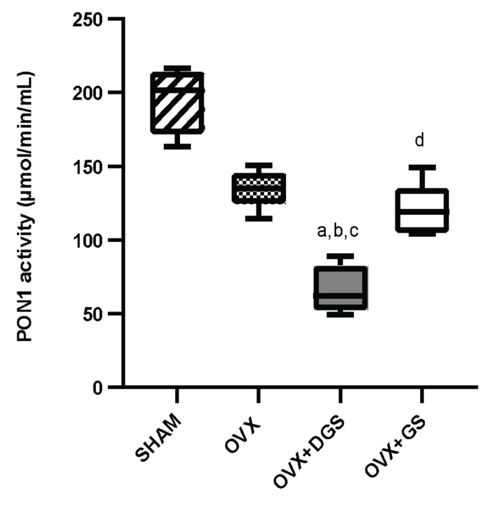

Table 1.
Biochemical parameters post-supplementation in rats.
| Parameter | SHAM | OVX | OVX+DGS | OVX+GS |
| Body weight gain (g) | 28.00±9.57 | 63.00±25.16c | 57.20±23.73d | 32.83±3.82 |
| Systolic blood pressure (mmHg) | 145.20±10.60 | 166.94±9.35b | 138.89±17.78 | 149.38±10.80 |
| Media blood pressure (mmHg) | 121.30±6.50 | 144.31±10.35a,b | 102.42±21.22 | 122.70±12.01 |
| Cholesterol (mg/dL) | 37.01±8.42 | 40.88±5.59 | 33.77±2.84 | 32.42±6.18 |
| Triglycerides (mg/dL) | 49.42±14.62 | 55.63±10.04b,c | 28.72±5.25d | 37.83±6.95 |
| Phospholipids (mg/dL) | 115.46±15.67 | 134.29±19.88b,c | 80.37±12.88d | 99.81±8.06 |
| Non-HDL-c (mg/dL) | 9.15±6.90 | 7.56±3.92 | 15.64±1.17 | 4.86±4.09 |
± Standard deviation. n=6. SHAM: SHAM-operated; OVX: ovariectomized; OVX+DGS: ovariectomized+defatted guava seeds OVX+GS: ovariectomized+guava seeds. Tukey test. ap<0.05 vs SHAM; bp<0.05 vs OVX+DGS; cp<0.05 vs OVX+GS, dOVX+DGS vs SHAM.
Table 2.
HDL lipid profile post-supplementation in rats.
| HDL lipid profile | SHAM | OVX | OVX+DGS | OVX+GS |
| HDL-c (mg/dL) | 27.86±4.59 | 33.32±2.42 | 18.13±2.31a,b,c | 27.55±6.90 |
| HDL-Tg (mg/dL) | 11.65±3.18 | 10.42±1.88 | 8.48±1.35 | 7.32±2.57 |
| HDL-PPL (mg/dL) | 58.80±13.86 | 64.71±4.38 | 52.06±5.31 | 61.66±12.68 |
| HDL-c/HDL-PPL ratio | 0.44±0.08 | 0.52±0.05 | 0.35±0.04b | 0.52±0.06 |
| HDL-Tg/HDL-PPL ratio | 0.18±0.09 | 0.16±0.04 | 0.17±0.03 | 0.14±0.04 |
± Standard deviation. n=6. SHAM: SHAM-operated; OVX: ovariectomized; OVX+DGS: ovariectomized+defatted guava seeds OVX+GS: ovariectomized+guava seeds. Tukey test. ap<0.05 vs SHAM; bp<0.05 vs OVX; cp<0.05 vs OVX+GS.
Table 3.
Size and lipid composition of HDL subclasses.
| HDL subclasses | SHAM | OVX | OVX+DGS | OVX+GS |
| Protein (%) | ||||
| HDL 2b | 40.60±3.32 | 46.56±2.08 | 48.12±1.64a | 43.19±5.06 |
| HDL 2a | 13.06±2.46 | 12.03±1.20 | 13.26±2.12 | 12.23±2.28 |
| HDL 3a | 16.68±2.00 | 14.39±1.06 | 14.84±1.21 | 15.10±2.11 |
| HDL 3b | 8.25±0.92 | 8.23±0.73 | 7.48±1.21 | 8.29±1.14 |
| HDL 3c | 19.00±3.10 | 18.51±1.74 | 16.31±1.07 | 21.19±3.75c |
| Cholesterol (%) HDL 2b |
||||
| 40.53±1.36 | 43.46±2.33 | 36.82±1.60 | 39.74±2.93 | |
| HDL 2a HDL 3a HDL 3b HDL 3c |
12.09±0.53 | 11.77±0.44 | 11.04±1.38 | 11.79±0.82 |
| 17.05±1.42 | 15.65±2.10 | 19.71±4.05 | 17.19±1.63 | |
| 9.24±0.80 | 8.42±1.19 | 11.38±1.91b | 9.77±1.29 | |
| 21.09±3.20 | 20.69±2.26 | 27.18±1.30a,b | 23.83±2.52 | |
| Triglycerides (%) HDL 2b |
||||
| 34.90±3.37 | 36.94±2.34 | 39.53±3.08 | 36.50±2.75 | |
| HDL 2a | 11.13±1.13 | 11.15±0.65 | 11.75±0.53 | 10.58±1.28 |
| HDL 3a | 17.63±2.57 | 17.29±1.58 | 16.53±0.44 | 17.05±1.42 |
| HDL 3b | 9.49±0.41 | 9.28±0.95 | 9.00±0.54 | 9.63±1.19 |
| HDL 3c | 26.84±1.90 | 25.33±2.93 | 23.37±2.62 | 26.25±3.05 |
| Phospholipids (%) | ||||
| HDL 2b | 41.34±5.21 | 40.54±3.64 | 39.70±0.94 | 41.42±6.00 |
| HDL 2a | 10.87±0.76 | 10.39±0.78 | 11.97±1.06 | 10.01±0.88c |
| HDL 3a | 16.57±2.18 | 15.64±1.03 | 17.21±0.39 | 15.47±1.34 |
| HDL 3b | 8.99±1.01 | 9.27±0.80 | 8.92±0.74 | 9.00±0.78 |
| HDL 3c | 22.24±2.29 | 24.79±1.64 | 22.20±0.93 | 24.11±3.49 |
± Standard deviation. n=6. SHAM: SHAM-operated; OVX: ovariectomized; OVX+DGS: ovariectomized+defatted guava seeds OVX+GS: ovariectomized+guava seeds. Tukey test. ap<0.05 vs SHAM; bp<0.05 vs OVX; cp<0.05 vs OVX+GS.
Table 4.
Apolipoprotein percentage of the total HDL protein.
| Apolipoproteins | SHAM | OVX | OVX+DGS | OVX+GS |
| Apo AIV | 19.79±1.53 | 18.22±3.64 | 22.76±3.13 | 21.01±3.12 |
| Apo E | 19.92±3.26 | 18.95±1.85 | 13.70±3.38a,b,c | 21.68±3.33 |
| Apo AI | 44.17±3.20 | 45.60±5.38 | 41.79±7.65 | 39.93±8.10 |
| Apo AII | 8.71±1.12 | 8.99±1.58 | 8.34±3.28 | 9.18±1.30 |
| Apo C | 5.89±0.90 | 6.75±0.88 | 13.41±5.03a,b,c | 8.20±0.93 |
± Standard deviation. n=6. SHAM: SHAM-operated; OVX: ovariectomized; OVX+GS: ovariectomized+defatted guava seeds OVX+GS: ovariectomized+guava seeds. Tukey test. ap<0.05 vs SHAM; bp<0.05 vs OVX; cp<0.05 vs OVX+GS.
Disclaimer/Publisher’s Note: The statements, opinions and data contained in all publications are solely those of the individual author(s) and contributor(s) and not of MDPI and/or the editor(s). MDPI and/or the editor(s) disclaim responsibility for any injury to people or property resulting from any ideas, methods, instructions or products referred to in the content. |
© 2025 by the authors. Licensee MDPI, Basel, Switzerland. This article is an open access article distributed under the terms and conditions of the Creative Commons Attribution (CC BY) license (http://creativecommons.org/licenses/by/4.0/).
Copyright: This open access article is published under a Creative Commons CC BY 4.0 license, which permit the free download, distribution, and reuse, provided that the author and preprint are cited in any reuse.


