
Submitted:
23 September 2025
Posted:
25 September 2025
You are already at the latest version
Abstract
Yellow fever (YF) infection typically affects the liver, with death primarily attributed to acute hepatic failure. The autophagic process in Flavivirus infection may be a key factor in disease progression. We explored the role of autophagy in the pathogenesis of YF within the liver tissue of fatally affected individuals. Liver samples were collected from 21 Yellow Fever Virus (YFV)-positive patients who died from the disease and five flavivirus-negative controls who died from unrelated causes but had preserved liver parenchymal architecture. The cellular stress environment established in the hepatic parenchyma in fatal virus-positive cases was found to be highly intense. Quantitatively, all markers (iNOS, IL-1β, IL-18, IL-33, BECLIN 1 and RIP3) showed increased expression in relation to the control group, which revealed the involvement of these markers in the pathogenesis of YF. Through correlation tests, we confirmed that both enzymes and proteins collaborate to increase inflammatory activity in the liver parenchyma, mainly in the midzonal zone. These findings suggest that autophagy is associated with the robust inflammatory response in the liver during fatal cases of yellow fever in humans.
Keywords:
yellow fever
; autophagy
; liver parenchyma
; Arbovirus
1. Introduction
The Yellow Fever virus (YFV) is an arthropod-borne virus belonging to the Flaviviridae family, Flavivirus genus, positive-sense RNA. Its genome, comprising approximately 11,000 bases, consists of a single open reading frame flanked by 2 untranslated regions. Like other flaviviruses, it has the ability to induce the production of a polyprotein that is processed and cleaved into three structural and seven non-structural proteins, modulating the host immune response [1,2,3,4,5]. From a clinical perspective, manifestations range from asymptomatic infection to severe disease, defined as Yellow Fever Disease (YFD), where, with the progression of the inflammatory process, pan-systemic complications lead to devastating effects on organs with an intense vascular network such as the kidney, lung, liver, brain, and heart [6,7,8,9,10]. The exacerbated worsening in organs with selective virus tropism, such as liver, is considered. In this case, histological lesions coincide with areas of hemorrhage and adjacent inflammatory infiltration. Immune response in this organ indicates that both Th1 and Th2 lymphocytic modulation induces pro- and anti-inflammatory cytokines release, promoting the establishment of an inhospitable tissue environment that attempts to inhibit the replicative process. However, this exacerbates both tissue and cellular, culminating in the phenomenon of cell death, classically marked by necrosis and apoptosis [11,12,13,14,15].
With the advancement of investigations into cellular injury mechanisms, the study of autophagy as a regulatory process of cellular recycling aimed at maintaining cellular homeostasis still requires further clarification on how flaviviruses can manipulate this process and how the defense system acts to mediate cytokine production to contain the virus's immune evasion strategies. Thus, we investigated the role of autophagy and its implications in the immunopathogenesis of fatal cases of human yellow fever.
2. Materials and Methods
2.1. Patients, Samples, and Diagnosis of Yellow Fever Infection
This study analyzed a total of 26 human liver biopsies, comprising 21 samples from fatal cases of yellow fever (YF). Five control samples were collected from patients with preserved liver architecture, testing negative for YF and other circulating Flaviviruses in Brazil, as verified by the death verification service at the Renato Chaves Scientific Expertise Center in Belém, Pará, Brazil.
YF diagnoses confirmation was based on the methods outlined in the study by Olimpio et al. [16], which included histopathological, immunohistochemical, and reverse transcriptase quantitative real-time polymerase chain reaction(RT-qPCR) analyses. For histopathological diagnosis, the paraffin-embedded biopsies were sectioned into 5μm slices and stained using the hematoxylin-eosin method. Detailed information about the patients included in this study is presented in Table 1.
2.2. Ethics Statement
Patient samples were obtained and processed as part of the response measures to the surveillance of the YFV epidemic in Brazil on an emergency basis, as defined by the Ministry of Health. This study was approved (protocol code number 2.364.226) by the Research Ethics Committee (CEP) of the Evandro Instituto Chagas (IEC). All methods were performed in accordance with the relevant guidelines and regulations approved by the CEP / IEC and the Brazilian Ministry of Health rules and regulations for studies with biological samples.
2.3. Immunohistochemistry (IHC)
Immunostaining of the hepatic tissues was performed using the streptavidin-biotin peroxidase immunohistochemical method (SABC) adapted it according to Olímpio et al., [16]. Antibodies specific for iNOS, IL-1β, IL-18, IL-33, BECLIN 1 and RIP3 are listed in Table 2. Briefly, tissue samples were deparaffinized in xylene and hydrated in a decreasing series of ethanol (90%, 80%, and 70%). Liver sections were incubated in 3% hydrogen peroxide for 45 min to block endogenous peroxidase. Incubation in citrate buffer, pH 6.0, for 20 min at 90 °C was realized to antigen retrieval. Non-specific proteins were blocked by incubating the sections in 10% skim milk for 30 min. Histological sections were then incubated overnight with the primary antibodies diluted in 1% bovine serum albumin (supplementary file). The slides were immersed in 1 × PBS and incubated with the biotinylated secondary anti-body (LSAB; DakoCytomation, Glostrup, Denmark) in an oven for 30 min at 37 °C. The slides were immersed in 1 × PBS and incubated with streptavidin peroxidase (LSAB; DakoCytomation) for 30 min at 37 °C. For visualization, specimens were treated with a chromogenic solution (0.03% diaminobenzidine and 3% hydrogen peroxide). Finally, histological sections were washed in distilled water, counterstained with Harris hema-toxylin for 1 min, dehydrated in ethanol (70%, 80%, 90%), and deparaffinized in xylene.
2.4. Quantitative Analysis and Photo-Documentation
The markers used to characterize the in-situ autophagy profile were visualized us-ing an Axio Imager Z1 microscope (Zeiss). Immunostaining results were evaluated quantitatively by randomly selecting ten fields in the hepatic parenchyma (Z3: Centrilobular zone; Z2: Midzonal zone; Z1: Periportal zone; PT: Portal tract) of the fatal YF or negative control (NC) cases for viewing at high magnification. Each field was subdi-vided into 10 × 10 areas delimited by a 0.0625 mm2 grid.
2.5. Statistical Analysis
The data were stored in a Microsoft Excel 2016 spreadsheet and analyzed using GraphPadPrism 9.0. The numerical variables were expressed as the mean, median, standard deviation, and variance. One-way ANOVA, Tukey’stest, and Pearson correlation were also applied; results were considered statistically significant at p < 0.05.
3. Results
In fatal YFV, all analytes, IL-1β, IL-18, IL-33, iNOS, BECLIN-1 and RIP3, were markedly increased compared with flavivirus-negative controls across hepatic compartments (one-way ANOVA with Tukey’s post-hoc; Table 3; Figure 1 and Figure 2). Expression consistently peaked in the midzonal region (Z2) for every marker. For example, in Z2 we observed ~2.3× higher IL-1β (190.5 ± 75.0 vs. 83.2 ± 13.4 cells/field), ~3.9× higher IL-18 (365.0 ± 44.9 vs. 92.8 ± 30.8), ~3.9× higher IL-33 (351.2 ± 43.1 vs. 89.6 ± 21.5), ~3.9× higher iNOS (150.1 ± 22.3 vs. 38.4 ± 8.8), ~2.8× higher RIP3 (255.1 ± 90.9 vs. 92.8 ± 13.4), and ~2.1× higher BECLIN-1 (134.1 ± 19.3 vs. 64.0 ± 16.0) relative to controls (all p < 0.001 unless noted; Table 3). The zonal gradient (Z2 > Z3 ≈ Z1 > PT) was evident for most markers, and pairwise post-hoc contrasts within each zone remained significant (Table 3).
RIP3 was strongly up-regulated in hepatocytes, particularly in Z2, aligning with activation of necroptosis pathways in severe YFV (Table 3; Figure 1e,f). Correlation analyses further highlighted coordinated shifts between autophagy and inflammation (Table 4 / Table S4): a robust RIP3–iNOS axis (e.g., RIP3 [Z1]–iNOS [PT], r = 0.84; RIP3 [Z2]–iNOS [PT/Z2], r ≈ −0.83/−0.82), coupling between IL-18 and IL-33 in Z3 (r = 0.80), and zonal links between BECLIN-1 and cytokines (IL-33 [Z2] with BECLIN-1 [Z1/PT], r ≈ 0.79/0.76; IL-18 [Z2] with BECLIN-1 [Z1/Z2], r ≈ −0.75). Together, these patterns indicate an autophagy–inflammation crosstalk centered on Z2. (Table 4). The full correlation matrix, including FDR-adjusted q-values, is provided in Table S4.
4. Discussion
The YFV is a flavivirus that has raised significant systemic concerns due to the risk of reemergence and sporadic outbreaks in developing countries, with alarming repercussions for public health [17,18,19]. From the perspective of the host-pathogen relationship and tissue compartmentalization, the way YFV manages to infect cells within the hepatic parenchyma still requires further clarification. It is believed that both hepatocytes and Kupffer cells are primary targets, reflecting directly on how the defense system organizes the in situ immune response in the rapaport space to try to inhibit replication [20,21,22].
However, due to immune evasion strategies, the escape mediated by adaptive survival characteristics in the pathogen's tissue environment induces a series of cellular injury mechanisms culminating in increased expression of inflammatory cytokines and the phenomenon of cell death [23,24,25,26].
From the results, in our investigation of cytokines belonging to the IL-1 family (IL-1β, IL-18, and IL-33), enzymes like iNOS, protein kinase RIP3, and autophagy markers (BECLIN-1), we observed that the cellular stress environment established in the hepatic parenchyma in fatal virus-positive cases is very intense. Quantitatively, all markers showed an increase compared to the control, and through correlation tests, we verified that both the enzyme and proteins work together to increase inflammatory activity in the hepatic parenchyma, especially in the mediozonal zone.
The elevation of inflammatory cytokines such as IL-1β and IL-18 indicates that the pro-inflammatory response is substantially necessary to enhance not only the Th1 response but also oxidative stress. Another important aspect is the activation of the inflammasome, a macromolecule belonging to the innate immune response that is essential for orchestrating the cleavage and conversion of these cytokines into their active form [27,28,29,30].
Several studies with flaviviruses have shown, for example, in fatal cases of Zika virus-induced microcephaly, that such mechanisms trigger the exacerbation of tissue damage marked by neuronal depopulation, neuronal necrosis, and perivascular inflammatory infiltration [31,32,33,34].
In our results, a noteworthy fact is that these cytokines are widely expressed in areas of massive destruction of hepatocytes, with a predominance of markers like iNOS. Considering the cellular stress environment caused by cell damage, this enzyme that modulates the activity of Kupffer cells through the classical inflammatory pathway demonstrates that this scenario contributes to the modulation of the M1 phenotype, where iNOS leads to the formation of L-citrulline, NO, and consequently reactive oxygen and nitrogen intermediates harmful to the survival of liver cells [33,34,35].
Due to this immunological seesaw, studies on the immunopathogenesis ofYF demonstrate the paradoxical balance between the Th1 and Th2 profiles [36,37,38]. Amidst the intense tissue destruction characterizing YF, such as hemorrhage and coagulative hepatocellular necrosis, in an attempt to prevent the progression of an extremely inhospitable environment [26,35], cytokines like IL-33 have gained importance in immunological studies for flaviviruses. IL-33 modulates a series of mechanisms that contribute to the development of Th2 lymphocyte response, and in the liver, the protein may contribute to the modulation of the M2 macrophage phenotype, which is reparative [39,40,41]. Another significant point is that this cytokine strongly modulates apoptosis mechanisms, and in YF, this phenomenon is crucial as apoptosis prevents the leakage of enzymatic content, avoiding the progressive escalation of inflammation.
Advancing in this scenario, in investigating autophagy, we found a significant increase in both BECLIN-1 and RIP3 in fatal virus-positive cases compared to the control. This indicates that autophagy may be decisive in mediating the recycling of cellular components over the massive attack orchestrated by both virus and the immune response [42,43,44,45,46]. Another interesting finding in our study is that both RIP3 and BECLIN-1 positively or negatively correlate with other markers involved in the study, demonstrating that autophagy is in an influential area in YFV, where in situ cellular recycling is another mechanism contributing to the phenomenon of cellular injury in the hepatic parenchyma of fatal cases of human YF.
5. Conclusions
The study sheds light on the complex immunopathogenesis of YFV. The investigation into the host-pathogen relationship within the hepatic parenchyma reveals that YFV infection involves both hepatocytes and likely Kupffer cells, with a profound impact on the in situ immune response. The observed elevation of inflammatory cytokines, such as IL-1β, IL-18, and IL-33, indicates their involvement along with the activation of the inflammasome, leading to a substantial pro-inflammatory response essential for intensifying the Th1 response and oxidative stress. In essence, the research provides valuable insights into the immunological seesaw and the complex cellular stress environment in fatal cases of yellow fever in humans, emphasizing the need for a comprehensive understanding of these mechanisms to develop effective strategies for prevention and intervention.
6. Patents
Not applicable.
Supplementary Materials
The following supporting information can be downloaded at the website of this paper posted on Preprints.org. Table S4: Full correlation matrix among autophagy and inflammation markers with p-values and Benjamini–Hochberg FDR q-values.
Author Contributions
Author Contributions: Conceptualization, J.A.S.Q., P.F.C.V., M.D.W., V.S.C.M. and J.R.S.; methodology, M.L.G.C., L.F.M.F., A.C.R.C., R.S.S.A., M.D.W., L.C.Ma. and J.R.S.; software, L.C.Mo. and L.A.M.F.; validation, A.J.M.F., M.D.W., L.C.Ma. and J.R.S.; formal analysis, L.A.M.F. and V.S.C.M.; investigation, V.S.C.M., M.L.G.C., A.J.M.F., A.C.R.C., R.S.S.A. and P.R.N.S.; resources, P.F.C.V., L.F.M.F., R.S.S.A., M.D.W. and L.C.Ma.; data curation, L.C.Mo., L.A.M.F. and V.S.C.M.; writing—original draft preparation, V.S.C.M. and M.L.G.C.; writing—review and editing, all authors; visualization, R.M.M.R., L.C.Mo. and V.S.C.M.; supervision, J.A.S.Q., P.F.C.V. and M.D.W.; project administration, V.S.C.M., J.A.S.Q., P.F.C.V. and M.D.W.; funding acquisition, J.A.S.Q., P.F.C.V. and M.D.W.. All authors have read and agreed to the published version of the manuscript.
Funding
This research was funded by the National Institute of Science and Technology for Emerging and Re-emerging Viruses—INCT-VER/CNPq—406360/2022-7, and the National Council for Scientific and Technological Development (CNPq)/Brazil-308600/2022-3 (JASQ).
Clarification of Initials
L.C.Ma. = Lívia Carício Martins; L.C.Mo. = Lucas Corrêa Modesto.
Institutional Review Board Statement
This study was conducted in accordance with the Declaration of Helsinki and approved by the Institutional Review Board of Instituto Evandro Chagas (protocol code number 2.364.226 on 11 June 2020).
Informed Consent Statement
Patient consent was waived due to the retrospective use of de-identified postmortem tissue and the absence of contact with next of kin, as approved by the Ethics Committee.
Data Availability Statement
All data supporting the findings of this study are available within the article and its Supplementary Materials (Table S4). Additional de-identified raw counts, analysis spreadsheets, and representative whole-slide images can be made available by the corresponding author upon reasonable request, subject to ethical and privacy restrictions
Acknowledgments
The authors are grateful to the PAPQ program (UFPA). During the preparation of this manuscript, the authors used ChatGPT 5 to assist with language editing and formatting. The authors reviewed and edited the content and take full responsibility for the publication’s scientific accuracy and integrity.
Conflicts of Interest
The authors declare no conflicts of interest.
Abbreviations
The following abbreviations are used in this manuscript:
| YFV | Yellow fever virus |
| NC | Negative control |
| FFPE | Formalin-fixed, paraffin-embedded |
| HPF | High-power field |
| IHC | Immunohistochemistry |
| PT | Portal tract |
| Z1/Z2/Z3 | Periportal/Midzonal/Centrilobular zones |
| BECLIN-1 | Autophagy initiation marker |
| RIP3 | Receptor-interacting protein kinase 3 |
| iNOS | Inducible nitric oxide synthase |
| IL-1β/IL-18/IL-33 | Interleukins 1β/18/33 |
| ANOVA | Analysis of variance |
| FDR | False discovery rate |
| BH | Benjamini–Hochberg |
| SD | Standard deviation |
References
- Gardner, C.L.; Ryman, K.D. Yellow fever: A reemerging threat. Clin. Lab. Med. 2010, 30, 237–260. [Google Scholar] [CrossRef]
- Elton, N.W. Yellow fever in Panama; historical and contemporary. Am. J. Trop. Med. Hyg. 1952, 1, 436–456. [Google Scholar] [CrossRef] [PubMed]
- Abreu, F.V.S.; Ribeiro, I.P.; Ferreira-de-Brito, A.; Santos, A.A.C.D.; Miranda, R.M.; Bonelly, I.S.; Neves, M.S.A.S.; Bersot, M.I.; Santos, T.P.D.; Gomes, M.Q.; Silva, J.L.D.; Romano, A.P.M.; Carvalho, R.G.; Said, R.F.D.C.; Ribeiro, M.S.; Laperrière, R.D.C.; Fonseca, E.O.L.; Falqueto, A.; Paupy, C.; Failloux, A.-B.; Moutailler, S.; Castro, M.G.; Gómez, M.M.; Motta, M.A.; Bonaldo, M.C.; Lourenço-de-Oliveira, R. Haemagogus leucocelaenus and Haemagogus janthinomys are the primary vectors in the major yellow fever outbreak in Brazil, 2016–2018. Emerg. Microbes Infect. 2019, 8, 218–231. [Google Scholar] [CrossRef] [PubMed]
- Waggoner, J.J.; Rojas, A.; Pinsky, B.A. Yellow fever virus: Diagnostics for a persistent arboviral threat. J. Clin. Microbiol. 2018, 56, e00827–18. [Google Scholar] [CrossRef] [PubMed]
- Faggioni, G.; De Santis, R.; Moramarco, F.; Di Donato, M.; De Domenico, A.; Molinari, F.; Petralito, G.; Fortuna, C.; Venturi, G.; Rezza, G.; Lista, F. Pan-yellow fever virus detection and lineage assignment by real-time RT-PCR and amplicon sequencing. J. Virol. Methods 2023, 316, 114717. [Google Scholar] [CrossRef]
- Monath, T.P.; Barrett, A.D. Pathogenesis and pathophysiology of yellow fever. Adv. Virus Res. 2003, 60, 343–395. [Google Scholar] [CrossRef]
- Gould, C.V.; Free, R.J.; Bhatnagar, J.; Soto, R.A.; Royer, T.L.; Maley, W.R.; Moss, S.; Berk, M.A.; Craig-Shapiro, R.; Kodiyanplakkal, R.P.L.; Westblade, L.F.; Muthukumar, T.; Puius, Y.A.; Raina, A.; Hadi, A.; Gyure, K.A.; Trief, D.; Pereira, M.; Kuehnert, M.J.; Ballen, V.; Kessler, D.A.; Dailey, K.; Omura, C.; Doan, T.; Miller, S.; Wilson, M.R.; Lehman, J.A.; Ritter, J.M.; Lee, E.; Silva-Flannery, L.; Reagan-Steiner, S.; Velez, J.O.; Laven, J.J.; Fitzpatrick, K.A.; Panella, A.; Davis, E.H.; Hughes, H.R.; Brault, A.C.; St George, K.; Dean, A.B.; Ackelsberg, J.; Basavaraju, S.V.; Chiu, C.Y.; Staples, J.E. ; Yellow Fever Vaccine Virus Transplant and Transfusion Investigation Team. Transmission of yellow fever vaccine virus through blood transfusion and organ transplantation in the USA in 2021: Report of an investigation. Lancet Microbe 2023, 4, e711–e721. [Google Scholar] [CrossRef]
- Qian, X.; Wu, B.; Tang, H.; Luo, Z.; Xu, Z.; Ouyang, S.; Li, X.; Xie, J.; Yi, Z.; Leng, Q.; Liu, Y.; Qi, Z.; Zhao, P. Rifapentine is an entry and replication inhibitor against yellow fever virus both in vitro and in vivo. Emerg. Microbes Infect. 2022, 11, 873–884. [Google Scholar] [CrossRef] [PubMed]
- Chen, Z.; Liu, L.; Lv, Y.; Zhang, W.; Li, J.; Zhang, Y.; Di, T.; Zhang, S.; Liu, J.; Li, J.; Qu, J.; Hua, W.; Li, C.; Wang, P.; Zhang, Q.; Xu, Y.; Jiang, R.; Wang, Q; Chen, L. ; Wang, S. ; Pang, X.; Liang, M.; Ma, X.; Li, X.; Wang, Q.; Zhang, F.; Li, D. A fatal yellow fever virus infection in China: Description and lessons. Emerg. Microbes Infect. 2016, 5, e69. [Google Scholar] [CrossRef]
- Ferreira, M.S.; Júnior, P.S.B.; Cerqueira, V.D.; Rivero, G.R.C.; Júnior, C.A.O.; Castro, P.H.G.; Silva, G.A.D.; Silva, W.B.D.; Imbeloni, A.A.; Sousa, J.R.; Araújo, A.P.S.; Silva, F.A.E.; Tesh, R.B.; Quaresma, J.A.S.; Vasconcelos, P.F.D.C. Experimental yellow fever virus infection in the squirrel monkey (Saimiri spp.) I: Gross anatomical and histopathological findings in organs at necropsy. Mem. Inst. Oswaldo Cruz 2020, 115, e190501. [Google Scholar] [CrossRef]
- James, E.A.; LaFond, R.E.; Gates, T.J.; Mai, D.T.; Malhotra, U.; Kwok, W.W. Yellow fever vaccination elicits broad functional CD4+ T cell responses that recognize structural and nonstructural proteins. J. Virol. 2013, 87, 12794–12804. [Google Scholar] [CrossRef]
- Sandberg, J.T.; Ols, S.; Löfling, M.; Varnaitė, R.; Lindgren, G.; Nilsson, O.; Rombo, L.; Kalén, M.; Loré, K.; Blom, K.; Ljunggren, H.G. Activation and kinetics of circulating T follicular helper cells, specific plasmablast response, and development of neutralizing antibodies following yellow fever virus vaccination. J. Immunol. 2021, 207, 1033–1043. [Google Scholar] [CrossRef]
- Santos, A.P.; Matos, D.C.S.; Bertho, A.L.; Mendonça, S.C.F.; Marcovistz, R. Detection of Th1/Th2 cytokine signatures in yellow fever 17DD first-time vaccinees through ELISpot assay. Cytokine 2008, 42, 152–155. [Google Scholar] [CrossRef]
- Quaresma, J.A.; Duarte, M.I.; Vasconcelos, P.F. Midzonal lesions in yellow fever: A specific pattern of liver injury caused by direct virus action and in situ inflammatory response. Med. Hypotheses 2006, 67, 618–621. [Google Scholar] [CrossRef]
- Quaresma, J.A.; Barros, V.L.; Pagliari, C.; Fernandes, E.R.; Guedes, F.; Takakura, C.F.; Andrade, H.F., Jr.; Vasconcelos, P.F.; Duarte, M.I. Revisiting the liver in human yellow fever: Virus-induced apoptosis in hepatocytes associated with TGF-beta, TNF-alpha and NK cells activity. Virology 2006, 345, 22–30. [Google Scholar] [CrossRef]
- Olímpio, F.A.; Falcão, L.F.M.; Carvalho, M.L.G.; Lopes, J. da C. ; Mendes, C.C.H.; Filho, A.J.M.; da Silva, C.A.M.; Miranda, V.D.S.C.; dos Santos, L.C.; Vilacoert, F.S. da S.; et al. Endothelium activation during severe yellow fever triggers an intense cytokine-mediated inflammatory response in the liver parenchyma. Pathogens 2022, 11, 101. [Google Scholar] [CrossRef]
- Cavalcante, K.R.L.J.; Tauil, P.L. Risk of re-emergence of urban yellow fever in Brazil. Epidemiol. Serv. Saude 2017, 26, 617–620. [Google Scholar] [CrossRef] [PubMed]
- Couto-Lima, D.; Madec, Y.; Bersot, M.I.; Campos, S.S.; Motta, M.A.; Santos, F.B.D.; Vazeille, M.; Vasconcelos, P.F.D.C.; Lourenço-de-Oliveira, R.; Failloux, A.-B. Potential risk of re-emergence of urban transmission of yellow fever virus in Brazil facilitated by competent Aedes populations. Sci. Rep. 2017, 7, 4848. [Google Scholar] [CrossRef] [PubMed]
- Rosser, J.I.; Nielsen-Saines, K.; Saad, E.; Fuller, T. Reemergence of yellow fever virus in southeastern Brazil, 2017–2018: What sparked the spread? PLoS Negl. Trop. Dis. 2022, 16, e0010133. [Google Scholar] [CrossRef]
- De Brito, T.; Siqueira, S.A.; Santos, R.T.; Nassar, E.S.; Coimbra, T.L.; Alves, V.A. Human fatal yellow fever: Immunohistochemical detection of viral antigens in the liver, kidney and heart. Pathol. Res. Pract. 1992, 188, 177–181. [Google Scholar] [CrossRef] [PubMed]
- Marianneau, P.; Desprès, P.; Deubel, V. Connaissances récentes sur la pathogénie de la fièvre jaune et questions pour le futur [Recent knowledge on the pathogenesis of yellow fever and questions for the future]. Bull. Soc. Pathol. Exot. 1999, 92, 432–434. [Google Scholar]
- Bailey, A.L.; Kang, L.I.; de Assis Barros D'Elia Zanella, L.G.F.; Silveira, C.G.T.; Ho, Y.L.; Foquet, L. ; Bial, G; McCune, B. T.; Duarte-Neto, A.N.; Thomas, A.; Raué, H.P.; Byrnes, K.; Kallas, E.G.; Slifka, M.K.; Diamond, M.S. Consumptive coagulopathy of severe yellow fever occurs independently of hepatocellular tropism and massive hepatic injury. Proc. Natl. Acad. Sci. USA 2020, 117, 32648–32656. [Google Scholar] [CrossRef]
- Lopes, J. da C. ; Falcão, L.F.M.; Martins Filho, A.J.; Carvalho, M.L.G.; Mendes, C.C.H.; Olímpio, F.A.; do Socorro Cabral Miranda, V.; Dos Santos, L.C.; Chiang, J.O.; Cruz, A.C.R.; Galúcio, V.C.A.; do Socorro da Silva Azevedo, R.; Martins, L.C.; Duarte, M.I.S.; de Sousa, J.R.; da Costa Vasconcelos, P.F.; Quaresma, J.A.S. Factors involved in the apoptotic cell death mechanism in yellow fever hepatitis. Viruses 2022, 14, 1204. [Google Scholar] [CrossRef]
- Ribeiro, Y.P.; Falcão, L.F.M.; Smith, V.C.; de Sousa, J.R.; Pagliari, C.; Franco, E.C.S.; Cruz, A.C.R.; Chiang, J.O.; Martins, L.C.; Nunes, J.A.L.; Vilacoert, F.S.D.S.; Santos, L.C.D.; Furlaneto, M.P.; Fuzii, H.T.; Bertonsin Filho, M.V.; da Costa, L.D.; Duarte, M.I.S.; Furlaneto, I.P.; Martins Filho, A.J.; Aarão, T.L.S.; Vasconcelos, P.F.D.C.; Quaresma, J.A.S. Comparative analysis of human hepatic lesions in dengue, yellow fever, and chikungunya: Revisiting histopathological changes in the light of modern knowledge of cell pathology. Pathogens 2023, 12, 680. [Google Scholar] [CrossRef] [PubMed]
- Lemos, F.O.; França, A.; Lima Filho, A.C.M.; Florentino, R.M.; Santos, M.L.; Missiaggia, D.G.; Rodrigues, G.O.L.; Dias, F.F.; Souza Passos, I.B.; Teixeira, M.M.; Andrade, A.M.F.; Lima, C.X.; Vidigal, P.V.T.; Costa, V.V.; Fonseca, M.C.; Nathanson, M.H.; Leite, M.F. Molecular mechanism for protection against liver failure in human yellow fever infection. Hepatol. Commun. 2020, 4, 657–669. [Google Scholar] [CrossRef] [PubMed]
- Carvalho, M.L.G.; Falcão, L.F.M.; Lopes, J.D.C.; Mendes, C.C.H.; Olímpio, F.A.; Miranda, V.D.S.C.; Santos, L.C.D.; de Moraes, D.D.P.; Bertonsin Filho, M.V.; da Costa, L.D.; da Silva Azevedo, R.D.S.; Cruz, A.C.R.; Galúcio, V.C.A.; Martins, L.C.; Duarte, M.I.S.; Martins Filho, A.J.; Sousa, J.R.; Vasconcelos, P.F.D.C.; Quaresma, J.A.S. Role of Th17 cytokines in the liver's immune response during fatal yellow fever: Triggering cell damage mechanisms. Cells 2022, 11, 2053. [Google Scholar] [CrossRef] [PubMed]
- Gaucher, D.; Therrien, R.; Kettaf, N.; Angermann, B.R.; Boucher, G.; Filali-Mouhim, A.; Moser, J.M.; Mehta, R.S.; Drake, D.R., 3rd; Castro, E.; Akondy, R.; Rinfret, A.; Yassine-Diab, B.; Said, E.A.; Chouikh, Y.; Cameron, M.J.; Clum, R.; Kelvin, D.; Somogyi, R.; Greller, L.D.; Balderas, R.S.; Wilkinson, P.; Pantaleo, G.; Tartaglia, J.; Haddad, E.K.; Sékaly, R.P. Yellow fever vaccine induces integrated multilineage and polyfunctional immune responses. J. Exp. Med. 2008, 205, 3119–3131. [Google Scholar] [CrossRef]
- Marquardt, N.; Ivarsson, M.A.; Blom, K.; Gonzalez, V.D.; Braun, M.; Falconer, K.; Gustafsson, R.; Fogdell-Hahn, A.; Sandberg, J.K.; Michaëlsson, J. The human NK cell response to yellow fever virus 17D is primarily governed by NK cell differentiation independently of NK cell education. J. Immunol. 2015, 195, 3262–3272. [Google Scholar] [CrossRef]
- Kum, D.B.; Boudewijns, R.; Ma, J.; Mishra, N.; Schols, D.; Neyts, J.; Dallmeier, K. A chimeric yellow fever–Zika virus vaccine candidate fully protects against yellow fever virus infection in mice. Emerg. Microbes Infect. 2020, 9, 520–533. [Google Scholar] [CrossRef]
- Elliott, E.I.; Sutterwala, F.S. Initiation and perpetuation of NLRP3 inflammasome activation and assembly. Immunol. Rev. 2015, 265, 35–52. [Google Scholar] [CrossRef]
- He, Z.; Chen, J.; Zhu, X.; An, S.; Dong, X.; Yu, J.; Zhang, S.; Wu, Y.; Li, G.; Zhang, Y.; Wu, J.; Li, M. NLRP3 inflammasome activation mediates Zika virus-associated inflammation. J. Infect. Dis. 2018, 217, 1942–1951. [Google Scholar] [CrossRef]
- Wang, W.; Li, G.; Wu, D.; Luo, Z.; Pan, P.; Tian, M.; Wang, Y.; Xiao, F.; Li, A.; Wu, K.; Liu, X.; Rao, L.; Liu, F.; Liu, Y.; Wu, J. Zika virus infection induces host inflammatory responses by facilitating NLRP3 inflammasome assembly and interleukin-1β secretion. Nat. Commun. 2018, 9, 106. [Google Scholar] [CrossRef]
- de Sousa, J.R.; Azevedo, R.D.S.D.S.; Martins Filho, A.J.; de Araujo, M.T.F.; Cruz, E.D.R.M.; Vasconcelos, B.C.B.; Cruz, A.C.R.; de Oliveira, C.S.; Martins, L.C.; Vasconcelos, B.H.B.; Casseb, L.M.N.; Chiang, J.O.; Quaresma, J.A.S.; Vasconcelos, P.F.D.C. In situ inflammasome activation results in severe damage to the central nervous system in fatal Zika virus microcephaly cases. Cytokine 2018, 111, 255–264. [Google Scholar] [CrossRef]
- Azevedo, R.S.S.; de Sousa, J.R.; Araujo, M.T.F.; Martins Filho, A.J.; de Alcantara, B.N.; Araujo, F.M.C.; Queiroz, M.G.L.; Cruz, A.C.R.; Vasconcelos, B.H.B.; Chiang, J.O.; Martins, L.C.; Casseb, L.M.N.; da Silva, E.V.; Carvalho, V.L.; Vasconcelos, B.C.B.; Rodrigues, S.G.; Oliveira, C.S.; Quaresma, J.A.S.; Vasconcelos, P.F.C. In situ immune response and mechanisms of cell damage in central nervous system of fatal cases microcephaly by Zika virus. Sci. Rep. 2018, 8, 1. [Google Scholar] [CrossRef] [PubMed]
- Ferreira, M.S.; Sousa, J.R.; Bezerra Júnior, P.S.; Cerqueira, V.D.; Oliveira Júnior, C.A.; Rivero, G.R.C.; Castro, P.H.G.; Silva, G.A.; Muniz, J.A.P.C.; da Silva, E.V.P.; Casseb, S.M.M; Pagliari, C.; Martins, L.C.; Tesh, R.B.; Quaresma, J.A.S.; Vasconcelos, P.F.C. Experimental yellow fever in squirrel monkey: Characterization of liver in situ immune response. Viruses 2023, 15, 551. [Google Scholar] [CrossRef] [PubMed]
- DeGottardi, Q.; Gates, T.J.; Yang, J.; James, E.A.; Malhotra, U.; Chow, I.T.; Simoni, Y.; Fehlings, M.; Newell, E.W.; DeBerg, H.A.; Kwok, W.W. Ontogeny of different subsets of yellow fever virus-specific circulatory CXCR5+ CD4+ T cells after yellow fever vaccination. Sci. Rep. 2020, 10, 15686. [Google Scholar] [CrossRef]
- Azamor, T.; da Silva, A.M.V.; Melgaço, J.G.; Dos Santos, A.P.; Xavier-Carvalho, C.; Alvarado-Arnez, L.E.; Batista-Silva, L.R.; de Souza Matos, D.C.; Bayma, C.; Missailidis, S.; Ano Bom, A.P.D.; Moraes, M.O.; da Costa Neves, P.C. Activation of an effective immune response after yellow fever vaccination is associated with the genetic background and early response of IFN-γ and CLEC5A. Viruses 2021, 13, 96. [Google Scholar] [CrossRef]
- Bovay, A.; Fuertes Marraco, S.A.; Speiser, D.E. Yellow fever virus vaccination: An emblematic model to elucidate robust human immune responses. Hum. Vaccin. Immunother. 2021, 17, 2471–2481. [Google Scholar] [CrossRef]
- França, R.F.; Costa, R.S.; Silva, J.R.; Peres, R.S.; Mendonça, L.R.; Colón, D.F.; Alves-Filho, J.C.; Cunha, F.Q. IL-33 signaling is essential to attenuate viral-induced encephalitis development by downregulating iNOS expression in the central nervous system. J. Neuroinflammation 2016, 13, 159. [Google Scholar] [CrossRef] [PubMed]
- de Sousa, J.R.; Azevedo, R.S.S.; Martins Filho, A.J.; Araujo, M.T.F.; Moutinho, E.R.C.; Baldez Vasconcelos, B.C.; Cruz, A.C.R.; Oliveira, C.S.; Martins, L.C.; Baldez Vasconcelos, B.H.; Casseb, L.M.N.; Chiang, J.O.; Quaresma, J.A.S.; Vasconcelos, P.F.C. Correlation between apoptosis and in situ immune response in fatal cases of microcephaly caused by Zika virus. Am. J. Pathol. 2018, 188, 2644–2652. [Google Scholar] [CrossRef]
- Furukawa, S.; Moriyama, M.; Miyake, K.; Nakashima, H.; Tanaka, A.; Maehara, T.; Iizuka-Koga, M.; Tsuboi, H.; Hayashida, J.-N.; Ishiguro, N.; Yamauchi, M.; Sumida, T.; Nakamura, S. Interleukin-33 produced by M2 macrophages and other immune cells contributes to Th2 immune reaction of IgG4-related disease. Sci. Rep. 2017, 7, 42413. [Google Scholar] [CrossRef] [PubMed]
- Ke, P.Y. The multifaceted roles of autophagy in flavivirus–host interactions. Int. J. Mol. Sci. 2018, 19, 3940. [Google Scholar] [CrossRef]
- Choi, Y.; Bowman, J.W.; Jung, J.U. Autophagy during viral infection—a double-edged sword. Nat. Rev. Microbiol. 2018, 16, 341–354. [Google Scholar] [CrossRef]
- Liu, Y.; Gordesky-Gold, B.; Leney-Greene, M.; Weinbren, N.L.; Tudor, M.; Cherry, S. Inflammation-induced, STING-dependent autophagy restricts Zika virus infection in the Drosophila brain. Cell Host Microbe 2018, 24, 57–68. [Google Scholar] [CrossRef]
- McLean, J.E.; Wudzinska, A.; Datan, E.; Quaglino, D.; Zakeri, Z. Flavivirus NS4A-induced autophagy protects cells against death and enhances virus replication. J. Biol. Chem. 2011, 286, 22147–22159. [Google Scholar] [CrossRef] [PubMed]
- Chaudhary, N.; Srivastava, S.; Gupta, S.; Menon, M.B.; Patel, A.K. Dengue virus–induced autophagy is mediated by HMGB1 and promotes viral propagation. Int. J. Biol. Macromol. 2023, 229, 624–635. [Google Scholar] [CrossRef] [PubMed]
Figure 1.
Quantitative analysis of markers associated with the autophagy/inflammation response in fatal yellow fever versus negative controls. Number of positive cells per high-power field for (a) IL-1β, (b) IL-18, (c) IL-33, (d) iNOS, (e) RIP3 and (f) BECLIN-1 across Z3 (centrilobular), Z2 (midzonal), Z1 (periportal) and PT (portal tract). One-way ANOVA with Tukey’s post-hoc. *p < 0.05; **p < 0.001; ***p < 0.0001.
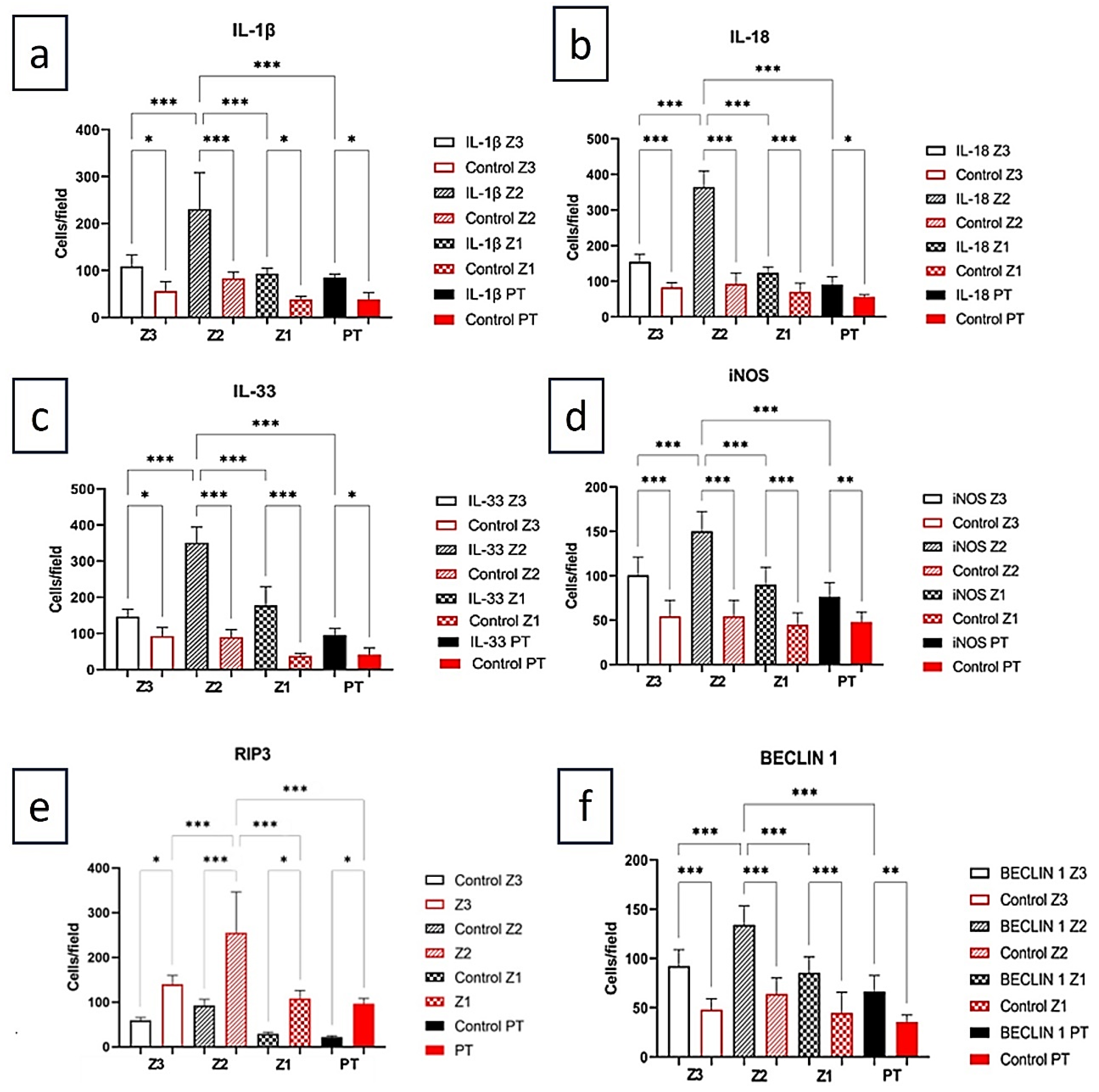
Figure 2.
Representative immunohistochemistry of autophagy/inflammation markers in fatal yellow fever versus negative controls. Micrographs for (A) IL-1β, (B) IL-18, (C) IL-33, (D) iNOS, (E) RIP3 and (F) BECLIN-1 in zones Z3, Z2, Z1 and PT. Insets highlight zonal patterns (Z2 peak). NC, negative control. Magnification: ×400.
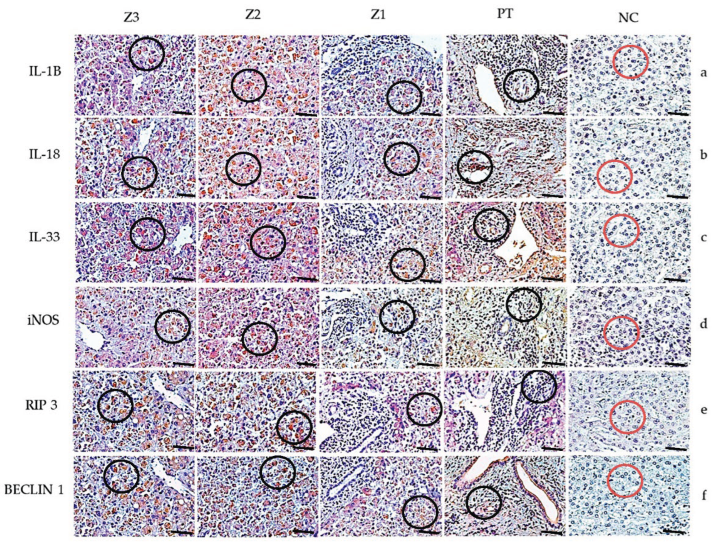

Table 1.
Characterization of patients with YF according to gender, age, state, year and duration of disease.
Table 1.
Characterization of patients with YF according to gender, age, state, year and duration of disease.
| Case | Patient | Gender | Age | State | Year | IT* |
|---|---|---|---|---|---|---|
| 1 | 001/00 | M | 25 | Tocantins | 2000 | 8 |
| 2 | 106/00 | M | 75 | Goiás | 2000 | NR** |
| 3 | 108/00 | M | 49 | Goiás | 2000 | 7 |
| 4 | 494/00 | M | NR** | Distrito Federal | 2000 | NR** |
| 5 | 251/00 | M | 16 | Mato Grosso do Sul | 2000 | 6 |
| 6 | 252/00 | M | 49 | Goiás | 2000 | NR** |
| 7 | 253/00 | M | 23 | Goiás | 2000 | NR** |
| 8 | 255/00 | M | NR** | Goiás | 2000 | NR** |
| 9 | 291/00 | M | NR** | Goiás | 2000 | NR** |
| 10 | 158/00 | M | 33 | Goiás | 2000 | NR** |
| 11 | 063/03 | M | NR** | Minas Gerais | 2003 | NR** |
| 12 | 339/04 | M | 36 | Amazonas | 2004 | 11 |
| 13 | 019/08 | M | 64 | Goiás | 2008 | 7 |
| 14 | 273/08 | M | 57 | Goiás | 2008 | 7 |
| 15 | 068/08 | F | 65 | Goiás | 2008 | 2 |
| 16 | 095/08 | M | 42 | Goiás | 2008 | 3 |
| 17 | 143/08 | M | 37 | Distrito Federal | 2008 | NR** |
| 18 | 361/15 | F | 53 | Rio grande do Norte | 2015 | 4 |
| 19 | 062/16 | M | 35 | Goiás | 2016 | NR** |
| 20 | 346/16 | M | 15 | Goiás | 2016 | 7 |
| 21 | 369/16 | M | 27 | Goiás | 2016 | 1 |
*IT= Illness time/ **NR= No registry.
Table 2.
Antibodies used in the study of autophagy profile in liver of fatal cases YFV-induced.
| Markers | Reference | Dilution |
|---|---|---|
| iNOS | Abcam/ab15323 | 1/200 |
| IL-1β | Abcam/ab9722 | 1/100 |
| IL-18 | Abcam/ab68435 | 1/100 |
| IL-33 | Abcam/ab118503 | 1/100 |
| BECLIN 1 | Abcam/ab210498 | 1/500 |
| RIP3 | Abcam/ab152130 | 1/50 |
Table 3.
Quantitative analysis of autophagy markers in the hepatic parenchyma in fatal YFV cases compared to control cases. Z3: Centrilobular zone; Z2: Midzonal zone; Z1: Periportal zone; PT: Portal tract. Tukey-test; ***p < 0.0001; ** p < 0.001; * p < 0.05.
Table 3.
Quantitative analysis of autophagy markers in the hepatic parenchyma in fatal YFV cases compared to control cases. Z3: Centrilobular zone; Z2: Midzonal zone; Z1: Periportal zone; PT: Portal tract. Tukey-test; ***p < 0.0001; ** p < 0.001; * p < 0.05.
| Markers | Z3 | Z2 | Z1 | PT | ANOVA (p ≤ 0.05) |
|---|---|---|---|---|---|
| IL-1β | 109.0±24.06 | 190.5±75.03 | 86.10±16.38 | 70.10± 16.38 | <0.0001*** |
| Control | 67.20±13.39 | 83.20±13.39 | 54.40±18.24 | 48.00± 11.31 | |
| Tukey (p ≤ 0.05) | <0.0001*** | <0.0001*** | 0.0002*** | 0.0098** | |
| IL-18 | 154.7±21.06 | 365.0±44.86 | 123.4±16.89 | 90.67±21.66 | <0.0001*** |
| Control | 83.20±13.39 | 92.80±30.78 | 70.40±24.27 | 60.80±7.259 | |
| Tukey (p ≤ 0.05) | 0.0001*** | <0.0001*** | <0.0001*** | 0.0011** | |
| IL-33 | 146.3 ± 20.42 | 351.2±43.07 | 177.2±51.64 | 89.14±21.21 | <0.0001*** |
| Control | 92.80 ± 23.73 | 89.60 ± 21.47 | 37.60 ± 7.79 | 41.60 ± 18.24 | |
| Tukey (p ≤ 0.05) | 0.0011** | 0.0045** | 0.0008*** | 0.0091** | |
| iNOS | 100.6±20.33 | 150.1±22.33 | 89.90±19.90 | 76.19±15.92 | <0.0001*** |
| Control | 38.40±18.24 | 38.40±8.764 | 44.80±13.39 | 48.00±19.60 | |
| Tukey (p ≤ 0.05) | <0.0001*** | <0.0001*** | <0.0001*** | 0.0062** | |
| RIP3 | 140.20 ± 19.41 | 255.10 ± 90.92 | 108.10 ± 18.13 | 96.33 ± 12.06 |
<0.0001*** |
| Control | 59.20 ± 6.57 | 92.80 ± 13.39 | 29.40 ± 3.84 | 22.20 ± 2.68 | |
| Tukey (p ≤ 0.05) | <0.0001*** | <0.0001*** | <0.0001*** | <0.0001*** | |
| BECLIN 1 | 92.19 ± 16.71 | 134.10 ± 19.25 | 85.33 ± 16.26 | 66.29 ± 16.23 | <0.0001*** |
| Control | 48.00 ± 11.31 | 64.00 ± 16.00 | 44.80 ± 20.86 | 35.60 ± 6.98 | |
| Tukey (p ≤ 0.05) | <0.0001*** | 0.0008*** | <0.0001*** | <0.0001 |
Table 4.
Key correlations (|r| ≥ 0.75) among autophagy and inflammation markers across hepatic zones in fatal YFV (Pearson). Pearson’s r (two-tailed). Exact p-values are shown; the complete set with Benjamini–Hochberg FDR (q) is provided in Table S4. Zones: Z1, periportal; Z2, midzonal; Z3, centrilobular; PT, portal tract. Markers: BECLIN-1, RIP3, iNOS, IL-1β, IL-18, IL-33.
| Pair (marker [zone] – marker [zone]) | R | p |
|---|---|---|
| RIP3 (Z1) – iNOS (PT) | 0.8402 | <0.0001 |
| RIP3 (Z2) – iNOS (PT) | -0.8333 | <0.0001 |
| RIP3 (Z2) – IL-1β (PT) | -0.8179 | <0.0001 |
| RIP3 (Z2) – iNOS (Z2) | -0.8160 | <0.0001 |
| IL-18 (Z3) – IL-33 (Z3) | 0.8044 | <0.0001 |
| iNOS (Z1) – IL-1β (PT) | 0.8011 | <0.0001 |
| IL-33 (Z2) – BECLIN-1 (Z1) | 0.7876 | <0.0001 |
| IL-33 (Z2) – BECLIN-1 (PT) | 0.7615 | 0.0002 |
| iNOS (Z2) – IL-18 (PT) | -0.7569 | <0.0001 |
| IL-18 (Z2) – BECLIN-1 (Z2) | -0.7557 | 0.0003 |
| IL-18 (Z2) – BECLIN-1 (Z1) | -0.7528 | <0.0001 |
Disclaimer/Publisher’s Note: The statements, opinions and data contained in all publications are solely those of the individual author(s) and contributor(s) and not of MDPI and/or the editor(s). MDPI and/or the editor(s) disclaim responsibility for any injury to people or property resulting from any ideas, methods, instructions or products referred to in the content. |
© 2025 by the authors. Licensee MDPI, Basel, Switzerland. This article is an open access article distributed under the terms and conditions of the Creative Commons Attribution (CC BY) license (http://creativecommons.org/licenses/by/4.0/).
Copyright: This open access article is published under a Creative Commons CC BY 4.0 license, which permit the free download, distribution, and reuse, provided that the author and preprint are cited in any reuse.


