
Submitted:
13 September 2025
Posted:
15 September 2025
You are already at the latest version
Abstract
In Gram-negative bacteria, the peptidoglycan layer is often thought to be a thin layer (even monolayer) that is in-between the two phospholipid bilayer of the bacterial double membrane cell envelope. However, preliminary evidence from scanning electron microscopy observations of the shape and morphology of Escherichia coli DH5α cells subjected to autoclave treatment at 121 oC for 20 minutes suggests that the peptidoglycan layer may be thicker, and it may be a non-porous layer rather than a perforated mesh. Specifically, the traditional view of the E. coli peptidoglycan layer as a thin or monolayer layer punctuated with ion channels and transporter does not explain the ability of E. coli cells to retain its cellular structure after autoclave treatment. On the other hand, the new proposed model of a thicker peptidoglycan layer which is non-porous may help explain the observation of intact E. coli cells (without any holes) after autoclave treatment. Future work may examine the compressive and tensile strength of E. coli cells both before and after autoclave treatment to gain a deeper understanding of its mechanical properties as well as draw inference on the structure of the peptidoglycan layer.
Keywords:
Escherichia coli
; peptidoglycan layer
; non-porous layer
; perforated mesh
; monolayer
Autoclave is the standard and reliable way for killing bacterial cells. Typically operated at elevated temperatures of 121 oC for 15 to 20 minutes, it is validated to kill most of the microbial cells in the sample. This work describes an accidental observation that Escherichia coli DH5α cells treated with standard autoclave decontamination still retain an intact morphological structure, and crucially, with no holes in the cell wall as examined by scanning electron microscopy.
In terms of sample preparation, the cell sample was taken from a 15 hour activated E. coli culture of 100 mL volume in a 250 mL conical shake flask with aerobic cultivation at 230 rpm shaking, that has undergone autoclave decontamination at 121 oC for 20 minutes. After autoclave treatment, the residual liquid was decanted off, and the remaining cell sample was put into a 15 mL polypropylene sample tube, and the sample was freeze dried for 24 hours to remove the residual liquid from the cell sample. The autoclaved freeze dried cells were then coated with platinum on a platinum sputter for 30 seconds, and viewed under a JEOL 5600 LV scanning electron microscope with 15 kV accelerating voltage.
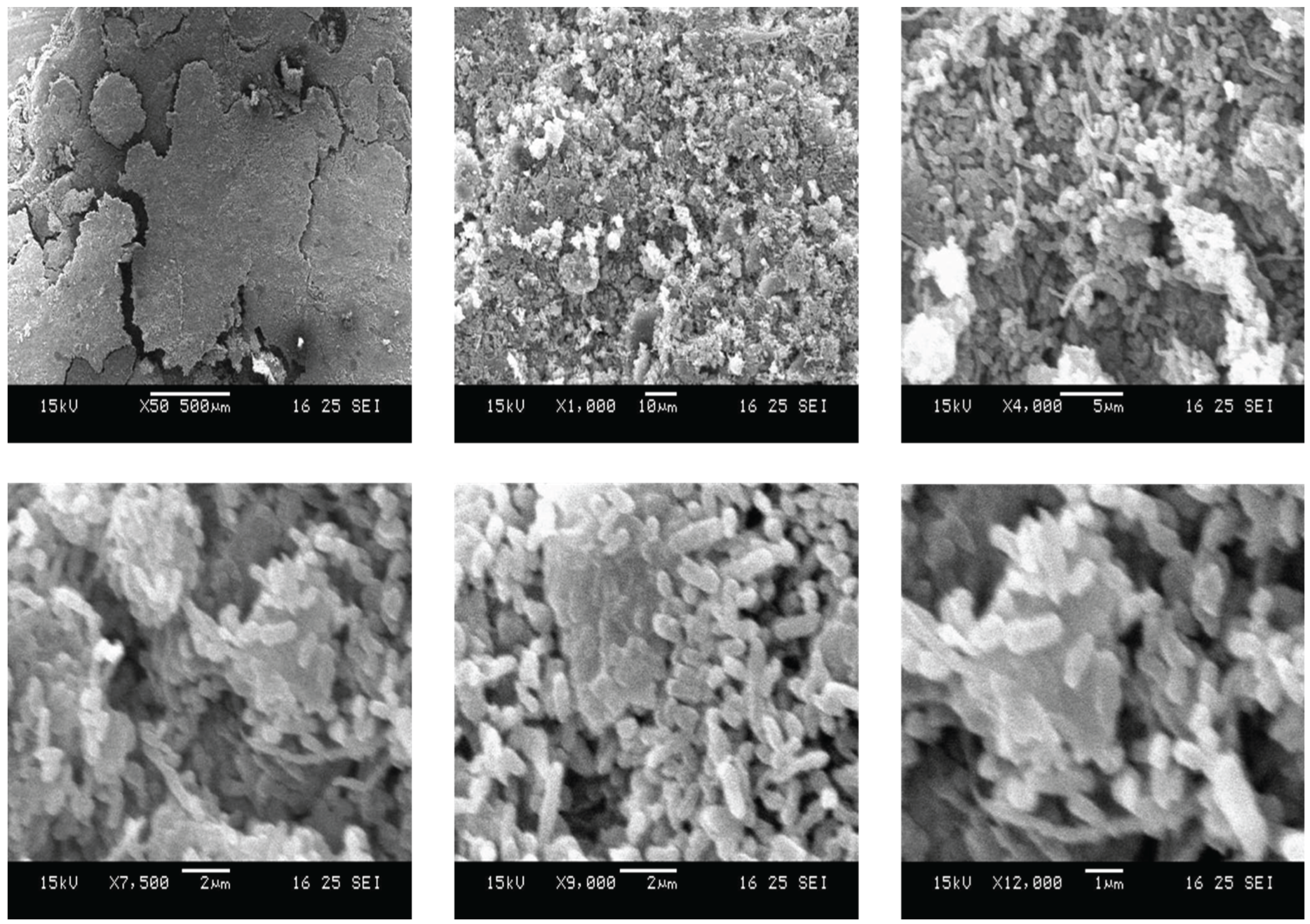
Figure 1 shows a series of scanning electron micrographs of E. coli DH5α of increasing magnification showing E. coli cells with no visible holes after autoclave treatment. In addition, the E. coli cells still retain their rod-shaped structure with smooth cell wall and surface. Hence, this contradicts conventional notion that autoclave treatment will cause cell wall breakage and puncture due to the high pressure environment in autoclave sterilisation.[1] Such a result suggests that the peptidoglycan layer in between E. coli double membrane cell wall is able to sustain a higher pressure than anticipated.
One possibility for this observation is that E. coli DH5α may possess a thicker peptidoglycan layer than commonly expected. This peptidoglycan layer is also likely to be non-porous and possess ion channels and transporters for transferring metabolites and biomolecules. Such a notion is in stark contrast to the textbook depiction of the peptidoglycan layer of Gram-negative bacteria as a perforated mesh with loosely held intertwined cross-linked filaments.[2,3,4]
In conclusion, this work describes an accidental observation that E. coli DH5α cells still retained their rod-shaped morphological structure with no holes in the cell wall after autoclave treatment at 121 oC for 20 minutes. Such an observation suggests that the peptidoglycan layer in E. coli cell wall is able to withstand higher pressure than previously thought, and is likely to be thicker than the single layer perforated mesh depicted in textbooks. Future work could validate this accidental observation, as well as compare the mechanical strength and properties of E. coli with and without autoclave treatment.
Funding
No funding was used in this work.
Conflicts of interest
The author declares no conflicts of interest.
References
- Calderón-Franco, D., Lin, Q., van Loosdrecht, M. C. M., Abbas, B. & Weissbrodt, D. G. Anticipating Xenogenic Pollution at the Source: Impact of Sterilizations on DNA Release From Microbial Cultures. Front. Bioeng. Biotechnol. Volume 8-2020, (2020). [CrossRef]
- Vollmer, W., Blanot, D. & De Pedro, M. A. Peptidoglycan structure and architecture. FEMS Microbiol. Rev. 32, 149–167 (2008). [CrossRef]
- Hammond, S. M., Lambert, P. A. & Rycroft, A. N. The Peptidoglycan Layer. in The Bacterial Cell Surface (eds Hammond, S. M., Lambert, P. A. & Rycroft, A. N.) 1–28 (Springer Netherlands, Dordrecht, 1984). [CrossRef]
- Pokhrel, R., Shakya, R., Baral, P. & Chapagain, P. Molecular Modeling and Simulation of the Peptidoglycan Layer of Gram-Positive Bacteria Staphylococcus aureus. J. Chem. Inf. Model. 62, 4955–4962 (2022). [CrossRef]
Figure 1.
Scanning electron microscopy images of autoclaved E. coli cells at increasing magnification showing clusters of rod-shaped E. coli bacterial cells with intact cell structure and morphology, and crucially without any holes in the cell wall ultrastructure.
Figure 1.
Scanning electron microscopy images of autoclaved E. coli cells at increasing magnification showing clusters of rod-shaped E. coli bacterial cells with intact cell structure and morphology, and crucially without any holes in the cell wall ultrastructure.
Disclaimer/Publisher’s Note: The statements, opinions and data contained in all publications are solely those of the individual author(s) and contributor(s) and not of MDPI and/or the editor(s). MDPI and/or the editor(s) disclaim responsibility for any injury to people or property resulting from any ideas, methods, instructions or products referred to in the content. |
© 2025 by the authors. Licensee MDPI, Basel, Switzerland. This article is an open access article distributed under the terms and conditions of the Creative Commons Attribution (CC BY) license (http://creativecommons.org/licenses/by/4.0/).
Copyright: This open access article is published under a Creative Commons CC BY 4.0 license, which permit the free download, distribution, and reuse, provided that the author and preprint are cited in any reuse.


