Preprint
Article

This version is not peer-reviewed.

Hypomethylation of the Dual Specificity Phosphatase (DUSP22) Promoter in Cell-Free DNA (cf-DNA) Is Associated with Rheumatoid Arthritis and Joint Space Narrowing

Submitted:

05 September 2025

Posted:

09 September 2025

You are already at the latest version

Abstract

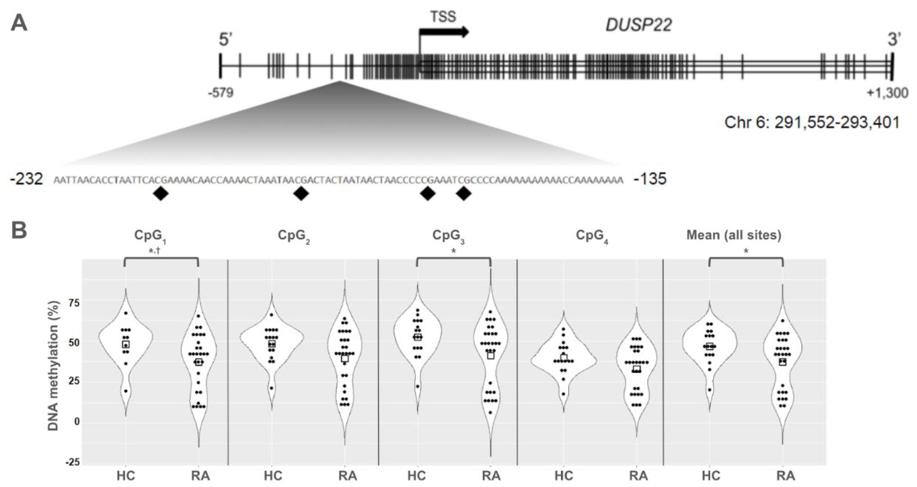
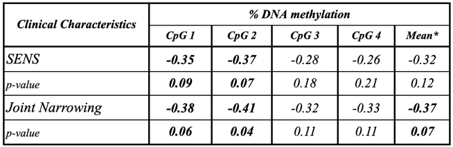
Background/Objective: While several advances have been made in the last decade, reliable biomarkers for diagnosis, prognosis, and especially for the treatment of rheumatoid arthritis (RA) have yet to be identified. In previous studies, DUSP22 DNA methylation changes were found to be associated with RA and erosive disease. We conducted a pilot study to investigate plasma cell-free DNA (cfDNA) methylation in DUSP22 in a cohort of RA patients and healthy controls. We also investigate DUSP22 DNA methylation associations with RA clinical characteristics and treatment. Methods: DNA was isolated from plasma from twenty-seven RA patients who satisfied the ACR criteria, and eighteen healthy controls. DUSP22 DNA methylation was determined by pyrosequencing. Statistical analysis identified group differences and associations with RA clinical measures. Results: RA patients had lower mean promoter cfDNA DUSP22 DNA methylation when compared to controls (36.47±16.17% vs. 47.05±10.28%, p=0.025). Hypomethylation of one CpG site in this region was also associated with increased joint space narrowing (ρCpG2=-0.41, p=0.04). Conclusion: Our pilot study is the first to show that cfDNA methylation might be an important biomarker in RA. Our hypothesis-generating findings suggest that hypomethylation of DUSP22 in cfDNA is associated with RA, and if replicated in future studies, our results point to the potential of cfDNA methylation to be a non-invasive biomarker for this disease.

Keywords: 
;  ;  ;  ;  ;  

1. Introduction

Rheumatoid arthritis (RA) is a chronic autoimmune disease that causes persistent inflammation, pain, and destruction of the joints [1]. The etiology of RA is complex due to the combined effect of genetic and environmental factors [2]. Thus, epigenetics has emerged as a key integrative mechanism, supported by epigenetic modifications observed in RA patients [3,4,5,6,7,8,9,10]. DNA methylation, the most studied epigenetic modification, is the addition of a methyl group to the fifth carbon of cytosine nucleotides when next to guanines in the DNA, called CpG sites [11]. This modification can regulate gene expression by limiting access to the promoters of genes. Disease-associated DNA methylation patterns have been identified in a variety of cell types from RA patients, including peripheral blood mononuclear cells (PBMCs), B-cells, T-cells, and fibroblast-like synoviocytes (FLS) [4,5,6,7,8,9,10]. Several differentially methylated genes have been identified in the synovium of individuals with RA, which encode proteins involved in inflammatory response [5,12,13]. Other research has found B- and T-lymphocyte DNA methylation of specific genes to be associated with established RA, early RA (RA in naïve patients) and response to disease-modifying antirheumatic drugs (DMARDs) [4,5,7]. Combined, these studies have shown that DNA methylation plays an important role in RA and potentially contributes to the persistent inflammation these patients experience.
Previous research identified DNA methylation changes in the Dual Specificity Phosphatase 22 (DUSP22) gene promoter to be associated with RA [4,9]. DUSP22, also known as JKAP, encodes for a phosphatase belonging to a family of enzymes that can dephosphorylate serine/threonine and tyrosine residues [14]. DUSP22 enzyme has an immunomodulator role, this evidenced by its regulation of c-Jun N-terminal kinases (JNK), signal transducer activator of transcription-3 (STAT3), and the lymphocyte-specific protein tyrosine kinase (LCK) [15,16,17]. Findings show that DUSP22 can activate JNK, one of the mammalian mitogen-activated protein kinases (MAPKs), in response to stress, growth, and apoptosis [15,18]. In addition, DUSP22 can dephosphorylate STAT3 and prevent it from translocating into the nucleus to promote the transcription of pro-inflammatory cytokines such as IL6 [16]. Finally, data suggests that DUSP22 inactivates LCK in the T-cell receptor (TCR) signaling pathway leading to autoimmunity and inflammation [17]. All these mechanisms have been identified as important in the etiology and progression of RA, making DUSP22 an important molecular target in this disease.
While most studies investigating epigenetic biomarkers of RA have focused on DNA extracted from lymphocytes and FLS, there is evidence that cell free-DNA (cfDNA) plays an important role in autoimmune diseases [19,20,21,22]. Data has shown that there is more cfDNA in the plasma of RA patients than in controls [23,24,25]. Studies have also found that better clinical outcomes are observed in individuals with RA in which the amounts of cfDNA increase after DMARDs therapy and that the use of cfDNA amounts along with a measure of anti-citrullinated peptide antibody (ACPA) might be a better diagnostic tool in RA than using ACPA alone [22,25]. However, studies have not investigated whether DNA methylation markers can be measured in the cfDNA of RA patients. This new area of research could improve our understanding of RA pathogenesis.
Here, we proposed to investigate DNA methylation biomarkers in the Epigenetics of Rheumatoid Arthritis (ERA) study. We researched whether DNA methylation biomarkers can be measured in plasma cfDNA, and whether levels of DNA methylation in DUSP22 differ between Hispanic RA and healthy individuals. We also explore its predictive potential and associations of this measure with clinical characteristics of the disease in this population.

2. Materials and Methods

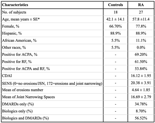
2.1. Study Participants

Twenty-seven RA patients, who meet the 2010 ACR classification criteria for RA [26], and eighteen healthy individuals, all 18 years or older, were recruited to the Epigenetics of Rheumatoid Arthritis (ERA) study. Recruitment took place from May 2016 to March 2018. The RA cohort attended the Rheumatology clinic of New York City Health + Hospitals/ Lincoln Hospital and healthy individuals learned about the study via open advertisement. RA patients with any of the following conditions were excluded: other autoimmune diseases, type 2 diabetes mellitus, entrapment neuropathies, radiculopathies, recent sepsis (period less than 6 months), chronic infections (Hepatitis C/B, HIV), previous and current intravenous drug use, chronic kidney disease (CKD 2), chronic heart failure, liver cirrhosis , patients taking more than 20 mg daily of corticosteroids at the time of recruitment, cancer, recent organ transplant, pregnancy, recent significant trauma (less than 6 months including admission to the hospital, fractures), recent major surgery (all recent surgeries within 4 months, except: cataracts, ambulatory or superficial procedures, breast lumpectomy, colonoscopy, and/or endoscopy). Informed consent was obtained from all subjects involved in the study. All participants completed a questionnaire including demographic data and in the case of RA patients, symptoms and clinical measures associated with RA were extracted from their patient records. Peripheral whole blood samples were collected in green top Heparin containing tubes and transported to the laboratory at John Jay College, College of the University of New York (CUNY) in sealed containers. The study was conducted according to the guidelines of the Declaration of Helsinki, and approved by the Institutional Review Board of New York City Health + Hospitals/ Lincoln Hospital and John Jay College at the City University of New York.

2.2. Clinical Measures

In this study, we define seropositive disease by the presence of either ACPA or rheumatoid factor (RF). Conversely, seronegative status meant neither factor was positive. RA patients were assessed utilizing the following clinical measures: Clinical Disease Activity Index (CDAI), Simple Erosive Narrowing Score (SENS), CDAI considers the number of swollen and tender joints as well as the patient and provider assessments of global disease activity to obtain a score ranging from 0.0 to 76.0, with disease activity increasing with higher values [27]. SENS is a scoring system ranging from 0 to 86 that determines the presence and number of joint erosions (32 for hands and 12 for feet) and the presence and number of joint space narrowing (30 for the hands and 12 for the feet) [28,29].

2.3. DNA Extraction and Methylation Analysis of DUSP22

Plasma was separated from whole blood by centrifugation. DNA was extracted from 1 mL of plasma using an UltraSens Virus Kit (Qiagen) according to the manufacturer's instructions. DNA was bisulfite converted using the EZ DNA Methylation-Gold kit (Zymo Research) following the kit's protocol. Bisulfite converted DNA was eluted in 15μl of water and 2.5-5μl were used for Pyromark PCR amplification (Qiagen). The assay targets a 177-bp DNA fragment in the DUSP22 promoter region [4] (Figure 1A). The forward primer 5’-GGTAGGGGGTTTTTAGATTTTTT-3’ was biotinylated in the 5’ end, and the sequence of the reverse primer was 5’-CCCCCAACCTAAATCTACC-3’. The annealing temperature used in amplification reactions was 56oC and 38 cycles. Pyrosequencing was carried out in a PyroMark Q24 (Qiagen) following the manufacturer’s instructions using 5’-CCCAAAAACCAAACCTCT-3’ as the sequencing primer and the following sequence to analyze 5’-AATTAACACCTAATTCACRAAAACAACCAAAACTAAATAACRACTACTAATAACTAACCCCCRAAATCRCCCCAAAAAAAAAAACCAAAAAAAA-3’. Pyrosequencing measured DNA methylation levels of four CpG sites (Figure 1). DNA methylation analysis was performed using the PyroMark Q24 Advanced 3.0.0 software. DNA methylation status was reported as the average percent methylation for the four CpG sites, or by individual CpG sites (is there a reason to pick these sites?) as indicated. Each amplification and pyrosequencing run included fully methylated and unmethylated DNA standards (Zymo Research) as controls. No-template controls were also included in all runs. Coefficient of variation for the assay was 6.50%.

2.4. Statistical Analysis
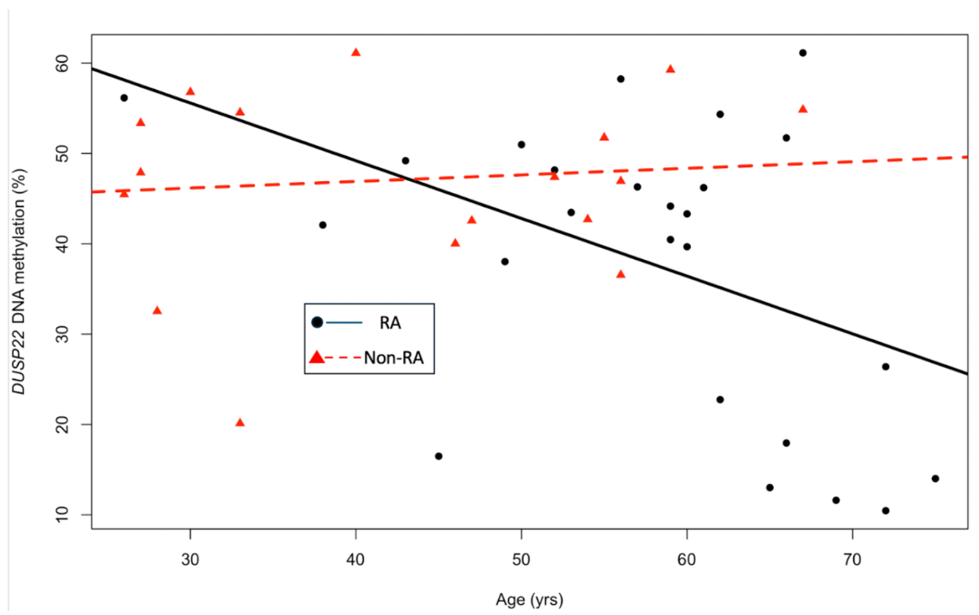
We used statistical tools to compare demographic characteristics, and to explore the associations of the percent of DNA methylation of DUSP22 in RA patients and controls and with clinical variables. We used t-test to compare demographic characteristics and a non-parametric Mann-Whitney test to compare DNA methylation levels at each site and across the region between RA patients and controls. In addition, we carried out linear regression modeling to examine the relationship between DNA methylation levels in both RA patients and healthy controls as a function of age. We logit-transformed DNA methylation percentages and carried out ANCOVA to compare DNA methylation levels between RA patients and controls while adjusting for age. Age was included as a covariate in these analyses, as it significantly differed between RA patients and the controls and could confound the association with DNA methylation levels. Pearson correlation coefficients were used to investigate associations between DUSP22 DNA methylation and clinical characteristics of RA. For interpretation, statistical significance was defined as p≤0.05, while results with p values between 0.05 and 0.10 were described as trends. SPSS v-31 was used for analysis.

3. Results

The demographic and clinical characteristics of the ERA study participants are shown in Table 1. Participants were mostly females and of Hispanic ethnicity (Table 1). Gender and race/ethnicity were similar between RA patients and controls; however, RA patients were significantly older than the controls (Table 1). Only three individuals in this sample were smokers; therefore, we did not include that variable in our analysis. Treatment data was available for twenty-six RA patients, 93.2% of them were on Disease-Modifying Antirheumatic Drugs (DMARDs) alone or in combination with biologics (Table 1). The most common DMARDs used was hydroxychloroquine (61.5%), followed by methotrexate (53.8%). 38.5% of participants were under combined hydroxychloroquine and methotrexate treatment. The most common biologic used was TNFα inhibitors and it was used by 23.1% of patients.
Figure 1 shows the location of all CpG sites of the DUSP22 promoter measured in this study (Figure 1A). The mean percentage of DNA methylation for all CpG sites was significantly lower in individuals with RA than in healthy individuals (36.48 ± 3.11% vs. 47.05 ± 2.42%, p=0.025; Figure 1B). We also compared the percentage of DNA methylation in DUSP22 in individual CpG sites measured in this study. Statistical significance remained for the comparison between RA patients and controls for two of the sites, CpG1 and CpG3 (Figures 1B). For CpG1, percent DNA methylation was 36.65 ± 3.25% for RA versus 46.94 ± 2.63% for controls (p=0.025, Figure 1C). The highest levels of DNA methylation were found in CpG3, with 40.40 ± 3.68% for RA patients and 52.74 ± 2.69% for controls (p=0.041, Figure 1B). The difference in DNA methylation of the two remaining sites, CpG2 and CpG4, were not different between RA patients and controls.
Because age was different between RA patients and healthy individuals, we used linear regression analysis to explore the association of DUSP22 DNA methylation with age in each group. For RA patients, age was associated with a significant decrease in DUSP22 DNA methylation. This association was observed for all individual sites (βCpG1=-0.64, p=0.03; βCpG2=-0.68, p=0.02; βCpG3=-0.73, p=0.03 and βCpG4=-0.51, p=0.03), and mean DUSP22 DNA methylation (βmean=-0.64, p=0.02; Figure 2). This trend was not observed for the control group at any of the specific sites or the mean DNA methylation in this region of the DUSP22 gene (β=0.07, p=0.69; Figure 2). To further explore the association of DUSP22 DNA methylation and RA, we carried out an ANCOVA analysis to remove the effect of age in the comparison of DUSP22 DNA methylation levels between RA patients and controls, and found that only for CpG1 DNA methylation levels the difference remained (p=0.049; Figure 1).
We analyzed the associations between clinical characteristics such as disease activity, and radiological findings with the average/individual CpG DUSP22 DNA methylation and found no significant associations for disease activity or CDAI. We found an inverse correlation between an increase in joint space narrowing with a decrease in the percentage of DNA methylation at CpG2 (ρCpG2=-0.41, p=0.04; Table 2) and a trend to hypomethylation in RA for CpG1 and the mean DUSP22 DNA methylation (ρCpG1=-0.38, p=0.06 and ρMean=-0.37, p=0.07; Table 3). Similarly, a DUSP22 hypomethylation trend was observed for SENS (ρCpG1=-0.35, p=0.09 and ρCpG2=-0.37, p=0.07; Table 2). No other associations were observed with clinical measures of RA and DNA methylation levels of DUSP22 in this population.
When exposure to methotrexate, or methotrexate and biologics were considered, no differences in mean DUSP22 DNA methylation was found (Table 3). Average DUSP22 DNA methylation in RA patients on methotrexate was 33.60 ± 4.15% and 40.78 ± 4.62% for those in other drugs (p=0.32, Table 3). Individual sites were also not significantly different for methotrexate treatment status (Table 3). When comparing DUSP22 DNA methylation levels between users and non-users of TNFα inhibitor, a trend towards hypomethylation of DUSP22 DNA was observed in cfDNA of individuals on TNFα inhibitor therapy than in those using other therapies (26.21 ± 6.28% vs 40.12 ± 3.54%, p=0.07; Table 3).

4. Discussion

In this pilot study, we investigated DNA methylation of the DUSP22 promoter in the plasma cfDNA of predominantly Hispanic RA patients and healthy controls. We found that DNA methylation at the DUSP22 promoter was lower in individuals with RA than controls, for RA patients we also observed a trend of lower DUSP22 DNA methylation with increasing age. These associations suggest that lower levels of DUSP22 methylation might be a feature of the disease and that plasma cfDNA has the potential to be a relevant biomarker in RA, if these findings are replicated in larger cohorts.
In contrast with our findings, a previous study identified T-lymphocyte DNA hypermethylation of the DUSP22 promoter in RA [4]. Studies performed on other autoimmune disorders support RA findings in T-lymphocytes [40,41]. Sjögren syndrome patients were found to have higher levels of DUSP22 DNA methylation in T-lymphocytes DNA when compared to controls [40]. Also a study carried out in systemic lupus erythematosus (SLE) patients found them to have less DUSP22 T-lymphocyte expression as well as a negative association between SLE disease activity and DUSP22 expression [41]. Less DUSP22 expression could be the result of epigenetic regulation of gene expression, in which higher DUSP22 DNA methylation would lead to lower expression of this gene. Our patient cohort consisted of 66.7% females mostly of Hispanic ethnicity. The RA and Sjögren syndrome studies both investigated Caucasian women, while the SLE study population studied Asians [9,40,41]. Future studies are needed to elucidate whether DUSP22 DNA methylation varies by race and/or ethnicity in this disease. Most importantly, DNA methylation here was measured in plasma cfDNA and previous studies measured this epigenetic biomarker in T-lymphocytes [9,30,31]. DNA methylation changes are tissue specific, and without previous studies investigating DUSP22 DNA methylation levels in cfDNA, the differential results among our work and this previous research might simply stem from the difference in specimens used.
The presence of cfDNA in RA patients has been found to be larger than in healthy individuals [22,23,24,25]. The origin of the cfDNA is not known. However, in one study that measured mitochondrial DNA in plasma and synovial fluid, only RA patients with large amounts of mitochondrial DNA in the synovial fluid had equally large amounts of plasma mitochondrial DNA [23]. It is possible that the DNA present in the plasma results from a breakdown of multiple tissues, one of which could be the synovium. Studies will have to assess the contribution of multiple tissues to the cfDNA fraction in RA patients and broaden our understanding of the role of cfDNA and cfDNA methylation in this disease.
The mechanism of action of DMARDs is not fully understood. However, research has shown that drugs such as methotrexate might reduce inflammation by epigenetic mechanisms. Previous studies have shown that methotrexate can alter global DNA methylation levels and DNA methylation at specific sequences [32,33]. We investigated here whether there was a difference in DUSP22 DNA methylation in methotrexate users. We did not find any difference between the DUSP22 level of RA patients using methotrexate and those treated with other DMARDs. These findings agree with previous results in RA CD4+ T cells of DMARDs naive patients and methotrexate users, in which DNA methylation at DUSP22 was not different between the groups [34]. Interestingly, that study identified other RA-relevant genes such as GALNT9 for which methotrexate use was associated with lower DNA methylation of the promoter region [34], suggesting that methotrexate effects on DNA methylation are limited to specific genomic regions.
A study investigating the effect of TNFα inhibitor on DUSP22 expression revealed that in inflammatory bowel disease patients DUSP22 mRNA expression was negatively correlated with TNFα [35]. In addition, the use of TNFα inhibitor was shown to significantly up-regulate DUSP22 mRNA and protein expression in the intestinal mucosa of Crohn’s disease patients [35]. In our study, we found a borderline significant decrease in DNA methylation of the DUSP22 promoter in RA patients using TNFα inhibitor therapy. Differences in DNA methylation have been observed after treatment with the anti-TNFα, Adalimumab, in skin biopsies of psoriatic patients [36]. The mechanisms through which TNFα inhibitors affect DUSP22 mRNA expression have not been elucidated, but we can hypothesize that TNFα inhibition reduces levels of DNA methylation at the DUSP22 promoter leading to increased expression.
The association between DUSP22 DNA hypomethylation at CpG2 and increased joint space narrowing and SENS found in our study is consistent with previously published reports, which demonstrated that lower DNA methylation in the promoter region of DUSP22 in monocytes, B-lymphocytes, naive CD4+ T and memory CD4+ T cells was associated with erosive disease [9]. The CpG2 measured here was also included in that study. However, the measures of erosive RA used in both studies were different. Mok and co-workers used the European League Against Rheumatism (EULAR) criteria and here we used SENS, which is indicative of erosive disease severity. On the other hand, the EULAR criteria identify the presence or absence of erosive disease. However, both studies agree that presence or severity of erosion in RA is associated with loss of DNA methylation at the DUSP22 promoter region. This strengthens the validity of our findings as similar associations were observed in our study as well.
Our findings show that decreased DNA methylation of DUSP22 at specific sites is associated with RA and with joint space narrowing is connected to some of the known functions of DUSP22. The DUSP22 protein activates JNK, which in turn promotes CD4+ T-cell differentiation [15]. It acts as a scaffold protein that supports the formation of the Apoptosis Signal-regulating Kinase-1 (ASK1), Mitogen-activated protein Kinase (MAPK) Kinase-7 (MKK7), and JNK complex [16]. In RA patients, the JNK pathway is characterized by heightened activity and has a broad role in different biological pathways relevant to the disease (reviewed in [37]). More recent studies have shown that JNK also promotes autophagy [38]. Current research has linked this cellular process to bone erosion in mice and has found that RA patients have increased expression of autophagy-related proteins [39,40]. Additional research is needed to understand the biological processes associated with DUSP22 and the pathogenesis of RA. Elucidating these mechanisms will help in the development of biomarkers that have the potential to improve response to treatment or the search for new therapies.
The main limitation of this pilot study is the small sample size. The age difference between our cases and controls limits the strength of our findings; however, the association with age and hypomethylation of DUSP22 is an important finding as genome wide methylation studies have failed to identify changes in DUSP22 DNA methylation with age in healthy individuals [41,42]. This is something we were able to confirm in our small sample as well. We had limited disease duration data, and could not carry out an analysis that highlighted whether the association of DUSP22 DNA methylation with age was dependent on how long the patient had RA. It is possible it represents a figure of the length of living with this chronic disease, providing further support for its potential as a biomarker. Future work should explore this question. We also could not measure DUSP22 protein levels, only DNA methylation levels in the DUSP22 promoter. However, previous data measuring DNA methylation and gene expression suggests that DUSP22 gene expression is regulated by DNA methylation of CpG sites in its promoter region [43]. In addition, we did not compare measurements of DNA methylation in plasma cfDNA with those on other tissues relevant in RA. Future studies in larger populations are needed to confirm our findings.
Our pilot study is the first one to investigate DNA methylation and RA associations in predominantly Hispanic patients. Between 1995 and 2014, the incidence of RA remained constant for the overall population; however, for Hispanic individuals it increased in that same period [44]. Another study comparing Caucasian, African American, Hispanic, and Asian RA patients have found that Hispanics had higher disease activity and African Americans were less likely to go on clinical remission than other groups [45]. In these works, the higher disease activity level among African American and Hispanic RA patients remained after adjusting for factors including socioeconomic status, type of clinical practice, treatment and disease severity, suggesting that disparities in RA are not resulting only from environmental influences [45]. Ethnic differences in genetic susceptibility to RA have also been observed, suggesting biological variation might contribute to RA health disparities [46,47]. Studies focused on minority RA populations are scarce, and addressing this gap is imperative to eliminating ethnic and racial disparities in RA.
In conclusion, we found DNA methylation in plasma cfDNA was associated with RA. Plasma cfDNA is very accessible and it provides a measure of systemic biomarker levels, facilitating diagnosis and analysis of response to therapy. Additionally, associations of clinical characteristics of the disease with measures in cfDNA confirm previous findings further supporting the relevance of epigenetic changes such as DNA methylation in RA. In addition, our results uncovered a possible role for epigenetic measures in RA affecting Hispanics and report for the first-time associations of epigenetic biomarkers in this population with clinical measures of the disease. Overall, this study adds to our current knowledge in these areas and proposes a new avenue of biomarker discovery and development for this disease.

Data Availability Statement

The Authors will make this material available upon request to interested researchers.

Conflicts of Interest

The authors declare no conflicts of interest.

References

  1. Mori, T.; Miyamoto, T.; Yoshida, H.; Asakawa, M.; Kawasumi, M.; Kobayashi, T.; Morioka, H.; Chiba, K.; Toyama, Y.; Yoshimura, A. IL-1β and TNFα -initiated IL-6-STAT3 pathway is critical in mediating inflammatory cytokines and RANKL expression in inflammatory arthritis. Int. Immunol. 2011, 23, 701–712. [Google Scholar] [CrossRef] [PubMed]
  2. Symmons, D.P. Epidemiology of rheumatoid arthritis: determinants of onset, persistence and outcome. Best Pr. Res. Clin. Rheumatol. 2002, 16, 707–722. [Google Scholar] [CrossRef]
  3. Kennedy, A.; Schmidt, E.M.; Cribbs, A.P.; Penn, H.; Amjadi, P.; Syed, K.; Read, J.E.; Green, P.; Gregory, B.; Brennan, F.M. A novel upstream enhancer of FOXP3, sensitive to methylation-induced silencing, exhibits dysregulated methylation in rheumatoid arthritis Treg cells. Eur. J. Immunol. 2014, 44, 2968–2978. [Google Scholar] [CrossRef]
  4. Glossop, J.R.; Emes, R.D.; Nixon, N.B.; E Haworth, K.; Packham, J.C.; Dawes, P.T.; A Fryer, A.; Mattey, D.L.; E Farrell, W. Genome-wide DNA methylation profiling in rheumatoid arthritis identifies disease-associated methylation changes that are distinct to individual T- and B-lymphocyte populations. Epigenetics 2014, 9, 1228–1237. [Google Scholar] [CrossRef]
  5. Glossop, J.R.; Emes, R.D.; Nixon, N.B.; Packham, J.C.; A Fryer, A.; Mattey, D.L.; E Farrell, W. Genome-wide profiling in treatment-naive early rheumatoid arthritis reveals DNA methylome changes in T and B lymphocytes. Epigenomics 2015, 8, 209–224. [Google Scholar] [CrossRef]
  6. Gomez-Cabrero, D.; Almgren, M.; Sjöholm, L.K.; Hensvold, A.H.; Ringh, M.V.; Tryggvadottir, R.; Kere, J.; Scheynius, A.; Acevedo, N.; Reinius, L.; et al. High-specificity bioinformatics framework for epigenomic profiling of discordant twins reveals specific and shared markers for ACPA and ACPA-positive rheumatoid arthritis. Genome Med. 2016, 8, 1–15. [Google Scholar] [CrossRef]
  7. Glossop, J.R.; Nixon, N.B.; Emes, R.D.; Sim, J.; Packham, J.C.; Mattey, D.L.; E Farrell, W.; A Fryer, A. DNA methylation at diagnosis is associated with response to disease-modifying drugs in early rheumatoid arthritis. Epigenomics 2016, 9, 419–428. [Google Scholar] [CrossRef]
  8. Meng, W.; Zhu, Z.; Jiang, X.; Too, C.L.; Uebe, S.; Jagodic, M.; Kockum, I.; Murad, S.; Ferrucci, L.; Alfredsson, L.; et al. DNA methylation mediates genotype and smoking interaction in the development of anti-citrullinated peptide antibody-positive rheumatoid arthritis. Arthritis Res. Ther. 2017, 19, 1–10. [Google Scholar] [CrossRef]
  9. Mok, A.; Rhead, B.; Holingue, C.; Shao, X.; Quach, H.L.; Quach, D.; Sinclair, E.; Graf, J.; Imboden, J.; Link, T.; et al. Hypomethylation of CYP2E1 and DUSP22 Promoters Associated With Disease Activity and Erosive Disease Among Rheumatoid Arthritis Patients. Arthritis Rheumatol. 2017, 70, 528–536. [Google Scholar] [CrossRef] [PubMed]
  10. Rhead, B.; Holingue, C.; Cole, M.; Shao, X.; Quach, H.L.; Quach, D.; Shah, K.; Sinclair, E.; Graf, J.; Link, T.; et al. Rheumatoid Arthritis Naive T Cells Share Hypermethylation Sites With Synoviocytes. Arthritis Rheumatol. 2016, 69, 550–559. [Google Scholar] [CrossRef] [PubMed]
  11. Hawtree, S.; Muthana, M.; Wilson, A.G. The role of histone deacetylases in rheumatoid arthritis fibroblast-like synoviocytes. Biochem. Soc. Trans. 2013, 41, 783–788. [Google Scholar] [CrossRef]
  12. Nakano, K.; Whitaker, J.W.; Boyle, D.L.; Wang, W.; Firestein, G.S. DNA methylome signature in rheumatoid arthritis. Ann. Rheum. Dis. 2013, 72, 110–117. [Google Scholar] [CrossRef]
  13. de la Rica, L.; et al. Identification of novel markers in rheumatoid arthritis through integrated analysis of DNA methylation and microRNA expression. J. Autoimmun. 2013, 41, 6–16. [Google Scholar] [CrossRef]
  14. Martell, K.J.; Angelotti, T.; Ullrich, A. The "VH1-like" Dual-Specificity Protein Tyrosine Phosphatases. Mol. Cells 1998, 8, 2–11. [Google Scholar] [CrossRef] [PubMed]
  15. Chen, A.J.; Zhou, G.; Juan, T.; Colicos, S.M.; Cannon, J.P.; Cabriera-Hansen, M.; Meyer, C.F.; Jurecic, R.; Copeland, N.G.; Gilbert, D.J.; et al. The Dual Specificity JKAP Specifically Activates the c-Jun N-terminal Kinase Pathway. J. Biol. Chem. 2002, 277, 36592–36601. [Google Scholar] [CrossRef] [PubMed]
  16. Sekine, Y.; Tsuji, S.; Ikeda, O.; Sato, N.; Aoki, N.; Aoyama, K.; Sugiyama, K.; Matsuda, T. Regulation of STAT3-mediated signaling by LMW-DSP2. Oncogene 2006, 25, 5801–5806. [Google Scholar] [CrossRef] [PubMed]
  17. Li, J.-P.; Yang, C.-Y.; Chuang, H.-C.; Lan, J.-L.; Chen, D.-Y.; Chen, Y.-M.; Wang, X.; Chen, A.J.; Belmont, J.W.; Tan, T.-H. The phosphatase JKAP/DUSP22 inhibits T-cell receptor signalling and autoimmunity by inactivating Lck. Nat. Commun. 2014, 5, 3618. [Google Scholar] [CrossRef]
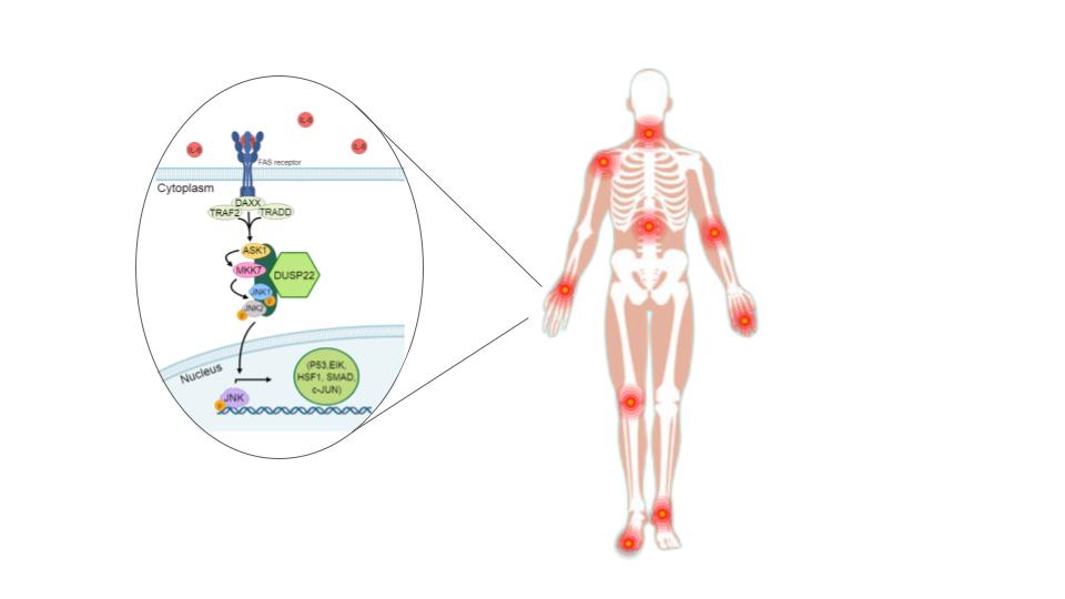
  18. Ju, A.; Cho, Y.-C.; Kim, B.R.; Park, S.G.; Kim, J.-H.; Kim, K.; Lee, J.; Park, B.C.; Cho, S.; Papa, S. Scaffold Role of DUSP22 in ASK1-MKK7-JNK Signaling Pathway. PLOS ONE 2016, 11, e0164259–e0164259. [Google Scholar] [CrossRef] [PubMed]
  19. Bartoloni, E.; Ludovini, V.; Alunno, A.; Pistola, L.; Bistoni, O.; Crinò, L.; Gerli, R. Increased levels of circulating DNA in patients with systemic autoimmune diseases: A possible marker of disease activity in Sjögren’s syndrome. Lupus 2011, 20, 928–935. [Google Scholar] [CrossRef]
  20. Chen, J.A.; Meister, S.; Urbonaviciute, V.; Rödel, F.; Wilhelm, S.; Kalden, J.R.; Manger, K.; Voll, R.E. Sensitive detection of plasma/serum DNA in patients with systemic lupus erythematosus. Autoimmunity 2007, 40, 307–310. [Google Scholar] [CrossRef]
  21. Mosca, M.; Giuliano, T.; Cuomo, G.; Doveri, M.; Tani, C.; Curcio, M.; Abignano, G.; De Feo, F.; Bazzichi, L.; Della Rossa, A.; et al. Cell-free DNA in the plasma of patients with systemic sclerosis. Clin. Rheumatol. 2009, 28, 1437–1440. [Google Scholar] [CrossRef]
  22. Rykova, E.; Sizikov, A.; Roggenbuck, D.; Antonenko, O.; Bryzgalov, L.; Morozkin, E.; Skvortsova, K.; Vlassov, V.; Laktionov, P.; Kozlov, V. Circulating DNA in rheumatoid arthritis: pathological changes and association with clinically used serological markers. Arthritis Res. Ther. 2017, 19, 1–10. [Google Scholar] [CrossRef]
  23. Hajizadeh, S.; DeGroot, J.; TeKoppele, J.M.; Tarkowski, A.; Collins, L.V. Extracellular mitochondrial DNA and oxidatively damaged DNA in synovial fluid of patients with rheumatoid arthritis. Arthritis Res. Ther. 2003, 5, R234–40. [Google Scholar] [CrossRef]
  24. Zhong, X.-Y.; von MühLenen, I.; Li, Y.; Kang, A.; Gupta, A.K.; Tyndall, A.; Holzgreve, W.; Hahn, S.; Hasler, P. Increased concentrations of antibody-bound circulatory cell-free DNA in rheumatoid arthritis. Clin. Chem. 2007, 53, 1609–1614. [Google Scholar] [CrossRef]
  25. Hashimoto, T.; Yoshida, K.; Hashimoto, N.; Nakai, A.; Kaneshiro, K.; Suzuki, K.; Kawasaki, Y.; Shibanuma, N.; Hashiramoto, A. Circulating cell free DNA: a marker to predict the therapeutic response for biological DMARDs in rheumatoid arthritis. Int. J. Rheum. Dis. 2016, 20, 722–730. [Google Scholar] [CrossRef]
  26. Aletaha, D.; Neogi, T.; Silman, A.J.; Funovits, J.; Felson, D.T.; Bingham, C.O., 3rd; Birnbaum, N.S.; Burmester, G.R.; Bykerk, V.P.; Cohen, M.D.; et al. 2010 Rheumatoid arthritis classification criteria: an American College of Rheumatology/European League Against Rheumatism collaborative initiative. Ann. Rheum. Dis. 2010, 69, 1580–1588. [Google Scholar] [CrossRef] [PubMed]
  27. Aletaha, D.; Smolen, J. The Simplified Disease Activity Index (SDAI) and the Clinical Disease Activity Index (CDAI): a review of their usefulness and validity in rheumatoid arthritis. Clin. Exp. Rheumatol. 2005, 23, S100–S108. [Google Scholar] [PubMed]
  28. Boini, S.; Guillemin, F. Radiographic scoring methods as outcome measures in rheumatoid arthritis: properties and advantages. Ann. Rheum. Dis. 2001, 60, 817–827. [Google Scholar] [CrossRef]
  29. van der Heijde, D.; Dankert, T.; Nieman, F.; Rau, R.; Boers, M. Reliability and sensitivity to change of a simplification of the Sharp/van der Heijde radiological assessment in rheumatoid arthritis. Rheumatology 1999, 38, 941–947. [Google Scholar] [CrossRef] [PubMed]
  30. Altorok, N.; et al. Genome-wide DNA methylation patterns in naive CD4+ T cells from patients with primary Sjögren ’ s syndrome. Arthritis Rheumatol. 2014, 66, 731–739. [Google Scholar] [CrossRef]
  31. Chuang, H.-C.; Chen, Y.-M.; Hung, W.-T.; Li, J.-P.; Chen, D.-Y.; Lan, J.-L.; Tan, T.-H. Downregulation of the phosphatase JKAP/DUSP22 in T cells as a potential new biomarker of systemic lupus erythematosus nephritis. Oncotarget 2016, 7, 57593–57605. [Google Scholar] [CrossRef]
  32. Cribbs, A.P.; Kennedy, A.; Penn, H.; Amjadi, P.; Green, P.; Read, J.E.; Brennan, F.; Gregory, B.; Williams, R.O. Methotrexate Restores Regulatory T Cell Function Through Demethylation of the FoxP3 Upstream Enhancer in Patients With Rheumatoid Arthritis. Arthritis Rheumatol. 2015, 67, 1182–1192. [Google Scholar] [CrossRef]
  33. de Andres, M.C.; et al. Assessment of global DNA methylation in peripheral blood cell subpopulations of early rheumatoid arthritis before and after methotrexate. Arthritis Res. Ther. 2015, 17, 233. [Google Scholar] [CrossRef]
  34. Guderud, K.; Sunde, L.H.; Flåm, S.T.; Mæhlen, M.T.; Mjaavatten, M.D.; Lillegraven, S.; Aga, A.-B.; Evenrød, I.M.; Norli, E.S.; Andreassen, B.K.; et al. Rheumatoid Arthritis Patients, Both Newly Diagnosed and Methotrexate Treated, Show More DNA Methylation Differences in CD4+ Memory Than in CD4+ Naïve T Cells. Front. Immunol. 2020, 11, 194. [Google Scholar] [CrossRef]
  35. Zhou, R.; Chang, Y.; Liu, J.; Chen, M.; Wang, H.; Huang, M.; Liu, S.; Wang, X.; Zhao, Q. JNK Pathway-Associated Phosphatase/DUSP22 Suppresses CD4+ T-Cell Activation and Th1/Th17-Cell Differentiation and Negatively Correlates with Clinical Activity in Inflammatory Bowel Disease. Front. Immunol. 2017, 8, 781. [Google Scholar] [CrossRef]
  36. Roberson, E.D.; et al. A subset of methylated CpG sites differentiate psoriatic from normal skin. J. Invest. Dermatol. 2012, 132 Pt 1, 583–592. [Google Scholar] [CrossRef]
  37. Guma, M.; Firestein, G.S. c-Jun N-Terminal Kinase in Inflammation and Rheumatic Diseases. Open Rheumatol. J. 2012, 2, 220–231. [Google Scholar] [CrossRef] [PubMed]
  38. Zhou, Y.-Y.; Li, Y.; Jiang, W.-Q.; Zhou, L.-F. MAPK/JNK signalling: a potential autophagy regulation pathway. Biosci. Rep. 2015, 35. [Google Scholar] [CrossRef] [PubMed]
  39. Vomero, M.; Barbati, C.; Colasanti, T.; Perricone, C.; Novelli, L.; Ceccarelli, F.; Spinelli, F.R.; Di Franco, M.; Conti, F.; Valesini, G.; et al. Autophagy and Rheumatoid Arthritis: Current Knowledges and Future Perspectives. Front. Immunol. 2018, 9, 1577. [Google Scholar] [CrossRef]
  40. Zhu, L.; Wang, H.; Wu, Y.; He, Z.; Qin, Y.; Shen, Q. The Autophagy Level Is Increased in the Synovial Tissues of Patients with Active Rheumatoid Arthritis and Is Correlated with Disease Severity. Mediat. Inflamm. 2017, 2017, 1–9. [Google Scholar] [CrossRef] [PubMed]
  41. McClay, J.L.; Aberg, K.A.; Clark, S.L.; Nerella, S.; Kumar, G.; Xie, L.Y.; Hudson, A.D.; Harada, A.; Hultman, C.M.; Magnusson, P.K.; et al. A methylome-wide study of aging using massively parallel sequencing of the methyl-CpG-enriched genomic fraction from blood in over 700 subjects. Hum. Mol. Genet. 2013, 23, 1175–1185. [Google Scholar] [CrossRef]
  42. Florath, I.; Butterbach, K.; Müller, H.; Bewerunge-Hudler, M.; Brenner, H. Cross-sectional and longitudinal changes in DNA methylation with age: an epigenome-wide analysis revealing over 60 novel age-associated CpG sites. Hum. Mol. Genet. 2013, 23, 1186–1201. [Google Scholar] [CrossRef] [PubMed]
  43. Jiang, L.; Pan, C.-L.; Wang, C.-Y.; Liu, B.-Q.; Han, Y.; Hu, L.; Liu, L.; Yang, Y.; Qu, J.-W.; Liu, W.-T. Selective suppression of the JNK-MMP2/9 signal pathway by tetramethylpyrazine attenuates neuropathic pain in rats. J. Neuroinflammation 2017, 14, 1–12. [Google Scholar] [CrossRef]
  44. Kawatkar, A.A.; Gabriel, S.E.; Jacobsen, S.J. Secular trends in the incidence and prevalence of rheumatoid arthritis within members of an integrated health care delivery system. Rheumatol. Int. 2019, 39, 541–549. [Google Scholar] [CrossRef]
  45. Greenberg, J. Racial and Ethnic Disparities in Disease Activity in Rheumatoid Arhtritis patients. Am J Med 2013, 126, 1089–1098. [Google Scholar] [CrossRef] [PubMed]
  46. Arya, R.; del Rincon, I.; Farook, V.S.; Restrepo, J.F.; A Winnier, D.; Fourcaudot, M.J.; Battafarano, D.F.; de Almeida, M.; Kumar, S.; E Curran, J.; et al. Genetic Variants Influencing Joint Damage in Mexican Americans and European Americans With Rheumatoid Arthritis. Genet. Epidemiology 2015, 39, 678–688. [Google Scholar] [CrossRef] [PubMed]
  47. del Rincón, I.; Battafarano, D.F.; Arroyo, R.A.; Murphy, F.T.; Fischbach, M.; Escalante, A. Ethnic variation in the clinical manifestations of rheumatoid arthritis: Role of HLA–DRB1 alleles. Arthritis Care Res. 2003, 49, 200–208. [Google Scholar] [CrossRef]
Figure 1. DUSP22 DNA methylation in the ERA study population. (A) Map of the DUSP22 gene including its promoter region. Gene locations correspond to genome assembly 37. Four sites were included in this study and are indicated in the insert. These sites were also measured in two other studies [4,9]. (B) Violin plots showing percent DNA methylation at each CpG site and in average for the region for healthy individuals (HC) and individuals with RA (RA). Significant differences between the groups are indicated for p<0.05, for Mann-Whitney (*) and ANCOVA (†).
Preprints 175520 g001
Figure 2. Mean DUSP22 DNA methylation and age in the ERA study population.
Preprints 175520 g002
Table 1. Demographic and clinical characteristics of ERA study participants.
Preprints 175520 i001
1 SE: Standard Error, ACPA: anti-citrullated peptide antibody, RF: rheumatoid factor, CDAI: clinical disease activity index, SENS: simple erosive narrowing score, DMARDS: disease-modifying anti-rheumatic drugs; *p <0.05.
Table 2. Pearson correlation coefficients of the associations of clinical characteristics and DNA methylation at the DUSP22 promoter in the RA patients of the ERA population.
Preprints 175520 i002
* Mean % DNA methylation is an average of the measurements at each site.
Table 3. Medication use and DNA methylation at the DUSP22 promoter in the RA patients of the ERA population.
Preprints 175520 i003
* Mean % DNA methylation is an average of the measurements at each site; TNFα: Tumor necrosis factor-α, † TNFα inhibitors included here: Etanercept and Adalimumab.
Disclaimer/Publisher’s Note: The statements, opinions and data contained in all publications are solely those of the individual author(s) and contributor(s) and not of MDPI and/or the editor(s). MDPI and/or the editor(s) disclaim responsibility for any injury to people or property resulting from any ideas, methods, instructions or products referred to in the content.
Copyright: This open access article is published under a Creative Commons CC BY 4.0 license, which permit the free download, distribution, and reuse, provided that the author and preprint are cited in any reuse.
Prerpints.org logo

Preprints.org is a free preprint server supported by MDPI in Basel, Switzerland.

Subscribe

Disclaimer

Terms of Use

Privacy Policy

Privacy Settings

© 2025 MDPI (Basel, Switzerland) unless otherwise stated