
Submitted:
04 September 2025
Posted:
05 September 2025
You are already at the latest version
Abstract
Background: Accurate transfer of implant positions is essential for the long-term success of full-arch prosthetic rehabilitation. Photogrammetry is widely regarded as the gold standard for accuracy, but its high cost and complexity limit widespread use. Recent developments in artificial intelligence (AI)-assisted intraoral scanning, such as Medit’s SmartX protocol combined with the Scan Ladder system, may offer a simplified and cost-effective alternative. Methods: This in vitro study evaluated the trueness and precision of full-arch implant scans obtained with the Medit i900 intraoral scanner using SmartX real-time library matching and Scan Ladder scan bodies. A 3Shape E2 desktop scanner served as the reference standard. Six implant positions (35, 33, 31, 41, 43, 45) were scanned across 20 SmartX datasets (n = 120 observations). Average surface deviations were calculated against the master STL using CloudCompare software, and descriptive statistics and two-way ANOVA were performed in SPSS. Results: The SmartX + Scan Ladder workflow achieved a mean deviation of 11.41 μm with a standard deviation of 12.16 μm across all implant positions. Anterior sites (31: 4.7 μm; 45: 6.1 μm) showed the lowest deviations, while posterior sites exhibited slightly higher values (43: 22.5 μm). No mean deviation exceeded 25 μm, and no individual measurement surpassed 45 μm. Implant position was a significant factor influencing accuracy (P < .001), whereas scan iteration had no effect (P > .05). Conclusions: SmartX combined with Scan Ladder achieved trueness and precision within the sub-20 μm range, comparable to reported values for photogrammetry. These findings suggest that high-fidelity, full-arch digital implant impressions can be achieved using this simplified workflow. Further in vivo studies are required to validate clinical applicability under real-world conditions.
Keywords:
Intraoral scanners
; Photogrammetry
; Edentulous arch
; Dental implants
; Scan Ladder system
; Accuracy and precision
; Full arch scanning
; All On X
1. Introduction
The process of fabricating a full-arch implant-supported prosthesis involves a series of critical steps, each capable of introducing variables that may influence the final fit and accuracy of the prosthetic outcome. Among these, the accurate recording of implant positions is essential and traditionally depends on the chosen impression material, technique, implant depth, angulation, and spatial distribution [1]. Conventional approaches have included open- and closed-tray impression techniques using polyvinyl siloxane or polyether, often supplemented by splinting methods to enhance stability [2,3]. Recent advancements in digital impression techniques, particularly with intraoral scanners, have the potential to improve accuracy and efficiency in capturing implant positions for full-arch prostheses [4,5,6]. However, the effectiveness of these digital methods compared to traditional techniques remains a topic of ongoing research and debate [7,8]. While digital implant impressions offer promising benefits, studies indicate that conventional techniques may still provide superior accuracy in certain contexts, necessitating further investigation to establish definitive conclusions [7,9,10]. Consequently, understanding the comparative accuracy of various impression techniques is crucial for optimising the fabrication process of implant-supported prostheses.
In recent years, digital workflows employing intraoral scanners (IOS) have transformed clinical protocols in implant dentistry. These digital alternatives offer increased patient comfort, enhanced communication with laboratories, and more streamlined clinical steps [11]. Although splinted open-tray impressions continue to be considered the clinical reference standard, numerous studies have validated that intraoral scanners can yield comparable levels of accuracy in both partially and fully edentulous implant cases [12,13]. Further research is essential to determine the long-term reliability and clinical applicability of IOS technologies in various implant scenarios, especially concerning complex anatomical considerations [14]. Intraoral scanners, such as the Medit i900, have demonstrated significant potential in achieving high precision and trueness for full-arch implant impressions, warranting further exploration in clinical settings [15].
Moreover, the integration of advanced technologies such as stereophotogrammetry and machine learning algorithms into the digital impression workflow has the potential to further enhance the accuracy and precision of implant positioning [16]. Stereophotogrammetry, for instance, has been shown to exhibit superior trueness and precision compared to conventional methods, meeting the misfit thresholds necessary for effective implant-supported prostheses [17]. Additionally, the implementation of automated analysis tools could mitigate human error during the scanning process, optimising the workflow and potentially leading to improved patient outcomes. As the field continues to evolve, it is essential to rigorously compare these modern techniques against traditional methods, not only to validate their effectiveness but also to explore their implications for clinical practice and patient satisfaction. Thus, ongoing research is crucial to fully understand how these innovations can be seamlessly integrated into existing protocols to maximise both efficiency and accuracy in implant dentistry.
Despite these advancements, the accuracy of IOS-based scans can be affected by a range of patient and operator dependent factors, including arch morphology, implant spacing, scanning sequence, scanner calibration, ambient conditions, software algorithms, and scan body design [18,19]. Traditionally, photogrammetry has emerged as a reliable chairside method for transferring implant positions due to its high accuracy and minimal operator influence [20]. However, the clinical adoption of photogrammetry systems is limited by their high cost, steep learning curve, and complexity, restricting widespread use [21]. In contrast, intraoral scanners like the Medit scanners are becoming increasingly popular due to their ability to simplify the impression process while maintaining accuracy, thus enhancing clinical workflows [22,23]. This study aims to evaluate the trueness and precision of the Medit SmartX AI workflow in comparison to traditional impression techniques, particularly focusing on its effectiveness in full-arch implant scanning accuracy. The findings will contribute to understanding how digital implant impression techniques using artificial intelligence can be optimised for clinical use, potentially reshaping implant prosthodontics.
The Medit SmartX protocol, recently introduced for use in Medit Link Scanning software for Medit i600/i700/i900 intraoral scanners, aims to democratise access to accurate full-arch implant scanning through real-time library matching and artificial intelligence-based recognition of scan bodies. This system digitally identifies and superimposes idealised virtual representations of scan bodies during the scan process, eliminating the need for external library alignment or costly photogrammetry hardware. The SmartX system uses AI-trained geometric matching to register scan body positions with high fidelity. This innovative approach not only streamlines the scanning process but also enhances the overall accuracy of full-arch implant impressions, making it a promising alternative to traditional techniques.
A recent study evaluated the in vitro performance of the Scan Ladder system used with two intraoral scanners (Primescan and Medit i900), comparing results against an intraoral photogrammetry device in edentulous patient cases. Their findings showed that the inclusion of Scan Ladder scan bodys significantly enhanced scan accuracy—achieving trueness and precision that were statistically indistinguishable from the photogrammetry gold standard [24].
Scan Ladder scan bodies, as shown in Figure 1, are uniquely suited for this workflow and feature novel attributes. Unlike traditional scan bodies, each Scan Ladder component is geometrically distinct in shape and length, allowing clinicians to customise positioning for each patient case to reduce gingival interference and improve scanning efficiency. The irregular surface geometry facilitates reliable AI recognition during scanning, while the shared library model standardises digital matching across all scan body positions. Furthermore, the Scan Ladder’s horizontal alignment structure reduces the frame distance between scan bodies, minimising distortion introduced during stitching and the irregular design and random surfaces unique to Scan Ladder which aims to enable precise cross-arch calibration. This study will assess the trueness and precision of the Medit SmartX using the Scan Ladder system as seen during scan in Figure 2, comparing its performance against traditional methods in capturing implant positions.The results of this study are anticipated to provide valuable insights into the effectiveness of AI-driven scanning techniques in enhancing the accuracy of full-arch implant prostheses and will question the concept that carefully designed scan body geometry, combined with appropriate digital workflows using artificial intelligence, can substantially close the accuracy gap between standard IOS systems and photogrammetric approaches.
To date, no published data exist comparing the trueness and precision of the SmartX real-time library matching protocol against validated reference data in full-arch implant models. In this study, a model with six implants restored using Scan Ladder scan bodies was scanned using a 3Shape E2 desktop scanner to establish a high-resolution reference model, representing the gold standard based on a clinically verified multi-unit jig. This was compared with twenty digital scans performed using the Medit i900 under SmartX protocol conditions.
This in vitro investigation aims to evaluate the spatial accuracy of the Medit SmartX scanning protocol in terms of both trueness and precision, as defined by ISO 5725-1 standards [25,26]. The null hypothesis is that no significant difference exists between the implant positions generated by Medit SmartX and those captured by the E2 desktop scanner. The findings of this study may clarify whether the SmartX system can serve as a clinically viable alternative to photogrammetry and traditional methods for recording full-arch implant positions.
2. Materials and Methods
2.1. Model Preparation
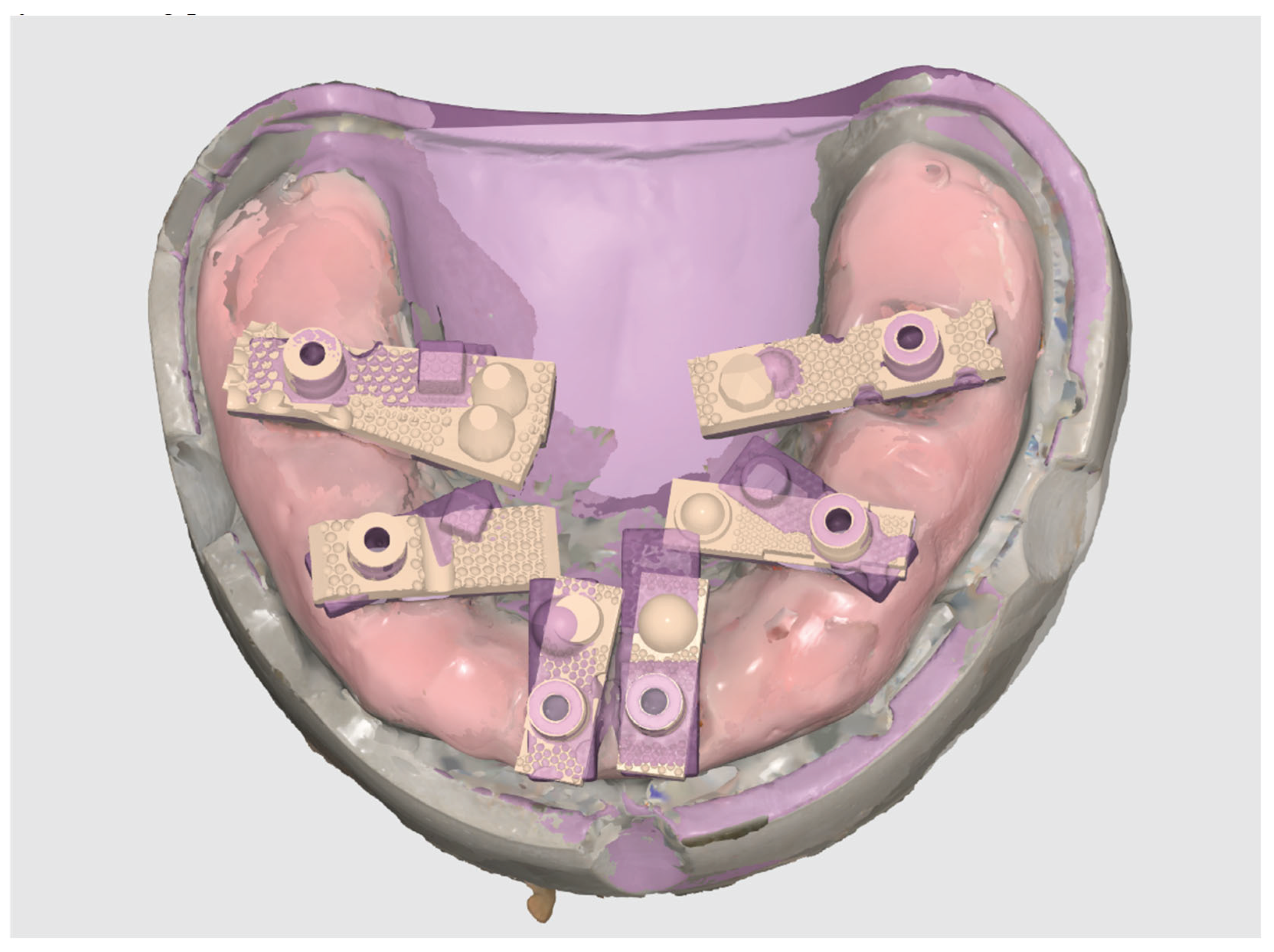
This in vitro study was conducted using a custom-fabricated mandibular full-arch model simulating an edentulous clinical scenario. The model incorporated six implant analogues, positioned to replicate a typical All-on-X distribution at sites 35, 33, 31, 41, 43, and 45. Multi-Unit Abutments (MUAs) were placed on each analogue, and Scan Ladder scan bodies were secured to the MUAs in their definitive positions.
To establish a gold standard reference geometry, a verification titanium bar jig was employed during analogue placement, ensuring consistent spacing and alignment across all implant sites. A silicone gingival mask was applied to the model to replicate soft tissue contours and introduce the scanning challenges encountered in vivo, such as sub-gingival interference and limited access around angled abutments.
Unlike retrospective clinical data analysis, this investigation involved direct experimental measurement under controlled conditions. A total of 20 independent intraoral scans were performed using a Medit i900 scanner equipped with the SmartX real-time library matching protocol. Each scan captured all six Scan Ladder positions, and the datasets were subsequently compared to a laboratory-grade reference scan generated with a 3Shape (Copenhagen, Denmark) E2 structured light scanner.
The study was designed in accordance with international standards for laboratory-based accuracy testing of intraoral scanners (ISO 12836). As no human participants were involved, no ethical approval was required.
2.2. Master STL Creation
A gold standard reference model was created using a mandibular full-arch model embedded with six implant analogues at positions 35, 33, 31, 41, 43, and 45. Multi-Unit Abutments (MUAs) were placed on each analogue, and Scan Ladder scan bodies were securely attached to replicate the definitive clinical scanning condition.
The model was digitised with a 3Shape E2 desktop structured light scanner as seen in Figure 3 below. (3Shape, Copenhagen, Denmark), which is accredited for high-precision dental metrology under ISO 12836 standards. The E2 scanner has a trueness specification of ≤7 μm and was therefore considered an appropriate reference for evaluating intraoral scanner accuracy.
The resulting STL dataset, representing the exact geometry and spatial relationships of the six implant positions, was designated as the Master STL. This file served as the gold standard against which all subsequent intraoral scans were compared.
The aim of creating the Master STL was to provide a validated, reproducible reference geometry for assessing the trueness (closeness to the reference) and precision (repeatability across scans) of the Medit SmartX protocol in conjunction with the Scan Ladder system. By ensuring that all deviations were measured against this high-fidelity reference, the study could isolate and quantify the contribution of the SmartX + Scan Ladder workflow to full-arch digital implant accuracy.
2.3. Scanning Procedure
All experimental scans were performed on the mandibular full-arch model with six implant analogues fitted with Multi-Unit Abutments and Scan Ladder scan bodies. Scanning was carried out using the Medit i900 intraoral scanner (Medit Corp., Seoul, South Korea) under the SmartX real-time library matching protocol.
Figure 4.
All Scans, Master and Test Data, Aligned and overlaid to view within Medit Link.
The Scan Ladder system provides detailed guidance for both the centralised positioning of scan bodies along the arch and for the recommended scanning sequence to optimise digital capture. These instructions were strictly adhered to in this study. The scan bodies were positioned in their prescribed horizontal alignment, ensuring their centroids were placed within a common reference plane. This arrangement is a defining feature of the Scan Ladder system and is designed to reduce cumulative stitching error during full-arch scanning.
For each acquisition, a standardised scan path was followed in accordance with the Scan Ladder protocol. Scanning commenced at the right posterior quadrant and proceeded sequentially across the occlusal surfaces towards the contralateral posterior region. Additional sweeps were made across buccal and lingual surfaces to capture gingival contours and ensure full visibility of each scan body. This workflow is specifically designed to maintain consistent reference points and optimise AI-based library matching within the SmartX system.
A total of 20 independent full-arch scans were acquired under identical conditions by a single experienced operator. Each SmartX dataset was automatically processed in real time, with virtual Multi-Unit Abutments (MUAs) replacing the physical Scan Ladder geometries during scanning. The resulting digital models were exported as STL files and imported into Exocad DentalCAD (Exocad GmbH, Darmstadt, Germany). Within Exocad, the six virtual MUAs were isolated and saved individually, creating 120 site-specific STL files for subsequent accuracy analysis.
Accuracy assessment was performed by importing each test STL alongside the Master STL (generated with the 3Shape E2 desktop scanner; see Section 2.2) into CloudCompare (3D Systems, Rock Hill, SC, USA), an open-source point-cloud and mesh processing software originally developed by Daniel Girardeau-Montaut at Télécom ParisTech/EDF R&D and now maintained by a global community under the GPL license. No best-fit superimposition was executed, and the average surface deviation (μm) was calculated between each test implant position and the reference in their relative 3D position as exported from exocad. This process produced one average deviation value per implant position per scan, yielding 20 values per site and 120 measurements in total.
Figure 5.
Aligning Scan Ladder Implant Library in Exocad.
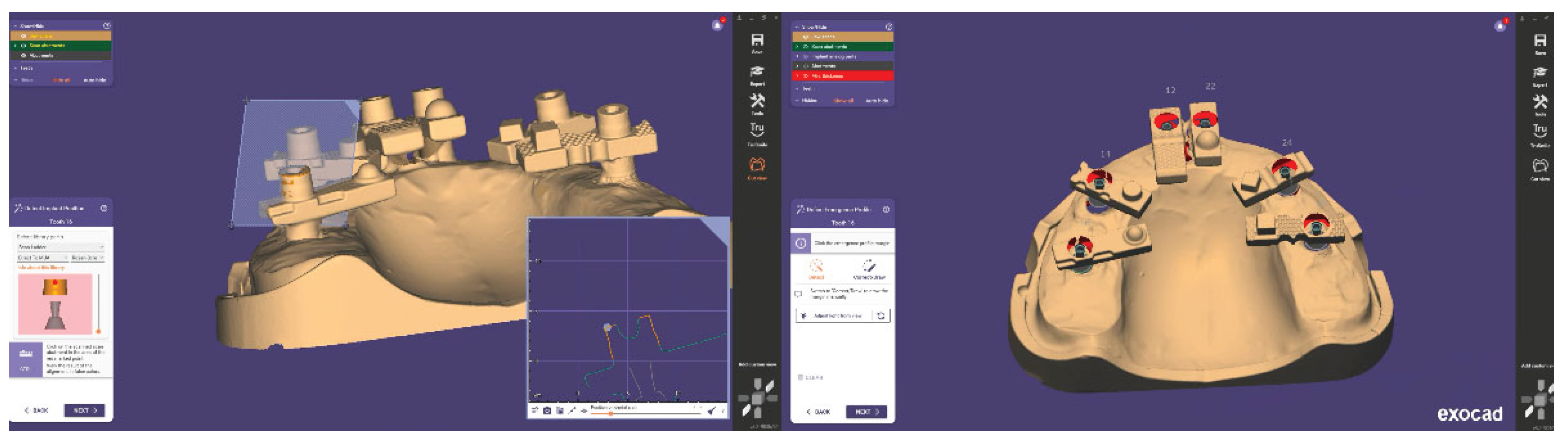
Figure 6.
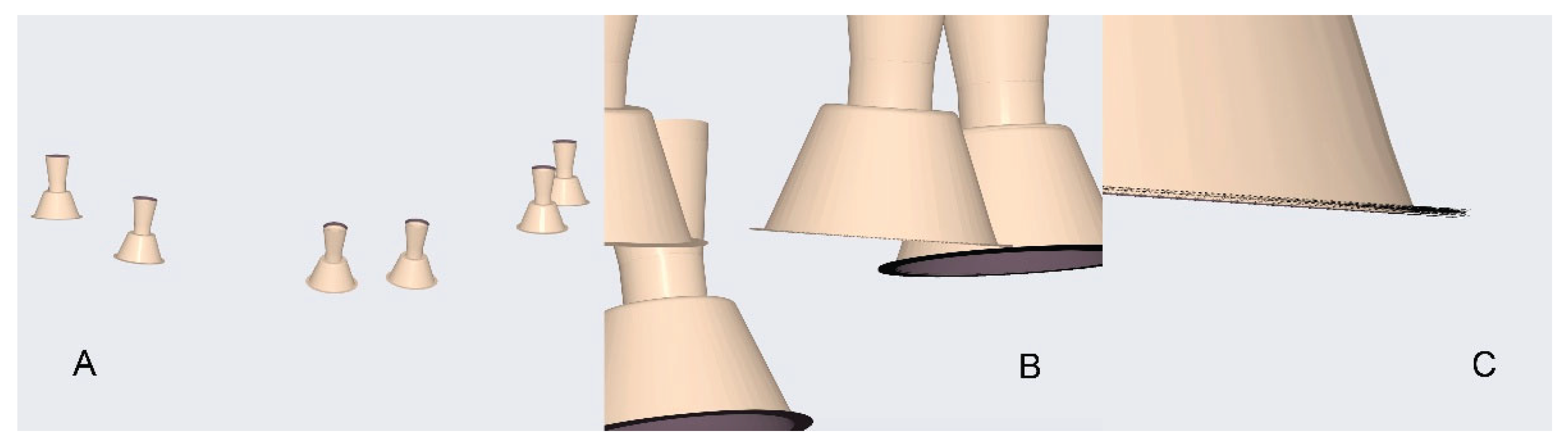
Zoom in of all Test Sets viewed overlaid. A. Whole Scene. B. Zoom into Multi Unit. C. Close up of all data.
Figure 6.
Zoom in of all Test Sets viewed overlaid. A. Whole Scene. B. Zoom into Multi Unit. C. Close up of all data.
2.3.1. Scanners in the Study
The scanners used in the present in vitro study are summarised in Table 1.

Table 1.
The Digital Scanners Used In This Study.
| Name | Manufacturer | Technology | STL Export | PLY/OBJ Colour Export | Photogrammetry |
|---|---|---|---|---|---|
| E2 | 3Shape, Copenhagen, Denmark | Structured white light desktop scanner. | YES | NO | NO |
| i900 | Medit, Seongbuk-gu, Seoul, Korea | Structured light-Active Speed 3D Video™ |
YES | YES | NO |
2.4. Design of the Study
2.4.1. Overview
This in vitro study evaluated the accuracy—defined in terms of trueness (closeness to the reference model) and precision(repeatability across scans)—of the Medit i900 intraoral scanner (Medit Corp., Seoul, South Korea) operating under the SmartX real-time library matching protocol in combination with Scan Ladder scan bodies.
The digital outputs from 20 repeated SmartX scans were compared against a high-accuracy Master STL generated using a 3Shape E2 desktop scanner (3Shape, Copenhagen, Denmark), accredited under ISO 12836 and specified to achieve ≤7 μm accuracy. The Master STL was considered the gold standard for all comparative analyses.
In total, 120 deviation measurements were collected across six implant positions (35, 33, 31, 41, 43, 45). This sample size was sufficient to provide robust statistical analysis, enabling evaluation of both systematic error (trueness) and variability across repeated acquisitions (precision).
2.5. Data Processing and Analysis
Following the scanning procedures, each SmartX-generated dataset was exported in STL format. Within Exocad DentalCAD (Exocad GmbH, Darmstadt, Germany), the automatically aligned virtual Multi-Unit Abutments (MUAs) were isolated from the full-arch model. Each implant position was exported as a separate STL file, resulting in six implant-specific files per scan and a total of 120 STL datasets across all 20 scans. This systematic segmentation ensured that each implant site could be assessed independently while maintaining consistent alignment with the overall arch geometry.
Each test STL was then imported into CloudCompare (3D Systems, Rock Hill, SC, USA) alongside the corresponding implant position from the Master STL (generated with the 3Shape E2 desktop scanner; Section 2.2). A best-fit alignment was performed using the software’s iterative closest point (ICP) algorithm. The average surface deviation (μm) between the test and reference STL was calculated for each implant site. This yielded 20 deviation values per implant position, providing a robust dataset for statistical evaluation of both trueness and precision.
2.5.1. Evaluating Trueness
Trueness was defined as the closeness of agreement between the test scan and the Master STL reference. For each implant position, the mean average deviation across the 20 scans was calculated, representing how accurately the SmartX workflow reproduced the known implant geometry. Descriptive statistics (mean, standard deviation, minimum, maximum) were compiled for each implant site as well as for the pooled dataset.
2.5.2. Evaluating Precision
Precision was defined as the repeatability of deviation measurements across multiple scans under identical conditions. For each implant site, the dispersion of deviation values across the 20 replicates was used to assess consistency. A lower standard deviation indicated higher precision.
2.5.3. Statistical Analysis
Statistical analysis was performed using SPSS version 22 (IBM, Chicago, IL, USA). A two-way analysis of variance (ANOVA) was applied to evaluate the effects of implant position and scan iteration on deviation values. Where significant effects were detected, post hoc multiple comparisons were conducted using Tukey’s HSD test, with the significance level set at α = 0.05. The analysis was designed to determine whether systematic differences existed between anterior and posterior implant positions and whether scan repetition introduced additional variability.
This methodology provided a rigorous framework for quantifying both trueness and precision of the Medit SmartX protocol with Scan Ladder scan bodies, ensuring reproducibility and comparability with existing literature on digital implant scanning accuracy.
3. Results
A total of 120 measurements (six implant positions across 20 Medit SmartX scans) were analysed and compared with the reference positions obtained using the 3Shape E2 desktop scanner. The reference STL file, generated with the aid of a verification jig, was treated as the gold standard against which all intraoral scanner-derived datasets were assessed.
Table 1.
Study Results.
| Block Number | Position 35 | Position 33 | Position 31 | Position 41 | Position 43 | Position 45 |
|---|---|---|---|---|---|---|
| 1 | 3.18 | 25.12 | -3.19 | 23.06 | 26.17 | 0.98 |
| 2 | 18.43 | 15.23 | -4.70 | 8.58 | 19.86 | 4.79 |
| 3 | 10.10 | 31.28 | -6.33 | 17.42 | 43.46 | 7.05 |
| 4 | 12.87 | 4.05 | 2.61 | 13.70 | 18.93 | 3.54 |
| 5 | 9.92 | 15.77 | -2.75 | 30.69 | 23.54 | -5.96 |
| 6 | 7.25 | 9.75 | 0.85 | 9.84 | 32.06 | 4.93 |
| 7 | 4.91 | 22.25 | 4.25 | 13.59 | 34.90 | 5.26 |
| 8 | 7.71 | 22.24 | 1.27 | 21.42 | 12.10 | -0.77 |
| 9 | 11.36 | -1.70 | 1.17 | 23.19 | 25.56 | 14.07 |
| 10 | 16.52 | 10.81 | -6.07 | 18.65 | 24.36 | 4.78 |
| 11 | 20.62 | 16.73 | -3.36 | 16.20 | 21.85 | 8.47 |
| 12 | 6.08 | -8.94 | -4.71 | 22.88 | 21.64 | 3.87 |
| 13 | 19.00 | 14.21 | -10.99 | 23.50 | 16.48 | 5.15 |
| 14 | 6.92 | 15.82 | -1.14 | 14.63 | 37.62 | 6.59 |
| 15 | 2.04 | 17.68 | -12.77 | 15.54 | 17.84 | -13.88 |
| 16 | 5.67 | 27.43 | -2.26 | 20.90 | 35.72 | 2.10 |
| 17 | 2.04 | 25.55 | 5.00 | 17.62 | 29.29 | -11.88 |
| 18 | 11.68 | 11.77 | -3.16 | 22.60 | 29.70 | -0.56 |
| 19 | 23.44 | 1.53 | -11.08 | 27.17 | 28.85 | 8.81 |
| 20 | 13.16 | -9.78 | -2.62 | 17.86 | 20.41 | 2.32 |
3.1. Trueness
When all positions were pooled, the Medit SmartX + Scan Ladder workflow achieved a mean average deviation of 11.41 μm with a standard deviation of 12.16 μm. These findings indicate exceptionally high trueness, with all values falling within a narrow range well below the widely accepted ±150 μm clinical threshold.
At the level of individual implant sites, the smallest mean deviations were observed at position 31 (4.7 μm) and position 45 (6.1 μm). The largest deviations occurred at position 43 (22.5 μm), with other posterior sites also demonstrating slightly greater error compared with anterior positions. Nevertheless, no mean site deviation exceeded 25 μm, and no individual test scan result exceeded 45 μm.
3.2. Precision
Repeated scans demonstrated strong reproducibility across the dataset. The pooled standard deviation of 12.16 μm reflects high precision, with tight clustering of values across the 20 replicate scans per position. Anterior positions in particular showed minimal variation, while posterior sites displayed slightly broader distributions yet remained within clinically acceptable limits.
3.3. Statistical Analysis
Two-way ANOVA confirmed that implant position significantly influenced deviation values (P < .001). Posterior implants yielded higher mean deviations than anterior sites, with the greatest discrepancies observed at position 43. In contrast, scan iteration (repeated scanning) showed no significant effect (P > .05), confirming reproducibility across all repeated SmartX scans.
No significant interaction was identified between implant position and scan iteration (P > .05), indicating that deviations were primarily influenced by anatomical and scanning-span factors rather than operator- or sequence-related variability.The results suggest that the Medit SmartX protocol, particularly when combined with Scan Ladder scan bodies, offers a promising alternative to traditional methods for achieving high trueness and precision in full-arch implant scanning.
The findings of this study may significantly influence the adoption of AI-driven scanning techniques in clinical practice, potentially reshaping standards for full-arch implant prosthetics.
Figure 7.
A Scan Overlay of one test scan STL from one position with the Master STL showing deviation between the two scans.
Figure 7.
A Scan Overlay of one test scan STL from one position with the Master STL showing deviation between the two scans.
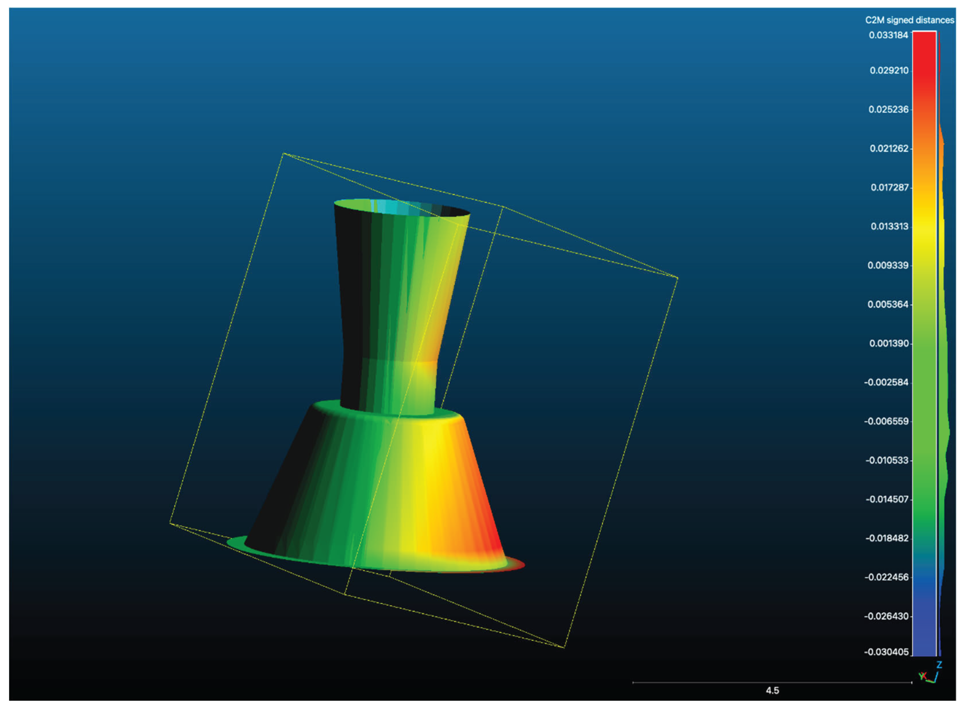
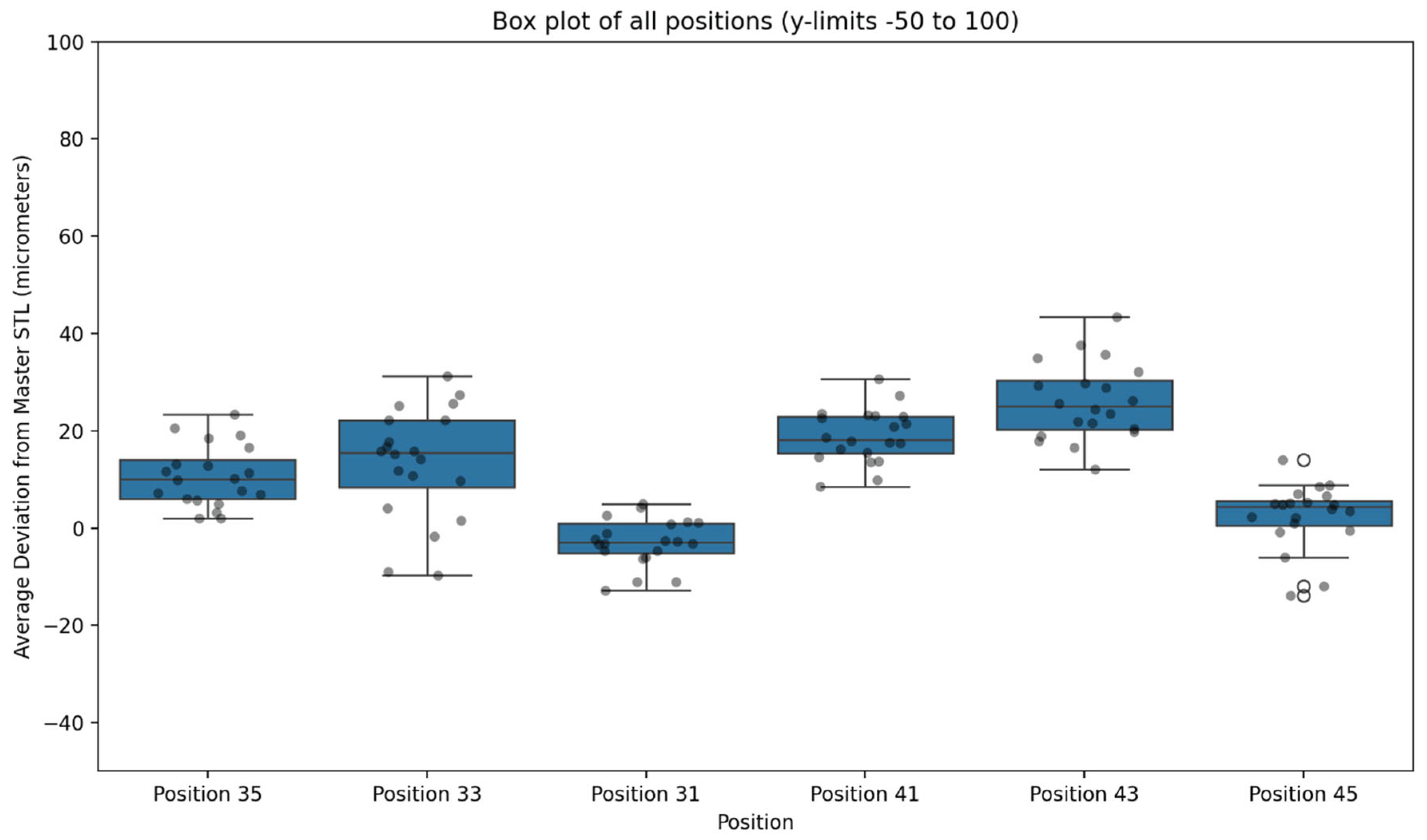
Figure 8.
Box Plot of Deviation in μm for Each Data Set for Each Scan Type in the Present Study.
4. Discussion
4.1. Evaluation of Trueness and Precision in Digital Intraoral Scanners
The purpose of this in vitro study was to evaluate the trueness and precision of full-arch implant scans obtained with the Medit SmartX real-time library matching protocol used in combination with Scan Ladder scan bodies, using a laboratory-grade desktop scanner (3Shape E2) as the reference standard. Across all positions, SmartX-generated digital models demonstrated a mean average deviation of 11.41 μm with a standard deviation of 12.16 μm, indicating both exceptional trueness and high precision. These results place the workflow within the same accuracy range typically reported for intraoral photogrammetry systems, which are regarded as the gold standard for full-arch implant transfer with values generally cited as below 20 μm. Importantly, even the most distal posterior sites—where deviations are usually greatest—remained well under 25 μm, confirming that the SmartX + Scan Ladder workflow can achieve a level of accuracy that is clinically indistinguishable from more complex and cost-intensive photogrammetry methods. The implications of these findings suggest that integrating advanced digital workflows could enhance clinical outcomes and patient satisfaction in implant dentistry.
4.2. Contribution to Existing Scientific Literature
These findings represent a significant advancement in chairside digital implant workflows. Photogrammetry has long been regarded as the gold standard for full-arch implant impression accuracy due to its extremely high spatial fidelity and minimal sensitivity to operator technique [20,28,29]. However, the clinical adoption of photogrammetry in dental implantology has been hindered by prohibitive costs, limited availability, and a steep learning curve [30,31]. In contrast, the SmartX protocol utilises artificial intelligence and integrated scan body libraries to automatically identify and align virtual components during intraoral scanning. The integration of artificial intelligence in this process offers a streamlined and cost-effective alternative that integrates seamlessly into existing Medit workflows without additional hardware or training overhead [32]. The results of this study indicate that the Medit SmartX protocol may provide a clinically viable alternative to traditional impression techniques, particularly in terms of accuracy and efficiency.
4.3. Enhancement in Accuracy with the Scan Ladder
The Scan Ladder scan bodies were developed to further enhance this process by addressing key limitations of traditional scan bodies and are currently the only scan body in the world which provides the full list of features and integrations present. First, the horizontally aligned structure of the Scan Ladder system ensures that scan body centroids lie within a common plane, reducing the angular distance and spatial distortion between implants—especially critical in full-arch scanning, where cumulative stitching errors can impact distant positions [33,34]. Second, each Scan Ladder scan body is individually identifiable by its unique shape and length, allowing the clinician to customise their position in the arch based on soft tissue contours and implant access. This variability helps minimise gingival interference and enhances scan capture in challenging sites, as previously identified as a source of error in deep or angled implants [35]. The findings suggest that the integration of the SmartX protocol with Scan Ladder scan bodies could significantly enhance the accuracy of full-arch implant scanning, providing a reliable alternative to traditional methods. [36] Furthermore, the results highlight the potential of AI-driven technologies in optimising dental workflows and improving patient outcomes in implant prosthodontics.
4.4. Comparison with Peer-Reviewed Literature
Previous research by Arcuri et al. highlighted the effect of scan body design and retention type (screw-retained vs snap-on) on full-arch digital scan accuracy [19] with Euclidean positional errors in some cases exceeding 100 μm, particularly at posterior or angled implant sites. In contrast, the present study observed a mean average deviation of just 11.41 μm across all implant positions, with no individual site exceeding 25 μm mean deviation. Even the most distal positions, which are typically the most error-prone, remained within this narrow range. These findings suggest that the combination of the SmartX algorithm with the unique geometry of the Scan Ladder system may effectively mitigate the spatial distortions often reported in traditional intraoral scanning workflows, providing accuracy levels that approach those of photogrammetry while retaining the accessibility of an intraoral scanner–based protocol.
Supporting this, Gómez-Polo et al. have demonstrated in recent literature that implant angulation, supra-gingival height, and scan body shape can all significantly influence intraoral scan fidelity. [34] The authors recommended optimisation of scan body design and workflow to improve full-arch results. In our study, the novel irregular surface morphology and height variation inherent to the Scan Ladder system may have played a pivotal role in enhancing geometric matching during the SmartX alignment process, thereby reinforcing their conclusion that well-designed scan bodies can substantially improve digital accuracy.
4.5. Implications for Clinical Practice
Importantly, the present study also confirms that trueness and precision were not affected by scan sequence or repetition, indicating a robust and repeatable performance of the SmartX protocol. Unlike photogrammetry, which typically requires calibration routines, operator training, and external software alignment,[37] the Medit system functions within a familiar clinical interface and automates the alignment process without user input, lowering the barrier to entry for full-arch digital workflows.
4.6. Challenges and Limitations of the Present Study
While the strong agreement with the laboratory-based reference model demonstrates the clinical potential of this workflow, several limitations should be recognised. This investigation was conducted in vitro under controlled conditions with static soft tissue simulation, optimal lighting, and a fixed reference geometry. Clinical reality introduces additional challenges such as patient movement, saliva, restricted access, and varying implant angulations, all of which may influence performance.
4.7. Future Directions
Future studies should therefore include in vivo validation to confirm the reliability of SmartX in routine practice. The long-term durability of the workflow, particularly when scan bodies are reused or applied in more complex implant scenarios, also warrants further investigation. Comparative trials against other AI-driven intraoral scanning protocols and established photogrammetry systems, ideally within multi-centre clinical settings, will be essential to contextualise the broader impact of SmartX and its integration with the Scan Ladder system.
5. Conclusions
This in vitro study demonstrated that the Medit SmartX real-time library matching protocol, when used in conjunction with Scan Ladder scan bodies, can achieve trueness and precision closely comparable to a laboratory-grade desktop scanner. Across all implant positions, the workflow produced a mean average deviation of 11.41 μm with a standard deviation of 12.16 μm, with no individual site exceeding 25 μm. These values place the system well within the sub-20 μm range typically associated with photogrammetry, the current gold standard for full-arch implant position transfer.
The findings support the clinical potential of SmartX as a simplified and cost-effective alternative to photogrammetry for full-arch digital implant scanning. The distinctive attributes of the Scan Ladder system—variable scan body geometries, a unified virtual library, and horizontally optimised positioning—appear to contribute substantially to the observed accuracy and reproducibility.
While these results highlight the promise of this approach, further clinical studies are required to confirm performance under real-world conditions, where biological and environmental variables may affect outcomes. Such investigations will be essential to validate whether SmartX with Scan Ladder can reliably deliver high-fidelity digital impressions in routine implant practice. The potential for integrating AI-driven technologies in dental workflows not only enhances accuracy but may also streamline the overall clinical process, ultimately benefiting patient care.
Supplementary Materials
The following supporting information can be downloaded at the website of this paper posted on Preprints.org.
Author Contributions
Conceptualisation, methodology, software, validation, formal analysis, investigation, resources, data curation, writing—original draft preparation, writing—review and editing, visualisation, supervision, project administration, funding acquisition, all completed by Adam Nulty. The author read and agreed to the published version of the manuscript.
Funding
This research received no external funding.
Data Availability Statement
The original contributions presented in the study are included in the article/supplementary material, further inquiries can be directed to the corresponding author/s.
Conflicts of Interest
One of the authors is the inventor of the Scan Ladder device, which was evaluated in this study, and holds a financial interest in its development and commercialization. To ensure objectivity, the study was conducted retrospectively using pre-existing, anonymized patient data that was collected during routine clinical procedures. The study’s design and outcomes were not influenced by any commercial interests or financial considerations. Additionally, while the Medit i900 intraoral scanner was employed as part of routine clinical practice alongside other data capture methods. All scanning technologies were evaluated objectively, and the study followed strict scientific and ethical standards to mitigate any potential bias. All potential conflicts of interest have been disclosed and managed in accordance with institutional policies. No other conflicts are declared.
References
- Zafiropoulos, G.-G.; Galil, A.A.; Deli, G. An Interocclusal Recording Method for the Fabrication of Full-Arch Implant-Retained Restorations. Journal of Oral Implantology 2014. [CrossRef]
- Jain, M. Impression Techniques for the Resorbed Mandibular Arch: A Guide to Increased Stability. Journal of the Scientific Society 2015. [CrossRef]
- Leão, M.P.; Pinto, C.P.; Sponchiado, A.P.; Ornaghi, B.P. Dimensional Stability of a Novel Polyvinyl Siloxane Impression Technique. Brazilian Journal of Oral Sciences 2014. [CrossRef]
- Rutkūnas, V.; Gečiauskaitė, A.; Jegelevičius, D.; Vaitiekūnas, M. Accuracy of Digital Implant Impressions with Intraoral Scanners. A Systematic Review. European Journal of Oral Implantology 2017.
- Di Fiore, A.; Meneghello, R.; Graiff, L.; Savio, G.; Vigolo, P.; Monaco, C.; Stellini, E. Full Arch Digital Scanning Systems Performances for Implant-Supported Fixed Dental Prostheses: A Comparative Study of 8 Intraoral Scanners. Journal of Prosthodontic Research 2019. [CrossRef] [PubMed]
- Ender, A.; Mehl, A. Influence of Scanning Strategies on the Accuracy of Digital Intraoral Scanning Systems. International journal of computerized dentistry 2013. [CrossRef]
- Drancourt, N.; Auroy, P.; Veyrune, J.-L.; Osta, N.E.; Nicolas, E. Accuracy of Conventional and Digital Impressions for Full-Arch Implant-Supported Prostheses: An In Vitro Study. Journal of Personalized Medicine 2023. [CrossRef]
- Fayad, noha E.H.; Abofadle, A.; Bahig, D. Accuracy of Different Digital Data Acquisition Work Flows for Full Arch Maxillary Implant Prostheses (an Invitro Study). Egyptian dental journal 2024. [CrossRef]
- Rutkunas, V.; Gedrimiene, A.; Adaškevičius, R.; Al-Haj Husain, N.; Özcan, M. Comparison of the Clinical Accuracy of Digital and Conventional Dental Implant Impressions. The European journal of prosthodontics and restorative dentistry 2020. [CrossRef]
- Papaspyridakos, P.; Papaspyridakos, P.; Vazouras, K.; Chen, Y.-W.; Kotina, E.; Natto, Z.S.; Natto, Z.S.; Kang, K.; Chochlidakis, K. Digital vs Conventional Implant Impressions: A Systematic Review and Meta-Analysis. Journal of Prosthodontics 2020. [CrossRef] [PubMed]
- Rahman, R.N.; Lee, A.; Lavasani, S.; Boehm, T. An Overview of Digital Workflows for Precision Implant Dentistry. Journal of the California Dental Association 2022. [CrossRef]
- Miyoshi, K.; Tanaka, S.; Yokoyama, S.; Sanda, M.; Baba, K. Effects of Different Types of Intraoral Scanners and Scanning Ranges on the Precision of Digital Implant Impressions in Edentulous Maxilla: An in Vitro Study. Clinical Oral Implants Research 2020. [CrossRef] [PubMed]
- Mangano, F.; Veronesi, G.; Hauschild, U.; Mijiritsky, E.; Mangano, C. Trueness and Precision of Four Intraoral Scanners in Oral Implantology: A Comparative in Vitro Study. PLOS ONE 2016. [CrossRef] [PubMed]
- Tallarico, M.; Lumbau, A.I.; Scrascia, R.; Demelas, G.; Sanseverino, F.; Amarena, R.; Meloni, S.M. Feasibility of Using a Prosthetic-Based Impression Template to Improve the Trueness and Precision of a Complete Arch Digital Impression on Four and Six Implants: An In Vitro Study. Materials 2020. [CrossRef]
- Sultanoğlu, E.G.; Keleş, B. Comparison of the Accuracy and Precision of Digital Scans for Implant-Supported Maxillary Hybrid Prosthesis: An in Vitro Study. Nigerian Journal of Clinical Practice 2024. [CrossRef]
- Giménez, B.; Özcan, M.; Martínez-Rus, F.; Pradíes, G. Accuracy of a Digital Impression System Based on Active Wavefront Sampling Technology for Implants Considering Operator Experience, Implant Angulation, and Depth. Clinical Implant Dentistry and Related Research 2015. [CrossRef]
- Cheng, J.; Zhang, H.; Liu, H.; Li, J.; Wang, H.-L.; Tao, X. Accuracy of Edentulous Full-Arch Implant Impression: An in Vitro Comparison between Conventional Impression, Intraoral Scan with and without Splinting, and Photogrammetry. Clinical Oral Implants Research 2024. [CrossRef]
- Çakmak, G.; Çakmak, G.; Yilmaz, H.; Treviño, A.; Kökat, A.M.; Yilmaz, B. The Effect of Scanner Type and Scan Body Position on the Accuracy of Complete-Arch Digital Implant Scans. Clinical Implant Dentistry and Related Research 2020. [CrossRef]
- Arcuri, L.; Pozzi, A.; Lio, F.; Rompen, E.; Zechner, W.; Nardi, A. Influence of Implant Scanbody Material, Position and Operator on the Accuracy of Digital Impression for Complete-Arch: A Randomized in Vitro Trial. Journal of Prosthodontic Research 2020. [CrossRef]
- Örtorp, A.; Jemt, T.; Bäck, T. Photogrammetry and Conventional Impressions for Recording Implant Positions: A Comparative Laboratory Study. Clinical Implant Dentistry and Related Research 2005. [CrossRef] [PubMed]
- Rivara, F.; Lumetti, S.; Calciolari, E.; Toffoli, A.; Forlani, G.; Manfredi, E. Photogrammetric Method to Measure the Discrepancy between Clinical and Software-Designed Positions of Implants. Journal of Prosthetic Dentistry 2016. [CrossRef]
- Richert, R.; Goujat, A.; Venet, L.; Viguie, G.; Viennot, S.; Robinson, P.; Farges, J.-C.; Fages, M.; Ducret, M. Intraoral Scanner Technologies: A Review to Make a Successful Impression. Journal of Healthcare Engineering 2017. [CrossRef]
- Accuracy of an Intraoral Scanner Based on Sleeve Type, Decontamination, and Calibration. 2023.
- Nulty, A.B. An In Vivo Comparison of Trueness and Precision of Two Novel Methods for Improving Edentulous Full Arch Implant Scanning Accuracy: A Pilot Study. Dent. J. 2024, 12, 367. [Google Scholar] [CrossRef] [PubMed]
- Menditto, A.; Patriarca, M.; Magnusson, B. Understanding the Meaning of Accuracy, Trueness and Precision. Accreditation and Quality Assurance 2007. [CrossRef]
- Bonnick, S.L.; Lewis, L.A. The Importance of Precision. In; 2013.
- Prenesti, E.; Gosmaro, F. Trueness, Precision and Accuracy: A Critical Overview of the Concepts as Well as Proposals for Revision. Accreditation and Quality Assurance 2015. [CrossRef]
- Bratos, M.; Bergin, J.M.; Rubenstein, J.E.; Sorensen, J.A. Effect of Simulated Intraoral Variables on the Accuracy of a Photogrammetric Imaging Technique for Complete-Arch Implant Prostheses. Journal of Prosthetic Dentistry 2018. [CrossRef]
- Bergin, J.M.; Rubenstein, J.E.; Mancl, L.; Brudvik, J.S.; Raigrodski, A.J. An in Vitro Comparison of Photogrammetric and Conventional Complete-Arch Implant Impression Techniques. Journal of Prosthetic Dentistry 2013. [CrossRef]
- Azevedo, L.; Molinero-Mourelle, P.; Antonaya-Martín, J.L.; del Río-Highsmith, J.; Correia, A.; Gómez-Polo, M. Photogrammetry Technique for the 3D Digital Impression of Multiple Dental Implants. In; 2019.
- Stuani, V. de T.; Ferreira, R.; Manfredi, G.G. do P.; Cardoso, M.V.; Sant’Ana, A.C.P. Photogrammetry as an Alternative for Acquiring Digital Dental Models: A Proof of Concept. Medical Hypotheses 2019. [CrossRef]
- De Angelis, F.; Pranno, N.; Franchina, A.; Di Carlo, V.; Brauner, E.; Ferri, A.; Pellegrino, G.; Grecchi, E.; Goker, F.; Stefanelli, L.V. Artificial Intelligence: A New Diagnostic Software in Dentistry: A Preliminary Performance Diagnostic Study. International Journal of Environmental Research and Public Health 2022. [CrossRef]
- Huang, R.; Liu, Y.; Huang, B.; Zhang, C.; Chen, Z.; Li, Z. Improved Scanning Accuracy with Newly Designed Scan Bodies: An in Vitro Study Comparing Digital versus Conventional Impression Techniques for Complete-Arch Implant Rehabilitation. Clinical Oral Implants Research 2020. [CrossRef]
- Gómez-Polo, M.; Sallorenzo, A.; Ortega, R.; Gomez-Polo, C.; Barmak, A.B.; Att, W.; Revilla-León, M. Influence of Implant Angulation and Clinical Implant Scan Body Height on the Accuracy of Complete Arch Intraoral Digital Scans. Journal of Prosthetic Dentistry 2022. [CrossRef]
- Ntovas, P.; Spanopoulou, M.; Martin, W.; Sykaras, N. Superimposition of Intraoral Scans of an Edentulous Arch with Implants and Implant-Supported Provisional Restoration, Implementing a Novel Implant Prosthetic Scan Body. Journal of Prosthodontic Research 2022. [CrossRef]
- Thanasrisuebwong, P.; Kulchotirat, T.; Anunmana, C. Effects of Inter-Implant Distance on the Accuracy of Intraoral Scanner: An in Vitro Study. The Journal of Advanced Prosthodontics 2021. [CrossRef] [PubMed]
- Jemt, T.; Bäck, T.; Petersson, A. Photogrammetry--an Alternative to Conventional Impressions in Implant Dentistry? A Clinical Pilot Study. International Journal of Prosthodontics 1999.
Figure 1.
The Scan Ladder Titanium Direct Scan Body Set.
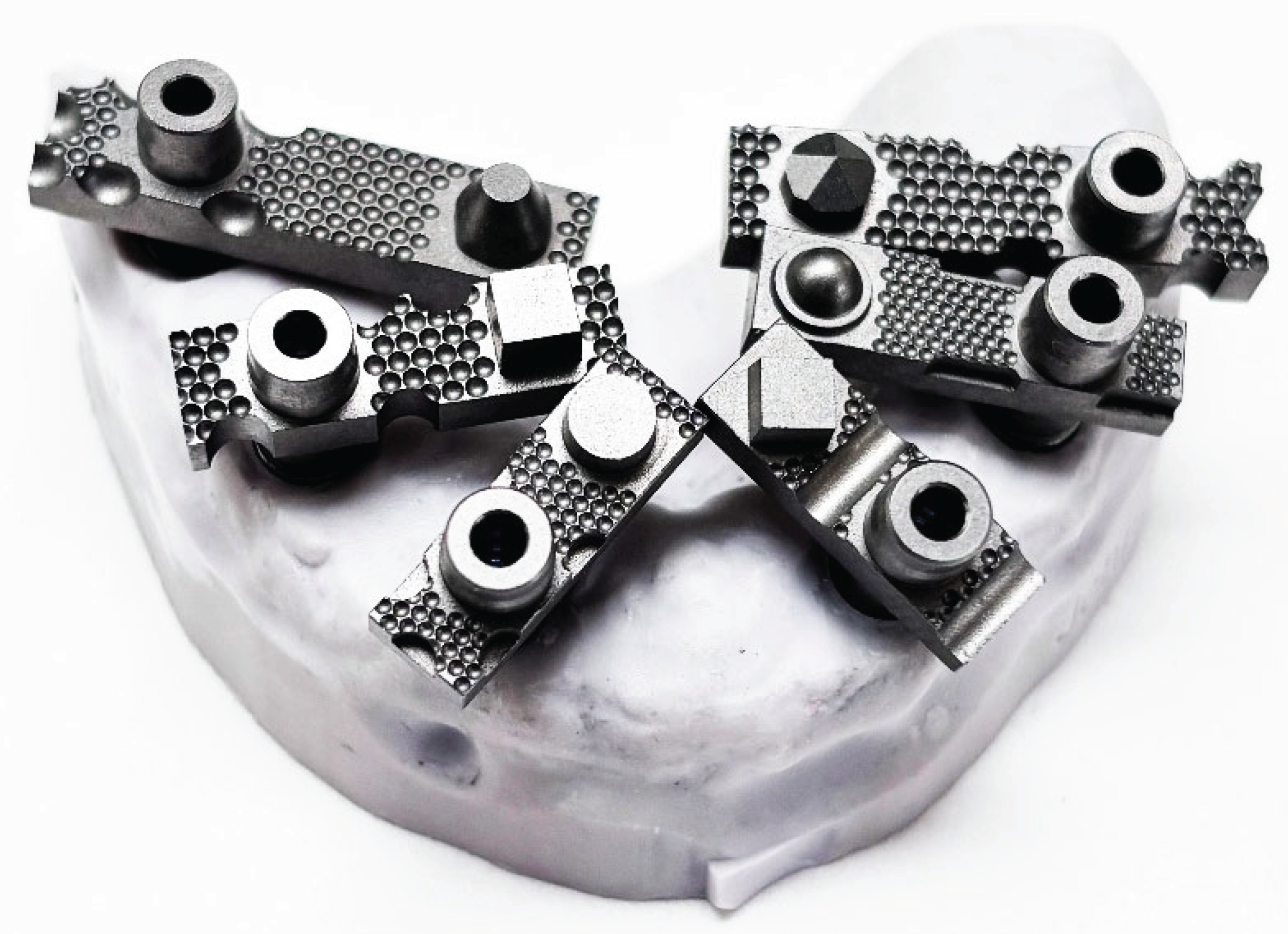
Figure 2.
Medit SmartX Real Time AI Smart Matching of the Scan Ladder system.
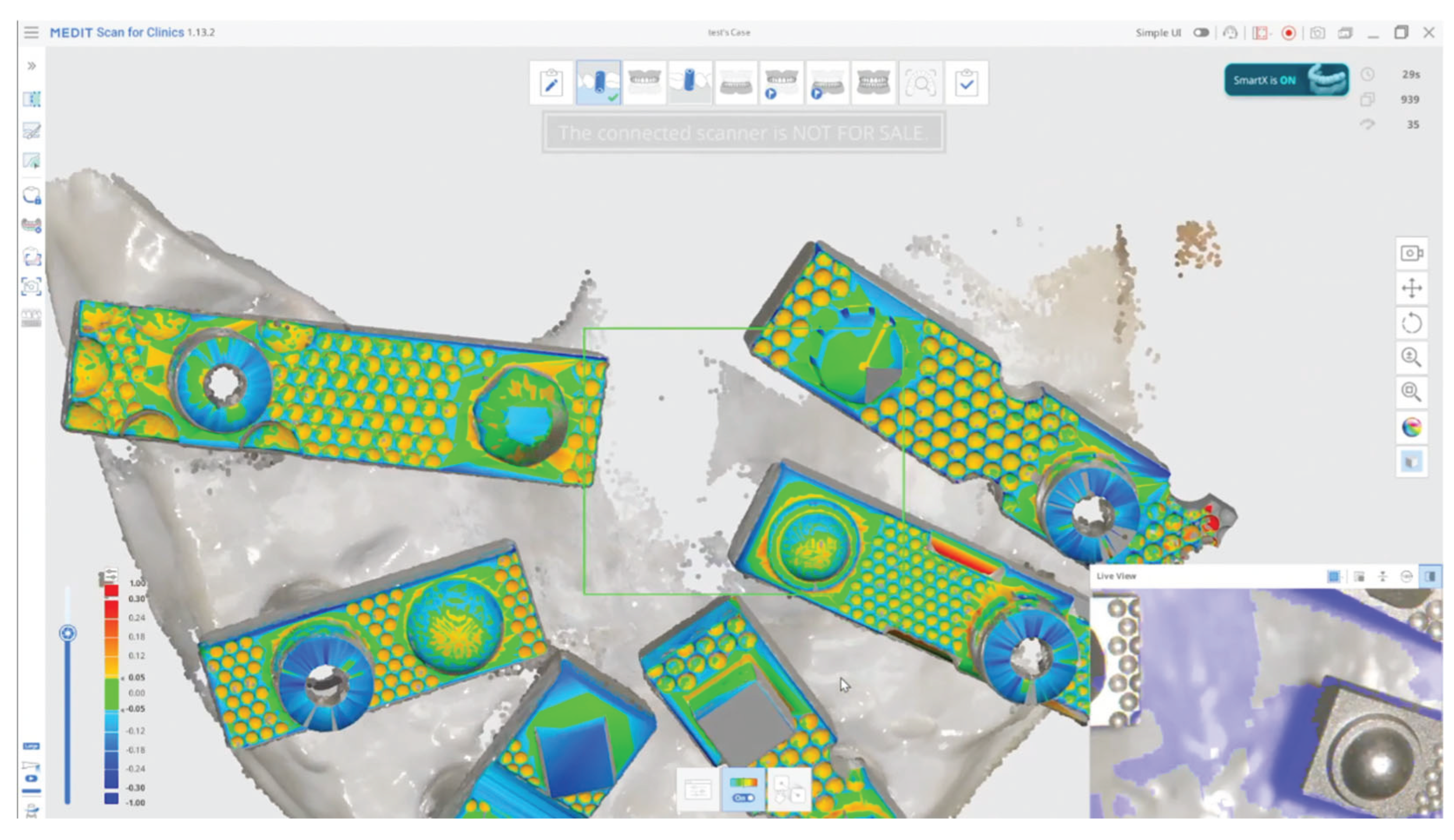
Figure 3.
Master Model being scanned with E2 Desktop Scanner.
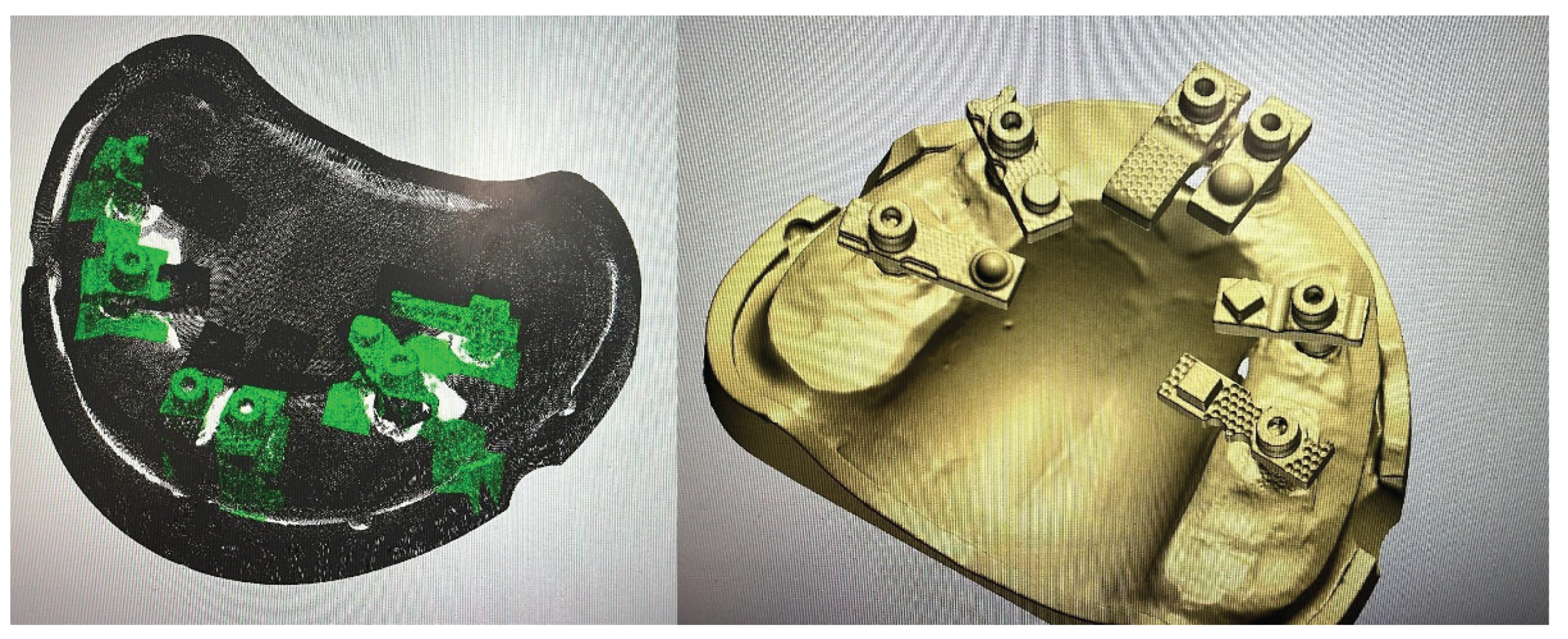
Table 2.
Mean Trueness and Standard Deviation of Each Position in comparison to the Master Scan from the E2.
Table 2.
Mean Trueness and Standard Deviation of Each Position in comparison to the Master Scan from the E2.
| Name | Mean (μm) | Std. Deviation (μm) | P Value |
|---|---|---|---|
| Position 35 | 10.65 | 6.32 | <000.1 |
| Position 33 | 13.34 | 11.51 | <000.1 |
| Position 31 | -3.00 | 4.89 | <000.1 |
| Position 41 | 18.95 | 5.57 | <000.1 |
| Position 43 | 26.02 | 7.94 | <000.1 |
| Position 45 | 2.48 | 6.69 | <000.1 |
Table 3.
Compared Means (Subset for alpha = 0.05).
| Position | N complete | Mean corr to others | Max corr to other | Max corr partner | Mean abs diff to others | Best linear R2 | Best linea partner |
| Position 45 | 20 | 0.09 | 0.56 | Position 35 | 14.39 | 0.31 | Position 35 |
| Position 43 | 20 | 0.08 | 0.37 | Position 33 | 18.28 | 0.14 | Position 33 |
| Position 33 | 20 | -0.07 | 0.37 | Position 43 | 14.08 | 0.14 | Position 43 |
| Position 35 | 20 | -0.07 | 0.56 | Position 45 | 12.08 | 0.31 | Position 45 |
| Position 31 | 20 | -0.07 | -0.41 | Position 35 | 18.10 | 0.17 | Position 35 |
| Position 41 | 20 | -0.10 | -0.28 | Position 31 | 13.67 | 0.08 | Position 31 |
Disclaimer/Publisher’s Note: The statements, opinions and data contained in all publications are solely those of the individual author(s) and contributor(s) and not of MDPI and/or the editor(s). MDPI and/or the editor(s) disclaim responsibility for any injury to people or property resulting from any ideas, methods, instructions or products referred to in the content. |
© 2025 by the authors. Licensee MDPI, Basel, Switzerland. This article is an open access article distributed under the terms and conditions of the Creative Commons Attribution (CC BY) license (http://creativecommons.org/licenses/by/4.0/).
Copyright: This open access article is published under a Creative Commons CC BY 4.0 license, which permit the free download, distribution, and reuse, provided that the author and preprint are cited in any reuse.


