Introduction
The advent of digital technologies in dentistry has revolutionized the process of dental implant treatment, transforming traditional workflows and enhancing the precision, efficiency, and patient experience of dental care. From diagnostic imaging using cone-beam computed tomography (CBCT) to intraoral scanning for impressions, digital tools have significantly improved the predictability and reproducibility of implant procedures. Among these advancements, digital impression techniques using intraoral scanners have gained widespread adoption due to their numerous advantages. These include improved patient comfort, reduced distortion compared to conventional impression materials, easy data storage, and efficient communication between clinicians and laboratory technicians. Furthermore, digital impressions shorten chairside time compared to traditional methods that rely on materials such as polyvinyl siloxane, making them an attractive option for both clinicians and patients [
1,
2,
3,
4,
5,
6].
In single-tooth implant prosthetics, the precision of implant impressions is critical for ensuring the passive fit and long-term success of the final restoration. Conventional techniques involve multiple steps, including physical impression-taking, model fabrication, and digital scanning of plaster models in the laboratory. Each step introduces potential sources of error, such as material shrinkage, tray movement, or inaccuracies during model fabrication, which can compromise the accuracy and fit of the final prosthesis. In contrast, digital impressions capture the implant position and surrounding structures directly in three dimensions, providing a streamlined workflow that minimizes cumulative errors. This direct digital capture not only enhances accuracy but also simplifies the process, reducing the need for physical models and enabling faster turnaround times for prosthetic fabrication [
7,
8,
9,
10].
Despite these advantages, the clinical effectiveness of digital impression techniques remains a subject of debate. While numerous studies have demonstrated comparable accuracy between digital and conventional methods, the impact on broader clinical outcomes—such as treatment time, patient satisfaction, and prosthetic fit—requires further exploration. For instance, although digital impressions are often faster and more comfortable for patients, the initial cost of acquiring digital equipment and the learning curve for clinicians can be barriers to widespread adoption. Additionally, patient preferences and familiarity with traditional methods may influence the acceptance of digital technologies in dental practice. These factors highlight the need for comprehensive research to evaluate the practical benefits of digital impressions in real-world clinical settings.
This study aims to compare treatment time and patient satisfaction between digital and conventional impression techniques in single-tooth implant prosthetics. By evaluating key metrics such as time efficiency, patient comfort, and overall satisfaction, this research seeks to provide evidence-based insights into the benefits of digital impressions. The findings will help clinicians make informed decisions about adopting digital technologies and support their integration into routine dental practice. Furthermore, this study will contribute to the growing body of literature on digital dentistry, addressing gaps in knowledge regarding the clinical outcomes and patient experiences associated with digital impression techniques.
Methods
Study Design
The study was open-label study, by approved by the Biomedical Research Ethics Committee of Can Tho University of Medicine and Pharmacy (IRB No. 23.336.HV/PCT-HDDD). The study was conducted at the Specialized Clinic of Odonto-Stomatology, Department of Odonto-Stomatology, Van Lang University, from April 2023 to April 2024
Study Subjects
Convenience sampling was used, following the study's inclusion and exclusion criteria. The participants were patients with posterior tooth loss who had undergone dental implant surgery at least four months prior and who consented to receive implant prosthetic restoration at the Department of Odonto-Stomatology, Van Lang University.
Inclusion Criteria
Patients aged 18 years or older.
Single-tooth loss in the posterior maxilla or mandible, with implants placed for at least four months and assessed for osseointegration via CBCT imaging.
The implant site must have healing abutments placed for a minimum of two weeks prior to the study.
Presence of at least one adjacent tooth or prosthetic restoration near the implant site, with an edentulous gap greater than 6 mm and prosthetic space greater than 5 mm.
The opposing teeth must be either natural teeth, restorations on natural teeth, or restorations on implants.
All patients selected for the study had implants from the same manufacturer (Naturactis, Euroteknika, France).
Exclusion Criteria
Patients with mental disorders, or those who are mute, deaf, or unable to communicate effectively with the interviewer.
Individuals with allergies to any components of the impression material.
Patients who are unable to return for follow-up visits within three months after prosthetic placement.
Patients who declined to participate in the study at any point.
Sample Size
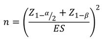
Based on the study by Gintaute et al. [
11], with Δ = 1.28 and
σ = 2.86, the calculated sample size was n > 39. Therefore, a sample size of 40 patients was chosen.
Procedures
The conventional impression technique involves capturing the implant position using light-body and heavy-body vinyl polysiloxane (VPS) impression materials (Panasyl Initial Contact Light and Panasyl Putty; Kettenbach Dental, Germany) with a one-step open-tray approach for the full dental arch. The impression tray was first tried in the patient’s mouth. The healing abutment was removed, and an impression coping was securely attached to the implant with a tightening torque of 10 Ncm. A periapical radiograph was taken using the parallel technique to confirm the precise seating of the impression coping and implant. The VPS impression material was then loaded into the tray and placed onto the dental arch. Once the material had fully set, the tray was carefully removed. The impression was meticulously inspected for accuracy, ensuring it was free of bubbles, distortions, or incomplete details. The impression coping remained firmly embedded in the impression material. A bite registration was recorded in maximum intercuspation using a bite registration material (Futar, Kettenbach Dental). An alginate impression (Tropicalgin; Zhermack Dental) was taken of the opposing arch to capture its anatomical details.
Digital impressions were taken using an intraoral scanner (3Shape Trios 3 Move, 3Shape A/S, Denmark) and the scanning process followed the manufacturer’s guidelines.it began by scanning the opposing arch relative to the implant site. The operator positioned themselves on the patient’s right side and performed a continuous scan, starting from the occlusal surface of the distalmost tooth, moving through the occlusal/incisal edges of the teeth, and ending at the distalmost tooth on the opposite side. The lingual and buccal surfaces were then scanned. For the maxillary arch, the scan included the relevant palatal area, distal surfaces of the terminal teeth, edentulous regions (if present), and both tuberosities. For the mandibular arch, the scan extended to the distal surfaces of the terminal teeth and edentulous regions (if present). The healing abutment was removed, and the entire dental arch, including the soft tissue emergence profile around the implant, was scanned. The digital impression coping was then connected to the implant with a tightening torque of 10 Ncm. A periapical radiograph was taken to verify the precise seating of the digital impression coping and implant. The scan was repeated until the TRIOS Patient Monitoring software confirms a successful scan. The digital impression coping was removed, and the bite registration was scanned in maximum intercuspation, starting from the right and then the left side.
Evaluation of Impression Time
Conventional Impression Time
The total time required to complete the conventional impression process was measured in seconds using a stopwatch. The timing began with the removal of the healing abutment and placement of the impression coping and ended when all clinical steps for obtaining the implant impression were completed. The process includes the following steps:
Removal of the healing abutment and attachment of the impression coping.
Taking an impression of the arch containing the implant.
Taking an impression of the opposing arch.
Recording the bite in maximum intercuspation.
Digital Impression Time
The total time required to complete the digital impression process was measured in seconds using a stopwatch. The timing commenced with the removal of the healing abutment and placement of the digital impression coping and concluded when all clinical steps for scanning the implant were completed. The steps involved in the process include:
Removal of the healing abutment and attachment of the digital impression coping.
Scanning of the arch containing the implant.
Scanning of the opposing arch.
Scanning of the bite in maximum intercuspation.
Evaluation of Patient Satisfaction
Patient satisfaction with the impression process was evaluated using a standardized questionnaire and a Visual Analog Scale (VAS) based on Joda’s study [
12]. The evaluation focuses on the following aspects:
Satisfaction with the time required for the impression process.
Anxiety level during the impression process.
Overall convenience of the impression technique.
Level of discomfort or pain during the impression.
Taste experience during and after the impression.
The degree of nausea caused by the impression.
Bias Control
To minimize potential errors during data collection and processing, the following rigorous measures were implemented:
Participant selection: Study participants were carefully selected based on predefined inclusion and exclusion criteria to ensure homogeneity and relevance to the research objectives.
Standardized procedures: All clinical procedures, including impression-taking, trial fittings, and final prosthesis placement, were performed exclusively by the principal dental researcher. This individual possesses a minimum of 5 years of experience in implant prosthetics and is proficient in the use of intraoral scanners (IOS), ensuring consistency and precision throughout the study.
Randomization: A randomization process was employed to determine the sequence of impression methods (digital vs. conventional) and the order in which the crowns were tried. Both crowns were fabricated by the same dental technician, following a randomized sequence to eliminate bias. Measurements were conducted using Geomagic Control X software, with all analyses performed by a single technician to maintain consistency and accuracy.
Questionnaire administration: The patient satisfaction questionnaire was pre-tested and refined to ensure clarity and reliability. Data collection was carried out by a single individual to minimize variability, and all research instruments were calibrated to guarantee precision and reproducibility.
Data Statistics
The data were presented as mean ± standard deviation and analyzed using SPSS 25.0. Analytical statistics included the Chi-square test and Fisher's exact test, with statistical significance defined as p < 0.05.
Results
Participants’ Characteristics
The mean age was 38.1 ± 13.24 years. Among the 40 participants, the number of females (n = 30) was three times greater than the number of males (n = 10). Regarding the edentulous regions receiving implants, the majority of cases were concentrated in the molar region, accounting for 92.5% (n = 37), while the premolar region represented only 7.5% (n = 3). The most common implant sites were tooth 36 (35.0%) and tooth 46 (30.0%), while teeth 27 and 37 had the lowest frequencies (2.5%/each). The average duration of tooth loss before implant treatment was 4.75 ± 4.5 years. The distribution of tooth loss duration was fairly balanced, with 32.5% of participants experiencing tooth loss for less than 1 year, 32.5% for 1–5 years, and 35% for more than 5 years.
Comparison of Impression Time
The comparison was based on four key procedures: implant position scanning, opposing arch scanning, centric occlusion scanning, and total impression time. Digital impression techniques were significantly faster than conventional impression methods at all steps (p < 0.001). For implant position scanning/rubber impression, conventional impressions took 373.9 ± 44.2 seconds, while digital impressions took 214.8 ± 47 seconds. In opposing arch scanning/opposing arch impression, conventional impressions took 180.2 ± 22.1 seconds, compared to 102.3 ± 25.2 seconds for digital impressions. For centric occlusion scanning/bite registration, conventional impressions took 86.8 ± 14.7 seconds, while digital impressions took 36.4 ± 10.2 seconds. In total impression time, conventional impressions took 640.8 ± 52.8 seconds, while digital impressions took 353.5 ± 62.6 seconds, with a time difference of 287.3 seconds (p < 0.001) (
Table 1).
Evaluation of Patient Satisfaction
Digital impressions offered a more comfortable, less stressful experience for patients compared to conventional impressions in all investigated aspects (p < 0.001). Digital impressions were associated with higher satisfaction in terms of impression time (87.4 ± 11.2 vs. 72 ± 15.2, p < 0.001), lower levels of anxiety (11.3 ± 13.4 vs. 19.7 ± 17.8, p < 0.001), and greater convenience (82.3 ± 14 vs. 68.9 ± 15.8, p < 0.001). Furthermore, patients reported less pain with digital impressions (12.8 ± 13 vs. 19.9 ± 14.3, p < 0.001), a more pleasant taste during and after the procedure (13.2 ± 15.3 vs. 31.8 ± 20, p < 0.001), and lower levels of nausea (14.3 ± 19.3 vs. 27 ± 21.4, p < 0.001) (
Table 2).
Discussion
The study compared the time required for implant position scanning using digital and conventional impression techniques and investigated patient satisfaction through a questionnaire. The results showed that the digital method took significantly less time compared to the conventional impression method. Moreover, patients reported higher satisfaction with the treatment time, convenience, comfort, and less pain, discomfort, nausea, and anxiety using the digital technique
The results indicated that the total working time for the digital impression process was 353.5 ± 62.6 seconds, significantly shorter than the conventional impression method using rubber materials, which took 640.8 ± 52.8 seconds. The average time difference between the two methods was 287.3 seconds (p < 0.001). Implant position scanning was the most time-consuming step in both methods, but digital impressions significantly reduce the overall time required. Furthermore, the time differences at each stage of the impression process further confirm that digital impressions were faster than conventional impressions.
Similarly, Lee et al. [
13]reported that the total time for the digital impression process was 676.8 seconds, which was shorter than the conventional rubber impression method, which took 846.6 seconds. Our study showed a shorter total implant scanning time using the IOS scanner compared to Sang J. Lee's study, due to differences in the time intervals considered during the impression process and the scanning systems used. In our study, the total time for digital implant scanning was the sum of the times for implant position scanning, opposing arch scanning, and centric occlusion scanning. In contrast, Sang J. Lee's study also included preparation time. The system used in Sang J. Lee's research was the iTero Element (Align Technology Inc.), while we used the Trios 3 Move+ (3Shape A/S). Joda et al. also [
12]used the Trios 3 IOS scanner for implant position scanning, with a total average time of 8.6 ± 1.6 minutes, faster than the IOS Virtua Vivo scanner by Dental Wings Inc (Montreal, Canada). The discrepancies in the time intervals considered in these studies may explain the shorter total time recorded in our study compared to those of Lee and Joda et al.’s studies [
12,
13,
14,
15].
One key advantage of digital impression techniques is the ability to take additional scans without needing to repeat the entire impression process [
13]. Digital techniques have been shown to be more efficient and reduce the total time for implant position scanning by as much as 50% compared to conventional methods [
16,
17].
Participants reported higher satisfaction with the impression time using the digital technique. The mean satisfaction score for the digital group was 87.4 ± 11.2, compared to 72 ± 15.2 for the conventional impression group (p < 0.001). This result was consistent with studies by Gintaute et al. [
18], Gallardo et al. [
19], Yuzbasioglu et al. [
20] and Oliveira et al. [
21]. Patients tend to be more satisfied when the time spent in the chair is shorter, and they prefer techniques that are less time-consuming [
13]. This was consistent to our findings, which was the total impression time for the digital method shorter than the traditional rubber impression method.
Pain perception may be related to the tools and materials used during the impression-taking process. Traditional techniques involve the use of rubber impression materials and trays that come into contact with teeth and gums, which can cause discomfort. The digital technique used a small scanner head to capture digital data, minimizing contact with oral tissues, resulting in a more comfortable experience for patients. Patients reported little to no unpleasant taste with the digital technique. The scanning head generated very little heat, which patients often did not notice, leading to minimal discomfort. Additionally, the digital technique causes less nausea compared to the traditional method, similar to findings of Yuzbasioglu et al. [
20]
These advantages make digital impressions a highly practical and patient-friendly option, especially in busy dental practices where time efficiency and patient experience are critical. The ability to take additional scans without repeating the entire process further underscores the flexibility and reliability of digital systems, reducing the likelihood of errors and the need for retakes. This feature is particularly beneficial in complex cases, such as full-arch restorations or cases involving multiple implants, where precision and efficiency are paramount. Additionally, the reduced physical discomfort and anxiety associated with digital impressions make them more appealing to patients, potentially improving compliance and overall treatment outcomes.
The study had some limitations. The sample size was relatively limited, which may affect the generalizability of the findings. A larger sample size across diverse demographics would provide more robust data. The study focused on a single type of digital impression system (Trios 3), which may not fully represent the performance of other digital impression systems available in the market. Further research comparing multiple digital impression systems would provide a more comprehensive understanding of the technology’s effectiveness. The study only assessed the time efficiency and patient satisfaction during the impression-taking process, without evaluating long-term outcomes such as the accuracy of the impressions or the success rate of treatments based on these impressions. Future studies should explore these aspects to provide a more holistic evaluation of digital versus conventional methods. Furthermore, the patient satisfaction assessment, while valuable, was based on subjective self-reporting, which may be influenced by individual biases. Objective measurements and longer follow-up periods would strengthen the findings. Finally, the experience and skill level of the practitioner performing the impressions could influence the results, and further research should address this by including multiple operators to assess the consistency of the outcomes.
Conclusion
The digital impression technique for single dental implant restorations can serve as an effective alternative to traditional rubber impression methods. This approach not only reduces the time spent by both the patient and the dentist but also enhances patient comfort, providing a more pleasant treatment experience.
Author Contributions
Conceptualization, T.H.L.; methodology, T.H.L.; software, P.V.C.; validation, V.H.B.T. and H.V.; formal analysis, X.X.; investigation, T.H.L.; resources, H.V.; data curation, PV.C.; writing—original draft preparation, H.V.; writing—review and editing, T.H.L.; visualization, PV.C.; supervision, T.H.L.; project administration, N.H.N.; funding acquisition, N.H.N. All authors have read and agreed to the published version of the manuscript.
Funding
No external funding.
Institutional Review Board Statement
by approved by the Biomedical Research Ethics Committee of Can Tho University of Medicine and Pharmacy (IRB No. 23.336.HV/PCT-HDDD).
Informed Consent Statement
Informed consent was obtained from all subjects involved in this study.
Data Availability Statement
The data presented in this study are available upon reasonable request, after the signature of a formal data-sharing agreement in an anonymous form, from the corresponding author because they are protected by privacy.
Conflicts of Interest
The authors declare no conflict of interest.
References
- Sanda, M.; Miyoshi, K.; Baba, K. Trueness and Precision of Digital Implant Impressions by Intraoral Scanners: A Literature Review. Int J Implant Dent 2021, 7, 97. [Google Scholar] [CrossRef] [PubMed]
- Viet, H.; Thi Nhu Thao, D.; Phuoc, T.H.; Quang Tien, N. A Multidisciplinary Approach to Managing Severe Gummy Smile Using 3D Simulation and Digital Surgical Guide: A Case Report. J Surg Case Rep 2024, 2024, rjae483. [Google Scholar] [CrossRef] [PubMed]
- Nguyen, V.A.; Nguyen, T.A.; Doan, H. Le; Pham, T.H.; Doan, B.N.; Pham, T.T.T.; Hoang, V. Transfer Accuracy of Partially Enclosed Single Hard Vacuum-Formed Trays with 3D-Printed Models for Lingual Bracket Indirect Bonding: A Prospective in-Vivo Study. PLoS One 2025, 20, e0316208. [Google Scholar] [CrossRef] [PubMed]
- Iturrate, M.; Minguez, R.; Pradies, G.; Solaberrieta, E. Obtaining Reliable Intraoral Digital Scans for an Implant-Supported Complete-Arch Prosthesis: A Dental Technique. J Prosthet Dent 2019, 121, 237–241. [Google Scholar] [CrossRef] [PubMed]
- Nguyen, P.N.; Tran, L.H.; Hoang, V. Full-Arch Implant-Supported Rehabilitation Using Reverse Scan Technique: A Case Report. J Oral Implantol 2025. [CrossRef] [PubMed]
- Viet, H.; Marya, A.; Apuzzo, F. d’; Nucci, L. The Clinical Applications and Outcomes of Digital MARPE in Orthodontics: A Scoping Review. Semin Orthod 2024. [CrossRef]
- Mangano, F.G.; Veronesi, G.; Hauschild, U.; Mijiritsky, E.; Mangano, C. Trueness and Precision of Four Intraoral Scanners in Oral Implantology: A Comparative in Vitro Study. PLoS One 2016, 11, e0163107. [Google Scholar] [CrossRef] [PubMed]
- Renne, W.; Ludlow, M.; Fryml, J.; Schurch, Z.; Mennito, A.; Kessler, R.; Lauer, A. Evaluation of the Accuracy of 7 Digital Scanners: An in Vitro Analysis Based on 3-Dimensional Comparisons. J Prosthet Dent 2017, 118, 36–42. [Google Scholar] [CrossRef] [PubMed]
- Su, T.; Sun, J. Comparison of Repeatability between Intraoral Digital Scanner and Extraoral Digital Scanner: An in-Vitro Study. J Prosthodont Res 2015, 59, 236–242. [Google Scholar] [CrossRef] [PubMed]
- Jeong, I.-D.; Lee, J.-J.; Jeon, J.-H.; Kim, J.-H.; Kim, H.-Y.; Kim, W.-C. Accuracy of Complete-Arch Model Using an Intraoral Video Scanner: An in Vitro Study. J Prosthet Dent 2016, 115, 755–759. [Google Scholar] [CrossRef] [PubMed]
- Gintaute, A.; Zitzmann, N.U.; Brägger, U.; Weber, K.; Joda, T. Patient-Reported Outcome Measures Compared to Professional Dental Assessments of Monolithic ZrO2 Implant Fixed Dental Prostheses in Complete Digital Workflows: A Double-Blind Crossover Randomized Controlled Trial. J Prosthodont 2023, 32, 18–25. [Google Scholar] [CrossRef] [PubMed]
- Joda, T.; Brägger, U. Patient-Centered Outcomes Comparing Digital and Conventional Implant Impression Procedures: A Randomized Crossover Trial. Clin Oral Implants Res 2016, 27, e185–e189. [Google Scholar] [CrossRef] [PubMed]
- Lee, S.J.; Jamjoom, F.Z.; Le, T.; Radics, A.; Gallucci, G.O. A Clinical Study Comparing Digital Scanning and Conventional Impression Making for Implant-Supported Prostheses: A Crossover Clinical Trial. J Prosthet Dent 2022, 128, 42–48. [Google Scholar] [CrossRef] [PubMed]
- Joda, T.; Brägger, U. Digital vs. Conventional Implant Prosthetic Workflows: A Cost/Time Analysis. Clin Oral Implants Res 2015, 26, 1430–1435. [Google Scholar] [CrossRef] [PubMed]
- Joda, T.; Brägger, U. Time-Efficiency Analysis Comparing Digital and Conventional Workflows for Implant Crowns: A Prospective Clinical Crossover Trial. Int J Oral Maxillofac Implants 2015, 30, 1047–1053. [Google Scholar] [CrossRef] [PubMed]
- Schepke, U.; Meijer, H.J.A.; Kerdijk, W.; Cune, M.S. Digital versus Analog Complete-Arch Impressions for Single-Unit Premolar Implant Crowns: Operating Time and Patient Preference. J Prosthet Dent 2015, 114, 403–6e1. [Google Scholar] [CrossRef] [PubMed]
- Lee, S.J.; Gallucci, G.O. Digital vs. Conventional Implant Impressions: Efficiency Outcomes. Clin Oral Implants Res 2013, 24, 111–115. [Google Scholar] [CrossRef]
- Gintaute, A.; Zitzmann, N.U.; Brägger, U.; Weber, K.; Joda, T. Patient-Reported Outcome Measures Compared to Professional Dental Assessments of Monolithic ZrO2 Implant Fixed Dental Prostheses in Complete Digital Workflows: A Double-Blind Crossover Randomized Controlled Trial. J Prosthodont 2023, 32, 18–25. [Google Scholar] [CrossRef]
- Gallardo, Y.R.; Bohner, L.; Tortamano, P.; Pigozzo, M.N.; Laganá, D.C.; Sesma, N. Patient Outcomes and Procedure Working Time for Digital versus Conventional Impressions: A Systematic Review. J Prosthet Dent 2018, 119, 214–219. [Google Scholar] [CrossRef] [PubMed]
- Yuzbasioglu, E.; Kurt, H.; Turunc, R.; Bilir, H. Comparison of Digital and Conventional Impression Techniques: Evaluation of Patients’ Perception, Treatment Comfort, Effectiveness and Clinical Outcomes. BMC Oral Health 2014, 14, 10. [Google Scholar] [CrossRef] [PubMed]
- de Oliveira, N.R.C.; Pigozzo, M.N.; Sesma, N.; Laganá, D.C. Clinical Efficiency and Patient Preference of Digital and Conventional Workflow for Single Implant Crowns Using Immediate and Regular Digital Impression: A Meta-Analysis. Clin Oral Implants Res 2020, 31, 669–686. [Google Scholar] [CrossRef] [PubMed]
Table 1.
Comparison of impression time between conventional and digital impressions.
Table 1.
Comparison of impression time between conventional and digital impressions.
| Time |
Conventional impressions (second) |
Digital impressions
(second) |
p |
| Implant position scanning / Rubber impression |
373.9 ± 44.2 |
214.8 ± 47 |
0.001 |
| Opposing arch scanning / Opposing arch impression |
180.2 ± 22.1 |
102.3 ± 25.2 |
0.001 |
| Centric occlusion scanning / Bite registration |
86.8 ± 14.7 |
36.4 ± 10.2 |
0.001 |
| Total impression time |
640.8 ± 52.8 |
353.5 ± 62.6 |
0.001 |
Table 2.
Evaluation of patient satisfaction.
Table 2.
Evaluation of patient satisfaction.
| |
Conventional impressions |
Digital impressions |
p |
| Satisfaction with impression time |
72 ± 15.2 |
87.4 ± 11.2 |
< 0.001 |
| Level of anxiety |
19.7 ± 17.8 |
11.3 ± 13.4 |
< 0.001 |
| Level of convenience during impression |
68.9 ± 15.8 |
82.3 ± 14 |
< 0.001 |
| Level of pain during impression |
19.9 ± 14.3 |
12.8 ± 13 |
< 0.001 |
| Unpleasant taste during and after impression |
31.8 ± 20 |
13.2 ± 15.3 |
< 0.001 |
| Level of nausea during impression |
27 ± 21.4 |
14.3 ± 19.3 |
< 0.001 |
|
Disclaimer/Publisher’s Note: The statements, opinions and data contained in all publications are solely those of the individual author(s) and contributor(s) and not of MDPI and/or the editor(s). MDPI and/or the editor(s) disclaim responsibility for any injury to people or property resulting from any ideas, methods, instructions or products referred to in the content. |
© 2025 by the authors. Licensee MDPI, Basel, Switzerland. This article is an open access article distributed under the terms and conditions of the Creative Commons Attribution (CC BY) license (http://creativecommons.org/licenses/by/4.0/).


