
Submitted:
29 August 2025
Posted:
01 September 2025
You are already at the latest version
Abstract
Counteracting neurodegenerative diseases (NDs) presents a multifaceted challenge in the aging societies of Western countries. Each year, millions of people worldwide are affected by such ailments as Parkinson's disease, PS; Alzheimer's disease, AD; Huntington’s disease, HD; multiple sclerosis, MS; spinal cord injury; ischemic stroke; motor neuron disease; spinal muscular atrophy; spinocerebellar ataxia; and amyotrophic lateral sclerosis, ALS. Advancements in modern biomaterial technologies present substantial opportunities for the field of regenerative medicine. Nevertheless, limitations arise from the requirement that biomaterial design be tailored to the specific biological parameters of the target cell types with which they are intended to interact. Such an opportunity creates nanomaterials involving nanoparticles. The surface chemistry of nanoparticles, especially when functionalized with bioactive agents, enhances biocompatibility and facilitates interactions with nervous cells. Herein, we review contemporary strategies in the application of biomaterials for nerve regeneration, with particular emphasis on nanomaterials and biocompatible polyelectrolyte layers, which the authors identify as having the most significant potential to drive transformative advances in regenerative medicine shortly.
Keywords:
polyelectrolyte membrane coating
; neurodegenerative diseases
; neural cells
; nanoparticles
1. Introduction
Counteracting neurodegenerative diseases (NDs) presents a multifaceted challenge in the aging societies of Western countries. Each year, millions of people worldwide are affected by such ailments as Parkinson's disease, PS; Alzheimer's disease, AD; Huntington’s disease, HD; multiple sclerosis, MS; spinal cord injury; ischemic stroke; motor neuron disease; spinal muscular atrophy; spinocerebellar ataxia; and amyotrophic lateral sclerosis, ALS [1,2,3,4]. A few years ago, it was estimated that approximately 15% of the Earth's population might suffer from neurodegenerative diseases [5,6]. In 2019 alone, NDs caused the death of 10 million individuals, whereas 349.2 million people were afflicted [7]. The latest reports are even more pessimistic; a study published in The Lancet Neurology shows that as many as 3.4 billion individuals could live with a neurological condition in 2021 [8]. Such a situation leaves a deep economic footprint as developed countries spend vast amounts of money on programs supporting the prevention and treatment of diseases of this type [5,9]. As a study from 2022 states, the total annual per capita cost of Alzheimer's disease alone ranged from US$468.28 in mild cases to US$171,283.80 in severe cases, with care-related expenses increasing nonlinearly with disease progression [10]. According to the report, including 93% of the world’s population, Alzheimer’s disease and other dementias are projected to impose an economic burden of $14,513 billion in international dollars from 2020 to 2050, which corresponds to an average of 0.421% of the annual global gross domestic product (GDP) [11].
Neurodegenerative diseases are characterized by progressive damage to the nervous system's structures. These disorders are currently essentially incurable, as mature cells of the nervous system do not tend to regenerate. In Figure 1, the schematic representation of nervous cells' degeneration according to Waller is presented.
Moreover, in addition to NDs, nerve damage can also occur as a result of traumatic brain injury, spinal cord injury, and peripheral nerve injury [12]. Nevertheless, regardless of the cause and type of damage, in each case, it is necessary to use regenerative medicine to restore the original, lost functions of nerve cells.
Neural tissue is the primary component of the nervous system and is found in the brain, spinal cord, and nerves. It is composed of two types of cells: neurons and neuroglial cells. There are three types of the latter: astrocytes, oligodendrocytes, and microglia, which are supposed to play a supporting role in neurons' activity. Moreover, they form myelin and provide neurons with nutrition. Neurons, the basic units of nervous tissue, generate and conduct impulses, which are responsible for cell-to-cell communication. The cells are composed of a soma, which includes the nucleus and other functional organelles, dendrites that receive signals from other neurons, and axons that receive signals from the cell body and transmit them to the axon terminals, where neurotransmitters are released to connect to muscles or glands. The dysfunction of neurons can be caused by neurodegeneration, resulting from the progressive loss of neurons and/or their functions, which is associated with dysfunction of the synapse and neural network [12,13].
As mentioned above, nervous tissue is characterized by poor regenerative capacity; therefore, all approaches aimed at supporting its growth are under extensive evaluation. For example, neural stem cell-based therapies for central nervous system restoration have attracted growing interest recently due to their potential for reconstructing damaged tissue through exogenous neural cell transplantation. The reason for this is that neural stem cells proliferate and are capable of long-term self-renewal, unlike mature neurons, which do not proliferate. Alternatively, some researchers place greater emphasis on advancements in materials engineering, proposing that the development of novel biomaterial surfaces capable of supporting neural regeneration may offer scalable and economically advantageous solutions for tissue repair. It may, in turn, enable the use of techniques from the field of nanotechnology and neurobiology, providing the opportunity to, among other things, thoroughly understand the structure and function of nervous tissue and influence the function of cells cultured on constructed substrates. Biomaterials engineering addresses nerve growth inhibition and loss of long-distance control problems, allowing for the expansion of therapy beyond cell-based approaches. Research has been conducted on materials that exhibit electrical and/or biomechanical properties, which trigger and enhance nerve regeneration. For that purpose, natural, synthetic, and conductive polymers are considered. Materials with incorporated nanoparticles are also employed; they are particularly valued for their ability to cross the blood-brain barrier (Figure 2 presents a diagram illustrating such a barrier). Moreover, biomaterial scaffolds in combination with growth factors, such as brain-derived neurotrophic factor (BDNF), basic fibroblast growth factor (bFGF), or vascular endothelial growth factor (VEGF), are reported to potentially stimulate neurogenesis directly at the injured site [14].
Herein, we review contemporary strategies in the application of biomaterials for nerve regeneration, with particular emphasis on nanomaterials and biocompatible polyelectrolyte layers, which the authors identify as having the most significant potential to drive transformative advances in regenerative medicine shortly.
2. Trouble in Paradise – Short Overview of the Neuronal Cell Cultures
Regarding the aspect of cell culture, it is worth noting that neuronal cell culture provides a basis for studying the functioning of the nervous system. The fact that mature neurons do not undergo cell division encourages the use of immortalized secondary cell lines derived from neuronal tumors. Although an unlimited number of these cells can be obtained, they show physiological differences from the cell type from which they originate. These lines can be induced to exhibit a more neuronal phenotype by modifying the culture conditions, e.g., by adding specific growth factors. Among these lines, in which the expression of neuronal markers can be induced, we can mention the human neuroblastoma SH-SY5Y cell line, NTera, a human neuronally committed teratocarcinoma cell line, and PC12, a pheochromocytoma of the adrenal medulla derived rat cell line. Also used as models for studying neuronal cells are mouse embryonic stem cells, as well as the F9 and P19 cell lines. Nevertheless, the chance to repeat the properties of neuronal cells in vivo is provided by the use of primary cell cultures, not derived from tumors. However, in this case, it is necessary to separate neuronal cells from other types of cells, such as astrocytes and oligodendrocytes [22]. In Figure 3, a and b, the exemplary pictures of murine neuronal cells isolated from the embryonic brain, immobilized on an alginate/chitosan polyelectrolyte bilayer, are presented.
Neural cells (both isolated and cell lines) are characterized by poor adhesion to the surface of culture vessels. Their culture is commonly performed using substrate surfaces, such as those produced by poly-L-lysine (PLL) and poly-D-lysine (PDL) [16,17,18], which are applied as monolayers. Nevertheless, studies have been conducted to approach the polyelectrolytes and their combinations for interaction with neural cells.
3. Nanomaterials in Nerve Regeneration
It has long been recognized that nanoparticles representing nanomaterials, which have revolutionized medical science, offer new horizons in regenerative therapies, particularly at the cellular level [26,27]. The benefits of using nanoelements go far beyond structural reinforcement. Nanoparticles' unique size affects their diffusion properties, enabling them to penetrate tissues and consequently influence cellular uptake, which is vital for regeneration. On the other hand, the surface chemistry of nanoparticles, especially when functionalized with bioactive agents, enhances biocompatibility and facilitates interactions with nervous cells. Surface charge also plays a crucial role in determining how cells interact with and internalize nanoparticles. Therefore, understanding how surface charge, chemistry, and size affect biological responses is essential to optimizing nanoparticle-based nerve regeneration therapies.
Additionally, the constantly increasing variety of available nanomaterials broadens the range of their possible usage in nerve regeneration therapies [28,29]. Figure 4 presents an overview of various examples of nanomaterials’ share among biomaterials applied in nerve regeneration.
One way to use nanoparticles is as carriers of active substances (like growth factors [30], neurotrophic agents [31], or genes [32,33]) in targeted therapies or as support facilitating regenerated tissue growth. Liposomes [34,35,36,37,38] and dendrimers [33,39,40,41,42] are excellent examples of nanoparticles designed for targeted drug delivery. On the other hand, nanofibers [43,44,45,46,47,48,49,50], nanowires [51,52,53,54], and graphene derivatives [55,56,57,58,59,60,61] serve as structures simulating the extracellular matrix to support nerve cell growth. Furthermore, systems based on the antibacterial properties of nanoparticles are often developed to limit inflammatory reactions [62,63,64]. A different approach employs nanoparticles in theranostics; fluorescent quantum dots are commonly used to enable imaging and tracking [65,66,67,68,69]. However, scientists have become cautious about the involvement of nanoparticles in nerve regeneration due to their potential toxicity [70,71,72], the risk of crossing the blood-brain barrier [73,74,75], and the accumulation of nanoparticles in organs during prolonged exposure [72,76,77,78].
3.1. The Influence of Nanomaterial Structure on Its Role in Neuroregeneration
The material's structure, characteristics, and toxicological profile determine the diverse functionalities of nanoparticles in neuroregeneration. For example, the specificity of metal nanoparticles (such as gold, silver, and iron) makes them suitable for nerve regeneration and stimulation, as they possess enhanced electrical conductivity that mimics natural conditions [79,80,81]. In contrast, polymeric nanoparticles, characterized by above-average biocompatibility, are excellent carriers for therapeutic agents as they allow precise and controlled drug release, supporting neuroprotection and modulation of neural growth simultaneously [82,83,84,85,86]. The separate class of materials is hybrid nanoparticles that combine metallic and polymeric elements to harness multiple functionalities. Such an amalgamate enables electrical conductivity and ensures direct delivery mechanisms, enhancing therapeutic efficacy in nerve renewal [87]. Combined metallic and polyelectrolyte elements, e.g., magnetic alginate microparticles obtained by water-in-oil emulsion crosslinking of sodium alginate and iron oxide nanoparticle mixture, can be applied as a sacrificial form of tubular structure to template construction composed of glycidyl methacrylate, hyaluronic acid, and collagen I to replicate the microarchitecture of the native nerve basal lamina [88].
Another critical aspect of nanoparticles' characteristics that should be considered before their application in nerve regeneration therapies is toxicity, as it influences the material's interaction with biological systems [89,90]. Material toxicity is a derivative of its composition, particle size, and surface properties [91]. Therefore, providing biocompatibility and minimizing potential adverse effects of applied nanoelements requires considering their physicochemical profile, which, in turn, is often determined by the chosen synthesis method. Two main strategies are applied to receive nanoparticles: bottom-up and top-down, each offering unique advantages for biomedical applications [92,93]. In the bottom-up approach, nanoparticles are constructed from the atomic or molecular level, allowing for precise control over their nature and simplified surface functionalization (functional groups, proteins, peptides, or antibodies can be attached to the surface to improve internalization, cellular uptake, and/or biocompatibility) [94]. Moreover, such modifications enable scientists to obtain stimuli-responsive materials, allowing for the precise release of drugs in response to environmental triggers, such as temperature or pH [95,96,97]. Among the bottom-up synthesis techniques used to obtain materials for biomedicine, chemical synthesis, emulsification, sol-gel methods, self-assembly layering, thin-film hydration, and chemical, physical, electrochemical, or atomic layer deposition can be listed [95,98,99]. For instance, gold and silver nanoparticles obtained by chemical synthesis have unique optical and electrical properties, which are beneficial for imaging and targeted delivery of therapeutic agents in nerve regeneration [100]. On the other hand, thin-film hydration of lipid-based nanoparticles provides carriers for bioactive molecules, facilitating their transport across the blood-brain barrier to support nerve reconstruction [99]. Finally, polymeric carriers of bioactive molecules, which enhance their transport across the blood-brain barrier while promoting cell growth, can be obtained by applying self-assembly or emulsion polymerization methods [101,102,103,104].
On the contrary, top-down synthesis techniques involve deconstructing bulk materials into nanoparticles by etching or managed fragmentation, offering scalability and precise size control. Examples include laser and electroablation, mechanical grinding (such as ball milling), sputtering, etching, and lithography, which are commonly used to fabricate nanoparticles with specific dimensions and shapes [105]. It is worth noting that top-down approaches are beneficial for creating nanoparticles with tailored physical properties, which are suitable for integration into scaffolds ideal for nerve reconstruction [106].
3.2. Multifaceted Approach to Nerve Regeneration Using Nanomaterials
Nanomaterials offer a multifaceted approach to nerve restoration by promoting axonal growth, supporting myelination, mitigating oxidative stress, modulating inflammation, and enhancing targeted drug delivery, which positions them as a key tool for advancing cell regeneration therapies. Selected examples of the usage of nanomaterials in different aspects serving nerve regeneration are collected in Table 1.
Nanoparticles' ability to scavenge reactive oxygen species (ROS) and simultaneously reduce inflammation is commonly employed in nerve repair after spinal cord injuries (SCI) [100,101,122,123]. In general, excessive ROS production stimulates macrophage activation, engendering inflammation and intensifying the development of SCI. It is worth noting that macrophages in SCI are categorized into two subpopulations: M1 (proinflammatory) and M2 (anti-inflammatory). A shift between M1 and M2 polarization alters the balance of inflammation, influencing the progression and recovery of the injury as different cytokine types are released. Nanoparticles are used to regulate the balance of macrophages [124]. For instance, it has been shown that selenium nanoparticles (SeNPs) derived from Proteus mirabilis YC801 reduce M1 macrophages by suppressing proinflammatory factors (TNF-α, IL-1β) and increase M2 macrophages by elevating anti-inflammatory cytokines (IL-4, IL-10), thereby modulating microglial responses to promote nerve recovery in rats with spinal cord injury [101]. A further example is curcumin/poly(-)-epigallocatechin-3-gallate-encapsulated nanoparticles (HA-CurNPs), which promote anti-inflammatory M2 microglial polarization and suppress proinflammatory M1 polarization by inhibiting CD74 expression. Studies have shown that such modulation reduces inflammation, facilitates neuronal repair, and supports motor function recovery in mice with spinal cord injury (SCI) [100]. In turn, scientists have demonstrated that mannose-coated magnetic nanoparticles (mSPIONs) reduce proinflammatory cytokines (IL-6 and TNF-α), thereby creating a conducive environment for nerve regeneration [102].
The promotion of remyelination and axonal growth after injury is an additional example of the beneficial effects of nanoparticles, which have applications in nerve regeneration [125]. Myelination is the biological process by which a protective myelin sheath is formed around axons, enhancing the speed and efficiency of electrical signal conduction along nerve fibers [126]. In Figure 3, the diagram shows the structure of a neuron with a myelin sheath and Schwann cells.
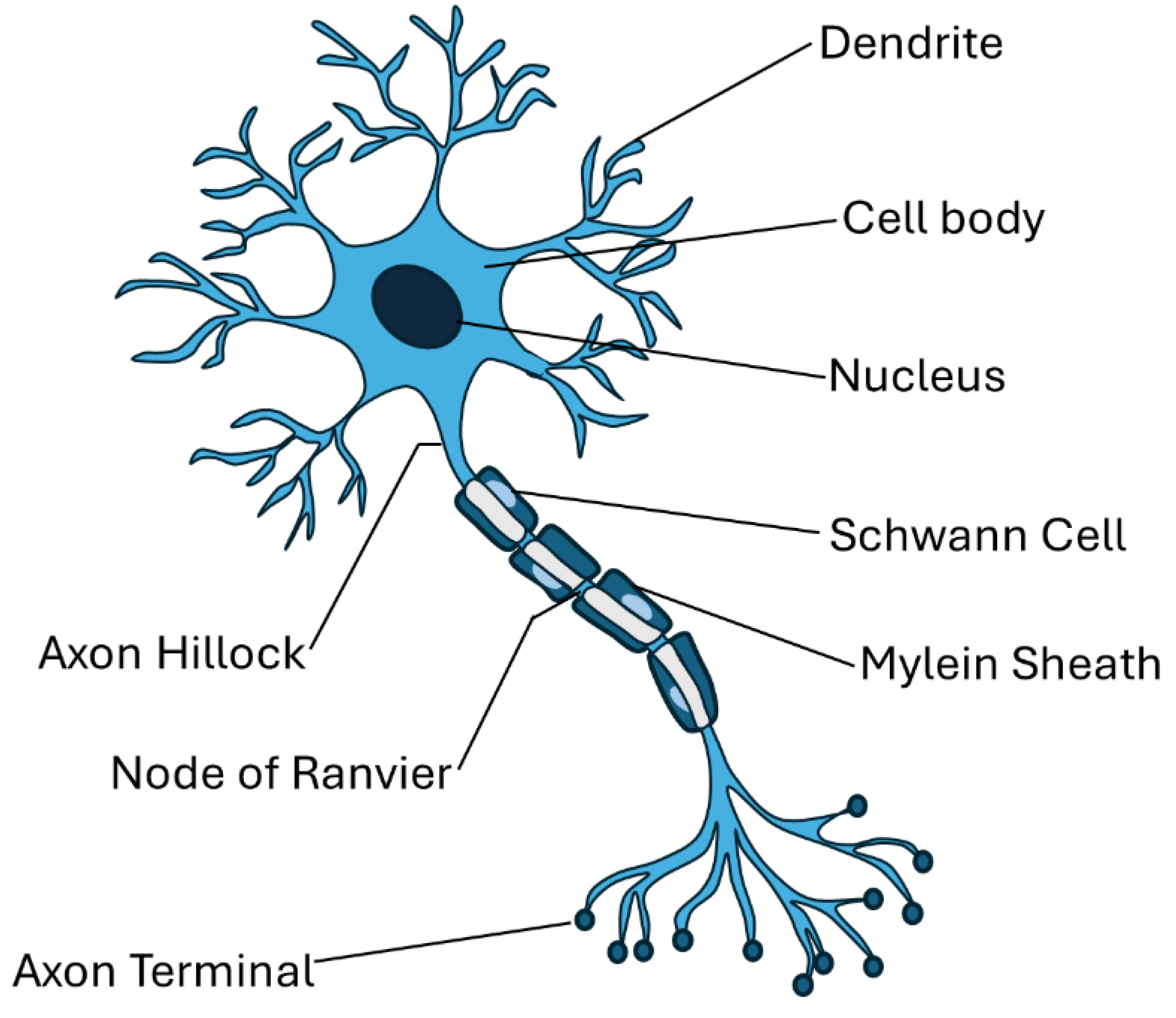
Figure 5.
Neuron structure with myelin sheath and Schwann cells.
The injury can disrupt or inhibit regular mechanisms, resulting in incomplete myelination in damaged areas, which leads to conduction disturbances of nerve impulses. Therefore, it is not surprising that to re-establish cell function, it is necessary to restore myelination. Although research typically prioritizes axon regeneration over remyelination, in some instances, such as specific hereditary disorders or toxin-induced neuropathies, the myelin is primarily damaged, while the axons remain relatively intact, at least in the initial phases of the degeneration [125]. Thus, as the level of knowledge about the specificity of cellular impairment increases, the need to analyze remyelination dynamics becomes emphasized, as it is crucial for the development of effective therapy.
Interestingly, not all types of cells exhibit the same capacity for remyelination. For example, peripheral nerves, in contrast to central nervous system cells, are not only characterized by their excellent regeneration potential but also by the extraordinary ability to remyelinate. It is worth noting that myelination in the peripheral nervous system (PNS) is primarily carried out by Schwann cells, which originate from neural crest cells. Moreover, Schwann cells differentiate into either myelinating or non-myelinating types, with each myelinating Schwann cell enveloping a single axon segment. This one-to-one relationship is key for the proper functioning and maintenance of peripheral nerves [125]. Besides transdifferentiating into a repair phenotype promoting axonal regrowth [127], Schwann cells' role after nerve injury also includes clearing degenerated myelin and axonal debris (during such processes as Wallerian degeneration) and axon remyelination [128]. However, the regenerated myelin sheaths are typically shorter and thinner than the original [129]. In Figure 5, a schematic representation of nerve degeneration and regeneration after the injury is presented.
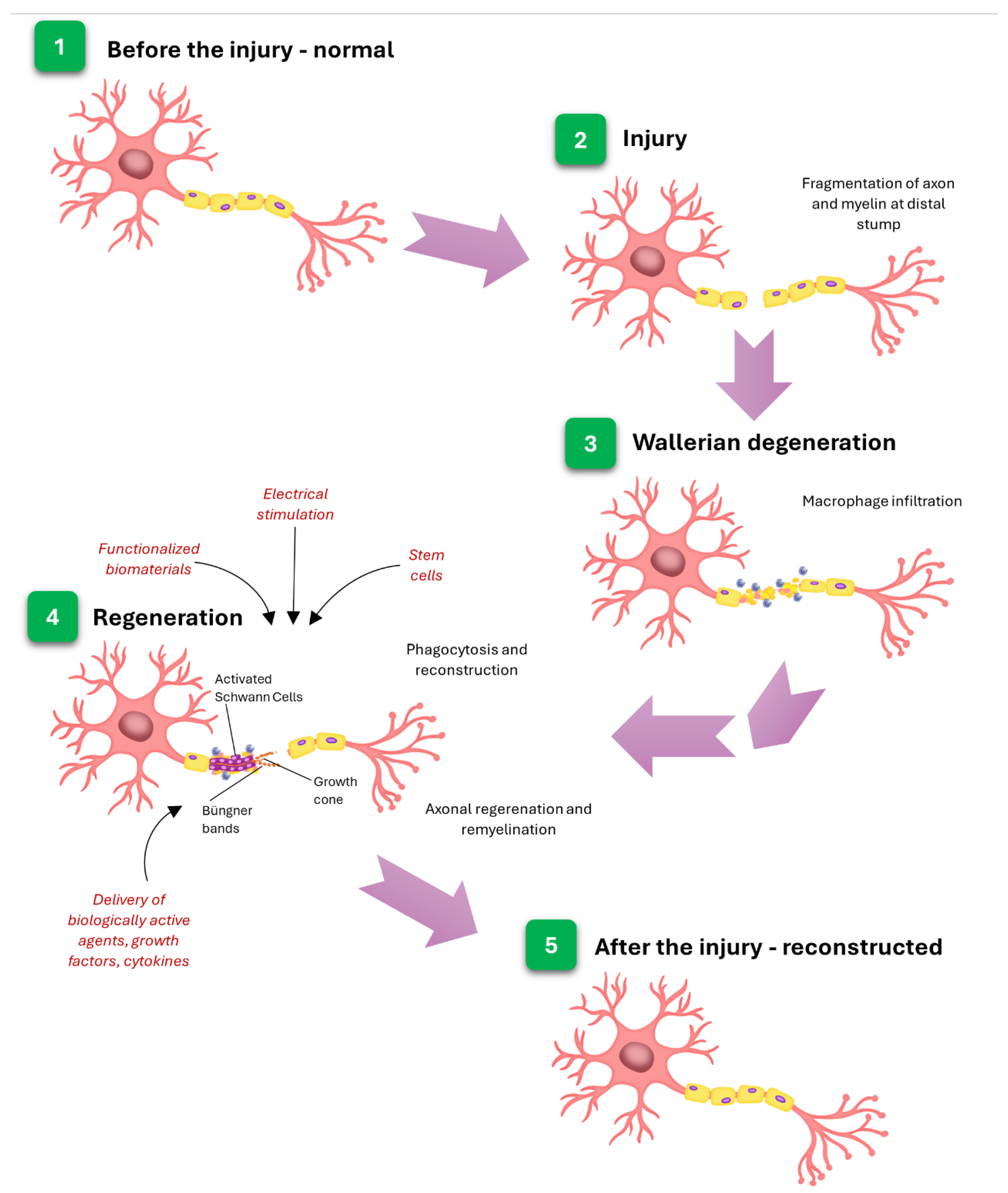
Figure 6.
Nerve regeneration after the injury.
While Schwann cells are crucial in nerve regeneration, their proper functioning requires intercellular interactions with endothelial cells and fibroblasts, which support the guidance of axons and Schwann cells across damaged sites [130].
Several approaches are being explored to augment the remyelination capacity of Schwann cells. Among these, gene- and cell-based therapies, pharmacological interventions (for instance, agents such as ascorbic acid have been demonstrated to facilitate myelination by supporting basal lamina formation [131]), and the application of biomaterial scaffolds can be listed. In the case of gene therapies, scientists target specific genetic mutations (e.g., the PMP22 gene, responsible for encoding peripheral myelin protein 22, which is critically implicated in the pathogenesis of Charcot-Marie-Tooth disease [132]) to correct aberrant protein expression. A different approach is to replace damaged Schwann cells via transplantation. Biomaterial scaffolds play a crucial role in the process, as they provide structural support for the transplanted cells, enhance their survival, and facilitate integration [133,134,135]. Nanoparticles, engineered to mimic the natural environment of the extracellular matrix, can act as scaffolds and directional cues, creating conditions that facilitate Schwann cells' ability to rebuild the insulating myelin layers essential for nerve function [136]. They might also be used as agents providing cellular alignment in a 3D environment, such as superparamagnetic oleic acid-coated iron oxide nanoparticles (SPIONs) incorporated into poly-l-lactic acid electrospun fiber scaffolds, which were employed to receive oriented guidance for dorsal root ganglion (DRG) neurites [137]. However, the range of nanomaterials in nerve injury applications goes far beyond the 'typical support' role. Zong and coworkers presented an interesting strategy: they developed a biological conduit composed of neurotrophin-3-transfected Schwann cells combined with poly(lactic-co-glycolic acid) (PLGA) copolymer and transplanted it into rats. Nanoparticle liposomes were used as a neurotrophin-3 carrier in that work. The study results show that the designed system effectively enhances sciatic nerve regeneration and decreases motor neuron apoptosis [138].
Undoubtedly, nanoparticles are most notably recognized for their drug delivery capabilities, a property that is also extensively exploited in the treatment of neurodegenerative diseases [99,110,139,140,141,142]. For instance, NPs are highly effective carriers of neuroactive factors due to their ability to encapsulate these agents. Moreover, after functionalization, nanoparticles can cross biological barriers and deliver therapeutic agents precisely to injury sites, minimizing off-target effects [142,143,144,145,146,147,148,149]. In their extensive publication, Zha and coworkers compiled nanoparticle-based platforms for drug delivery that can cross the blood-brain barrier (BBB) through various transport mechanisms, including diffusion, carrier-, receptor-, or absorptive-mediated transcytosis, efflux pump, and osmosis, which are applied in the treatment of neurodegenerative diseases and cancer therapies. Authors devote considerable space for discussing functionalized polymeric, lipid, and metal nanomaterials that cross the BBB. Such materials are exceptionally suitable for the role thanks to their unique physicochemical characteristics and high biocompatibility [147]. As a result, they have attracted considerable attention within the scientific research community. An example is the assembled BBB-crossing lipid nanoparticles, which were applied to enable mRNA delivery to neurons and astrocytes in broad brain regions [150]. On the contrary, one non-standard approach to the subject is the engagement of microorganisms. Sun's team developed the so-called 'Trojan bacteria' – a drug delivery system based on bacteria (attenuated Salmonella strain VNP20009 and Escherichia coli 25922 were tested) loaded with glucose polymer (GC) and photosensitive indocyanine green (ICG)- silicon nanoparticles (GP-ICG-SiNPs) aimed at glioblastoma (GBM) photothermal immunotherapy. The studies on the orthotopic GBM mouse model have proven that the platform can bypass the BBB and penetrate cancer tissues, making them susceptible to treatment. Additionally, scientists observed that the designed system promotes innate immune responses due to the tumor-associated antigens (TAAs) released in response to applied irradiation [151].
On the other hand, nanoparticles provide sustained and controlled release of therapeutics, reducing the need for frequent dosing and maintaining effective drug concentrations at the injury site [152,153]. Lu and coworkers proposed a ligand-mediated drug/gene co-delivery platform for glioblastoma multiforme, which allows for controlled and sustained pH-triggered release of therapeutics. Co-encapsulating therapeutic agents developed the delivery system within a thermosensitive chitosan-g-poly(N-isopropylacrylamide) (CPN) hydrogel matrix. Specifically, stomatin-like protein 2 (SLP2) short hairpin RNA (shRNA) was combined with irinotecan (CPT-11) and loaded onto cetuximab (CET)-conjugated graphene oxide (GO-CET/CPT11), resulting in the formulation of a composite hydrogel designated as CPN@GO-CET/CPT11@shRNA. The effectiveness of the construct was proven both in vitro and in vivo (with a xenograft of U87 tumor-bearing nude mice). The results not only demonstrate the system's effectiveness in extended controlled drug release but also highlight its potential for broader applications [154].
Finally, due to the functionalization, nanoparticles demonstrate suitability for localized administration, offering improved selectivity while minimizing off-target effects [155,156,157]. Bai and coworkers presented phosphatidylserine (PS)– and transferrin (TF)-modified liposomes encapsulating dexamethasone (DSS), designated as TF/PS/DSS-LPs, to enhance the treatment of ischemic stroke. Initially, TF molecules targeted the overexpressed transferrin receptor (TfR) in the blood-brain barrier. Upon entry into the brain, the PS modification enabled the liposomes to bind to phosphatidylserine-specific receptors (PSRs) on the surfaces of microglia and astrocytes. This modification also facilitated the uptake of TF/PS/DSS-LPs by these cells. Additionally, it promoted the polarization of astrocytes from the A1 to A2 phenotype and microglia from the M1 to M2 phenotype, thereby decreasing neuronal inflammation and boosting cerebral ischemic injury [158]. An illustrative example of a platform designed for targeted drug delivery to the central nervous system is the one developed by Vilella et al., comprising glycopeptide-functionalized poly(lactide-co-glycolide) nanoparticles (g7-NPs). This delivery system exhibited enhanced cellular uptake by both neurons and glial cells, accompanied by a region- and cell–type–specific distribution within the brain. Researchers also proved that the nanoparticles undergo clathrin-mediated endocytosis and localize within Rab5-positive early endosomes, with a direct correlation observed between Rab5 expression levels and nanoparticle accumulation [159].
Metal-based nanoparticles, particularly iron oxide and gold nanoparticles, represent a prominent class of nanomaterials that have been extensively explored for targeted drug delivery in the treatment of neurological disorders [160,161,162,163]. A recent study by Choi et al. introduces a transferrin-conjugated nanoparticle-based system designed to enhance delivery specificity to brain regions affected by amyloid aggregation in Alzheimer’s disease. The platform, termed Tf-MeLioNs—comprising melittin and L-arginine–coated iron oxide nanoparticles—demonstrated reduced cytotoxicity and hemolytic activity in vitro. Moreover, in vivo experiments using 5XFAD transgenic mice revealed a significant reduction in amyloid plaque burden, particularly within the hippocampal region [164]. In a complementary approach, another research group synthesized gold nanoparticles (AuNPs) engineered for the selective targeting of tight junctions and showed that transcranial picosecond laser stimulation, following intravenous administration, significantly enhances blood-brain barrier permeability. Additionally, the applied modulation is dependent on laser intensity, reversible in nature, and promotes enhanced paracellular diffusion without impairing vasomotion or the structural integrity of the neurovascular unit. The method facilitates the trans-BBB delivery of viral vectors, immunoglobulins, and liposome-encapsulated therapeutics, presenting a promising nanotechnological strategy for drug delivery to light-accessible brain regions and expanding the scope for CNS drug screening and therapeutic applications [165].
Despite the encouraging outcomes associated with targeted therapies utilizing metallic nanoparticles, their clinical application remains constrained by concerns regarding potential toxicity. To address challenges related to bioavailability, a widely adopted strategy involves encapsulating these nanoparticles within biocompatible materials, such as polyelectrolytes. This approach not only improves biocompatibility and stability but also enables the functionalization of the nanoparticle surface with targeting ligands, thereby enhancing the precision of both imaging and therapeutic interventions.
4. Polyelectrolytes Materials
Among the diverse classes of biomaterials designed for cell interaction, polyelectrolytes (PEs) have emerged as up-and-coming candidates. Polyelectrolyte-based biomaterials have been employed for several decades across diverse industries, including pharmaceuticals, chemical processing, and food technology. Furthermore, they are employed in biomedicine for tissue engineering, bioimaging, biosensing, and drug delivery [166]. Polyelectrolytes participate in delivering regulatory factors, thereby facilitating cell functioning and tissue development, as well as stimulating processes such as differentiation, proliferation, and adhesion [167,168]. Another beneficial application is the use of polyelectrolytes as delivery systems for therapeutic agents (e.g., drugs and proteins) [169], as antibacterial factors [170], or in gene delivery systems [171].
Polyelectrolyte (PE) gels and complexes have gained considerable attention due to their potential to facilitate direct cell contact. Notably, the presence of ionic groups within PEs promotes the formation of stable aqueous dispersions, wherein the ionized polymer chains function as surface-active centers.
The immobilization of cells within polyelectrolyte-based biomaterials has garnered substantial interest in the field of biomedical engineering, owing to its capacity to enhance the regeneration of damaged tissues through the localized production of biologically active molecules, including cytokines and growth factors, which are often deficient or absent due to pathological dysfunction.
Considering the origin, two classes of polyelectrolytes are distinguished – natural and synthetic ones (Figure 6). Natural polyelectrolytes include polysaccharides (e.g., sodium alginate (ALG), chitosan (CHIT), hyaluronic acid (HA), cellulose sulfate, heparin, dextran sulfate, carboxymethylcellulose, proteins, polypeptides (like poly-L-aspartic acid (ASP), poly-L-glutamic acid (PGA), poly-L-lysine (PLL), poly-L-arginine (ARG), and nucleic acids [172]. On the contrary, among the synthetic polyelectrolytes poly(styrene sulfonate) (PSS), poly(allylamine) (PAH), poly(acrylic acid) (PAA), poly(methacrylic acid) (PMA), poly(ethyleneimine) (PEI), poly(N-isopropyl acrylamide (PNIPAM), poly(dimethyldiallylammonium chloride) (PDDA) and poly(vinyl sulfate) (PVS) can be enumerated [173,174].
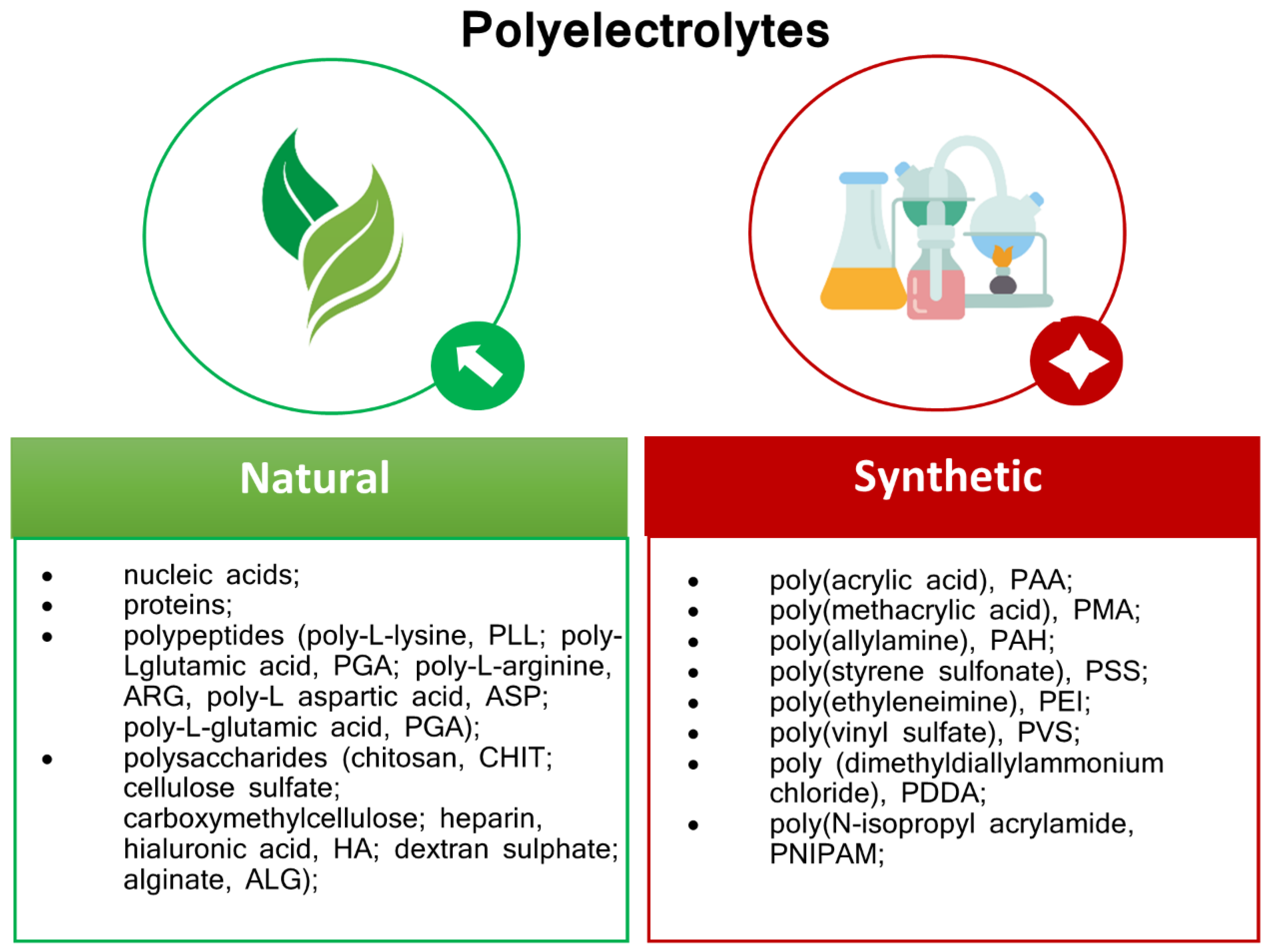
Figure 7.
Polyelectrolytes classification.
4.1. Mechanism Of Self-Assembly
Layer-by-layer (LbL) self-assembly can be governed by various intermolecular forces, including electrostatic interactions between oppositely charged species [175], van der Waals forces [176,177], hydrogen interactions [178,179,180], hydrophobic interactions [181,182,183], covalent linkages [181], or synergistic combinations thereof. Multilayer formation through combined mechanisms may be initiated via hydrophobic interactions and subsequently stabilized by hydrogen bonding, resulting in the formation of ultrathin membrane layers. Moreover, electrostatic and hydrophobic forces, as well as electrostatic-hydrogen bonding [184], are commonly associated with the self-assembly process [185]. The morphology of individual monolayers is predominantly influenced by the competitive balance between hydrogen interactions and van der Waals forces, with the resulting two-dimensional nanostructure reflecting this equilibrium [186]. Among these, electrostatic interactions are most widely utilized, enabling the sequential adsorption of oppositely charged polyions onto charged substrates to fabricate polyelectrolyte (PE) multilayer films [175,187] with adjustable thickness and surface topography [188,189,190]. Covalent bonding is typically reserved for specialized applications, such as cell immobilization, where it facilitates the attachment of polymers to cell surfaces via chemical or enzymatic conjugation [191,192,193]. Additionally, metabolic introduction may be employed. Amphiphilic polymers, such as temperature-sensitive succinylated pullulan-g-oligo(L-lactide) [194], have been demonstrated to mediate hydrophobic conjugation. Experimental evidence suggests that the interaction between ionic and hydrophobic forces is a crucial factor in both guiding self-assembly and modulating the functional characteristics of the resulting bioactive interface [195].
4.2. Polyelectrolytes for Neural Cell Regeneration in Terms of Mechanical Properties and Potential
The limitations of newly developed materials primarily arise from the requirement that biomaterial design be tailored to the specific biological parameters of the target cell types with which they are intended to interact. The example here might be the potential. The action potential of neurons during the depolarization and repolarization cycle (occurring on a time scale of approximately 2 ms) ranges from 30 mV at the transmembrane potential when the neuron is depolarized to a resting potential of −70 mV when the neuron is at rest. In the resting state, the increased number of positively charged sodium ions outside the cell compared to inside creates a concentration gradient. Voltage-gated sodium channels open, leading to the passive transport of positively charged sodium ions from the outside of the cell to the inside, thereby shifting the transmembrane potential toward a positive value. After depolarization, voltage-gated sodium channels close and voltage-gated potassium channels open, causing potassium ions to move from the inside of the cell to the outside, after which the cell repolarizes to its resting potential [196]. Therefore, it can be assumed that the most beneficial would be to work with biomaterials that provide potential values within the range of neuronal function.
The use of polyelectrolytes with various potential values for interaction with neural cells has been reported. Lastly, capsule shells built of poly(allylamine hydrochloride) and poly(4-styrene sulfonate) sodium salt (PAH/PSS) or biodegradable poly-L-arginine and dextran sulphate sodium salt (PArg/DS) with zeta-potential of 3-bilayered samples meanly −18.2 mV and −16.3 mV respectively were applied for enhance electrostatic interactions with the plasma membrane, potentially facilitating cellular uptake by neuroblastoma N2A cells [197]. Moreover, polyethylenimine-poly-L-lysine PEI/PLL bilayers, which involve mainly hydrogen interactions between layers, with a positive potential of both layers, meanly +14.9 mV, and +14.1 mV respectively; furthermore, alginate-chitosan bilayers, with a potential meanly −37.60 and +27.44 [mV] respectively were assessed for cooperation with cells isolated from embryonic (E19) Wistar rat brains. Evaluation indicates the importance of the potential of the material-cell interface itself, and not the entire membrane system [198].
Conversely, the mechanical properties of biomaterial scaffolds can alter the function of neural cells. Evidence suggested that the soft materials (0.1–1 kPa) promote the differentiation of neurons, whereas the stiffer ones (7–10 kPa) promote the glial differentiation [199,200]. Hydrogels represent a widely utilized class of biomaterials in the treatment of neural dysfunction, mainly due to their controllable mechanical properties. These properties can be precisely modulated by varying the concentration of polymer precursors during hydrogel preparation, a crucial feature for potential clinical applications [201,202].
4.3. Polyelectrolytes for Interface with Neural Cells
Several studies have been conducted to explore the usage of polyelectrolytes and their combinations in conjunction with neural cells. As mentioned above, for neuronal cell culture, the commonly applied supports are poly-L-lysine (PLL) and poly-D-lysine (PDL) [16,17,18], which are applied as monolayers. Poly-D-lysine and poly-L-lysine, enantiomeric forms of synthetic polylysine, are positively charged molecules known to enhance electrostatic interactions with negatively charged components of the cell membrane. Regarding the charge-related properties of these molecules, both are the same; however, there are reports that poly-L-lysine is subject to deterioration due to the action of proteases released by certain types of cells. Nevertheless, polymers such as HA [203], PAH, PAA, and PSS [204] have been identified as potential candidates for such applications. Dąbkowska and co-workers [205] utilized positively charged poly(diallyldimethylammonium chloride) (PDADMAC) and polyanion heparin sodium salt (HP) in their research to construct a polyelectrolyte membrane (PEM) scaffold for the adsorption of brain-derived neurotrophic factor (BDNF). The PEM scaffolds were fabricated using the layer-by-layer (LbL) technique, which relies on the successive deposition of polyelectrolytes with opposite charges. The impact of Brain-derived neurotrophic factor (BDNF) immobilized within poly(diallyldimethylammonium chloride)/ heparin PEMs was studied on the SH-SY5Y human neuroblastoma cell line. The SH-SY5Y cell line is heterogeneous, consisting of S-type (epithelial-like) and N-type (neuroblastic) undifferentiated cells. Studies have demonstrated that multilayers terminated with PDADMAC/HP may facilitate the formation of spheroid-like three-dimensional (3D) cell cultures, presenting a promising platform for future investigations as an in vitro model of neurodegenerative diseases. Moreover, polymers such as CHIT, ALG, PEI, and PLL were used to construct multilayer films to examine their interaction with rat neural cells [206]. PEMs multilayer scaffold constructed using the LbL technique, built of PLL as a polycation and poly-l-glutamine acid (PLGA) as a polyanion, was reported by Lee and Wu [16] to promote the differentiation of neural stem/progenitor cells (NSPCs) into neurons with synaptic functions. Those studies showed that NSPCs cultured within PEMs scaffolds without adding serum and growth factors could differentiate into functional neurons with significant neurite outgrowth. NSPCs are multipotent, self-renewing cells that can differentiate into the three principal neural lineages: astrocytes, oligodendrocytes, and neurons. Cerebral cortical NSPCs isolated from 14– to 15–day–old Wistar rat embryos were used to study the upregulation of the number of differentiated neurons and synaptic function with a PLL terminal layer. Some other studies reported the impact of PEMs scaffolds on neural stem/progenitor cells (NSPCs) derived from Wistar rat embryos on days 14-15 [207]. PEMs applied in the research were composed of cationic PAH and anionic PSS or PAA. It was observed that the PAH/PSS multilayer scaffolds stimulate extensive outgrowth and differentiation of NSPCs, primarily into neurons. The neurospheres were unable to attach and differentiate on the PAA-ended multilayers. That work indicates the NSPCs' dependence on the surface charge of PEM films.
However, the above-mentioned materials are not among the FDA- and/or CE-approved natural and synthetic materials used in clinical trials as components of DDS systems or as catheters for the delivery of cells intended to produce CNTF. To date, nerve conduits, wraps, and cuffs that have received FDA or CE approval are collagen or polyester – based, like RevolNerv® (Orthomed S.A.S., Lyon, France); NeuraGen® and NeuraWrap™ (Integra Life Sciences Corp., Plainsboro, NJ, USA); NeuroMatrix™, Neuroflex™ and NeuroMend™ (Collagen Matrix, Inc., Franklin Lakes, NJ, USA) or Neurolac® nerve guidance (Polyganics B.V., Groningen, The Netherlands) and Neurotube® nerve guidance (Synovis Micro Companies Alliance. Birmingham, AL, USA) respectively [208,209].
5. PE and PE-Based Nanocomposites Application for Neuronal Cell Immobilization
It is noteworthy that, among various scaffolding materials, polymeric hydrogel scaffolds are employed owing to their biocompatibility and structural resemblance to tissue [210]; however, they are distinguished by low mechanical strength and limited biological activity. Thus, investigations have been conducted on materials with enhanced physicochemical properties and/or bioactive features. In scaffolds made of polyelectrolyte membranes, various substances can be incorporated, including anticancer factors [211], DNA [212], graphene [213,214], inorganic materials [215], drugs [190,216], and proteins [217], as well as different types of nanomaterials.
There has been a marked increase in research activity concerning the incorporation of metallic nanoparticles into biomaterial systems. Metal-based NPs employed as additives allow nanocomposites to produce improved mechanical strength, involving additional biological features like antibacterial and antiviral activity [218,219]. Bacteriostatic materials for use in conjunction with neuronal cells have been continually developed. For example, incorporating polyethylenimine CuNPs has been verified to cooperate with the mice's neural stem cell line NE-4C. The effect of different concentrations of CuNPs incorporated within the membrane shells was verified. The results of the mitochondrial activity evaluation of NE-4C cells indicated that the involvement of CuNPs up to 100 ppm did not decline mitochondrial activity by more than 13% compared with the control (cells cultured without membrane addition) (Figure 8).
It is worth noting that some metal-based NPs may be considered as electrostimulating factors for theranostic applications. For example, copper nanoparticles (CuNP), which show high conductivity (Cu, σ = 5.96 × 10⁷ Sm−1) [219].
Electrical stimulation (ES) is a widely discussed strategy for treating the nervous tissue. The principle of ES involves the application of electrical impulses to activate neural cells, which are inherently responsive to electrical signals. The demonstrated efficacy of electrical stimulation in modulating neural cell activity and enhancing tissue regeneration presents promising implications for clinical applications. Thus, conductive materials like graphene, carbon nanotubes, silk fibroin-based biomaterials, and metallic nanoparticles can be considered. Besides metallic nanoparticles, various polymers are used as conductive scaffolds, including HA, ALG, poly(pyrrole) (Ppy), polyaniline (PANI), poly(3,4-ethylene dioxythiophene) (PEDOT), and PSS, which are also utilized as components of PEM construction [220,221,222,223].
It can be noted, that those conductive materials for nerves bridging in case of peripheral nerve injury with minor defects have the form of nerve guidance conduits (NGCs) that can mimic the conductivity of natural nerves or and can be applied with electrical stimulation E.g. the conductive NGCs in form of fiber formed by coprocessing the conductive polymers PEDOT/ PSS and CHIT via electrospinning technology [224].
The integration of these two approaches—facilitating the directed differentiation of nerve cells on polyelectrolyte substrates and applying electrostimulation—could efficiently promote nerve tissue reconstruction and advance the elucidation of nerve cell regeneration mechanisms. Furthermore, metallic nanoparticles not only support electrostimulation but also serve as effective antimicrobial agents or drug delivery factors.
Microscopic observation of the morphology of neural cells in a system with immobilizing material can be a simple way to provide insight into their interaction with the immobilizing substrate used.
Below, we present an exemplary microscopic analysis of the function of neural cells immobilized in membranes composed of selected polyelectrolytes with incorporated metallic nanoparticles.
5.1. Visualization of Layer Coating Scaffold-Neuronal Cells Systems
Scanning electron microscopy (SEM) is a valuable technique for examining the morphological features of layer membrane coating scaffold-cells systems, particularly polyelectrolyte (PE) membrane–immobilized cells. Figure 9 presents the example morphology of PE membranes, particularly a PEI-based coating layer incorporating copper nanoparticles (CuNPs) at different concentrations (10, 50, and 100 ppm), after 3-, 6-, and 10-day culture periods. No noticeable alterations in cell morphology may be observed during a 10-day culture period in the presence of nanocomposites containing varying CuNPs content. SEM analysis of cellular morphology corroborated the findings of the MTT assay (Figure 8), indicating no significant differences in cellular function in the presence of composites with differing CuNP share.
The application of selective dyes that distinguish between neurons and astrocytes enables visualization of the functional architecture of neural cells within the scaffolds. For example, cells can be simultaneously labeled with anti-microtubule-associated protein 2 (MAP2), a marker for the neural skeleton, and anti-glial fibrillary acidic protein (GFAP), an indicator of astrocytes. Furthermore, fluorescent labeling can be accomplished by conjugating fluorochromes such as Alexa Fluor 488 and Alexa Fluor 555, facilitating the identification and delineation of specific cellular components under fluorescence microscopy. Figure 5 illustrates the neural cells encapsulated within a nanocomposite scaffolding matrix following one week of culture (5% CO2, 37◦C). An examination of the system using immunostaining demonstrated the presence of neural cell populations. The system layer coating scaffold, designed explicitly for NE-4C cells, supports the growth of neurons within the neuronal cell population. The light green fluorescence demonstrates the presence of cells containing glial fibrillary protein, and the red fluorescence reveals the presence of neurons. The obtained results indicate that the demonstrated scaffolds enable the survival of neurons during culture, which can be considered for regenerating neural tissue.
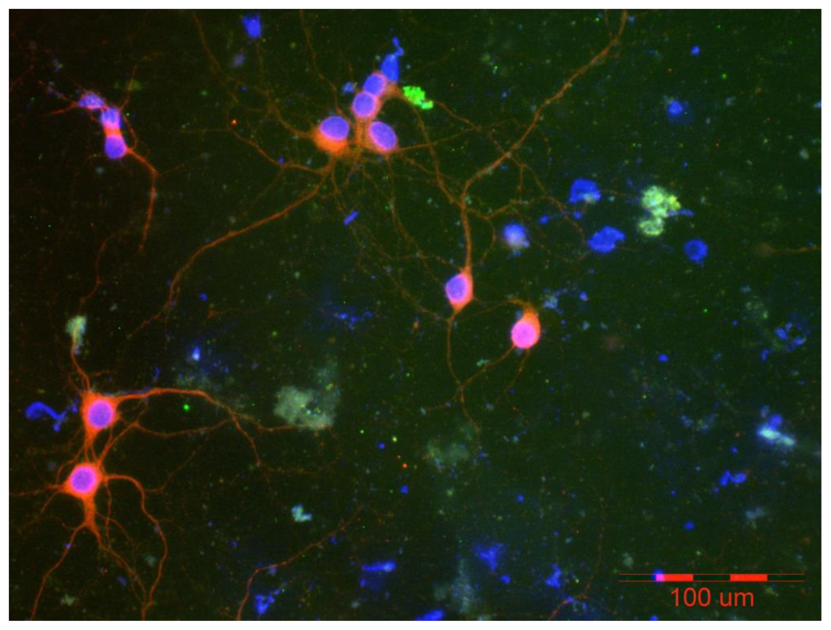
Figure 10.
Visualization of cells immobilized within PEM membrane scaffolds after a week of culture. The red fluorescence indicates neurons stained with anti-microtubule-associated protein 2 (MAP2), using a secondary antibody conjugated with the fluorochrome Alexa Fluor 555. The light green fluorescence shows astrocytes stained with anti-glial fibrillary acidic protein (GFAP) and a secondary antibody conjugated with Alexa Fluor 488 fluorochrome. Cell nuclei were visualized using Hoechst 33342, a fluorescent dye that emits blue fluorescence upon binding to DNA. Photos were taken using an Olympus IX71 fluorescence microscope.
Poly-L-lysine may be another example of a polyelectrolyte, evaluated for its potential use in conjunction with neural cells.
The efficacy assessment of scaffolds composed of poly-L-lysine incorporating copper or iron oxide nanoparticles is presented below. Figure 11 shows the neural cells immobilized within the nanocomposite scaffolding shell after two weeks of culture (5% CO2, 37◦C). An examination of the system using immunostaining demonstrated the presence of neural cell populations. The light green fluorescence indicates the presence of cells containing glial fibrillary protein, while the red fluorescence reveals the presence of neurons.
However, microscopic observations confirmed the presence of both astrocytes and neurons on all selected scaffolds; the morphology of the neurons differed from the typical characteristics of this cell type. Cells with a bundle shape predominate, with a few showing a tendency to form a spindle shape. The culture of the material containing Fe3O4NPs indicates a more significant share of neurons in the cell population than the material containing CuNPs.
It should be noted that the use of selected metallic nanoparticles affects both the morphology and function of neural cells, in addition to the anticipated antibacterial effects and the possibility of mediating electrical stimulation of neural cells.
6. Perspectives
Repairing the nervous system is a constant challenge for modern medicine. Research is focused on both cell therapies and biomaterials, as well as their associated technologies. The goal in the latter case is to obtain materials that influence the function, growth, and differentiation of neural cells. Polyelectrolyte materials offer some promise in this regard.
Polyelectrolyte layer membrane coatings offer a large number of possibilities for differing membrane properties, e.g., thickness, morphology, or wettability, allowing the control of cell culture outgrowth, differentiation, and development, which may serve as elements of the scaffolds for immobilization of cells, giving the ability to adjust cell interaction with the material at the material-host interface. Polyelectrolyte-based scaffolds incorporating bacteriostatic components can be considered an integral part of the system for amplifying nervous tissue and supporting the reconstruction of functions lost by neural tissue.
It is worth noting that layered coating presents some challenges to material stability. The PE materials may undergo deterioration by endocytosis. Also, dissociation from the surrounding medium or body fluids may occur. Thus, only the temporary usability of the systems may be considered. The spatial complexity of neural cells and the optimization of operating time are also factors that limit the usefulness of materials. However, the use of nanotechnology in the construction of nanocomposite materials based on polyelectrolytes allows for the modification and strengthening of interactions at the material-cell interface.
At the same time, continuous assessment of toxicity or unintended immune reactions is necessary to protect against adverse effects.
It can be assumed that future work on systems of polyelectrolyte membrane nanocomposites with immobilized cells will focus on developing functional materials incorporating biological activity to enhance/modulate cell function, as well as novel cell types.
Author Contributions
Conceptualization, A.G., A.K., L.H.G.; Methodology, A.G.; Validation, L.H.G.; Formal analysis, L.H.G., and A.K.; Investigation, A.G., A.L., B.K.; Writing—original draft preparation, A.K., L.H.G., A.G.; Writing—review and editing, L.H.G.; Visualization, A.K.; A.G.; Supervision, L.H.G. All authors have read and agreed to the published version of the manuscript.
Funding
This research received no external funding.
Conflicts of Interest
The authors declare no conflict of interest.
References
- The Molecular and Cellular Basis of Neurodegenerative Diseases | ScienceDirect. Available online: https://www.sciencedirect.com/book/9780128113042/the-molecular-and-cellular-basis-of-neurodegenerative-diseases?via=ihub= (accessed on 31 May 2025).
- Ciurea, A.V.; Mohan, A.G.; Covache-Busuioc, R.A.; Costin, H.P.; Glavan, L.A.; Corlatescu, A.D.; Saceleanu, V.M. Unraveling Molecular and Genetic Insights into Neurodegenerative Diseases: Advances in Understanding Alzheimer’s, Parkinson’s, and Huntington’s Diseases and Amyotrophic Lateral Sclerosis. Int. J. Mol. Sci. 2023, 24, 10809. [Google Scholar] [CrossRef]
- Ayers, J.I.; Borchelt, D.R. Phenotypic diversity in ALS and the role of poly-conformational protein misfolding. Acta Neuropathol. 2021, 142, 41–55. [Google Scholar] [CrossRef] [PubMed]
- Gadhave, D.G.; Sugandhi, V. V.; Jha, S.K.; Nangare, S.N.; Gupta, G.; Singh, S.K.; Dua, K.; Cho, H.; Hansbro, P.M.; Paudel, K.R. Neurodegenerative disorders: Mechanisms of degeneration and therapeutic approaches with their clinical relevance. Ageing Res. Rev. 2024, 99, 102357. [Google Scholar] [CrossRef] [PubMed]
- Feigin, V.L.; Vos, T.; Nichols, E.; Owolabi, M.O.; Carroll, W.M.; Dichgans, M.; Deuschl, G.; Parmar, P.; Brainin, M.; Murray, C. The global burden of neurological disorders: translating evidence into policy. Lancet Neurol. 2020, 19, 255–265. [Google Scholar] [CrossRef] [PubMed]
- Van Schependom, J.; D’haeseleer, M. Advances in Neurodegenerative Diseases. J. Clin. Med. 2023, 12, 1709. [Google Scholar] [CrossRef]
- Huang, Y.; Li, Y.; Pan, H.; Han, L. Global, regional, and national burden of neurological disorders in 204 countries and territories worldwide. J. Glob. Health 2023, 13, 04160. [Google Scholar] [CrossRef]
- Steinmetz, J.D.; Seeher, K.M.; Schiess, N.; Nichols, E.; Cao, B.; Servili, C.; Cavallera, V.; Cousin, E.; Hagins, H.; Moberg, M.E.; et al. Global, regional, and national burden of disorders affecting the nervous system, 1990–2021: a systematic analysis for the Global Burden of Disease Study 2021. Lancet Neurol. 2024, 23, 344–381. [Google Scholar] [CrossRef]
- Thakur, R.; Saini, A.K.; Taliyan, R.; Chaturvedi, N. Neurodegenerative diseases early detection and monitoring system for point-of-care applications. Microchem. J. 2025, 208, 112280. [Google Scholar] [CrossRef]
- Tay, L.X.; Ong, S.C.; Tay, L.J.; Ng, T.; Parumasivam, T. Economic Burden of Alzheimer’s Disease: A Systematic Review. Value Heal. Reg. Issues 2024, 40, 1–12. [Google Scholar] [CrossRef]
- Chen, S.; Cao, Z.; Nandi, A.; Counts, N.; Jiao, L.; Prettner, K.; Kuhn, M.; Seligman, B.; Tortorice, D.; Vigo, D.; et al. The global macroeconomic burden of Alzheimer’s disease and other dementias: estimates and projections for 152 countries or territories. Lancet Glob. Heal. 2024, 12, e1534–e1543. [Google Scholar] [CrossRef]
- Hammam, I.A.; Winters, R.; Hong, Z. Advancements in the application of biomaterials in neural tissue engineering: A review. Biomed. Eng. Adv. 2024, 8, 100132. [Google Scholar] [CrossRef]
- Kovacs, G.G. Molecular pathology of neurodegenerative diseases: Principles and practice. J. Clin. Pathol. 2019, 72, 725–735. [Google Scholar] [CrossRef] [PubMed]
- Collins, M.N.; Zamboni, F.; Serafin, A.; Escobar, A.; Stepanian, R.; Culebras, M.; Reis, R.L.; Oliveira, J.M. Emerging scaffold- and cellular-based strategies for brain tissue regeneration and imaging. Vitr. Model. 2022 12 2022, 1, 129–150. [Google Scholar] [CrossRef]
- Gordon, J.; Amini, S. General Overview of Neuronal Cell Culture. Methods Mol. Biol. 2021, 2311, 1–8. [Google Scholar] [CrossRef]
- Lee, I.C.; Wu, Y.C. Facilitating neural stem/progenitor cell niche calibration for neural lineage differentiation by polyelectrolyte multilayer films. Colloids Surfaces B Biointerfaces 2014, 121, 54–65. [Google Scholar] [CrossRef]
- Landry, M.J.; Gu, K.; Harris, S.N.; Al-Alwan, L.; Gutsin, L.; De Biasio, D.; Jiang, B.; Nakamura, D.S.; Corkery, T.C.; Kennedy, T.E.; et al. Tunable Engineered Extracellular Matrix Materials: Polyelectrolyte Multilayers Promote Improved Neural Cell Growth and Survival. Macromol. Biosci. 2019, 19. [Google Scholar] [CrossRef]
- Roach, P.; Parker, T.; Gadegaard, N.; Alexander, M.R. Surface strategies for control of neuronal cell adhesion: A review. Surf. Sci. Rep. 2010, 65, 145–173. [Google Scholar] [CrossRef]
- Shah, S.; Solanki, A.; Lee, K.B. Nanotechnology-Based Approaches for Guiding Neural Regeneration. Acc. Chem. Res. 2016, 49, 17–26. [Google Scholar] [CrossRef]
- Rahimi Darehbagh, R.; Mahmoodi, M.; Amini, N.; Babahajiani, M.; Allavaisie, A.; Moradi, Y. The effect of nanomaterials on embryonic stem cell neural differentiation: a systematic review. Eur. J. Med. Res. 2023, 28, 1–18. [Google Scholar] [CrossRef]
- Liaw, K.; Zhang, Z.; Kannan, S. Neuronanotechnology for brain regeneration. Adv. Drug Deliv. Rev. 2019, 148, 3–18. [Google Scholar] [CrossRef]
- Krsek, A.; Jagodic, A.; Baticic, L. Nanomedicine in Neuroprotection, Neuroregeneration, and Blood–Brain Barrier Modulation: A Narrative Review. Medicina (B. Aires). 2024, 60, 1384. [Google Scholar] [CrossRef]
- Wei, Z.; Hong, F.F.; Cao, Z.; Zhao, S.Y.; Chen, L. In Situ Fabrication of Nerve Growth Factor Encapsulated Chitosan Nanoparticles in Oxidized Bacterial Nanocellulose for Rat Sciatic Nerve Regeneration. Biomacromolecules 2021, 22, 4988–4999. [Google Scholar] [CrossRef]
- Zhang, Q.; Wu, P.; Chen, F.; Zhao, Y.; Li, Y.; He, X.; Huselstein, C.; Ye, Q.; Tong, Z.; Chen, Y. Brain Derived Neurotrophic Factor and Glial Cell Line-Derived Neurotrophic Factor-Transfected Bone Mesenchymal Stem Cells for the Repair of Periphery Nerve Injury. Front. Bioeng. Biotechnol. 2020, 8, 563158. [Google Scholar] [CrossRef]
- dos Santos Rodrigues, B.; Kanekiyo, T.; Singh, J. ApoE-2 Brain-Targeted Gene Therapy Through Transferrin and Penetratin Tagged Liposomal Nanoparticles. Pharm. Res. 2019, 36, 161. [Google Scholar] [CrossRef]
- Arora, S.; Layek, B.; Singh, J. Design and Validation of Liposomal ApoE2 Gene Delivery System to Evade Blood-Brain Barrier for Effective Treatment of Alzheimer’s Disease. Mol. Pharm. 2021, 18, 714–725. [Google Scholar] [CrossRef] [PubMed]
- dos Santos Rodrigues, B.; Oue, H.; Banerjee, A.; Kanekiyo, T.; Singh, J. Dual functionalized liposome-mediated gene delivery across triple co-culture blood brain barrier model and specific in vivo neuronal transfection. J Control Release 2018, 286, 264–278. [Google Scholar] [CrossRef] [PubMed]
- Lakkadwala, S.; dos Santos Rodrigues, B.; Sun, C.; Singh, J. Dual Functionalized Liposomes for Efficient Co-delivery of Anti-cancer Chemotherapeutics for the Treatment of Glioblastoma. J. Control. Release 2019, 307, 247. [Google Scholar] [CrossRef] [PubMed]
- Ying, X.; Wen, H.; Lu, W.L.; Du, J.; Guo, J.; Tian, W.; Men, Y.; Zhang, Y.; Li, R.J.; Yang, T.Y.; et al. Dual-targeting daunorubicin liposomes improve the therapeutic efficacy of brain glioma in animals. J. Control. Release 2010, 141, 183–192. [Google Scholar] [CrossRef]
- Sharma, G.; Modgil, A.; Sun, C.; Singh, J. Grafting of cell-penetrating peptide to receptor-targeted liposomes improves their transfection efficiency and transport across blood-brain barrier model. J. Pharm. Sci. 2012, 101, 2468–2478. [Google Scholar] [CrossRef]
- dos Santos Rodrigues, B.; Banerjee, A.; Kanekiyo, T.; Singh, J. Functionalized Liposomal Nanoparticles for Efficient Gene Delivery System to Neuronal Cell Transfection. Int. J. Pharm. 2019, 566, 717. [Google Scholar] [CrossRef]
- Vidal, F.; Guzman, L. Dendrimer nanocarriers drug action: perspective for neuronal pharmacology. Neural Regen. Res. 2015, 10, 1029. [Google Scholar] [CrossRef] [PubMed]
- Pérez-Carrión, M.D.; Posadas, I. Dendrimers in Neurodegenerative Diseases. Process. 2023, Vol. 11, Page 319 2023, 11, 319. [Google Scholar] [CrossRef]
- Ma, J.; Zhan, M.; Sun, H.; He, L.; Zou, Y.; Huang, T.; Karpus, A.; Majoral, J.P.; Mignani, S.; Shen, M.; et al. Phosphorus Dendrimers Co-deliver Fibronectin and Edaravone for Combined Ischemic Stroke Treatment via Cooperative Modulation of Microglia/Neurons and Vascular Regeneration. Adv. Healthc. Mater. 2024, 13. [Google Scholar] [CrossRef] [PubMed]
- Escobar, A.; Carvalho, M.R.; Maia, F.R.; Reis, R.L.; Silva, T.H.; Oliveira, J.M. Glial Cell Line-Derived Neurotrophic Factor-Loaded CMCht/PAMAM Dendrimer Nanoparticles for Peripheral Nerve Repair. Pharmaceutics 2022, 14. [Google Scholar] [CrossRef]
- Xue, J.; Wu, T.; Qiu, J.; Xia, Y. Spatiotemporally Controlling the Release of Biological Effectors Enhances Their Effects on Cell Migration and Neurite Outgrowth. Small Methods 2020, 4. [Google Scholar] [CrossRef]
- Donsante, A.; Xue, J.; Poth, K.M.; Hardcastle, N.S.; Diniz, B.; O’Connor, D.M.; Xia, Y.; Boulis, N.M. Controlling the Release of Neurotrophin-3 and Chondroitinase ABC Enhances the Efficacy of Nerve Guidance Conduits. Adv. Healthc. Mater. 2020, 9. [Google Scholar] [CrossRef]
- Du, W.; Wang, T.; Hu, S.; Luan, J.; Tian, F.; Ma, G.; Xue, J. Engineering of electrospun nanofiber scaffolds for repairing brain injury. Eng. Regen. 2023, 4, 289–303. [Google Scholar] [CrossRef]
- Baiguera, S.; Del Gaudio, C.; Lucatelli, E.; Kuevda, E.; Boieri, M.; Mazzanti, B.; Bianco, A.; Macchiarini, P. Electrospun gelatin scaffolds incorporating rat decellularized brain extracellular matrix for neural tissue engineering. Biomaterials 2014, 35, 1205–1214. [Google Scholar] [CrossRef]
- Ramesh, B.; Chandrasekaran, J.; Cherian, K.M.; Fakoya, A.O.J. Biodegradable nanofiber coated human umbilical cord as nerve scaffold for sciatic nerve regeneration in albino Wistar rats. Folia Morphol. (Warsz). 2024, 83, 72–82. [Google Scholar] [CrossRef]
- Xie, J.; MacEwan, M.R.; Schwartz, A.G.; Xia, Y. Electrospun nanofibers for neural tissue engineering. Nanoscale 2010, 2, 35–44. [Google Scholar] [CrossRef]
- Shi, S.; Ou, X.; Cheng, D. How Advancing is Peripheral Nerve Regeneration Using Nanofiber Scaffolds? A Comprehensive Review of the Literature. Int. J. Nanomedicine 2023, 18, 6763. [Google Scholar] [CrossRef]
- Sedaghati, T.; Seifalian, A.M. Nanotechnology and bio-functionalisation for peripheral nerve regeneration. Neural Regen. Res. 2015, 10, 1191. [Google Scholar] [CrossRef] [PubMed]
- Alla, K.; Yuri, C.; Anatoliy, L.; Volodymyr, L.; Yuriy, S.; Alla, K.; Yuri, C.; Anatoliy, L.; Volodymyr, L.; Yuriy, S. Interface Nerve Tissue-Silicon Nanowire for Regeneration of Injured Nerve and Creation of Bio-Electronic Device. Neurons - Dendrites Axons 2019. [Google Scholar] [CrossRef]
- Lichodievskiy, V.; Vysotskaya, N.; Ryabchikov, O.; Korsak, A.; Chaikovsky, Y.; Klimovskaya, A.; Pedchenko, Y.; Lutsyshyn, I.; Stadnyk, O. Application of Oxidized Silicon Nanowires for Nerve Fibers Regeneration. Adv. Mater. Res. 2014, 854, 157–163. [Google Scholar] [CrossRef]
- Vidu, R.; Rahman, M.; Mahmoudi, M.; Enachescu, M.; Poteca, T.D.; Opris, I. Nanostructures: a platform for brain repair and augmentation. Front. Syst. Neurosci. 2014, 8, 91. [Google Scholar] [CrossRef]
- Bechara, S.; Wadman, L.; Popat, K.C. Electroconductive polymeric nanowire templates facilitates in vitro C17.2 neural stem cell line adhesion, proliferation and differentiation. Acta Biomater. 2011, 7, 2892–2901. [Google Scholar] [CrossRef]
- Joo, J.; Kwon, E.J.; Kang, J.; Skalak, M.; Anglin, E.J.; Mann, A.P.; Ruoslahti, E.; Bhatia, S.N.; Sailor, M.J. Porous silicon–graphene oxide core–shell nanoparticles for targeted delivery of siRNA to the injured brain. Nanoscale horizons 2016, 1, 407. [Google Scholar] [CrossRef]
- Convertino, D.; Trincavelli, M.L.; Giacomelli, C.; Marchetti, L.; Coletti, C. Graphene-based nanomaterials for peripheral nerve regeneration. Front. Bioeng. Biotechnol. 2023, 11, 1306184. [Google Scholar] [CrossRef]
- Ławkowska, K.; Pokrywczyńska, M.; Koper, K.; Kluth, L.A.; Drewa, T.; Adamowicz, J. Application of Graphene in Tissue Engineering of the Nervous System. Int. J. Mol. Sci. 2022, Vol. 23, Page 33 2021, 23, 33. [Google Scholar] [CrossRef]
- Aydin, T.; Gurcan, C.; Taheri, H.; Yilmazer, A. Graphene based materials in neural tissue regeneration. Adv. Exp. Med. Biol. 2018, 1107, 129–142. [Google Scholar] [CrossRef]
- Hui, Y.; Yan, Z.; Yang, H.; Xu, X.; Yuan, W.E.; Qian, Y. Graphene Family Nanomaterials for Stem Cell Neurogenic Differentiation and Peripheral Nerve Regeneration. ACS Appl. Bio Mater. 2022, 5, 4741–4759. [Google Scholar] [CrossRef]
- Qian, Y.; Wang, X.; Song, J.; Chen, W.; Chen, S.; Jin, Y.; Ouyang, Y.; Yuan, W.E.; Fan, C. Preclinical assessment on neuronal regeneration in the injury-related microenvironment of graphene-based scaffolds. npj Regen. Med. 2021, 6, 1–8. [Google Scholar] [CrossRef]
- Aleemardani, M.; Zare, P.; Seifalian, A.; Bagher, Z.; Seifalian, A.M. Graphene-Based Materials Prove to Be a Promising Candidate for Nerve Regeneration Following Peripheral Nerve Injury. Biomedicines 2021, 10, 73. [Google Scholar] [CrossRef] [PubMed]
- Wang, J.; Lin, Y.; Li, C.; Lei, F.; Luo, H.; Jing, P.; Fu, Y.; Zhang, Z.; Wang, C.; Liu, Z.; et al. Zein-Based Triple-Drug Nanoparticles to Promote Anti-Inflammatory Responses for Nerve Regeneration after Spinal Cord Injury. Adv. Healthc. Mater. 2024, 13, 2304261. [Google Scholar] [CrossRef] [PubMed]
- Lang, Y.; Liu, H.; Zhang, C. Study on antibacterial activity and repair of peripheral nervous system injury by Zno nanocomposites. Mater. Lett. 2023, 340, 134180. [Google Scholar] [CrossRef]
- Qu, Y.; Ma, Y.; Zhang, M.; Haoran, J.; Xing, B.; Wang, B.; Heng, A.; Wen, Y.; Peixun, Z. Design of AgNPs Loaded γ-PGA Chitosan Conduits with Superior Antibacterial Activity and Nerve Repair Properties. Front. Bioeng. Biotechnol. 13 1561330. [CrossRef]
- Demirdöğen, B.C. Theranostic potential of graphene quantum dots for multiple sclerosis. Mult. Scler. Relat. Disord. 2022, 68. [Google Scholar] [CrossRef]
- Chakraborty, P.; Das, S.S.; Dey, A.; Chakraborty, A.; Bhattacharyya, C.; Kandimalla, R.; Mukherjee, B.; Gopalakrishnan, A.V.; Singh, S.K.; Kant, S.; et al. Quantum dots: The cutting-edge nanotheranostics in brain cancer management. J. Control. Release 2022, 350, 698–715. [Google Scholar] [CrossRef]
- Xu, G.; Mahajan, S.; Roy, I.; Yong, K.T. Theranostic quantum dots for crossing blood-brain barrier in vitro and providing therapy of HIV-associated encephalopathy. Front. Pharmacol. 2013, 4 NOV, 66969. [Google Scholar] [CrossRef]
- Hu, Y.; Wang, X.; Niu, Y.; He, K.; Tang, M. Application of quantum dots in brain diseases and their neurotoxic mechanism. Nanoscale Adv. 2024, 6, 3733–3746. [Google Scholar] [CrossRef]
- Villalva, M.D.; Agarwal, V.; Ulanova, M.; Sachdev, P.S.; Braidy, N. Quantum Dots as A Theranostic Approach in Alzheimer’s Disease: A Systematic Review. Nanomedicine 2021, 16, 1595–1611. [Google Scholar] [CrossRef]
- Xue, Y.; Wu, J.; Sun, J. Four types of inorganic nanoparticles stimulate the inflammatory reaction in brain microglia and damage neurons in vitro. Toxicol. Lett. 2012, 214, 91–98. [Google Scholar] [CrossRef]
- Zia, S.; Islam Aqib, A.; Muneer, A.; Fatima, M.; Atta, K.; Kausar, T.; Zaheer, C.N.F.; Ahmad, I.; Saeed, M.; Shafique, A. Insights into nanoparticles-induced neurotoxicity and cope up strategies. Front. Neurosci. 2023, 17, 1127460. [Google Scholar] [CrossRef]
- Czajka, M.; Sawicki, K.; Sikorska, K.; Popek, S.; Kruszewski, M.; Kapka-Skrzypczak, L. Toxicity of titanium dioxide nanoparticles in central nervous system. Toxicol. Vitr. 2015, 29, 1042–1052. [Google Scholar] [CrossRef] [PubMed]
- Tang, J.; Xiong, L.; Zhou, G.; Wang, S.; Wang, J.; Liu, L.; Li, J.; Yuan, F.; Lu, S.; Wan, Z.; et al. Silver nanoparticles crossing through and distribution in the blood-brain barrier in vitro. J. Nanosci. Nanotechnol. 2010, 10, 6313–6317. [Google Scholar] [CrossRef]
- Tang, J.; Xiong, L.; Wang, S.; Wang, J.; Liu, L.; Li, J.; Yuan, F.; Xi, T. Distribution, translocation and accumulation of silver nanoparticles in rats. J. Nanosci. Nanotechnol. 2009, 9, 4924–4932. [Google Scholar] [CrossRef] [PubMed]
- Hu, Y.L.; Gao, J.Q. Potential neurotoxicity of nanoparticles. Int. J. Pharm. 2010, 394, 115–121. [Google Scholar] [CrossRef] [PubMed]
- Loeschner, K.; Hadrup, N.; Qvortrup, K.; Larsen, A.; Gao, X.; Vogel, U.; Mortensen, A.; Lam, H.R.; Larsen, E.H. Distribution of silver in rats following 28 days of repeated oral exposure to silver nanoparticles or silver acetate. Part. Fibre Toxicol. 2011, 8. [Google Scholar] [CrossRef]
- Zhang, Y.N.; Poon, W.; Tavares, A.J.; McGilvray, I.D.; Chan, W.C.W. Nanoparticle–liver interactions: Cellular uptake and hepatobiliary elimination. J. Control. Release 2016, 240, 332–348. [Google Scholar] [CrossRef]
- Soltysova, A.; Ludwig, N.; Diener, C.; Sramkova, M.; Kozics, K.; Jakic, K.; Balintova, L.; Bastus, N.G.; Moriones, O.H.; Liskova, A.; et al. Gold and titania nanoparticles accumulated in the body induce late toxic effects and alterations in transcriptional and miRNA landscape. Environ. Sci. Nano 2024, 11, 1296–1313. [Google Scholar] [CrossRef]
- Rahman, M.; Mahady Dip, T.; Padhye, R.; Houshyar, S. Review on electrically conductive smart nerve guide conduit for peripheral nerve regeneration. J. Biomed. Mater. Res. Part A 2023, 111, 1916–1950. [Google Scholar] [CrossRef]
- Qian, Y.; Lin, H.; Yan, Z.; Shi, J.; Fan, C. Functional nanomaterials in peripheral nerve regeneration: Scaffold design, chemical principles and microenvironmental remodeling. Mater. Today 2021, 51, 165–187. [Google Scholar] [CrossRef]
- Wang, J.; Fang, J.; Weng, Z.; Nan, L.; Chen, Y.; Shan, J.; Chen, F.; Liu, J. Advanced development of conductive biomaterials for enhanced peripheral nerve regeneration: a review. RSC Adv. 2025, 15, 12997–13009. [Google Scholar] [CrossRef] [PubMed]
- Xia, H.; Gao, X.; Gu, G.; Liu, Z.; Hu, Q.; Tu, Y.; Song, Q.; Yao, L.; Pang, Z.; Jiang, X.; et al. Penetratin-functionalized PEG–PLA nanoparticles for brain drug delivery. Int. J. Pharm. 2012, 436, 840–850. [Google Scholar] [CrossRef] [PubMed]
- Pan, Z.; Ye, H.; Wu, D. Recent advances on polymeric hydrogels as wound dressings. APL Bioeng. 2021, 5, 11504. [Google Scholar] [CrossRef]
- Zhu, J.; Fu, Q.; Song, L.; Liu, L.; Zheng, Z.; Xu, Y.; Zhang, Z. Advances in Peripheral Nerve Injury Repair with the Application of Nanomaterials. J. Nanomater. 2022, 2022, 7619884. [Google Scholar] [CrossRef]
- Sharifi, M.; Farahani, M.K.; Salehi, M.; Atashi, A.; Alizadeh, M.; Kheradmandi, R.; Molzemi, S. Exploring the Physicochemical, Electroactive, and Biodelivery Properties of Metal Nanoparticles on Peripheral Nerve Regeneration. ACS Biomater. Sci. Eng. 2023, 9, 106–138. [Google Scholar] [CrossRef]
- Pan, T.; Huang, Y.; Wei, J.; Lai, C.; Chen, Y.; Nan, K.; Wu, W. Implantation of biomimetic polydopamine nanocomposite scaffold promotes optic nerve regeneration through modulating inhibitory microenvironment. J. Nanobiotechnology 2024, 22, 683. [Google Scholar] [CrossRef]
- Kim, H.J.; Lee, J.S.; Park, J.M.; Lee, S.; Hong, S.J.; Park, J.S.; Park, K.H. Fabrication of Nanocomposites Complexed with Gold Nanoparticles on Polyaniline and Application to Their Nerve Regeneration. ACS Appl. Mater. Interfaces 2020, 12, 30750–30760. [Google Scholar] [CrossRef]
- Lacko, C.S.; Singh, I.; Wall, M.A.; Garcia, A.R.; Porvasnik, S.L.; Rinaldi, C.; Schmidt, C.E. Magnetic particle templating of hydrogels: Engineering naturally derived hydrogel scaffolds with 3D aligned microarchitecture for nerve repair. J. Neural Eng. 2020, 17. [Google Scholar] [CrossRef]
- Garcés, M.; Cáceres, L.; Chiappetta, D.; Magnani, N.; Evelson, P. Current understanding of nanoparticle toxicity mechanisms and interactions with biological systems. New J. Chem. 2021, 45, 14328–14344. [Google Scholar] [CrossRef]
- Egbuna, C.; Parmar, V.K.; Jeevanandam, J.; Ezzat, S.M.; Patrick-Iwuanyanwu, K.C.; Adetunji, C.O.; Khan, J.; Onyeike, E.N.; Uche, C.Z.; Akram, M.; et al. Toxicity of Nanoparticles in Biomedical Application: Nanotoxicology. J. Toxicol. 2021, 2021, 9954443. [Google Scholar] [CrossRef] [PubMed]
- Fard, J.K.; Jafari, S.; Eghbal, M.A. A Review of Molecular Mechanisms Involved in Toxicity of Nanoparticles. Adv. Pharm. Bull. 2015, 5, 447. [Google Scholar] [CrossRef] [PubMed]
- Cook, A.B.; Clemons, T.D. Bottom-Up versus Top-Down Strategies for Morphology Control in Polymer-Based Biomedical Materials. Adv. NanoBiomed Res. 2022, 2, 2100087. [Google Scholar] [CrossRef]
- Abid, N.; Khan, A.M.; Shujait, S.; Chaudhary, K.; Ikram, M.; Imran, M.; Haider, J.; Khan, M.; Khan, Q.; Maqbool, M. Synthesis of nanomaterials using various top-down and bottom-up approaches, influencing factors, advantages, and disadvantages: A review. Adv. Colloid Interface Sci. 2022, 300, 102597. [Google Scholar] [CrossRef]
- Shimomura, M.; Sawadaishi, T. Bottom-up strategy of materials fabrication: a new trend in nanotechnology of soft materials. Curr. Opin. Colloid Interface Sci. 2001, 6, 11–16. [Google Scholar] [CrossRef]
- Municoy, S.; Álvarez Echazú, M.I.; Antezana, P.E.; Galdopórpora, J.M.; Olivetti, C.; Mebert, A.M.; Foglia, M.L.; Tuttolomondo, M. V.; Alvarez, G.S.; Hardy, J.G.; et al. Stimuli-Responsive Materials for Tissue Engineering and Drug Delivery. Int. J. Mol. Sci. 2020, Vol. 21, Page 4724 2020, 21, 4724. [Google Scholar] [CrossRef]
- Redolfi Riva, E.; Özkan, M.; Stellacci, F.; Micera, S. Combining external physical stimuli and nanostructured materials for upregulating pro-regenerative cellular pathways in peripheral nerve repair. Front. Cell Dev. Biol. 2024, 12, 1491260. [Google Scholar] [CrossRef]
- Yang, J.; des Rieux, A.; Malfanti, A. Stimuli-Responsive Nanomedicines for the Treatment of Non-cancer Related Inflammatory Diseases. ACS Nano 2025. [Google Scholar] [CrossRef]
- Laurent, S.; Forge, D.; Port, M.; Roch, A.; Robic, C.; Vander Elst, L.; Muller, R.N. Magnetic iron oxide nanoparticles: Synthesis, stabilization, vectorization, physicochemical characterizations and biological applications. Chem. Rev. 2008, 108, 2064–2110. [Google Scholar] [CrossRef]
- Mehta, M.; Bui, T.A.; Yang, X.; Aksoy, Y.; Goldys, E.M.; Deng, W. Lipid-Based Nanoparticles for Drug/Gene Delivery: An Overview of the Production Techniques and Difficulties Encountered in Their Industrial Development. ACS Mater. Au 2023, 3, 600–619. [Google Scholar] [CrossRef]
- Hammami, I.; Alabdallah, N.M.; jomaa, A. Al; kamoun, M. Gold nanoparticles: Synthesis properties and applications. J. King Saud Univ. - Sci. 2021, 33, 101560. [Google Scholar] [CrossRef]
- Asem, H.; Zheng, W.; Nilsson, F.; Zhang, Y.; Hedenqvist, M.S.; Hassan, M.; Malmström, E. Functional Nanocarriers for Drug Delivery by Surface Engineering of Polymeric Nanoparticle Post-Polymerization-Induced Self-Assembly. ACS Appl. Bio Mater. 2021, 4, 1045–1056. [Google Scholar] [CrossRef]
- Hochreiner, E.G.; van Ravensteijn, B.G.P. Polymerization-induced self-assembly for drug delivery: A critical appraisal. J. Polym. Sci. 2023, 61, 3186–3210. [Google Scholar] [CrossRef]
- Varma, L.T.; Singh, N.; Gorain, B.; Choudhury, H.; Tambuwala, M.M.; Kesharwani, P.; Shukla, R. Recent Advances in Self-Assembled Nanoparticles for Drug Delivery. Curr. Drug Deliv. 2020, 17, 279–291. [Google Scholar] [CrossRef]
- Floyd, T.G.; Gurnani, P.; Rho, J.Y. Characterisation of polymeric nanoparticles for drug delivery. Nanoscale 2025, 17, 7738–7752. [Google Scholar] [CrossRef]
- Baig, N.; Kammakakam, I.; Falath, W.; Kammakakam, I. Nanomaterials: a review of synthesis methods, properties, recent progress, and challenges. Mater. Adv. 2021, 2, 1821–1871. [Google Scholar] [CrossRef]
- Shi, S.; Ou, X.; Cheng, D. Nanoparticle-Facilitated Therapy: Advancing Tools in Peripheral Nerve Regeneration. Int. J. Nanomedicine 2024, 19, 19. [Google Scholar] [CrossRef]
- Chen, T.; Wan, L.; Xiao, Y.; Wang, K.; Wu, P.; Li, C.; Huang, C.; Liu, X.; Xue, W.; Sun, G.; et al. Curcumin/pEGCG-encapsulated nanoparticles enhance spinal cord injury recovery by regulating CD74 to alleviate oxidative stress and inflammation. J. Nanobiotechnology 2024, 22, 1–21. [Google Scholar] [CrossRef]
- Liu, X.; Mao, Y.; Huang, S.; Li, W.; Zhang, W.; An, J.; Jin, Y.; Guan, J.; Wu, L.; Zhou, P. Selenium nanoparticles derived from Proteus mirabilis YC801 alleviate oxidative stress and inflammatory response to promote nerve repair in rats with spinal cord injury. Regen. Biomater. 2022, 9. [Google Scholar] [CrossRef]
- Liu, H.; Qing, X.; Peng, L.; Zhang, D.; Dai, W.; Yang, Z.; Zhang, J.; Liu, X. Mannose-coated nanozyme for relief from chemotherapy-induced peripheral neuropathic pain. iScience 2023, 26. [Google Scholar] [CrossRef]
- Li, X.; Han, Z.; Wang, T.; Ma, C.; Li, H.; Lei, H.; Yang, Y.; Wang, Y.; Pei, Z.; Liu, Z.; et al. Cerium oxide nanoparticles with antioxidative neurorestoration for ischemic stroke. Biomaterials 2022, 291, 121904. [Google Scholar] [CrossRef]
- Chen, X.; Wang, B.; Mao, Y.; Al Mamun, A.; Wu, M.; Qu, S.; Zhang, X.; Zhang, J.; Pan, J.; Zhu, Y.; et al. Zein nanoparticles loaded with chloroquine improve functional recovery and attenuate neuroinflammation after spinal cord injury. Chem. Eng. J. 2022, 450, 137882. [Google Scholar] [CrossRef]
- Yuan, Y.; Xu, M.; Feng, L.; Zhong, W.; Zhang, L.; Du, R.; Sun, J.; Wang, C.; Du, J. Nanozyme Hydrogels Promote Nerve Regeneration in Spinal Cord Injury by Reducing Oxidative Stress. ACS Appl. Mater. Interfaces 2024. [Google Scholar] [CrossRef] [PubMed]
- Zhang, K.; Tu, M.; Gao, W.; Cai, X.; Song, F.; Chen, Z.; Zhang, Q.; Wang, J.; Jin, C.; Shi, J.; et al. Hollow Prussian Blue Nanozymes Drive Neuroprotection against Ischemic Stroke via Attenuating Oxidative Stress, Counteracting Inflammation, and Suppressing Cell Apoptosis. Nano Lett. 2019, 19, 2812–2823. [Google Scholar] [CrossRef] [PubMed]
- Hu, Y.; Chen, Z.; Wang, H.; Guo, J.; Cai, J.; Chen, X.; Wei, H.; Qi, J.; Wang, Q.; Liu, H.; et al. Conductive Nerve Guidance Conduits Based on Morpho Butterfly Wings for Peripheral Nerve Repair. ACS Nano 2022, 16, 1868–1879. [Google Scholar] [CrossRef] [PubMed]
- Zhou, X.; Tang, A.; Xiong, C.; Zhang, G.; Huang, L.; Xu, F. Oriented Graphene Oxide Scaffold Promotes Nerve Regeneration in vitro and in vivo. Int. J. Nanomedicine 2024, 19, 2573–2589. [Google Scholar] [CrossRef]
- Hanwright, P.J.; Qiu, C.; Rath, J.; Zhou, Y.; von Guionneau, N.; Sarhane, K.A.; Harris, T.G.W.; Howard, G.P.; Malapati, H.; Lan, M.J.; et al. Sustained IGF-1 delivery ameliorates effects of chronic denervation and improves functional recovery after peripheral nerve injury and repair. Biomaterials 2022, 280. [Google Scholar] [CrossRef]
- Lopes, C.D.F.; Gonçalves, N.P.; Gomes, C.P.; Saraiva, M.J.; Pêgo, A.P. BDNF gene delivery mediated by neuron-targeted nanoparticles is neuroprotective in peripheral nerve injury. Biomaterials 2017, 121, 83–96. [Google Scholar] [CrossRef]
- Pal, A.; Kumar, S.; Jain, S.; Nag, T.C.; Mathur, R. Neuroregenerative Effects of Electromagnetic Field and Magnetic Nanoparticles on Spinal Cord Injury in Rats. J. Nanosci. Nanotechnol. 2018, 18, 6756–6764. [Google Scholar] [CrossRef]
- Papastefanaki, F.; Jakovcevski, I.; Poulia, N.; Djogo, N.; Schulz, F.; Martinovic, T.; Ciric, D.; Loers, G.; Vossmeyer, T.; Weller, H.; et al. Intraspinal Delivery of Polyethylene Glycol-coated Gold Nanoparticles Promotes Functional Recovery After Spinal Cord Injury. 2015. [Google Scholar] [CrossRef]
- Rittchen, S.; Boyd, A.; Burns, A.; Park, J.; Fahmy, T.M.; Metcalfe, S.; Williams, A. Myelin repair invivo is increased by targeting oligodendrocyte precursor cells with nanoparticles encapsulating leukaemia inhibitory factor (LIF). Biomaterials 2015, 56, 78–85. [Google Scholar] [CrossRef]
- Robinson, A.P.; Zhang, J.Z.; Titus, H.E.; Karl, M.; Merzliakov, M.; Dorfman, A.R.; Karlik, S.; Stewart, M.G.; Watt, R.K.; Facer, B.D.; et al. Nanocatalytic activity of clean-surfaced, faceted nanocrystalline gold enhances remyelination in animal models of multiple sclerosis. Sci. Rep. 2020, 10, 1–16. [Google Scholar] [CrossRef]
- Luo, W.; Wang, Y.; Lin, F.; Liu, Y.; Gu, R.; Liu, W.; Xiao, C. Selenium-doped carbon quantum dots efficiently ameliorate secondary spinal cord injury via scavenging reactive oxygen species. Int. J. Nanomedicine 2020, 15, 10113–10125. [Google Scholar] [CrossRef]
- Antman-Passig, M.; Giron, J.; Karni, M.; Motiei, M.; Schori, H.; Shefi, O. Magnetic Assembly of a Multifunctional Guidance Conduit for Peripheral Nerve Repair. Adv. Funct. Mater. 2021, 31, 2010837. [Google Scholar] [CrossRef]
- Manoukian, O.S.; Rudraiah, S.; Arul, M.R.; Bartley, J.M.; Baker, J.T.; Yu, X.; Kumbar, S.G. Biopolymer-nanotube nerve guidance conduit drug delivery for peripheral nerve regeneration: In vivo structural and functional assessment. Bioact. Mater. 2021, 6, 2881–2893. [Google Scholar] [CrossRef] [PubMed]
- Sandoval-Castellanos, A.M.; Claeyssens, F.; Haycock, J.W. Bioactive 3D Scaffolds for the Delivery of NGF and BDNF to Improve Nerve Regeneration. Front. Mater. 2021, 8, 734683. [Google Scholar] [CrossRef]
- Dinis, T.M.; Vidal, G.; Jose, R.R.; Vigneron, P.; Bresson, D.; Fitzpatrick, V.; Marin, F.; Kaplan, D.L.; Egles, C. Complementary Effects of Two Growth Factors in Multifunctionalized Silk Nanofibers for Nerve Reconstruction. PLoS One 2014, 9, e109770. [Google Scholar] [CrossRef]
- Xia, B.; Lv, Y. Dual-delivery of VEGF and NGF by emulsion electrospun nanofibrous scaffold for peripheral nerve regeneration. Mater. Sci. Eng. C 2018, 82, 253–264. [Google Scholar] [CrossRef]
- Chen, X.; Ge, X.; Qian, Y.; Tang, H.; Song, J.; Qu, X.; Yue, B.; Yuan, W.E. Electrospinning Multilayered Scaffolds Loaded with Melatonin and Fe3O4 Magnetic Nanoparticles for Peripheral Nerve Regeneration. Adv. Funct. Mater. 2020, 30. [Google Scholar] [CrossRef]
- Wu, C.; Chen, S.; Zhou, T.; Wu, K.; Qiao, Z.; Zhang, Y.; Xin, N.; Liu, X.; Wei, D.; Sun, J.; et al. Antioxidative and Conductive Nanoparticles-Embedded Cell Niche for Neural Differentiation and Spinal Cord Injury Repair. ACS Appl. Mater. Interfaces 2021, 13, 52346–52361. [Google Scholar] [CrossRef]
- Yang, Q.; Lu, D.; Wu, J.; Liang, F.; Wang, H.; Yang, J.; Zhang, G.; Wang, C.; Yang, Y.; Zhu, L.; et al. Nanoparticles for the treatment of spinal cord injury. Neural Regen. Res. 2024, 20, 1665. [Google Scholar] [CrossRef] [PubMed]
- Voiţă-Mekereş, F.; Mekeres, G.M.; Voiță, I.B.; Galea-Holhoș, L.B.; Manole, F. A Review of the Protective Effects of Nanoparticles in the Treatment of Nervous System Injuries. Int. J. Pharm. Res. Allied Sci. 2023, 12, 149–155. [Google Scholar] [CrossRef]
- Zhou, Y.; Notterpek, L. Promoting peripheral myelin repair. Exp. Neurol. 2016, 283, 573. [Google Scholar] [CrossRef] [PubMed]
- Vigil, P.; Orellana, R.F.; Cortés, M.E.; Molina, C.T.; Switzer, B.E.; Klaus, H. Endocrine Modulation of the Adolescent Brain: A Review. J. Pediatr. Adolesc. Gynecol. 2011, 24, 330–337. [Google Scholar] [CrossRef]
- Arthur-Farraj, P.J.; Latouche, M.; Wilton, D.K.; Quintes, S.; Chabrol, E.; Banerjee, A.; Woodhoo, A.; Jenkins, B.; Rahman, M.; Turmaine, M.; et al. c-Jun Reprograms Schwann Cells of Injured Nerves to Generate a Repair Cell Essential for Regeneration. Neuron 2012, 75, 633–647. [Google Scholar] [CrossRef]
- Scheib, J.; Höke, A. Advances in peripheral nerve regeneration. Nat. Rev. Neurol. 2013, 9, 668–676. [Google Scholar] [CrossRef]
- Griffin, J.W.; Thompson, W.J. Biology and pathology of nonmyelinating schwann cells. Glia 2008, 56, 1518–1531. [Google Scholar] [CrossRef]
- Cattin, A.L.; Burden, J.J.; Van Emmenis, L.; MacKenzie, F.E.; Hoving, J.J.A.; Garcia Calavia, N.; Guo, Y.; McLaughlin, M.; Rosenberg, L.H.; Quereda, V.; et al. Macrophage-Induced Blood Vessels Guide Schwann Cell-Mediated Regeneration of Peripheral Nerves. Cell 2015, 162, 1127–1139. [Google Scholar] [CrossRef]
- Eldridge, C.F.; Bunge, M.B.; Bunge, R.P.; Wood, P.M. Differentiation of axon-related Schwann cells in vitro. I. Ascorbic acid regulates basal lamina assembly and myelin formation. J. Cell Biol. 1987, 105, 1023–1034. [Google Scholar] [CrossRef]
- Yoshioka, Y.; Taniguchi, J.B.; Homma, H.; Tamura, T.; Fujita, K.; Inotsume, M.; Tagawa, K.; Misawa, K.; Matsumoto, N.; Nakagawa, M.; et al. AAV-mediated editing of PMP22 rescues Charcot-Marie-Tooth disease type 1A features in patient-derived iPS Schwann cells. Commun. Med. 2023 31 2023, 3, 1–15. [Google Scholar] [CrossRef]
- Bozkurt, A.; Boecker, A.; Tank, J.; Altinova, H.; Deumens, R.; Dabhi, C.; Tolba, R.; Weis, J.; Brook, G.A.; Pallua, N.; et al. Efficient bridging of 20 mm rat sciatic nerve lesions with a longitudinally micro-structured collagen scaffold. Biomaterials 2016, 75, 112–122. [Google Scholar] [CrossRef]
- Marquardt, L.M.; Sakiyama-Elbert, S.E. Engineering peripheral nerve repair. Curr. Opin. Biotechnol. 2013, 24, 887–892. [Google Scholar] [CrossRef]
- Hajimirzaei, P.; Tabatabaei, F.S.A.; Nasibi-Sis, H.; Razavian, R.S.; Nasirinezhad, F. Schwann cell transplantation for remyelination, regeneration, tissue sparing, and functional recovery in spinal cord injury: A systematic review and meta-analysis of animal studies. Exp. Neurol. 2025, 384, 115062. [Google Scholar] [CrossRef] [PubMed]
- Rao, Z.; Lin, Z.; Song, P.; Quan, D.; Bai, Y. Biomaterial-Based Schwann Cell Transplantation and Schwann Cell-Derived Biomaterials for Nerve Regeneration. Front. Cell. Neurosci. 2022, 16, 926222. [Google Scholar] [CrossRef] [PubMed]
- Johnson, C.D.L.; Ganguly, D.; Zuidema, J.M.; Cardinal, T.J.; Ziemba, A.M.; Kearns, K.R.; McCarthy, S.M.; Thompson, D.M.; Ramanath, G.; Borca-Tasciuc, D.A.; et al. Injectable, Magnetically Orienting Electrospun Fiber Conduits for Neuron Guidance. ACS Appl. Mater. Interfaces 2018, 11, 356. [Google Scholar] [CrossRef] [PubMed]
- Zong, H.; Zhao, H.; Zhao, Y.; Jia, J.; Yang, L.; Ma, C.; Zhang, Y.; Dong, Y. Nanoparticles carrying neurotrophin-3-modified Schwann cells promote repair of sciatic nerve defects. Neural Regen. Res. 2013, 8, 1262. [Google Scholar] [CrossRef]
- Toader, C.; Dumitru, A.V.; Eva, L.; Serban, M.; Covache-Busuioc, R.A.; Ciurea, A.V. Nanoparticle Strategies for Treating CNS Disorders: A Comprehensive Review of Drug Delivery and Theranostic Applications. Int. J. Mol. Sci. 2024, Vol. 25, Page 13302 2024, 25, 13302. [Google Scholar] [CrossRef]
- Nguyen, T.T.; Dung Nguyen, T.T.; Vo, T.K.; Tran, N.M.A.; Nguyen, M.K.; Van Vo, T.; Van Vo, G. Nanotechnology-based drug delivery for central nervous system disorders. Biomed. Pharmacother. 2021, 143, 112117. [Google Scholar] [CrossRef]
- Kreuter, J. Drug delivery to the central nervous system by polymeric nanoparticles: What do we know? Adv. Drug Deliv. Rev. 2014, 71, 2–14. [Google Scholar] [CrossRef]
- Wu, D.; Chen, Q.; Chen, X.; Han, F.; Chen, Z.; Wang, Y. The blood–brain barrier: structure, regulation, and drug delivery. Signal Transduct. Target. Ther. 2023, 8, 1–27. [Google Scholar] [CrossRef]
- Teleanu, D.M.; Chircov, C.; Grumezescu, A.M.; Volceanov, A.; Teleanu, R.I. Blood-Brain Delivery Methods Using Nanotechnology. Pharmaceutics 2018, 10, 269. [Google Scholar] [CrossRef]
- Guillama Barroso, G.; Narayan, M.; Alvarado, M.; Armendariz, I.; Bernal, J.; Carabaza, X.; Chavez, S.; Cruz, P.; Escalante, V.; Estorga, S.; et al. Nanocarriers as Potential Drug Delivery Candidates for Overcoming the Blood-Brain Barrier: Challenges and Possibilities. ACS Omega 2020, 5, 12583–12595. [Google Scholar] [CrossRef]
- Hersh, A.M.; Alomari, S.; Tyler, B.M. Crossing the Blood-Brain Barrier: Advances in Nanoparticle Technology for Drug Delivery in Neuro-Oncology. Int. J. Mol. Sci. 2022, 23, 4153. [Google Scholar] [CrossRef] [PubMed]
- Wu, J.R.; Hernandez, Y.; Miyasaki, K.F.; Kwon, E.J. Engineered nanomaterials that exploit blood-brain barrier dysfunction for delivery to the brain. Adv. Drug Deliv. Rev. 2023, 197, 114820. [Google Scholar] [CrossRef] [PubMed]
- Zha, S.; Liu, H.; Li, H.; Li, H.; Wong, K.L.; All, A.H. Functionalized Nanomaterials Capable of Crossing the Blood-Brain Barrier. ACS Nano 2024, 18, 1820–1845. [Google Scholar] [CrossRef]
- Sadat Razavi, Z.; Sina Alizadeh, S.; Sadat Razavi, F.; Souri, M.; Soltani, M. Advancing neurological disorders therapies: Organic nanoparticles as a key to blood-brain barrier penetration. Int. J. Pharm. 2025, 670, 125186. [Google Scholar] [CrossRef]
- Luo, Q.; Yang, J.; Yang, M.; Wang, Y.; Liu, Y.; Liu, J.; Kalvakolanu, D. V.; Cong, X.; Zhang, J.; Zhang, L.; et al. Utilization of nanotechnology to surmount the blood-brain barrier in disorders of the central nervous system. Mater. Today Bio 2025, 31, 101457. [Google Scholar] [CrossRef]
- Wang, C.; Xue, Y.; Markovic, T.; Li, H.; Wang, S.; Zhong, Y.; Du, S.; Zhang, Y.; Hou, X.; Yu, Y.; et al. Blood–brain-barrier-crossing lipid nanoparticles for mRNA delivery to the central nervous system. Nat. Mater. 2025, 1–11. [Google Scholar] [CrossRef]
- Sun, R.; Liu, M.; Lu, J.; Chu, B.; Yang, Y.; Song, B.; Wang, H.; He, Y. Bacteria loaded with glucose polymer and photosensitive ICG silicon-nanoparticles for glioblastoma photothermal immunotherapy. Nat. Commun. 2022 131 2022, 13, 1–15. [Google Scholar] [CrossRef]
- Nagri, S.; Rice, O.; Chen, Y. Nanomedicine strategies for central nervous system (CNS) diseases. Front. Biomater. Sci. 2023, 2, 1215384. [Google Scholar] [CrossRef]
- Shi, J.; Tan, C.; Ge, X.; Qin, Z.; Xiong, H. Recent advances in stimuli-responsive controlled release systems for neuromodulation. J. Mater. Chem. B 2024, 12, 5769–5786. [Google Scholar] [CrossRef] [PubMed]
- Lu, Y.J.; Lan, Y.H.; Chuang, C.C.; Lu, W.T.; Chan, L.Y.; Hsu, P.W.; Chen, J.P. Injectable Thermo-Sensitive Chitosan Hydrogel Containing CPT-11-Loaded EGFR-Targeted Graphene Oxide and SLP2 shRNA for Localized Drug/Gene Delivery in Glioblastoma Therapy. Int. J. Mol. Sci. 2020, Vol. 21, Page 7111 2020, 21, 7111. [Google Scholar] [CrossRef] [PubMed]
- Li, J.; Wei, Y.; Zhang, C.; Bi, R.; Qiu, Y.; Li, Y.; Hu, B. Cell-Membrane-Coated Nanoparticles for Targeted Drug Delivery to the Brain for the Treatment of Neurological Diseases. Pharm. 2023, Vol. 15, Page 621 2023, 15, 621. [Google Scholar] [CrossRef]
- Pinheiro, R.G.R.; Coutinho, A.J.; Pinheiro, M.; Neves, A.R. Nanoparticles for Targeted Brain Drug Delivery: What Do We Know? Int. J. Mol. Sci. 2021, Vol. 22, Page 11654 2021, 22, 11654. [Google Scholar] [CrossRef]
- Wang, K.; Yang, R.; Li, J.; Wang, H.; Wan, L.; He, J. Nanocarrier-based targeted drug delivery for Alzheimer’s disease: addressing neuroinflammation and enhancing clinical translation. Front. Pharmacol. 2025, 16, 1591438. [Google Scholar] [CrossRef]
- Bai, M.; Cui, N.; Liao, Y.; Guo, C.; Li, L.; Yin, Y.; Wen, A.; Wang, J.; Ye, W.; Ding, Y. Astrocytes and microglia-targeted Danshensu liposomes enhance the therapeutic effects on cerebral ischemia-reperfusion injury. J. Control. Release 2023, 364, 473–489. [Google Scholar] [CrossRef]
- Vilella, A.; Tosi, G.; Grabrucker, A.M.; Ruozi, B.; Belletti, D.; Vandelli, M.A.; Boeckers, T.M.; Forni, F.; Zoli, M. Insight on the fate of CNS-targeted nanoparticles. Part I: Rab5-dependent cell-specific uptake and distribution. J. Control. Release 2014, 174, 195–201. [Google Scholar] [CrossRef]
- Zhao, L.; Peng, T.; Wu, S.Y.; Zhao, L.; Peng, T.; Wu, S.Y. Iron Oxide Nanoparticles Application in Toxicity Therapeutics of CNS Disorders Indicated by Molecular MRI. 2025. [Google Scholar] [CrossRef]
- Ibarra-Ramírez, E.; Montes, M.; Urrutia, R.A.; Reginensi, D.; Segura González, E.A.; Estrada-Petrocelli, L.; Gutierrez-Vega, A.; Appaji, A.; Molino, J. Metallic Nanoparticles Applications in Neurological Disorders: A Review. Int. J. Biomater. 2025, 2025, 4557622. [Google Scholar] [CrossRef]
- Agarwal, R.; Adhikary, S.; Bhattacharya, S.; Goswami, S.; Roy, D.; Dutta, S.; Ganguly, A.; Nanda, S.; Rajak, P. Iron oxide nanoparticles: a narrative review of in-depth analysis from neuroprotection to neurodegeneration. Environ. Sci. Adv. 2024, 3, 635–660. [Google Scholar] [CrossRef]
- Qiao, R.; Fu, C.; Forgham, H.; Javed, I.; Huang, X.; Zhu, J.; Whittaker, A.K.; Davis, T.P. Magnetic iron oxide nanoparticles for brain imaging and drug delivery. Adv. Drug Deliv. Rev. 2023, 197, 114822. [Google Scholar] [CrossRef] [PubMed]
- Choi, M.; Ryu, J.; Vu, H.D.; Kim, D.; Youn, Y.J.; Park, M.H.; Huynh, P.T.; Hwang, G. Bin; Youn, S.W.; Jeong, Y.H. Transferrin-Conjugated Melittin-Loaded L-Arginine-Coated Iron Oxide Nanoparticles for Mitigating Beta-Amyloid Pathology of the 5XFAD Mouse Brain. Int. J. Mol. Sci. 2023, 24, 14954. [Google Scholar] [CrossRef] [PubMed]
- Li, X.; Vemireddy, V.; Cai, Q.; Xiong, H.; Kang, P.; Li, X.; Giannotta, M.; Hayenga, H.N.; Pan, E.; Sirsi, S.R.; et al. Reversibly Modulating the Blood–Brain Barrier by Laser Stimulation of Molecular-Targeted Nanoparticles. Nano Lett. 2021, 21, 9805. [Google Scholar] [CrossRef] [PubMed]
- Zheng, M.; Pan, M.; Zhang, W.; Lin, H.; Wu, S.; Lu, C.; Tang, S.; Liu, D.; Cai, J. Poly(α-l-lysine)-based nanomaterials for versatile biomedical applications: Current advances and perspectives. Bioact. Mater. 2020, 6, 1878–1909. [Google Scholar] [CrossRef]
- Xi, Y.; Song, T.; Tang, S.; Wang, N.; Du, J. Preparation and Antibacterial Mechanism Insight of Polypeptide-Based Micelles with Excellent Antibacterial Activities. Biomacromolecules 2016, 17, 3922–3930. [Google Scholar] [CrossRef]
- Fenton, O.S.; Olafson, K.N.; Pillai, P.S.; Mitchell, M.J.; Langer, R. Advances in Biomaterials for Drug Delivery. Adv. Mater. 2018, 30. [Google Scholar] [CrossRef]
- Do, A.V.; Khorsand, B.; Geary, S.M.; Salem, A.K. 3D Printing of Scaffolds for Tissue Regeneration Applications. Adv. Healthc. Mater. 2015, 4, 1742–1762. [Google Scholar] [CrossRef]
- Shi, B.; Zheng, M.; Tao, W.; Chung, R.; Jin, D.; Ghaffari, D.; Farokhzad, O.C. Challenges in DNA Delivery and Recent Advances in Multifunctional Polymeric DNA Delivery Systems. Biomacromolecules 2017, 18, 2231–2246. [Google Scholar] [CrossRef]
- Derakhshanfar, S.; Mbeleck, R.; Xu, K.; Zhang, X.; Zhong, W.; Xing, M. 3D bioprinting for biomedical devices and tissue engineering: A review of recent trends and advances. Bioact. Mater. 2018, 3, 144–156. [Google Scholar] [CrossRef]
- Hudalla, G.A.; Murphy, W.L. Biomaterials that regulate growth factor activity via bioinspired interactions. Adv. Funct. Mater. 2011, 21, 1754–1768. [Google Scholar] [CrossRef]
- Luo, R.; Neu, B.; Venkatraman, S.S. Surface functionalization of nanoparticles to control cell interactions and drug release. Small 2012, 8, 2585–2594. [Google Scholar] [CrossRef]
- Jain, S.; Kumar, D.; Swarnakar, N.K.; Thanki, K. Polyelectrolyte stabilized multilayered liposomes for oral delivery of paclitaxel. Biomaterials 2012, 33, 6758–6768. [Google Scholar] [CrossRef] [PubMed]
- Iler, R.K. Multilayers of colloidal particles. J. Colloid Interface Sci. 1966, 21, 569–594. [Google Scholar] [CrossRef]
- Sano, M.; Kamino, A.; Okamura, J.; Shinkai, S. Noncovalent Self-Assembly of Carbon Nanotubes for Construction of “Cages. ” Nano Lett. 2002, 2, 531–533. [Google Scholar] [CrossRef]
- Sato, M.; Sano, M. Van der Waals layer-by-layer construction of a carbon nanotube 2D network. Langmuir 2005, 21, 11490–11494. [Google Scholar] [CrossRef]
- Clark, S.; Hammond, P. The role of secondary interactions in selective electrostatic multilayer deposition. Langmuir 2000, 16, 10206–10214. [Google Scholar] [CrossRef]
- Fu, Y.; Bai, S.; Cui, S.; Qiu, D.; Wang, Z.; Zhang, X. Hydrogen-bonding-directed layer-by-layer multilayer assembly: Reconformation yielding microporous films. Macromolecules 2002, 35, 9451–9458. [Google Scholar] [CrossRef]
- Kharlampieva, E.; Kozlovskaya, V.; Tyutina, J.; Sukhishvili, S.A. Hydrogen-bonded multilayers of thermoresponsive polymers. Macromolecules 2005, 38, 10523–10531. [Google Scholar] [CrossRef]
- Teramura, Y.; Iwata, H. Cell surface modification with polymers for biomedical studies. Soft Matter 2010, 6, 1081–1091. [Google Scholar] [CrossRef]
- Kotov, N.A. Layer-by-layer self-assembly: the contribution of hydrophobic interactions. Nanostructured Mater. 1999, 12, 789–796. [Google Scholar] [CrossRef]
- Zhao, J.; Pan, F.; Li, P.; Zhao, C.; Jiang, Z.; Zhang, P.; Cao, X. Fabrication of ultrathin membrane via layer-by-layer self-assembly driven by hydrophobic interaction towards high separation performance. ACS Appl. Mater. Interfaces 2013, 5, 13275–13283. [Google Scholar] [CrossRef] [PubMed]
- Huang, G.Q.; Cheng, L.Y.; Xiao, J.X.; Han, X.N. Preparation and characterization of O-carboxymethyl chitosan–sodium alginate polyelectrolyte complexes. Colloid Polym. Sci. 2015, 293, 401–407. [Google Scholar] [CrossRef]
- Li, C.; Zhang, J.; Yang, S.; Li, B.L.; Li, Y.Y.; Zhang, X.Z.; Zhuo, R.X. Controlling the growth behaviour of multilayered films vialayer-by-layer assembly with multiple interactions. Phys. Chem. Chem. Phys. 2009, 11, 8835–8840. [Google Scholar] [CrossRef]
- Liao, L.Y.; Li, Y.B.; Xu, J.; Geng, Y.F.; Zhang, J.Y.; Xie, J.L.; Zeng, Q.D.; Wang, C. Competitive influence of hydrogen bonding and van der Waals interactions on self-assembled monolayers of stilbene-based carboxylic acid derivatives. J. Phys. Chem. C 2014, 118, 28625–28630. [Google Scholar] [CrossRef]
- Hammond, P.T. Recent explorations in electrostatic multilayer thin film assembly. Curr. Opin. Colloid Interface Sci. 2000, 4, 430442. [Google Scholar] [CrossRef]
- Decher, G. Fuzzy Nanoassemblies: Toward Layered Polymeric Multicomposites. Science (80-. ). 1997, 277, 1232–1237. [Google Scholar] [CrossRef]
- Lynn, D.M. Peeling back the layers: Controlled erosion and triggered disassembly of multilayered polyelectrolyte thin films. Adv. Mater. 2007, 19, 4118–4130. [Google Scholar] [CrossRef]
- Choi, D.; Hong, J. Layer-by-layer assembly of multilayer films for controlled drug release. Arch. Pharm. Res. 2014, 37, 79–87. [Google Scholar] [CrossRef]
- Stabler, C.L.; Sun, X.L.; Cui, W.; Wilson, J.T.; Haller, C.A.; Chaikof, E.L. Surface re-engineering of pancreatic islets with recombinant azido-thrombomodulin. Bioconjug. Chem. 2007, 18, 1713–1715. [Google Scholar] [CrossRef]
- Rabuka, D.; Forstner, M.B.; Groves, J.T.; Bertozzi, C.R. Noncovalent cell surface engineering: incorporation of bioactive synthetic glycopolymers into cellular membranes. J. Am. Chem. Soc. 2008, 130, 5947–5953. [Google Scholar] [CrossRef]
- Yun Lee, D.; Hee Nam, J.; Byun, Y. Functional and histological evaluation of transplanted pancreatic islets immunoprotected by PEGylation and cyclosporine for 1 year. Biomaterials 2007, 28, 1957–1966. [Google Scholar] [CrossRef]
- Jung, Y.S.; Park, W.; Na, K. Temperature-modulated noncovalent interaction controllable complex for the long-term delivery of etanercept to treat rheumatoid arthritis. J. Control. Release 2013, 171, 143–151. [Google Scholar] [CrossRef]
- Lorena Cortez, M.; De Matteis, N.; Ceolín, M.; Knoll, W.; Battaglini, F.; Azzaroni, O. Hydrophobic interactions leading to a complex interplay between bioelectrocatalytic properties and multilayer meso-organization in layer-by-layer assemblies. Phys. Chem. Chem. Phys. 2014, 16, 20844–20855. [Google Scholar] [CrossRef]
- Khadria, A. Tools to measure membrane potential of neurons. Biomed. J. 2022, 45, 749. [Google Scholar] [CrossRef]
- Kopach, O.; Sindeeva, O.A.; Zheng, K.; McGowan, E.; Sukhorukov, G.B.; Rusakov, D.A. Brain neurons internalise polymeric micron-sized capsules: Insights from in vitro and in vivo studies. Mater. Today Bio 2025, 31. [Google Scholar] [CrossRef]
- Grzeczkowicz, A.; Gruszczynska-Biegala, J.; Czeredys, M.; Kwiatkowska, A.; Strawski, M.; Szklarczyk, M.; Koźbiał, M.; Kuźnicki, J.; Granicka, L.H. Polyelectrolyte membrane scaffold sustains growth of neuronal cells. J. Biomed. Mater. Res. A 2019, 107, 839–850. [Google Scholar] [CrossRef] [PubMed]
- Tseng, T.C.; Tao, L.; Hsieh, F.Y.; Wei, Y.; Chiu, I.M.; Hsu, S.H. An injectable, self-healing hydrogel to repair the central nervous system. Adv. Mater. 2015, 27, 3518–3524. [Google Scholar] [CrossRef] [PubMed]
- Liu, Y.; Hsu, S.H. Biomaterials and neural regeneration. Neural Regen. Res. 2020, 15, 1243. [Google Scholar] [CrossRef] [PubMed]
- Discher, D.E.; Mooney, D.J.; Zandstra, P.W. Growth factors, matrices, and forces combine and control stem cells. Science (80-. ). 2009, 324, 1673–1677. [Google Scholar] [CrossRef]
- Fan, B.; Zhang, K.; Liu, Q.; Eelkema, R. Self-Healing Injectable Polymer Hydrogel via Dynamic Thiol-Alkynone Double Addition Cross-Links. ACS Macro Lett. 2020, 9, 776–780. [Google Scholar] [CrossRef] [PubMed]
- Wu, Z.R.; Ma, J.; Liu, B.F.; Xu, Q.Y.; Cui, F.Z. Layer-by-layer assembly of polyelectrolyte films improving cytocompatibility to neural cells. J. Biomed. Mater. Res. A 2007, 81, 355–362. [Google Scholar] [CrossRef] [PubMed]
- Forry, S.P.; Reyes, D.R.; Gaitan, M.; Locascio, L.E. Facilitating the culture of mammalian nerve cells with polyelectrolyte multilayers. Langmuir 2006, 22, 5770–5775. [Google Scholar] [CrossRef] [PubMed]
- Dąbkowska, M.; Stukan, I.; Kowalski, B.; Donerowicz, W.; Wasilewska, M.; Szatanik, A.; Stańczyk-Dunaj, M.; Michna, A. BDNF-loaded PDADMAC-heparin multilayers: a novel approach for neuroblastoma cell study. Sci. Reports 2023 131 2023, 13, 1–18. [Google Scholar] [CrossRef]
- Grzeczkowicz, A.; Gruszczynska-Biegala, J.; Czeredys, M.; Kwiatkowska, A.; Strawski, M.; Szklarczyk, M.; Koźbiał, M.; Kuźnicki, J.; Granicka, L.H. Polyelectrolyte membrane scaffold sustains growth of neuronal cells. J. Biomed. Mater. Res. Part A 2019, 107, jbm.a.36599. [Google Scholar] [CrossRef]
- Tsai, H.C.; Wang, J.H.; Chen, Y.A.; Tsai, L.K.; Young, T.H.; Ethan Li, Y.C. A self-assembled layer-by-layer surface modification to fabricate the neuron-rich model from neural stem/precursor cells. J. Formos. Med. Assoc. 2020, 119, 430–438. [Google Scholar] [CrossRef]
- Wong, F.S.Y.; Chan, B.P.; Lo, A.C.Y. Carriers in Cell-Based Therapies for Neurological Disorders. Int. J. Mol. Sci. 2014, 15, 10669. [Google Scholar] [CrossRef]
- Yu, X.; Zhang, T.; Li, Y. 3D Printing and Bioprinting Nerve Conduits for Neural Tissue Engineering. Polym. 2020, Vol. 12, Page 1637 2020, 12, 1637. [Google Scholar] [CrossRef]
- Dhandayuthapani, B.; Yoshida, Y.; Maekawa, T.; Kumar, D.S. Polymeric Scaffolds in Tissue Engineering Application: A Review. Int. J. Polym. Sci. 2011, 2011, 290602. [Google Scholar] [CrossRef]
- Kwiatkowska, A.; Granicka, L.H.; Grzeczkowicz, A.; Stachowiak, R.; Kamiński, M.; Grubek, Z.; Bielecki, J.; Strawski, M.; Szklarczyk, M. Stabilized nanosystem of nanocarriers with an immobilized biological factor for anti-tumor therapy. PLoS One 2017, 12, e0170925. [Google Scholar] [CrossRef]
- He, P.; Bayachou, M. Layer-by-layer fabrication and characterization of DNA-wrapped single-walled carbon nanotube particles. Langmuir 2005, 21, 6086–6092. [Google Scholar] [CrossRef]
- Byon, H.R.; Lee, S.W.; Chen, S.; Hammond, P.T.; Shao-Horn, Y. Thin films of carbon nanotubes and chemically reduced graphenes for electrochemical micro-capacitors. Carbon N. Y. 2011, 49, 457–467. [Google Scholar] [CrossRef]
- Lee, D.W.; Hong, T.K.; Kang, D.; Lee, J.; Heo, M.; Kim, J.Y.; Kim, B.S.; Shin, H.S. Highly controllable transparent and conducting thin films using layer-by-layer assembly of oppositely charged reduced graphene oxides. J. Mater. Chem. 2011, 21, 3438–3442. [Google Scholar] [CrossRef]
- Srivastava, S.; Kotov, N.A. Composite Layer-by-Layer (LBL) assembly with inorganic nanoparticles and nanowires. Acc. Chem. Res. 2008, 41, 1831–1841. [Google Scholar] [CrossRef] [PubMed]
- Ariga, K.; Lvov, Y.M.; Kawakami, K.; Ji, Q.; Hill, J.P. Layer-by-layer self-assembled shells for drug delivery. Adv. Drug Deliv. Rev. 2011, 63, 762–771. [Google Scholar] [CrossRef] [PubMed]
- Qu, X.; Lu, G.; Tsuchida, E.; Komatsu, T. Protein nanotubes comprised of an alternate layer-by-layer assembly using a polycation as an electrostatic glue. Chemistry 2008, 14, 10303–10308. [Google Scholar] [CrossRef]
- Nikolova, M.P.; Chavali, M.S. Metal Oxide Nanoparticles as Biomedical Materials. Biomimetics 2020, Vol. 5, Page 27 2020, 5, 27. [Google Scholar] [CrossRef]
- Kanaan, A.F.; Piedade, A.P. Electro-responsive polymer-based platforms for electrostimulation of cells. Mater. Adv. 2022, 3, 2337–2353. [Google Scholar] [CrossRef]
- Dybowska-Sarapuk, Ł.; Sosnowicz, W.; Grzeczkowicz, A.; Krzemiński, J.; Jakubowska, M. Ultrasonication effects on graphene composites in neural cell cultures. Front. Mol. Neurosci. 2022, 15, 992494. [Google Scholar] [CrossRef]
- Zhuang, A.; Huang, X.; Fan, S.; Yao, X.; Zhu, B.; Zhang, Y. One-Step Approach to Prepare Transparent Conductive Regenerated Silk Fibroin/PEDOT:PSS Films for Electroactive Cell Culture. ACS Appl. Mater. Interfaces 2022, 14, 123–137. [Google Scholar] [CrossRef]
- Bhaskar, N.; Kachappilly, M.C.; Bhushan, V.; Pandya, H.J.; Basu, B. Electrical field stimulated modulation of cell fate of pre-osteoblasts on PVDF/BT/MWCNT based electroactive biomaterials. J. Biomed. Mater. Res. A 2023, 111, 340–353. [Google Scholar] [CrossRef]
- Rocha, I.; Cerqueira, G.; Varella Penteado, F.; Córdoba de Torresi, S.I. Electrical Stimulation and Conductive Polymers as a Powerful Toolbox for Tailoring Cell Behaviour in vitro. Front. Med. Technol. 2021, 3, 670274. [Google Scholar] [CrossRef]
- Qi, T.; Wang, X.; Zhang, J.; Gou, M.; He, C. Percutaneous electrical stimulation combined with conductive nerve guidance conduits for peripheral nerve regeneration. Chem. Eng. J. 2025, 511, 162124. [Google Scholar] [CrossRef]
Figure 1.
Nerve degeneration according to Waller.
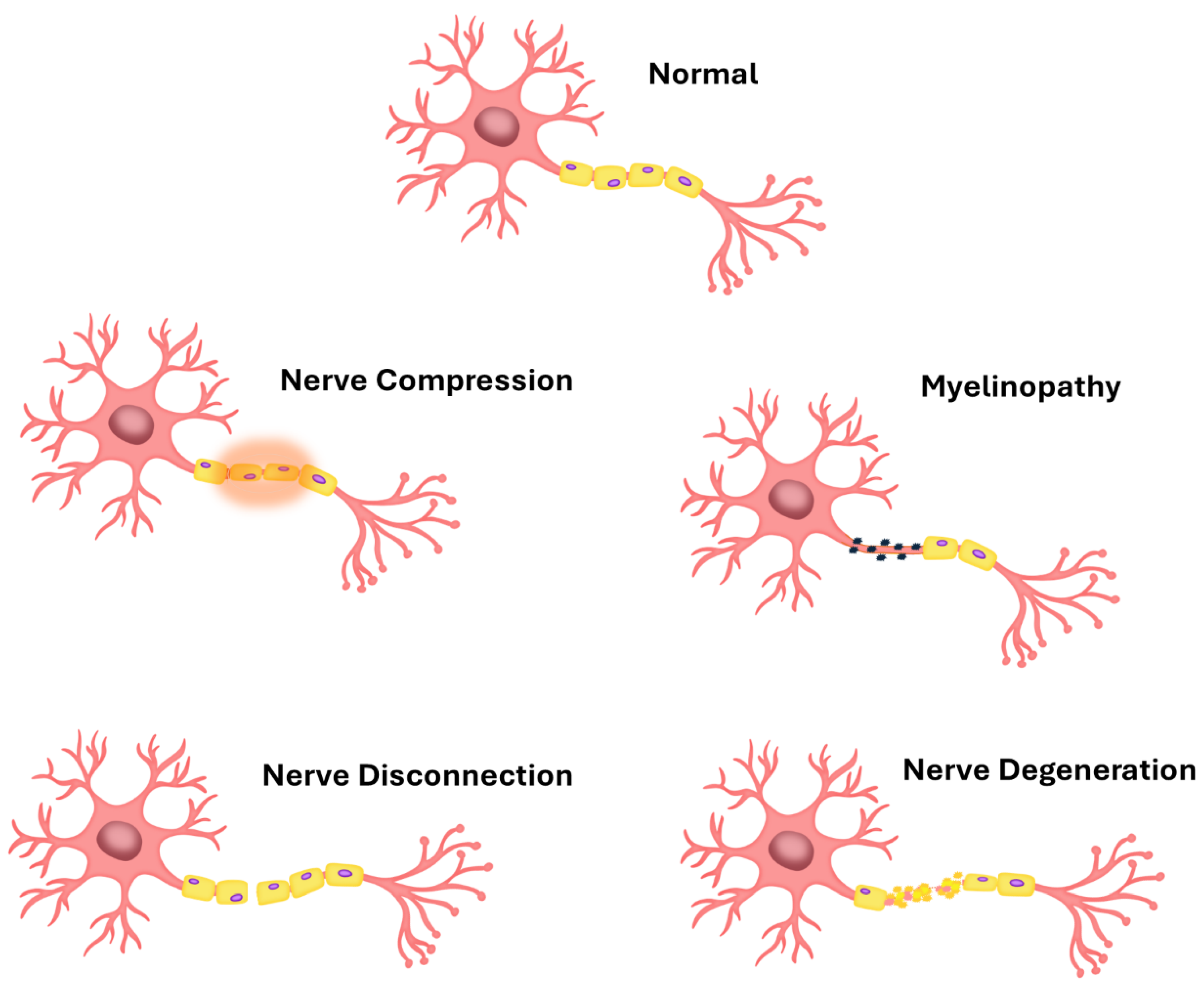
Figure 2.
Blood-brain barrier.
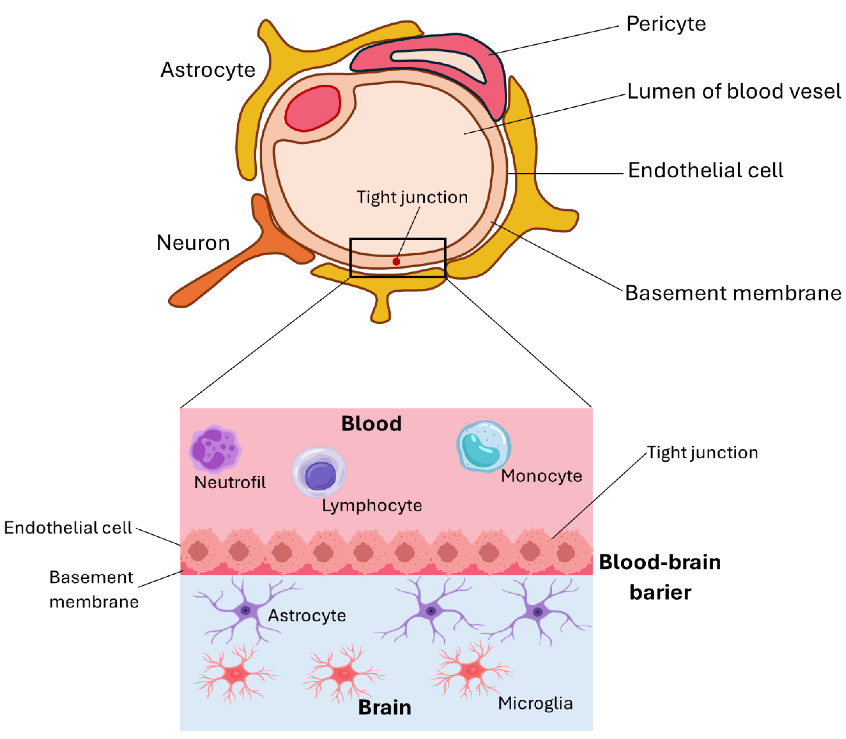
Figure 3.
Visualization of murine neuronal cells immobilized on alginate/chitosan polyelectrolyte bilayer after two weeks of culture. The red fluorescence shows neurons stained with anti-microtubule-associated protein 2 (MAP2), using a secondary antibody conjugated with the fluorochrome Alexa Fluor 555. The light green fluorescence indicates astrocytes stained with anti-glial fibrillary acidic protein (GFAP) and a secondary conjugated antibody labeled with Alexa Fluor 488 fluorochrome. Nuclei were dyed blue with Hoechst 33342 to enhance contrast under microscopy. Photos were taken using an Olympus IX71 fluorescence microscope.
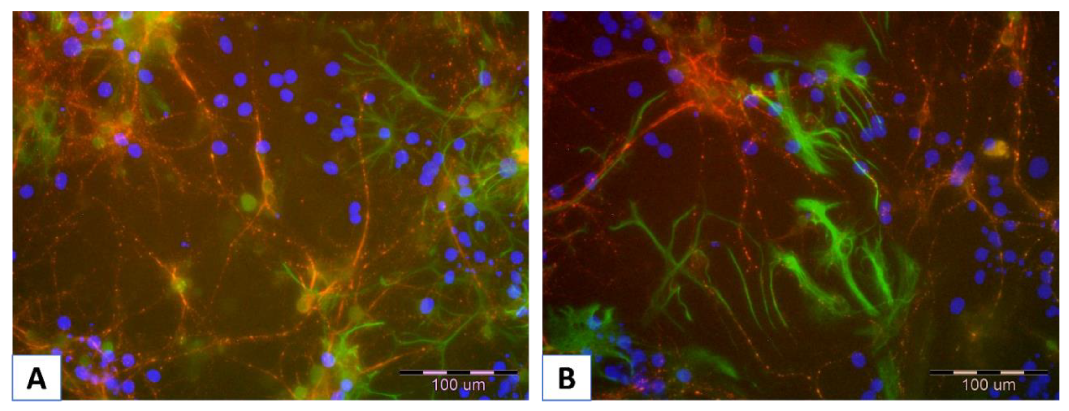
Figure 4.
Different examples of biomaterial applications in nerve regeneration.
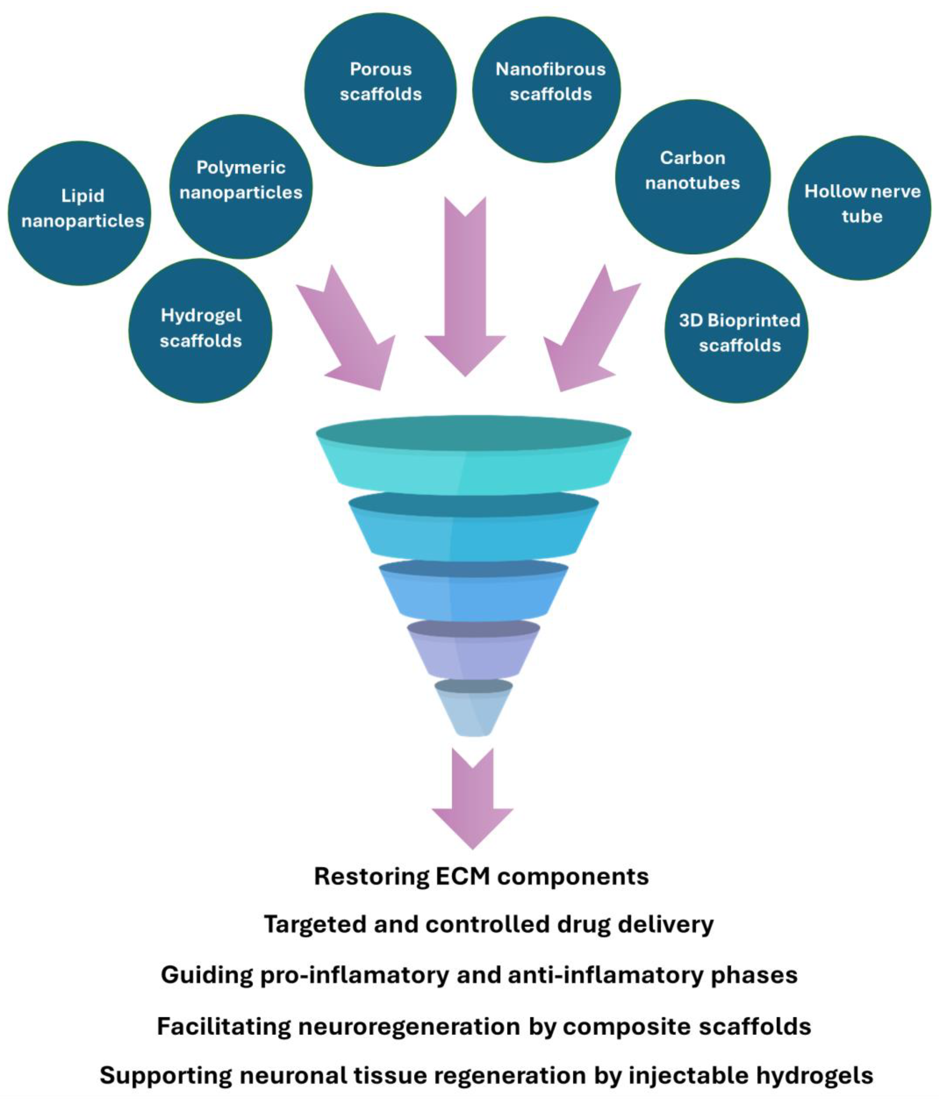
Figure 8.
The mitochondrial activity of NE-4C cells immobilized on the coating layer assessed over a 10-day culture period, with results expressed as the ratio relative to the control based on formazan production, as measured by absorbance. Key to the symbols: PEI – polyethyleneimine, PEI Cu10ppm – polyethylenimine 10ppm CuNPs incorporating, PEI Cu50ppm – polyethylenimine 50ppm CuNPs incorporating, PEI Cu 100ppm – polyethyleneimine 100ppm CuNPs incorporating. The values are presented as mean ± SD.
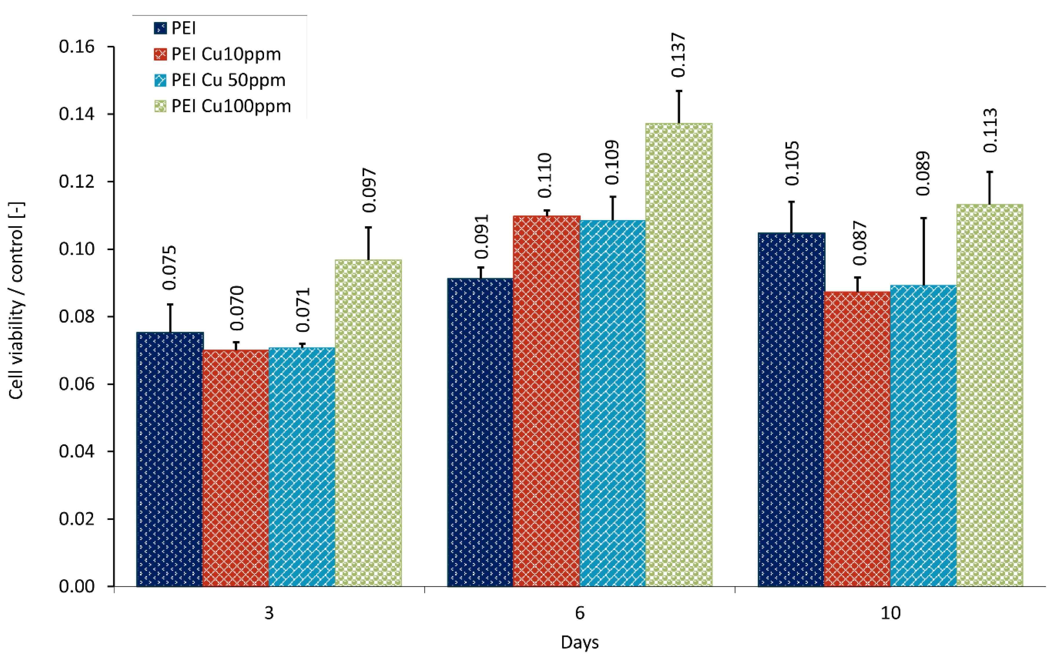
Figure 9.
SEM images of NE-4C cells adsorbed within the designed membranes after a third day of culture. Key to the symbols: PEI – polyethyleneimine, PEI-Cu10ppm – polyethylenimine 10ppm CuNPs incorporating, PEI-Cu50ppm – polyethylenimine 50ppm CuNPs incorporating, PEI-Cu100ppm – polyethyleneimine 100ppm CuNPs incorporating.
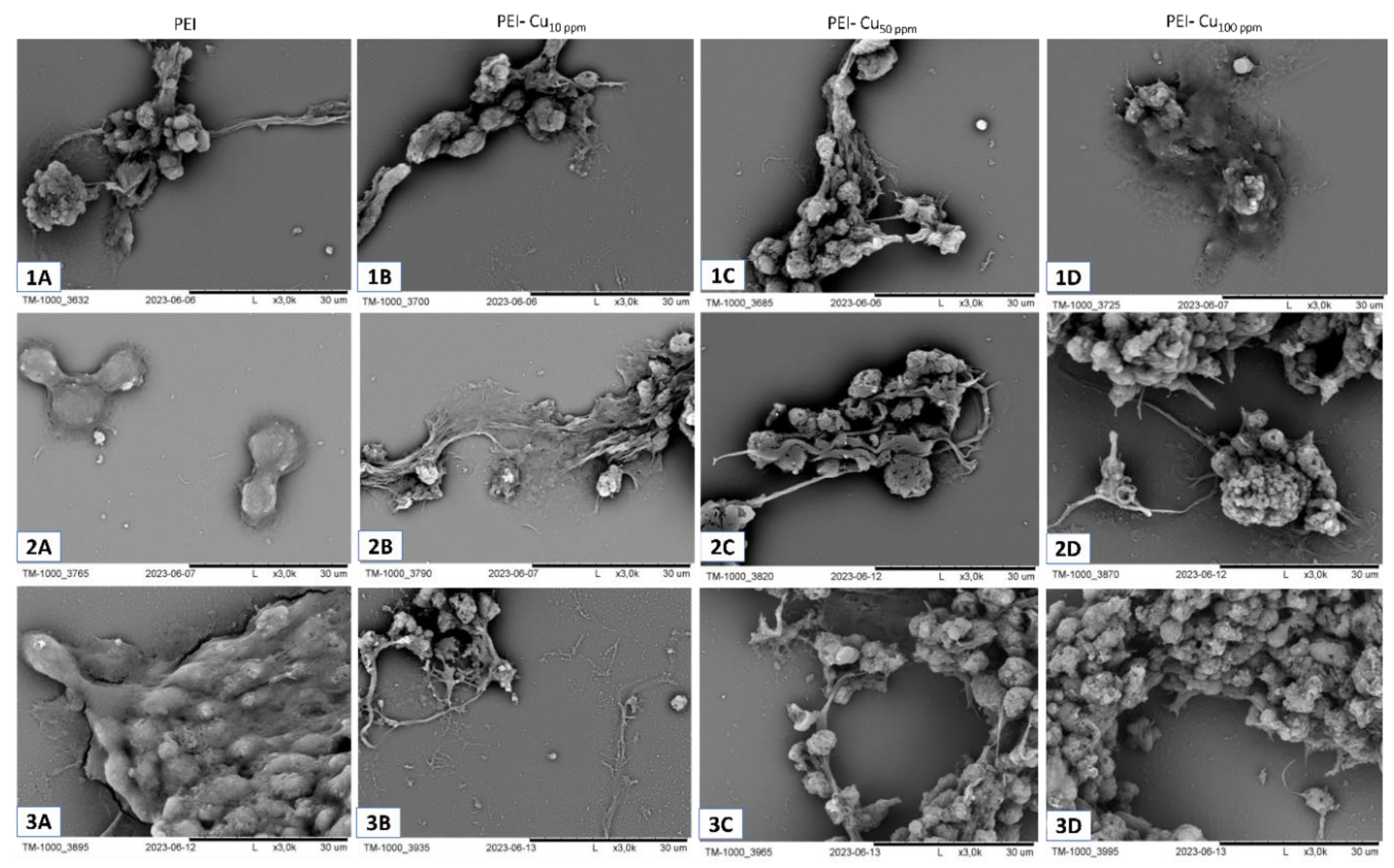
Figure 11.
Visualization after two-week culture of cells immobilized within layer membrane scaffold built of poly-L-lysine with incorporated: CuNPs in share of 10ppm (a, b) and 50ppm ( c, d ); and with incorporated Fe3O4NPs in share of 10ppm share (e, f), and 50ppm (g, h), and poly-L-lysine alone (i, j). The red fluorescence indicates neurons stained with anti-microtubule-associated protein 2 (MAP2), using a secondary antibody conjugated with the fluorochrome Alexa Fluor 555. The light green fluorescence shows astrocytes stained with anti-glial fibrillary acidic protein (GFAP) and a secondary antibody conjugated with Alexa Fluor 488 fluorochrome. The nuclei were tagged with Hoechst 33342 for imaging. Photos were taken using an Olympus IX71 fluorescence microscope.
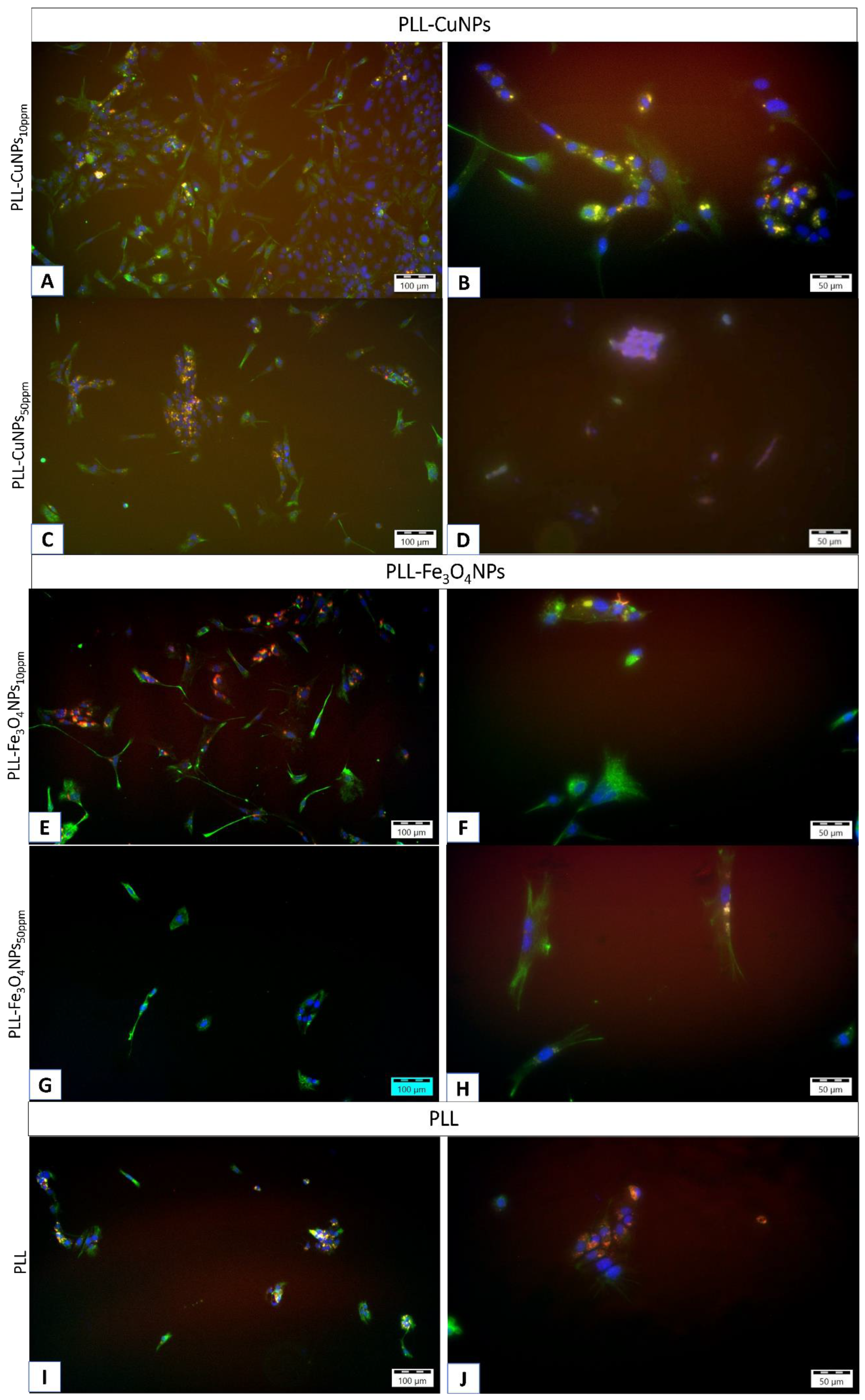

Table 1.
Key Mechanisms of Neuroprotection/Regeneration.
| Action | Description | Examples |
| Antioxidant and Anti-Inflammatory Actions | ||
| Oxidative Stress Mitigation | NPs can scavenge reactive oxygen species (ROS), protecting neurons from oxidative damage. |
|
| Inflammation Modulation | NPs can decrease proinflammatory cytokines, creating a favorable environment for nerve regeneration. | |
| Enhancing neuronal viability | NPs can reduce cellular apoptosis. | |
| Promotion of Axonal Growth and Myelination | ||
| Axonal Regeneration | Surface-modified NPs can guide axonal growth by mimicking extracellular matrix structures. |
|
| Myelination Support | NPs can promote axon remyelination. | |
| Enhanced Drug Delivery | ||
| Targeted Delivery | Functionalized NPs can cross biological barriers and deliver therapeutic agents directly to injury sites, minimizing off-target effects. |
|
| Controlled Release | NPs can provide sustained release of therapeutics, reducing the need for frequent dosing and maintaining effective drug concentrations at the injury site. | |
Disclaimer/Publisher’s Note: The statements, opinions and data contained in all publications are solely those of the individual author(s) and contributor(s) and not of MDPI and/or the editor(s). MDPI and/or the editor(s) disclaim responsibility for any injury to people or property resulting from any ideas, methods, instructions or products referred to in the content. |
© 2025 by the authors. Licensee MDPI, Basel, Switzerland. This article is an open access article distributed under the terms and conditions of the Creative Commons Attribution (CC BY) license (http://creativecommons.org/licenses/by/4.0/).
Copyright: This open access article is published under a Creative Commons CC BY 4.0 license, which permit the free download, distribution, and reuse, provided that the author and preprint are cited in any reuse.


