Submitted:
25 August 2025
Posted:
25 August 2025
You are already at the latest version
Abstract
Nanoparticle-based transdermal drug delivery systems (TDDS) have emerged as a revolutionary approach for antiparasitic therapy, addressing key challenges such as poor bioavailability, systemic toxicity and drug resistance. This review highlights the advancements in nanotechnology-driven TDDS for combating zoonotic parasitic diseases, including leishmaniasis, malaria, and infections treated by broad-spectrum drugs like ivermectin and albendazole. By leveraging nanocarriers such as liposomes, nanoemulsions, and microneedles, which enhance skin permeation, enable controlled drug release, and improve targeting specificity. For instance, deformable transfersomes and ethosomes achieve high transdermal efficiency without chemical adjuvants, while microneedle arrays physically bypass the stratum corneum for precise delivery. Furthermore, sustained-release hydrogels and stimuli-responsive nanoparticles optimize therapeutic efficacy and reduce adverse effects. Despite promising results, clinical translation faces challenges in manufacturing scalability, long-term safety, and accessibility in resource-limited settings. Future directions include bioinspired nanocarriers, artificial intelligence (AI)-driven design, and integration with global health initiatives like "One Health", all aimed at ensuring equitable implementation. This review highlights the transformative potential of nanotechnology in achieving sustainable antiparasitic solutions for zoonotic diseases.
Keywords:
nanoparticles
; transdermal drug delivery
; antiparasitic drug
; leishmaniasis
; malaria
; one health
; zoonotic diseases
1. Introduction
According to the World Health Organization’s 2024 report on Neglected Tropical Diseases (NTDs), parasitic infections remain a devastating global health burden, affecting over a billion people worldwide and causing significant morbidity, mortality, and socioeconomic disruption, particularly in resource-limited regions [1]. Among these, zoonotic parasitic diseases—transmitted between animals and humans—pose a critical and escalating threat. The report emphasizes that factors such as climate change, population migration, and uncertain funding outlooks may lead to the spread of many NTDs beyond their traditional endemic areas in low-income countries and tropical regions [1]. The Corona Virus Disease 2019 (COVID-19) pandemic has fundamentally reshaped global public health awareness, and the “One Health” concept has become a key framework for addressing complex infectious diseases due to its core value in coordinating human-animal-environment health synergy[2]. Notably, shifts in vector distribution due to climate change, coupled with health services not yet fully recovering from disruptions caused by the COVID-19 pandemic, are reshaping disease transmission patterns[1]. The report indicates that NTDs cause billions of dollars in losses annually in low- and middle-income countries, while the limitations of traditional treatment options continue to drive this figure upward[1] (Figure 1). Therefore, it is imperative to develop innovative and sustainable solutions to prevent and treat zoonotic parasitic diseases.
Among the various strategies to combat parasitic diseases, transdermal drug delivery systems have received a lot of attention due to their unique advantages[3]. Compared with oral or injectable drug delivery, transdermal drug delivery exhibits several advantages, such as avoiding the first-pass effect of the liver, no gastrointestinal irritation, good compliance, able to reduce the animal stress, convenient use and high safety[4]. How to make the drug transdermal absorption through the stratum corneum is the focus of transdermal drug delivery system. Enhancing the permeability of transdermal drugs can be achieved by both physical and chemical methods. Common physical methods include iontophoresis, electroporation, ultrasound introduction, pressure wave, laser cautery, and thermal promotion of permeation, while chemical methods are generally used to enhance transdermal penetration of drugs through chemical permeation promoters[5,6,7]. However, the traditional physical and chemical methods to promote transdermal delivery tend to enhance the rate of transdermal penetration by increasing drug solubility, diffusion coefficient and reservoir effect, which often leads to the overuse of chemical enhancers and the production of toxic side effects, and thus unmask more drawbacks[8]. Confronted with these difficulties in transdermal drug delivery, nanotechnology may represent a good choice.
Nanotechnology is often defined as the ability to produce and process materials at the nanoscale or to manipulate nano-objects. The nanoscale is often specified as 1 to 100 nm in the field of physics because the electromagnetic and quantum characteristics of nano-objects are more prominent in this scale[9]. Based on the special properties of nano-objects, nanotechnology has been used in various fields of science, and one important application area is nanodrug carriers. Nanodrug carriers include liposomes, transfersomes, ethosomes and nanoemulsions in which drugs can be encapsulated by dissolution, dispersion, encapsulation, adsorption and coupling[10]. In the context of transdermal drug delivery, nanodrug carriers offer innovative solutions to overcome the skin’s natural barrier, the stratum corneum, which is a major obstacle to prevent drug penetration. The ex vivo and in vivo behaviours of drugs are determined by the physicochemical properties of the nano carriers, which can be used to enhance drug efficacy, reduce side effects, enhance therapeutic index, and improve drug compliance.
This review focuses on the application of nanotechnology in transdermal treatment systems for parasitic diseases, summarizing its mechanisms of action and the important role it plays in malaria, leishmaniasis, and broad-spectrum antiparasitic drugs. By placing this discussion within the framework of the “One Health” approach, we aim to provide insights that can inform future health policy directions and contribute to the development of innovative and sustainable strategies for combating zoonotic parasitic diseases.
This figure illustrates the One Health concept via Toxoplasma gondii and arthropod-borne diseases. Blue: Foodborne T. gondii infects human (e.g., pregnant women) via contaminated meat/produce, potentially causing neurological damage or miscarriage. Green: Cats (definitive hosts) shed oocysts, which contaminate water and soil via rain. Intermediate hosts (e.g., pigs) ingest oocysts; humans risk infection through environmental contact or undercooked meat. Red: Arthropods (ticks, mosquitoes) cause rashes, fever, itching, and transmit diseases (Lyme, dengue) and pathogens (babesiosis, endemic typhus), embodying the interconnection of human, animal, and environmental health—the core of One Health.
2. Advantages of Nano Transdermal Delivery System
2.1. Transdermal Mechanism of Nanoformulations
As the primary physical barrier protecting the human body against external stimuli, the brick-and-mortar structure of the stratum corneum in the outermost epidermal layer severely restricts the transdermal permeation efficiency of conventional drugs through the highly ordered arrangement of intercellular lipids. Nanocarriers break this barrier through a multidimensional synergistic mechanism, significantly enhancing the delivery efficacy of anti-parasitic drugs. The specific mechanisms are as follows and summarized in Figure 2.
The transdermal mechanisms of liposomes involve synergistic effects through multiple pathways: First, the phospholipids in liposomes exhibit high compatibility with stratum corneum lipids, leading to structural disintegration of liposomes and subsequent release of phospholipids. These released phospholipids infiltrate the stratum corneum and reorganize into lamellar structures, thereby widening lipid intercellular channels and enhancing drug permeation[11,12,13]. Second, their hydration effect increases stratum corneum water content, alters the arrangement of intercellular lipid layers in corneocytes, and reduces barrier density. Additionally, intact liposomes can directly penetrate through corneocytes or utilize skin appendage pathways such as hair follicles or sweat glands for delivery[14,15]. Surfactant-driven deformable carriers create transient permeation channels by disrupting stratum corneum lipid organization. Transfersomes are formed by modifying liposomal composition. Surfactants embedded in the vesicle membrane enable extreme deformability, allowing penetration through pores several times smaller than their own size[16,17]. Ethosomes are multilamellar vesicles incorporating high-concentration ethanol into conventional liposomal formulations. The high ethanol concentration enhances the mobility of polar lipid heads in membrane components, thereby increasing lipid fluidity and membrane flexibility. It also reduces the density of intercellular lipid domains, facilitating vesicle deformation during permeation. Consequently, higher ethanol content correlates with greater permeation flux. Drug release and absorption in deeper skin layers result from liposome-skin lipid fusion, which persists throughout the transdermal process[18,19]. For instance, trifluralin-loaded transfersomes demonstrate the potential of deformable nanocarriers for targeted cutaneous leishmaniasis treatment, achieving high-efficiency transdermal delivery without permeation enhancers[20]. Co-loaded nitazoxanide-quercetin nanotransfersomal exhibit a 4-fold enhancement in skin permeation compared to conventional transfersomes[21]. In addition, an ethosomal patch, an innovative antimalarial transdermal nanosystem has been developed for increasing the cumulative permeation of artesunate by 1.57-fold compared to conventional patches after 8 h of administration[22].
Nanoemulsions significantly enhance transdermal drug permeation through a triple synergistic mechanism. First, surfactant components disrupt the lipid arrangement in the stratum corneum while expanding intercellular spaces through hydration, creating permeation pathways for drug molecules[23]. Second, the nanoemulsion structure enables effective solubilization of drugs, especially lipophilic drugs, thereby increasing the drug concentration gradient across the skin and enhancing permeation[24]. At last, nanoscale particle size combined with superior wettability improves skin contact and permeation efficiency[8]. This layer-by-layer mechanism establishes nanoemulsions as ideal transdermal carriers. For example, nanoemulsions effectively deliver chalcone to deep skin layers (dermis), the parasitic site of Leishmania pathogens[25]. Studies have demonstrated enhanced skin permeation using nanoemulsions loaded with C6I/TC1/TC2, which are promising antileishmanial drugs for treating Leishmania infections[26].
Microneedle arrays (MNs), composed of needle-like structures with diameters of several micrometers, penetrate the skin surface to create microchannels, providing an innovative solution for transdermal drug delivery. MNs overcome the limitations of traditional methods through precise targeting capability, offering higher safety, efficacy, and painless administration[27,28,29,30]. For instance, a novel dissolving microneedle patch has been successfully designed to enhance the skin permeability of amphotericin B. The microneedles penetrated rat skin to a depth of 303±8 µm, dissolved rapidly, and formed micropores that caused no significant cellular damage, with natural healing within 30 minutes[31]. Similarly, a hydrogel-forming microneedle combined with a polyethylene glycol (PEG) reservoir has been developed for cystic Echinococcosis treatment, and in vitro permeation tests showed an albendazole permeation amount of 4584.43 ± 26.61 µg/cm²[32]. Liposomes[13] (including transfersomes[17] and ethosomes[8]), nanoemulsions[23], and microneedle arrays[27] overcome the inherent barrier limitations of the stratum corneum through their distinctive multidimensional synergistic mechanisms—such as the structural compatibility and deformability adaptation of liposomes, the triple synergistic enhancement of permeability in nanoemulsions, and the physical microchannel creation by microneedle arrays. This breakthrough significantly augments the transdermal delivery efficiency of antiparasitic drugs, serving as a critical foundation for subsequent controlled release and targeted delivery systems.
This figure illustrates transdermal delivery modalities of nanoformulations. Polymeric nanocarriers, nanoemulsions, liposomes, niosomes, solid lipid nanoparticles (SLNs), and microneedles enable penetration across the skin strata (epidermis and dermis). Leveraging follicular and transcellular/paracellular pathways, these systems exemplify advanced nanocarrier - mediated strategies for percutaneous therapeutic administration.
2.2. Controlled Release and Targeted Delivery of Nanoformulations
The success of antiparasitic therapy relies not only on efficient drug penetration through the skin barrier but also on achieving long-acting and low-toxicity therapeutic outcomes through precise controlled-release[33] and targeting strategies[34]. Nanoparticle-based drug delivery systems employ multidimensional collaborative mechanisms to prolong therapeutic duration temporally[35] and enhance lesion targeting efficiency spatially[36], providing comprehensive solutions spanning from localized permeation to dynamic regulation for parasitic infections. This section systematically elaborates the core mechanisms of sustained-release depot construction, and targeting synergy, elucidating their pivotal roles in reducing recurrence risk and minimizing host damage.
The sustained-release mechanisms of transdermal nanoformulations achieve high-efficiency antiparasitic therapy through multi-mechanism synergy. Controlled-release carriers such as liposomes[37] and nanoemulsions[34] regulate drug release via matrix diffusion or degradation-controlled processes, forming long-acting drug depots in the skin to reduce dosing frequency. The synergistic interaction between reservoir effects and gradient-driven permeation further optimizes drug delivery: depot-accumulated high-concentration gradients drive drug penetration into subcutaneous tissues, preventing systemic plasma concentration spikes[36]. Building on this, multistage release strategies—employing biphasic rapid-sustained release designs or combination drug loading—enable insecticidal-immunomodulatory synergy, lowering recurrence risks[38,39]. This hierarchical sustained-release system, transitioning from local retention to dynamic permeation and from monotherapy to combinatorial regulation, provides precise long-acting solutions for parasitic infections. Based on this mechanism, a topical ivermectin formulation has been successfully developed where solid lipid nanoparticles (SLNs) confers superior sustained-release efficacy for treating scabies[40]. Moreover, a novel electrospun wound dressing has been designed and evaluated with excellent antileishmanial activity, achieving sustained drug release for up to two weeks[41,42].
Additionally, hydrogels have emerged as ideal topical drug delivery vehicles. Hydrogels have garnered significant attention due to their unique properties, including low cost, excellent biocompatibility, non-toxicity, stimuli-responsive behavior, and tunable drug release kinetics[43,44]. Nanostructured lipid carrier (NLC)-based hydrogels represent a transformative approach for localized antileishmanial therapy. Inspired by the relevance theory, glucantime-loaded NLC hydrogels has been generated with controlled drug release performance[45].
The passive targeting effect of transdermal nanoformulations primarily optimizes skin penetration efficiency through the physical properties of carriers (e.g., particle size, deformability). For example, trifluralin-loaded transfersomes, with a nanoscale particle size of 140.3 ± 2.3 nm and a high deformation index (DI = 43.5 ± 1.0), can permeate through microscopic pores in the stratum corneum[20]. Additionally, the negatively charged surface of TFS may enhance interactions with macrophage phagosomal membranes, promoting intracellular drug delivery[20]. Experimental validation revealed that rhodamine-labeled TFS exhibited a 4.13-fold increase in endocytosis efficiency compared to free solutions, directly attributed to the adaptive membrane penetration of their elastic vesicular structures. This property demonstrates significant advantages in antileishmanial therapy: trifluralin-loaded transfersomes achieved 90.87% inhibition of intracellular amastigotes in macrophages at 50 µg/mL, showing a 1.68-fold efficacy enhancement over conventional TFL solutions that exhibited 54% inhibition[20]. In a similar strategy, rifampicin-loaded nanotransfersomes also rely on nanoscale size and elasticity for passive targeting, further validating the universality of this mechanism in transdermal antiparasitic therapy[46]. Furthermore, elastic liposomes are demonstrated to enhance immune responses via transdermal delivery of the Plasmodium falciparum surface antigen (PfMSP-119), effectively activating Langerhans cells and promoting their migration to draining lymph nodes[47,48].
The deep integration of these mechanisms and advantages marks the transition of nanocarrier-based transdermal delivery systems into a new era of precision and long-acting performance in antiparasitic therapy. Currently, this technology has displayed significant clinical value in zoonotic parasitic diseases such as leishmaniasis and malaria. Its specific applications and future challenges will be systematically discussed below.
3. Application of Nanoformulations in the Treatment of Parasitic Diseases
Nanotechnology-based systems, leveraging their unique technological advantages, provide highly promising solutions for antiparasitic drug delivery. The application of nanocarriers significantly enhances drug targeting specificity, effectively protects drugs or immunotherapeutic agents from extracellular degradation, and optimizes biopharmaceutical and pharmacological properties, thereby improving therapeutic efficacy while reducing side effects[49]. Currently, nanocarriers-based therapy has been extensively used in the control of parasitic diseases, among which Leishmaniasis and Malaria are the most noteworthy aspects. Herein, we take Leishmaniasis and Malaria as examples to discuss the application of nanoformulations for parasitic diseases control.
3.1. Leishmaniasis
Leishmaniasis, caused by protozoan parasites of the genus Leishmania (family Trypanosomatidae), is transmitted through the bite of infected sand flies, resulting in a spectrum of clinical manifestations including visceral (VL), cutaneous (CL), mucosal (ML), and mucocutaneous leishmaniasis (MCL)[50,51]. The severity of illness is determined by the virulence of specific Leishmania species and the host immune response, ranging from self-resolving cutaneous lesions to life-threatening visceral organ failure[51,52,53]. Macrophages, serving as the primary host cells for Leishmania parasites, play a pivotal role in pathogenesis. Despite substantial research efforts, challenges such as drug resistance and limited therapeutic accessibility persist, necessitating accelerated development of novel therapies to address this ongoing global health burden[54].
Current pharmacotherapy for Leishmaniasis faces multifaceted limitations, including drug toxicity, pathogen resistance, and restricted access to treatment resources, particularly in low-income regions. First-line pentavalent antimonials have been progressively phased out due to widespread resistance[55,56]. Alternative agents such as miltefosine, paromomycin, and pentamidine are further constrained by emerging resistance, prohibitive costs, and severe adverse effects. Therefore, the development of innovative, safe, and efficacious anti-leishmanial agents remains a critical clinical imperative. Herein, we systematically examine recent advances in therapeutic strategies against Leishmaniasis focusing on transdermal penetration enhancement, safety optimization, wound healing promotion, and immune modulation, while addressing translational challenges and future research trajectories.
Transdermal delivery technologies have undergone multistage iterative optimization. Early research focused on nanoemulsions: a chalcone nanoemulsion has been developed using a mixed system of anionic and nonionic surfactants, which enhanced dermal permeation of trans-chalcone and 3’-(trifluoromethyl)-chalcone by 1.26-fold and 2.41-fold, respectively[25,57,58]. The fluorinated derivative achieved 88.31 μg/g skin retention (vs. 45.86 μg/g for the free form) due to fluorine-enhanced hydrophobicity[25]. Garcia et al. further validated this potential: an ethanol-oleic acid/Tween®-80 nanoemulsion with particle size modulation of 147-273 nm enabled sustained release of anti-leishmanial compounds (first-order kinetics and Weibull model), demonstrating 2-fold increased parasiticidal activity and reduced macrophage cytotoxicity[26]. Recent advances highlight deformable nanocarriers for enhancer-free transdermal delivery. Second-generation systems achieve precision delivery through structural engineering: trifluralin-loaded transfersomes with 140 nm particle size and ultralow polydispersity index (PDI, 0.006) achieved 86% encapsulation efficiency and 24 h sustained release without permeation enhancers[20]. This reduced Leishmania IC50 values by 2.86 to 3.07-fold, suppressed amastigotes by 90.87% (vs. 54% for free drug), and demonstrated efficient macrophage internalization and biocompatibility[20]. Furthermore, it is reported that nitazoxanide-quercetin nanotransfersomal gel (NTZ-QUR-NTG) enhanced skin permeation 4-fold, increased macrophage uptake 10-fold, elevated concentration of cytotoxicity 50% (CC50) from 49.77 μg/mL to 71.95 μg/mL, and significantly reduced lesion size in mice with minimized systemic toxicity[21]. Interestingly, microneedle technology physically bypasses the stratum corneum barrier. Based on this mechanism, a PVP-carboxymethylcellulose microneedle patch has been successfully developed with 303±8 μm penetration depth, releasing amphotericin B within 30 minutes through self-sealing micropores, achieving 86% anti-leishmanial activity[31]. Additionally, engineered electrospun core-shell nanofibers have been demonstrated sustainably to release 84% glucantime over 9 h, maintaining therapeutic drug concentrations for prolonged efficacy[59].
To enhance the safety profile and immunomodulatory efficacy of nanoformulations, breakthroughs have been achieved across multiple dimensions. In nanomaterial innovation, silymarin-selenium nanoparticles exhibit skin deposition rate of 82.21%, which reduced local toxicity by inhibiting trypanothione reductase activity and improving cytocompatibility [60] (Figure 3). Rabia team has engineered rifampicin-loaded nanotransfersomes combined with a chitosan-based gel, achieving 3-fold enhanced skin permeability and reduced IC50 values through passive macrophage targeting, thereby minimizing systemic exposure while boosting therapeutic efficacy via apoptosis induction[46]. In novel delivery system design, a glucantime-loaded nanostructured lipid carrier (NLC) hydrogel has been formulated that enabled sustained drug release, enhanced skin retention in murine models, and compared to injectable formulations, this system significantly reduced lesion size, parasite burden, and systemic toxicity, offering a non-invasive therapeutic strategy[45]. For immune modulation, Lanza et al. pioneered a LiHyp1 recombinant antigen-based vaccine delivered via dissolving microneedles (DMNs) and cationic liposomes (CL). This approach elicited high-titer antibody responses in mice, suppressed L. donovani infection, and established a novel paradigm for anti-leishmanial immunotherapy[61].
Functionalized nanomaterials synergize intelligent drug release with multimodal mechanisms to promote chronic ulcer repair. On basis of this mechanism, engineered chitosan-polyethylene oxide nanofibers loaded with berberine has been successfully designed, achieving gradient release over 14 days (80% released within the first 3 days). This system demonstrated potent larvae growth inhibition (IC50=0.24 µg/mL) and excellent biocompatibility[41]. Furthermore, its efficacy has been validated in accelerating Leishmania ulcer healing, noting stable berberine release kinetics coupled with dual antibacterial activity against Staphylococcus aureus and Escherichia coli and sensor compatibility for real-time wound microenvironment monitoring[42]. Similarly, a PVP-PVA-clay hydrogel has been shown that achieved 15 h sustained release of N-methyl glucamine under 1.5% clay conditions, reducing lesions by 99% in L. amazonensis-infected models[62]. A cobalt-60 radiation-crosslinked PVP-PEG-agar hydrogel loaded with amphotericin B exhibited 12 h continuous drug release with thermal/radiation stability, achieving 100% parasitic growth inhibition within 48 h while integrating sterilization and long-term anti-infection functions[63].
Nanoparticle-based transdermal delivery technologies, through strategies such as deformable nanocarriers, microneedle patches, and smart hydrogels, have significantly enhanced transdermal efficiency and targeting specificity in the treatment of Leishmaniasis, while reducing systemic toxicity via sustained-release designs. However, their clinical translation remains constrained by manufacturing complexity, insufficient long-term biosafety validation, and accessibility challenges in resource-limited regions. Future efforts should prioritize the development of intelligent responsive systems (e.g., enzyme/pH dual-sensitive hydrogels), artificial intelligence (AI)-driven carrier optimization, and multimechanistic combination therapies. Concurrently, leveraging global health governance frameworks such as the “One Health” initiative will be critical to advancing technological equity and accelerating the elimination of cutaneous leishmaniasis.
This figure illustrates nanotherapeutic mechanisms against Leishmaniasis (left) and Malaria (right). Silymarin-selenium nanoparticles enhance drug loading, promote cellular uptake, and inhibit trithiol reductase for Leishmaniasis. Artemisininone solid lipid nanoparticles boost epidermal drug concentration in the stratum corneum to treat Malaria, showcasing nano - based strategies for parasitic diseases.
3.2. Malaria
Malaria is a major infectious disease that causes severe morbidity, mortality, and economic burden globally[64]. Although significant efforts have been made to eradicate Malaria, the number of malaria cases worldwide still reached 249 million in 2022, resulting in 608,000 deaths[65]. The burden of Malaria remains particularly severe in countries such as Nigeria, Uganda, the Democratic Republic of the Congo, and Mozambique[65]. Malaria is caused by a single-celled parasite known as Plasmodium, which is transmitted to humans through the bites of female Anopheles mosquitoes, continuously threatening the lives of millions of people worldwide. There are five known species of Plasmodium that can cause human malaria, including Plasmodium falciparum, Plasmodium malariae, Plasmodium vivax, Plasmodium ovale, and Plasmodium knowlesi, with Plasmodium falciparum having the highest mortality rate[66].
The treatment of malaria mainly depends on chemical drugs such as chloroquine, lumefantrine, primaquine, artemisinin and its derivatives. While these drugs have achieved certain effectiveness in controlling malaria, their effectiveness is increasingly influenced by various factors, including the emergence of multidrug resistance, nonspecific targeting of intracellular parasites, low solubility and bioavailability of the drugs, and significant toxicity of the medications[67,68]. Increasing resistance manifests itself in the ability of the parasite to survive and multiply at a certain concentration of the drug that would normally be killed, so it is important to seek drug combinations and new strategies. The WHO recommends the use of artemisinin-based combination therapies (ACTs) for the treatment of malaria, including artesunate-amodiaquine, artemether-lumefantrine, artesunate-sulfadoxine-pyrimethamine, dihydroartemisinin-piperaquine and artesunate-mefloquine[69]. However, they also face issues such as high costs, differences in drug metabolism characteristics, potential toxicity, and insufficient availability[69]. Furthermore, problems such as high transmission rate of Plasmodium parasites, resistance of mosquito vectors to insecticides, logistical challenges in implementing control measures, and population mobility pose greater difficulties for malaria control efforts. To overcome these difficulties, in recent years, innovations in nanoparticle-based drug delivery technology have provided breakthrough solutions[65]. Next, we systematically review relevant advances, focusing on three key dimensions: enhancement of targeting specificity, reversal of drug resistance, and mitigation of toxicity, aiming to provide scientific evidence for optimizing antimalarial strategies.
Improving drug targeting specificity represents a core breakthrough direction in nanotechnology. Ethosomes containing artesunate and febrifugine into a patch matrix to form a composite transdermal patch has been prepared, achieving 8 h cumulative permeation 1.57 times higher than conventional patches, with 100% parasite clearance and zero recurrence, demonstrating the high efficiency of transdermal delivery[22]. Zech team further optimized an artemisone-based microemulsion spray, achieving a blood concentration exceeding 500 ng/mL in a cerebral malaria model, doubling the efficacy compared to artesunate microemulsions, and enabling complete pathogen eradication[70]. Similarly, artemisone-loaded solid lipid nanoparticles, through precise regulation of particle size and crystallinity, elevated drug concentration at the stratum corneum-epidermis junction to 62.632 μg/mL (versus 12.792 μg/mL for artemisone-loaded niosomes), demonstrating the significant efficacy of nanoparticles in optimizing dermal drug delivery concentration[71] (Figure 3). In the field of immunomodulation, an elastic liposomal vaccine delivering Plasmodium falciparum antigen PfMSP-119 has been developed, which markedly enhanced humoral and cell-mediated immune responses[48], maintained peak IgG-specific antibody titers for 70 days post-immunization, exhibited more sustained immune responses compared to conventional liposomes and intramuscular injection groups, and significantly increased IFN-γ secretion levels (approximately 9-fold higher than controls)[47]. This immuno-based system provides a novel strategy for developing needle-free malaria vaccines[47,48].
To combat drug resistance, scientists have significantly enhanced therapeutic efficacy through co-delivery systems and novel mechanism designs. Aderibigbe’s team has developed an arabic hydrogel dual-drug loading system co-encapsulating 4-aminoquinoline and curcumin that, by leveraging differential release kinetics (short-term anomalous transport and long-term quasi-Fickian diffusion), achieved synergistic effects, providing a new strategy for antiparasitic combination therapies[38]. Further studies revealed that carbomer-polyacrylamide-soy protein composite hydrogels, through temporal regulation of chloroquine and curcumin diphosphate release via a super case II transport mechanism, enabled phased drug release to overcome current antimalarial drug resistance challenges[72]. To address resistance limitations of conventional drugs, a hydroxypropyl methylcellulose-polyvinylpyrrolidone hydrogel nanoparticle loaded with curcumin has been successfully designed and prepared and significantly enhanced curcumin’s antimalarial activity (exhibiting doubled potency compared to free drug at 25 mg/kg), prolonged drug circulation time, and improved bioavailability for enhanced efficacy[73]. Moreover, Mavondo’s team has developed a pectin hydrogel-based percutaneous patch that not only suppressed parasitemia but also ameliorated inflammatory responses and anemia symptoms in malaria-infected Sprague-Dawley rats, offering a multifunctional solution for drug-resistant malaria treatment[74].
In addressing systemic toxicity reduction, nanotechnology strategies focusing on localized controlled release and biocompatibility optimization have demonstrated remarkable efficacy. Nnamani et al. developed an artemether-loaded nanostructured lipid carrier hydrogel, which forms a localized drug reservoir through transdermal penetration, achieved a 26% cumulative permeation rate over 48 h, significantly enhanced drug stability, release controllability, and demonstrated no observable systemic exposure[75]. Conventional oral administration of primaquine (PMQ) may induce adverse effects and undergo significant first-pass metabolism in the liver, thereby compromising therapeutic efficacy. Ananda’s team pioneered the exploration of PMQ transdermal patch technology combined with solid microneedles (Dermaroller®)[76]. Their comprehensive evaluation included multiple parameters such as thickness, weight uniformity, and pH values. In vitro and ex vivo tests on PMQ release and permeation through rat skin confirmed the system’s safety profile, with all formulations demonstrating optimal performance, establishing a safe paradigm for long-term medication[76].
Looking forward, although nanoparticle-based drug delivery technologies have demonstrated advantages in targeting specificity and safety profiles, their large-scale application still requires overcoming cost constraints and manufacturing barriers to provide sustainable technological support for global malaria eradication. Beyond the prominent examples of leishmaniasis and malaria, the versatility of nanotransdermal delivery systems holds significant promise for addressing a broader spectrum of zoonotic parasitic infections. For instance, helminthic infections such as cystic echinococcosis[77] and lymphatic filariasis[78] face challenges similar to those discussed, including poor drug bioavailability, systemic toxicity, and the need for prolonged therapy. As highlighted in section four, nanotechnological approaches, particularly microneedle-integrated systems, demonstrate remarkable efficacy in enhancing transdermal delivery, achieving targeted deposition (e.g., lymphatic targeting), and enabling sustained release, thereby improving therapeutic outcomes while mitigating adverse effects[29,79]. Other neglected tropical diseases with zoonotic components, such as onchocerciasis and strongyloidiasis, could similarly benefit from the advancements in nanocarrier design (liposomes, nanoemulsions, SLNs) tailored for transdermal delivery of ivermectin, albendazole, or other relevant anthelmintics[80,81]. The core principles of overcoming the stratum corneum barrier, enhancing local drug concentration, enabling controlled release, and reducing systemic exposure are universally applicable. The success seen with leishmaniasis, malaria, and broad-spectrum drugs provides a strong foundation for expanding the application of this technology across the neglected zoonotic parasitic disease landscape.
4. Nanoformulations in Broad-Spectrum Antiparasitic Drugs
Beyond targeted therapies against specific pathogens (e.g., Leishmania, Plasmodium), nanoparticle-based transdermal delivery technologies have provided revolutionary solutions for enhancing the application of conventional broad-spectrum antiparasitic drugs. Taking avermectin and albendazole as examples, both exhibit high efficacy against a wide range of endo- and ectoparasites, however, their conventional administration routes such as oral administration is limited by low bioavailability, systemic toxicity, and poor patient compliance[32,40]. Nanoparticle-based reformulation significantly enhances transdermal permeation efficiency, targeting specificity, and therapeutic window of avermectin- and Benzimidazole-class drugs. We next review and discuss the updates related nanoformulation-driven anthelmintic drugs (Table 1).
4.1. Avermectin-Class Drugs
Avermectin-class drugs such as avermectin, ivermectin and eprinomectin play an indispensable role in veterinary and human parasitic disease control due to their exceptional broad-spectrum antiparasitic activity [82]. For instance, ivermectin represents a landmark therapeutic for treating onchocerciasis [80], lymphatic filariasis [83], scabies [84], and strongyloidiasis [81], and is included in the World Health Organization Essential Medicines List [85]. However, they have long been constrained in their antiparasitic efficacy due to low solubility, insufficient transdermal permeation efficiency, and systemic toxicity risks [85]. Multifaceted innovations in nanoparticle-based delivery technologies—through formulation design optimization and enhancement of controlled-release strategies—are progressively advancing their translation from laboratory research to clinical practice.
Early studies focused on solubility optimization and enhancement of transdermal permeation efficiency of avermectin-class drugs. For instance, Das et al. pioneered a breakthrough with ivermectin-loaded microemulsion-based gel formulations using nanoscale technology, where in vitro membrane permeability assays demonstrated superior permeation advantages, enabling low-dose drugs to achieve equivalent or better efficacy compared to conventional formulations [86]. Subsequently, an advanced ivermectin nanocrystal technology through nanocrystallization combined with lyophilization, achieving a 730-fold increase in equilibrium solubility, a 24-fold acceleration in dissolution rate, and threefold higher drug deposition in the deep dermis, significantly reducing side effect risks [87]. Building upon these advancements, formulation diversity exploration has enabled tailored solutions for different infection scenarios. A recent study systematically compared three ivermectin-loaded formulations: nanoemulsion (NE), nanoemulsion gel (NEG), and colloidal system (CS)[85]. NE exhibited the highest drug concentration at the stratum corneum and epidermis/dermis junction, making it ideal for localized therapy. NEG demonstrated superior adhesiveness, solubilization capacity, and occlusiveness, achieving the fastest delivery rate. CS showed the highest drug diffusion percentage, enabling efficient delivery via the follicular route at a 0.35% low dose (versus 2% in other systems) [85]. This study not only highlights formulation design diversity but also provides a theoretical foundation for precision therapy by using conventional antiparasitic drugs (Figure 4.).
To balance efficacy and safety of antiparasitic drugs, controlled-release technology has become a key research focus. Ivermection-loaded solid lipid nanoparticles exhibiting excellent sustained-release efficacy, with their targeting capability significantly reducing systemic toxicity and side effects [40]. With the accelerating of technology development, Mao et al. designed hybrid micelles that demonstrated 1.92-fold higher transdermal efficiency compared to the commercial pour-on formulation (Eprinex®), and in vivo trials support its favorable safety profiles [88]. In addition, the release profiles of mesoporous silica nanoparticles (IVM-MCM) and polymeric nanocapsules (IVM-NC) have been further studied. IVM-MCM facilitates rapid drug release suitable for acute infection management, whereas IVM-NC provides sustained release ideal for chronic prevention requirements [89]. This rational optimization of ivermectin nanoformulations provides a novel strategy to accelerate its clinical translation [89].
From solubility breakthroughs in microemulsion-based gels [86] to transdermal optimization of hybrid micelles [88], and from explorations in formulation diversity [85] to modulation of release kinetics [40], avermectin-class nanoformulations are progressively overcoming some of the key limitations inherent in conventional formulations. However, their full clinical translation still requires overcoming barriers in manufacturing scalability, safety profiles, and therapeutic validation. Going forward, cross-disciplinary collaboration and technological innovation will be pivotal to translating the theoretical advantages of nanoformulations into practical clinical benefits for conventional anthelmintic drugs.
This figure illustrates the mechanism of ivermectin nanoemulsion (IVM - NE) in treating strongyloidiasis. IVM - NE, with favorable drug - release ability, releases ivermectin (IVM). IVM acts on glutamate - gated Cl⁻ channels of Strongyloides organisms. It shows strong dermal retention, good safety, and is suitable for topical administration to combat strongyloidiasis.
4.2. Benzimidazole-Class Drugs
Benzimidazole-class drugs constitute the cornerstone of antihelminthic therapy [90], exhibiting highly effective broad-spectrum activity against diverse intestinal nematodes, cestodes, and certain trematodes [91]. However, oral formulations of conventional benzimidazoles are severely limited in antiparasitic efficacy and safety due to poor aqueous solubility (only 48.11 μg/mL in PBS), significant first-pass metabolism (bioavailability of less than 5%), and hepatotoxicity induced by systemic exposure [32]. Recent innovative strategies based on nanotechnology and transdermal delivery systems have successfully overcome these limitations, with key advancements manifested in these followed aspects.
First, drug morphology reconstruction using carriers like solid lipid nanoparticles, the solubility kinetics of albendazole have been significantly improved. Permana et al. formulated albendazole into nanocrystals, achieving 89.92% cumulative drug release over 24 h in vitro with a 3.2-fold accelerated release rate [77]. Similar with that, a polyethylene glycol reservoir stabilize albendazole in an amorphous state, achieving a solubility of 283.62 μg/mL, representing a 5-fold increase [32]. Therefore, nanonization not only overcomes benzimidazole’s intrinsic hydrophobicity but also enables rapid therapeutic onset through accelerated dissolution.
Second, the application of dissolving microneedles (DMNs) and hydrogel-forming microneedles (HFM) has overcome skin barrier limitations, enabling precision drug delivery. Based on this nanoformulation, an albendazole-loaded dissolving microneedles-nanocrystals system achieved a penetration depth of 505.29 μm in ex vivo porcine skin, with a peak dermal drug concentration of 1891.53 μg/cm³[77]. For albendazole sulfoxide delivered via a solid lipid nanoparticle - dissolving microneedle system, both drug targeting efficiency and direct transport percentage were significantly enhanced, reaching 11.99-fold and 91.66%, respectively [79] (Figure 5). Additionally, HFM-PEG integrated system delivering albendazole demonstrated a 24 h transdermal permeation of 4584.43 μg/cm²—tens of times higher than oral formulations [32]. Taken together, microneedle technology facilitates localized high-concentration exposure and lymphatic-targeted uptake, directly acting on parasite habitats (e.g., subcutaneous or lymphatic systems) while bypassing systemic metabolic losses.
Third, the sustained-release design of solid lipid nanoparticles and nanocrystals significantly prolongs drug action duration. Permana et al. demonstrated that the dissolving microneedles-nanocrystals system extended the drug half-life to 136 h (versus 3.54 h for oral nanocrystals)[77]. The SLN-based system achieved 65.35% sustained release of albendazole sulfoxide (ABZ-OX) over 48 h, while reducing the generation of metabolite ABZ-ON by 65%. Furthermore, drug distribution levels in the liver and kidneys were only 27.5% and 44.5%, respectively, compared to the oral administration group [79]. Mahfufah’s study further revealed that the nanosystem exhibited a hemolysis rate of less than 5%, maintained skin integrity, and a surface pH of 5.43–5.51 compatible with the physiological pH range, demonstrating significantly enhanced safety over conventional formulations [32].
Collectively, nanotransdermal formulations for benzimidazole class drugs through a synergistic mechanism integrating ‘dissolution enhancement, delivery potentiation, and toxicity control’ have achieved breakthrough in both relative bioavailability and lymphatic targeting efficiency, providing highly effective, long-acting, and low-toxicity innovative solutions for cystic echinococcosis [32,77] and lymphatic filariasis [79]. Future efforts should prioritize advancing clinical translation trials to evaluate the feasibility of scaled-up production and long-term safety profile.
This figure illustrates the mechanism of solid lipid nanoparticle (SLN) - based dissolving microneedles for lymphatic filariasis treatment. ABZ-OX-loaded SLNs penetrate the skin via microneedles, target lymphatic vessels. Advantages include high drug-targeting efficiency and direct transport rate, disrupting microtubules and inhibiting energy metabolism of filarial parasites for effective therapy.
5. Summary and Perspectives
Nanoparticle-based transdermal drug delivery systems, leveraging their unique delivery mechanisms and precise controlled-release capabilities, have pioneered revolutionary pathways for antiparasitic drug development. Nanoformulations including liposomes, nanoemulsions, and microneedles, nanocarriers overcome the physical barrier of the stratum corneum, significantly enhancing local drug permeation efficiency and targeting specificity, while decreasing first-pass metabolism and systemic toxicity associated with conventional administration routes. However, the full-scale translation of this technology still faces multidimensional challenges, necessitating systematic integration of interdisciplinary innovations and resources to achieve the transition from laboratory study to clinical practice.
Current challenges primarily revolve around the conflict between skin barrier penetration efficiency and targeting precision. Despite the ability of nanocarriers to partially penetrate the intercellular spaces of the stratum corneum through their submicron particle size and elastic deformation properties, their permeation efficiency remains constrained by the complex skin architecture and dynamic physiological microenvironment [8]. For instance, inflammatory microenvironments induced by parasitic infections may compromise nanocarrier stability, leading to premature drug leakage. Furthermore, the multistage life cycle and tissue distribution heterogeneity of parasites (e.g., the hepatic stage and blood stage of Plasmodium) impose heightened demands on targeted delivery. Current environmentally responsive carriers (e.g., pH-sensitive or enzyme-triggered systems) still face challenges in precisely aligning their release kinetics with pathological microenvironmental changes at infection sites.
Barriers in manufacturing processes and scalable applications further hinder the clinical translation of nanoformulations in the control of parasitic diseases. Challenges such as variability in nanocarrier encapsulation efficiency (with batch-to-batch variation reaching 20%–30%), insufficient long-term storage stability (e.g., Ostwald ripening), and complex quality control standards (e.g., particle size distribution and surface charge characterization) collectively pose significant challenges to industrial-scale production. In resource-limited regions, high production costs and low-temperature storage requirements (e.g., lyophilization processes) further limit technological accessibility. Meanwhile, the long-term biosafety of nanomaterials remains incompletely characterized. Potential risks such as dermal irritation, immunogenicity, and systemic accumulation effects require validation through more comprehensive evaluation systems, including 3D skin models and computational toxicology.
From a public health perspective, the core dilemma in developing nano-based antiparasitic drugs lies in the imbalance between economic accessibility and market incentives. While malaria and leishmaniasis receive prioritized support from international organizations, nanopharmaceutical research and development for most neglected tropical diseases (NTDs) (e.g., onchocerciasis, schistosomiasis) remains reliant on non-profit funding with weak commercial drivers. Strategically leveraging policy instruments (e.g., the Medicines Patent Pool, MPP) and regional collaborations to reduce costs, coupled with advancing generic drug manufacturing at scale, has become pivotal to achieving equitable technological accessibility.
Looking forward, breakthroughs in nanotechnology-based transdermal drug delivery systems will require multidimensional innovation strategies. At the scientific frontier, the development of bioinspired nanocarriers (e.g., parasite membrane-camouflaged systems) and intelligent responsive materials (e.g., thermo/enzyme-sensitive hydrogels) holds promise for achieving spatiotemporal dynamic drug release. The integration of microneedle arrays with nanotechnology could mechanically breach the skin barrier while enabling precise modulation of drug release kinetics. Technologically, artificial intelligence (AI)-driven carrier design (e.g., machine learning-optimized lipid compositions) and modular manufacturing processes (e.g., microfluidic continuous-flow technology) are expected to significantly shorten research and development cycles and reduce costs. Furthermore, advancing the global health governance framework—particularly through the “One Health” initiative—could accelerate technology translation via public-private partnerships, while developing region-adapted formulations (e.g., prefilled microneedle patches) for low-resource settings.
Transdermal delivery of nanoformulation represents not merely a technological innovation, but a critical practice in advancing global health equity. Only by overcoming the systemic barriers among penetration efficiency, targeting precision, manufacturing feasibility, and health policy alignment can we translate this potential into life-saving therapeutics, ultimately advancing the vision of the elimination of neglected tropical diseases.
Author Contributions
Yuan Zhao: Data curation, Writing – original draft, Investigation. Ruoxuan Xiu: Writing – original draft, Data curation, Investigation. Chengxiang Wang: Data curation, Writing – review & editing. Junqi Wang: Formal analysis, Writing – review & editing. Dawei Guo: Writing – review & editing, Validation. Wanhe Luo: Writing – review & editing, Validation. Shanxiang Jiang: Supervision, Writing – review & editing. Zhiyi Ge: Conceptualization, Supervision, Writing – review & editing, Validation. Xiuge Gao: Conceptualization, Writing – original draft, Writing – review & editing, Validation.
Acknowledgments
This study was funded by the opening project of Engineering Laboratory for Tarim Animal Diseases Diagnosis and Control of Xinjiang Production & Construction Corps (ELDC202403) and a Project Funded by the Priority Academic Program Development of Jiangsu Higher Education Institutions (PAPD). We thank BioRender team for providing elements in drawing schematic diagrams.
Conflict of Interest
The authors declare no conflict of interest.
References
- Global Report on Neglected Tropical Diseases 2024.
- Otranto, D.; Strube, C.; Xiao, L. Zoonotic Parasites: The One Health Challenge. Parasitol Res 2021, 120, 4073–4074. [Google Scholar] [CrossRef]
- Alkilani, A.Z.; McCrudden, M.T.C.; Donnelly, R.F. Transdermal Drug Delivery: Innovative Pharmaceutical Developments Based on Disruption of the Barrier Properties of the Stratum Corneum. Pharmaceutics 2015, 7, 438–470. [Google Scholar] [CrossRef] [PubMed]
- Bruno, B.J.; Miller, G.D.; Lim, C.S. Basics and Recent Advances in Peptide and Protein Drug Delivery. Ther Deliv 2013, 4, 1443–1467. [Google Scholar] [CrossRef] [PubMed]
- Patlolla, R.R.; Desai, P.R.; Belay, K.; Singh, M.S. Trans Location of Cell Penetrating Peptide Engrafted Nanoparticles across Skin Layers. Biomaterials 2010, 31, 5598–5607. [Google Scholar] [CrossRef]
- Ashtikar, M.; Langelüddecke, L.; Fahr, A.; Deckert, V. Tip-Enhanced Raman Scattering for Tracking of Invasomes in the Stratum Corneum. Biochimica et Biophysica Acta (BBA) - General Subjects 2017, 1861, 2630–2639. [Google Scholar] [CrossRef] [PubMed]
- Schoellhammer, C.M.; Blankschtein, D.; Langer, R. Skin Permeabilization for Transdermal Drug Delivery: Recent Advances and Future Prospects. Expert Opin. Drug Deliv. 2014, 11, 393–407. [Google Scholar] [CrossRef]
- Zhou, X.; Hao, Y.; Yuan, L.; Pradhan, S.; Shrestha, K.; Pradhan, O.; Liu, H.; Li, W. Nano-Formulations for Transdermal Drug Delivery: A Review. Chinese Chemical Letters 2018, 29, 1713–1724. [Google Scholar] [CrossRef]
- Weng, Y.; Liu, J.; Jin, S.; Guo, W.; Liang, X.; Hu, Z. Nanotechnology-Based Strategies for Treatment of Ocular Disease. Acta Pharmaceutica Sinica B 2017, 7, 281–291. [Google Scholar] [CrossRef]
- Kayser, O.; Kiderlen, A.F. Delivery Strategies for Antiparasitics. Expert Opin. Investig. Drugs 2003. [Google Scholar] [CrossRef]
- Laouini, A.; Charcosset, C.; Fessi, H.; Holdich, R.G.; Vladisavljević, G.T. Preparation of Liposomes: A Novel Application of Microengineered Membranes–From Laboratory Scale to Large Scale. Colloids and Surfaces B: Biointerfaces 2013, 112, 272–278. [Google Scholar] [CrossRef]
- Kato, A.; Ishibashi, Y.; Miyake, Y. Effect of Egg Yolk Lecithin on Transdermal Delivery of Bunazosin Hydrochloride. Journal of Pharmacy and Pharmacology 1987, 39, 399–400. [Google Scholar] [CrossRef]
- Kirjavainen, M.; Urtti, A.; Jääskeläinen, I.; Marjukka Suhonen, T.; Paronen, P.; Valjakka-Koskela, R.; Kiesvaara, J.; Mönkkönen, J. Interaction of Liposomes with Human Skin in Vitro — The Influence of Lipid Composition and Structure. Biochimica et Biophysica Acta (BBA) - Lipids and Lipid Metabolism 1996, 1304, 179–189. [Google Scholar] [CrossRef]
- Kirjavainen, M.; Mönkkönen, J.; Saukkosaari, M.; Valjakka-Koskela, R.; Kiesvaara, J.; Urtti, A. Phospholipids Affect Stratum Corneum Lipid Bilayer Fluidity and Drug Partitioning into the Bilayers. Journal of Controlled Release 1999, 58, 207–214. [Google Scholar] [CrossRef] [PubMed]
- El Maghraby, G.M.M.; Williams, A.C.; Barry, B.W. Can Drug-Bearing Liposomes Penetrate Intact Skin? Journal of Pharmacy and Pharmacology 2006, 58, 415–429. [Google Scholar] [CrossRef] [PubMed]
- Akram, M.W.; Jamshaid, H.; Rehman, F.U.; Zaeem, M.; Khan, J.Z.; Zeb, A. Transfersomes: A Revolutionary Nanosystem for Efficient Transdermal Drug Delivery. AAPS PharmSciTech 2021, 23, 7. [Google Scholar] [CrossRef] [PubMed]
- Sala, M.; Diab, R.; Elaissari, A.; Fessi, H. Lipid Nanocarriers as Skin Drug Delivery Systems: Properties, Mechanisms of Skin Interactions and Medical Applications. International Journal of Pharmaceutics 2018, 535, 1–17. [Google Scholar] [CrossRef]
- Raj, A.; C. , S.C.; V., A.N.; Ivon, A.; M., N.F.N.; P., N.N.P. Lipid-Based Vesicles: A Non-Invasive Tool for Transdermal Drug Delivery. J Pharm Innov 2022, 17, 1039–1052. [Google Scholar] [CrossRef]
- Zhou, X.; Hao, Y.; Yuan, L.; Pradhan, S.; Shrestha, K.; Pradhan, O.; Liu, H.; Li, W. Nano-Formulations for Transdermal Drug Delivery: A Review. Chinese Chemical Letters 2018, 29, 1713–1724. [Google Scholar] [CrossRef]
- Khan, A.U.; Jamshaid, H.; Din, F. ud; Zeb, A.; Khan, G.M. Designing, Optimization and Characterization of Trifluralin Transfersomal Gel to Passively Target Cutaneous Leishmaniasis. J. Pharm. Sci. 2022, 111, 1798–1811. [Google Scholar] [CrossRef]
- Bashir, S.; Shabbir, K.; Din, F.U.; Khan, S.U.; Ali, Z.; Khan, B.A.; Kim, D.W.; Khan, G.M. Nitazoxanide and Quercetin Co-Loaded Nanotransfersomal Gel for Topical Treatment of Cutaneous Leishmaniasis with Macrophage Targeting and Enhanced Anti-Leishmanial Effect. Heliyon 2023, 9, e21939. [Google Scholar] [CrossRef]
- Shen, S.; Liu, S.-Z.; Zhang, Y.-S.; Du, M.-B.; Liang, A.-H.; Song, L.-H.; Ye, Z.-G. Compound Antimalarial Ethosomal Cataplasm: Preparation, Evaluation, and Mechanism of Penetration Enhancement. Int. J. Nanomed. 2015, 10, 4239–4253. [Google Scholar] [CrossRef]
- A. , N.; Kovooru, L.; Behera, A.K.; Kumar, K.P.P.; Srivastava, P. A Critical Review of Synthesis Procedures, Applications and Future Potential of Nanoemulsions. Advances in Colloid and Interface Science 2021, 287, 102318. [Google Scholar] [CrossRef]
- Sghier, K.; Mur, M.; Veiga, F.; Paiva-Santos, A.C.; Pires, P.C. Novel Therapeutic Hybrid Systems Using Hydrogels and Nanotechnology: A Focus on Nanoemulgels for the Treatment of Skin Diseases. Gels 2024, 10, 45. [Google Scholar] [CrossRef]
- Coelho, D.; Veleirinho, B.; Mazzarino, L.; Alberti, T.; Buzanello, E.; Oliveira, R.E.; Yunes, R.A.; Moraes, M.; Steindel, M.; Maraschin, M. Polyvinyl Alcohol-Based Electrospun Matrix as a Delivery System for Nanoemulsion Containing Chalcone against Leishmania (Leishmania) Amazonensis. Colloids and Surfaces B: Biointerfaces 2021, 198, 111390. [Google Scholar] [CrossRef] [PubMed]
- Garcia, D.J.; Fernandez-Culma, M.; Upegui, Y.A.; Rios-Vasquez, L.A.; Quinones, W.; Ocampo-Cardona, R.; Echeverri, F.; Velez, I.D.; Robledo, S.M. Nanoemulsions for Increased Penetrability and Sustained Release of Leishmanicidal Compounds. Arch. Pharm. 2023, 356, e202300108. [Google Scholar] [CrossRef] [PubMed]
- Lee, J.W.; Han, M.-R.; Park, J.-H. Polymer Microneedles for Transdermal Drug Delivery. Journal of Drug Targeting 2013, 21, 211–223. [Google Scholar] [CrossRef] [PubMed]
- Li, R.; Zhang, L.; Jiang, X.; Li, L.; Wu, S.; Yuan, X.; Cheng, H.; Jiang, X.; Gou, M. 3D-Printed Microneedle Arrays for Drug Delivery. Journal of Controlled Release 2022, 350, 933–948. [Google Scholar] [CrossRef]
- Gunawan, M.; Bestari, A.N.; Ramadon, D.; Efendi, A.; Boonkanokwong, V. Combination of Lipid-Based Nanoparticles with Microneedles as a Promising Strategy for Enhanced Transdermal Delivery Systems: A Comprehensive Review. Journal of Drug Delivery Science and Technology 2025, 107, 106807. [Google Scholar] [CrossRef]
- Dumitriu Buzia, O.; Păduraru, A.M.; Stefan, C.S.; Dinu, M.; Cocoș, D.I.; Nwabudike, L.C.; Tatu, A.L. Strategies for Improving Transdermal Administration: New Approaches to Controlled Drug Release. Pharmaceutics 2023, 15, 1183. [Google Scholar] [CrossRef]
- Zare, M.R.; Khorram, M.; Barzegar, S.; Sarkari, B.; Asgari, Q.; Ahadian, S.; Zomorodian, K. Dissolvable Carboxymethyl Cellulose/Polyvinylpyrrolidone Microneedle Arrays for Transdermal Delivery of Amphotericin B to Treat Cutaneous Leishmaniasis. International Journal of Biological Macromolecules 2021, 182, 1310–1321. [Google Scholar] [CrossRef]
- Mahfufah, U.; Fitri Sultan, N.A.; Nurul Fitri, A.M.; Elim, D.; Sya’ban Mahfud, M.A.; Wafiah, N.; Ardita Friandini, R.; Chabib, L.; Aliyah; Permana, A. D. Application of Multipolymers System in the Development of Hydrogel-Forming Microneedle Integrated with Polyethylene Glycol Reservoir for Transdermal Delivery of Albendazole. European Polymer Journal 2023, 183, 111762. [Google Scholar] [CrossRef]
- Sharma, S.; Rawat, K.; Bohidar, H.B. Role of Nanomedicines in Controlling Malaria: A Review. Curr. Top. Med. Chem. 2023, 23, 1477–1488. [Google Scholar] [CrossRef]
- Nahanji, M.K.; Mahboobian, M.M.; Harchegani, A.L.; Mohebali, M.; Fallah, M.; Nourian, A.; Motavallihaghi, S.; Maghsood, A.H. Enhancing the Efficacy of Fluconazole against Leishmania Major: Formulation and Evaluation of FLZ-Nanoemulsions for Topical Delivery. Biomedicine & Pharmacotherapy 2024, 178, 117109. [Google Scholar] [CrossRef] [PubMed]
- Deshmukh, R. Exploring the Potential of Antimalarial Nanocarriers as a Novel Therapeutic Approach. Journal of Molecular Graphics and Modelling 2023. [Google Scholar] [CrossRef]
- Nemati, S.; Mottaghi, M.; Karami, P.; Mirjalali, H. Development of Solid Lipid Nanoparticles-Loaded Drugs in Parasitic Diseases. Discov. Nano. 2024, 19, 7. [Google Scholar] [CrossRef]
- Azim, M.; Khan, S.A.; Osman, N.; Sadozai, S.K.; Khan, I. Ameliorated Delivery of Amphotericin B to Macrophages Using Chondroitin Sulfate Surface-Modified Liposome Nanoparticles. Drug Development and Industrial Pharmacy 2025, 51, 38–49. [Google Scholar] [CrossRef] [PubMed]
- Aderibigbe, B.; Sadiku, E.; Jayaramudu, J.; Sinha Ray, S. Controlled Dual Release Study of Curcumin and a 4-aminoquinoline Analog from Gum Acacia Containing Hydrogels. J of Applied Polymer Sci 2015, 132, app.41613. [Google Scholar] [CrossRef]
- Aderibigbe, B.A.; Mhlwatika, Z. Dual Release Kinetics of Antimalarials from Soy Protein Isolate-Carbopol-Polyacrylamide Based Hydrogels. Journal of Applied Polymer Science 2016, 133. [Google Scholar] [CrossRef]
- Guo, D.; Dou, D.; Li, X.; Zhang, Q.; Bhutto, Z.A.; Wang, L. Ivermection-Loaded Solid Lipid Nanoparticles: Preparation, Characterisation, Stability and Transdermal Behaviour. Artificial Cells, Nanomedicine, and Biotechnology 2018, 46, 255–262. [Google Scholar] [CrossRef]
- Rahimi, M.; Seyyed Tabaei, S.J.; Ziai, S.A.; Sadri, M. Anti-Leishmanial Effects of Chitosan-Polyethylene Oxide Nanofibers Containing Berberine: An Applied Model for Leishmania Wound Dressing. Iran J Med Sci 2020, 45, 286–297. [Google Scholar] [CrossRef]
- Seyyed Tabaei, S.J.; Rahimi, M.; Akbaribazm, M.; Ziai, S.A.; Sadri, M.; Shahrokhi, S.R.; Rezaei, M.S. Chitosan-Based Nano-Scaffolds as Antileishmanial Wound Dressing in BALB/c Mice Treatment: Characterization and Design of Tissue Regeneration. Iranian Journal of Basic Medical Sciences 2020, 23, 788–799. [Google Scholar] [CrossRef] [PubMed]
- Golshirazi, A.; Mohammadzadeh, M.; Labbaf, S. The Synergistic Potential of Hydrogel Microneedles and Nanomaterials: Breaking Barriers in Transdermal Therapy. Macromolecular Bioscience 2025, 25, 2400228. [Google Scholar] [CrossRef]
- Alex, M.; Alsawaftah, N.M.; Husseini, G.A. State-of-All-the-Art and Prospective Hydrogel-Based Transdermal Drug Delivery Systems. Applied Sciences 2024, 14, 2926. [Google Scholar] [CrossRef]
- Dehghani, F.; Farhadian, N.; Goyonlo, V.M.; Ahmadi, O. A Novel Topical Formulation of the Leishmaniasis Drug Glucantime as a Nanostructured Lipid Carrier-Based Hydrogel. Am. J. Trop. Med. Hyg. 2023, 109, 301–314. [Google Scholar] [CrossRef] [PubMed]
- Rabia, S.; Khaleeq, N.; Batool, S.; Dar, M.J.; Kim, D.W.; Din, F.-U.; Khan, G.M. Rifampicin-Loaded Nanotransferosomal Gel for Treatment of Cutaneous Leishmaniasis: Passive Targeting Via Topical Route. Nanomedicine (Lond.) 2020, 15, 183–203. [Google Scholar] [CrossRef]
- Tyagi, R.K.; Garg, N.K.; Jadon, R.; Sahu, T.; Katare, O.P.; Dalai, S.K.; Awasthi, A.; Marepally, S.K. Elastic Liposome-Mediated Transdermal Immunization Enhanced the Immunogenicity of P. Falciparum Surface Antigen, MSP-119. Vaccine 2015, 33, 4630–4638. [Google Scholar] [CrossRef]
- Tyagi, R.K.; Garg, N.K.; Dalai, S.K.; Awasthi, A. Transdermal Immunization of P-Falciparum Surface Antigen (MSP-119) via Elastic Liposomes Confers Robust Immunogenicity. Human Vaccines Immunother. 2016, 12, 990–992. [Google Scholar] [CrossRef]
- Puttappa, N.; Kumar, R.S.; Yamjala, K. Artesunate-Quercetin/Luteolin Dual Drug Nanofacilitated Synergistic Treatment for Malaria: A Plausible Approach to Overcome Artemisinin Combination Therapy Resistance. Medical Hypotheses 2017, 109, 176–180. [Google Scholar] [CrossRef]
- Courtenay, O.; Peters, N.C.; Rogers, M.E.; Bern, C. Combining Epidemiology with Basic Biology of Sand Flies, Parasites, and Hosts to Inform Leishmaniasis Transmission Dynamics and Control. PLOS Pathogens 2017, 13, e1006571. [Google Scholar] [CrossRef]
- Burza, S.; Croft, S.L.; Boelaert, M. Leishmaniasis. The Lancet 2018, 392, 951–970. [Google Scholar] [CrossRef]
- Van Assche, T.; Deschacht, M.; da Luz, R.A.I.; Maes, L.; Cos, P. Leishmania–Macrophage Interactions: Insights into the Redox Biology. Free Radical Biology and Medicine 2011, 51, 337–351. [Google Scholar] [CrossRef]
- Hussain, H.; Al-Harrasi, A.; Al-Rawahi, A.; Green, I.R.; Gibbons, S. Fruitful Decade for Antileishmanial Compounds from 2002 to Late 2011. Chem. Rev. 2014, 114, 10369–10428. [Google Scholar] [CrossRef] [PubMed]
- Reithinger, R.; Dujardin, J.-C.; Louzir, H.; Pirmez, C.; Alexander, B.; Brooker, S. Cutaneous Leishmaniasis. The Lancet Infectious Diseases 2007, 7, 581–596. [Google Scholar] [CrossRef] [PubMed]
- Olliaro, P.L.; Guerin, P.J.; Gerstl, S.; Haaskjold, A.A.; Rottingen, J.-A.; Sundar, S. Treatment Options for Visceral Leishmaniasis: A Systematic Review of Clinical Studies Done in India, 1980–2004. The Lancet Infectious Diseases 2005, 5, 763–774. [Google Scholar] [CrossRef] [PubMed]
- Sundar, S.; Chakravarty, J. An Update on Pharmacotherapy for Leishmaniasis. Expert Opinion on Pharmacotherapy 2015, 16, 237–252. [Google Scholar] [CrossRef]
- De Souza, A.; Marins, D.S.S.; Mathias, S.L.; Monteiro, L.M.; Yukuyama, M.N.; Scarim, C.B.; Löbenberg, R.; Bou-Chacra, N.A. Promising Nanotherapy in Treating Leishmaniasis. International Journal of Pharmaceutics 2018, 547, 421–431. [Google Scholar] [CrossRef]
- Santos, S.S.; de Araújo, R.V.; Giarolla, J.; Seoud, O.E.; Ferreira, E.I. Searching for Drugs for Chagas Disease, Leishmaniasis and Schistosomiasis: A Review. International Journal of Antimicrobial Agents 2020, 55, 105906. [Google Scholar] [CrossRef]
- Alishahi, M.; Khorram, M.; Asgari, Q.; Davani, F.; Goudarzi, F.; Emami, A.; Arastehfar, A.; Zomorodian, K. Glucantime-Loaded Electrospun Core-Shell Nanofibers Composed of Poly(Ethylene Oxide)/Gelatin-Poly(Vinyl Alcohol)/Chitosan as Dressing for Cutaneous Leishmaniasis. International Journal of Biological Macromolecules 2020, 163, 288–297. [Google Scholar] [CrossRef]
- Mahmoud Abd-Alaziz, D.; Mansour, M.; Nasr, M.; Sammour, O. Tailored Green Synthesized Silymarin-Selenium Nanoparticles: Topical Nanocarrier of Promising Antileishmanial Activity. International Journal of Pharmaceutics 2024, 660, 124275. [Google Scholar] [CrossRef]
- Lanza, J.S.; Vucen, S.; Flynn, O.; Donadei, A.; Cojean, S.; Loiseau, P.M.; Fernandes, A.P.S.M.; Frézard, F.; Moore, A.C. A TLR9-Adjuvanted Vaccine Formulated into Dissolvable Microneedle Patches or Cationic Liposomes Protects against Leishmaniasis after Skin or Subcutaneous Immunization. International Journal of Pharmaceutics 2020, 586, 119390. [Google Scholar] [CrossRef]
- Maria Jose Alves De, O.; Regina, M.; Lucia Almeida, B.; Ademar Benevolo, L.; Valdir Sabbaga, A.; Duclerc Fernandes, P. Topical Treatment of Cutaneous Leishmaniasis: Wound Reduction in Mice Using N-Methyl Glucamine from PVP and Nano Clay Membranes. J Dermatol Res Ther 2016, 2. [Google Scholar] [CrossRef]
- Oliveira, M.J.A.D.; Villegas, G.M.E.; Motta, F.D.; Fabela-Sánchez, O.; Espinosa-Roa, A.; Fotoran, W.L.; Peixoto, J.C.; Tano, F.T.; Lugão, A.B.; Vásquez, P.A.S. Influence of Gamma Radiation on Amphotericin B Incorporated in PVP Hydrogel as an Alternative Treatment for Cutaneous Leishmaniosis. Acta Tropica 2021, 215, 105805. [Google Scholar] [CrossRef]
- Puttappa, N.; Kumar, R.S.; Kuppusamy, G.; Radhakrishnan, A. Nano-Facilitated Drug Delivery Strategies in the Treatment of Plasmodium Infection. Acta Tropica 2019, 195, 103–114. [Google Scholar] [CrossRef]
- Mishra, A.; Qamar, F.; Ashrafi, K.; Fatima, S.; Samim, M.; Mohmmed, A.; Abdin, M.Z. Emerging Nanotechnology-Driven Drug Delivery Solutions for Malaria: Addressing Drug Resistance and Improving Therapeutic Success. International Journal of Pharmaceutics 2025, 670, 125163. [Google Scholar] [CrossRef]
- Baruah, U.K.; Gowthamarajan, K.; Ravisankar, V.; Karri, V.V.S.R.; Simhadri, P.K.; Singh, V. Optimisation of Chloroquine Phosphate Loaded Nanostructured Lipid Carriers Using Box-Behnken Design and Its Antimalarial Efficacy. J. Drug Target. 2018, 26, 576–591. [Google Scholar] [CrossRef]
- Kuntworbe, N.; Martini, N.; Shaw, J.; Al-Kassas, R. Malaria Intervention Policies and Pharmaceutical Nanotechnology as a Potential Tool for Malaria Management. Drug Development Research 2012, 73, 167–184. [Google Scholar] [CrossRef]
- Santos-Magalhães, N.S.; Mosqueira, V.C.F. Nanotechnology Applied to the Treatment of Malaria. Advanced Drug Delivery Reviews 2010, 62, 560–575. [Google Scholar] [CrossRef] [PubMed]
- Eastman, R.T.; Fidock, D.A. Artemisinin-Based Combination Therapies: A Vital Tool in Efforts to Eliminate Malaria. Nat Rev Microbiol 2009, 7, 864–874. [Google Scholar] [CrossRef]
- Zech, J.; Dzikowski, R.; Simantov, K.; Golenser, J.; Maeder, K. Transdermal Delivery of Artemisinins for Treatment of Pre-Clinical Cerebral Malaria. Int. J. Parasitol.-Drugs Drug Resist. 2021, 16, 148–154. [Google Scholar] [CrossRef] [PubMed]
- Dwivedi, A.; Mazumder, A.; Fox, L.T.; Brümmer, A.; Gerber, M.; Du Preez, J.L.; Haynes, R.K.; Du Plessis, J. In Vitro Skin Permeation of Artemisone and Its Nano-Vesicular Formulations. International Journal of Pharmaceutics 2016, 503, 1–7. [Google Scholar] [CrossRef]
- Aderibigbe, B.A.; Mhlwatika, Z. Dual Release Kinetics of Antimalarials from Soy Protein Isolate-carbopol-polyacrylamide Based Hydrogels. J of Applied Polymer Sci 2016, 133, app.43918. [Google Scholar] [CrossRef]
- Dandekar, P.P.; Jain, R.; Patil, S.; Dhumal, R.; Tiwari, D.; Sharma, S.; Vanage, G.; Patravale, V. Curcumin-Loaded Hydrogel Nanoparticles: Application in Anti-Malarial Therapy and Toxicological Evaluation. Journal of Pharmaceutical Sciences 2010, 99, 4992–5010. [Google Scholar] [CrossRef]
- Alfrd Mavondo, G.A.; Tagumirwa, M.C. Asiatic Acid-Pectin Hydrogel Matrix Patch Transdermal Delivery System Influences Parasitaemia Suppression and Inflammation Reduction in P. Berghei Murine Malaria Infected Sprague–Dawley Rats. Asian Pacific Journal of Tropical Medicine 2016, 9, 1172–1180. [Google Scholar] [CrossRef]
- Nnamani, P.O.; Hansen, S.; Windbergs, M.; Lehr, C.-M. Development of Artemether-Loaded Nanostructured Lipid Carrier (NLC) Formulation for Topical Application. International Journal of Pharmaceutics 2014, 477, 208–217. [Google Scholar] [CrossRef] [PubMed]
- Ananda, P.W.R.; Elim, D.; Zaman, H.S.; Muslimin, W.; Tunggeng, M.G.R.; Permana, A.D. Combination of Transdermal Patches and Solid Microneedles for Improved Transdermal Delivery of Primaquine. International Journal of Pharmaceutics 2021, 609, 121204. [Google Scholar] [CrossRef] [PubMed]
- Permana, A.D.; Paredes, A.J.; Zanutto, F.V.; Amir, Muh. N.; Ismail, I.; Bahar, Muh.A.; Sumarheni; Palma, S.D.; Donnelly, R.F. Albendazole Nanocrystal-Based Dissolving Microneedles with Improved Pharmacokinetic Performance for Enhanced Treatment of Cystic Echinococcosis. ACS Appl. Mater. Interfaces 2021, 13, 38745–38760. [Google Scholar] [CrossRef] [PubMed]
- Devineni, J.; Pravallika, Ch.D.; Sudha Rani, B.; Nalluri, B.N. IJPER 2020, 54, s492–s504. 54. [CrossRef]
- Permana, A.D.; Tekko, I.A.; McCrudden, M.T.C.; Anjani, Q.K.; Ramadon, D.; McCarthy, H.O.; Donnelly, R.F. Solid Lipid Nanoparticle-Based Dissolving Microneedles: A Promising Intradermal Lymph Targeting Drug Delivery System with Potential for Enhanced Treatment of Lymphatic Filariasis. J. Control. Release 2019, 316, 34–52. [Google Scholar] [CrossRef]
- Manna, O.; Nditanchou, R.; Siewe Fodjo, J.; Colebunders, R. Utilization of Health Facilities and Schools in Onchocerciasis Elimination Efforts. IJID Regions 2025, 16, 100677. [Google Scholar] [CrossRef]
- Okumura, N.; Torii, A.; Suzuki, D.; Ito, K.; Iwade, K.; Takahashi, K. A Case Report of Disseminated Strongyloidiasis Following Chemoradiotherapy for Lung Cancer in a Patient Living in a Non-Endemic Area in Japan. Intern. Med. 2025. [Google Scholar] [CrossRef]
- Berger, S.N.; Rustum, A.M. A Comprehensive Study to Identify Major Degradation Products of Avermectin Active Ingredient Using High Resolution LCMS and NMR. J. Liq. Chromatogr. Relat. Technol. 2025. [Google Scholar] [CrossRef]
- de Souza, D.K.; Bockarie, M.J. Current Perspectives in the Epidemiology and Control of Lymphatic Filariasis. Clin. Microbiol. Rev. 2025, 38. [Google Scholar] [CrossRef]
- Karakoyun, Ö.; Ayhan, E.; Yıldız, İ. Effect of Ivermectin on Scabies: A Retrospective Evaluation. BMC Infect Dis 2025, 25, 937. [Google Scholar] [CrossRef] [PubMed]
- Steenekamp, E.M.; Liebenberg, W.; Lemmer, H.J.R.; Gerber, M. Formulation and Ex Vivo Evaluation of Ivermectin Within Different Nano-Drug Delivery Vehicles for Transdermal Drug Delivery. Pharmaceutics 2024, 16, 1466. [Google Scholar] [CrossRef] [PubMed]
- Das, S.; Lee, S.H.; Chia, V.D.; Chow, P.S.; Macbeath, C.; Liu, Y.; Shlieout, G. Development of Microemulsion Based Topical Ivermectin Formulations: Pre-Formulation and Formulation Studies. Colloids and Surfaces B: Biointerfaces 2020, 189, 110823. [Google Scholar] [CrossRef] [PubMed]
- Awad, H.; Rawas-Qalaji, M.; El Hosary, R.; Jagal, J.; Ahmed, I.S. Formulation and Optimization of Ivermectin Nanocrystals for Enhanced Topical Delivery. Int. J. Pharm. X 2023, 6, 100210. [Google Scholar] [CrossRef]
- Mao, Y.; Hao, T.; Zhang, H.; Gu, X.; Wang, J.; Shi, F.; Chen, X.; Guo, L.; Gao, J.; Shen, Y.; et al. Penetration Enhancer-Free Mixed Micelles for Improving Eprinomectin Transdermal c Efficiency in Animal Parasitic Infections Therapy. Int. J. Nanomed. 2024, 19, 11071–11085. [Google Scholar] [CrossRef]
- Velho, M.C.; Funk, N.L.; Deon, M.; Benvenutti, E.V.; Buchner, S.; Hinrichs, R.; Pilger, D.A.; Beck, R.C.R. Ivermectin-Loaded Mesoporous Silica and Polymeric Nanocapsules: Impact on Drug Loading, In Vitro Solubility Enhancement, and Release Performance. Pharmaceutics 2024, 16, 325. [Google Scholar] [CrossRef]
- Karpstein, T.; Kalamatianou, A.; Keller, S.; Spane, P.; Haberli, C.; Odermatt, A.; Blacque, O.; Cariou, K.; Gasser, G.; Keiser, J. Synthesis and Multidisciplinary Preclinical Investigations of Ferrocenyl, Ruthenocenyl, and Benzyl Derivatives of Thiabendazole as New Drug Candidates against Soil-Transmitted Helminth Infections. ACS Infect. Dis. 2025, 11, 2037–2047. [Google Scholar] [CrossRef]
- Bai, S.; Zhang, M.; Tang, S.; Li, M.; Wu, R.; Wan, S.; Chen, L.; Wei, X.; Li, F. Research Progress on Benzimidazole Fungicides: A Review. Molecules 2024, 29, 1218. [Google Scholar] [CrossRef]
Figure 1.
Zoonotic Parasitic Diseases under the One Health Framework (Take Toxoplasma gondii as an example).
Figure 1.
Zoonotic Parasitic Diseases under the One Health Framework (Take Toxoplasma gondii as an example).
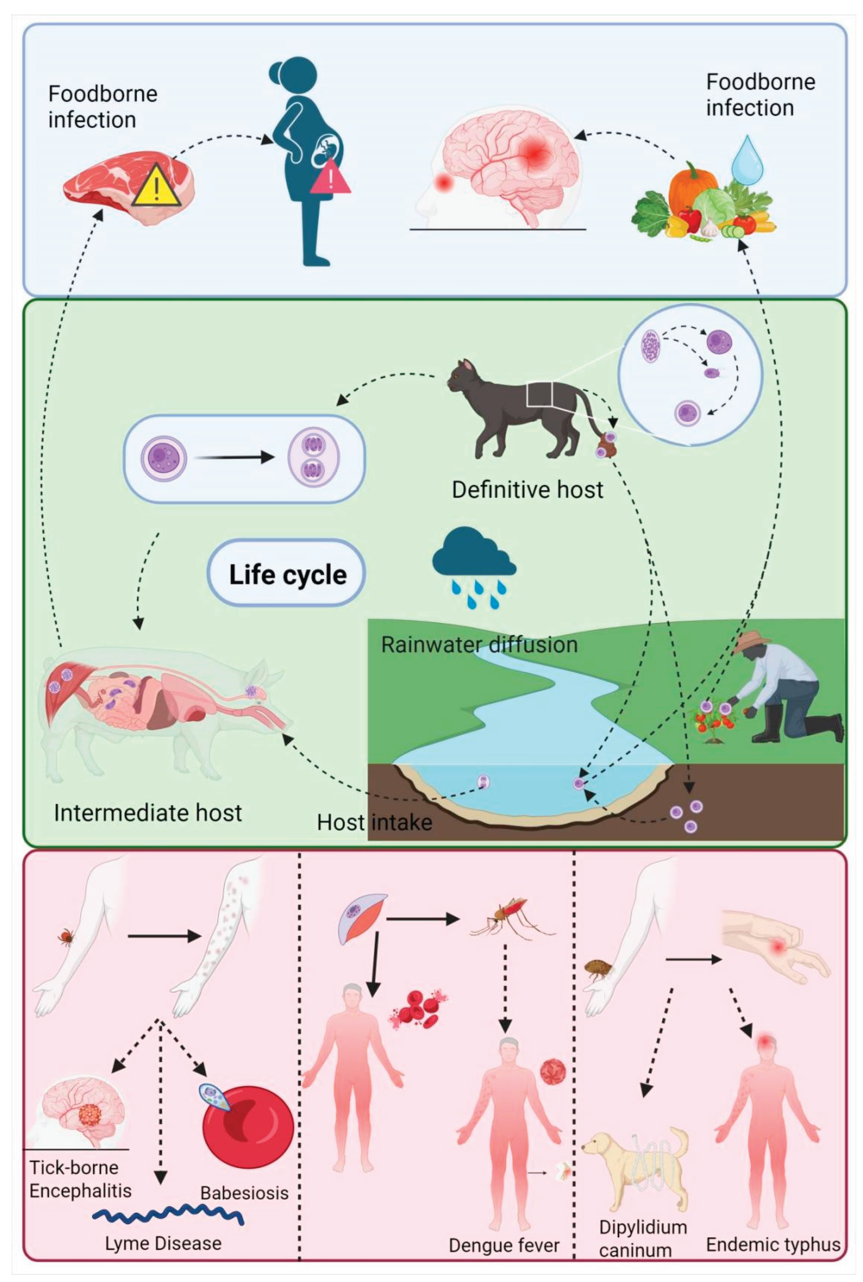
Figure 2.
Pathways of transdermal absorption of nanoformulated drugs.
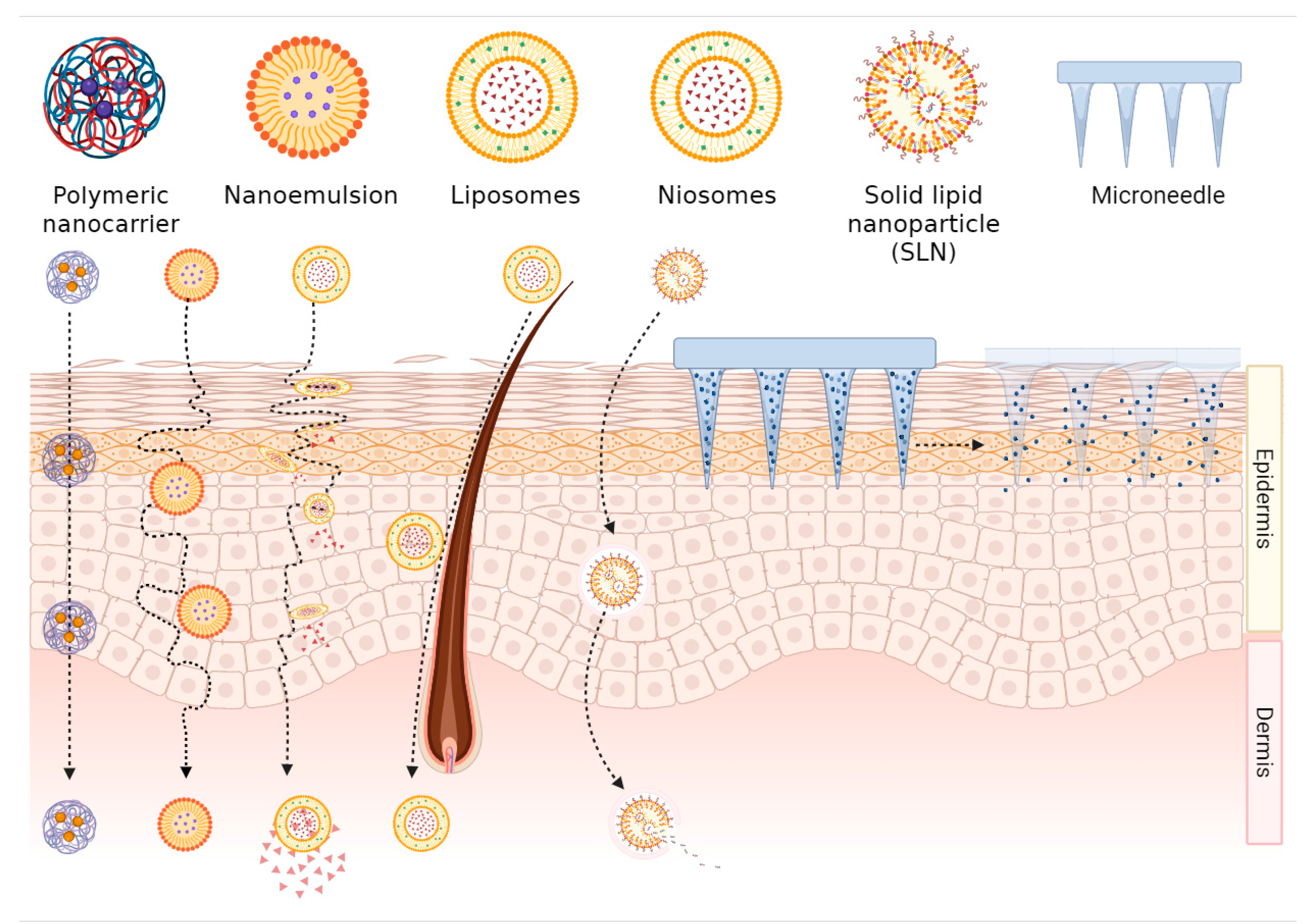
Figure 3.
Examples for transdermal nanoformulations against leishmaniasis and malaria.
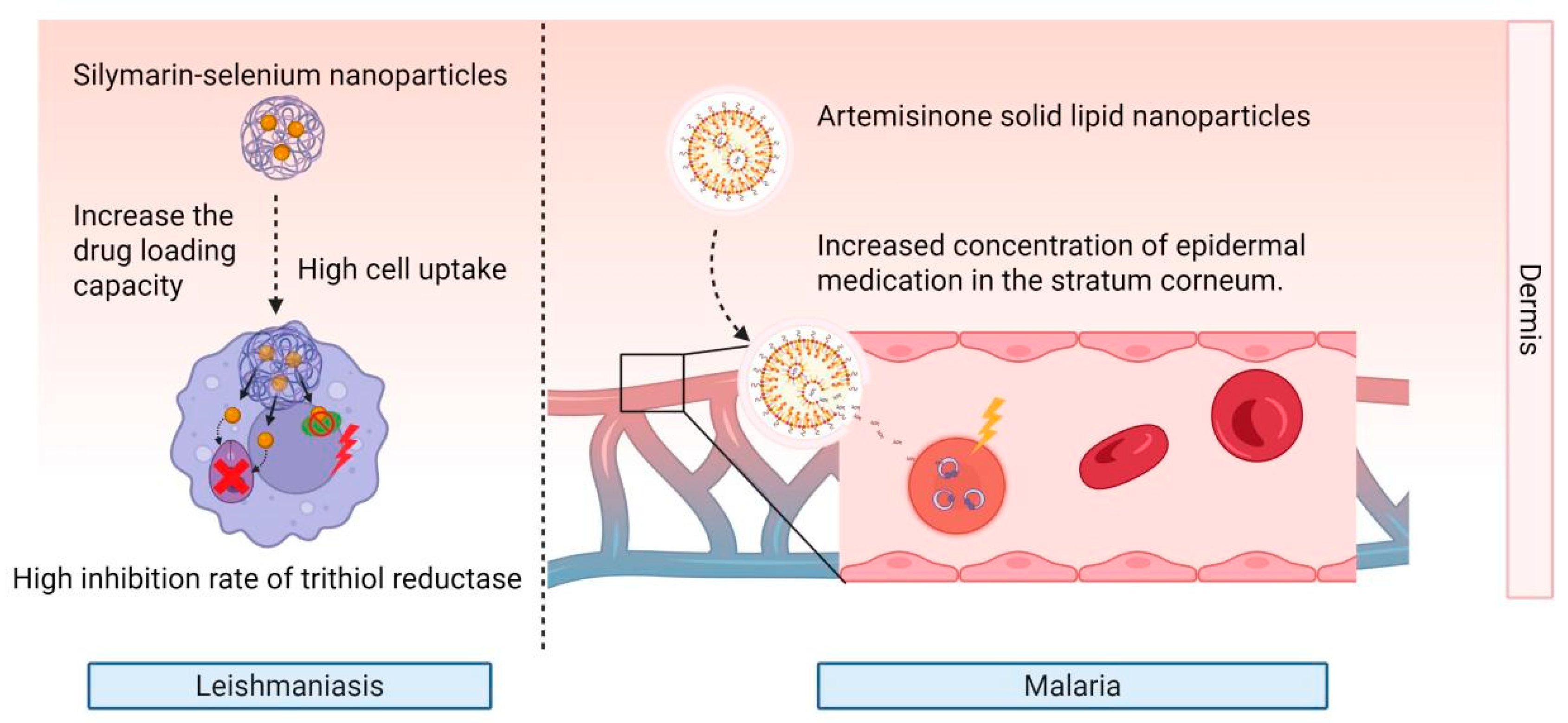
Figure 4.
Mechanism of ivermectin nanoemulsion in treating strongyloidiasis.
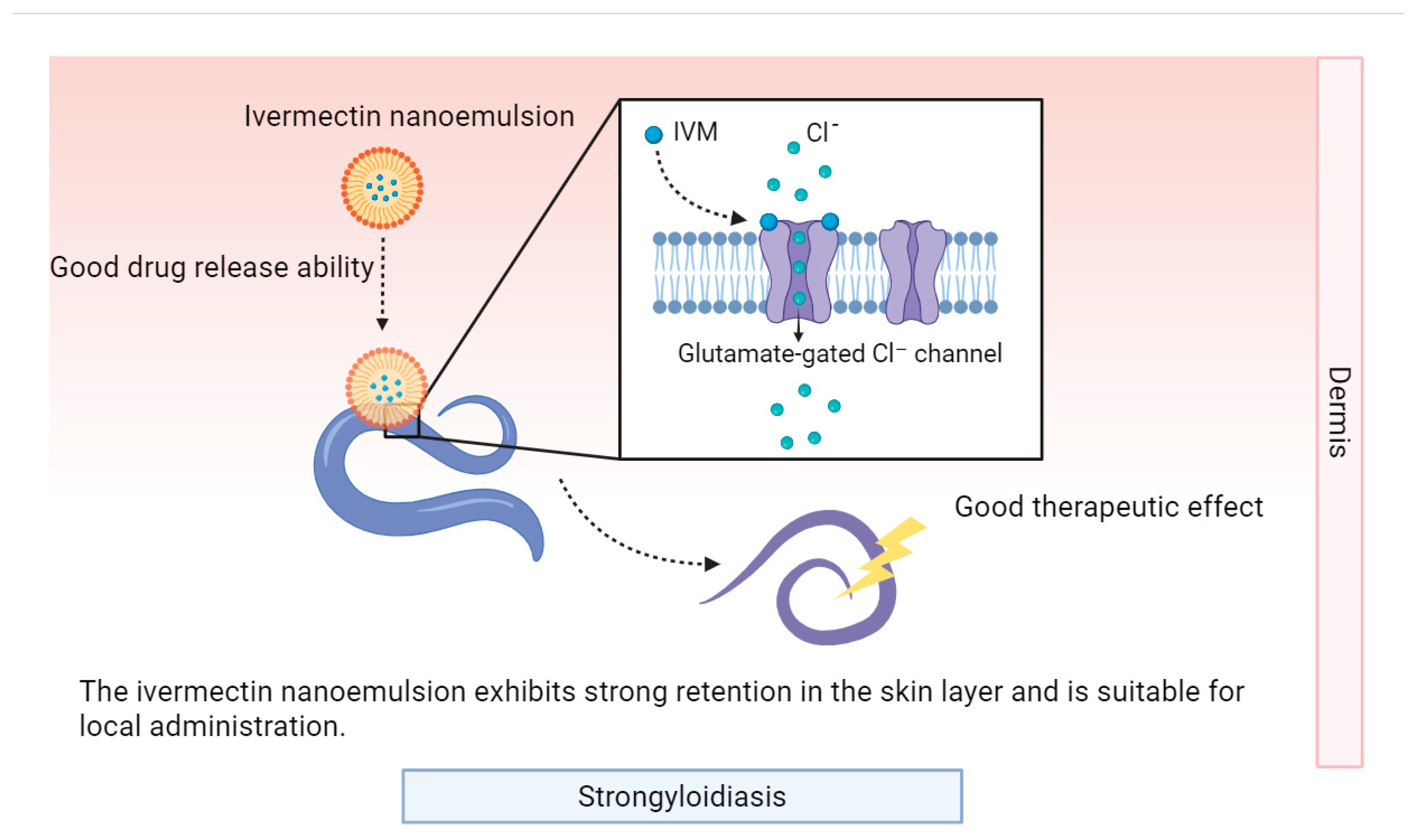
Figure 5.
Mechanism of solid lipid nanoparticle-dissolving microneedle system of albendazole.
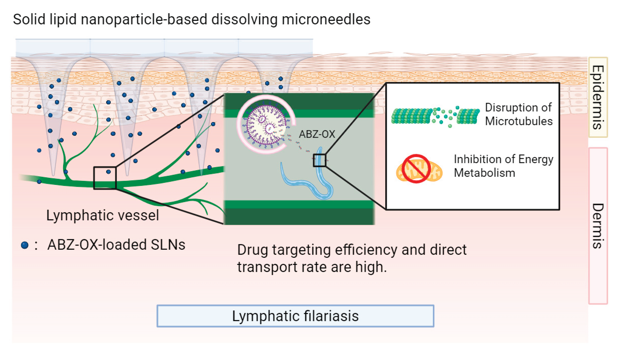

Table 1.
Nanoformulations of antiparasitic drugs for zoonotic diseases.
| Indication | Nanoformulation | Compounds | Advantages | References |
|---|---|---|---|---|
| Leishmaniasis | Transfersomes | Trifluralin | High transdermal efficiency, high encapsulation efficiency, sustained release, reduced IC50 for Leishmania pathogens, high inhibition rate of amastigotes | [20] |
| Transfersomes | Nitazoxanide-quercetin | Enhanced skin permeation, increased macrophage uptake, higher CC50, smaller lesion size, low systemic toxicity | [21] | |
| Nanoemulsions | 3’-(Trifluoromethyl)-chalcone | High dermal permeation amount, high skin retention | [25] | |
| Nanoemulsions | C6I/TC1/TC2 | Enhanced skin permeation, Sustained release, high parasiticidal activity, low macrophage cytotoxicity | [26] | |
| Electrospun core-shell nanofibers | Glucantime | Stable drug release, maintenance of effective drug concentration for a long time, sustained therapeutic effect | [59] | |
| Nanoparticles | Silymarin-selenium | High drug loading capacity, high skin deposition rate, significant reduction of local treatment toxicity | [60] | |
| Transfersomes | Rifampicin | High skin permeability, targeted reduction of IC50 value | [46] | |
| Nanostructured lipid carrier | Glucantime | Controlled drug release, enhanced skin retention, reduced systemic toxicity | [45] | |
| Nanofibers | Berberine | Gradient release, excellent biocompatibility, stable release rate | [33,34] | |
| Microneedle | Aphotericin B | Improved skin permeability, minimal cellular damage | [31] | |
| Malaria | Ethosomes | Artesunate and Febrifugine | High cumulative permeation, high efficiency | [22] |
| Solid lipid nanoparticles | Artemisone | High skin delivery concentration | [71] | |
| Elastic liposomes | PfMSP-119 | Efficient targeting, long-lasting immune response | ||
| Nanoparticles | Curcumin | High antimalarial activity, prolonged drug circulation time, high bioavailability, enhanced therapeutic efficacy | [47,48,73] | |
| Nanostructured lipid carrier | Artemether | High cumulative permeation rate, high drug stability, high release controllability | [75] | |
| Microneedles | Primaquine | Optimal performance, safe for long-term medication | [76] | |
| Cystic Echinococcosis | Microneedle | Albendazole | High transdermal permeation amount, safe | [32] |
| Microneedles-Nanocrystals system | Albendazole | High transdermal depth, high peak drug concentration, long half-life | [77] | |
| Lymphatic filariasis | Solid lipid nanoparticle - microneedle system | Albendazole | High drug targeting efficiency, high direct transport rate, Sustained release, reduced metabolite generation, low drug distribution in liver and kidney | [79] |
|
Scabies, Rosacea, Head lice, Trichuriasis, Onchocerciasis, Lymphatic filariasis, Strongyloidiasis |
Microemulsion | Ivermectin | Improved membrane permeability, high solubility | [86] |
| Nanocrystals | Ivermectin | High equilibrium solubility, fast dissolution rate, high dermal deposition, low side effects | [87] | |
| Nanoemulsion (NE) | Ivermectin | Highest drug concentration in stratum corneum and epidermis/dermis junction | [85] | |
| Nanoemulsion gel (NEG) | Ivermectin | Fastest delivery rate | ||
| Colloidal system (CS) | Ivermectin | Highest drug diffusion percentage, efficient delivery | ||
| Solid lipid nanoparticles | Ivermectin | Sustained-release efficacy, targeting, low systemic toxicity, few side effects | [40] | |
| Nanoparticles | Ivermectin | suitable for acute infection management | [89] | |
| Nanocapsules | Ivermectin | suitable for chronic prevention | ||
| Hybrid micelles | Eprinomectin | High permeability, low toxicity | [88] |
Disclaimer/Publisher’s Note: The statements, opinions and data contained in all publications are solely those of the individual author(s) and contributor(s) and not of MDPI and/or the editor(s). MDPI and/or the editor(s) disclaim responsibility for any injury to people or property resulting from any ideas, methods, instructions or products referred to in the content. |
© 2025 by the authors. Licensee MDPI, Basel, Switzerland. This article is an open access article distributed under the terms and conditions of the Creative Commons Attribution (CC BY) license (http://creativecommons.org/licenses/by/4.0/).
Copyright: This open access article is published under a Creative Commons CC BY 4.0 license, which permit the free download, distribution, and reuse, provided that the author and preprint are cited in any reuse.


