
Submitted:
19 August 2025
Posted:
21 August 2025
You are already at the latest version
Abstract
Camelids are increasingly recognized as important livestock species. They are valuable sources of meat, fiber, and milk. Despite their growing popularity, many aspects of their reproductive physiology and pathology remain unclear. Their reproductive performance is reported to be low in many countries. Advances in camelid veterinary care have identified several disorders, some of which are species-specific. This article describes an approach and diagnosis of infertility and subfertility cases in alpacas, llamas, and camels referred to the authors over the past 35 years. Ultrasonography, endometrial cytology, and biopsy are the primary diagnostic tools for practitioners. However, laparoscopy, hysteroscopy, and cytogenetics are indicated for cases referred to theriogenologists. The incidence of congenital and acquired reproductive disorders is presented. A high incidence of congenital defects of the reproductive tract is found in South American camelids, which raises concerns about animal welfare. Acquired disorders are similar to those described in other species. Endometritis and endometrosis are major disorders contributing to infertility and early pregnancy loss. However, studies on uterine defense mechanisms and the pathogenesis of these disorders are lacking. Hydrobursitis, a common cause of infertility in dromedary camels, warrants further research. The implications of some contagious diseases (tuberculosis, campylobacteriosis, and brucellosis) in female infertility are discussed. These findings emphasize the importance of including camelid medicine in veterinary education to ensure a high standard of care for this species.
Keywords:
dromedary
; alpacas
; llamas
; breeding soundness exam
; infertility
; pregnancy loss
Introduction
Camelids have been recognized for a long time as essential production animals in many areas of the world. However, seasonal pregnancy and birthing rates in traditionally managed herds are generally lower compared to those observed in other domestic species. In camels, the calving rates in extensive herds vary between 25 to 50% [16,40,68,85]. The estimated seasonal pregnancy rate in Peruvian alpaca and llama herds ranges between 40 and 60% [39,46]. On the other hand, intensively managed herds, under good veterinary care, achieve pregnancy rates of 70% or more both in South American Camelids (SACs) and dromedary camels [82,87]. The discrepancy in reproductive performance between traditionally and intensively managed herds is due to several factors, including nutrition and the prevalence of infectious and parasitic diseases. These factors affect both ovarian activity and the ability to maintain pregnancy (i.e., high incidence of early pregnancy loss) [23,30,46,57,61].
The role of individual male and female infertility or subfertility in the overall herd reproductive performance has been understudied until recently. Until the 1990s, most observations on reproductive disorders and infertility in camelids were derived from a single case report or abattoir studies [77,78]. The growing interest in performance camels (racing and show) and SACs outside of South America emphasized the importance of reproductive soundness of the female. Elite females have increased in value and are often maintained in production for a longer time. Additionally, many of the genetically superior females are routinely enrolled in multiple ovulation and embryo transfer programs [23,32]. These developments have increased the importance of individual medicine and the need to develop appropriate approaches for the diagnosis of the cause of subfertility and infertility in females.
This paper outlines the approach used by the authors for the diagnostic work-up of infertility or subfertility in female camelids. It provides a retrospective analysis of reproductive disorders in females referred for infertility over the past 35 years. During this time, the average number of cases seen annually is 80 for SACs (USA) and 3,000 for dromedary camels (UAE, Qatar, and Morocco). Only cases with complete medical records were included in this report. Incomplete records were primarily due to a lack of precise history (dromedaries) and the owner declining further diagnostics. Dromedary camels will be referred to as 'camels' throughout the paper.
Breeding Soundness Examination of the Subfertile Female
An approach to the investigation of female infertility and subfertility with a complete description of the examination techniques was published in 1997 [77] and updated more recently [83,87]. The female breeding soundness includes a complete medical and reproductive history, followed by a physical examination, and a thorough evaluation of the reproductive tract [84]. The main complaints in female camelids with complete medical records are summarized in Table 1. Repeat breeding is the main complaint, followed by early pregnancy loss. These results are consistent with previously published reports in camelids [13,84,87].
A detailed reproductive history is crucial for clinical case definition. Unfortunately, one of the significant constraints in camels is the lack of precise information on breeding management. This differs from SACs in our practice, where the breeding management and results of each mating (i.e., pregnancy diagnosis) are recorded.
Reproductive examination techniques have improved significantly due to the knowledge gained in reproductive physiology, in particular follicular dynamics, and the use of transrectal ultrasonography, vaginoscopy, as well as endometrial cytology and culture [71,75,76]. The objective of the initial examination is to rule out major congenital and acquired causes of infertility. This examination is often sufficient to establish a diagnosis in most infertility cases [13,84,87]. It is imperative to inform clients that often the cause of infertility cannot be determined in a single theriogenology visit. Follow-up reproductive evaluations are necessary to ascertain that the female can ovulate and conceive. More advanced techniques such as endometrial biopsy, hysteroscopy, cytogenetics, endocrinology, and laparoscopy are indicated in cases of ovulation failure, fertilization failure, or recurrent early embryo loss [77,83].
Complete reproductive examination records were available for 413 SACs and 2387 camels presented for infertility. A diagnosis of the cause of infertility was made for 92.7% of the camel cases and 100% of the SAC cases. The lack of diagnosis in 7.3% of the camel cases can be explained by the inability to perform more advanced examination techniques, such as endometrial biopsy and hysteroscopy. For discussion purposes, reproductive disorders will be divided into two categories: congenital and acquired defects.
Congenital Defects
All the congenital disorders were observed in infertile maiden females. The main complaints were repeat breeding and visible abnormalities of the external genitalia. Overall, congenital disorders of the reproductive tract were more commonly found in SACs compared to camels. Congenital disorders of the reproductive tract and their relative frequency are presented in Table 2. The most common diagnosed disorders were ovarian dysgenesis, segmental aplasia of the tubular genitalia (uterus unicornis, persistent hymen, hydrosalpinx, vaginal aplasia), and atresia vulvi.
Ovarian dysgenesis/hypoplasia was diagnosed in 36.3% of maiden SACs presented for infertility. This is higher than the 16.8% incidence reported in slaughtered alpacas that were culled for infertility [72]. The high incidence in our data may be due to the inability of referring veterinarians to diagnose these defects. Affected females are generally presented for repeat breeding without evidence of ovulation. In rare cases, the female may be presented due to male rejection in the absence of a pregnancy or a rise in progesterone level [64]. The disorder is characterized by the lack of normal follicular activity, difficulty in visualizing the ovary by ultrasonography, and an infantile reproductive tract [24,87]. Confirmation of the diagnosis is achieved via laparoscopy (Figure 1). Hormonal diagnosis (i.e., serial serum estradiol concentration, anti-mullerian hormone (AMH) concentration) is possible but rarely performed. The ovaries are extremely small, measuring 2 to 3 mm in length. Histologically, this syndrome can be categorized as ovarian hypoplasia (i.e., germ cell deficiency or low germ cell resistance), ovarian dysgenesis (i.e., defective embryonic development of the gonad), or ovarian dysplasia (i.e., abnormal follicular development) [24].
Several chromosomal abnormalities have been associated with ovarian hypoplasia/dysgenesis in SACs, including X0, XX/XY, and XXX [54,87]. A unique cytogenetic abnormality, known as the minute chromosome syndrome, has been observed in alpacas and llamas, affecting over 50% of cases of ovarian dysgenesis. This abnormality is characterized by a significant size discrepancy between chromosome 36 homologs, suspected to result from a translocation [25,45,66].
Ovarian follicular inactivity has been documented in cases of infertility in camels, but no cytogenetic studies are available [13,60,84].
Cystic formations of the caudal mesonephric tubules (Wolffian ducts), also called paraovarian cysts or paroophoron, are commonly found in all camelids but were not included here because they do not affect fertility. However, they should be differentiated from hydrosalpinx and hydrobursitis, which can impair fertilization and oviductal embryo transport.
Other congenital abnormalities of the ovary include cystic rete ovarii and congenital ovarian teratomas [1,4,43,74,84,87].
Segmental aplasia can affect any part of the tubular genitalia. An imperforate (persistent) hymen is the most common of these abnormalities. Affected females are presented with discomfort or tenesmus during mating. Transrectal ultrasonography reveals mucocolpos and mucometra (Figure 2). Other forms of segmental aplasia of the tubular genitalia include vaginal aplasia, uterus unicornis, and congenital hydrosalpinx due to segmental aplasia of the uterine tube (Figure 3) [13,48,82,87].
Acquired Disorders of the Ovaries and Ovarian Bursa
The relative frequency of disorders of the ovaries and uterine tube diagnosed in infertile female SACs and camels is presented in Table 3.
Cystic structures are the most common ovarian abnormalities found in both SACs and camels. They have been reported previously in SACs, [3,31] dromedaries [7,41,47,76], and Bactrian camels [35]. Their role in infertility is not clear. A higher incidence of ovarian cysts was reported in infertile (8.3%) compared to fertile (4.7%) alpacas [72]. A large proportion (30 to 40%) of dromedary females develop anovulatory follicles in the absence of copulation [76]. The incidence of anovulatory follicles varies from 0.9% to 43% depending on the study [6,41,43,47,55,88]. An increase in the incidence of anovulatory follicles was observed in females with clinical endometritis, ovarian hydrobursitis, and vaginal/cervical adhesions [6]. Some females appear to be more susceptible to developing anovulatory follicles. This has been attributed to hormonal imbalances, metabolic disorders, and oxidative stress [41]. However, studies comparing follicular fluid and serum biochemical profiles in females with normal follicles and those with anovulatory follicles did not show any significant differences [26]. On ultrasonography, these structures are large, reaching a diameter of up to 5 cm in llamas and alpacas, and up to 12 cm in dromedaries. They can be completely anechoic or present variable degrees of hemorrhage and luteinization (Figure 5) [43,75,76]. A rise in serum progesterone level is observed in cases of luteinized hemorrhagic follicles [19]. It is noteworthy that embryo recipients with a luteinized anovulatory follicle can maintain the pregnancy and deliver a live newborn [23].
Lack of ovarian follicular activity was observed in 24.7% of SACs and 29.1% of dromedaries referred for infertility. It is important to note that despite the absence of follicular activity, female camelids can still be receptive to the male. Ovarian inactivity is often associated with loss of body condition during lactation or after a debilitating disease. In camels, seasonal inactivity is marked under harsh conditions and poor nutrition [69]. The incidence of inactive ovaries is substantially higher (57.6% vs. 5.8%) in maiden than in multiparous racing camels [60]. Endocrine abnormalities (i.e., hypothyroidism) and administration of anabolic steroids have been suspected as a potential cause of ovarian inactivity. Aging is also an important factor in the depletion of ovarian follicular reserves. In alpacas, females aged 16 years or older had lower serum AMH concentrations compared to younger females [67].
Oophoritis has been described primarily in slaughtered camels [40,43,47,56] and can be associated with abscesses and hydrobursitis. In cases of oophoritis and ovarian abscess, adhesions may extend to the intestines.
Hydrobursitis, or ovariobursitis, is a peculiar disorder in camels characterized by the accumulation of varying amounts of fluid within the ovarian bursa [80]. This pathology has been reported worldwide but appears to have a higher incidence in the Middle East [16,27,47,55,60,80]. The etiopathogenesis remains unclear. However, based on biochemical analysis of the fluid content, in some cases, the cause is attributed to recurrent ruptures of hemorrhagic follicles [10,80]. A variety of bacteria have been isolated (Actinobacillus spp., Escherichia coli, Klebsiella pneumoniae, Pseudomonas aeruginosa, Staphylococcus spp., and Streptococcus spp., as well as others) [5,10,18]. More recently, Chlamydophila abortus (Chlamydia abortus) infection has been demonstrated in several cases of hydrobursitis [17]. The role of Chlamydia spp., Brucella spp., and Campylobacter spp. in this pathology merits further investigation. Clinically, hydrobursitis is suspected when uterine retraction is not possible despite normal size of the uterus and cervix. Confirmation is easily obtained by transrectal or transcutaneous inguinal ultrasonography (Figure 6). This condition appears to occur predominantly on the left side. Bilateral development is more frequent in females with long-standing infertility (more than 2 years) [80]. The main signs include repeat breeding, early embryo loss, and abortion [13,16,27,80]. Endometritis, uterine adhesions, and pyometra may also be diagnosed in cases of hydrobursitis [6,10,13]. Treatment with oxytetracycline (20 mg/kg IM for 7 days) combined with intrauterine infusion of metacresol sulfonic acid and formaldehyde solution was shown to be effective in restoring fertility if the lesion is small (<3 cm) [11]. Surgical excision of the affected side is the only treatment for large lesions (Figure 7) [80].
Persistence of luteal activity in the absence of pregnancy has been reported following mating-induced ovulation, spontaneous ovulation, and follicular luteinization [2]. An increased incidence is observed in females with endometritis [19].
Ovarian neoplasms are rare in camelids [20,87]. Most cases described are from slaughterhouse studies. In camels, the reported incidence of ovarian tumors varies from 0.06% to 6.8% [28,43,47]. In alpacas, an incidence of 3.2% of all genital abnormalities, with most being teratomas, was reported in Peru [72]. Granulosa cell tumors (GCT) and Granulosa-Theca cell tumors (GTCT) have been described in camels [12,20,43] and llamas [22,34]. Females may be presented for failure to conceive or for displaying male-like behavior [22,34]. On transrectal palpation, the affected ovary is enlarged with an irregular surface, while the contralateral ovary is inactive. On ultrasonography, the ovary has a multilocular appearance. There are no reports on the use of inhibin or anti-Mullerian hormone (AMH) serum levels for the diagnosis of GTCT in camelids. Metastasis of GTCT is rare, although it was suspected in two llamas [22,34]. Other ovarian tumors reported in camelids include interstitial cells (Leydig cells) [51,89] and Sertoli-Leydig cell tumor (arrhenoblastomas) [15], which are associated with the elevation of serum androgen levels. Other types of ovarian neoplasia have been reported in dromedary slaughterhouse specimens, including papillary cystadenoma, fibroadenoma, luteoma, thecoma, fibroma, cavernous hemangioma, and dysgerminoma [4,43].
Acquired Uterine Tube Disorders
Occlusion of the uterine tubes, resulting in hydrosalpinx and pyosalpinx, has been reported in camelids with chronic salpingitis [55,56,79,81,84,87]. The incidence of uterine tube pathology in slaughterhouse specimens is 0.7% for alpacas [72] and 6.1% for camels [63]. The etiopathogenesis of camelid salpingitis has not been studied, but it is suspected to result from systemic or ascending infection from the uterus following abortion and metritis. In a clinical study, the incidence of pyosalpinx in infertile female camels was 1.8% [12]. Accumulation of fluid or pus in the uterine tube can be diagnosed by transrectal palpation and ultrasonography. This pathology may be associated with ovario-bursal adhesions. Inflammatory changes and the presence of microabscesses are evident on histopathology (Figure 8). Some cases of salpingitis that extend to the uterotubal junction are evident on hysteroscopy. However, many salpingitis cases may go unnoticed in clinical practice [62,84]. Techniques for the evaluation of uterine patency have not been studied in camelids. The authors have used primarily two approaches: embryo collection and surgical normograde flushing of the uterine tube [83].
Acquired Uterine Disorders
Inflammatory and degenerative uterine pathologies are the most common causes of subfertility or infertility in camelids (Table 4) [82].
Endometritis has been reported by several authors, mainly in slaughterhouse camel specimens [29,40,47]. Endometrial cytology, culture, and biopsy have all been used for the diagnosis of endometritis in camelids [9,33,82,83,86,87]. The endometrial cytology sample is best obtained with either low-volume flushing or a cytobrush® (Figure 9) [82,83]. Post-mating uterine inflammation is always present in camelids due to intrauterine copulation. Healthy polynuclear neutrophils can be present in the sample for up to 3 days after mating. It is crucial to consider both cytological and bacteriological findings to increase the sensitivity and specificity of diagnosis in cases of subclinical endometritis [38,70,77,82,87]. Studies on the local defense mechanism of the uterus in camelids are lacking. Our clinical observations suggest that the uterus of camelids has efficient mechanisms to clear this post-breeding inflammation and infection. These mechanisms may fail due to overbreeding, degenerative uterine changes, systemic illness, and inappropriate intrauterine manipulation.
The most common bacterial isolates in cases of camelid endometritis are Streptococcus equi zooepidemicus, β-hemolytic Streptococci, Enterococcus spp., coagulase-negative Staphylococcus spp., Proteus spp., Enterobacter aerogenes, Klebsiella pneumoniae, Pseudomonas aeruginosa, and Trueperella pyogenes [13,38,43,44,70,86,92]. Samples from camels should also be examined for Ureaplasma spp., Mycoplasma spp., and fungi [38,43,44,91,92], and Campylobacter fetus venerealis, which has been suspected to cause infertility [86,90,91,92]. Endometrial biopsy is considered the gold standard for the diagnosis of endometritis and should be an integral part of the evaluation of the barren female (camel [44,50,70,83,84], llama, and alpaca [65,81,87]). The endometrium shows widespread polymorphonuclear neutrophils in acute endometritis (Figure 10).
In chronic endometritis, the infiltration consists predominantly of lymphocytes, and occasional plasmacytes, macrophages, eosinophils, or mast cells (Figure 11).
A granulomatous chronic endometritis has been described in camels. Similar lesions have been described in cattle with campylobacteriosis, tuberculosis, or chronic fungal infections (Figure 12) [44,52,77,82].
Controlled trials on the treatment of endometritis in camelids are scarce. Therapeutic approaches have been largely adapted from those used in the equine and bovine species. These consist of uterine lavages with saline or weak antiseptic solution, followed by daily systemic or intrauterine antibiotic treatment [33,82]. A first-generation cephalosporin (cephapirin) used for intrauterine infusion in cattle has also been successfully used in camels [49,73]. It is important to note that camelids are food-producing animals in many countries, and veterinarians should adhere to rules of antibiotic stewardship and educate clients about withdrawal times for milk and meat for each drug. Antiseptic solutions formulated for cattle (1% policresulen, 4% metacresol sulphonic acid and formaldehyde, 0.1% solution of acriflavine) have been used successfully to treat uterine infections in camels [8]. Additional therapies used in equine practice, such as uterine infusion with mucolytic or chelating agents (N-acetylcysteine 20%, tris-EDTA, and Tricide®) to dissolve biofilm and improve antimicrobial activity, have been used anecdotally for the treatment of endometritis in camelids. A preliminary clinical trial was conducted on eight camels using ozone foam for the treatment of chronic endometritis with satisfactory results (A. Tibary, personal observation). Future studies should include the use of antimicrobial peptides and platelet-rich plasma for the treatment of endometritis in camelids.
The incidence of endometritis can be reduced by minimizing the risk of contamination of the uterus during mating, obstetrical interventions, and other reproductive manipulations. In our experience, the leading causes of uterine infections are improper intrauterine treatments and overbreeding. The reproductive tract microbiome and the role of the vaginal flora changes in the pathophysiology of endometritis in camelids merit more investigation [42].
Other uterine disorders include degenerative changes, pyometra/mucometra, and uterine tumors. Chronic degenerative changes or endometrosis are a common histopathological finding in camelids with a history of repeat breeding or recurrent pregnancy losses. Its pathophysiology remains poorly studied in camelids, but it is often a consequence of chronic uterine inflammation. Although a classification of endometrial biopsy has been published for camelids, there is no data correlating this biopsy grade to the probability of a female carrying a pregnancy to term [65,83,87].
Pyometra and non-congenital mucometra are often due to cervical or vaginal adhesions resulting from poor handling of dystocia or fetotomy [13,29,87]. Uterine cysts and intraluminal adhesions have also been observed following dystocia or postpartum uterine prolapse (Figure 13).
Uterine neoplasms are rare in camelids. Reported cases include adenocarcinomas, leiomyomas, and hemangiomas [1,20,58,65,82,87]. Females are presented for repeat breeding or post-mating vaginal bleeding. Most cases are suspected on transrectal palpation or ultrasonography and confirmed by biopsy or postmortem examination of the uterus. Long-term prognosis for all uterine neoplasms is poor. Metastases to the lymph nodes, mesentery, and lungs have been reported in SACs [1,58].
Other uterine diseases found in camelids include uterine cysts, uterine abscesses, peri-uterine adhesions, intraluminal adhesions, uterine wall perforation, foreign bodies, polyps, fibromas, and endometrial gland dysgenesis [28,48,82,87]. Most of these disorders are suspected on transrectal palpation and ultrasonography, and can be confirmed by uterine biopsy, hysteroscopy, or laparoscopy. They generally carry a poor prognosis for fertility [82].
Acquired Disorders of the Cervix
The most common acquired pathological condition of the cervix diagnosed in our practice is cervical trauma (adhesions, tears) (Table 5). These lesions are often the result of unattended dystocia and inadequate obstetrical or gynaecological manipulations [9,13,87]. Females with these cervical pathologies are presented for repeat breeding or recurrent pregnancy loss. Mucometra is always present in cases of non-patent cervixes [9,28,37,44,84]. Cervical fibrosis and stenosis occur in some cases and are suspected when catheterization of the cervix is not possible. In alpacas, hysteroscopy or contrast fluoroscopy are the best methods for diagnosis [87]. Breaking cervical adhesions manually has been suggested as a treatment [37]. However, the adhesions tend to recur and are often more severe. Placing a cervical stent may provide a better way to prevent large fluid buildup and help maintain the reproductive capacity of valuable females, enabling them to be used for oocyte collection and in vitro embryo production.
Acquired Disorders of the Vulva and Vagina
In camelids, acquired disorders of the vulva and vagina include inflammation (vulvitis, vaginitis), trauma, and neoplasia (Table 6). Vulvar edema and vulvitis may result from irritation (mating, diarrhea, or chemical) or parasite infestation (i.e., mange, ticks, myiasis due to Wohlfahrtia Magnifica) [21,82,83]. A form of viral granular vulvovaginitis has been described in camels and attributed to Bovine herpes virus-1 [36]. Vulvovaginitis is also observed in camel pox virus infections. Non-specific or zinc-responsive vulvar and perineal hyperkeratosis is often seen in alpacas and llamas.
Vaginitis is uncommon and associated with poor breeding management and inadequate gynecological manipulations. Necrotic vaginitis is often seen following dystocia and may progress to vaginal adhesions leading to mucometra [37,83,87]. A few cases of vaginal tumors (leiomyosarcoma in an alpaca [53], adenocarcinoma in camels [14]) have been reported.
Rectovaginal tears are common following dystocia due to the small perineal body in camelids. In camels, lesions on or around the vulva may be due to "traditional infertility treatment” (firing, clitoridectomy) [83]. Vulvar neoplasia is rare. Two cases of vulvar squamous cell carcinoma have been diagnosed by the authors in llamas (Figure 14).
Conclusion
This article aimed to provide an overview of the advances in the diagnosis of reproductive disorders in female camelids. The distribution of disorders shown here may not accurately represent their actual occurrence in the general population, as our cases are drawn from referrals to specialized centers and are thus selected samples. However, our data illustrate the wide range of disorders that may affect the camelid reproductive system. Significant progress has been made in developing methods to diagnose these disorders. However, there is a considerable discrepancy between the standard of care for SACs and camels. In camels, advanced techniques such as endometrial biopsy and endocrinology are often not used, and cytogenetic evaluations are entirely lacking.
In our experience, camelid veterinary care is often not included in the standard veterinary curriculum in many countries. This deficit frequently results in a significant shortage of veterinarians equipped with the clinical and laboratory skills necessary to perform accurate reproductive evaluations. Failure to properly diagnose reproductive disorders can have serious consequences for animal welfare, especially for females with congenital defects.
Uterine disorders, endometritis in particular, appear to be a major contributor to reproductive loss. Research on camelid uterine defense mechanisms, the role of the reproductive microbiome, and studies on factors predisposing to uterine infection in these species are urgently needed. In many regions of the world, improper diagnosis of uterine infection leads to the overuse of antibiotics with subsequent increases in the risk for antibiotic resistance, which poses a significant health concern for both animal and human populations.
Veterinarians should also consider assisted reproductive technologies (i.e., embryo collection, oocyte collection, and in vitro fertilization) as alternative methods for reproduction of valuable females with severe acquired uterine and utero-tubal disorders.
Author Contributions
Conceptualization, A.S., M.C., S.M.W., A.A., A.T.; writing—original draft preparation, A.S., M.C., A.T.; writing—review and editing, A.S., M.C., S.M.W., A.A., A.T.; Illustrations, A.T., A.A. All authors have read and agreed to the published version of the manuscript.
Funding
This research received no external funding.
Informed Consent Statement
Written informed consent has been obtained from the owner of the animals involved in this study.
Conflicts of Interest
The authors declare no conflicts of interest.
Abbreviations
| SACs | South American Camelids |
| USA | United States of America |
| UAE | United Arab Emirates |
| AMH | Anti-Mullerian Hormone |
| SPP | Species |
| GCT | Granulosa Cell Tumor |
| GTCT | Granulosa Theca Cell Tumor |
| EDTA | Ethylenediaminetetraacetic acid |
| IE | Id Est |
References
- Aboellail, T.A.; Waugh, M.; Harvey, A.; Fisher, J.; Vilander, A.C. Neoplasia and Proliferative Lesions of New World Camelids: A Systematic Literature Review and Retrospective Study of Cases Submitted to Colorado State University From 1995 to 2020. Front. Vet. Sci. 2021, 8. [CrossRef]
- Adam, C.L.; Moir, C.E.; Shiach, P. Plasma progesterone concentrations in pregnant and non-pregnant llamas (Lama glama). Vet Rec. 1989;125:618-620.
- Adams, G.P.; Sumar, J.; Ginther, O.J. Hemorrhagic ovarian follicles in llamas. Theriogenology 1991, 35, 557-568. [CrossRef]
- Al-Afaleq, A.I.; Homeida, A.M., Barakat, S.; Moqbel, M. Gross and Microscopic Lesions of the Ovaries of Camels from Abattoirs in Eastern Province of Kingdom of Saudi Arabia. Open J. Path. 2022, 12. [CrossRef]
- Al-Sultan, A.A.; Al-Eknah, M.M.; Al-Dougym, A.M; Elsayed, A. Detection of Klebsiella pneumoniae, Pseudomonas aeruginosa and ESBL Producing Escherichia coli Associated with Ovarian Hydrobursitis Syndrome in Female Camels (Camelus dromedarius). Asian J. Anim. Vet. Adv. 2014, 9, 499-505. [CrossRef]
- Ali, A; Derar, D.R. Ovary and Ovarian Bursa in Dromedary Camels: Clinical Relevance of the Topographical Features. Anat. Histol. Embryol. 2020, 49, 325-332. [CrossRef]
- Ali, A.; Tharwat, M.; Al-Sobayil, F.A. Hormonal, Biochemical, and Hematological Profiles in Female Camels (Camelus dromedarius) Affected with Reproductive Disorders. Anim. Reprod. Sci. 2010,118, 372-376. [CrossRef]
- Ali, A.; Al-sobayil, F.A.; Al-Hawas, A. Evaluating the Effectiveness of Different Treatments of Uterine Infections in Female Camels (Camelus dromedarius). Theriogenology 2010,74,40-44. [CrossRef]
- Ali, A., Derar, D.R., Almundarij, T.I. Infertility in Female Dromedary Camels. J.f Camel Pract. Res. 2021, 28, 267-276. [CrossRef]
- Ali, A.; Al-Sobayil, F.A.; Tharwat, M.; Hassanein, K.M.. Ovarian Hydrobursitis in Female Camels (Camelus dromedarius): Biochemical, Bacterial and Protozoal evaluation. Theriogenology 2011, 75, 734-741. [CrossRef]
- Ali, A., Al-Sobayil, F.A., Hassanein, K.M., Al Hawas A.. Ovarian Hydrobursitis in Female Camels (Camelus dromedarius): The Role of Chlamydophila abortus and a Trial for Medical Treatment. Theriogenology 2012, 77,1754-1758. [CrossRef]
- Ali, A.; Derar, D.; Alsamri, A., Al Sobayil F. Echography of Clinically Relevant Disorders in the Genital Tract of Female Dromedary Camels. Anim. Reprod. Sci. 2017, 182, 123-133. [CrossRef]
- Ali, A., Derar, R., Al-Sobayil, F., Al Hawas A.; Hassanein, K. A Retrospective Study on Clinical Findings of 7300 Cases (2007-2014) of Barren Female Dromedaries. Theriogenology 2015, 84, 452-456. [CrossRef]
- Ali, A.; Derar, D.; Al Sobayil, F. Tharwat M.; Fathy, A.; Khodei, M. Adenocarcinoma in the Genital Tract of Infertile Female Dromedary Camels. J Camel Pract. Res. 2018, 25, 181-187. [CrossRef]
- Ali, A.; Derar, D.; Hassanein, K.M.A.; Abdella, A.H.; Madeh, S; El Sayed E, Al Sobayil F.A. Sertoli-Leydig Cell Tumour in a Female Dromedary Camel. J. Camel Pract. Res. 2019, 26, 287-290.
- Ali, A.; Derar, D.; Alsharari, A; Alsharari, A.; Khalil, R.; Amindarij, T.I.; Alboti, Y.; Al Sobayil, F. Factors Affecting Reproductive Performance in Dromedary Camel Herds in Saudi Arabia. Trop. Anim. Health Prod. 2018, 50,1155-1160.
- Ali, A.; Derar, D.R.; Mousa, H.A.; Osman, S.A.; Refaai, W., Almudarij, T.I; Al Dubaib, M.A., Allam, S.A.. First Report on the Isolation of Chlamydia abortus from Female Dromedary Camels with Ovarian Hydrobursitis. Theriogenology 2022, 191,102-108. [CrossRef]
- Ali, L.M.; Shalaby, S.I.A.; Shalash, M.R.; Nawito, M.F.; Afify, M.M. Bacterial Status of Abnormal Genitalia of the Camels. Egyptian J. Vet. Sci. 1987, 24, 41-44.
- Almushawwah, K.S.; Derar, D.R.; Ali, A. Pseudopregnancy in Dromedary Camels: Characterization, Prevalence, and Clinical and Hormonal Properties. Theriogenology 2025, 235, 196-202. [CrossRef]
- Alsobavil, F.A.; Ali, A.; Derar, D.R.; Thawrat, M.; Ahmed, A.F.; Khodeir, M. Tumours in Dromedary Camels: Prevalence, Types and Locations. J. Camel Pract. Res. 2018, 25, 189-197. [CrossRef]
- An, X.; Yang, B.; Bao, H.; Oyun, G.; Wang, X.; Er, D. Morphological Observation of the Larva of the Alxa bactrian Camel Vaginal Myiasis. J. Camel Pract. Res. 2019, 26, 57-62. [CrossRef]
- Anderson, D.E., Couto, C.G.; Oglesbee, M. Granulosa Theca Cell Tumor with Erythrocytosis in a Llama. Can. Vet. J. 2010, 51,1157-1158, 1160.
- Anouassi, A.; Tibary, A. Development of a Large Commercial Camel Embryo Transfer Program: 20 years of Scientific Research. Anim. Reprod. Sci. 2013, 136, 211-221. [CrossRef]
- Arroyo, E.; Patiño, C.; Ciccarelli, M.; Raudsepp, A.; Conley, A., Tibary, A. Clinical and Histological Features of Ovarian Hypoplasia/Dysgenesis in Alpacas. Front. Vet. Sci. 2022;8. [CrossRef]
- Avila, F., Baily, M.P., Merriwether, D.A., Trifonov, V.A; Rubes, J.; Kuyzler, M.A.; Chowdhary, E.; Janecka, J.; Raudsepp, R. A Cytogenetic and Comparative Map of Camelid Chromosome 36 and the Minute in Alpacas. Chromosome Res, 2015, 23, 237-251. [CrossRef]
- Bekkouche, A.; Miroud, K.; Mimoune, N.; Benmour, B.; Kaidi, R.; Benaissa, M.H. Follicular Fluid and Serum Biochemical and Hormonal Profiles of Normal and Cystic Dromedary Camel Breeds. Vet. World 2022,15,2511-2516. [CrossRef]
- Benaissa, M.H., Faye, B., Kaidi, R. Ovarian Hydrobursitis in Slaughtered Female Camels (Camelus dromedarius) in Southeast Algeria. Emir. J. Food Agric. 2014, 26, 915-920. [CrossRef]
- Benaissa, M.H.; Faye, B.; Kaidi, R. Reproductive Abnormalities in Female Camel (Camelus dromedarius) in Algeria: Relationship with Age, Season, Breed and Body Condition Score. J. Camel Pract. Res. 2015, 22, 67-73. [CrossRef]
- Benaissa, M.H.; Mimoune, N.; Kaidi, R. Prevalence and Anatomohistopathologic Studies of Uterine Anomalies in She-Camels in Southeast Algeria. HVM Bioflux 2017, 9, 137-145.
- Bravo, P.W.; Sumar, J.; Bravo, W.; Sumar, J. Factores Que Determinan la Fertilidad en Alpacas. In: Anales de la V Convencion Internacional sobre Camelidos Sudamericanos Ogranizado por IVITA de la Universidad Nacional Mayor de San Marcos y Universidad San Antonio Abad del Cusco (Cusco, Peru) 1985:4.
- Bravo, P.W.; Stabenfeldt, G.H.; Fowler, M.E. Lasley, B.L. Ovarian and Endocrine Patterns Associated with Reproductive Abnormalities in Llamas and Alpacas. J. Am. Vet. Med. Assoc. 1993, 202, 268-272. [CrossRef]
- Campbell, A.; Tibary, A. Embryo Transfer in Camelids. Clinical Theriogenology 2017, 9, 321-327.
- Campbell, A.; Tibary, A. Infertility of Female and Male Camelids. Clinical Theriogenology 2017, 9, 311-319.
- Carrasco, R.A.; Hutt, K.D.; McKenzie, C.M.; Adams, G.P. Granulosa Cell Tumour in a Llama (Lama glama). Reprod Domest Anim 2023;58:459-464. [CrossRef]
- Chen, B.X.; Yuen, Z.X. Reproductive Pattern of the Bactrian Camel. Uppsala: Scandinavian Institute of African Studies, 1984;364-386.
- Darwish, S.F.; Hassanien, T.K., Salem, H.A. Patho-molecular Studies on the Occurrence of Bovine Herpes Virus Type 1 in the Genitalia of She Camels. Alexandria J. Vet. Sci. 2015, 46, 130-137. [CrossRef]
- Derar, R.; Ali. A.; Al-Sobayil. F.; Al Hawas, A. Clinical Findings and Reproductive Performance of Female Dromedary Affected with Vaginal and Cervical Adhesions and Stenosis. J. Camel Pract .Res. 2016, 23,179-184. [CrossRef]
- Derar, R.; Ahmed, A.; Saeed, E.M., Al Sobayl, F., Al Samri, A.; Wlbehiry, A. Diagnostic Evaluation of Subclinical Endometritis in Dromedary Camels. Anim. Reprod. Sci. 2020, 215. [CrossRef]
- Díaz, C.A.; Huamán, U.H.; Camacho. S.J.; Ampuero A.B.; Quispe, D.M; Huanca, W.L. Nivel Sérico de Estradiol a la Monta y su Efecto Sobre la Tasa de Concepción en Alpacas. Rev. Investig. Vet. del Perú 2011;22:312-317.
- Dinaol, B.; Amare, E.; Sisay, A.;Shaso, B; Mohammed, T.; Mohammed, A.; Mummed, B.; Regassa, D. Reproductive Diseases and Disorders of Female Camels: An Assessment and Pathological and Bacteriological Study in Eastern Ethiopia. Vet. Med. Int. 2021. [CrossRef]
- El-Badry, D.A.; Ibrahim, M.A.; Leil, A.Z. Hormonal and Biochemical Studies on Female Dromedary Camels Affected with Multiple Ovarian Cysts. Small Rumin. Res. 2020,188. [CrossRef]
- El-Deeb, W.M.; Fayez, M., Elsohaby, I.; Ghoneim, I, Al Marrii T, Kandeel, M.El Gioushy, M. Isolation and Characterization of Vaginal Lactobacillus spp. in Dromedary Camels (Camelus dromedarius): in vitro Evaluation of Probiotic Potential of Selected Isolates. Peerj 2020,8. OI: ARTN e8500. [CrossRef]
- Elshazly, M.O.; El-Rahman, S.S.A.; Hamza, D.A.; Ali, E. Prevalence of Non-neoplastic Ovarian Disorders in Non-pregnant She-Camels (Camelus dromedarius) and their Correlation to Bacteriological Isolation. Int. J. Vet. Sci. 2019, 8:20-27.
- Elshazly, M.O., Abd El-Rahman, S.S.; Hamza, D.A.; Ali, M.E. Pathological and Bacteriological Studies on Reproductive Tract Abnormalities of She-camels (Camelus dromedarius), Emphasizing on Zoonotic Importance. J. Adv. Vet. Anim.Res. 2020,7,633-646. [CrossRef]
- Fellows, E; Kutzler, M.; Avila, F.; Das, P.J; Raudsepp, T. Ovarian Dysgenesis in an Alpaca with a Minute Chromosome 36. J Hered 2014,105,870-874. [CrossRef]
- Fernandez-Baca, S.; Hansel ,W.; Novoa, C. Embryonic Mortality in the Alpaca. Biol Reprod 1970, 3, 243-251. [CrossRef]
- Gherissi, D.E.; Bouzebda, Z.; Afri, F.B.; Lamraoui, R., Chacha F. Cross-sectional Survey of Congenital and Acquired Genital Disorders in Sahraoui Female Camels (Camelus dromedarius) at El Oued Abattoir, South-East Algeria. Anim. Res. Int. 2019,16,3450-3462.
- Gherissi, D.E.; Lamraoui, R.; Chacha, F.; Bouzebda, Z.; Bouzebda F.A.; Hanzen, C. Genital Abnormalities Associated to Lack of Uterine Adenogenesis or Endometrial Gland Dysgenesis of Female Dromedary Camels (Camelus dromedarius). Open Vet. J. 2020,10:44-52. [CrossRef]
- Ghoneim, I.M.; El-Deeb, W.; Al-Fehaed, H.F. Efficiency of Single-dose Adminitration Therapies for Treatment of Endometritis in the Camels (Camelus dromedarius). J. Camel Pract. Res. 2018, 25, 291-294. [CrossRef]
- Ghoneim, I.M.; Waheed, M.M.; Hamouda, M.A. Al Eknah, M.M, Al Fehaed, H.F. Endometrial Biopsy Technique and Histopathological Dindings of Endometritis in Camels (Camelus dromedarius). J. Camel Pract. Res. 2013, 20, 65-70.
- Gilbert, R., Kutzler. M., Valentine, B.A. Semevolos, S. Hyperandrogenism from an Ovarian Interstitial-cell tumor in an Alpaca. J. Vet. Diag. Investig. 2006,18,605-607. [CrossRef]
- Gimbo, A, Zanghi, A. Diffuse Granulomatous Endometritis in the Dromedary. Histopathological Findings. Schweizer Archiv fur Tierheilkunde 1981,123,249-261.
- Hardefeldt, L.Y.; Poulsen, K.P.; McGuirk, S.M.;Livesey, A; Kich, C.; Perrier, MP.; Pinkerton, M.E. Urogenital leiomyosarcoma in an alpaca. Can. Vet. J. 2010, 51, 1387-1390.
- Hinrichs, K.; Horin, S.E.; Buoen, L.C.;Ting, Q., Ruth, G.R. X-Chromosome Monosomy in an Infertile Female Llama. J. Am. Vet. Med. Assoc. 1997, 210, 1503-1504. [CrossRef]
- Hussein, F.M.; Metwelly, K.K. Gross and Histopathological Affections of Slaughtered She-camel Genitalia. Proceeding of the International Camel Conference, Al Hasa, Saudi Arabia, Bikaner: Camel Publishing House, 2015, pp. 245-250.
- Hussein, N.A., Saad, A.M. Pathological Disorders of the Ovaries and Uterine tubes in Camels (Camelus dromedarius) Slaughtered at Tamboul Abattoir, Sudan. J. Camel Pract. Res. 2017, 24, 251-256. [CrossRef]
- Kapustka, J.; Budzyńska, M. Reproductive Losses and Their causes in Alpacas - a Survey-based Study. Animals 2022,12. [CrossRef]
- Klopfleisch, R., Grinten, E.V.D.; Gruber, A.D. Metastatic Uterine Adenocarcinoma and Hepatic Lipomatosis in a Llama (Lama glama). J. Vet. Diagn. Invest. 2009, 21, 280-282. [CrossRef]
- Mahdy, M.A.A.; Eldeen, M.S.N. True Uterus Didelphys in She-camel: A Case Report and Review of Literature. Front. Vet. Sci. 2024,11. [CrossRef]
- Mansour, N., Karen, A. Ovarian Inactivity in Female Dromedary Camels. Emir. J. Food Agric. 2021,33.171-177. [CrossRef]
- Nagy, P.; Reiczigel, J.; Gupta, A.D.; Barua, R.; Juhász, J. Pregnancy and Parturition in Dromedary Camels II. Incidence, Timing and Factors Affecting Early Pregnancy Loss (EPL) and the Outcome of Twin Pregnancies. Theriogenology 2021, 172, 289-299.
- Nourani, H.; Tafti, A.K.; Kafi, M. Pathological Study of the Uterine Tubes in Non-Pregnant Camels (Camelus dromedarius) Slaughtered in Yazd Province, Iran. Iranian J. Vet. Res. 2005;6:79-83.
- Nur, H.M. Some Reproductive Aspects and Breeding Patterns of the Somali Camel (Camelus dromedarius): Somali Academy of Sciences and Arts, 1983, 91-114.
- Pearson, L.K.; Tibary, A. Reproductive failure in female camelids. Clinical Theriogenology 2014, 6, 555-570.
- Powers, B.E.; Johnson, L.W.; Linton, L.B.; Garry, F.; Smith, J. Endometrial Biopsy Technique and Uterine Pathologic Findings in Llamas. J. Am. Vet. Med. Assoc. 1990, 197,1157-1162. [CrossRef]
- Raudsepp, T.; Mendoza, M.; Tibary, A. Clinical and Molecular Cytogenetics of Camelids: A Comprehensive Review. Cabi Agriculture and Bioscience 2025;6. [CrossRef]
- Schwartz, D.; Patino, C.; Waqas, S.; Attoyo, E; Conley, A.; Tibary, A. Effect of Age on Serum AMH Levels in Female Alpacas Clinical Theriogenology 2023, 15. [CrossRef]
- Sghiri, A. Evaluation Des Performances de Reproduction d'un Troupeau Camelin à Laayoune (Camelus dromedarius). These de Doctorat Vétérinaire, Institut Agronomique et Vététinaire Hass II., Rabat-institut , Morocco. 1988.
- Sghiri, A., Driancourt, M.A. Seasonal Effects on Fertility and Ovarian Follicular Growth and Maturation in Camels (Camelus dromedarius). Anim. Reprod. Sci. 1999, 55, 223-237. [CrossRef]
- Shawaf, T.; Hussen, J.; Ghoneim, I. Associations of Bacterial Isolation with Endometrial Cytology in Camels (Camelus dromedarius) with Endometritis. J. Camel Pract. Res. 2019, 26,173-177. [CrossRef]
- Skidmore, J.A., Billah, M., Allen, W.R. The Ovarian Follicular Wave Pattern and Induction of Ovulation in the Mated and Non-mated One-Humped Camel (Camelus dromedarius). J. Reprod. Fert. 1996,106,185-192. [CrossRef]
- Sumar, J. Studies on Reproductive Pathology in Alpacas. MS Thesis, Dept Obstet and Gynaec, College of Vet Med, Swedish Univ of Agric Sci, Upsala 1983. 1983.
- Swelum, A.A.A. Comparison of the Use of Cephapirin and Oxytetracycline for the Treatment of Clinical Endometritis in the Camel (Camelus dromedarius): A Field Study. J. Anim. Vet. Adv. 2013,12,527-532.
- Tafti, A.K.; Nourani, H. The Report of Benign Cystic Teratoma (Dermoid Cyst) in Camel Ovaries (Camelus dromedarius). Iranian J. Vet. Res. 2004, 5,168-173.
- Tibary, A. Monitoring and Controlling Follicular Activity in Camelids. Theriogenology 2018, 109,22-30. [CrossRef]
- Tibary, A., Anouassi, A. Ultrasonographic Changes of the Reproductive Tract in the Female Camel (Camelus dromedarius) During the Follicular Cycle and Pregnancy. J. Camel Pract. Res. 1996, 3, 71-90.
- Tibary, A.; Anouassi, A. Breeding Soundness Examination of the Female Camelidae In: Tibary A, ed. Theriogenology in Camelidae: Anatomy, Physiology, BSE, Pathology and artificial breeding. Abu Dhabi, UAE: Actes Editions, Institut Agronomique et Veterinaire Hassan II, 1997;243-310.
- Tibary, A.; Anouass,i A. Reproductive Disorders of the Female Camelidae In: Tibary A, ed. Theriogenology in Camelidae: Anatomy, Physiology, BSE, Pathology and artificial breeding. Abu Dhabi, UAE: Actes Editions, Institut Agronomique et Veterinaire Hassan II, 1997;317-368.
- Tibary, A.; Anouassi, A. Reproductive Disorders in the Female Camelid In: Skidmore J,Adams GP, eds. Recent Advances in Camelid Reproduction. Ithaca, New York: International Veterinary Information Service, 2000;A1007.1100. 2000.
- Tibary, A.; Anouassi, A. Retrospective Study on An Unusual Form of Ovario-bursal Pathology in the Camel (Camelus dromedarius). Theriogenology 2001, 56, 415-424.
- Tibary, A.; Ruiz, A. Diagnóstico de Infertilidad y Subfertilidad en la Hembra In: Mellisho E, ed. Technicas de Investigation Reproductive en Camelidos. Peru, 2018;52-72.
- Tibary, A.; Anouassi, A. Cchapter 14. Infertility and subfertility in the female. In Theriogenology in Camelidae; Tibary, A., Anouassi, A., Eds.; Advanced Scientific Group: Abu Dhabi, UAE, 2024. pp 373-424.
- Tibary, A.; Anouassi, A. Cchapter 13. Breeding Soundness Examination of the Female. In Theriogenology in Camelidae; Tibary, A., Anouassi, A., Eds.; Advanced Scientific Group: Abu Dhabi, UAE, 2024. Pp.333-372.
- Tibary, A.; Anouassi, A.; Memon, M.A. Approach to the Diagnosis of Infertility in Camelids: Retrospective Study in Alpacas, Llamas and Camels. J. Camel Pract. Res. 2001, 8, 167-179.
- Tibary, A.; Anouassi, A.; Sghiri, A. Factors Affecting Reproductive Performance of Camels at the Herd and Individual Level. Desertification Combat and Food Safety: The Added Value of Camel Producers 2005;362:97-114.
- Tibary, A.; Fite, C.; Anouassi, A.; Sghiri, A. Infectious causes of reproductive loss in camelids. Theriogenology 2006, 66, 633-647. [CrossRef]
- Tibary, A.; Campbell, A.; Rodriguez, J.S.; Ruiz, A.; Patino, C.; Ciccarelli, M. Investigation of Male and Female Infertility in Llamas and Alpacas. Reprod. Fert. Devel. 2021, 33, 20-30. [CrossRef]
- Waheed, M.M.; Ghoneim, I.M.; Hasseeb, M.M.; Al Muhasen, F.M. Behaviroual, Hormonal, and Histolopathological Changes Accompanying the Oversized Follicles in Camels (Camelus dromedarius). J. Camel Pract. Res. 2017, 24, 27-34.
- Waters, K.; Hopper, R.; Stocler, J.; Passler, T.; Cofield, L.; Boakari, Y.; Rush, J.; Gard Schnuelle, J. Hypertestosteronism in an Intact Female Alpaca Secondary to an Interstitial Cell Tumor. Clinical Theriogenology 2020;12:398.
- Wernery, U. The Barren Camel with Endometritis - Isolation of Trichomonas fetus and Different Bacteria. Journal of Veterinary Medicine Series B 1991, 38:523-528. [CrossRef]
- Wernery, U.; Ali, S.A. Bacterial Infertility in Camels (Camelus dromedarius): Isolation of Campylobacter fetus. Deutsche Tierärztliche Wochenschrift 1989, 96:497-498.
- Wernery, U., Wernery, R. Uterine Infections in the Dromedary Camel - A Review. Proceedings of the First International Camel Conference, Dubai, 2nd-6th February 1992. Newmarket: R & W Publications Ltd (Newmarket), 1992;155-158.
Figure 1.
(a) Reproductive tract (scale bar: 1cm) and (b) laparoscopic view of an alpaca with ovarian dysgenesis. Arrows indicate ovaries; (c) Histology of ovarian hypoplasia in an alpaca (scale bar: 2mm).
Figure 1.
(a) Reproductive tract (scale bar: 1cm) and (b) laparoscopic view of an alpaca with ovarian dysgenesis. Arrows indicate ovaries; (c) Histology of ovarian hypoplasia in an alpaca (scale bar: 2mm).
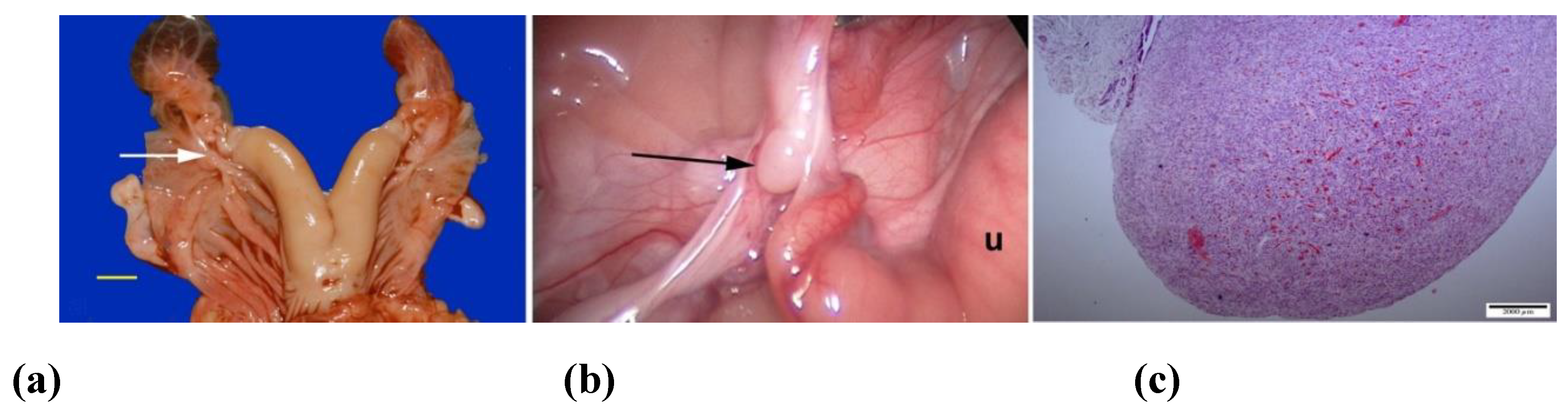
Figure 2.
Ultrasonograms of mucometra due to an imperforate hymen in an alpaca. (a) cranial vagina; (b) cervical os (arrow) and uterine body.
Figure 2.
Ultrasonograms of mucometra due to an imperforate hymen in an alpaca. (a) cranial vagina; (b) cervical os (arrow) and uterine body.
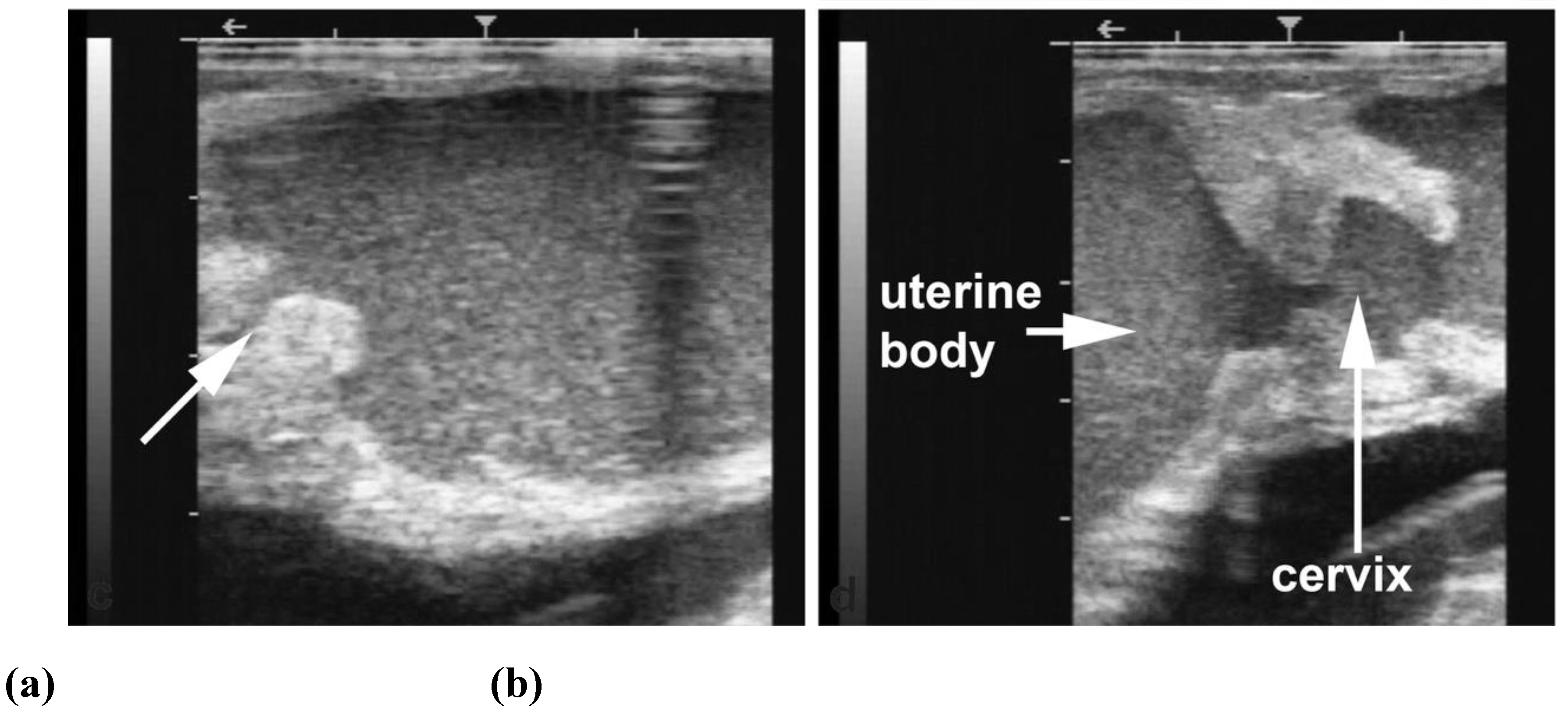
Figure 3.
(a) Hydrosalpinx due to segmental aplasia in an infertile dromedary female (scale bar: 1cm); (b) Ultrasonographic image of a case of the hydrosalpinx.
Figure 3.
(a) Hydrosalpinx due to segmental aplasia in an infertile dromedary female (scale bar: 1cm); (b) Ultrasonographic image of a case of the hydrosalpinx.
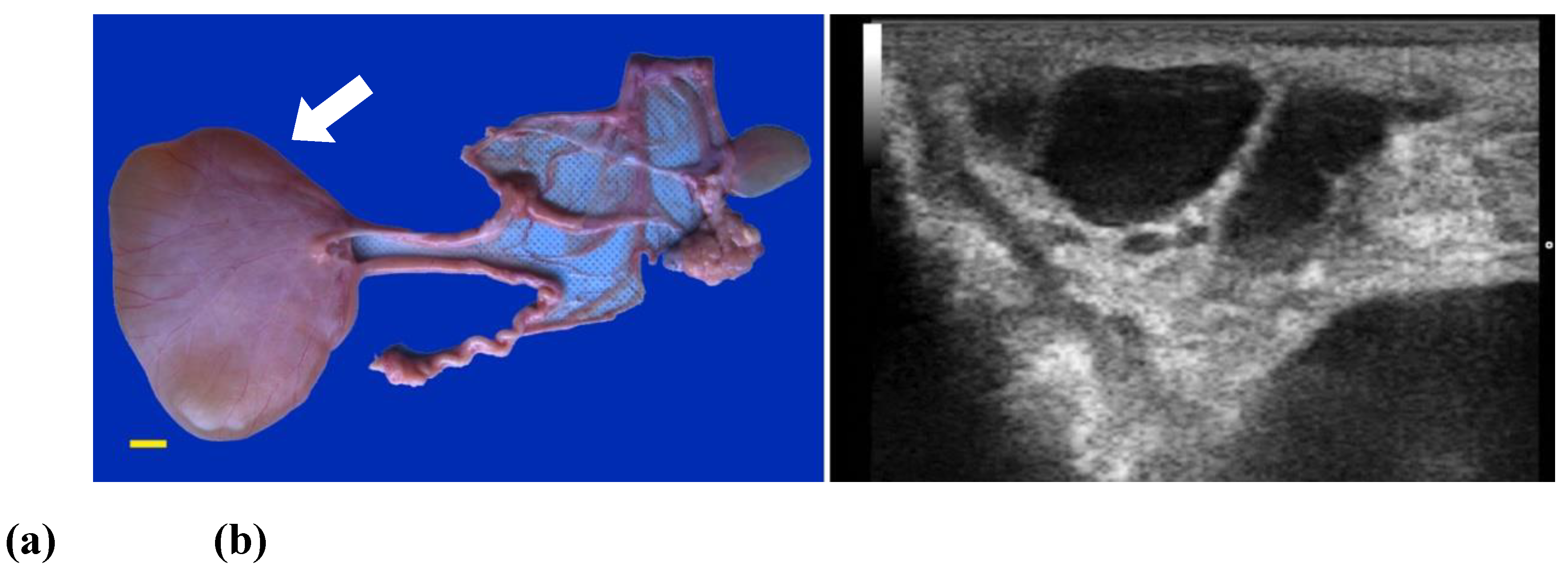
Figure 4.
Vulvar atresia in a camel.
Figure 5.
Gross appearance and ultrasonograms of anovulatory follicles in camels. (a) Thin-walled anovulatory follicle; (b) Thick-walled anovulatory follicle with some fibrin; (c) Hemorrhagic anovulatory follicle; (d) Luteinized anovulatory follicle.
Figure 5.
Gross appearance and ultrasonograms of anovulatory follicles in camels. (a) Thin-walled anovulatory follicle; (b) Thick-walled anovulatory follicle with some fibrin; (c) Hemorrhagic anovulatory follicle; (d) Luteinized anovulatory follicle.
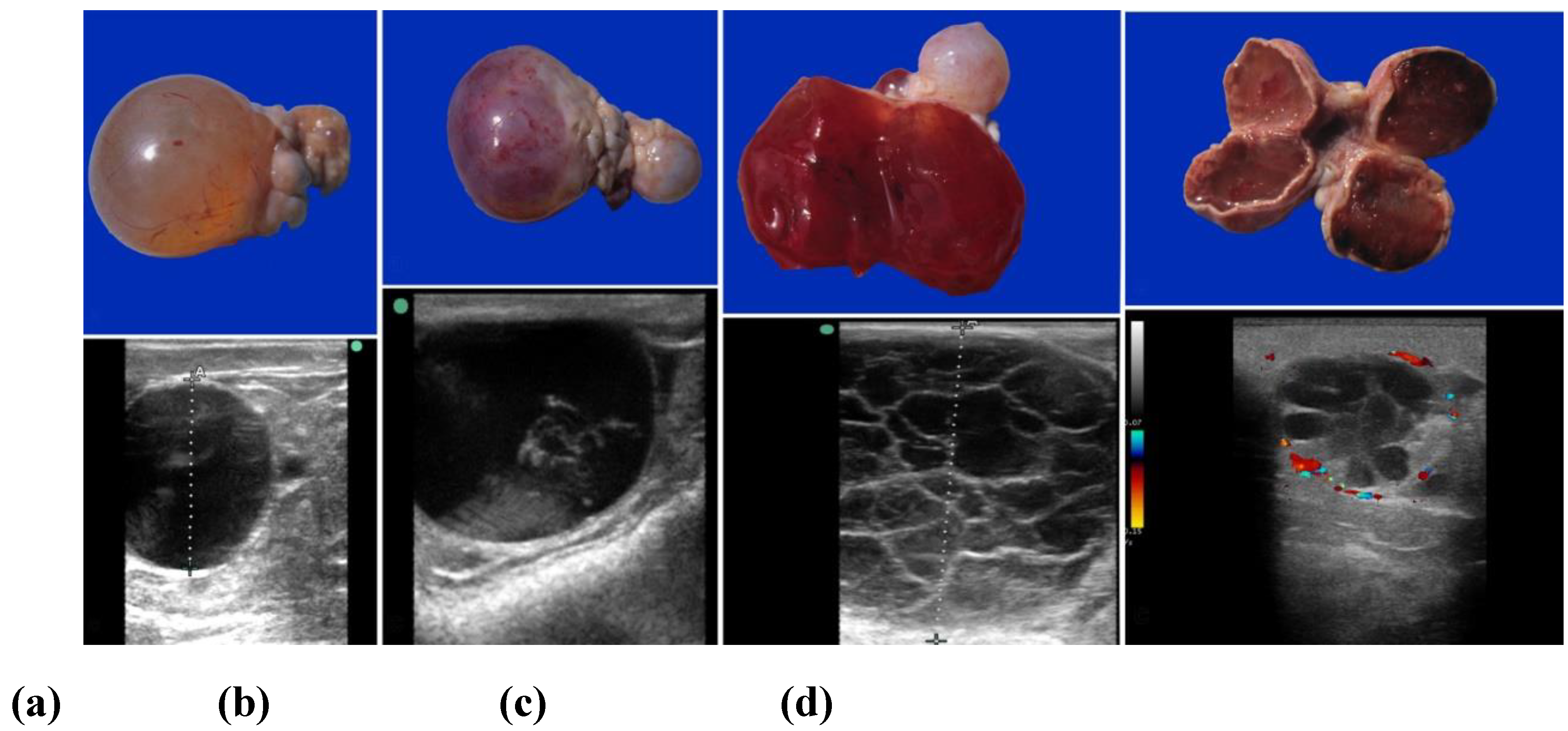
Figure 6.
Ultrasonographic images of hydrobursitis in camels. (a) Arrow: echogenic fluid within the bursa; (b) Arrow: abnormal ovary contained in the fluid-filled bursa.
Figure 6.
Ultrasonographic images of hydrobursitis in camels. (a) Arrow: echogenic fluid within the bursa; (b) Arrow: abnormal ovary contained in the fluid-filled bursa.
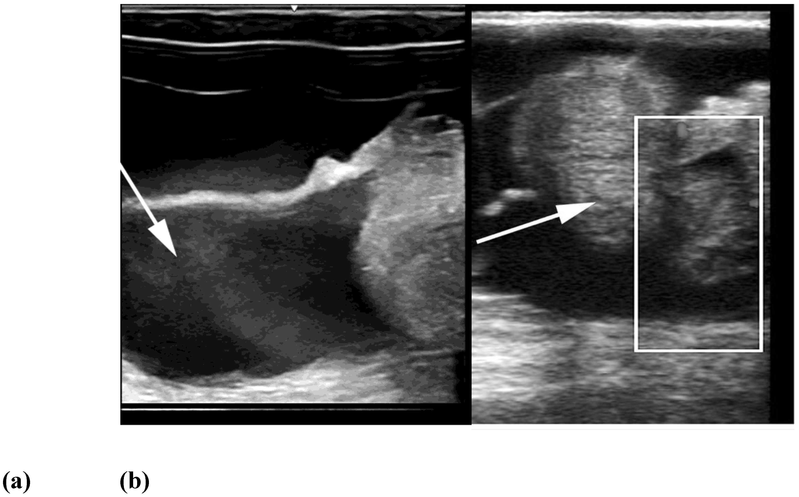
Figure 7.
Gross appearance of ovariobursitis surgically exposed by flank laparotomy in a camel.
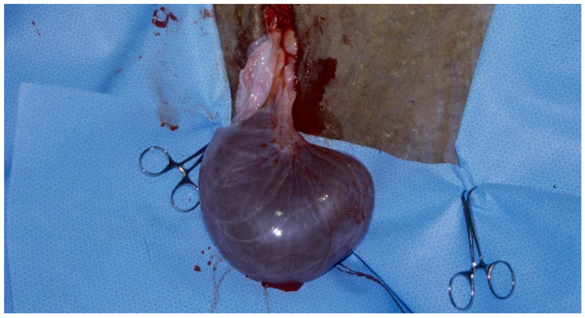
Figure 8.
Gross pathology and histopathology of disorders of the uterine tubes in camels. (a) Pyosalpinx; (b) Hydrosalpinx with ovarian-bursal adhesions.
Figure 8.
Gross pathology and histopathology of disorders of the uterine tubes in camels. (a) Pyosalpinx; (b) Hydrosalpinx with ovarian-bursal adhesions.

Figure 9.
Alpaca endometrial cytology showing polynuclear neutrophils (arrows).

Figure 10.
Camel endometrial biopsy exhibiting acute inflammatory reaction (acute endometritis).
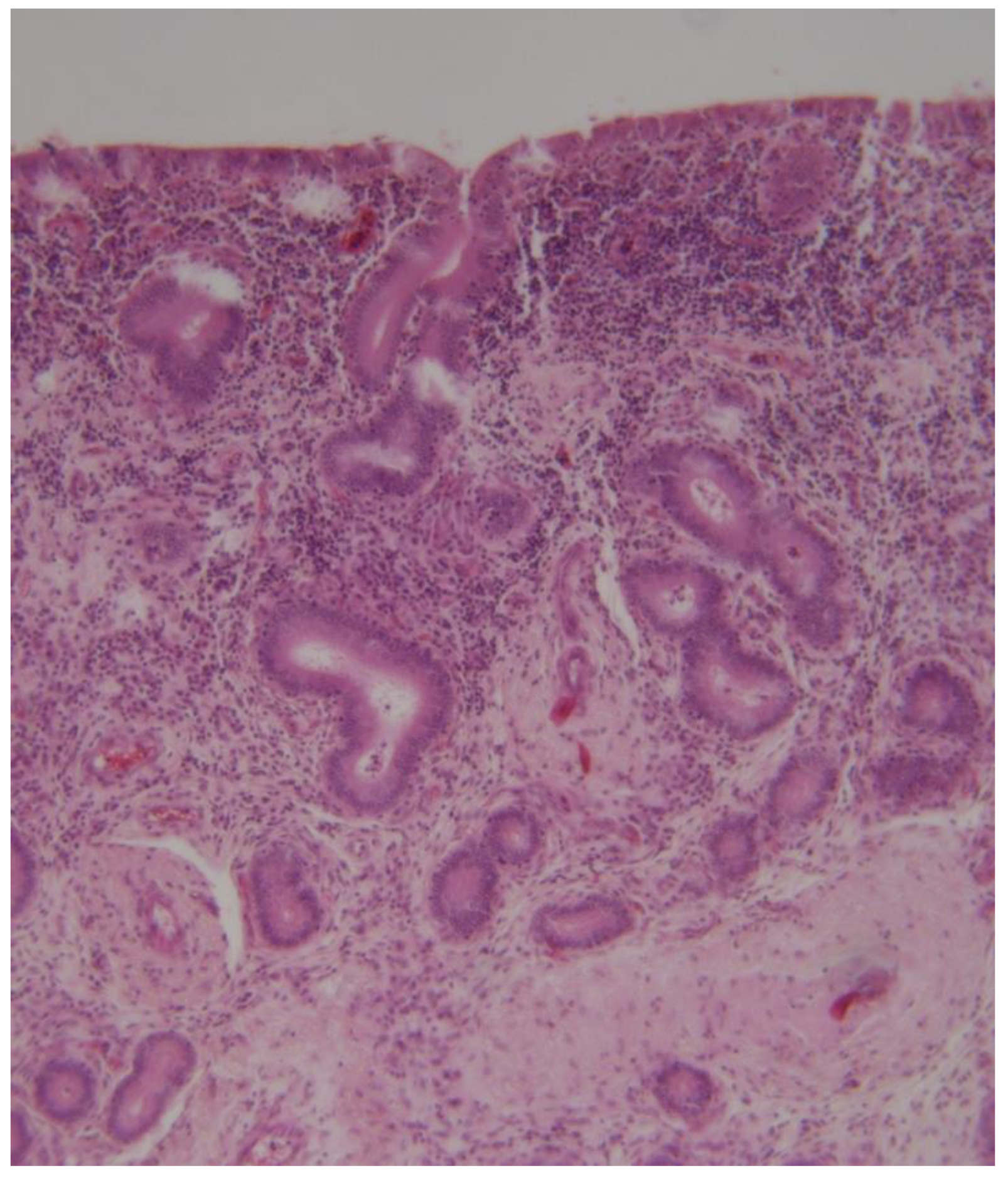
Figure 11.
Camel endometrial biopsy exhibiting mixed inflammation in the stratum compactum and periglandular fibrosis with cystic dilation (chronic endometritis).
Figure 11.
Camel endometrial biopsy exhibiting mixed inflammation in the stratum compactum and periglandular fibrosis with cystic dilation (chronic endometritis).
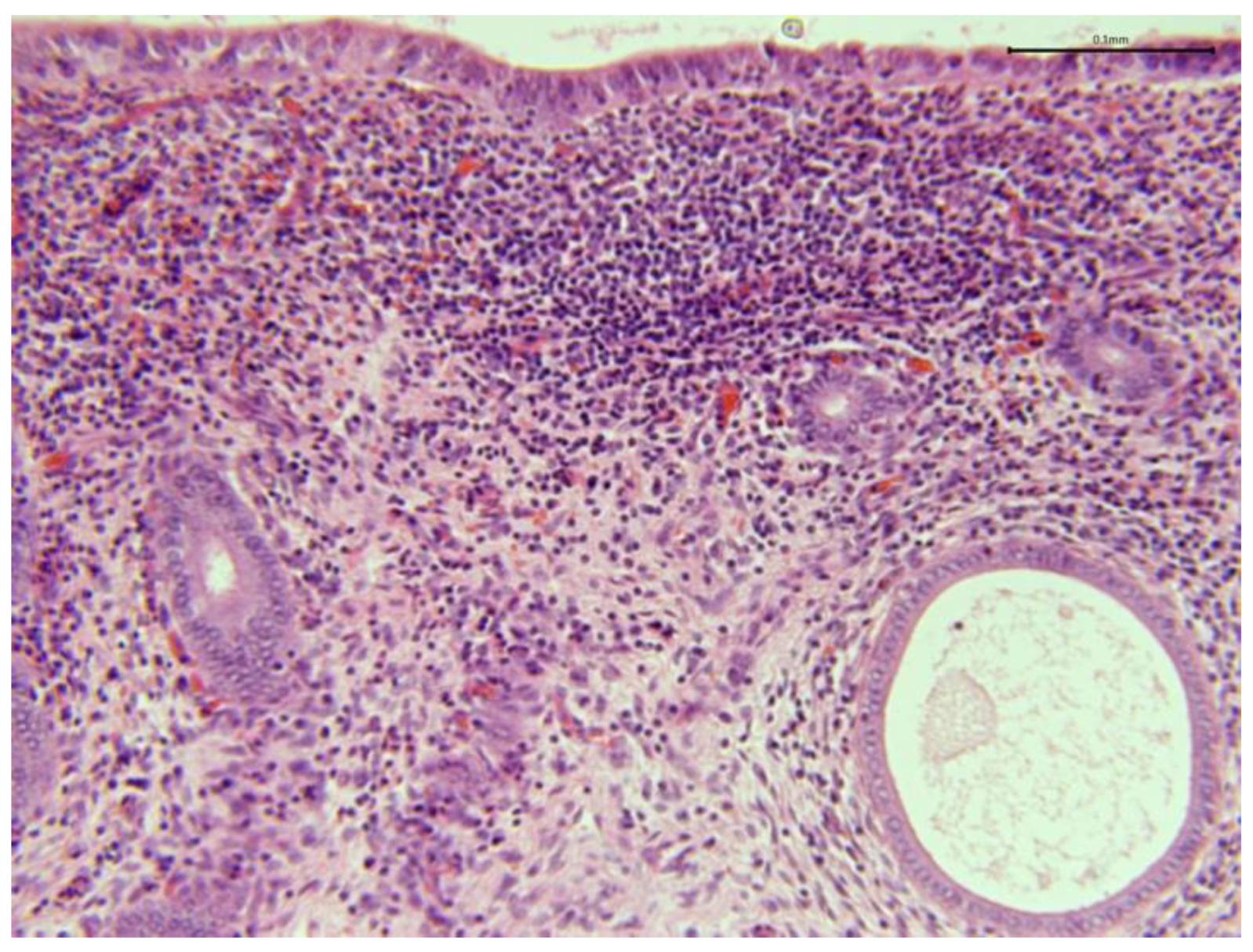
Figure 12.
Camel endometrial biopsy exhibiting chronic granulomatous endometritis (scale bar: 100 μm).
Figure 12.
Camel endometrial biopsy exhibiting chronic granulomatous endometritis (scale bar: 100 μm).
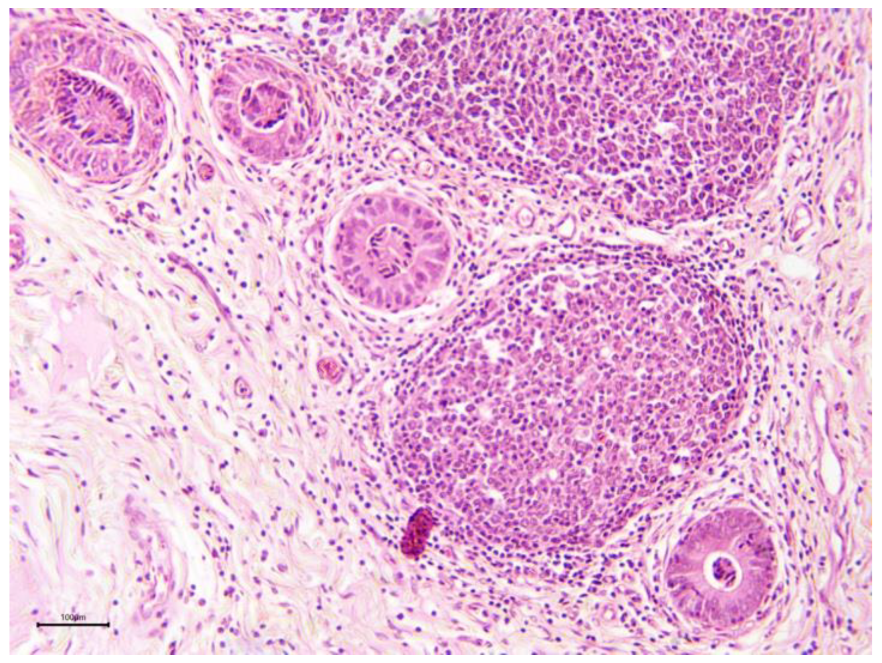
Figure 13.
Gross pathology (scale bar: 1 cm) and ultrasonogram of intraluminal uterine cysts due to intraluminal uterine adhesions in an alpaca.
Figure 13.
Gross pathology (scale bar: 1 cm) and ultrasonogram of intraluminal uterine cysts due to intraluminal uterine adhesions in an alpaca.
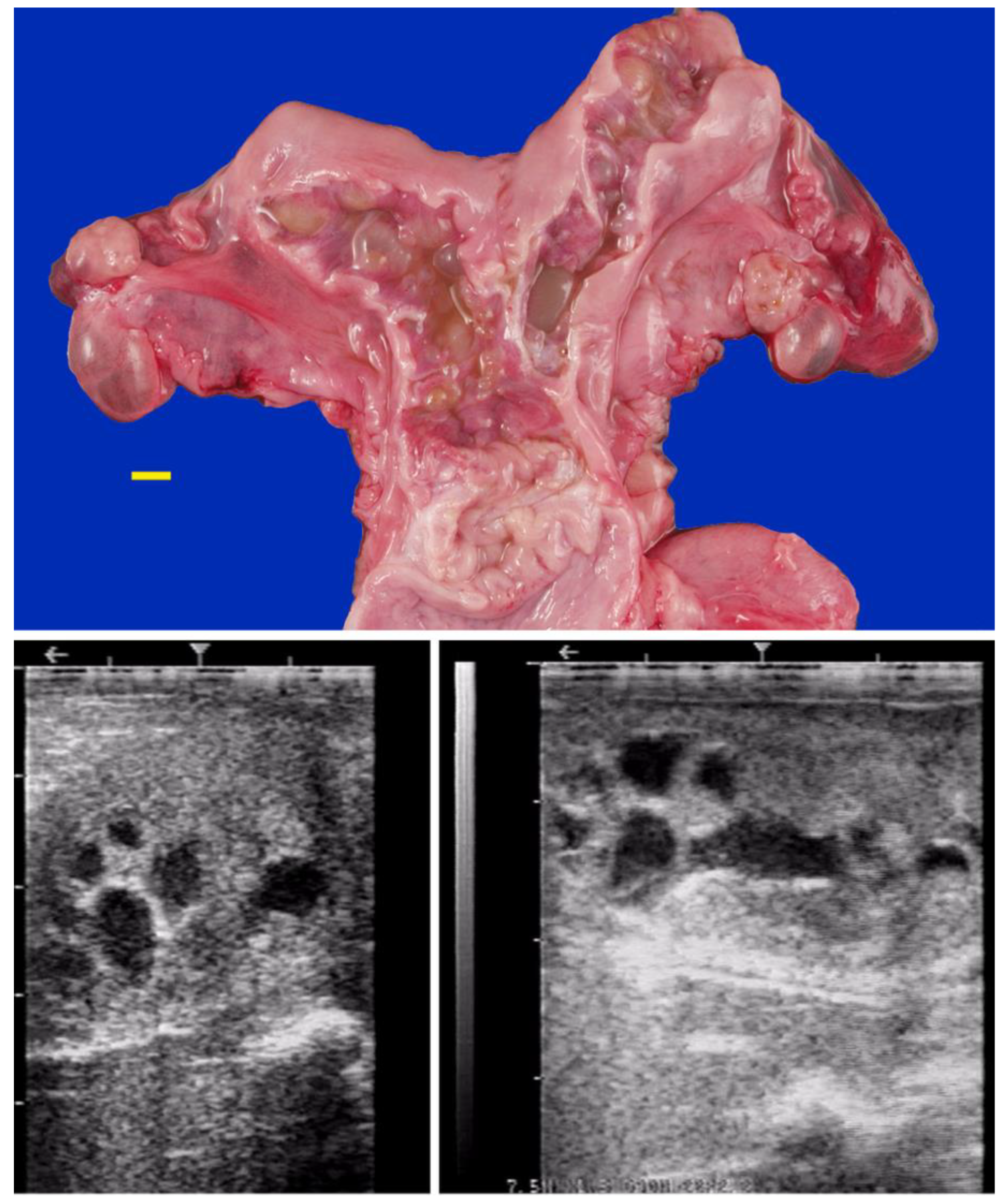
Figure 14.
Vulvar squamous cell carcinoma in a llama.
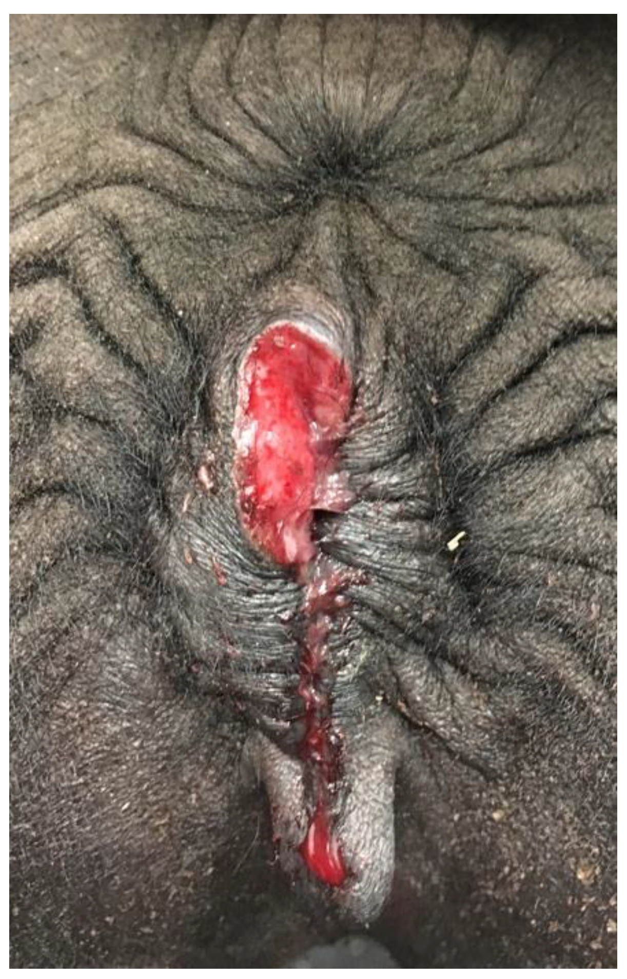

Table 1.
Proportion of different reproductive complaints in female camels (n= 542) and South American camelids (SACs) (n=2495).
Table 1.
Proportion of different reproductive complaints in female camels (n= 542) and South American camelids (SACs) (n=2495).
| Primary complaint | Camels* (%) | SACs** (%) |
|---|---|---|
| Repeat breeding | 411 (75.83) | 2003 (80.30) |
| Early pregnancy loss | 88 (16.23) | 448 (18.00) |
| Behavioral problems | 15 (2.77) | 16 (0.64) |
| Visible genital abnormalities | 28 (5.17) | 28 (1.12) |
*Cases seen in UAE, Qatar, Morocco, **Cases seen in USA.
Table 2.
Congenital abnormalities of the reproductive tract diagnosed in infertile SACs (n=102) and camels (n=25).
Table 2.
Congenital abnormalities of the reproductive tract diagnosed in infertile SACs (n=102) and camels (n=25).
| Congenital abnormality | SACs (%) | Camels (%) |
|---|---|---|
| Atresia vulvi/vulvar hypoplasia | 23 (22.54) | 3 (12.00) |
| Double cervix | 2 (1.96) | 2 (8.00) |
| Double vagina | 2 (1.96) | 1 (4.00) |
| Hydrosalpinx (segmental aplasia) | 2 (1.96) | 5 (20.00) |
| Imperforate hymen | 15 (14.70) | 3 (12.00) |
| Intersex/ambiguous gender | 5 (4.90) | 1 (4.00) |
| Ovarian dysgenesis | 37 (36.27) | 6 (24.00) |
| Ovarian teratoma | 1 (0.98) | 1 (4.00) |
| Vaginal aplasia | 10 (9.80) | - |
| Uterus unicornis | 5 (4.90) | 3 (12.00) |
| Total number of animals | 102 | 25 |
Table 3.
Distribution of acquired disorders of the ovaries, ovarian bursa, and uterine tube in SACs and camels presented for infertility.
Table 3.
Distribution of acquired disorders of the ovaries, ovarian bursa, and uterine tube in SACs and camels presented for infertility.
| Abnormality | SACs (%) | Camels (%) |
|---|---|---|
| Anovulatory/Hemorrhagic follicles | 42 (47.19) | 669 (54.34) |
| Ovarian adhesions | 3 (3.37) | 8 (6.49) |
| Ovulation failure | 8 (8.98) | 10 (0.81) |
| Ovarian inactivity | 22 (24.71) | 358 (29.08) |
| Ovarian tumors | 2 (2.24) | 4 (0.32) |
| Ovario-bursal adhesions | 3 (3.37) | 149 (12.10) |
| Persistent luteal structures | 16 (17.97) | 33 (2.68) |
| Pyosalpinx/salpingitis | 1 (1.12) | 82 (6.66) |
| Total number of animals | 89* | 1231* |
*The total percentage is greater than 100% because some animals presented a combination of abnormalities.
Table 4.
Distribution of acquired uterine disorders in SACs and camels.
| Abnormality | SACs (%) | Camels (%) |
|---|---|---|
| Endometrial degeneration | 33 (20.00) | 371 (40.77) |
| Endometritis | 84 (50.91) | 501 (55.05) |
| Intestinal-uterine adhesions | 2 (1.21) | 14 (1.54) |
| Luminal adhesions | 12 (7.27) | 3 (0.33) |
| Mucometra-pyometra | 18 (10.91) | 10 (1.10) |
| Mummification | 1 (0.61) | 1 (0.11) |
| Neoplasia | 5 (3.03) | 1 (0.11) |
| Uterine abscess | 2 (1.21) | 6 (0.66) |
| Uterine cysts | 8 (4.85) | 4 (0.44) |
| Total number of animals | 165 | 910 |
Table 5.
Distribution of acquired disorders of the cervix in SACs and dromedary camels.
| Abnormality | SACs (%) | Camels (%) |
|---|---|---|
| Cervical stenosis/adhesions | 10 (34.48) | 6 (54.54) |
| Cervicitis | 7 (24.14) | 3 (27.27) |
| Cervical tears | 12 (41.38) | 2 (18.18) |
| Total number of animals | 29 | 11 |
Table 6.
Distribution of acquired disorders of the vaginal and vulva in SACs and dromedary camels.
| Abnormality | SACs (%) | Camels (%) |
|---|---|---|
| Recto-vaginal tears | 11 (39.28) | 10 (27.77) |
| Squamous cell carcinoma | 2 (7.14) | - |
| Vaginal adhesions | 13 (46.42) | 6 (16.66) |
| Vaginitis | 1 (3.57) | 2 (5.55) |
| Vulvitis | 1 (3.57) | 18 (50.00) |
| Total number of animals | 28 | 36 |
Disclaimer/Publisher’s Note: The statements, opinions and data contained in all publications are solely those of the individual author(s) and contributor(s) and not of MDPI and/or the editor(s). MDPI and/or the editor(s) disclaim responsibility for any injury to people or property resulting from any ideas, methods, instructions or products referred to in the content. |
© 2025 by the authors. Licensee MDPI, Basel, Switzerland. This article is an open access article distributed under the terms and conditions of the Creative Commons Attribution (CC BY) license (http://creativecommons.org/licenses/by/4.0/).
Copyright: This open access article is published under a Creative Commons CC BY 4.0 license, which permit the free download, distribution, and reuse, provided that the author and preprint are cited in any reuse.


