Preprint
Article

This version is not peer-reviewed.

A New Peyritschiella Species (Laboulbeniales, Ascomycota) on Staphylinidae (Coleoptera, Insecta) from the Tropical Montane Cloud Forest of Mexico

A peer-reviewed article of this preprint also exists.

Submitted:

19 August 2025

Posted:

20 August 2025

You are already at the latest version

Abstract

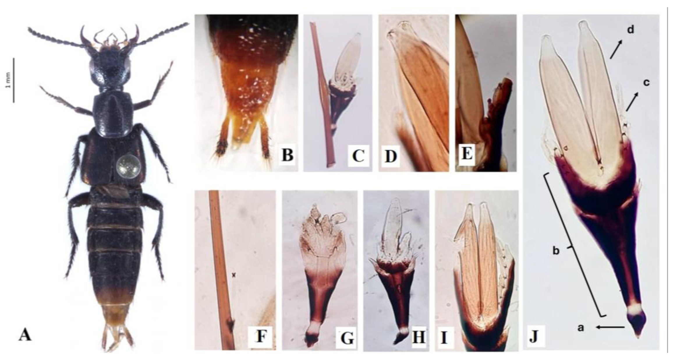
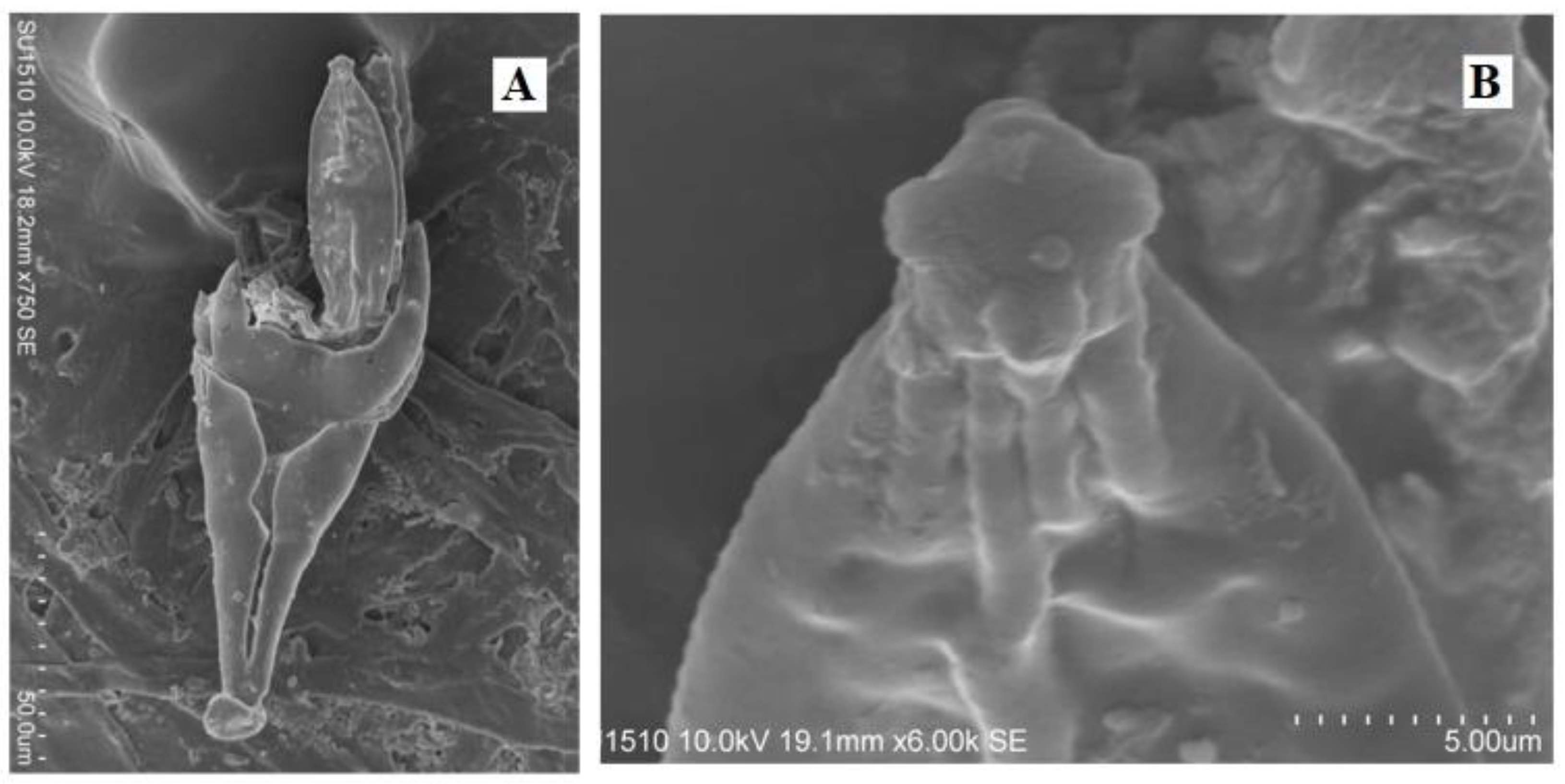
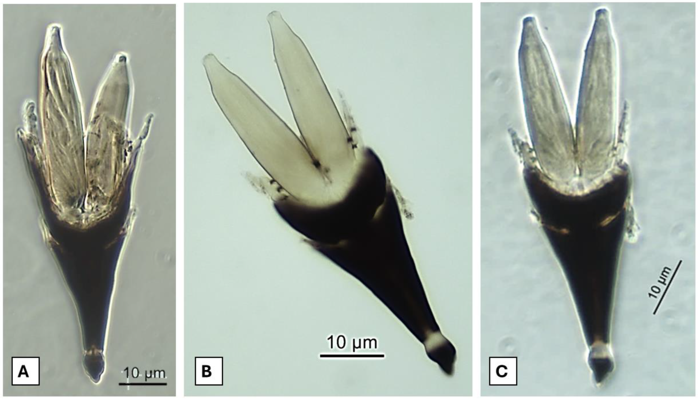
One new species of Laboulbeniaceae, Peyritschiella styngeti, is described and illustrated. Characterized by appendages with a black constriction at the base, perithecia with four papillae on the apical zone, cruciform bilateral symmetry, and an extremely melanized receptacle. This species was observed on the stylus of the staphylinid Styngetus deyrollei, highly specific to this rove beetle species, which are distributed in the tropical montane cloud forest in Mexico. Currently, the Laboulbeniales mycobiota in Mexico comprises 82 species, with 11 described growing on species of the Staphylinidae family. Additionally, a compilation of the Laboulbeniales species reported for Mexico and for Staphylinidae species is presented.

Keywords: 
;  ;  ;  

1. Introduction

Peyritschiella Thaxt. 1890 [1] is a genus taxonomically located in the Laboulbeniaceae family, some of its diagnosis characteristics are that it presents a receptacle conformed by four overlapped areas; a basal area, a sub basal area, a subterminal area, and a terminal area where the perithecia develops, which are symmetric and possesses papillae on the apical zone. The antheridia are conical-shaped, continuous, divided from the receptacle by a constricted dark-colored basal zone, and the ascospores are septate [2]. Thaxter [3,4] states that it is a very particular group because there is no other that possesses compound antheridium and the receptacle's bilateralism, as these characteristics are only shared with Dimorphomyces Thaxt. 1890 [1] Haelewaters et al. [4] mention that the order to which Peyritschiella is part of, the Laboulbeniales, has representative hosts from the Insecta class, subphylum Chelicerata, and Myriapoda.
Laboulbeniales are considered obligate ectoparasites that rely on their hosts and exhibit a high degree of specificity. As stated by Van der Linde and Rhong [5], their distribution corresponds to that of their hosts, and they depend on these hosts to acquire the necessary resources for their life cycle, sustenance, and survival within insect populations. The transmission method has been observed to be highly effective, involving direct contact between insects, including sexual reproduction and chance encounters among organisms in large groups displaying social behaviors. This facilitates the spores' contact with non-infected organisms, allowing them to penetrate the cuticle and successfully develop their thallus. The environment may also serve as a secondary intermediary, albeit with a lower success rate, maintaining spore viability through soil humidity [6,7]. All these factors contribute to the specificity of Laboulbeniales, which grow only in certain regions of their hosts, resulting in a wide range of morphologies based on the area of infection they develop [8].
In Mexico, studies of Laboulbeniomycetes have been absent, almost nonexistent. The first study on this group [9] was done over genus Rickia Cavara 1899 [10] growing on passalid beetles (Coleoptera: Passalidae). However, Mexican records have been mentioned in studies worldwide [2,11,12,13,14,15,16,17,18,19,20,21,22], which are summarized in (Appendix A, Table A1), in which only ten species parasitizing rove beetles have been reported (Appendix B, Table A2). The records state 81 species of Laboulbeniomycetes in the country, of which 78 are located in the Laboulbeniales order and three are part of the Herpomycetales order. Until now, there have been no specific studies whatsoever about Laboulbeniales found parasitizing Staphylinidae in Mexico; hence, the objective of the present study is to describe a new species of Peyritschiella for the world´s mycobiota.

2. Materials and Methods

Host Examination and Thalli Removal

The specimens were obtained from Staphylinidae (rove beetles) organisms that were part of the entomological collection integrated into the CC-UAEH (Universidad Autónoma del Estado de Hidalgo); which were revised on the head, thorax and abdomen, there was a higher focus on the host's stylus located in the abdomen’s basal area under the stereoscopic microscope; for this revision the insects were placed on a Petri’s dish, with the help of a few drops of 70% alcohol; the thallus were extracted using a periodontal probe by doing light sweeps over the host’s stylus.

Morphological Characterization

The samples were set as fixed specimens using Hoyer's reactive, and subsequently sealed. The thallus was characterized, measured, and described in form, color, and number of perithecia, antheridia, and appendages, as well as the foot, and visible cells of the receptacle. The taxonomic identification was followed up to the genus using Thaxter’s taxonomic key. The fixed specimens’ preparations of the Laboulbeniales are deposited in the ENCB fungi collection located in the Instituto Politécnico Nacional.

3. Results

Ascomycota
Pezizomycotina
Laboulbeniomycetes
Laboulbeniomycetidae
Laboulbeniales
Laboulbeniaceae
Peyritschiella styngeti E.L. Ortiz-Pacheco, Raymundo & Baut.-Hern. sp. nov.
MycoBank No. 857590
Diagnosis. Three-leveled receptacle, subtriangular and flattened; hyaline appendages; constricted at the base with an evident dark-brown coloration; symmetrical or almost symmetrical perithecia, presence of papillae on the apical area of the perithecia; hyaline, bicellular ascospores (Figure 1).
Figure 1. Peyritschiella styngeti, A. dorsal view of the host Styngetus deyrollei Solski 1866 [23]; B. stylus of the host which were infected; C. lateral view of the thallus on a seta from the stylus of the host; D. Apical zone of the perithecia, showing the four papillae, characteristic of the species; E. Antheridia, growing on the apical zone, besides the perithecia, growing apically on the appendages; F. First stage of the growing thallus, growing on a seta from the stylus of the host, the foot cell has developed and the bottom cell of the ascospore has elongated, it is visible the darkened division between booth cell of the ascospore in a dark brown, looking like a small cross, apical cell of the ascospore has not yet to grow, the thallus has not developed and the structures are highly hyaline; G. Thallus developing, foot cell has grown and the receptacle’s cells are developing, apical structures which include perithecia and appendages have yet developed as well as the receptacle has yet been fully defined; H. growing thallus, upper level of the receptacle is developing, perithecia growing on the apical are of the receptacle, coloration has darkened on the receptacle, appendages have been yet developed; I. apical zone of the receptacle, appendages on the right side, hyaline and darkened at the base, two symmetric perithecia are seen, the left one broken, a fusiform, highly hyaline ascospore is seen sideways in the formed opening; J. Frontal view of the thallus, a. foot cell, b. receptacle, c. appendages, d. perithecia.
Preprints 173035 g001
Figure 2. Scanning electron microscopy (SEM) of P. styngeti, A. young specimen of the thallus, only one perithecium has developed, the upper level of the receptacle is uneven because it has not developed fully, structures are seen; B. apical zone of the perithecium, showing the four papillae, diagnostic characteristic for the species.
Preprints 173035 g002
Etymology. The term styngeti corresponds to the genus in which the host is taxonomically located (Styngetus).
Holotype. Mexico • Hidalgo, Molango, Acautitlán; tropical montane cloud forest; 1715 m.a.s.l.; N 20° 45’38.4”, W 88°42’50.7”; squid-baited pitfall trap; 15 to 24 May 2011; J. Márquez, ENCB
Description. Thallus 69-280 × 20-64 µm (Figure 1J). Receptacle 50-100 × 20-64 µm, subtriangular and flattened, narrow at the base, wider under the perithecia, of superficial insertion. Receptacle cells are covered by a dark brown color, the last row of a cell arranged in transversal form and curved inwards, building a margin that covers longitudinally the basal area of the perithecia (Figure 1Jb). Foot cell 4-20 × 9-16 µm, dark colored (Figure 1Ja). Hyaline appendages, constricted at the base with an evident dark-brown coloration, arranged to the external sides of the perithecia over the longitudinal margin of the receptacle, 10-80 × 2-5 µm (Figure 1Jc); compound antheridia, arranged laterally over the appendages, only observed in one 15-20 × 6-9 µm thallus (Figure 1E). Perithecia 15-150 × 5-37 µm, located apically (Figure 3; Figure 4A,B), symmetrical or almost symmetrical, arranged one beside the other, presence of four papillae, cruciform, on the apical area of the perithecia (Figure 1D, Figure 2B); hyaline, bicellular ascospores 40-56 × 4-5 µm, fusiform, septate and covered with a mucilage which help adhesion to the host (Figure 1I; Figure 4C,D).
Specimens examined. Mexico • Puebla, Zacapoaxtla, Apulco, Cascada La Gloria; tropical montane cloud forest; squid-baited pitfall trap; 18 May to 28 May 2021; J. Márquez and J. Asiain. • Hidalgo, Tlanchinol, camino a Apantazol, El Pozo; tropical montane cloud forest, 1391 m.a.s.l.; squid-baited pitfall trap No. 2; 1 to 22 October 2011; J. Márquez. • Hidalgo, Zacualtipán; squid-baited pitfall trap No. 2; 20 September to 4 October 2011. • Hidalgo, Zacualtipán; tropical montane cloud forest; N 20°38’44.5”, W 98° 36’7.2”; squid-baited pitfall trap No. 6; 14 to 28 April 2011; MA. • Hidalgo, Zacualtipán, camino a Tizapán; tropical montane cloud forest; N 20°38’44.5”, W 98°36’7.2”; squid-baited pitfall trap; 14 to 28 May 2011; J. Márquez. • Hidalgo, Molango, Acuatitlán; tropical montane cloud forest; 1715 m.a.s.l.; N 20°45’38.4”, W 88°42’50.7”; flight interception trap No. 1; 15 to 29 April 2011; J. Márquez. • Hidalgo, La Misión, Los Naranjos - Palo Hueco; tropical montane cloud forest; 1715 m.a.s.l.; N 21°08’06”, W 99°05’02”; 10 to 24 March 2011; J. Márquez. • Hidalgo, Molango, Acautitlán; tropical montane cloud forest; N 20°45’38.4”, W 88°42’50.7”; 15 to 24 May 2011, J. Márquez. • Hidalgo, La Misión, Las Pilas; tropical montane cloud forest; flight interception trap; 12 to 19 July 2018; A. Lora and J. Márquez. • Hidalgo, Tlanchinol, camino a Apantazol, El Pozo; tropical montane cloud forest; 1391 m.a.s.l.; N 20°59’6.73”, W 98°37’39.86”; flight interception trap No, 1; 16 to 30 April 2011; J. Márquez. • Hidalgo, Tlanchinol; tropical montane cloud forest; 1391 m.a.s.l.; N 20°59’6.73”, W 98°37’39.86”;squid-baited pitfall trap No. 2; 16 to 30 April 2011. • Hidalgo, Molango, Acuatitlán; tropical montane forest; 1715 m.a.s.l.; N20°45’38.4”, W 88°42’50.7”; squid-baited pitfall trap No. 4; 15 to 29 April 2011; J. Márquez. • Hidalgo, Tlanchinol, camino a Apantazol, El Pozo; tropical montane cloud forest; 1391 m.a.s.l.; N 20°59’6.73”, W 98°32’39.86”; squid-baited pitfall trap; 16 to 30 April 2011; J. Márquez. • Hidalgo, La Misión, Lagunita de Pilas; tropical montane cloud forest; squid-baited pitfall trap No. 2; 17 February to 17 March 2019; A, Lora. • Hidalgo, Zacualtipán, camino a Tizapán; tropical montane cloud forest; 1790 m.a.s.l.; N 20°38’44.5”, W 98°36’7.2”; squid-baited pitfall trap; 14 to 28 April 2011; J. Márquez. • Hidalgo, La Misión, Lagunita de Pilas; tropical montane cloud forest; 1770 m.a.s.l.; N 21°7’38”, W 99°6’53”; dung-baited pitfall trap; 11 June to 10 July 2019; J. A. Lora. • Hidalgo, Tenango de Doria, La Viejita; tropical montane cloud forest; 1163 m.a.s.l.; N 22°47’58”, W 57°66’07”; squid-baited pitfall trap; 12 August 2010; C. Cornejo. • San Luis Potosí, Las Pozas de James; tropical montane cloud forest; squid-baited pitfall trap; 17 July 2007; J. Asiain, APMP, IAA, MY, J. Márquez • Hidalgo, Tlanchinol, camino a Apantazol, El Pozo; tropical montane cloud forest; 1391 m.a.s.l.; N 20°59’6.732”, W 98°37’39.86”; squid-baited pitfall trap No. 1; 16 to 30 April 2011; J. Márquez.
Host. Obligate ectoparasite of Styngetus deyrollei, specifically located on the stylus on both males and females (Figure 1A); host with scavenger predaceous habitat in the tropical montane cloud forest.
Distribution. Distributed in the states of Hidalgo, Puebla, and San Luis Potosí. It is a new species for the worldwide mycobiota.
Notes. Peyritschiella styngeti sp. nov. is characterized by the presence of appendages with a dark constriction at the base (Figure 1I), two perithecia with four papillae on the apical area, cruciform aspect (Figure 1D; 2B), bilateral symmetry, a receptacle conformed by three highly pigmented levels, making it look like it is only one level (Figure 1J). This species is highly specific because it is only found on S. deyrolley’s (Figure 1A) stylus (Figure 1B). The characteristics, as mentioned earlier, differentiate this species from other taxonomically close ones like P. cafiana (Thaxt.) I.I. Tav. 1985 [18], which presents three well-defined leveled receptacle. Furthermore, the pigmented area in this same structure is only observed on the foot cell and the adjacent area, short appendages; although both species present two lobulated perithecia, differentiating it from P. styngeti in this melanization pattern.
When compared with P. angolensis (Thaxt.) I.I. Tav. 1985 [18], P. styngeti presents the same dark-colored foot cell and two lobulated perithecia, however, the receptacle in P. angolensis exhibits three row of cells of the same width, overlaped, making it look like simetrical and a little bit flattened, only darkened at the margin and over the lower row of cells [21], meanwhile, the aforementioned one presents the three rows of highly pigmented cells, giving it a one-leveled receptacle appearance with the upper row being wider than the other two, surrounding the perithecia.
Peyritschiella exilis (Thaxt.) I.I. Tav. 1985 [18], unlike P. styngeti, presents two highly pigmented perithecia which are similar to the receptacle in size. This structure is divided into three highly differentiated levels, and the pigmentation is only observed on the basal area of it and the foot cell. Meanwhile, P. vulgata (Thaxt.) I.I. Tav. 1985 [18] exhibits a difference mostly because the aforementioned has three rows of cells, pigmented on the first rows of the cell but not in the last two rows, the appendages are constricted, with two terminal perithecia which have an appendiculate tip, most prominent difference when compared with P. styngeti [21].
In the case of P. furcifera (Thaxt.) I.I. Tav. 1985 [18] it exhibits lesser-developed levels, similar to the ones in P. styngeti, although the apical cells are bigger in comparison with the last taxon, they also do not possess any pigmentation on the apical area nor the foot cell, as well as appendages which are short and unicellular [24].
With P. australiensis (Thaxt.) I.I. Tav. 1985 [18] it is observed that the present pigmentation on the base and sides of the receptacle, as well as appendages arranged at the sides and in between the apical perithecia, also has an appendiculated tip. Meanwhile, P. mexicana (Thaxt.) I.I. Tav. 1985 [18] has well-differentiated levels, with evident pigmentation only on the sides of the base of the receptacle, two appendiculate perithecia and multicellular appendages arranged laterally and constricted, alongside the aforementioned genres, which are differentiated from P. styngeti because of the lack of the elongate margin under the perithecia, leaving hem “bare” [24].
P. madagascariensis (Thaxt.) I.I. Tav. 1985 [18], on the other hand, presents a pigmented receptacle with lateral projections which seem to be protecting the perithecia, however, these projections are long and are separated from the receptacle, compared to P. styngeti, moreover, it exhibits a slim receptacle with only one level and appendiculate perithecia [24].

4. Discussion

Peyritschiella is a genus that has been reported to be parasitic on rove beetles. In Mexico’s case, it has been cited P. exilis over Belonuchus formosus Gravenhorst 1806 [25] (current valid name B. rufipennis Fabricius 1801 [26] and Philonthus oxysporinus Sharp 1885 [27], P. furcifera on Philonthus centralis Sharp 1885 [27], P. mexicana on Philonthus atriceps Sharp 1885 [27], P. princeps (Thaxt.) I.I. Tav. 1985 [18] on Philonthus sp. and Quediomacrus puniceipennis Solski 1868 [28], finally, P. vulgata on Philonthus flavolimbatus Erichson 1840 [29] Considering P. styngeti on Styngetus deyrollei, the taxonomic richness increases to six species.
In this study, 787 organisms of rove beetles were examined, from which 476 were identified as Styngetus deyrollei. Among these, 104 were infected with thallus at different developmental stages, representing 22% of infected organisms in this genus and 13.21% of all organisms revised.
The Staphylinidae family represents a group of arthropods highly parasitized by Laboulbeniales. Worldwide, there have been reported incidents of almost every family but Ceratomycetaceae; regardless, one important characteristic of this organism that needs to be highlighted is its specificity towards their hosts, which has made it clear for the fungi to be described as species-specific in most cases [18,30]. Frank [31] put Peyritschiella in the family Peyritschiellaceae as rove beetles’ parasites by excellence, before they were translated to their correct family, Laboulbeniaceae.
The number of rove beetles described worldwide goes up to 56000 species, out of this, 1656 have been registered in Mexico, and only 80 of them are endemic to the territory, these numbers make Mexico the second country with the most diversity of rove beetles in Latin America [32], hence, considering that the genus Peyritschiella is frequent and presents specificity for this host, it can be expected a high richness of species. So far, only 10 species of Laboulbeniales are known to grow on Staphylinidae: Cucujomyces bilateralis (R.K. Bnj.) W. Rossi & A. Weir 2008 [33], Corethromyces brasilianus Thaxt. 1900 [34], Laboulbenia cristata Thaxt. 1893 [15], L. philonthi Thaxt. 1893 [15], Mimeomyces quedionuchi Thaxt. 1931 [2], Peyritschiella exilis, P. furcifera, P. mexicana, P. princeps, and P. vulgata, [18,35], which represents only 12.34% of the 81 species known for the country; the aforementioned shows the necessity of maintaining the mycological and entomological studies on those that are the most diverse organism groups.

5. Conclusions

Peyritschiella styngeti is a species found on the stylus of Styngetus deyrollei, predatory beetles that can be found between the leaf litter in the tropical montane cloud forest. They are also distributed in Hidalgo, Puebla, San Luis Potosí, and Veracruz.
With this study, the number of Laboulbeniales species for Mexico goes up to 82, out of which eleven are found to be parasitizing rove beetles. For the Peyritschiella genus, the number rises to 51, out of the 50 previously known worldwide.
The studied species were obtained from specimens from an entomological collection, lent by the CC-UAEH, which highlights the importance of investigating recent and new biological collections, especially in the case of Laboulbeniales, which are a lesser-known group and possibly have a high level of diversity and endemism in Mexico.

Author Contributions

Conceptualization, ELOP, SBH and TR; methodology, ELOP, and SBH; validation, SBH, ELOP, JM, and JA; investigation, ELOP, SBH; resources, SBH and TR; data curation, ELOP; writing—original draft preparation, ELOP; writing—review and editing, ELOP, SBH, TR, JM and JA; visualization, SBH, TR and ELOP; supervision, SBH, TR, JM and JA; project administration, TR and SBH; funding acquisition, SBH and TR. All authors have read and agreed to the published version of the manuscript.

Funding

This research was financed by the Instituto Politécnico Nacional (SIP20251281).

Data Availability Statement

All of the data that support the findings of this study are available in the main text and two appendices listed below.

Acknowledgments

We acknowledge the Instituto Politécnico Nacional for the financial support of this research. We thank the CC-UAEH (Universidad Autónoma del Estado de Hidalgo, México) for the materials to review and the taxonomic guidance during the species identifications, and all the extra resources provided regarding the hosts studied, to M.Sc. Berenit Mendoza Garfias for the scanning electron microscope (SEM) and the Phase-contrast microscopy (PCM) images (LANABIO, Instituto de Biología, UNAM), and last but not least, to the Mycology Laboratory, part of the Escuela Nacional de Ciencias Biológicas (ENCB), IPN, for the equipment and materials used during the research process. The authors have reviewed and edited the output and take full responsibility for the content of this publication.

Conflicts of Interest

The authors have declared that no competing interests exist. Ethical statement: No ethical statement was reported.

Abbreviations

The following abbreviations are used in this manuscript:
CC-UAEH Coleoptera Collection of Universidad Autónoma del Estado de Hidalgo
ENCB Escuela Nacional de Ciencias Biológicas (IPN)
IPN Instituto Politécnico Nacional

Appendix A

Table A1. Species of Laboulbeniales reported in Mexico (ordered alphabetically).
Genus Host The host’s family Vegetation Reference
Order: Herpomycetales
Herpomyces paranensis Thaxt. Blabera Blaberidae (Blattodea) Tropical Forest [1,13,14,15]
Herpomyces periplanetae Thaxt. Periplaneta Blattidae (Blattodea) Tropical Forest [1,13,14,15]
Herpomyces platyzosteriae Thaxt. Platyzosteria ingens Scudder Blaberidae (Blattodea) Tropical Forest [1,13,14,15]
Order: Laboulbeniales
Ceratomyces ansatus Thaxt. Tropisternus Hydrophilidae (Coleoptera) Tropical forest
Temperate Forest
Boreal Forest
[1,13,14,15,18]
Ceratomyces confusus Thaxt. Tropisternus glaber Herbst.
T. nimbatus Fabricius
Hydrophilidae (Coleoptera) Tropical Forest
Temperate Forest
Boreal Forest
[1,13,14,15,18]
Ceratomyces filiformis Thaxt. Tropisternus
Pleurohomus obscurus Shp.
Hydrophilidae (Coleoptera)
Dryopidae (Coleoptera)
Tropical Forest
Temperate Forest
Boreal Forest
[1,13,14,15]
Ceratomyces mexicanus Thaxt. Tropisternus nitidus Sharp
T. chalybeus Cast
Hydrophilidae (Coleoptera) Tropical Forest
Temperate Forest
Boreal Forest
[1,13,14,15,16]
Ceratomyces miriabilis Thaxt. Tropisternus
T. xantophus Sharp
Hydrophilidae (Coleoptera) Tropical Forest
Temperate Forest
Boreal Forest
[1,13,14,15]
Ceratomyces spiniger Thaxt. Tropisternus apicipalpis Cast. Hydrophilidae (Coleoptera) Tropical Forest
Temperate Forest
Boreal Forest
[1,13,14,15,16]
Dimeromyces forficulae Thaxt. Doru lineare Esch. Forficulidae (Dermaptera) Tropical Forest
Temperate Forest
Boreal Forest
[1,13,14,15,16]
Dimeromyces parasii Thaxt. Parasitus Parasitidae (Mesostigmata) Tropical Forest
Temperate Forest
Boreal Forest
[1,13,14,15,16]
Dixomyces clivinae (Thaxt.) I.I. Tav. Clivina dentifermorata Putz
Clivina
Carabidae (Coleoptera) Tropical Forest
Temperate Forest
Boreal Forest
[1,13,14,15,16]
Dixomyces pallescens (Thaxt.) I.I. Tav. Clivina Carabidae (Coleoptera) Tropical Forest
Temperate Forest
Boreal Forest
[16]
Eucantharomyces casnoniae Thaxt. Casnonia subdistincta Chaud. Carabidae (Coleoptera) Tropical Forest
Temperate Forest
Boreal Forest
[1,13,14,15,16]
Eucantharomyces diaphorii Thaxt. Diaphorus tenuicornis Chaud. Carabidae (Coleoptera) Tropical Forest
Temperate Forest
Boreal Forest
[1,13,14,15,16]
Homaromyces epieri R.K. Benj. Unespecified Histeridae (Coleoptera) Tropical Forest
Temperate Forest
Boreal Forest
[16]
Laboulbenia arietina Thaxt. Disonychia Carabidae (Coleoptera) Tropical Forest
Temperate Forest
Boreal Forest
[1,13,14,15]
Laboulbenia armata Thaxt. Oedionychus sublineatus Jac. Chrysomelidae (Coleoptera) Tropical Montane Cloud Forest [1,13,14,15]
Laboulbenia barbata Thaxt. Morio georgiae Pal. Leiodidae (Coleoptera) Tropical Montane Cloud Forest [1,13,14,15,16]
Laboulbenia brachini Thaxt. Brachinus elongatus Tourn.
B. mexicanus Dej.
B. rhytiderus Chd.
Carabidae (Coleoptera) Tropical Forest
Temperate Forest
Boreal Forest
[1,13,14,15]
Laboulbenia bruchii (Speg.) Thaxt. Lema albini Lac.
L. sallei Jac.
L. dimidiaticornis Jac.
Chrysomelidae (Coleoptera) Oak Forest
Pine Tree-Oak Forest
[1,13,14,15]
Laboulbenia catascopi Thaxt. Catoscopus
Pinacodera atrata Chev.
Coptodera arcuata Chev.
Colpodes auratus Chd.
Carabidae (Coleoptera) Tropical Forest
Temperate Forest
Boreal Forest
[1,13,14,15]
Laboulbenia decipiens Thaxt. Galerita nigra Chev. Carabidae (Coleoptera) Tropical Forest
Temperate Forest
Boreal Forest
[1,13,14,15]
Laboulbenia diabrotica Thaxt. Diabrotica fairmairei Baly Chrysomelidae (Coleoptera) Tropical Forest
Temperate Forest
Boreal Forest
[1,13,14,15]
Laboulbenia disonichae Thaxt. Disonycha figurata Jac. Chrysomelidae (Coleoptera) Tropical Forest
Temperate Forest
Boreal Forest
[1,13,14,15]
Laboulbenia egae Thaxt. Ega Carabidae (Coleoptera) Temperate Forest [1,13,14,15]
Laboulbenia elongata Thaxt. Colpodes Carabidae (Coleoptera) Tropical Forest
Temperate Forest
Coniferous Tree Forest
[1,13,14,15]
Laboulbenia erecta Thaxt. Colpodes agilis Chd.
C. evanescens Bates
Carabidae (Coleoptera) Tropical Forest
Temperate Forest
Coniferous Tree Forest
[1,13,14,15]
Laboulbenia flaccida Thaxt. Casonia subdistincta Chaud. Carabidae (Coleoptera) Tropical Forest
Temperate Forest
Boreal Forest
[1,13,14,15]
Laboulbenia flagellata Peyr. Onypterigia pusilla Chaud. Carabidae (Coleoptera) Tropical Forest
Temperate Forest
Boreal Forest
[1,13,14,15]
Laboulbenia galeritae Thaxt. Galerita forreri Bates
G. mexicana Chaud.
Carabidae (Coleoptera) Tropical Forest
Temperate Forest
Boreal Forest
[1,13,14,15]
Laboulbenia guerinii Thaxt. Gyretes
G. boreandri Chev.
G. immarginatus Chev.
G. leionatus Duby
Gyrinidae (Coleoptera) Tropical Forest
Temperate Forest
[1,13,14,15]
Laboulbenia gyrinidarum Thaxt. Gyrinus Gyrinidae (Coleoptera) Tropical Forest
Temperate Forest
Boreal Forest
[1,13,14,15]
Laboulbenia homophoetae Speg. Asphaera transversofasciata Jac.
Oedionychus sublineata Jac.
Systema littera Linn.
Chrysomelidae (Coleoptera) Tropical Forest
Subtropical Forest
Temperate Forest
Deciduous Forest
[1,13,14,15]
Laboulbenia mexicana Thaxt. Galerita
G. mexicana Chaud.
G. aequinoctialis Chd.
G. nigra Chev.
Carabidae (Coleoptera) Tropical Forest
Temperate Forest
Boreal Forest
[1,13,14,15]
Laboulbenia minima Thaxt. Callida Carabidae (Coleoptera) Tropical Forest
Temperate Forest
Tropical Montane Cloud Forest
[1,13,14,15]
Laboulbenia morionis Thaxt. Morio georgiae Pal.
M. monillicornis Latr.
Moriosomus sylvestris Motsch.
Carabidae (Coleoptera) Tropical Montane Cloud Forest [1,13,14,15,16]
Laboulbenia pachytelis Thaxt. Pachyteles mexicanus Chaud.
P. longicornis Chaud.
P. seriatoporus Chaud.
Carabidae (Coleoptera) Tropical Forest
Temperate Forest
Tropical Montane Cloud Forest
[1,13,14,15]
Laboulbenia pallescens Thaxt. Clivina dilutipennis Putz. Carabidae (Coleoptera) Tropical Forest
Temperate Forest
Boreal Forest
[1,13,14,15]
Laboulbenia parvula Thaxt. Pelmatellus obtusus Bates Carabidae (Coleoptera) Tropical Forest
Temperate Forest
Boreal Forest
Tropical Montane Cloud Forest
[1,13,14,15]
Laboulbenia pheropsophi Thaxt. Pheropsophus aequinoctialis Linn.
P. biplagiatus Chaud.
Carabidae (Coleoptera) Tropical Forest
Temperate Forest
Boreal Forest
Tropical Montane Cloud Forest
[1,13,14,15]
Laboulbenia polyphaga Thaxt. Phlaeotheratus quadricollis Chaud.
Stenognatus quadricollis Chad.
Carabidae (Coleoptera) Tropical Forest
Pine tree-Oak Forest
Tropical Montane Cloud Forest
[1,13,14,15]
Laboulbenia pygmaea Thaxt. Galerita Carabidae (Coleoptera) Tropical Forest
Temperate Forest
Boreal Forest
[1,13,14,15]
Laboulbenia sbordonii W. Rossi & Cesari Mexaphaenops intermedius Barr Carabidae (Coleoptera) Temperate Forest
Coniferous Tree Forest
[10]
Laboulbenia texana Thaxt. Brachinus lateralis Dej. Carabidae (Coleoptera) Pine Tree-Oak Forest
Tropical Forest
[1,13,14,15]
Laboulbenia variabilis Thaxt. Paecilus mexicanus Chd. Carabidae (Coleoptera) Tropical Forest
Temperate Forest
Boreal Forest
[1,13,14,15]
Laboulbenia vulgaris Peyr. Bembidium mexicanum Dej. Carabidae (Coleoptera) Coniferous Tree Forest [1,13,14,15]
Limnaiomyces tropisterni Thaxt. Tropisternus Hydrophilidae (Coleoptera) Tropical Forest
Temperate Forest
Boreal Forest
[1,13,14,15]
Prolixandromyces corniculatus R.K. Benj. Velia Veliidae (Hemiptera) Tropical Forest
Temperate Forest
Boreal Forest
[11,12]
Prolixandromyces veliae R.K. Benj. Velia Veliidae (Hemiptera) Tropical Forest
Temperate Forest
Boreal Forest
[11,12,16]
Rhachomyces magrinii W. Rossi & M. Leonardi Mexaphaenops elegans Barr Carabidae (Coleoptera) Temperate Forest
Coniferous Tree Forest
[19]
Rhachomyces mateui Balazuc Xendromius brachinoides Mateu Hydrophilidae (Coleoptera) Tropical Forest
Temperate Forest
Boreal Forest
[9,16]
Rhachomyces quetzalcoatl Balazuc Paratrechus mexicanus Putz. Carabidae (Coleoptera) Coniferous Forest [9,16]
Rhachomyces velatus Thaxt. Gynandropus mexicanus Putz.
Colpodes agilis Chaud.
Carabidae (Coleoptera) Coniferous Forest
Tropical Forest
[1,13,14,15,16]
Rhachomyces zuphii Thaxt. Zuphium mexicanum Chaud. Carabidae (Coleoptera) Coniferous Forest [1,13,14,15,16]
Rhizopodomyces basifurcatus R.K. Benj. Hebrus sp. Hebridae (Hemiptera) Tropical Forest
Temperate Forest
Boreal Forest
[11,12,16]
Rhizopodomyces merragate Thaxt. Hebrus bilineatus Champion Hebridae (Hemiptera) Tropical Forest
Temperate Forest
Boreal Forest
[11,12,16]
Rhizopodomyces mexicanus R.K. Benj. Hebrus bilineatus Champion Hebridae (Hemiptera) Tropical Forest
Temperate Forest
Boreal Forest
[11,12,16]
Rhizopodomyces polhemi R.K. Benj. Herbus sp. Hebridae (Hemiptera) Tropical Forest
Temperate Forest
Boreal Forest
[11,12,16]
Rickia apiculifera Thaxt. Chondrocephalus debilis Bates
Other unspecified species of Passalidae
Passalidae (Coleoptera) Coniferous Forest [1,13,14,15,16]
Rickia bifida Thaxt. Passalus
P. punctiger Lep. Et Serv.
Passalidae (Coleoptera) Coniferous Forest [1,13,14,15,16]
Rickia furcata Thaxt. Euzercon Euzerconidae (Mesostigmata) Tropical Forest
Coniferous Forest
Deciduous Forest
[1,13,14,15,16]
Rickia parasiti Thaxt. Parasitus Parasitidae (Mesostigmata) Tropical Forest
Temperate Forest
Boreal Forest
[1,13,14,15,16]
Rickia passalina Thaxt. Chondrocephalus debilis Bates
Passalidae
Passalidae (Coleoptera) Coniferous Forest [1,13,14,15,16]
Stigmatomyces benjaminii W. Rosii & A. Weir Spilochroa polita Malloch Heleomyzidae (Diptera) Temperate Forest
Tropical Montane Cloud Forest
[17]
Stigmatomyces indentatus Thaxt. Psilopa Veliidae (Hemiptera) Coniferous Forest
Tropical Forest
Deciduous Forest
[1,13,14,15]
Stigmatomyces inflatus Thaxt. Sapromyza Lauxaniidae (Diptera) Coniferous Forest
Tropical Forest
Deciduous Forest
[1,13,14,15]
Stigmatomyces limnophorae Thaxt. Onesia Calliphoridae (Diptera) Pine Tree-Oak Forest
Tropical Forest
Deciduous Forest
[1,13,14,15]
Stigmatomyces limosinae Thaxt. Limosina Sphaeroceridae (Diptera) Pine Tree-Oak Forest
Tropical Forest
Deciduous Forest
[1,13,14,15]

Appendix B

Table A2. Laboulbeniales species reported parasitizing rove beetles of the family Staphylinidae in Mexico.
SPECIES HOST VEGETATION REFERENCE
Balazucia bilateralis R.K. Benj. Phloeomus sp. Tropical Forest
Temperate Forest
Boreal Forest
[16]
Corethromyces brasilianus Thaxt. Cryptobium flohri Sharp.
C. venustum Sharp.
C. similipenne Say
Tropical Forest
Temperate Forest
Boreal Forest
[1,13,14,15,16]
Laboulbenia cristata Thaxt. Paederus sp.
P. erythroderus Erich.
Tropical Forest
Temperate Forest
Boreal Forest
[1,13,14,15]
Laboulbenia philonthi Thaxt. Philonthus incertus Solsk
P. furvus var. flohrii Sharp.
Tropical Forest
Temperate Forest
Boreal Forest
Tropical Montane Cloud Forest
[1,13,14,15]
Mimeomyces quedionuchi Thaxt. Quedius sp. Deciduous Forest
Coniferous Forest
[16]
Peyritschiella exilis (Thaxt.) I.I. Tav. Belonuchus rufipennis Erichson
Philonthus oxysporinus Sharp.
Deciduous Forest
Tropical Montane Cloud Forest
[1,13,14,15,16]
Peyritschiella mexicana (Thaxt.) I.I. Tav. Philonthus atriceps Sharp. Tropical Forest
Temperate Forest
Boreal Forest
Tropical Montane Cloud Forest
[1,13,14,15,16]
Peyritschiella princeps (Thaxt.) I.I. Tav. Philonthus sp.
Quediomacrus puniceipennis Solsky.
Tropical Forest
Temperate Forest
Boreal Forest
Tropical Cloud Forest
[1,13,14,15,16]
Peyritschiella vulgata (Thaxt.) I.I. Tav. Philonthus flavolimbatus Erichson. Tropical Forest
Temperate Forest
Boreal Forest
Tropical Cloud Forest
[1,13,14,15,16]
Sphaleromyces quedionuchi Thaxt. Quedionuchus impuctus Sharp. Deciduous Forest [1,13,14,15]
Peyritschiella styngeti (E.L. Ortiz-Pacheco, Raymundo, Baut.-Hern.) Styngetus deyrollei Solsky Tropical Montane Cloud Forest This paper

References

  1. Thaxter, R. On some North American species of Laboulbeniaceae. Proc. Am. Acad. Arts Sci. 1890, 25: 5-14. https://www.biodiversitylibrary.org/part/246995.
  2. Thaxter, R. Contribution towards~ monograph of the Laboulbeniaceae, Reimpresion 1971, Bibl. Myc. Cramer, Lehre, 1896-1931.
  3. Thaxter, R. Contribution towards a monograph of the Laboulbeniaceae, Part V, Mem. Amer. Acad. Arts. 1931, 16: 1–435.
  4. Healewaters, D.; Blackwell, M.; Pfister, D.H. Laboulbeniomycetes: Intimare fungal associates of arthropods, Annu. Rev. Entomol. 2021, 66: 257-276. [CrossRef]
  5. Van der Linde, E. J.; Rhong, I. H. New and interesting records of South African fungi. XV. Two new Laboulbeniales records from South Africa. Afr. J. Bot. 1997, 63(2): 109-110. [CrossRef]
  6. Santamaria, S. Los Laboulbeniales, un grupo enigmático de hongos parásitos de insectos. Lazarroa. 2001, 22: 3-19.
  7. Lindroth, C.H. Notes on the ecology of Laboulbeniaceae infesting carabid beetles. Svensk Bot. Tidskr. 1948, 42: 34-41.
  8. Peyritsch, J. Über Vorkommen und Biologie von Laboulbeniaceen. Sitzungsber. Kaiserl. Akad. Wiss., Math. -Naturiwss. Cl., Abt. 1875, 1. 72: 377-385.
  9. Luna-Zendejas, H.; Pérez-Silva, E.; Reyes-Castillo, P. The Laboulbeniales of Mexico and a study on three new records of Rickia parasiting beetles (Passalidae). Rev. Mex. Mic. 1988, 4: 303-316.
  10. Cavara, F. Funghetti parassiti raccolti nella Repubblica Argentina dal Dr. Carlo Spegazzini. Malpighia, . 1899. 13: 177–193.
  11. Balazuc, J. Recherches sur les Laboulbeniomycetes. II. Description de cinq especes nouvelles de Rhachomyces, parasites de Coleopteres Carabiques. Rev. Mycol. 1973, 38: 218- 227.
  12. Rossi, W.; Cesari, G. Laboulbeniales (Ascomycetes) parassite di Coleotteri cavernicoli del Messico. Problemi Attuali Di Scienza E Di Cultura, Sezione Missioni Ed Esplorazioni, Section 1. 1997, 171: 373.
  13. Benjamin, R.K. Laboulbeniales on semiaquatic Hemiptera, III. Rhizopodomyces. Aliso. 1979, 9: 379-409. [CrossRef]
  14. Benjamin, R.K. Laboulbeniales on semiaquatic Hemiptera, IV. Addenda to Prolixandromyces. Aliso. 1981, l: 1-17.
  15. Thaxter, R. New species of Laboulbeniaceae from various localities. Proc. Am. Acad. Arts Sci. 1893, 10: 156-188. [CrossRef]
  16. Thaxter, R. Preliminary diagnoses of new species of Laboulbeniaceae, VI. Proc. Am. Acad. Arts Sci. 1905, I. 41: 303- 318.
  17. Thaxter, R. Laboulbeniales parasitic on Chrysomelidae. Proc. Am. Acad. Arts Sci. 1914, 50: 17- 50. [CrossRef]
  18. Tavares, I.I. Laboulbeniales (Fungi, Ascomycota). Mycologia memori No. 9. J. Cramer. The New York Botanical Garden. Germany. 1985. 627 pp.
  19. Rossi, W.; Weir, A. New Species of Stigmatomyces from various continents. Mycologia. 2007, 99:139–143.
  20. Kaishian, P.J. Insects and their Laboulbeniales (Ascomycota, Fungi) of Lake Eustis and Emeralda Marsh Conservation Area: A case study on urbanization and diversity. Ecol. Evol. 2021, 11:23, 16618–16633. [CrossRef]
  21. Rossi, W.; Leonardi, M. Rhachomyces magrinii (Note 1586). In Jayawardena, R.S.; Thambugala, N.; Dissanayake, D.; et al. (Eds.), Fungal diversity notes. 2023, 1512–1610: Taxonomic and phylogenetic contributions on genera and species of fungal taxa (Fungal Diversity, 117, 89–285). Springer. [CrossRef]
  22. Index Fungorum. 2025. http://www.indexfungorum.org/Names/Names.asp (Accessed July 14, 2025).
  23. Solsky, S. Deux Staphylins nouveaux du Mexique. Hor. Ent. Ross. BHL, 1866. 4: 314–316.
  24. Thaxter, R. Contribution towards a monograph of the Laboulbeniaceae. Part II. Mem. Amer. Acad. Arts. 1908, 13: 217-469. [CrossRef]
  25. Gravenhorst, J.L.C. (1806). Monographia Coleopterorum Micropterorum. Typis Henrici Dieterich, Gottingae. 1–236.
  26. Fabricius, J.C. Systema eleutheratorum secundum ordines, genera, species: adiectis synonymis, locis, observationibus, descriptionibus. Volumen II. Acad. Novi, Kiel], 1801. 687 pp.
  27. Sharp, D. Biologia Centrali-Americana. Insecta. Coleoptera. Volume I, Part 2: Staphylinidae. R.H. Porter, London, 1885. 1–824.
  28. Solsky, S. Études sur les Staphylinides du Mexique. Hor. Ent. Ross., 1868. 5: 3–112.
  29. Erichson, W.F. Genera et species Staphylinorum insectorum coleopterorum familiae. F.H. Morin, Berlin. 1840. 401–954, pls. 1–5.
  30. Thaxter, R. Contribution towards a monograph of the Laboulbeniaceae. Mem. Amer. Acad. Arts. 1895, 12: 187-429. https://www.jstor.org/stable/41550031.
  31. Frank, J.H. The parasites of Staphylinidae (Coleoptera) are a contribution towards an encyclopedia of the Staphylinidae. Encyclopedia of Staphylinidae. Bulletin Agricultural Experiment Stations, University of Florida. 1982, 118 pp.
  32. Navarrete-Heredia, J.L.; Newton, A.F. Biodiversidad de Staphylinidae (Insecta: Coleoptera) en México. Rev. Mex. Biodivers. Supl. 2014, 85: S332-S338. [CrossRef]
  33. Rossi, W.; Weir, A. New species of Cucujomyces (Laboulbeniales) on Chilean Leiodidae. Aliso, 2008. 26(1): 9–14. [CrossRef]
  34. Thaxter, R. Preliminary diagnoses of new species of Laboulbeniaceae. Proc. Amer. Acad. Arts, 1900. 35: 429–442.
  35. Navarrete-Heredia, J.L.; Newton, A.; Thayer, M.; Ashe, J.; Chandler, D. Guía ilustrada para los géneros de Staphylinidae (Coleoptera) de México. Illustrated guide to the genera of Staphylinidae (Coleoptera) of Mexico. 2002. 401 pp.
Figure 3. Phase-contrast microscopy (PCM) of P. styngeti, A. Front view of the thalli. Melanized receptacle, two perithecia and margin developing, hence the evident asymmetry of the perithecia, appendages have yet to be seen from this view, asci visible inside the left perithecia; B. Frontal view of the thalli, appendages are developing on each side of the perithecia, whom are lobulated in the apical zone, melanized receptacle overlapping looking almost as one-leveled, not fully developed, high pigmentation in both receptacle and foot cell; C. front view, not fully developed, perithecia with papillae containing asci, appendages starting to develop on each side of the perithecia, last row of cell of the receptacle also developing, receptacle and foot cell highly pigmented.
Preprints 173035 g003
Figure 4. Optic Microscope view of P. styngeti´s perithecia, A. Left perithecia, ascospores visible; B. Right perithecia asci containing ascospores slightly visible; C-D. Frontal view of ascospore, bicellular, septate, fusiform, highly hyaline, and covered with mucilage.
Preprints 173035 g004
Disclaimer/Publisher’s Note: The statements, opinions and data contained in all publications are solely those of the individual author(s) and contributor(s) and not of MDPI and/or the editor(s). MDPI and/or the editor(s) disclaim responsibility for any injury to people or property resulting from any ideas, methods, instructions or products referred to in the content.
Copyright: This open access article is published under a Creative Commons CC BY 4.0 license, which permit the free download, distribution, and reuse, provided that the author and preprint are cited in any reuse.
Prerpints.org logo

Preprints.org is a free preprint server supported by MDPI in Basel, Switzerland.

Subscribe

Disclaimer

Terms of Use

Privacy Policy

Privacy Settings

© 2026 MDPI (Basel, Switzerland) unless otherwise stated