Preprint
Article

This version is not peer-reviewed.

Genome-Wide Variation Profile of the Genus Tobamovirus

A peer-reviewed article of this preprint also exists.

Submitted:

18 August 2025

Posted:

19 August 2025

You are already at the latest version

Abstract

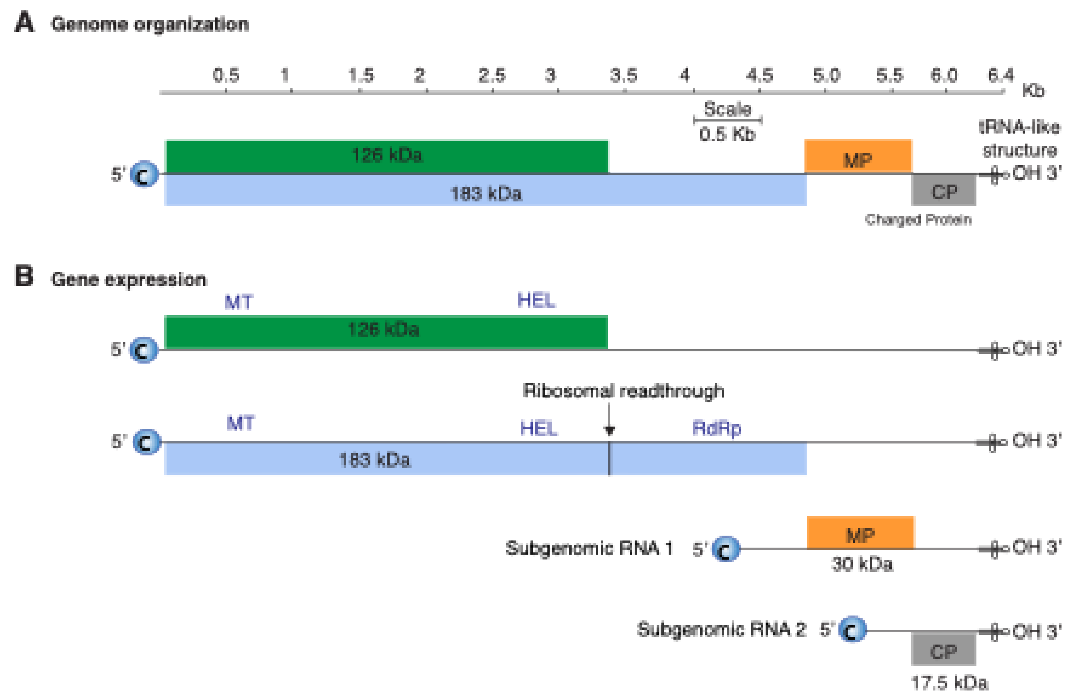
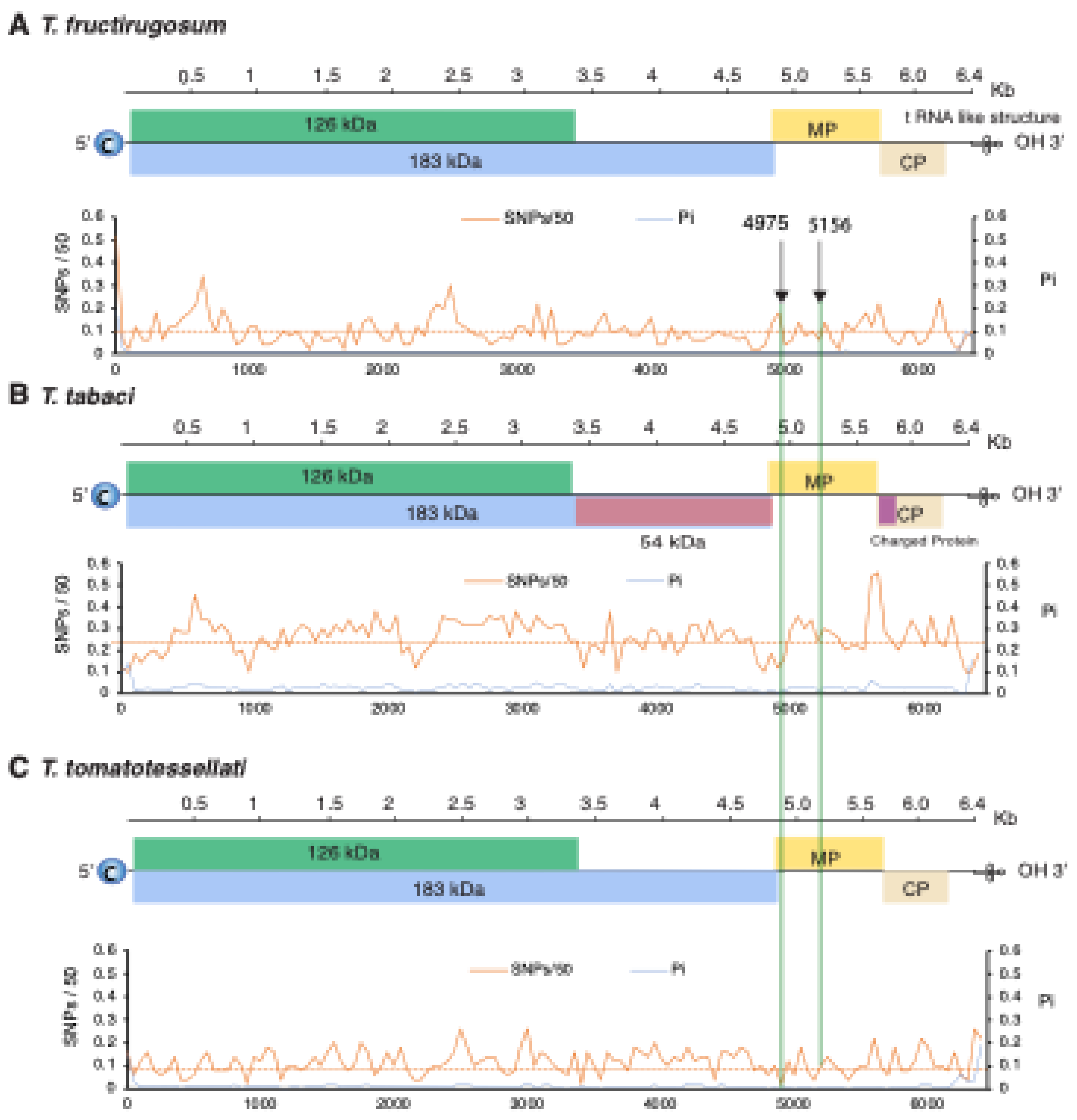
The genus Tobamovirus belongs to the family Virgaviridae, and the genome consists of monopartite, positive, single-strand RNA. Most species have four open reading frames encoding four essential proteins. Transmission occurs by mechanical contact between plants and sometimes by seed. Tobamovirus fructirugosum (Tomato brown rugose fruit virus, ToBRFV) is the most recent species in the genus, was first reported in 2015, broke genetic resistance that had been effective in tomato for sixty years, has caused devastating damage to tomato production worldwide, and highlights the importance of understanding genomic variation and evolution of tobamoviruses. In this study, we measured and characterized nucleotide variation for the entire genome and for all species in the genus Tobamovirus and measured the selection pressure acting on each open reading frame. Results showed that low nucleotide diversity and negative selection pressure are general features of tobamoviruses, with values that are approximately the same across open reading frames and without hypervariable areas. A comparison of nucleotide diversity between T. fructirugosum and its close relatives, T. tomatotessellati (Tomato mosaic virus, ToMV) and T. tabaci (Tobacco mosaic virus, TMV), showed low nucleotide diversity in the movement protein region harboring the resistance-breaking mutation. Furthermore, phylogenetic and diversity analyses showed that T. fructirugosum continues to evolve, and geographical distribution and host influence genomic diversity.

Keywords: 
;  ;  ;  ;  ;  ;  

1. Introduction

The genus Tobamovirus (tobamoviruses) belongs to the family Virgaviridae, includes several species that cause important diseases in crops [1,2] , such as Tobamovirus fructirugosum (Tomato brown rugose fruit virus, ToBRFV), a species first described in 2015 that broke Tm22-mediated resistance to tobamoviruses in tomato [3,4]. The general features of tobamoviruses include monopartite, positive, single-strand RNA, rigid, rod-shaped particles [5], are transmitted mechanically and through seed contamination, can persist in the soil for extended periods in infected plant residues [6], and can be transmitted by pollinator insects [7,8].
The tobamoviral genome consists of approximately 6,400 nucleotides [9]. The genome is organized into four core open reading frames (ORFs), and two additional ORFs that have been described in some species (Figure 1). The genomic RNA expresses a 126 kDa protein that contains a methyltransferase and a helicase domain [10,11,12]. The second ORF encodes a 183 kDa protein, produced by readthrough of the 126 kDa termination signal, and includes an RNA-dependent RNA polymerase domain. The movement protein and coat protein are expressed from subgenomic RNAs [5,13]. Two additional proteins have been described for some species, including T. tabaci (Tobacco mosaic virus, TMV): a 54 kDa protein representing the RNA-dependent RNA polymerase (RdRp) domain of the 183 kDa protein, and a small 4-6 kDa protein (P6 protein) also known as charged protein [14]. The length of the open reading frames and their overlap vary among species, and the size of the replicase protein varies between strains, even within the same species [14,15].
Figure 1. Tobamovirus genome organization and gene expression. (A) Genome organization based on T. tabaci (Tobacco mosaic virus, TMV accession number NC_001367.1). Single lines represent non-coding regions, and labeled boxes represent open reading frames. (B) Gene expression strategies include the formation of sub-genomic RNAs and ribosomal readthrough. Functional methyltransferase (MT), helicase (HEL) and RNA-dependent RNA polymerase (RdRp) domains for 126 kDa and 183 kDa proteins are indicated.
Preprints 172966 g001
Breeding for genetic resistance to tobamoviruses has been a valuable management practice [5]. T. fructirugosum (ToBRFV) was first detected in Israel and Jordan in 2014 and 2015 [3,4], and has spread globally [16]. According to the current working model, ToBRFV emerged from a recombination event between T. tomatotessellati (Tomato mosaic virus, ToMV) and T. tabaci (Tobacco mosaic virus, TMV) [4]. After selection and adaptation, ToBRFV broke Tm22-mediated resistance to tobamoviruses in tomato that had been effective for sixty years [3,4], causing widespread economic losses and becoming a major threat to tomato production globally [17]. The emergence of ToBRFV highlights the need to better understand genomic diversity and evolutionary mechanisms of tobamoviruses [18]. Moreover, the global dissemination of tobamoviruses through trade and seed distribution has contributed to their rapid spread, making it imperative to study the geographical and temporal dynamics of their populations [18,19].
Previous studies have focused on identification of genomic variation within individual tobamoviral species in particular areas of the world, or within areas of the genome [19,20,21,22]. In this study, we took a general, genome-wide, and comparative approach to measure genomic variation for the entire genus Tobamovirus. After determining variation patterns, due to its current economic impact, we focused on the global evolution patterns of ToBRFV. Results identified key characteristics of tobamovirus diversity and evolution. Nucleotide diversity and selection patterns across species showed that low genomic variation, negative selection pressure, and lack of hypervariable areas are general features of tobamoviruses, including ToBRFV. However, influenced by geographical distribution and hosts, ToBRFV is still mutating and evolving into new strains due to variation at the single amino acid level instead of specific regions or at the protein or domain levels.

2. Materials and Methods

All computational analyses were performed using a high-performance computing nodding system available through the Holland Computing Center (HCC) at the University of Nebraska Lincoln. In-house Python, Bash, and R scripts used are available upon request.

2.1. Tobamovirus Genomic Sequences

Genomic sequences for all species in genus Tobamovirus were retrieved from the National Center for Biotechnology Information (NCBI) database on 18 June 2024, using a customized Python script incorporating Batch Entrez and SeqI0 based on Entrez Programming Utilities. For each species, a reference genome was chosen (Table 1). If NCBI had an officially defined reference genome, it was used; otherwise, the longest nucleotide sequence was selected. For each species, the reference genome was used to determine the ORF coordinates. All downloaded accessions were filtered using a Bash script to remove partial sequences and retain only complete genomes and accessions measuring at least 95% the length of the reference genome. Accessions with less than 95% of the reference genome length were excluded, and accessions containing more than 2.5% unknown characters were also excluded. In tobamovirus taxonomy, the primary criterion for species demarcation is a nucleotide difference of at least 10%. Accordingly, if two tobamoviruses have more than 90% whole-genome nucleotide sequence identity, they are considered strains of the same species. Conversely, if they have less than 90% sequence identity, they are considered different species [23]. Thus, for each species, accessions were compared to the reference accession and discarded if their nucleotide identity was below 90% [24]. To ensure robust statistical comparisons, variation analyses were done only for species with a minimum of three accessions meeting the criteria described above [25]. The final number of sequences retained after these filtering steps is indicated in Table 1.
Table 1. Tobamovirus species downloaded from NCBI and number of accessions used in this analysis.
Scheme 1. Virus name ICTV Abbreviation No. of accessions Reference genome1 Length (nt) 95% length Accessions (≥ 95%)3
T. maculacapsici Bell pepper mottle virus BPMV 5 NC 009642.1 6375 6056 4
T. brugmansiae Brugmansia mild mottle virus BrMMV 2 NC 010944.1 6381 6062 2
T. cacti Cactus mild mottle virus CMMoV 5 NC 011803.1 6449 6127 3
NA2 Cactus tobamovirus 1 2 MW938767.1 6458 6135 2
NA2 Cactus tobamovirus 2 2 MW938766.1 6368 6050 2
NA2 Chili pepper mild mottle virus 14 MN164455.1 6383 6064 2
T. clitoriae Clitoria yellow mottle virus CliYMV 2 NC 016519.1 6514 6188 2
T. maculafructi Cucumber fruit mottle mosaic virus CFMMV 19 NC 002633.1 6562 6234 6
T. viridimaculae Cucumber green mottle mosaic virus CGMMV 484 NC 001801.1 6424 6103 198
T. cucumeris Cucumber mottle virus CMoV 2 NC 008614.1 6485 6161 2
T. frangipani Frangipani mosaic virus FrMV 9 NC 014546.1 6643 6311 4
T. fortpiercense Hibiscus latent Fort Pierce virus HLFPV 26 NC 025381.1 6431 6109 10
T. singaporense Hibiscus latent Singapore virus HLSV 9 NC 008310.2 6485 6161 6
NA2 Hoya chlorotic spot virus 2 NC 034509.1 6386 6067 2
NA2 Hoya necrotic spot virus 4 LC807720.1 6425 6104 3
T. kyuri Kyuri green mottle mosaic virus KGMMV 51 NC 003610.1 6514 6188 4
T. maracujae Maracuja mosaic virus MarMV 2 NC 008716.1 6794 6454 2
T. obudae Obuda pepper virus ObPV 4 NC 003852.1 6507 6182 4
T. odontoglossi Odontoglossum ringspot virus ORSV 241 NC 001728.1 6618 6287 14
NA2 Opuntia virus 2 19 NC 040685.2 6453 6130 8
T. paprikae Paprika mild mottle virus PaMMV 19 NC 004106.1 6524 6198 9
T. passiflorae Passion fruit mosaic virus PFMV 2 NC 015552.1 6791 6451 2
T. capsici Pepper mild mottle virus PMMoV 662 NC 003630.1 6357 6039 86
NA2 Piper chlorosis virus 3 ON924221.1 6237 5925 2
T. plumeriae Plumeria mosaic virus PluMV 4 NC 026816.1 6688 6354 4
T. muricaudae Rattail cactus necrosis-associated virus RCNaV 22 NC 016442.1 6506 6181 5
T. rehmanniae Rehmannia mosaic virus RheMV 75 NC 009041.1 6395 6075 8
T. plantagonis Ribgrass mosaic virus RMV 41 NC 002792.2 6311 5995 11
T. streptocarpi Streptocarpus flower break virus SFBV 6 NC 008365.1 6279 5965 5
T. mititessellati Tobacco mild green mosaic virus TMGMV 398 NC 001556.1 6355 6037 40
T. tabaci Tobacco mosaic virus TMV 653 NC 001367.1 6395 6075 111
T. fructirugosum Tomato brown rugose fruit virus TBRFV 451 NC 028478.1 6393 6073 248
T. tomatotessellati Tomato mosaic virus ToMV 372 NC 002692.1 6383 6064 81
T. maculatessellati Tomato mottle mosaic virus ToMMV 62 NC 022230.1 6398 6078 28
T. tropici Tropical soda apple mosaic virus TSAMV 13 NC 030229.1 6350 6033 11
T. rapae Turnip vein clearing virus TVCV 29 NC 001873.1 6311 5995 12
T. wasabi Wasabi mottle virus WMoV 6 NC 003355.1 6298 5983 6
NA2 Watermelon green mottle mosaic virus WGMMV 6 MH837097.1 6482 6158 6
T. anthocercis Yellow tailflower mild mottle virus YTMMV 97 NC 022801.1 6379 6060 4
T. youcai Youcai mosaic virus YoMV 129 NC 004422.1 6303 5988 33
T. cucurbitae Zucchini green mottle mosaic virus ZGMMV 10 NC 003878.1 6513 6187 5
1The accession used as reference for each species. 2 Indicates virus name in NCBI that is not listed by ICTV. 3 Number of accessions with at least 95% the length of the reference genome.

2.2. Tobamovirus Phylogeny

Consensus sequences were generated for each virus species using the genome sequences that passed the filters indicated above. Consensus sequences were then combined into a single FASTA file and aligned using MAFFT version 7.4 (Multiple Alignment using Fast Fourier Transform). A neighbor-joining phylogenetic tree was constructed from the aligned sequences, and the resulting alignment was exported in Newick format. The phylogenetic tree was visualized using Figtree version 1.4.3 http://tree.bio.ed.ac.uk/software/figtree/ accessed on 10 Jan 2025 [26].

2.3. Single Nucleotide Polymorphism and Nucleotide Diversity

For each virus species with more than three complete accessions, alignment files (.aln) based on forward reads from MAFFT were analyzed for single nucleotide polymorphism (SNPs) using bash scripts as described [27]. SNPs were normalized across the genome using a 50-nt sliding window. Average pairwise nucleotide diversity (π = Pi) was calculated using TASSEL Version 5.0 [28] from alignment files (.aln) for each species, also employing a 50-nt sliding window.
Nucleotide diversity captures variation at the population level by normalizing the number of polymorphic sites to the number of accessions [29]. Thus, based on nucleotide diversity, the five most diverse viruses were used for further analyses and to map variation cross their genomes. Nucleotide diversity for each viral open reading frame (ORF) was estimated by calculating the weighted average of the Pi values from the sliding windows within each gene. This was done by summing the Pi values for all windows covering the gene, multiplying each by the window length (50 nt), and then dividing by the total gene length [30]. T. fructirugosum and its ancestors T. tabaci and T. tomatotessellati [4], were included in the analyses due to their current importance. For both SNPs and Pi, the mean values along with the 99% confidence intervals (P-value < 0.01) were estimated, and a genome-wide map was obtained for each species.

2.4. Selection Analyses

For each species, accession numbers were used to retrieve coding sequences (CDS) from the NCBI database using Batch Entrez. A custom Bash script was used to separate CDS for each gene. The stop codon in the ribosomal read-through region of the 126 kDa was removed by a Python script to obtain the complete sequence for the 183 kDa replicase gene. The rates of synonymous substitutions (dS) and non-synonymous substitutions (dN) [31] were calculated for each gene’s CDS using the SLAC (Single-Likelihood Ancestor Counting) and MEME (Mixed Effects Model of Evolution) tools on Datamonkey https://www.datamonkey.org, last accessed on 10 Jan 2025. A significance threshold of ≤0.05 and a posterior probability >0.95 were applied for both SLAC and MEME [27,32]. The dN/dS ratio was calculated, and a selection pressure map for the entire genome was created by stitching the ORFs together. The expected and observed numbers of sites under selection were calculated using the model previously described in [27,33,34]. In which the expected number of sites under positive or negative selection for each ORF was estimated by dividing the length of the ORF by the total length of the genome, then multiplying this proportion by the total number of sites under negative or positive selection genome-wide. This expected value was then compared to the observed number of sites in each ORF.

2.5. Maximum Likelihood Phylogenetic Tree for T. fructirugosum

We constructed a maximum likelihood phylogenetic tree for the alignment sequences of all available T. fructirugosum in NCBI, used IQtree http://www.iqtree.org. The tree was subjected to 1000 bootstrap replicates [35]. The best-fit nucleotide substitution model was determined by Modeltest [36]. The phylogenetic tree was visualized using an R script.

2.6. Multidimensional Scaling

To understand factors (geographical distribution, host, or year of isolation) contributing to variation in T. fructirugosum, we use multidimensional scaling (MDS) [37] to visualize the virus sequences in two-dimensional Euclidean space. MDS, a non-linear dimensionality reduction technique, transforms a pairwise distance matrix into a lower-dimensional representation while best recapitulating the original distance matrix. The distance matrix was generated using the dist.dna function from the dplyr package in R, with the alignment file containing all complete T. fructirugosum accessions as input. MDS analysis was then performed using a custom R script.

3. Results

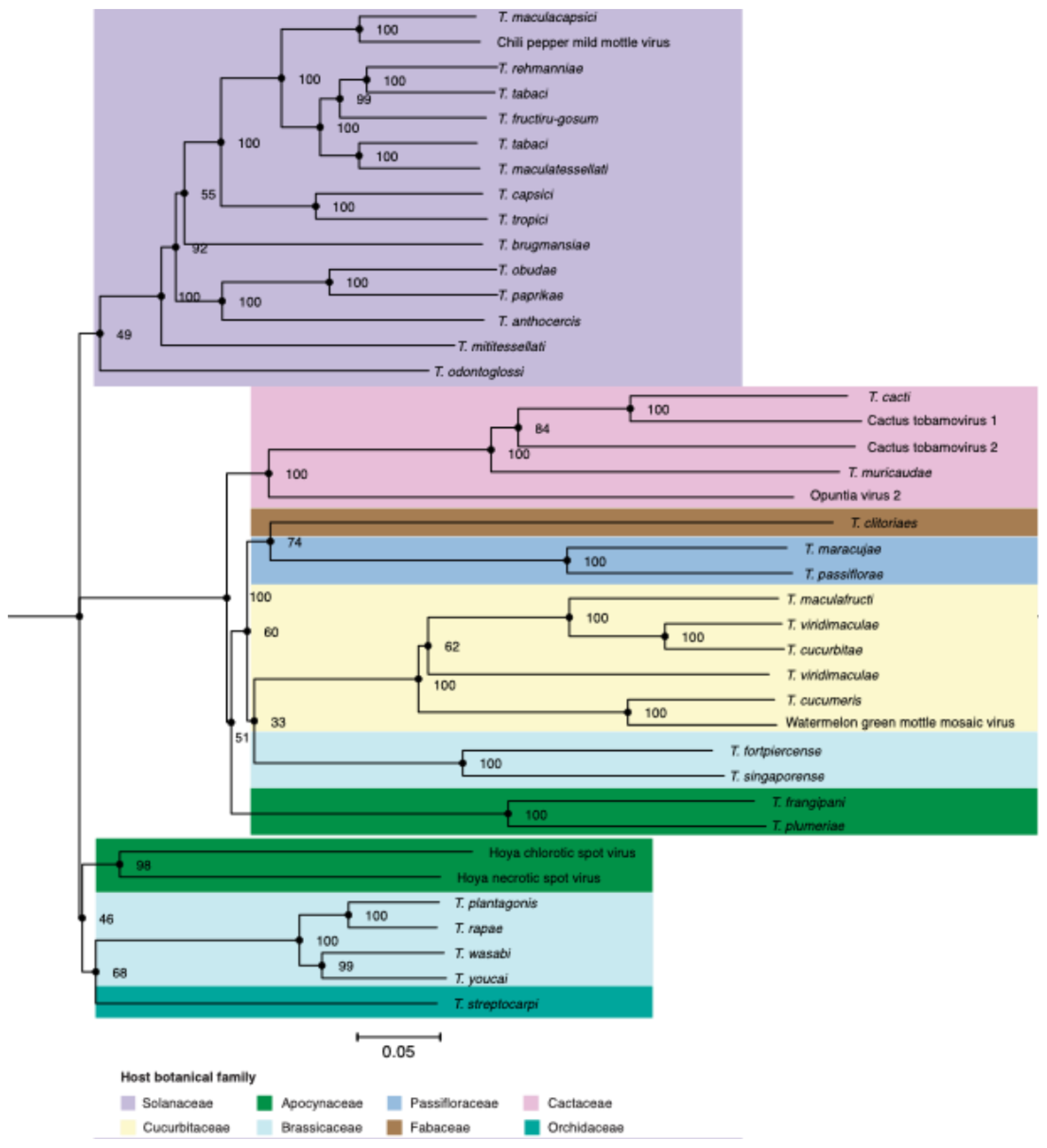
3.1. Tobamoviruses Group According to the Botanical Family of Their Host

Genetic relationships among tobamovirus species were determined by constructing a phylogenetic tree based on nucleotide sequences. All species with at least two complete genomes were included. The resulting tree comprised thirty-three classified species and seven unclassified species, which clustered into three clades. The botanical family of the hosts was added to each virus species (Figure 2). The first clade was formed by tobamoviruses that predominantly infect plants in the family Solanaceae. In a second clade, more than half of the species mainly infect plants in the families Cucurbitaceae and Cactaceae.
The third clade is dominated by species that infect plants in the family Brassicaceae. This phylogenetic organization highlights that sequence similarity between tobamovirus species correlates with the botanical family of their host and indicates that host selection pressure is a determinant of virus adaptation.

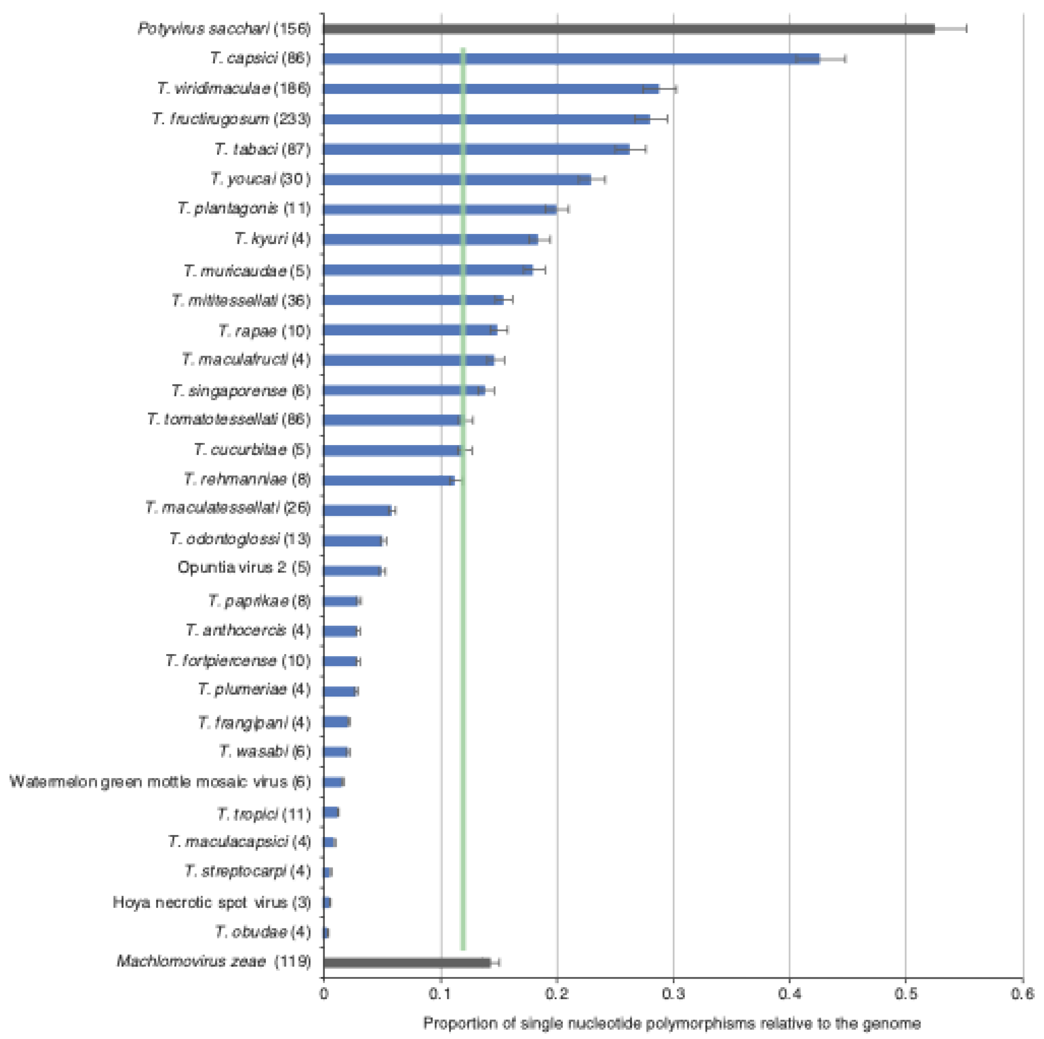
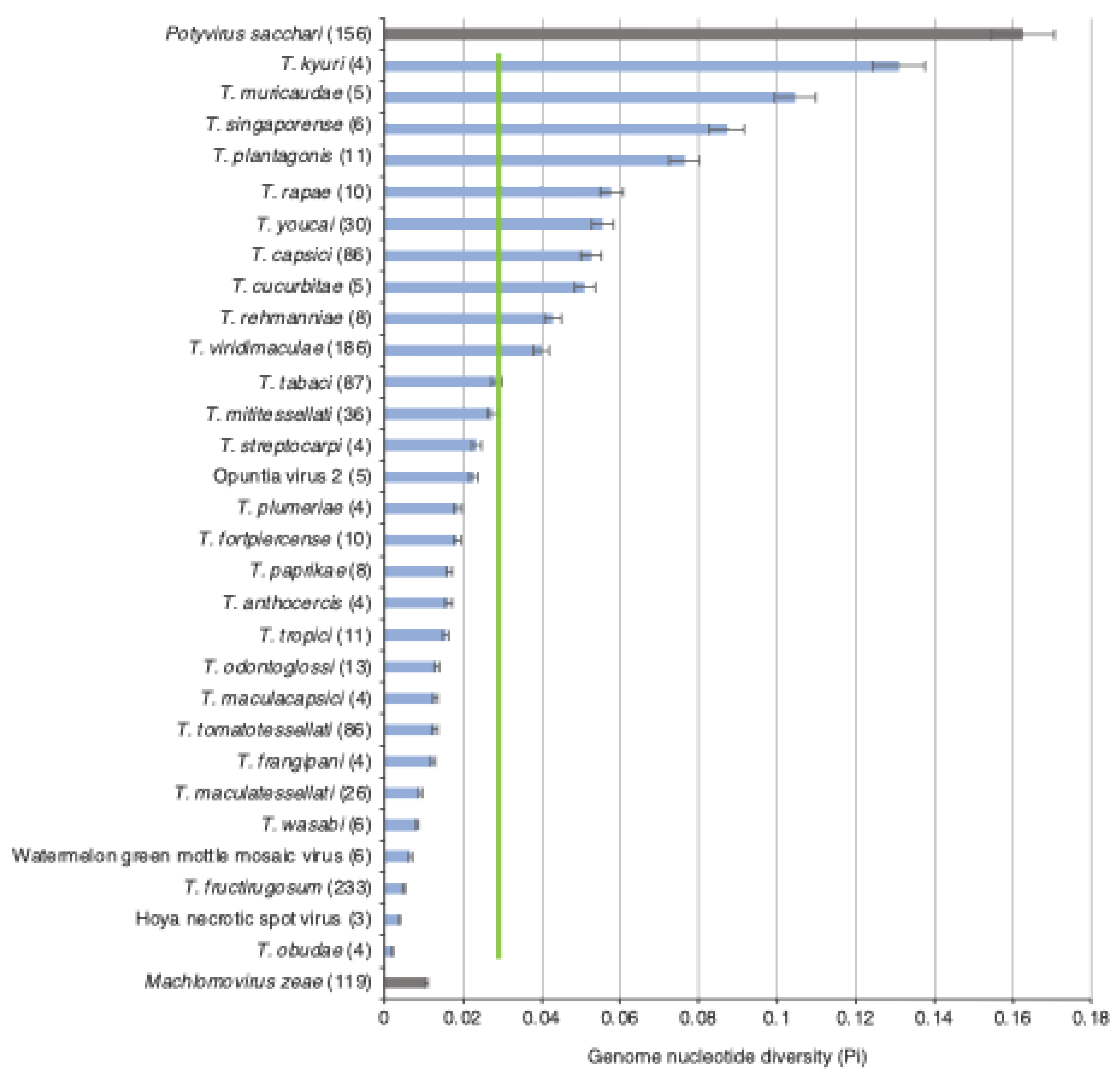
3.2. Tobamovirus Genome Diversity

Using a 50-nucleotide window, genomic diversity in the genus Tobamovirus was assessed by comparing single nucleotide polymorphism (SNP) relative to genome length (genomic variation) for all species with three or more complete genome sequences [27]. Highly variable Potyvirus sacchari (Sugarcane mosaic virus, SCMV) and low-diversity Machlomovirus zeae (maize chlorotic mottle virus, MCMV) [38] were included as internal controls for comparison. Results showed that the average genomic variation of tobamoviruses is 0.11± 0.2 (Figure 3), which means that approximately 11% of positions in the genome are polymorphic. This level of genomic variation is 4.8 times lower than the observed for P. sacchari and approximately the same (1.1 times) as the observed for M. zeae (Figure 3).
Figure 2. Tobamovirus phylogeny as inferred from consensus nucleotide sequences from all accessions in Table 1 that were ≥95% of the reference accession length and species with at least two complete genomes. Consensus sequences were aligned using MAFFT, and the phylogenetic tree was generated using FigTree v1.4.4. Colors in the tree indicate the botanical family of the primary host. Branches are labeled to indicate bootstrap values based on 1000 replicates. Binomial names are used for classified species; virus names are used for species not classified by ICTV.
Preprints 172966 g002
Tobamoviruses with the highest genomic variation were T. capsici, T. viridimaculae, T. fructirugosum, and T. tabaci. While all tobamoviruses have lower genomic variation than P. sacchari, 18 tobamovirus species showed lower genomic variation than M. zeae (Figure 3). These results show that low genomic variation is a general feature of tobamoviruses.
In a complementary approach, nucleotide diversity (Pi) was used to measure genetic variation in tobamoviruses. Nucleotide diversity accounts for the number of polymorphic sites and the frequency of different nucleotides at each site, while also normalizing variation to the number of accessions [27,39]. Nucleotide diversity was calculated using Tassel software [25,27]. Results show that the average nucleotide diversity for tobamoviruses is 0.03, which is 5.5 times lower than the observed for P. sacchari and only two times higher than the observed for M. zeae (Figure 4). The five most diverse species were T. kyuri, T. muricaudae, T. singaporense, T. plantagonis, and T. rapae (Figure 4). These species were chosen for further variation analysis. T. fructirugosum was included due to its current importance. Collectively, using two complementary approaches, the results described above show that low genomic variation is a general feature of tobamoviruses (Figure 3 and Figure 4).
Figure 3. Genomic variation in tobamoviruses. For species with three or more accessions, genomic variation was determined as single nucleotide polymorphisms relative to the genome and estimated in a 50-nt window. Bars represent the average, and standard error of the proportion of polymorphic sites (number of single nucleotide polymorphisms/length of the genome). For each species, the number of nucleotide accessions is indicated in parentheses. A green vertical line represents the mean and a 99% confidence interval (p-value < 0.01). Species binomial names indicate classified tobamoviruses, while viruses with common names were unclassified. For comparison, Potyvirus sacchari (Sugarcane mosaic virus) and Machlomovirus zeae (Maize chlorotic mottle virus) were used as hypervariable and genetically stable, respectively [27].
Preprints 172966 g003

3.3. Nucleotide Diversity by Open Reading Frame

We used nucleotide diversity to measure variation per open reading frame (ORF) for the five most variable species and T. fructirugosum (Figure 5A). No significant differences were detected among the four core open reading frames. The highest value was observed for the ORF coding for the 126 kDa protein, which on average was 0.1. For comparison, nucleotide variation in Potyvirus sacchari P1 was 0.22, which is 2.2 times higher.
Figure 4. Nucleotide diversity (Pi) in tobamoviruses. Species with three or more accessions were included. Bars represent the proportion of variable positions with respect to the length of the genome and normalized to the number of accessions. Labels are as in Figure 3.
Preprints 172966 g004

3.4. Selection Analysis by Open Reading Frame

The ratio of non-synonymous to synonymous genetic changes is used to determine how natural selection affects the evolution of viral populations [40]. This ratio is described as dN/dS. A dN/dS ratio less than 1 indicates negative selection pressure, where harmful mutations are removed, preserving the function of essential genes. Neutral selection occurs when the dN/dS ratio equals 1, meaning changes have no effect on fitness and accumulate randomly. A ratio greater than 1 indicates positive selection, where changes in the gene sequence are beneficial and favored, allowing the virus to adapt to different hosts or overcome immune defenses [41,42]. For comparison, we estimated negative and positive selection in P. sacchari P1. The proportion of sites under negative selection in tobamoviruses, normalized by ORF length, was on average 0.01, which was approximately 23 times lower than that observed in P. sacchari P1 (Figure 5B). Positive selection was barely above background and approximately 10 times lower than that observed in P. sacchari P1 (Figure 5C). Within tobamoviral ORFs, the frequency of sites under negative selection was six times higher than the frequency of sites under positive selection (Figure 5B,C). No significant differences in positive or negative selection were observed among the four core open reading frames in tobamoviruses (Figure 5B,C).
Figure 5. Nucleotide diversity, positive and negative selection by open reading frame for the top five most variable tobamoviruses and ToBRV. Parameters were estimated by ORF and normalized to the corresponding length. Virus names are color-coded. Potyvirus sacchari P1 was used as a control [27]. (A) Cumulative nucleotide diversity across ORF for each virus species. There is no significant difference between nucleotide diversity disruption across the four ORFs, with a p-value of 0.99 as calculated with the Chi-square test. (B) Sites under negative selection normalized to the length of the ORF. (C) Sites under positive selection normalized to the length of the ORF. (D) Expected (E) and observed (O) number of sites under negative selection. Expected values were determined assuming a random distribution in the genome. (E) Expected and observed number of sites under positive selection. The * denotes significant differences with p-value ≤0.5, ** for p-value ≤0.001, as calculated by the Chi-square test.
Preprints 172966 g005
The frequency of sites observed under positive or negative selection per open reading frame was compared to the frequency expected in a random way. Results showed that in all open reading frames, the observed frequency of sites under negative selection was significantly lower than that expected randomly (Figure 5D). Across the top viruses analyzed, the 183 kDa, 126 kDa, MP, and CP ORFs all showed significantly fewer sites under purifying selection than expected (p≤ 0.001, Figure 5D) [33]. Positive selection was low across the genome. Only in the ORF coding for the 126kDa protein the expected was higher than the observe frequency (Figure 5E). Collectively, these results show that tobamoviruses are under negative selection.

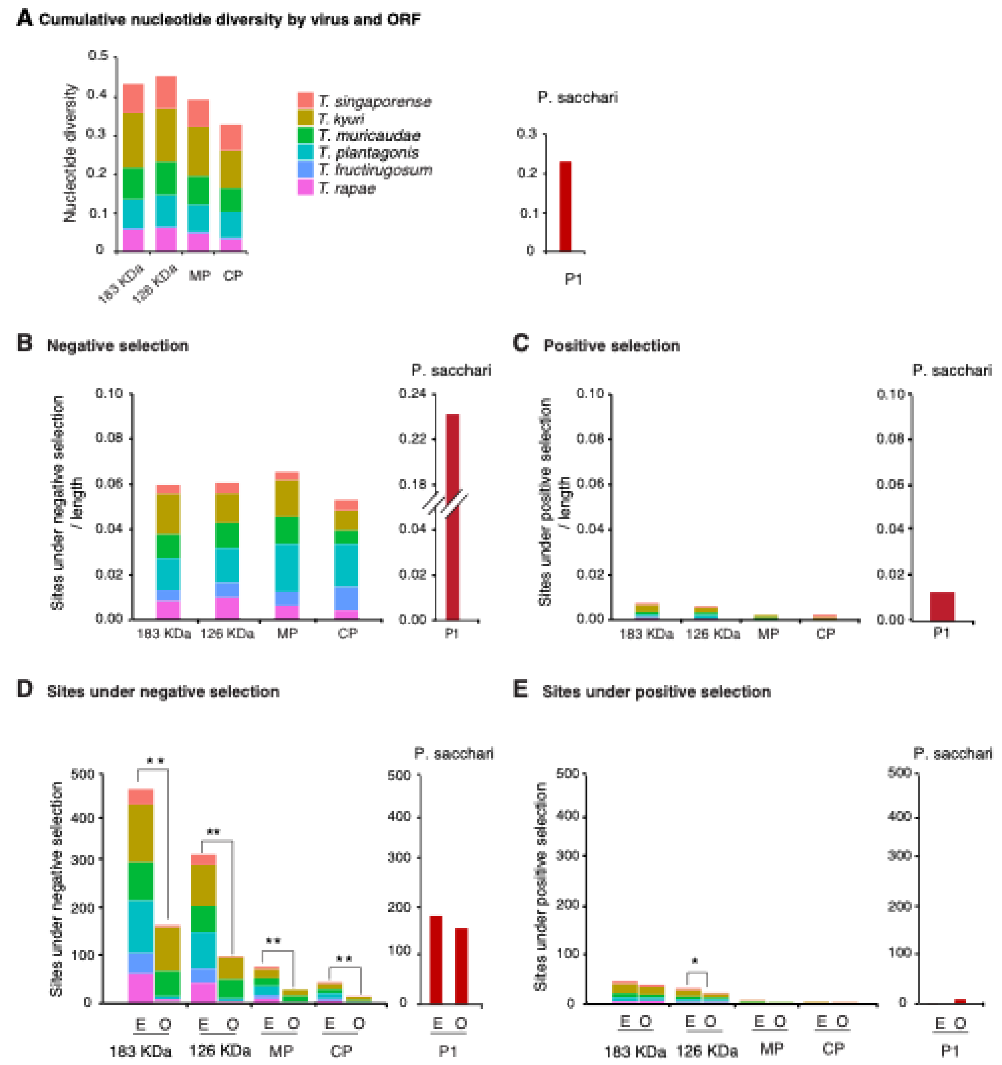
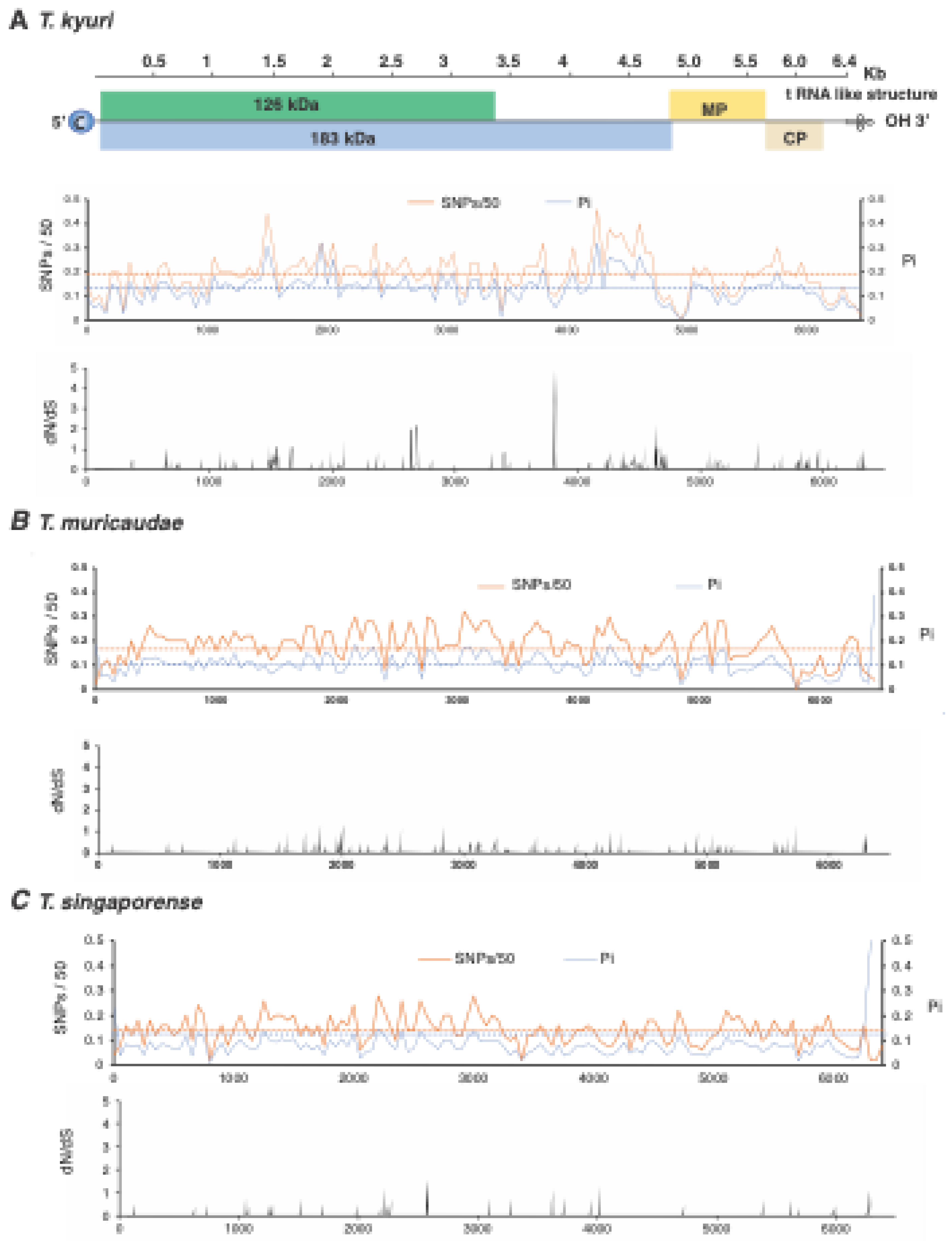
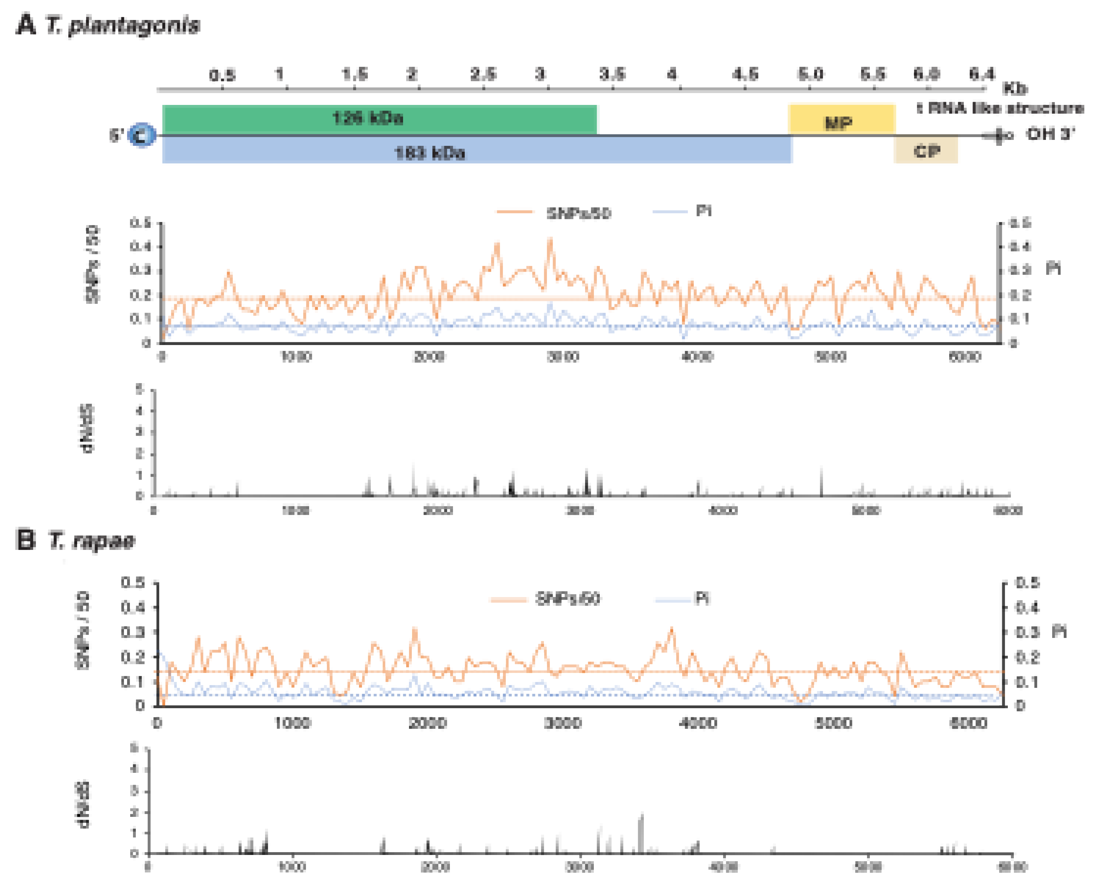
3.5. No Hypervariable Areas Were Detected in the Tobamovirus Genome

Genome-wide variation of the five most variable species was done to identify whether variation concentrates in areas of the genome or is distributed randomly within the genome. The ratio of non-synonymous to synonymous changes (dN/dS) was estimated in a 50-nucleotide (nt) window to identify the region that accumulates mutations in the genome. No hypervariable areas were consistently detected across species (Figure 6 and Figure 7).
Figure 6. Genome-wide variation in the top tree most variable tobamoviruses. Single nucleotide polymorphism (SNP), nucleotide diversity (Pi), and the ratio of non-synonymous to synonymous changes (dN/dS) were estimated in a 50-nt window. The average and a 99% confidence interval (p-value < 0.01) are indicated as a horizontal line. ORFs are color-coded and labeled with the name of the protein they encode. The coordinates for T. kyuri are based on reference accession NC_003610.1, for T. muricaudae based on NC_016442.1, and for T. singaporense based on NC_008310.2.
Preprints 172966 g006
Figure 7. Genome-wide variation in the fourth and fifth most variable tobamoviruses. Labels are as in Figure 6. The coordinates for T. plantagonis are based on reference accession NC_002792.2, and for T. rapae based on NC_001873.1.
Preprints 172966 g007

3.6. Variation in T. fructirugosum

While there is low genomic variation and predominantly negative selection across the entire genus Tobamovirus, T. fructirugosum, the most recently identified species, has broken resistance in tomato varieties due to variation in a single amino acid within the movement protein. Similar mutations occurred independently twice in two different strains [43,44]. These positions map to nucleotides 4975 and 5156 [43,44], which correspond to nucleotide changes from thymine (T) to adenine (A) at the two positions. In ToBRFV-Tom2M-Jo-MZ2438228.1, these nucleotide substitution resulted in amino acid changes at position 22 (change from Phe to Tyr) and position 82 (change from Asp to Lys) in the movement protein, respectively (Figure 8) [43]. In strain ToBRFV_G78_RB-OR760199, nucleotide 5156 mutated from T to guanine (G) resulting in an amino acid change from Asn to Lys at position 82 of the MP [44]. In T. fructirugosum and its closest relatives, T. tabaci and T. tomatotessellati, this region of the movement protein does not accumulate mutations at a particularly high rate (Figure 8).
Figure 8. Genome-wide variation in T. fructirugosum and its closest relatives. (A) Black arrows indicate resistance-breaking mutations in the movement protein (MP) of ToBRFV at nucleotide positions 4975 and 5156. At both sites, T is substituted by A in strain ToBRFV-Tom2M-Jo-MZ2438228.1, resulting in amino acid changes at amino acids 22 (Phe to Tyr) and 82 (Asp tp Lys) [43], while in strain ToBRFV_G78_RB-OR760199, at position 5156, T was replaced by G resulting in a Asn to Lys mutation in amino acid 88 [44]. B) and C) The green lines mark the equivalent positions of MP in T. tabaci and T. tomatotessellati where a breaking resistance mutation was reported in T. fructirugosum.
Preprints 172966 g008

3.7. Phylogenetic Analysis of T. fructirugosum

To gain insight into the evolution of T. fructirugosum (ToBRFV), a phylogenetic tree was constructed using all complete genome accessions available and the maximum likelihood model with IQtree [35]. Results indicate the presence of four clades (Figure 9). The country of origin of each accession was mapped in the tree. One clade is dominated by accessions from the Netherlands, another by accessions from the Middle East and China, and a third by accessions from North America. Most of the accessions were isolated from Solanum sp. and Capsicum sp. Interestingly, differences in host affect the phylogeny of ToBRFV, suggesting that the virus has mutated and evolved to adapt to each host. The year of isolation for each accession was also mapped, showing that isolates from the same year tend to cluster together.
Figure 9. Phylogenetic tree based on ToBRFV accessions with complete genome using IQ-TREE maximum likelihood. The tree was generated with 1,000 bootstrap replicates. The country of origin is indicated by colored triangles, and the host species is indicated by the color of each accession. The four main clades are labeled.
Preprints 172966 g009

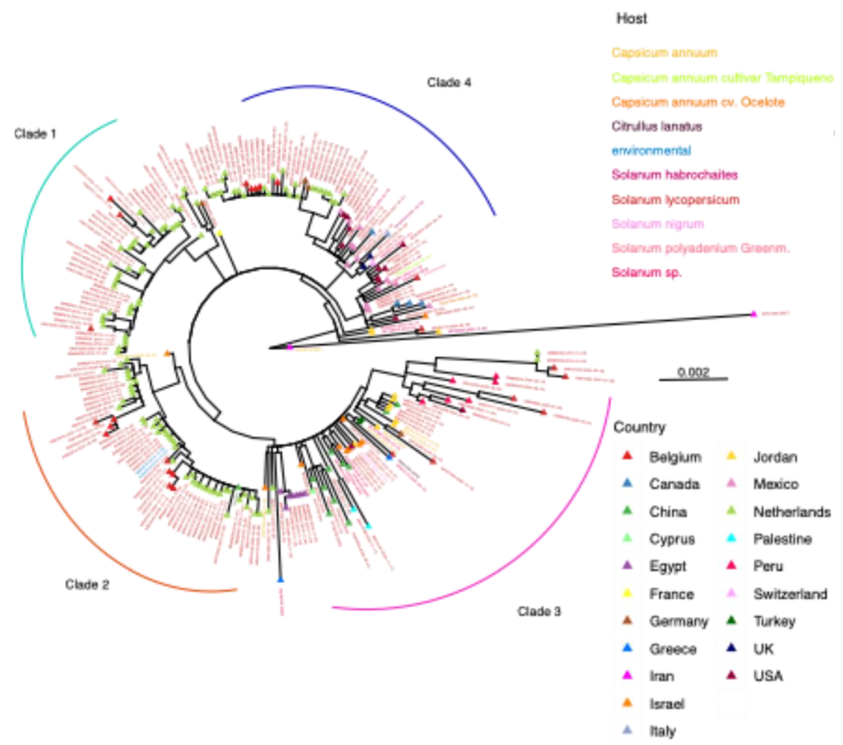
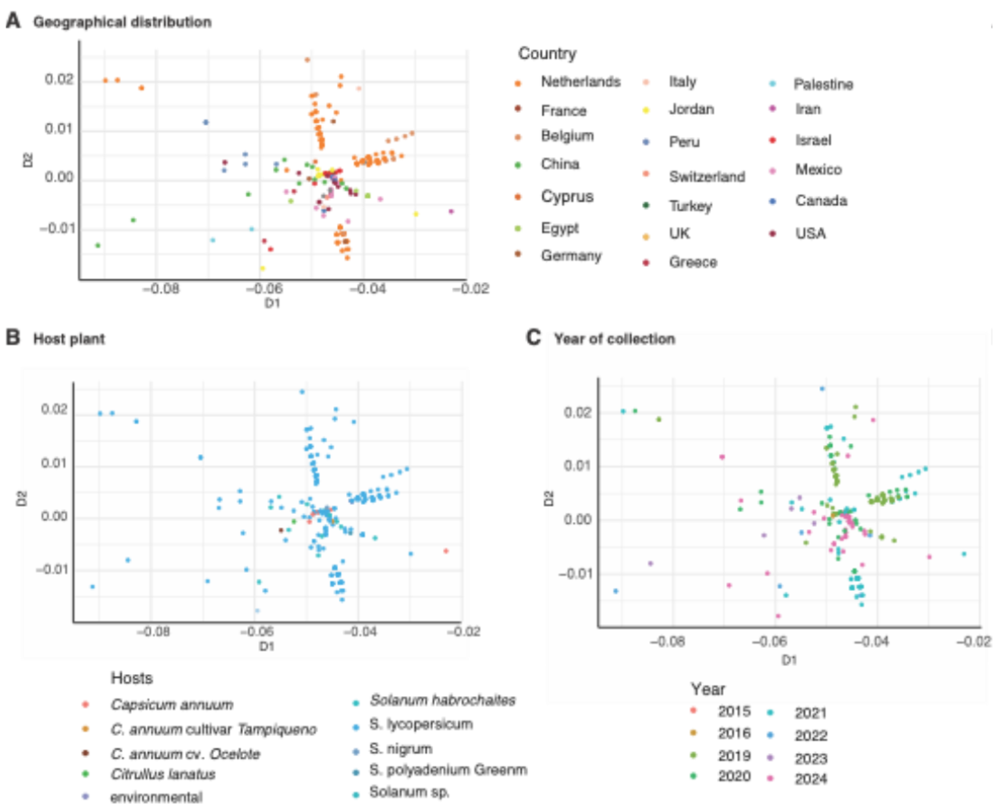
3.8. Geographical Distribution Correlates with Virus Variation

As indicated above, phylogenetic organization of ToBRFV accessions seems to be determined by the geographical origin, the host species, and the year. We used multidimensional scaling (MDS) to identify the most significant factor. The results visually showed that accessions clustered by geographical origin. Three clusters were formed by accessions from Netherlands and other neighboring European countries, including Belgium (Figure 10a). MDS scatter plot was color-coded according to the host from which each accession was isolated (Figure 10b). Accessions dominated by Solanum (tomato) are indicated with the blue color, and there is less domination of Capsicum sp. (pepper). Therefore, there was no distinct clustering according to the host due to the domination of Solanum sp.. This indicates that diversity between accessions is not due to the host. For the effect of collection date (Figure 10c), the same year of collection clusters together, which supports the hypothesis that the virus is not changing rapidly within the same collection area.
Figure 10. Multidimensional scaling clustering generated from the genomic distance matrix of 232 ToBRFV accessions. A) diversity based on geographical distribution; the color of circles represents the origin country of accessions. B) Diversity based on host plant. C) Diversity based on the year of collection for each accession.
Preprints 172966 g010

4. Discussion

Published analyses of isolates in geographical areas and of individual species suggest that tobamoviruses exhibit limited genetic diversity across their genomes [20,45]. In contrast with this observation, the emergence of new strains and even new species [22,46] shows that tobamoviruses are actively mutating and evolving. The most recent species in the genus is T. fructirugosum, which affects global tomato production. A notable feature of T. fructirugosum is that it broke genetic resistance based on the Tm-22 gene, which had been effective for 60 years against tobamoviruses, including T. tomatotessellati and T. tabaci [47,48]. The discrepancy between low genetic diversity in some tobamovirus species and the emergence of new strains and species highlights the need to better understand genomic variation and its relationship to tobamovirus evolution.
After measuring nucleotide variation for the entire genome and for all members of the genus, our results showed that tobamoviruses exhibit low genetic diversity and are under strong negative selection (Figure 5). On average, tobamoviruses harbor nucleotide diversity in 11% of the nucleotide positions on the genome. This is 4.8 times lower than the observed for Potyvirus sacchari (Figure 3), 2.3 times lower than the observed for poleroviruses [25], 3.3 times lower than the observed for orthotospoviruses [29], and 2.6 times lower than the observed for betacoronaviruses [49]. Tobamovirus variation was approximately the same as that observed in M. zeae (Figure 3), a virus considered genetically stable [38]. In contrast to what was observed in potyviruses [27], orthotospoviruses [29], poleroviruses [25] , and betacoronaviruses [49] , tobamoviruses do not harbor hypervariable areas (Figure 7 and 8).
Tobamoviruses have been evolving alongside their eudicotyledonous hosts since their establishment in the asterid, rosid, and caryophyllid lineages approximately 112.9 million years ago [50]. Currently, tobamoviruses infect hosts across eight botanical families (Figure 2), and our phylogenetic analyses showed that tobamoviruses cluster according to the botanical family of their host. Fifteen virus species that infect plants in the family Solanaceae form a clade, while viruses infecting plants in the family Cucurbitacea form a separate clade (Figure 2). This pattern of host-specific clustering supports the hypothesis of long-term co-evolution, adaptation, and specificity of viruses in general, and tobamoviruses in particular, to their host plants [22].
The low genomic variation for tobamoviruses described here (Figure 3 and Figure 4) is in contrast with previous findings that show a rapid evolutionary rate in this genus [22]. In that previous study, Bayesian coalescent methods were used to reveal the nucleotide substitution, and results prove that the substitution level is similar to that observed in animal and plant RNA viruses; therefore, they concluded that the rapid evolutionary rates [22]. Our findings are consistent with the variation analysis of T. tomatotessellati, which indicates the conserved evolution and limited variation in the genome of this species [51].
To further explore mutation distribution, we analyzed the five most variable tobamoviruses. Across the genome, nucleotide substitutions accumulated at relatively similar rates across open reading frames (ORFs) (Figure 5A). The most variable species, T. kyuri exhibited a region within the 183 kDa replicase ORF where mutations accumulated at levels exceeding the genome-wide average (Figure 6). Similarly, T. plantagonis showed a distinct mutation accumulation region in the 183 kDa replicase ORF (Figure 7). In contrast, the other three viral species displayed overall low variation with no specific regions of concentrated mutations.
For the five most variable viruses and T. fructirugosum , ORFs are predominantly under negative selection, as the number of negatively selected sites normalized to ORF length exceeds that of positively selected sites (Figure 5). Additionally, the randomly expected number of sites under negative selection is significantly higher than the observed, further supporting the low genomic variation rate in tobamoviruses. Fewer sites under purifying selection than expected suggest relaxed functional constraint or tolerance of variation in this region [33]. This is consistent with previous analysis of T. fructirugosum [52,53], and can be explained by the model that strains harboring deleterious mutations are unlikely to be fixed in the population [54] and thus not present in the sequence data set.
Despite low genetic variation, there is evidence of adaptive evolution in T. mititessellati, T. tabaci, T. capsica, and T. youcai [20,51]. For instance, a single nucleotide change in the overlapping of MP and CP open reading frames resulted in altered symptoms in T. youcai [55]. Furthermore, the low genomic variation and negative selection pressure observed in tobamoviruses do not preclude their ability to adapt and overcome host resistance. In tomato, resistance against tobamoviruses is conferred by Tm-1, Tm-2, and Tm-22 [21]. However, the first documented case of resistance breaking occurred in 2014 in Israel, where a tobamovirus successfully infected tomato and pepper [3], and infected tomato in the greenhouse during the spring of 2015 in Jordan [4]. A notable example of resistance breakdown is strain ToBRFV-Tom2M-Jo-MZ2438228.1, where resistance was overcome due to two amino acid substitutions in the movement protein at amino acids 22 (Phe replaced by Asn) and 82 (Tyr replaced by Lys) [43]. These two amino acid changes resulted from single nucleotide substitutions at positions 4975 and 5156, respectively (T replaced by A in both cases) [43]. More recently, on a second strain (ToBRFV_G78_RB-OR760199), a single amino acid substitution ( Asn82Lys), resulting from a single nucleotide substitution at position 5156 (T is replaced by G) in the movement protein, also broke resistance [44]. This site maps to an area with a small variation peak that is not maintained in other relative species (Figure 8). These observations support the hypothesis that tobamoviruses can mutate and evolve even with limited variation in nucleotides and highlight the need to better understand interactions between plant and viral proteins [56].
The emergence of T. fructirugosum is linked to a recombination event between T. tomatotessellati and T. tabaci [4,57]. After the recombination event, one or two mutations in the movement protein were identified as responsible for overcoming Tm-22 resistance [47,48,58]. Since RNA recombination requires co-infection of the same plant, and even the same cell [54,59], it is unlikely that recombination between T. tomatotessellati and T. tabaci occurred in commercial tomato. It is most likely that the recombination event occurred in an alternate host, such as wild tomato, breeding lines, or other species that serve as a host for both T. tomatotessellati and T. tabaci. In this scenario, following the recombination event, commercial tomato imposed selection pressure, allowing only the strain capable of breaking resistance to infect and replicate.
To better understand the factors influencing genomic diversity in T. fructirugosum, we generated a phylogenetic tree relating geographical distribution, host plant, and date of collection to the diversity observed in this species (Figure 9). Our phylogenetic analysis revealed that T. fructirugosum isolates cluster into four major groups that could correlate with the host, the country of origin, or the year of collection. Accessions from alternative hosts, such as Capsicum spp., clustered separately, indicating the emergence of variation associated with host adaptation. The multidimensional scaling analysis illustrated the formation of three distinct clusters, primarily explained by geographical origin and including accessions from the Netherlands and Belgium. While geographical separation played a significant role in defining viral clusters, the host and year of collection had a minor impact (Figure 10). Clusters of isolates from the same collection year imply that T. fructirugosum does not undergo rapid mutation within localized outbreaks. This is consistent with previous reports indicating that T. fructirugosum exhibits a relatively low evolutionary rate compared to other plant RNA viruses [19,52].
The results described here show that low genetic variation, strong negative selection, and the absence of hypervariable areas are general features of the genus Tobamovirus. The emergence of new strains and species, such as T. fructirugosum implicate alternative hosts as a source of genetic variation that is later selected and influenced by geographical distribution more than host specificity. Accordingly, these results highlight the importance of minimizing tobamovirus spread by monitoring global seed trade.

Author Contributions

Conceptualization, H.GR.; methodology, A.E.G. and H.GR.; formal analysis, A.E.G.; resources, A.E.G. and H.GR.; data curation, A.E.G.; writing—original draft preparation, A.E.G.; writing—review and editing, H.GR.; supervision, H.GR.; project administration, H.GR.; funding acquisition, A.E.G. and H.GR. All authors have read and agreed to the published version of the manuscript.

Funding

Amany E. Gomaa was funded by the Ministry of Higher Education of the Arab Republic of Egypt for her Ph.D. scholarship (GM 1147) at the University of Nebraska Lincoln, USA.

Institutional Review Board Statement

Not applicable.

Informed Consent Statement

Not applicable.

Data Availability Statement

In-house Python, bash, and R scripts used in this analysis are available upon request.

Acknowledgments

The authors thank the Holland Computing Center for technical assistance. We express gratitude to Prof. James C. Schnable for providing guidance and to Dr. J. Vladimir Torres-Rodríguez for technical assistance.

Conflicts of Interest

The authors declare no conflict of interest. The founding sponsors had no role in the design of the study, the collection and analyses or interpretation of data, in the writing of the manuscript, or in the decision to publish the results.

References

  1. Dorokhov, Y.L.; Sheshukova, E.V.; Komarova, T.V. , Tobamoviruses and their diversity. In Plant Viruses, CRC Press: 2018; pp 65-80.
  2. Spiegelman, Z.; Dinesh-Kumar, S.P. , Breaking Boundaries: The Perpetual Interplay Between Tobamoviruses and Plant Immunity. 2024, 58, 24–24.
  3. Luria, N.; Smith, E.; Reingold, V.; Bekelman, I.; Lapidot, M.; Levin, I.; Elad, N.; Tam, Y.; Sela, N.; Abu-Ras, A. , A new Israeli Tobamovirus isolate infects tomato plants harboring Tm-22 resistance genes. PloS one 2017, 12, e0170429–e0170429. [Google Scholar] [CrossRef]
  4. Salem, N.; Mansour, A.; Ciuffo, M.; Falk, B.W.; Turina, M. , A new tobamovirus infecting tomato crops in Jordan. Archives of virology 2016, 161, 503–506. [Google Scholar] [CrossRef] [PubMed]
  5. Carr, J.P. , Engineered Resistance to Tobamoviruses. In Viruses, Multidisciplinary Digital Publishing Institute (MDPI): 2024; Vol. 16.
  6. Dombrovsky, A.; Smith, E. , Seed Transmission of Tobamoviruses: Aspects of Global Disease Distribution. In Advances in Seed Biology, InTech: 2017.
  7. Darzi, E.; Smith, E.; Shargil, D.; Lachman, O.; Ganot, L.; Dombrovsky, A. , The honeybee Apis mellifera contributes to Cucumber green mottle mosaic virus spread via pollination. Plant Pathology 2018, 67, 244–251. [Google Scholar] [CrossRef]
  8. Okada, K.; Kusakari, S.-i.; Kawaratani, M.; Negoro, J.-I.; Ohki, S.T.; Osaki, T. , Tobacco mosaic virus is transmissible from tomato to tomato by pollinating bumblebees. Journal of General Plant Pathology 2000, 66, 71–74. [Google Scholar] [CrossRef]
  9. Ishibashi, K.; Ishikawa, M. , Replication of tobamovirus RNA. Annual Review of Phytopathology 2016, 54, 55–78. [Google Scholar] [CrossRef]
  10. Ahola, T.; Laakkonen, P.; Vihinen; Helena; Kääriäinen, L., Critical residues of Semliki Forest virus RNA capping enzyme involved in methyltransferase and guanylyltransferase-like activities. Journal of virology 1997, 71, 392–397. [CrossRef] [PubMed]
  11. Ahola, T.; den Boon, J.A.; Ahlquist, P. , Helicase and capping enzyme active site mutations in brome mosaic virus protein 1a cause defects in template recruitment, negative-strand RNA synthesis, and viral RNA capping. Journal of virology 2000, 74, 8803–8811. [Google Scholar] [CrossRef]
  12. Fernández, A.; Laín, S.; García, J.A. , RNA helicase activity of the plum pox potyvirus CI protein expressed in Escherichia coli. Mapping of an RNA binding domain. Nucleic Acids Res 1995, 23, 1327–1332. [Google Scholar] [CrossRef]
  13. Goregaoker, S.P.; Culver, J.N. , Oligomerization and activity of the helicase domain of the tobacco mosaic virus 126- and 183-kilodalton replicase proteins. J Virol 2003, 77, 3549–3556. [Google Scholar] [CrossRef]
  14. Palukaitis, P.; Akbarimotlagh, M.; Astaraki, S.; Shams-Bakhsh, M.; Yoon, J.Y. , The Forgotten Tobamovirus Genes Encoding the 54 kDa Protein and the 4–6 kDa Proteins. In Viruses, Multidisciplinary Digital Publishing Institute (MDPI): 2024; Vol. 16.
  15. Lewandowski, D.J.; Dawson, W.O. , Functions of the 126-and 183-kDa proteins of tobacco mosaic virus. Virology 2000, 271, 90–98. [Google Scholar] [CrossRef]
  16. van de Vossenberg, B.T.L.H.; Dawood, T.; Woźny, M.; Botermans, M. , First Expansion of the Public Tomato Brown Rugose Fruit Virus (ToBRFV) Nextstrain Build; Inclusion of New Genomic and Epidemiological Data. PhytoFrontiers™ 2021, 1, 359–363. [Google Scholar] [CrossRef]
  17. Chanda, B.; Gilliard, A.; Jaiswal, N.; Ling, K.S. , Comparative Analysis of Host Range, Ability to Infect Tomato Cultivars with Tm-22 Gene, and Real-Time Reverse Transcription PCR Detection of Tomato Brown Rugose Fruit Virus. Plant Disease 2021, 105, 3643–3652. [Google Scholar] [CrossRef] [PubMed]
  18. Zhang, S.; Griffiths, J.S.; Marchand, G.; Bernards, M.A.; Wang, A. , Tomato brown rugose fruit virus: An emerging and rapidly spreading plant RNA virus that threatens tomato production worldwide. Molecular Plant Pathology 2022, 23, 1262–1277. [Google Scholar] [CrossRef] [PubMed]
  19. Ghorbani, A. , Genetic analysis of tomato brown rugose fruit virus reveals evolutionary adaptation and codon usage bias patterns. Scientific Reports, 2024; 14. [Google Scholar]
  20. Zamfir, A.D.; Babalola, B.M.; Fraile, A.; McLeish, M.J.; García-Arenal, F. , Tobamoviruses Show Broad Host Ranges and Little Genetic Diversity Among Four Habitat Types of a Heterogeneous Ecosystem. Phytopathology 2023, 113, 1697–1707. [Google Scholar] [CrossRef]
  21. Maayan, Y.; Pandaranayaka, E.P.J.; Srivastava, D.A.; Lapidot, M.; Levin, I.; Dombrovsky, A.; Harel, A. , Using genomic analysis to identify tomato Tm-2 resistance-breaking mutations and their underlying evolutionary path in a new and emerging tobamovirus. Archives of Virology 2018, 163, 1863–1875. [Google Scholar] [CrossRef]
  22. Pagán, I.; Firth, C.; Holmes, E.C. , Phylogenetic analysis reveals rapid evolutionary dynamics in the plant RNA virus genus tobamovirus. Journal of Molecular Evolution 2010, 71, 298–307. [Google Scholar] [CrossRef]
  23. Adams, M.J.; Adkins, S.; Bragard, C.; Gilmer, D.; Li, D.; MacFarlane, S.A.; Wong, S.M.; Melcher, U.; Ratti, C.; Ryu, K.H.; Ictv Report, C. , ICTV Virus Taxonomy Profile: Virgaviridae. J Gen Virol 2017, 98, 1999–2000. [Google Scholar] [CrossRef]
  24. Shen, Y.; Wan, Z.; Coarfa, C.; Drabek, R.; Chen, L.; Ostrowski, E.A.; Liu, Y.; Weinstock, G.M.; Wheeler, D.A.; Gibbs, R.A. , A SNP discovery method to assess variant allele probability from next-generation resequencing data. Genome research 2010, 20, 273–280. [Google Scholar] [CrossRef]
  25. LaTourrette, K.; Holste, N.M.; Garcia-Ruiz, H. , Polerovirus genomic variation. Virus Evolution 2021, 7, veab102. [Google Scholar] [CrossRef]
  26. Rambaut, A., FigTree V1.4.3. In 2009.
  27. Nigam, D.; LaTourrette, K.; Souza, P.F.N.; Garcia-Ruiz, H. , Genome-Wide Variation in Potyviruses. Frontiers in Plant Science 2019, 10. [Google Scholar] [CrossRef]
  28. Hazra, A. , Using the confidence interval confidently. Journal of thoracic disease 2017, 9, 4125–4125. [Google Scholar] [CrossRef]
  29. Nigam, D.; Garcia-Ruiz, H. , Variation Profile of the Orthotospovirus Genome. Pathogens 2020, 9. [Google Scholar] [CrossRef] [PubMed]
  30. Korunes, K.L.; Samuk, K. , pixy: Unbiased estimation of nucleotide diversity and divergence in the presence of missing data. Molecular ecology resources 2021, 21, 1359–1368. [Google Scholar] [CrossRef] [PubMed]
  31. Delport, W.; Poon, A.F.; Frost, S.D.; Kosakovsky Pond, S.L. , Datamonkey 2010: a suite of phylogenetic analysis tools for evolutionary biology. Bioinformatics 2010, 26, 2455–2457. [Google Scholar] [CrossRef] [PubMed]
  32. Murrell, B.; Wertheim, J.O.; Moola, S.; Weighill, T.; Scheffler, K.; Kosakovsky Pond, S.L. , Detecting individual sites subject to episodic diversifying selection. PLoS genetics 2012, 8, e1002764–e1002764. [Google Scholar] [CrossRef]
  33. Kryazhimskiy, S.; Plotkin, J.B. , The population genetics of dN/dS. PLoS Genet 2008, 4, e1000304. [Google Scholar] [CrossRef]
  34. Mugal, C.F.; Wolf, J.B.W.; Kaj, I. , Why Time Matters: Codon Evolution and the Temporal Dynamics of dN/dS. Molecular Biology and Evolution 2013, 31, 212–231. [Google Scholar] [CrossRef]
  35. Nguyen, L.-T.; Schmidt, H.A.; Von Haeseler, A.; Minh, B.Q. , IQ-TREE: a fast and effective stochastic algorithm for estimating maximum-likelihood phylogenies. Molecular biology and evolution 2015, 32, 268–274. [Google Scholar] [CrossRef]
  36. Posada, D.; Crandall, K.A. , MODELTEST: testing the model of DNA substitution. Bioinformatics (Oxford, England) 1998, 14, 817–818. [Google Scholar] [CrossRef]
  37. Kruskal, J.B. , Multidimensional scaling. Murry Hill 1978. [Google Scholar]
  38. Braidwood, L.; Quito-Avila, D.F.; Cabanas, D.; Bressan, A.; Wangai, A.; Baulcombe, D.C. , Maize chlorotic mottle virus exhibits low divergence between differentiated regional sub-populations. Scientific reports 2018, 8, 1173–1173. [Google Scholar] [CrossRef] [PubMed]
  39. Rubio, L.; Galipienso, L.; Ferriol, I. , Detection of plant viruses and disease management: Relevance of genetic diversity and evolution. Frontiers in plant science 2020, 11, 1092. [Google Scholar] [CrossRef] [PubMed]
  40. Bhatt, S.; Katzourakis, A.; Pybus, O.G. , Detecting natural selection in RNA virus populations using sequence summary statistics. Infection, Genetics and Evolution 2010, 10, 421–430. [Google Scholar] [CrossRef] [PubMed]
  41. Yang, Z.; Nielsen, R.; Goldman, N.; Pedersen, A.-M. K. , Codon-substitution models for heterogeneous selection pressure at amino acid sites. Genetics 2000, 155, 431–449. [Google Scholar] [CrossRef]
  42. Nei, M.; Gojobori, T. , Simple methods for estimating the numbers of synonymous and nonsynonymous nucleotide substitutions. Molecular biology and evolution 1986, 3, 418–426. [Google Scholar]
  43. Jewehan, A.; Kiemo, F.W.; Salem, N.; Tóth, Z.; Salamon, P.; Szabó, Z. , Isolation and molecular characterization of a tomato brown rugose fruit virus mutant breaking the tobamovirus resistance found in wild Solanum species. Archives of Virology 2022, 167, 1559–1563. [Google Scholar] [CrossRef]
  44. Zisi, Z.; Ghijselings, L.; Vogel, E.; Vos, C.; Matthijnssens, J. , Single amino acid change in tomato brown rugose fruit virus breaks virus-specific resistance in new resistant tomato cultivar. Frontiers in Plant Science 2024, 15. [Google Scholar] [CrossRef]
  45. Gibbs, A. , Evolution and origins of tobamoviruses. Philosophical Transactions of the Royal Society of London. Series B: Biological Sciences 1999, 354, 593–602. [Google Scholar] [CrossRef]
  46. Fraile, A.; García-Arenal, F. , Tobamoviruses as models for the study of virus evolution. Advances in virus research 2018, 102, 89–117. [Google Scholar]
  47. Yan, Z.Y.; Ma, H.Y.; Wang, L.; Tettey, C.; Zhao, M.S.; Geng, C.; Tian, Y.P.; Li, X.D. , Identification of genetic determinants of tomato brown rugose fruit virus that enable infection of plants harbouring the Tm-22 resistance gene. Molecular Plant Pathology 2021, 22, 1347–1357. [Google Scholar] [CrossRef]
  48. Hak, H.; Raanan, H.; Schwarz, S.; Sherman, Y.; Dinesh-Kumar, S.P.; Spiegelman, Z. , Activation of Tm-22 resistance is mediated by a conserved cysteine essential for tobacco mosaic virus movement. Molecular Plant Pathology 2023, 24, 838–848. [Google Scholar] [CrossRef]
  49. LaTourrette, K.; Holste, N.M.; Rodriguez-Peña, R.; Leme, R.A.; Garcia-Ruiz, H. , Genome-Wide Variation in Betacoronaviruses. J Virol 2021, 95, e0049621. [Google Scholar] [CrossRef] [PubMed]
  50. Gibbs, A.J.; Wood, J.; Garcia-Arenal, F.; Ohshima, K.; Armstrong, J.S. , Tobamoviruses have probably co-diverged with their eudicotyledonous hosts for at least 110 million years. Virus Evolution, 2015; 1. [Google Scholar]
  51. Lyu, J.; Yang, Y.; Sun, X.; Jiang, S.; Hong, H.; Zhu, X.; Liu, Y. , Genetic Variability and Molecular Evolution of Tomato Mosaic Virus Populations in Three Northern China Provinces. Viruses, 2023; 15. [Google Scholar]
  52. Abrahamian, P.; Cai, W.; Nunziata, S.O.; Ling, K.S.; Jaiswal, N.; Mavrodieva, V.A.; Rivera, Y.; Nakhla, M.K. , Comparative Analysis of Tomato Brown Rugose Fruit Virus Isolates Shows Limited Genetic Diversity. Viruses, 2022; 14. [Google Scholar]
  53. Esmaeilzadeh, F.; Santosa, A.I.; Çelik, A.; Koolivand, D. , Revealing an Iranian Isolate of Tomato Brown Rugose Fruit Virus: Complete Genome Analysis and Mechanical Transmission. Microorganisms, 2023; 11. [Google Scholar]
  54. LaTourrette, K.; Garcia-Ruiz, H. , Determinants of virus variation, evolution, and host adaptation. Pathogens 2022, 11, 1039. [Google Scholar] [CrossRef] [PubMed]
  55. Ju, H.-K.; Kim, I.-H.; Hu, W.-X.; Kim, B.; Choi, G.-W.; Kim, J.; Lim, Y.P.; Domier, L.L.; Hammond, J.; Lim, H.-S. , A single nucleotide change in the overlapping MP and CP reading frames results in differences in symptoms caused by two isolates of Youcai mosaic virus. Archives of virology 2019, 164, 1553–1565. [Google Scholar] [CrossRef]
  56. Gomaa, A.E.; El Mounadi, K.; Parperides, E.; Garcia-Ruiz, H. , Cell Fractionation and the Identification of Host Proteins Involved in Plant–Virus Interactions. Pathogens 2024, 13, 53. [Google Scholar] [CrossRef]
  57. Caruso, A.G.; Bertacca, S.; Parrella, G.; Rizzo, R.; Davino, S.; Panno, S. , Tomato brown rugose fruit virus: A pathogen that is changing the tomato production worldwide. Annals of Applied Biology 2022, 181, 258–274. [Google Scholar] [CrossRef]
  58. Jewehan, A.; Salem, N.; Tóth, Z.; Salamon, P.; Szabó, Z. , Screening of Solanum (sections Lycopersicon and Juglandifolia) germplasm for reactions to the tomato brown rugose fruit virus (ToBRFV). Journal of Plant Diseases and Protection 2022, 1–7. [Google Scholar] [CrossRef]
  59. Garcia-Ruiz, H.; Diaz, A.; Ahlquist, P. , Intermolecular RNA recombination occurs at different frequencies in alternate forms of brome mosaic virus RNA replication compartments. Viruses 2018, 10, 131. [Google Scholar] [CrossRef]
Disclaimer/Publisher’s Note: The statements, opinions and data contained in all publications are solely those of the individual author(s) and contributor(s) and not of MDPI and/or the editor(s). MDPI and/or the editor(s) disclaim responsibility for any injury to people or property resulting from any ideas, methods, instructions or products referred to in the content.
Copyright: This open access article is published under a Creative Commons CC BY 4.0 license, which permit the free download, distribution, and reuse, provided that the author and preprint are cited in any reuse.
Prerpints.org logo

Preprints.org is a free preprint server supported by MDPI in Basel, Switzerland.

Subscribe

Disclaimer

Terms of Use

Privacy Policy

Privacy Settings

© 2025 MDPI (Basel, Switzerland) unless otherwise stated