
Submitted:
18 August 2025
Posted:
21 August 2025
You are already at the latest version
Abstract
Gingival wound healing involves tightly regulated inflammatory and regenerative re-sponses. Marine-derived (6Z)-Octadecenoic acid, isolated from Stichopus hermanii, is a promising candidate for topical wound therapy due to its dual anti-inflammatory and pro-regenerative properties. This study combined in silico molecular docking with in vivo evaluation to assess its therapeutic potential. Docking analysis revealed strong binding affinities between (6Z)-Octadecenoic acid and TNF-α, NF-κB, IL-1β, and JAK1. In vivo, gingival wounds were induced in male Wistar rats and treated topically. His-tological assessments on Days 3 and 7 showed significantly increased fibroblast density and collagen deposition in the treated group. ADME-Tox predictions indicated high absorption and no hepatotoxicity, supported by stable SGOT and SGPT levels. Statisti-cal analysis confirmed significant improvements in fibroblast proliferation and colla-genization (p < 0.05), with no hepatic enzyme elevation. These findings suggest that (6Z)-Octadecenoic acid enhances early wound healing by modulating inflammation and stimulating tissue remodeling, supporting its potential use in periodontal regener-ative applications.
Keywords:
(6Z)-Octadecenoic acid
; Stichopus hermanii
; marine product
; wound healing
; anti-inflammatory
1. Introduction
Wound healing is a highly orchestrated biological process that fundamentally responds to tissue injury. This process universally proceeds through four overlapping but distinct phases: hemostasis, inflammation, proliferation, and remodeling, each regulated in a temporally coordinated manner to restore tissue integrity [1,2]. Successful healing depends on the precise timing, sequence, and intensity of these phases. In oral tissues, particularly the gingiva, disruption of this process—often due to persistent inflammation—can impede regeneration and prolong wound closure [3,4].
Periodontal wounds, including those in gingival tissues, are typically marked by heightened inflammatory reactions triggered by microbial pathogens [5]. The host immune system responds by releasing a broad spectrum of pro-inflammatory mediators, including cytokines [6], chemokines [7], prostaglandins [8], and matrix-degrading enzymes [9]. While these responses are essential for initial defense, their dysregulation can delay tissue repair and exacerbate clinical outcomes. Modulating the inflammatory cascade has thus emerged as a critical target for promoting more efficient wound healing, especially in the oral environment [10,11]. However, conventional anti-inflammatory therapies often present limitations, such as systemic side effects or insufficient efficacy in localized mucosal applications [12,13,14,15]. These challenges underscore the need for safer, more targeted, and biologically compatible alternatives to address the complex inflammatory milieu of periodontal tissues [16,17,18].
Due to the limited effectiveness and potential side effects of conventional anti-inflammatory treatments for oral mucosal wounds, there is growing interest in alternative therapies that are safer, more natural, and offer multiple benefits. In this context, the exploration of natural resources, especially bioactive compounds derived from marine organisms, has garnered growing interest as a promising alternative strategy. Natural compounds, especially those derived from marine organisms, are increasingly being explored for their potential in wound care, either as complementary or standalone treatments [19]. According to the World Health Organization (WHO), more than 170 countries now incorporate traditional medicine into their healthcare systems, and nearly 40% of modern drugs originate from natural sources [20]. Despite this, marine-based bioresources—particularly those found in Southeast Asia—are still underused, even though they offer significant pharmacological potential [21,22,23].
One promising yet underexplored marine organism is Stichopus hermanii, commonly known as the golden sea cucumber, which is abundantly found in Indonesia[24]. Traditionally consumed for its nutritional value, Stichopus hermanii has recently gained scientific interest due to its rich composition of bioactive compounds with potential therapeutic effects [25,26]. These include triterpene glycosides [27], carotenoids [28], Omega-3 and -6 fatty acids [29], collagen peptides [30], chondroitin sulphate [31], and glycosaminoglycans (GAGs) [32]. These compounds have been associated with various biological functions, including antioxidant, anti-inflammatory, and tissue-regenerative activities [33,34]. Among these bioactive constituents, one molecule that has garnered increasing attention for its targeted pharmacological activity, particularly in inflammatory modulation, is (6Z)-Octadecenoic acid [35,36].
Petroselinic acid, or (6Z)-octadecenoic acid, has garnered attention for its diverse pharmacological properties, particularly in the nutraceutical and pharmaceutical sectors [35,37]. Several investigations have shown that this fatty acid has a wide range of biological potentials, including antidiabetic, antibacterial, and antifungal activities. This monounsaturated fatty acid has demonstrated strong anti-inflammatory properties by inhibiting the synthesis of arachidonic acid metabolites [38], reducing the expression of intracellular adhesion molecules [39], and suppressing key inflammatory pathways such as NF-κB and MAPKs [40]. However, despite these promising attributes, its mechanistic role in modulating gingival inflammation and wound healing remains poorly defined, particularly in the context of local application to oral soft tissues.
No prior study has systematically evaluated its molecular interaction with key inflammatory mediators in oral tissue. In light of the limitations associated with conventional anti-inflammatory therapies and the growing demand for biocompatible, nature-derived alternatives in oral regenerative medicine, the therapeutic investigation of (6Z)-Octadecenoic acid isolated from Stichopus hermanii represents a timely and pertinent research direction. This study seeks to bridge the existing knowledge gap by employing a comprehensive in silico and in vivo framework to evaluate the compound’s efficacy and safety in modulating inflammatory responses and enhancing gingival tissue repair.
2. Results
2.1. In Silico Binding Profile of (6Z)-Octadecenoic Acid Against Key Inflammatory Targets
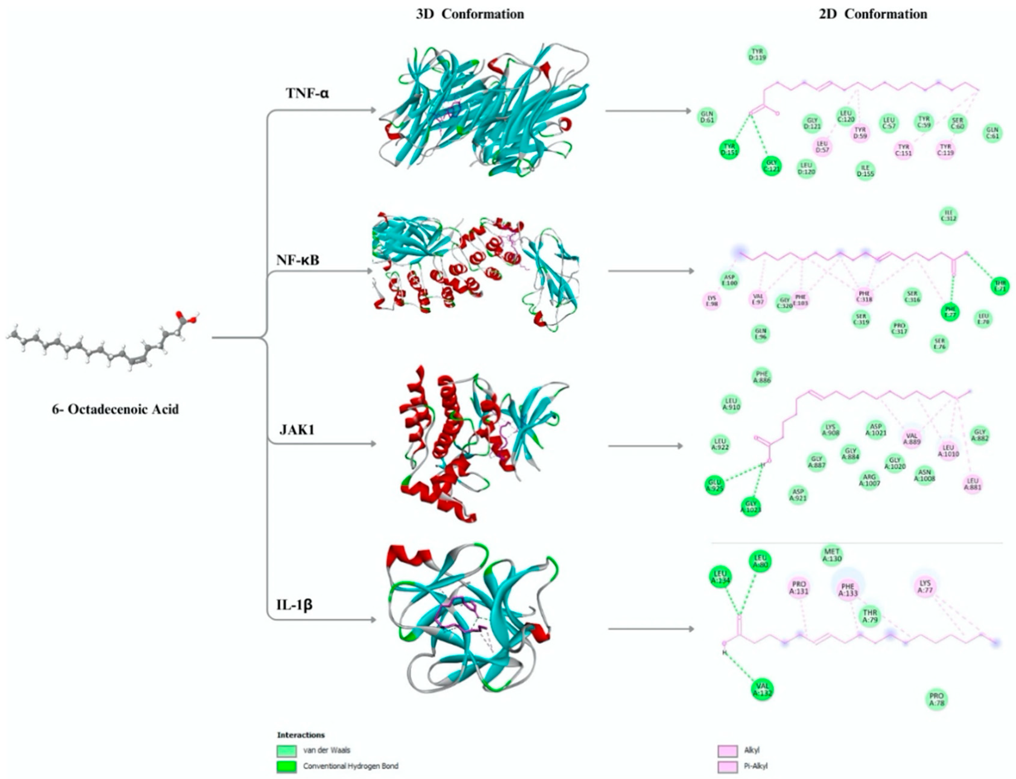
A molecular docking study assessed the binding characteristics of (6Z)-Octadecenoic acid with four primary inflammatory target proteins TNF-α, NF-κB, JAK1, and IL-1β. The 3D structures of these targets were retrieved from the RCSB Protein Data Bank with respective PDB IDs and resolutions as summarized in Table 1. These proteins were selected based on their key roles in inflammatory signaling pathways relevant to gingival wound healing.
Docking simulations revealed favorable binding affinity values ranging from –4.4 to –5.4 kcal/mol (Table. 2). Among the targets, JAK1 demonstrated the strongest interaction (–5.4 kcal/mol), followed by TNF-α (–5.3 kcal/mol), NF-κB (–5.0 kcal/mol), and IL-1β (–4.4 kcal/mol), as summarized in Table 2. Hydrophobic contacts and hydrogen bonds were observed at key residues within each protein’s binding pocket.
For TNF-α, key hydrophobic interactions were noted at TYR119, TYR151, TYR59, and LEU57, with hydrogen bonds formed at TYR151 and GLY121. In the NF-κB complex, hydrophobic residues included VAL97, PHE318, PHE77, and PHE103, while TYR71 and PHE77 contributed to hydrogen bonding. In JAK1, hydrophobic interactions involved VAL889, LEU1010, and LEU881, with GLU925 and GLY1023 forming hydrogen bonds. IL-1β showed hydrophobic contacts with LYS77, PRO131, and PHE133, and hydrogen bonding with VAL132, LEU134, and LEU80.
Figure 1.
3D and 2D molecular docking conformations of (6Z)-Octadecenoic acid with four key inflammatory target proteins: TNF-α, NF-κB, JAK1, and IL-1β. The left panel shows the 3D structure of (6Z)-Octadecenoic acid; the middle panel displays the ligand–protein binding interactions in 3D conformations; and the right panel illustrates 2D interaction diagrams showing van der Waals forces (green), conventional hydrogen bonds (blue), alkyl interactions (pink), and π-alkyl interactions (purple).
2.2. Histological Features of Fibroblast Proliferation in Gingival Wound Healing
Histological examination of gingival tissues was performed on Days 3 and 7 using Hematoxylin and Eosin (H&E) staining to assess fibroblast proliferation following topical application of Na-CMC (negative control), aspirin (positive control), and Stichopus hermanii extract. Representative micrographs for each group at both time points are presented in Figure 2.
On Day 3, fibroblast distribution in the Na-CMC group (Figure 2a) appeared sparse, with loosely arranged connective tissue architecture. The aspirin-treated group (Figure 2b) displayed increased cellular density relative to the negative control. The Stichopus hermanii extract group (Figure 2c) showed a denser accumulation of fibroblast cells within the granulation tissue. By Day 7, fibroblast presence increased in all groups. The Na-CMC group (Figure 2d) showed a slight increase in cellular presence, while the aspirin group (Figure 2e) demonstrated linear alignment of fibroblasts within organized connective tissue. The Stichopus hermanii group (Figure 2f) exhibited tightly packed fibroblasts and dense connective matrix throughout the wound site.
2.3. Collagen Fiber Visualization Using Masson's Trichrome Staining
In control and treatment groups, Masson's Trichrome staining was performed to observe collagen fiber distribution during gingival wound healing on Days 3 and 7. Collagen fibers are visualized in blue, whereas cytoplasmic and muscle tissues appear red (Figure 3).
On Day 3, the Na-CMC group (Figure 3a) exhibited limited blue staining, indicating low collagen presence and a loosely arranged extracellular matrix. The aspirin group (Figure 3b) showed more localized blue areas, and early fiber organization was visible in the granulation tissue. Blue staining was observed along the wound margin in the Stichopus hermanii extract group (Figure 3c), with a denser distribution than the other groups. By Day 7, collagen deposition increased in all groups. The Na-CMC group (Figure 3d) presented with wider, yet disorganized collagen fibers. The aspirin group (Figure 3e) demonstrated thicker blue-stained regions with moderate alignment. The Stichopus hermanii group (Figure 3f) showed pronounced collagen accumulation, with dense, organized blue-stained fibers throughout the connective tissue layer.
2.4. Quantitative Evaluation of Fibroblast Density and Collagen Fiber Thickness
Quantitative assessments of fibroblast cell counts and collagen fiber thickness were performed on Days 3 and 7 across all experimental groups, including Na-CMC (control), aspirin, and Stichopus hermanii extract treatments. The compiled data are presented in Table 3.
On Day 3, fibroblast counts were recorded at 119 ± 5.38 in the Na-CMC group, 177 ± 14.62 in the aspirin group, and 213 ± 13.2 in the Stichopus hermanii group. Collagen thickness measurements showed values of 112.00 µm in the Na-CMC group, 238.50 µm in the aspirin group, and 177.25 µm in the Stichopus hermanii group. On Day 7, fibroblast counts increased across all groups, reaching 166 ± 13.8 in the Na-CMC group, 350 ± 24.54 in the aspirin group, and 251 ± 21.7 in the Stichopus hermanii group. Collagenization values were measured at 175.75 µm, 452.75 µm, and 717.25 µm in the Na-CMC, aspirin, and Stichopus hermanii groups, respectively.
One-way ANOVA analysis indicated statistically significant differences in both fibroblast counts and collagenization across treatment groups on both Day 3 and Day 7, with p-values ranging from 0.000 to 0.002 (p < 0.05), as detailed in Table 3.
2.5. ADME-Tox Profiling of (6Z)-Octadecenoic Acid
The pharmacokinetic and toxicity properties of (6Z)-Octadecenoic acid were evaluated through in silico prediction (Table 4). The compound demonstrated Caco-2 permeability of 1.563 nm·s⁻¹ and human intestinal absorption of 91.82%. The volume of distribution at steady state (VDss) was calculated at –0.558 log L/kg. The compound was not predicted to be a CYP2D6 inhibitor. Total systemic clearance was recorded at 1.884. Toxicity analysis showed negative predictions for AMES mutagenicity and hepatotoxicity. The oral acute toxicity (LD50) was predicted at 1.417 mol/kg, and the oral chronic toxicity (LOAEL) was 3.259 log mg/kg bw/day. These values are summarized in Table 4.
Additional drug-likeness assessment using Lipinski’s Rule of Five indicated a molecular weight of 282.5 Da, two hydrogen bond acceptors (HBA), one hydrogen bond donor (HBD), a partition coefficient (log P) of 6.11, and a molar refractivity (Rm) of 87.08. One rule violation was noted. Detailed chemical properties are presented in Table 5.
2.6. Serum Liver Enzyme Levels: SGOT and SGPT
Serum levels of liver enzymes SGOT and SGPT were measured on Days 0 and 7 to evaluate systemic effects following topical application of Stichopus hermanii extract at concentrations of 25%, 50%, and 100%, alongside the Na-CMC control group. The results are presented in Figure 4. In Figure 4a, SGOT levels are shown for each group across the two time points. Initial values (Day 0) varied across groups, with an increase observed in all extract-treated groups by Day 7, while the control group remained constant. Figure 4b presents SGPT levels. Similar to SGOT, an increase in enzyme levels was noted in the treatment groups from Day 0 to Day 7, with the control group showing no change.
3. Discussion
Authors should discuss the results and how they can be interpreted from the perspective of previous studies and of the working hypotheses. The findings and their implications should be discussed in the broadest context possible. Future research directions may also be highlighted.
This study investigated the potential of (6Z)-Octadecenoic acid, a major fatty acid component of Stichopus hermanii, in modulating inflammatory pathways and enhancing gingival wound healing. A combined in silico and in vivo approach was used to evaluate molecular interactions, tissue responses, and toxicity, providing a comprehensive preclinical assessment.
The molecular docking results revealed stable binding interactions between (6Z)-Octadecenoic acid and four key pro-inflammatory mediators: TNF-α [41], NF-κB [42], IL-1β [43], and JAK1 [44]. These proteins orchestrate early inflammatory signaling and tissue degradation in periodontal wounds [45,46]. The observed binding affinities and interaction patterns—particularly hydrogen bonding and hydrophobic contacts—indicate structural compatibility between (6Z)-Octadecenoic acid and key pro-inflammatory targets. Although molecular docking does not equate to confirmed biological activity, these findings align with existing literature demonstrating the regulatory abilities of unsaturated fatty acid derivatives [47,48].
For instance, Cui et al. [49] demonstrated that oleic acid nitroalkenes (OA-NO₂) inhibit NF-κB by alkylating the p65 subunit—specifically at Cys38—thereby repressing NF-κB-mediated gene expression. Likewise, Khoo et al.[50] reported that electrophilic fatty acid nitroalkenes reduce NF-κB activation while activating the antioxidant regulator Nrf2 in macrophages. Moreover, Wang et al. [51] found that OA-NO₂ suppresses both NF-κB and STAT3 phosphorylation in murine models of psoriasiform dermatitis. These converging findings support the hypothesis that (6Z)-Octadecenoic acid may exert similar anti-inflammatory mechanisms through direct interaction with inflammatory mediators, consistent with our docking analysis.
Histological analysis further substantiated the anti-inflammatory and regenerative potential of (6Z)-Octadecenoic acid. The extract-treated group demonstrated increased fibroblast proliferation and enhanced collagen deposition on Days 3 and 7, as observed through both H&E and Masson’s Trichrome staining. These findings are consistent with a previous study that reported that bioactive marine-derived fatty acids accelerated granulation tissue formation and fibroblast recruitment in wound models [52,53]. The alignment of histological improvements with molecular docking insights strengthens the plausibility of the compound's dual role in modulating inflammatory signals and promoting early tissue remodeling.
Quantitative assessments further supported these histological observations. On Day 7, fibroblast counts and collagen fiber thickness in the Stichopus hermanii extract group were significantly higher than those in both the Na-CMC and aspirin-treated groups. These increases suggest a progressive and sustained effect of the extract on the wound healing microenvironment. Similar trends have been observed by Valente et al. [54], who found that sea cucumber glycosaminoglycans stimulated fibroblast proliferation and extracellular matrix synthesis in dermal injury models. Moreover, Sun et al. [55] demonstrated that marine bioactive lipids enhance collagen deposition via modulation of the TGF-β/Smad signaling pathway. The congruence between our results and these prior studies reinforces the biological activity of marine-derived fatty acids in soft tissue regeneration. The congruence between these previous reports and the current study underscores the regenerative potential of marine-derived fatty acids in oral tissue repair.
In addition, the in silico pharmacokinetic prediction via pkCSM revealed high intestinal absorption, non-inhibition of CYP2D6, and a negative profile for AMES toxicity and hepatotoxicity. These characteristics are comparable to a previous study that highlighted favorable ADMET parameters for structurally related long-chain unsaturated fatty acids [61]. Correspondingly, serum SGOT and SGPT levels in our in vivo toxicity evaluation remained within physiological ranges across all treatment groups, further supporting the compound’s systemic safety profile when administered topically [56]. These findings collectively suggest a favorable risk-benefit profile that may support further translational development.
From a translational perspective, the dual anti-inflammatory and pro-regenerative effects of (6Z)-Octadecenoic acid may offer clinical advantages in managing gingival wounds, post-extraction sockets, or periodontal surgery sites. Current topical anti-inflammatory agents, such as NSAID-based gels, may attenuate inflammation but are not designed to support tissue remodeling actively [57]. In contrast, compounds like (6Z)-Octadecenoic acid demonstrate the potential to target inflammatory signaling while simultaneously promoting fibroblast activity and matrix restoration. This functional duality aligns with the clinical need for integrated wound management in the oral environment, where rapid healing and resolution of inflammation are essential for maintaining periodontal health and minimizing complications [13].
Recent evidence highlights the therapeutic potential of (6Z)-Octadecenoic acid, a bioactive compound isolated from Stichopus hermanii, in promoting gingival wound healing. This fatty acid exhibits targeted anti-inflammatory effects through direct interactions with key pro-inflammatory mediators such as TNF-α and NF-κB, suggesting a more active mechanism of action compared to conventional agents. In our study, topical application of (6Z)-Octadecenoic acid led to enhanced fibroblast proliferation and collagen deposition, outperforming typical outcomes reported for standard therapies. Moreover, its favorable safety profile—demonstrated by stable serum liver enzyme levels and predictive in silico ADME-Tox modeling—supports its potential for clinical application in oral wound management.
In comparison, hyaluronic acid (HA) gel has been widely recognized for its supportive role in wound healing due to its viscoelasticity, mucoadhesiveness, and ability to retain moisture [58]. HA promotes fibroblast migration, angiogenesis, and epithelial proliferation, improving healing rates in periodontal surgeries and oral ulcers [59]. However, its biological activity remains largely passive, functioning primarily as a hydrating and protective matrix without directly modulating inflammatory signaling [60].
Similarly, chlorhexidine gel is commonly employed as an oral antiseptic owing to its broad-spectrum antimicrobial effects and frequent use in post-surgical care [61]. Despite its efficacy in reducing bacterial colonization, chlorhexidine lacks regenerative potential and may exert cytotoxic effects on fibroblasts and keratinocytes when used excessively, potentially delaying wound repair [62,63].
Taken together, the dual anti-inflammatory and regenerative actions of (6Z)-Octadecenoic acid—along with its non-toxic profile—position this compound as a promising alternative to existing agents like HA and chlorhexidine, offering a more comprehensive approach to oral wound healing.
Limitation Study
This study, while offering important preliminary insights, is subject to several limitations. The short-term observation period, limited to seven days, allowed for the assessment of early inflammatory and proliferative responses but did not capture later stages of healing such as epithelialization, angiogenesis, or tensile strength restoration. The in silico molecular docking results revealed promising interactions between (6Z)-Octadecenoic acid and key inflammatory mediators; however, further validation is required to strengthen the mechanistic interpretation. Additionally, the use of a single formulation vehicle—Na-CMC gel—may have constrained local bioavailability and residence time at the wound site, particularly in the absence of advanced drug delivery systems designed to prolong mucosal retention or control release kinetics.
As an initial study, we acknowledge these limitations as important considerations for future research. The continuity of investigation and the consistency of the findings in this study serve as a foundation for the further development and refinement of subsequent research.
4. Materials and Methods
4.1. In-Silico Studies
We utilized the Protein Data Bank (https://www.rcsb.org) to investigate target inflammatory proteins.
4.1.1. Quantitative Structure-Activity Relationship Analysis
The anti-inflammatory potential of (6Z)-Octadecenoic acid derived from Stichopus hermanii was evaluated using PyRx version 0.8. A redocking procedure was performed using the natural ligands of the respective target proteins to validate the docking protocol. The reliability of the docking results was determined based on the root mean square deviation (RMSD), with values ≤ 2 Å considered acceptable for predictive accuracy. The results confirmed the validity of the docking methodology and parameters, allowing for their application in docking the test compound. Molecular interactions between the active compound and target protein residues were further visualized using Biovia Discovery Studio Visualizer version 21.1.0.20298.
4.1.2. Pharmacokinetics and Toxicity Prediction
The pharmacokinetic and toxicity profiles of (6Z)-Octadecenoic acid were predicted using the pkCSM online platform (http://biosig.unimelb.edu.au/pkcsm/), which enables in silico assessment of ADMET properties (Absorption, Distribution, Metabolism, Excretion, and Toxicity) of chemical compounds. Evaluation of drug-likeness was performed based on Lipinski’s Rule of Five (http://www.scfbio-iitd.res.in/software/drugdesign/lipinski.jsp), a widely used criterion to differentiate between drug-like and non-drug-like molecules. This rule serves as a benchmark in early-phase drug discovery, indicating the compound’s likelihood of success based on compliance with established physicochemical parameters.
4.2. Preparation of Stichopus hermanii Extract
We employed a laboratory-based experimental design using a post-test control group approach. Fibroblast analysis was conducted to evaluate cellular proliferation, while collagenase evaluation was performed to measure collagen fiber thickness within the gingival tissue. In addition, molecular docking simulations were carried out to identify interactions between active compounds and target inflammatory proteins
Fresh Stichopus hermanii specimens were thoroughly washed, trimmed to remove extraneous tissues, and cut into small sections. The cleaned samples were then oven-dried for 24 hours. Subsequently, 500 mL of methanol was used to macerate the dried material in a closed vessel for 48 hours. The maceration mixture was filtered using Whatman filter paper to separate the filtrate from the solid residue. The methanolic extract was concentrated using a rotary evaporator to isolate the bioactive constituents. The resulting concentrate was centrifuged at 6,000 rpm for 10 minutes at 40°C to maintain compound stability. Finally, the extract was formulated into a gel by dissolving it in sodium carboxymethyl cellulose (Na-CMC) for topical administration in the animal model.
4.2.1. Preparation Controlled Variables
The aspirin solution was prepared by dissolving 18 mg of aspirin in 1 mL of 0.5% sodium carboxymethyl cellulose (Na-CMC). Separately, 50 g of Na-CMC was dissolved in sterile distilled water to a final volume of 100 mL. Na-CMC was used as the negative control for the fibroblast proliferation and collagenization assays, while the aspirin solution served as the positive control. In the toxicity evaluation, only Na-CMC was used as the negative control group [64].
4.3. Preparation of Animal Studies (Rattus Norvegicus)
This study allocated twelve male Wistar rats into three experimental groups, each comprising four animals. The groups received topical applications of Stichopus hermanii extract at concentrations of 25%, 50%, and 100%, respectively, followed prior study [65], along with a Na-CMC control group. All rats were maintained under standard conditions with unrestricted access to food and water and observed for seven days.
The incision, about 3 mm in length, was made on the front part of the mandible. The study used 12 male Wistar rats as the experimental animal model. The rats were kept in standard polycarbonate cages (45 cm x 35 cm x 18 cm) with wood shavings as bedding material. The cages were placed in a temperature-controlled room maintained at 22 ± 2°C, with a 12-hour light/dark cycle. Before incision, all samples were anesthetized using a single intramuscular injection of 0.2 ml of ketamine. The gingiva of the sample was then smeared with povidone iodine 10%. Using a 5 mm long scalpel number 11, an incision about 3 mm long was made on the anterior part of the mandible. After the incision, the area was irrigated with aquades. Each group comprised four rats [66].
4.3.1. Blood Collection for Toxicity Testing
To extract the blood from each white rat, the rat is first placed in a jar containing ether-soaked cotton until it faints. Blood is then drawn using a capillary tube and transferred into a vacuum. The blood is then centrifuged, followed by an examination of the initial blood plasma to assess SGOT and SGPT.
4.3.2. Fibroblast Test and Collagen Test
Twelve male Wistar rats, aged approximately 3 months and weighing around 250 grams, were randomly assigned to three groups (n = 4 per group): the Na-CMC group, the aspirin group, and the Stichopus hermanii extract group. Each treatment was topically applied to the gingival wounds twice within 24 hours, with a 6-hour interval between applications [67].
4.3.3. Tissue Collection and Histological Examination
On days three and seven, the animals were euthanized under anesthesia, followed by cervical dislocation. Gingival tissue samples were harvested from the wound sites and immediately fixed in 10% neutral buffered formalin (NBF). The fixed specimens were processed at the anatomical pathology laboratory, where they were placed in plastic cassettes and subjected to graded alcohol dehydration (70%, 80%, 90%, and absolute ethanol), followed by clearing and infiltration using xylene. Tissues were then embedded in paraffin blocks, stored under refrigeration, and sectioned using a microtome of 5–6 µm thickness. The tissue sections were floated on a 60 °C water bath to prevent folding, then mounted onto glass slides. Fibroblast evaluation was performed using Hematoxylin and Eosin (H&E) staining, while collagen fiber analysis was conducted using Masson’s Trichrome staining. Fibroblast counts were quantified per field of view using an Olympus C-21 microscope equipped with an Optilab Advance digital camera at 200× magnification. Lesion length was measured, and collagen fiber structure was further analyzed using electron microscopy to assess the extent of wound healing. To minimize observational bias, all histological examinations were blinded; the histopathologist was not informed of the group allocation of each specimen.
4.4. Ethical Clearance
All experimental procedures were conducted at the Phytochemistry, Pharmacology, Pharmacognosy, and Clinical Pharmacy Laboratories of the Faculty of Pharmacy, as well as the Anatomical Pathology Laboratory at Hasanuddin University General Hospital. Ethical clearance for this study was obtained from the Ethics Committee of Hasanuddin University under registration number UH23020115.
4.5. Statistical Analysis
All quantitative data were presented as mean ± standard deviation (SD). Statistical analysis was performed using one-way Analysis of Variance (ANOVA) to evaluate differences among the treatment groups. A p-value of less than 0.05 (p < 0.05) was considered statistically significant. Data analysis was conducted using IBM SPSS Statistics for Windows, Version 25.0 (IBM Corp., Armonk, NY, USA).
5. Conclusions
This study demonstrates that (6Z)-Octadecenoic acid, a bioactive compound isolated from Stichopus hermanii, exhibits both anti-inflammatory and regenerative properties relevant to oral wound healing. Through molecular docking, the compound showed favorable binding affinity toward key pro-inflammatory mediators, including TNF-α, NF-κB, IL-1β, and JAK1. Histological and quantitative assessments further confirmed its ability to promote fibroblast proliferation and collagen deposition in gingival tissues. Pharmacokinetic and toxicity evaluations revealed acceptable safety and drug-likeness profiles, with no adverse hepatic effects observed in vivo. Taken together, these findings suggest that (6Z)-Octadecenoic acid has promising therapeutic potential as a topical agent for managing gingival wounds. Further long-term and molecular studies are warranted to validate these preclinical results and to explore clinical applications in oral tissue regeneration.
Supplementary Materials
The following supporting information can be downloaded at the website of this paper posted on Preprints.org, The following supporting information can be downloaded at Protein Data Bank (https://www.rcsb.org)
Author Contributions
Conceptualization, A.A. and A.M.T.; methodology, A.A., A.M.T., K.E.G, and B.T software, A.A. and K.F.; validation, A.A and T.S.; formal analysis, A.A and K.F.; investigation, A.A., A.M.T. and K.E.G; resources, A.A.; data curation, A.A. and K.F; writing—original draft preparation, A.A. and A.M.T.; writing—review and editing, A.A., A.MT., K.E.G., B.T., T.S., and K.F.; visualization, A.A.; supervision, A.A. and T.S. All authors have read and agreed to the published version of the manuscript.
Funding
This research was funded by Hasanuddin Univesity, grant number 00773/UN4.22/PT.01.03/2028 and The APC was funded by A.A.
Institutional Review Board Statement
The animal study protocol was approved by Ethics Committee of Hasanuddin University under registration number UH23020115.
Data Availability Statement
The original contributions presented in the study are included in the article, further inquiries can be directed to the corresponding author.
Acknowledgments
The authors would like to express their sincere gratitude to the Institute for Research and Community Service, Hasanuddin University, for facilitating the research grant, and to the Anatomical Pathology Laboratory at Hasanuddin University General Hospital for providing laboratory facilities. The authors have reviewed and edited the manuscript and take full responsibility for the content of this publication.
Conflicts of Interest
The authors declare no conflicts of interest
References
- Guo, S.; Dipietro, L.A. Factors affecting wound healing. J Dent Res 2010, 89, 219–229. [Google Scholar] [CrossRef] [PubMed]
- Hosgood, G. Stages of wound healing and their clinical relevance. Vet Clin North Am Small Anim Pract 2006, 36, 667–685. [Google Scholar] [CrossRef] [PubMed]
- Smith, P.C.; Cáceres, M.; Martínez, C.; Oyarzún, A.; Martínez, J. Gingival wound healing: an essential response disturbed by aging? J Dent Res 2015, 94, 395–402. [Google Scholar] [CrossRef]
- Kaner, D.; Soudan, M.; Zhao, H.; Gaßmann, G.; Schönhauser, A.; Friedmann, A. Early Healing Events after Periodontal Surgery: Observations on Soft Tissue Healing, Microcirculation, and Wound Fluid Cytokine Levels. International Journal of Molecular Sciences 2017, 18, 283. [Google Scholar] [CrossRef]
- Visentin, D.; Gobin, I.; Maglica, Ž. Periodontal Pathogens and Their Links to Neuroinflammation and Neurodegeneration. Microorganisms 2023, 11. [Google Scholar] [CrossRef]
- Cavaillon, J.M.; Adib-Conquy, M. The Pro-Inflammatory Cytokine Cascade. In Immune Response in the Critically Ill; Marshall, J.C., Cohen, J., Eds.; Springer Berlin Heidelberg: Berlin, Heidelberg, 2002; pp. 37–66. [Google Scholar]
- Esche, C.; Stellato, C.; Beck, L.A. Chemokines: Key Players in Innate and Adaptive Immunity. Journal of Investigative Dermatology 2005, 125, 615–628. [Google Scholar] [CrossRef]
- Wautier, J.L.; Wautier, M.P. Pro- and Anti-Inflammatory Prostaglandins and Cytokines in Humans: A Mini Review. Int J Mol Sci 2023, 24. [Google Scholar] [CrossRef]
- Galvão, I.; Sugimoto, M.; Vago, J.P.; Gomes Machado, M.; Sousa, L. Mediators of Inflammation. 2018; 3–32. [Google Scholar]
- Batool, F.; Özçelik, H.; Stutz, C.; Gegout, P.Y.; Benkirane-Jessel, N.; Petit, C.; Huck, O. Modulation of immune-inflammatory responses through surface modifications of biomaterials to promote bone healing and regeneration. J Tissue Eng 2021, 12, 20417314211041428. [Google Scholar] [CrossRef]
- Morand, D.N.; Davideau, J.L.; Clauss, F.; Jessel, N.; Tenenbaum, H.; Huck, O. Cytokines during periodontal wound healing: potential application for new therapeutic approach. Oral Dis 2017, 23, 300–311. [Google Scholar] [CrossRef]
- Martu, M.A.; Maftei, G.A.; Luchian, I.; Stefanescu, O.M.; Scutariu, M.M.; Solomon, S.M. The Effect of Acknowledged and Novel Anti-Rheumatic Therapies on Periodontal Tissues-A Narrative Review. Pharmaceuticals (Basel) 2021, 14. [Google Scholar] [CrossRef]
- Hasturk, H.; Kantarci, A. Activation and resolution of periodontal inflammation and its systemic impact. Periodontol 2000 2015, 69, 255–273. [Google Scholar] [CrossRef] [PubMed]
- Kim, W.J.; Soh, Y.; Heo, S.M. Recent Advances of Therapeutic Targets for the Treatment of Periodontal Disease. Biomol Ther (Seoul) 2021, 29, 263–267. [Google Scholar] [CrossRef] [PubMed]
- Łasica, A.; Golec, P.; Laskus, A.; Zalewska, M.; Gędaj, M.; Popowska, M. Periodontitis: etiology, conventional treatments, and emerging bacteriophage and predatory bacteria therapies. Front Microbiol 2024, 15, 1469414. [Google Scholar] [CrossRef] [PubMed]
- Mukherjee, B.; Barman, M.; Hota, S. Present and Future Treatment Modalities for the Mitigation and Cure of Periodontal Diseases. Online Journal of Dentistry & Oral Health 2023, 7, 1–13. [Google Scholar] [CrossRef]
- Arampatzis, A.S.; Karra, A.; Kyrilas, E.; Kampasakali, E.; Tsalikis, L.; Barmpalexis, P.; Christofilos, D.; Assimopoulou, A. Bioactive 3D printed scaffolds for the treatment of periodontal diseases, 2022; Volume 88.
- Chen, P.; Zhang, C.; He, P.; Pan, S.; Zhong, W.; Wang, Y.; Xiao, Q.; Wang, X.; Yu, W.; He, Z.; et al. A Biomimetic Smart Nanoplatform as "Inflammation Scavenger" for Regenerative Therapy of Periodontal Tissue. Int J Nanomedicine 2022, 17, 5165–5186. [Google Scholar] [CrossRef]
- Attiq, A.; Jalil, J.; Husain, K.; Ahmad, W. Raging the War Against Inflammation With Natural Products. Front Pharmacol 2018, 9, 976. [Google Scholar] [CrossRef]
- World Health, O. WHO global report on traditional and complementary medicine 2019; World Health Organization: Geneva, 2019. [Google Scholar]
- Agrawal, S.; Acharya, D.; Adholeya, A.; Barrow, C.J.; Deshmukh, S.K. Nonribosomal Peptides from Marine Microbes and Their Antimicrobial and Anticancer Potential. Front Pharmacol 2017, 8, 828. [Google Scholar] [CrossRef]
- Almaliti, J.; Gerwick, W.H. Methods in marine natural product drug discovery: what's new? Expert Opin Drug Discov 2023, 18, 687–691. [Google Scholar] [CrossRef]
- Ahmed, I.; Asgher, M.; Sher, F.; Hussain, S.M.; Nazish, N.; Joshi, N.; Sharma, A.; Parra-Saldívar, R.; Bilal, M.; Iqbal, H.M.N. Exploring Marine as a Rich Source of Bioactive Peptides: Challenges and Opportunities from Marine Pharmacology. Mar Drugs 2022, 20. [Google Scholar] [CrossRef]
- Adam, M.; Achmad, H.; Tanumihardja, M.; Ramadhan, S.R.J.; Masyta, N. The benefits of golden sea cucumber (Stichopus hermanni) as an alternative antimicrobial material in oral health. Journal of International Dental and Medical Research 2022, 15, 1806–1815. [Google Scholar]
- Mercier, A.; Purcell, S.W.; Montgomery, E.M.; Kinch, J.; Byrne, M.; Hamel, J.F. Revered and Reviled: The Plight of the Vanishing Sea Cucumbers. Ann Rev Mar Sci 2025, 17, 115–142. [Google Scholar] [CrossRef]
- Pangestuti, R.; Arifin, Z. Medicinal and health benefit effects of functional sea cucumbers. Journal of Traditional and Complementary Medicine 2018, 8, 341–351. [Google Scholar] [CrossRef]
- Kalinin, V.I.; Silchenko, A.S.; Avilov, S.A.; Stonik, V.A.; Smirnov, A.V. Sea Cucumbers Triterpene Glycosides, the Recent Progress in Structural Elucidation and Chemotaxonomy. Phytochemistry Reviews 2005, 4, 221–236. [Google Scholar] [CrossRef]
- Galasso, C.; Corinaldesi, C.; Sansone, C. Carotenoids from Marine Organisms: Biological Functions and Industrial Applications. Antioxidants (Basel) 2017, 6. [Google Scholar] [CrossRef] [PubMed]
- Wen, J.; Hu, C.; Fan, S. Chemical composition and nutritional quality of sea cucumbers. J Sci Food Agric 2010, 90, 2469–2474. [Google Scholar] [CrossRef] [PubMed]
- Shaik, M.I.; Kadir, A.N.A.; Sarbon, N.M. Physicochemical and thermal properties of pepsin- and acid-soluble collagen isolated from the body wall of sea cucumbers (Stichopus hermanni). J Food Sci 2024, 89, 320–329. [Google Scholar] [CrossRef]
- Sari, R.P.; Larashati, D.I.D.; Aldiana, C.; Nafi'ah, N.; Damaiyanti, D.W.; Kurniawati, A. Application of Stichopus hermanni Nanoparticle Gel in the Healing of Traumatic Ulcers. Eur J Dent 2023, 17, 330–336. [Google Scholar] [CrossRef]
- Li, X.; Luo, L.; Cai, Y.; Yang, W.; Lin, L.; Li, Z.; Gao, N.; Purcell, S.W.; Wu, M.; Zhao, J. Structural Elucidation and Biological Activity of a Highly Regular Fucosylated Glycosaminoglycan from the Edible Sea Cucumber Stichopus herrmanni. J Agric Food Chem 2017, 65, 9315–9323. [Google Scholar] [CrossRef]
- Bahrami, Y.; Franco, C.M. Acetylated Triterpene Glycosides and Their Biological Activity from Holothuroidea Reported in the Past Six Decades. Mar Drugs 2016, 14. [Google Scholar] [CrossRef]
- Hosseini, S.F.; Masoud, R.; McClements, D.J. Bioactive functional ingredients from aquatic origin: a review of recent progress in marine-derived nutraceuticals. Critical Reviews in Food Science and Nutrition 2022, 62, 1242–1269. [Google Scholar] [CrossRef]
- Revol-Cavalier, J.; Quaranta, A.; Newman, J.W.; Brash, A.R.; Hamberg, M.; Wheelock, C.E. The Octadecanoids: Synthesis and Bioactivity of 18-Carbon Oxygenated Fatty Acids in Mammals, Bacteria, and Fungi. Chem Rev 2025, 125, 1–90. [Google Scholar] [CrossRef]
- Quaranta, A.; Revol-Cavalier, J.; Wheelock, C.E. The octadecanoids: an emerging class of lipid mediators. Biochem Soc Trans 2022, 50, 1569–1582. [Google Scholar] [CrossRef] [PubMed]
- Yang, Z.; Li, C.; Jia, Q.; Zhao, C.; Taylor, D.C.; Li, D.; Zhang, M. Transcriptome Analysis Reveals Candidate Genes for Petroselinic Acid Biosynthesis in Fruits of Coriandrum sativum L. J Agric Food Chem 2020, 68, 5507–5520. [Google Scholar] [CrossRef] [PubMed]
- Weber, N.; Richter, K.D.; Schulte, E.; Mukherjee, K.D. Petroselinic acid from dietary triacylglycerols reduces the concentration of arachidonic acid in tissue lipids of rats. J Nutr 1995, 125, 1563–1568. [Google Scholar] [CrossRef] [PubMed]
- Kang, M.C.; Ham, Y.M.; Heo, S.J.; Yoon, S.A.; Cho, S.H.; Kwon, S.H.; Jeong, M.S.; Jeon, Y.J.; Sanjeewa, K.; Yoon, W.J.; et al. Anti-inflammation effects of 8-oxo-9-octadecenoic acid isolated from Undaria peterseniana in lipopolysaccharide-stimulated macrophage cells. Excli j 2018, 17, 775–783. [Google Scholar] [CrossRef]
- Fatwati, K.; Amin, A.; Indriani, L.; Ladju, R.B.; Akbar, F.H.; Hamrun, N. GC–MS analysis and in silico approaches to Stichopus hermanii as anti-inflammatory through PKC-β inhibition. Results in Chemistry 2025, 14, 102086. [Google Scholar] [CrossRef]
- Brierly, G.; Celentano, A.; Breik, O.; Moslemivayeghan, E.; Patini, R.; McCullough, M.; Yap, T. Tumour Necrosis Factor Alpha (TNF-α) and Oral Squamous Cell Carcinoma. Cancers (Basel) 2023, 15. [Google Scholar] [CrossRef]
- Pathak, J.L.; Fang, Y.; Chen, Y.; Ye, Z.; Guo, X.; Yan, Y.; Zha, J.; Liang, D.; Ke, X.; Yang, L.; et al. Downregulation of Macrophage-Specific Act-1 Intensifies Periodontitis and Alveolar Bone Loss Possibly via TNF/NF-κB Signaling. Frontiers in Cell and Developmental Biology 2021, 9. [Google Scholar] [CrossRef]
- Lee, C.H.; Chang, J.S.; Syu, S.H.; Wong, T.S.; Chan, J.Y.; Tang, Y.C.; Yang, Z.P.; Yang, W.C.; Chen, C.T.; Lu, S.C.; et al. IL-1β promotes malignant transformation and tumor aggressiveness in oral cancer. J Cell Physiol 2015, 230, 875–884. [Google Scholar] [CrossRef]
- Spinelli, F.R.; Colbert, R.A.; Gadina, M. JAK1: Number one in the family; number one in inflammation? Rheumatology (Oxford) 2021, 60, ii3–ii10. [Google Scholar] [CrossRef]
- Plemmenos, G.; Evangeliou, E.; Polizogopoulos, N.; Chalazias, A.; Deligianni, M.; Piperi, C. Central Regulatory Role of Cytokines in Periodontitis and Targeting Options. Curr Med Chem 2021, 28, 3032–3058. [Google Scholar] [CrossRef] [PubMed]
- Souza, J.A.; Rossa, C., Jr.; Garlet, G.P.; Nogueira, A.V.; Cirelli, J.A. Modulation of host cell signaling pathways as a therapeutic approach in periodontal disease. J Appl Oral Sci 2012, 20, 128–138. [Google Scholar] [CrossRef] [PubMed]
- Gheibi, N.; Ghorbani, M.; Shariatifar, H.; Farasat, A. Effects of unsaturated fatty acids (Arachidonic/Oleic Acids) on stability and structural properties of Calprotectin using molecular docking and molecular dynamics simulation approach. PLoS One 2020, 15, e0230780. [Google Scholar] [CrossRef] [PubMed]
- Vetrivel, U.; Ravichandran, S.B.; Kuppan, K.; Mohanlal, J.; Das, U.N.; Narayanasamy, A. Agonistic effect of polyunsaturated fatty acids (PUFAs) and its metabolites on brain-derived neurotrophic factor (BDNF) through molecular docking simulation. Lipids in Health and Disease 2012, 11, 109. [Google Scholar] [CrossRef]
- Cui, T.; Schopfer, F.J.; Zhang, J.; Chen, K.; Ichikawa, T.; Baker, P.R.; Batthyany, C.; Chacko, B.K.; Feng, X.; Patel, R.P.; et al. Nitrated fatty acids: Endogenous anti-inflammatory signaling mediators. J Biol Chem 2006, 281, 35686–35698. [Google Scholar] [CrossRef]
- Khoo, N.K.H.; Li, L.; Salvatore, S.R.; Schopfer, F.J.; Freeman, B.A. Electrophilic fatty acid nitroalkenes regulate Nrf2 and NF-κB signaling:A medicinal chemistry investigation of structure-function relationships. Sci Rep 2018, 8, 2295. [Google Scholar] [CrossRef]
- Wang, P.; Killeen, M.E.; Sumpter, T.L.; Ferris, L.K.; Falo, L.D., Jr.; Freeman, B.A.; Schopfer, F.J.; Mathers, A.R. Electrophilic nitro-fatty acids suppress psoriasiform dermatitis: STAT3 inhibition as a contributory mechanism. Redox Biol 2021, 43, 101987. [Google Scholar] [CrossRef]
- Ontoria-Oviedo, I.; Amaro-Prellezo, E.; Castellano, D.; Venegas-Venegas, E.; González-Santos, F.; Ruiz-Saurí, A.; Pelacho, B.; Prósper, F.; Pérez Del Caz, M.D.; Sepúlveda, P. Topical Administration of a Marine Oil Rich in Pro-Resolving Lipid Mediators Accelerates Wound Healing in Diabetic db/db Mice through Angiogenesis and Macrophage Polarization. Int J Mol Sci 2022, 23. [Google Scholar] [CrossRef]
- McDaniel, J.C.; Belury, M.; Ahijevych, K.; Blakely, W. Omega-3 fatty acids effect on wound healing. Wound Repair Regen 2008, 16, 337–345. [Google Scholar] [CrossRef]
- Rizzo-Valente, V.S.; Fusco, M.A.; Cruz, R.M.M.L.; Santos, R.A.; Silva, L.S.; Escaleira, R.C.; Schulz, D.F.; Barroso, S.P.C.; Miranda, B.L.; Santos, D.Z.; et al. Effects of Dermatan Sulfate from Marine Invertebrate Styela plicata in the Wound Healing Pathway: A Natural Resource Applied to Regenerative Therapy. Marine Drugs 2022, 20, 676. [Google Scholar] [CrossRef]
- Sun, X.; Yang, Y.; Sun, X.; Meng, H.; Hao, W.; Yin, J.; Ma, F.; Guo, X.; Du, L.; Sun, L.; et al. Krill Oil Turns Off TGF-β1 Profibrotic Signaling in the Prevention of Diabetic Nephropathy. J Agric Food Chem 2022, 70, 9865–9876. [Google Scholar] [CrossRef] [PubMed]
- Ulrich, C.; Bichel, J.; Euvrard, S.; Guidi, B.; Proby, C.M.; van de Kerkhof, P.C.; Amerio, P.; Rønnevig, J.; Slade, H.B.; Stockfleth, E. Topical immunomodulation under systemic immunosuppression: results of a multicentre, randomized, placebo-controlled safety and efficacy study of imiquimod 5% cream for the treatment of actinic keratoses in kidney, heart, and liver transplant patients. Br J Dermatol 2007, 157 Suppl 2, 25–31. [Google Scholar] [CrossRef] [PubMed]
- Brewer, A.R.; McCarberg, B.; Argoff, C.E. Update on the use of topical NSAIDs for the treatment of soft tissue and musculoskeletal pain: a review of recent data and current treatment options. Phys Sportsmed 2010, 38, 62–70. [Google Scholar] [CrossRef] [PubMed]
- Bhati, A.; Fageeh, H.; Ibraheem, W.; Fageeh, H.; Chopra, H.; Panda, S. Role of hyaluronic acid in periodontal therapy (Review). Biomed Rep 2022, 17, 91. [Google Scholar] [CrossRef]
- Polizzi, A.; Leanza, Y.; Belmonte, A.; Grippaudo, C.; Leonardi, R.; Isola, G. Impact of Hyaluronic Acid and Other Re-Epithelializing Agents in Periodontal Regeneration: A Molecular Perspective. Int J Mol Sci 2024, 25. [Google Scholar] [CrossRef]
- Sprott, H.; Fleck, C. Hyaluronic Acid in Rheumatology. Pharmaceutics 2023, 15. [Google Scholar] [CrossRef]
- Asbi, T.; Hussein, H.A.; Horwitz, J.; Gabay, E.; Machtei, E.E.; Giladi, H.Z. A single application of chlorhexidine gel reduces gingival inflammation and interleukin 1-β following one-stage implant placement: A randomized controlled study. Clin Implant Dent Relat Res 2021, 23, 726–734. [Google Scholar] [CrossRef]
- Fiorillo, L.; D'Amico, C.; Mehta, V.; Cicciù, M.; Cervino, G. Chlorhexidine cytotoxicity on oral Behaviors: Last 20 Years systematic review. Oral Oncology Reports 2024, 9, 100245. [Google Scholar] [CrossRef]
- Pucher, J.J.; Daniel, J.C. The effects of chlorhexidine digluconate on human fibroblasts in vitro. J Periodontol 1992, 63, 526–532. [Google Scholar] [CrossRef]
- Adam, M.; Thahir, H.; Supiaty; Achmad, H.; Putri, S.W.; Azizah; Satya, D.E. The Potential of Golden Sea Cucumber (Stichopus Hermanii) in the Regeneration of Periodontal Tissues: A Literature Review. Annals of the Romanian Society for Cell Biology 2021, 25, 4407–4418. [Google Scholar]
- Ujianti, I.; Lakhsmi, B.S.; Nurusshofa, Z.; Sukarya, W.S. Evaluation of the potential of Stichopus Herrmanni extract in inhibiting cervical cancer cell proliferation. Phytomedicine Plus 2024, 4, 100577. [Google Scholar] [CrossRef]
- Prasetyaningrum, N.; Priana, S. Star fruit leaves (Averrhoa bilimbi) extract and shrimp shell chitosan gel improves neovascularization in gingival wound healing in vivo. Indonesian Journal of Dental Medicine 2024, 7, 25–29. [Google Scholar] [CrossRef]
- Amin, A.; Thalib, B.; Natsir, N.; Thalib, A.; Hasyim, R. The increase of fibroblast cells number in rat (rattus norvegicus) gingival wound after the application of moringa (moringa oleifera lam) fruit oil. Journal of Dentomaxillofacial Science 2020, 5, 173. [Google Scholar] [CrossRef]
Figure 2.
Histological micrographs of gingival wound tissue stained with Hematoxylin and Eosin (H&E) on Day 3 (a–c) and Day 7 (d–f). (a, d) represent the negative control (Na-CMC), (b, e) represent the positive control (aspirin), and (c, f) represent the Stichopus hermanii extract group. Black arrows indicate fibroblast presence and tissue organization. Images captured at 200× magnification.
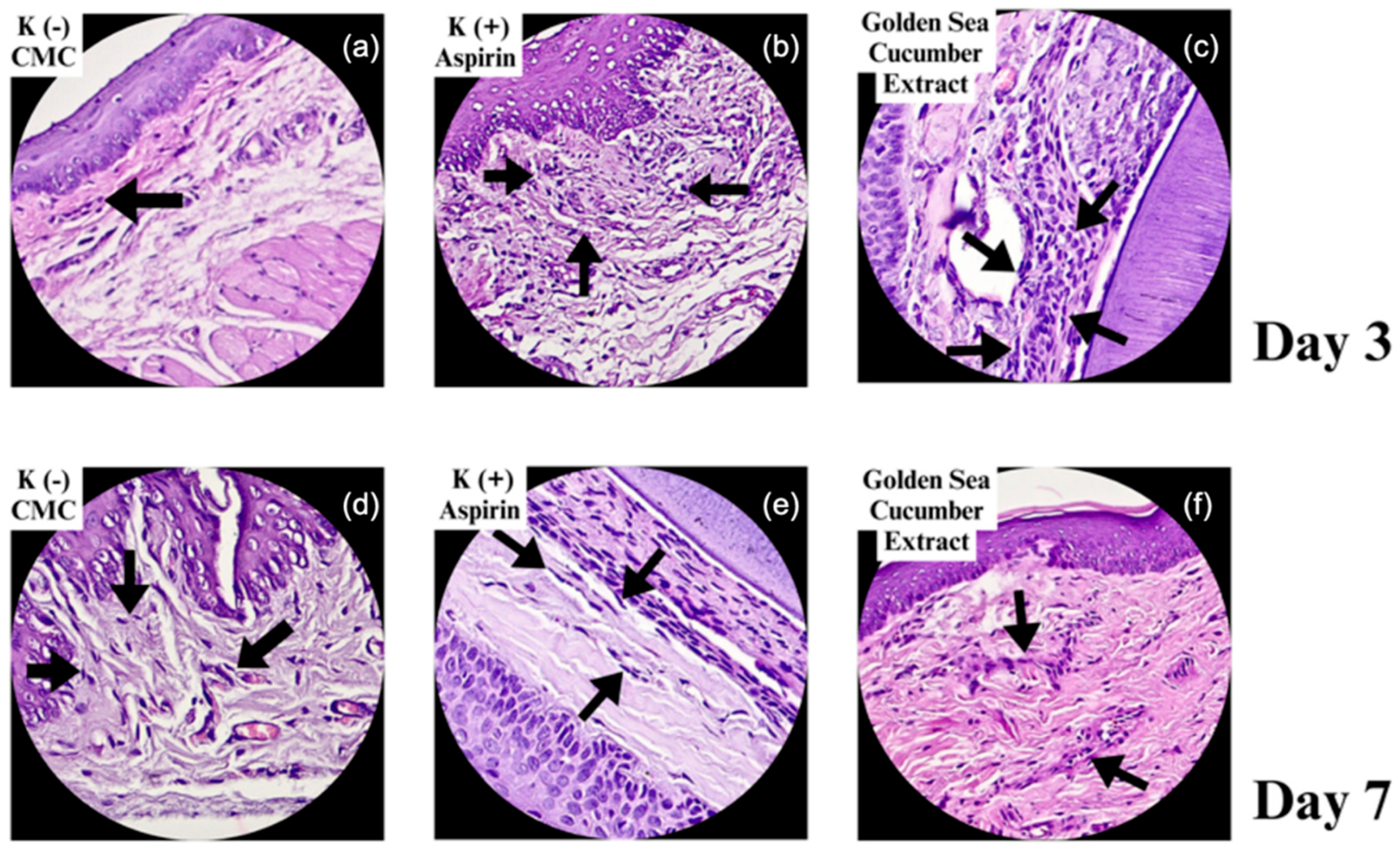
Figure 3.
Histological micrographs of gingival wound tissue stained with Hematoxylin and Eosin (H&E) on Day 3 (a–c) and Day 7 (d–f). (a, d) represent the negative control (Na-CMC), (b, e) represent the positive control (aspirin), and (c, f) represent the Stichopus hermanii extract group. Black arrows indicate fibroblast presence and tissue organization. Images captured at 200× magnification.
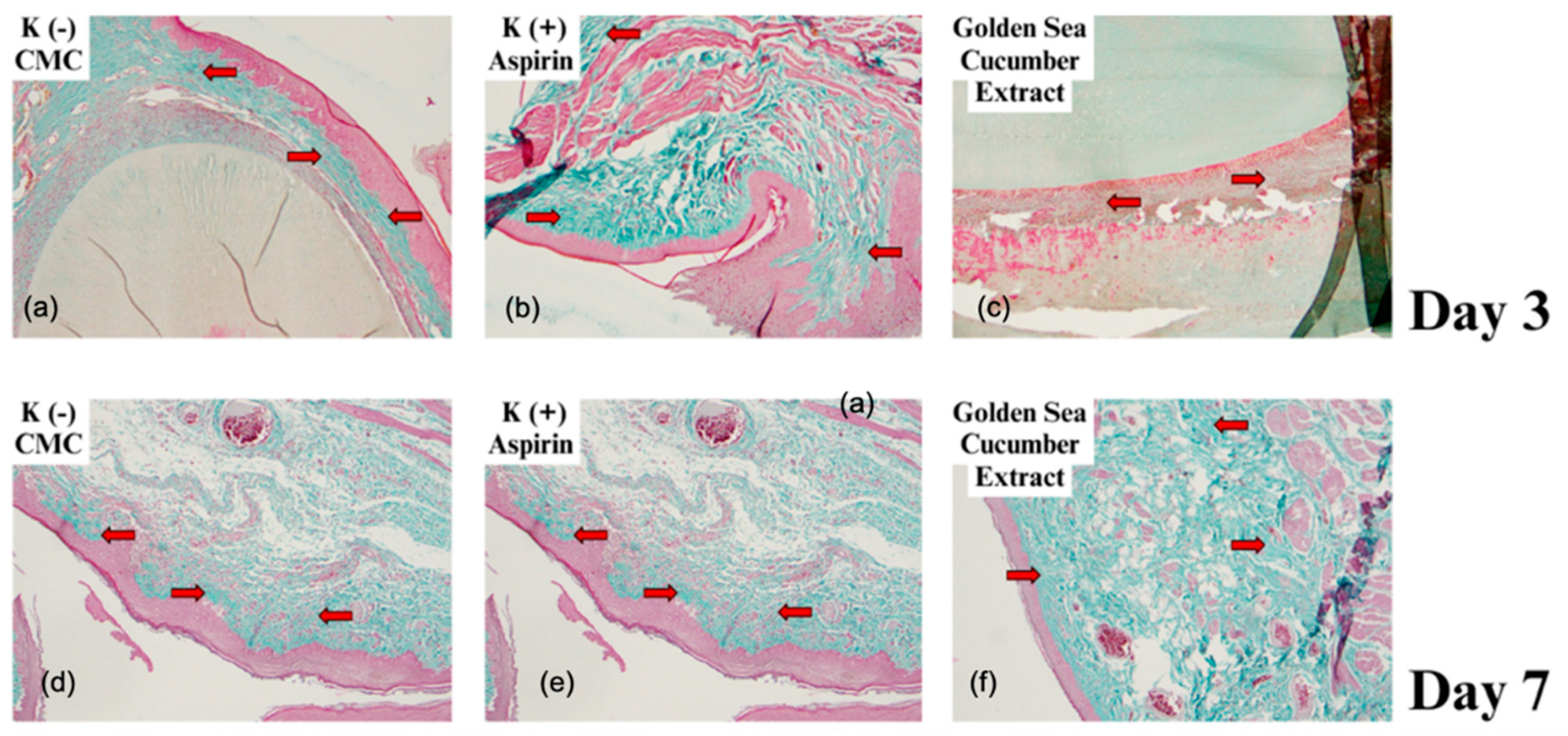
Figure 4.
Serum SGOT levels and (b) serum SGPT levels on Days 0 and 7 in the Na-CMC control group and treatment groups receiving Stichopus hermanii extract at 25%, 50%, and 100% concentrations.
Figure 4.
Serum SGOT levels and (b) serum SGPT levels on Days 0 and 7 in the Na-CMC control group and treatment groups receiving Stichopus hermanii extract at 25%, 50%, and 100% concentrations.
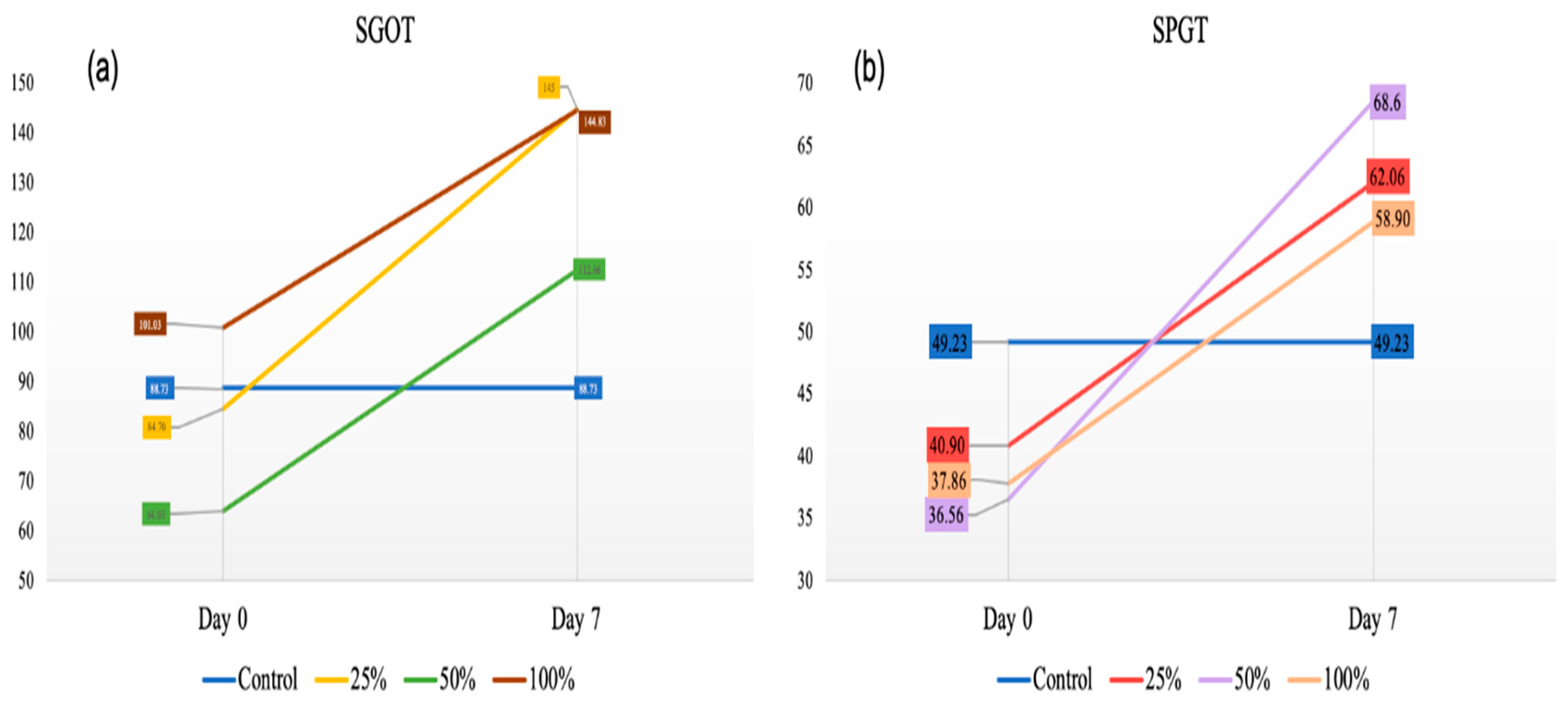

Table 1.
3D structure and resolution of selected inflammatory protein targets used for molecular docking simulations.
Table 1.
3D structure and resolution of selected inflammatory protein targets used for molecular docking simulations.
| Receptor | PDB ID | Resolution | 3D Structure |
|---|---|---|---|
| TNF-α | 6RMJ | 2.65 Å | |
| NF-κB | 1NFI | 2.7 Å | |
| JAK-1 | 6N79 | 2.27 Å | |
| IL-1β | 7JWQ | 2 Å |
Table 2.
Interaction of Protein and (6Z)-Octadecenoic acid.
| Compound | Protein | Binding affinity (Kcal/mol) | Interacting Residu in the Binding Pocket | |
|---|---|---|---|---|
| Hydrophobic interactions | Hydrogen Bond | |||
| (6Z)-Octadecenoic acid | TNF-α | -5.3 | TYR119, TYR151, TYR59, LEU57 | TYR151, GLY121 |
| NF-κB | -5 | VAL97, PHE318, PHE77, PHE103 | TYR71, PHE77 | |
| JAK-1 | -5.4 | VAL889, LEU1010, LEU881 | GLU925, GLY1023 | |
| IL-1β | -4.4 | LYS77, PRO131, PHE133 | VAL132, LEU134, LEU 80 | |
Table 3.
Analysis of fibroblast cells and the level of collagenization on day 3 and 7.
| Group | n | Day 3 | Day 7 | ||
|---|---|---|---|---|---|
| Fibroblasts (Mean ± SD) |
Collagenization | Fibroblasts (Mean ± SD) |
Collagenization | ||
| Na-CMC | 2 | 119 ± 5.38 | 112.00 μm | 166 ± 13.8 | 175.75 μm |
| Aspirin | 2 | 177 ± 14.62 | 238.50 μm | 350 ± 24.54 | 452.75 μm |
| Stichopus hermanii | 2 | 213 ± 13.2 | 177.25 μm | 251 ± 21.7 | 717.25 μm |
| P value | 0.001* | 0.002* | 0.001* | 0.000* | |
| *One-way ANOVA test (p<0.05) | |||||
Table 4.
ADME-Tox Prediction of Stichopus hermanii bioactive compound.
| Compound | ADME-Tox | |
|---|---|---|
| Properties | Value | |
| (6Z)-Octadecenoic acid | Caco-2 permeability (nm.sec-1) | 1.563 |
| Human Intestinal Absorpstion (%) | 91.82 | |
| VDss (log L/kg) | 0.558 | |
| CYP2D6 Inhibitor | No | |
| Total Clearance | 1.884 | |
| AMES Toxicity | No | |
| Hepatotoxicity | No | |
| Oral Rate Acute Toxicity (LD50) (mol/kg) | 1.417 | |
| Oral Rate Chronic Toxicity (LOAEL) (log mg/kg_bw/day) | 3.259 | |
Table 5.
Physicochemical properties and Lipinski’s Rule of Five evaluation og (6Z)-Octadecenoic acid.
Table 5.
Physicochemical properties and Lipinski’s Rule of Five evaluation og (6Z)-Octadecenoic acid.
| Compound | Molecular Formula | SMILES | Lipinski’s Rule of Five | |
|---|---|---|---|---|
| Properties | Value | |||
| (6Z)-Octadecenoic acid | C18H34O2 | CCCCCCCCCCC/C=C\CCCCC(=O)O | WM | 282.5 |
| HBA | 2 | |||
| HBD | 1 | |||
| Log P | 6.11 | |||
| Rm | 87.08 | |||
| Violation | 1 | |||
| *Violation of criteia, maximum violation of criteria is two. MW: Weight of mass (<500 Da); HBA: Hydrogen Bond Acceptors (<10); HBD : Hydrogen Bond Donor (<5); LogP: partition coefficient (5); Rm: Refractivity of Molar (40-130 cm3/mol). | ||||
Disclaimer/Publisher’s Note: The statements, opinions and data contained in all publications are solely those of the individual author(s) and contributor(s) and not of MDPI and/or the editor(s). MDPI and/or the editor(s) disclaim responsibility for any injury to people or property resulting from any ideas, methods, instructions or products referred to in the content. |
© 2025 by the authors. Licensee MDPI, Basel, Switzerland. This article is an open access article distributed under the terms and conditions of the Creative Commons Attribution (CC BY) license (http://creativecommons.org/licenses/by/4.0/).
Copyright: This open access article is published under a Creative Commons CC BY 4.0 license, which permit the free download, distribution, and reuse, provided that the author and preprint are cited in any reuse.


