Preprint
Article

This version is not peer-reviewed.

Multi-Gene Analysis, Morphology, and Species Delimitation Methods Reveal a New Species of Melanothamnus, M. Coxsbazarensis sp. nov. (Rhodomelaceae, Ceramiales) for the Marine Red Algal Flora from Bangladesh

A peer-reviewed article of this preprint also exists.

Submitted:

13 August 2025

Posted:

14 August 2025

You are already at the latest version

Abstract

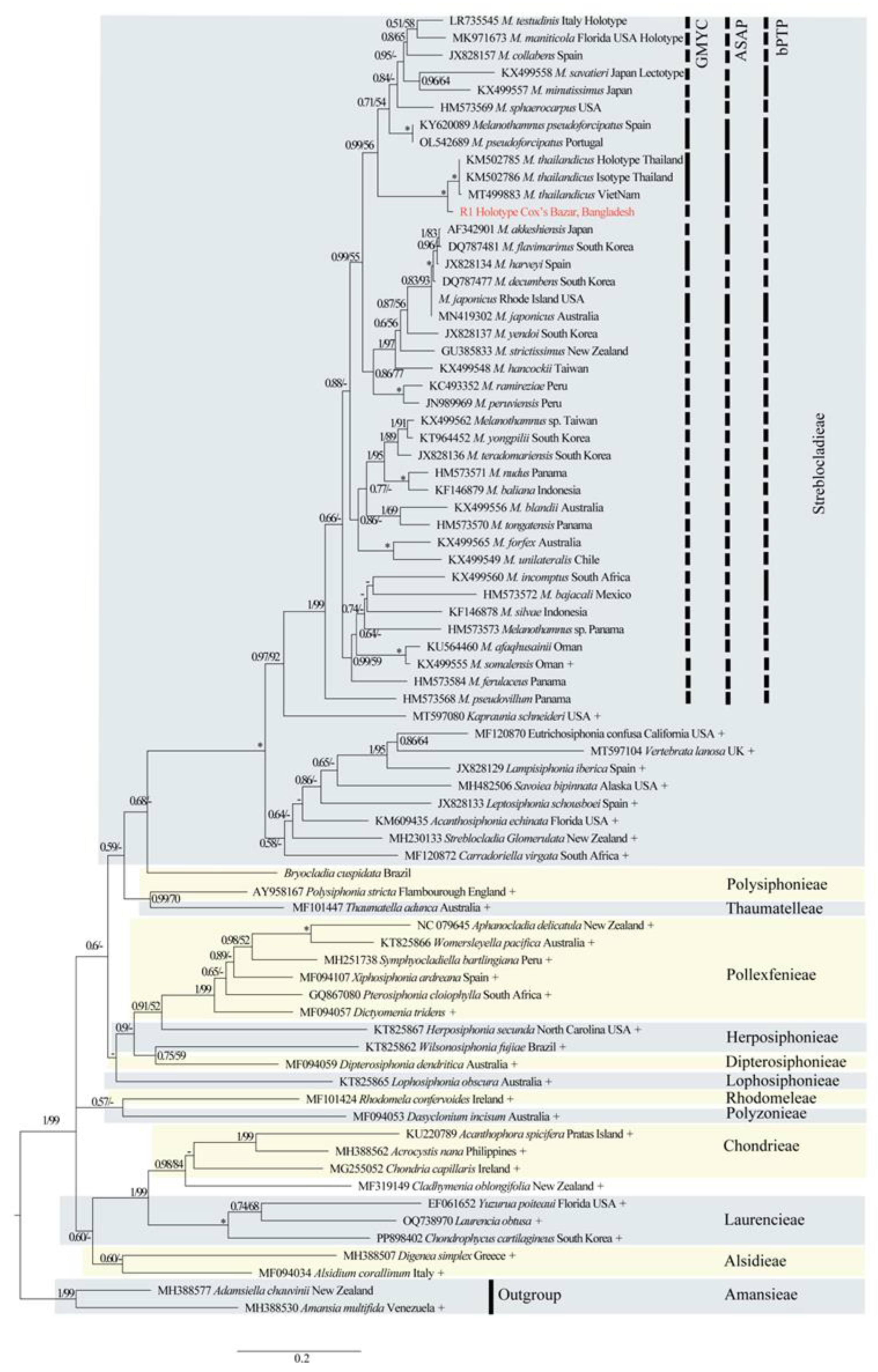
Turf-forming Polysiphonia-looking algae were collected from a small (< 1.0 km2 area) Agarophyton tenuistipitata (Gracilariaceae, Gracilariales) farm on the East coast of the Bay of Bengal. DNA was extracted from silica gel-preserved specimens, and plastid-encoded rbcL, nuclear-encoded small subunit SSU, large subunit LSU, and universal plastid amplicon (UPA) were amplified and sequenced. Maximum likelihood (ML) and Bayesian inference were performed for the phylogenetic analysis. Four single-locus species delimitation methods (SDMs), namely the Generalized Mixed Yule-Coalescent (GMYC) method, a Poisson Tree Processes (PTP) model, the Automatic Barcode Gap Discovery (ABGD), and the Assemble Species by Automatic Partitioning (ASAP) method, were performed to segregate the putative species from other taxa in the Polysiphonia sensu lato clades. Our results revealed that rbcL had 1.4% interspecific genetic divergence, whereas LSU, UPA, and SSU had 1.6%, 2.5%, and 5.4% genetic divergence, respectively, from the nearest neighbors. Both comparative genetic and distinct morphological data revealed that the collected Bay of Bengal specimens comprise a species new to science. In addition, the above-mentioned SDMs supported the genetic data and segregated our specimens as Melanothamnus coxsbazarensis sp. nov. as a distinct species.

Keywords: 
;  ;  ;  ;  ;  ;  

1. Introduction

Members of the red algal genus Melanothamnus Bornet & Falkenberg [1] (Rhodomelaceae) are predominantly found in the Indo-Pacific regions [2]. These turf algae form macroscopic clumps featuring unique filiform thalli [3,4]. Indo-Pacific species of Melanothamnus were originally described as Neosiphonia by Kim and Lee [5] based on specimens collected from Bangpo on the western coast of Korea. Subsequently, Díaz-Tapia et al. [2] provided the newly treated name Melanothamnus with new combinations based on nomenclatural priority, transferring 46 species of Fernandosiphonia and Neosiphonia into Melanothamnus. This transfer was based on 14 consistent morphological features and a well-supported molecular rbcL and 18S phylogeny. This polysiphonous genus often grows epiphytically on other algae, becoming entangled with each other to form dense mats and turfs or grows epilithically [2,3, 6−11]. The most common and diverse group of epiphytic macroalgae found in seaweed farms consists of members of the order Ceramiales, including two widespread genera, Polysiphonia and Melanothamnus [12,13]. For instance, M. savatieri and M. thailandicus have been reported from the Gulf of Thailand, growing epiphytically on Gracilaria in seaweed farms [14,15]. Additionally, M. thailandicus has also been reported to grow epiphytically on Kappaphycus alvarezii (Solieriaceae) in seaweed farms in Vietnam [16]. Some Melanothamnus species (e.g., M. maniticola, M. testudinis) have also been documented as growing epizoically on animals [17,18]. For example, M. manaticola was reported from the skin of Indian Manatees in Florida [17].
Melanothamnus is characterized by having erect or prostrate thalli mostly < 10.0 cm in length (except for M. afaqhusainii which can reach >1.0 m in length); 4 – 9 pericentral cells; rhizoids cut off from pericentral cells; single trichoblast per segment; segments spirally arranged and moderately developed at the apical region of the branchlets; spermatangial branches cylindrical, sometimes with a sterile tip, arising from 1 trichoblast fork; procarps with a 3-celled carpogonial branch; cystocarps scattered on branchlets, not aggregated, globose or ovoid, very shortly pedicellate. Tetrasporangia tetrahedrally divided, forming spiral series in upper branchlets [2,5,11].
Molecular investigations have been performed to delimit the taxonomic position of Melanothamnus and to infer their evolutionary relationships [19−21]. Molecularly, Melanothamnus belongs to the polyphyletic group Polysiphonia sensu lato of the Streblocladieae tribe of the Rhodomelaceae. Currently, this tribe contains a total of 14 genera, namely: Acanthosiphonia, Aiolocolax, Carradoriella, Erythrocystis, Eutrichosiphonia, Kapraunia, Lampisiphonia, Leptosiphonia, Melanothamnus, Pterochondria, Savoiea, Streblocladia, Tolypiocladia, and Vertebrata [11,22−25]. At present, 57 species of Melanothamnus are taxonomically accepted in the Streblocladieae tribe [11]. Whereas species of Melanothamnus are reported from the Indo-Pacific regions such as Korea, Japan, Philippines, and Hawaii, few molecular data of species have been documented from the Indian coasts [11]. In addition, some species have also been catalogued from South Africa (M. incomptus), Oman (M. somalensis and M. afaqhusainii), Thailand (M. thailandicus), and India (M. platycarpus) [2].
To date, there are about 337 species of seaweeds in Bangladesh listed in 61 families, of which 73 species belong to Chlorophyta, 89 species to Ochrophyta, and 175 species to Rhodophyta [26−29]. Among them, Islam[30] reported three species of Polysiphonia sensu lato from Saint Martin Island, Bangladesh, namely Polysiphonia mollis, Carradoriella denudata [as P. denudata], and Tolypiocladia glomerulata. In addition, Aziz and Rahman [6] revealed Melanothamnus harveyi [as P. harveyi] epiphytic on Liagora sp. in the Northeast coast of the Bay of Bengal. To the best of our knowledge, no molecular data on Melanothamnus species have been disclosed for the algal flora of Bangladesh. For the present study, a turf of Polysiphonia-like algae from a small (< 1.0 km2 area) Agarophyton (Gracilaria) tenuistipitata farm on the East coast of the Bay of Bengal was collected for their identification on the basis of a a multi-gene analysis, morphological features, and several species delimitation methods (SDMs).

2. Materials and Methods

2.1. Study Area and Sample Collections

On March 07, 2021, a small chunk of a reddish Polysiphonia-like sample was collected manually growing in an Agarophyton (Gracilaria) tenuistipitata farm attached to the twisted nylon ropes during low tide in the Bakkhali River estuary, Cox’s Bazar district, Bangladesh (Fig. S1). The algae were collected at 28.8 ˚C and 29.54 PSU salinity. Specimens were kept in flow-through seawater tanks at ambient water temperature (~12 °C) until processed for molecular and morphological analyses. The vouchers were examined using a ZEISS Stemi 2000-C dissecting scope, and the branching patterns, thallus length, rhizoidal attachment, and pericentral cell numbers were observed. A clean piece of the sample was preserved in silica gel for DNA extraction, and the remainder of samples were pressed on herbarium paper and finally brought to the Phycology Lab at the University of Louisiana at Lafayette Hebarium (LAF) for further assessment.

2.2. DNA Extraction, PCR Amplification, and Sequencing Protocols

DNA extraction, PCR amplification, and sequencing were performed as follows: DNA was extracted from the preserved silica gel-preserved materials using the Quick-DNA Plant/Seed Miniprep Kit (Zymo Research, Irvine, CA, USA). PCR was performed using the plastid-encoded Rubisco Large subunit (rbcL) primers (F753/RrbcS-start and F57/R893 [19,31], 23 S (UPA), primers (p23SrV_f1and p23SrV_r1) [32] nuclear-encoded 18S small subunit (SSU) L (G01-G10) fragment [33], 28S large subunit (LSU) barcoding primers (nu28SF and nu28SR) [34], following the primers protocol. Amplified products were checked for yield and proper length using 1 % agarose gels stained with ethidium bromide. Successful amplifications were purified and sequenced by Epoch Life Science (Missouri City, TX, USA).

2.3. Sequence Alignment and Phylogenetic Analysis

Sequencher (V. 5.4.6, Gene Codes Corp., Ann Arbor, MI, USA) was used to assemble chromatograms, edit sequence reactions, and build contigs. Resulting sequences were BLASTed in NCBI GenBank (https://www.ncbi.nlm.nih.gov/) and the closest worldwide sequences were initially aligned using Muscle in Mega X [35]. Identical sequences were removed from the rbcL, SSU, LSU, and UPA alignments. The phylogenies were derived from rbcL (80 sequences; 1387 bp), SSU (59 sequences; 494 bp), LSU (31 sequences; 509 bp), and UPA (36 sequences; 378 bp) loci. The best-fitting nucleotide substitution model was selected using the program PartitionFinder 2 [36]. The best partition strategy and model of sequence evolution were selected based on the Bayesian Information Criterion (BIC), Akaike Information Criterion corrected (AICc), and Akaike Information Criterion (AIC) scores. The general time-reversible nucleotide substitution model with a gamma distribution and a proportion of invariable sites (GTR + Γ + I) was selected for the rbcL, SSU, LSU, and UPA data. Maximum likelihood (ML) analyses were performed with the RAxML-HPC2 on ACCESS (8.2.12) [37] through 1000 replications of rapid bootstrap. In addition, Bayesian inference (BI) was performed with MrBayes v3.2.7 software [38] using Metropolis coupled Markov chain Monte Carlo (MCMC) and the GTR + Γ model. To evaluate posterior probabilities, two runs, each with four chains (three hot and one cold) for 100,000,000 generations, sampling trees every 1,000 generations. We plotted likelihood vs. generation using the Tracer v1.7.2 program [39] to reach a likelihood plateau and set the burn-in value. The convergence of both runs was evaluated using Tracer to observe whether the runs reached an effective sample size (ESS) >200. A burn-in of 25% was used to avoid suboptimal trees in the final consensus tree. The “.TRE” file was imported into FigTree 1.4.4 [39] as a starting point for further editing in Adobe Illustrator (Version 28.1).

2.4. Assessing the Species Delimitation Methods

Single-marker species delimitation methods (SDMs) were applied on rbcL, SSU, LSU, and UPA datasets, i.e., the Automatic Barcode Gap Discovery (ABGD) [40], Assemble Species by Automatic Partitioning (ASAP) [41], Multispecies Coalescent Model, Poisson Tree Processes model (PTP) [42], and the Generalized Mixed Yule-Coalescent model (GMYC) [43], respectively. ABGD was performed through the web interface (https://bioinfo.mnhn.fr/abi/public/abgd/abgdweb.html) using the Kimura-2 model parameters and 100 screening steps [40]. The relative gap width, maximum (Pmax), and minimum (Pmin) variability (P) were set following Laboni’s protocol [44]. For ASAP, branch lengths were extracted from the rbcL, SSU, LSU, and UPA RAxML tree with the function cophenetic.phylo of the package “APE” in R [45] to produce a Cox’s Bazar, Bangladesh Melanothamnus sp. distance matrix as input and run through the web-based interface (https://bioinfo.mnhn.fr/abi/public/asap/) using the default parameters [41]. GMYC was performed with the package SPLITS in R [46], with the single threshold method based on an ultrametric tree generated in BEAST v1.10.4 [47] using a relaxed log-normal clock with an exponential growth coalescent as prior, and a GTR+I+G model of evolution partitioned per codon position. MCMC chains were run for 100 million generations (sampled every 1000th generation) and the quality of the run assessed in the Tracer v1.7.2 program [48] to ensure that ESS values were >200 with the 10% burn-in values. The PTP model was estimated to putative species boundaries on a given phylogenetic input of RAxML tree by relying on the branch lengths, assessing the number of substitutions between branching events [42,49]. The main assumption of this model is that the number of substitutions between species is significantly higher than the number of substitutions within species [50]. The Bayesian PTP (bPTP) species delimitation was analyzed on the web server (http://species. h-its.org/) using the above-generated rooted ML tree as input and implemented maximum likelihood (mlPTP) and heuristic search (hsPTP) algorithms. The maximum MCMC generations, thinning, and burn-in values were applied by Laboni’s protocol [44], and the outgroup was removed to improve species delimitation [42]. Finally, the best convergence MCMC chain was visually confirmed as Zhang et al. [42] recommended.

2.5. Morphological Observations

The distinct morphological characteristics found in this study for the holotype and isotype specimens were recorded and analyzed. Furthermore, unstained and stained (1% aqueous aniline blue acidified with 0.1% diluted HCl) microscopic images were captured using an Olympus BX60 compound microscope attached with a Canon DS126271 camera.

3. Results

3.1. Phylogenetic Results

A total of 40 rbcL, 23 SSU, 9 LSU, and 19 UPA Melanothamnus specimens were compared and used to construct the gene trees, including the Cox’s Bazar sample in this study. Phylogenetically, Melanothamnus formed a moderately to strongly supported intergeneric sister clade with the Kapraunia species (Figure 1, Figures S2 and S3). Among them, rbcL was highly informative, forming a fully supported clade (*) among the Melanothamnus species (Figure 1). Melanothamnus coxsbazarensis sp. nov. was phylogenetically distinct from other species and formed a fully supported sister clade with M. thailandicus with 1.4% rbcL sequence divergence and grouped with moderate to weakly supported clade with higher sequence divergence (%) M. pseudoforcipatus (6.3–6.9), M. testudinis (7.2), M. minutissimus (7.6), and M. collabens (7.9), M. teradomariensis (7.9) M. sphaerocarpus (8.2), M. maniticola (8.7), M. harveyi (8.8), M. flavimarinus (9.3) M. savatieri (9.9) (Figure 1). In addition, Melanothamnus formed a strongly supported SSU clade (79/1) of 23 Melanothamnus specimens, whereas M. coxsbazarensis sp. nov. formed a weakly supported clade (0.5/-) with 5.4% sequence divergence from M. collabens (Figure S2). However, the UPA region of the 23S rRNA gene formed a weakly supported clade (0.82/-) between 19 Melanothamnus specimens and Kapraunia, where our studied sample (R1) formed a strongly supported clade (1/90) with the Hawaiian Melanothamnus sp. (Accession HQ421163, HQ421164, HQ421175, and HQ421282) with 0.8% sequence divergence (Figure S3). In addition, the Panama sample (KY573941 Melanothamnus sp.) had 1.9% UPA sequence divergence from our studied materials (Figure S3). Furthermore, our Cox’s Bazar specimen had 2.5–2.8% UPA sequence divergence with M. harveyi (Figure S3). In addition, only 9 LSU samples analyzed were available in GenBank, and the Melanothamnus species formed a strongly supported clade (1/99) (Figure S4). However, since no Kapraunia samples were assessed for LSU, it formed a weakly supported clade (0.72/-) with Carradoriella (Figure S4). The Hawaiian Melanothamnus sp. (Accession HQ422097) formed a fully supported clade (*) for LSU with 1.0% sequence divergence, whereas 1.6% sequence divergence characterized M. Harveyi, M. akkeshiensis, M. japonicus, Melanothamnus sp. M. upolensis and Melanothamnus sp. (Figure S4). M. afaqhusainii formed a separate clade with 3.7% LSU sequence divergence, whereas Carradoriella elongata had 3.3% sequence divergence (Figure S4).

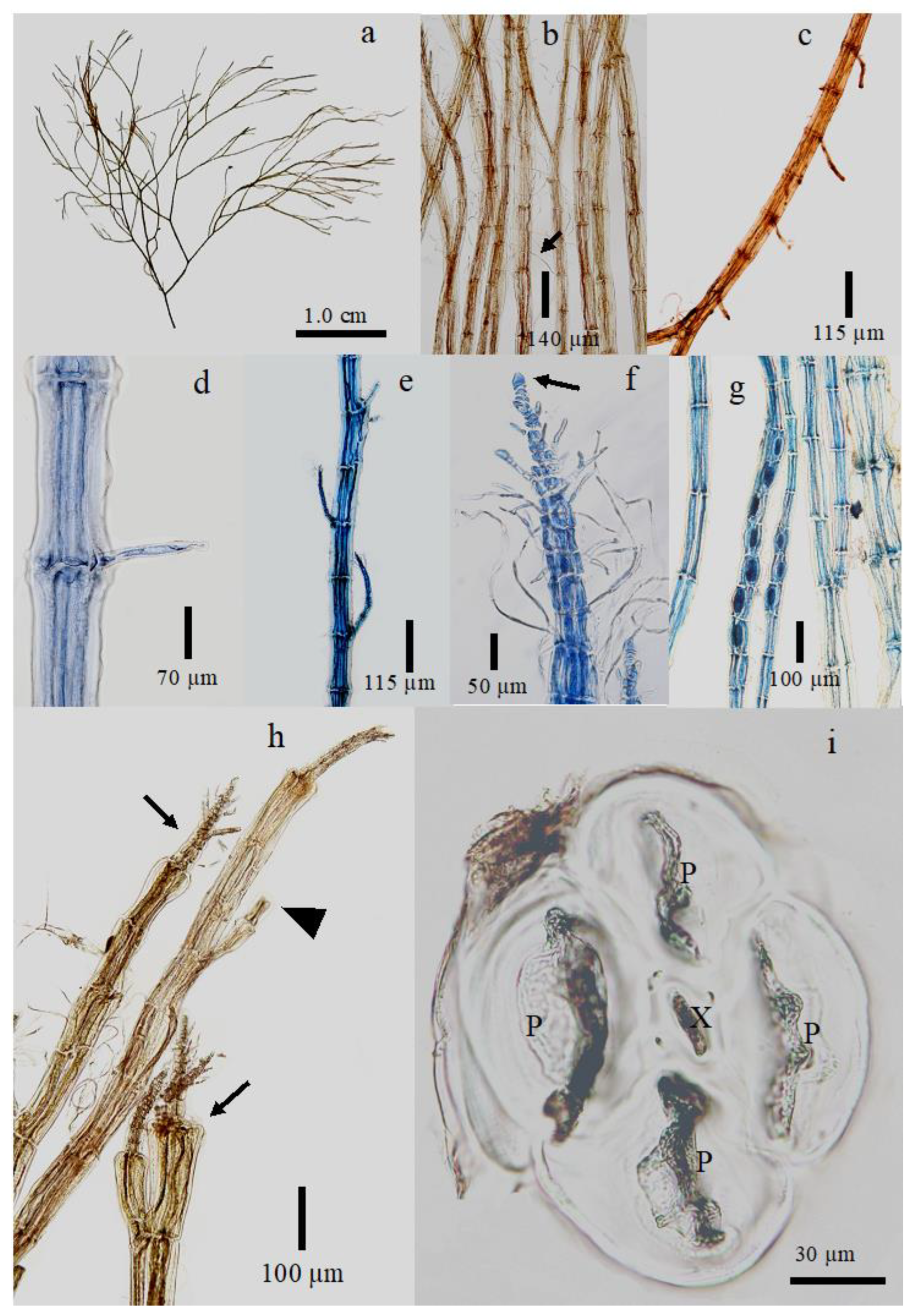
3.2. New Species Descriptions

Melanothamnus Coxsbazarensis M.A.Islam and Fredericq sp. nov. Figure 2
DESCRIPTION: Thalli grow epiphytically on nylon ropes in an Agarophyton (Gracilaria) tenuistipitata farm. Prostrate thalli sub-dichotomously branched and reddish brown. Rhizoids distributed throughout the thallus, except at the apical regions, are cut off from the pericentral cells. The lower part of the thallus is usually thicker in diameter than the upper part of the axes and occasionally contains 5 pericentral cells, but more frequently 4 pericentral cells. Trichoblasts present in the upper middle part of the thallus but abundant in the apical regions. Dome-shaped or acute apical cell. Adventitious endogenous branches rarely present and alternately arranged. Tetrasporangia arranged in spiral series. No carposporophytes or spermatangial specimens were found.
HOLOTYPE: LAF-3-7-21-1-1 (R1), collected March 07, 2021, by Md. Ariful Islam and Mohammad Khairul Alam Sobuj, deposited in the Phycology Lab, Department of Biology, University of Louisiana at Lafayette, LA, USA.
ISOTYPES: LAF-03-7-21-1-2 (R1_1); LAF-03-7-21-1-3 (R1_2); LAF-03-7-21-1-4 (R1_3), collected March 07, 2021, by Md. Ariful Islam and Mohammad Khairul Alam Sobuj, deposited in the Phycology Lab, Department of Biology, University of Louisiana at Lafayette, LA, USA.
TYPE LOCALITY: Bakkhali River estuary (21°28'27.7"N 91°57'55.2" E), Cox’s Bazar, Bangladesh.
ETYMOLOGY: The species name was chosen for the collection locality, the Cox’s Bazar district.
OTHER SPECIMENS EXAMINED: LAF-03-7-21-1-5 (R1_4); LAF-03-7-21-1-6 (R1_5); LAF-03-7-21-1-7 (R1_6), LAF-03-7-21-1-8 (R1_7); LAF-03-7-21-1-9 (R1_8); LAF-03-7-21-1-10 (R1_9).
VEGETATIVE FEATURES: Prostrate turf thalli, 3.5–5 cm high, epiphytically growing on nylon ropes in the Agarophyton (Gracilaria) tenuistipitata farm, reddish brown, sub-dichotomous to dichotomously branched (Figure 2a,b). The basal regions, including the main and lateral axes comparatively thicker (95–110 μm in diameter) than the middle (65–100 μm in diameter) to upper middle (60–95 μm in diameter) region of the thalli (Figure 2a,b). Fewer trichoblasts found in the middle part of the thalli, but frequently present in the upper middle to the apical regions (Figure 2b). Rhizoidal attachments found throughout the axes except in the apical region of the thalli (Figure 2c,d). Single cellular rhizoids cut off from the pericentral cells (Figure 2d). Rhizoidal origin mostly unilateral and sometimes random but lack digitate or multicellular tips (Figure 2c,d, Figure S5a). Adventitious endogenous branches scarcely present (found only in a single thallus among the examined samples) and alternately arranged (Figure 2e). Both rhizoids and endogenous branches originated between the branch internodes (Figure 2c, Figure S5a).
When the single-cell young branchlet divides, it forms an adventitious endogenous branch, but when it does not divide, it forms single-cellular rhizoids (Figure S5a). Radially arranged abundant trichoblasts (0.4–0.7 mm long) present around the apical regions and subapical lateral branches available to the thallus, and scar cells are found between the internodes (Figure 2f, Figure S5b). The apical cells dominantly dome-shaped but sometimes acute in shape without pointed tips (Figure 2f). Some trichoblast cells dichotomously branched at the second segment (Figure S5b). Sub-apical lateral branches bear roundly shaped tip cells (Figure S5b). Threadlike main axes surrounded by 4-pericentral cells extend throughout the thallus, and the protruding internodes are connected by a primary pit connection (Figure S5b). Branches bear young tetrasporangia arranged in a spiral series (Figure 2g) and tetrahedrally divided (Figure S5c). The first pericentral cell of the lateral branch starts with 1/3rd to 1/4th length of the regular pericentral cells (Figure S5d,e). Whereas some branches sporadically stop regular growth, they subsequently cut off pericentral cells, trichoblast cells, and apical cells (Figure 2h). Cross section of the main axes dominantly surrounded by 4 pericentral cells; however, rarely 5 pericentral cells found (Figure 2i, Figure S5f,g). No cystocarps or spermatangial branches were found among the studied specimens.

3.3. Species Delimitation Methods (SDMs)

A total of four SDMs (i.e., ABGD, ASAP, GMYC, and bPTP) were analyzed for all the molecular data (rbcL, SSU, LSU, and UPA) in this study. Among them, the three best ones were counted for each gene. The selected three SDMs, i.e., GMYC, ASAP, and bPTP) delineated 35 Melanothamnus species for rbcL (Figure 1). All three SDMs of rbcL delimited M. coxsbazarensis as a separate species from M. thailandicus. In case of SSU, ABGD and ASAP delineated 12 Melanothamnus species, whereas bPTP segregated 18 species (Figure S2). All these SSU SDMs segregated M. coxsbazarensis from the nearest representatives. In case of UPA, ASAP (14 species) segregated a higher number of Melanothamnus species than bPTP (11 species) and ABGD (8 species) (Figure S3). All three SDMs segregated M. coxsbazarensis from the nearest neighbors (Figure S3). For LSU, bPTP (5 species) segregated a higher number of Melanothamnus species than ABGD (3 species) and ASAP (3 species) (Figure S4). Although ABGD and ASAP segregated M. coxsbazarensis from the nearest neighbors, bPTP was unable to segregate the closest Hawaiian Melanothamnus species (Accession: HQ422097) (Figure S4). All the selected genes (i.e., rbcL, SSU, LSU, and UPA) sequenced in this study delimited our M. coxsbazarensis from their nearest neighbors, except the Hawaiian Melanothamnus species (Accession: HQ422097) for bPTP of LSU gene, where the intraspecific sequence divergence between these taxa is 1.0% (Figure 1, Figure S2–S4).

4. Discussion

Based on the multiple gene analysis, distinct morphological features, and SDMs, the specimens collected from the Cox’s Bazar coast of Bangladesh are distinguishable from other described species and support the proposal of a new species of Melanothamnus, Melanothamnus coxsbazarensis sp. nov. The rbcL phylogeny of M. coxsbazarensis shows a fully supported clade (*) to M. thailandicus, which was originally described from the Gulf of Thailand [15] and later recorded from Viet Nam [16]. In addition, these algae moderate to strongly assembled (1/65) with M. pseudoforcipatus, M. sphaerocarpus, M. savatieri, M. minutissimus, M. collabens, M. testudinis and M. maniticola with higher sequence divergence (Figure 1). For SSU, our M. coxsbazarensis formed a weakly supported sister clade with M. ferulaceus Panama, and Melanothamnus sp. and M. japonicus from Japan (Figure S2). The Melanothamnus genus was reinstated due to nomenclature priority that formed a fully supported rbcL and a strongly supported 18S SSU clade by Díaz-Tapia et al. [2]. Likewise, we assessed 40 Melanothamnus specimens that also formed a fully supported clade (*) with rbcL and a strongly supported clade (1/80) for 23 SSU samples analyzed in this study (Figure 1, Figure S2). The rbcL genetic distance between the M. coxsbazarensis from the Bay of Bengal and M. thailandicus from the Gulf of Thailand materials is 1.4%, whereas the Vietnamese specimens have 1.6% sequence distance. The intraspecific divergence of M. thailandicus between the Gulf of Thailand and Vietnam materials is 0.2%. A 1.4% rbcL sequence divergence was mentioned between Omani type species M. somalensis and M. afaqhusainii [2]. In addition, their morphological features also distinguished these species [2,51]. However, our rbcL phylogeny shows 1.2% genetic divergence (p-distance) between these two taxa. In addition, Vertebrata foetidissima and V. isogona had 1.7–1.8% interspecific divergence [52]. Furthermore, the Carradoriella sp. was resolved as 1.8% interspecific divergence to their sister clades [24]. The rbcL interspecific divergence between two Ceramiales species, Wilsonosiphonia howei and W. indica, had 1.8% [53]. Although 2.1% rbcL intraspecific divergence is thought to be enough to segregate the Polysiphonia sensu lato species even for red algae, the sequence divergence is genus-specific [54]. Since only rbcL sequence data are available for M. thailandicus, it could be better to compare the sequence divergence with SSU, LSU, and UPA data. Our M. coxsbazarensis had 1.0% LSU sequence divergence with the Hawaiian (Accession HQ422097) sample [55], which could indicate a separate species. A 0.8% UPA sequence divergence was found between the M. coxsbazarensis and four Hawaiian Melanothamnus sp. (Figure S3). Since the V domain of the 23S UPA gene is highly conserved with ~380 nucleotides, it is normal to have a low interspecific sequence divergence. Additionally, UPA is less promising or even fails to delimit the closely related species [56]. In addition, the LSU and UPA have been widely used for the Hawaiian red algae. These genes are useful to segregate the genera but may not be very helpful to delimit closely related species [34]. The LSU phylogeny shows that M. afaqhusainii formed a separate clade from the Melanothamnus genus clade (Figure S4), although this Omani material formed a sister clade with M. somalensis in the rbcL phylogeny (Figure S4). As only rbcL and 28S LSU M. afaqhusainii data are available in GenBank, comparison with the type species M. somalensis (only rbcL and COI-5P data available in GenBank) would be conducive to understanding the M. afaqhusainii LSU phylogenetic position. We tried to sequence our samples with COI-5P but were unable to get sequence data.
To date, four Polysiphonia sensu lato species have been reported from the Bay of Bengal coasts of Bangladesh based on thallus morphology [6,30]. Whereas Bangladeshi 1-cm tall Polysiphonia mollis was collected epiphytically attached to the substratum with a discoid holdfast and with trichoblast cells, Carradoriella denudata, 10 – 15 cm tall, has 4 – 6 pericentral cells without trichoblast cells in the apical region [30]. Only M. harveyi was reported epiphytically on Liagora sp. from the Bay of Bengal coasts [6]. Most members of Melanothamnus algae are epiphytic on other algae, entangled with each other forming dense mats and turfs (e.g. M. collabens, M. harveyi, M. minutissimus, M. pseudoforcipatus, M. sphaerocarpus) [3,6−10), and often growing as fouling organisms in aquaculture farms (e.g. M. savatieri, M. thailandicus) [14−16,57], and also epizoic (e.g. M. maniticola, M. testudinis) on other animals [17,18]. The Bangladeshi M. harveyi was reddish when young but subsequently became light brown to blackish [6]. This small thallus (3.44 mm tall) is erect, with the main axes having 4 pericentral cells, and abundant trichoblast cells [6]. In addition, M. harveyi has a greater rbcL sequence divergence (> 8%) than our studied M. coxsbazarensis.
Morphologically and genetically M. coxsbazarensis is related to M. thailandicus (Figure 1; Table 1).
Both algae are reddish brown and sub-dichotomous to dichotomously branched (Table 1). However, the M. coxsbazarensis thallus is smaller (Figure 2a) and prostrate axes have 4 – 5 pericentral cells (Figure 2i, Figure S5f,g), whereas M. thailandicus is larger with erect thalli and always with 4 pericentral cells (Table 1). Most importantly, M. coxsbazarensis has abundant trichoblast cells in the apical regions and the rhizoids are distributed throughout the basal to upper middle region of the thallus (Figure 2f, Figure S5b), whereas rhizoids form in the basal part as a discoid holdfast with scarce trichoblast cells in the apical regions for M. thailandicus (Table 1). In addition, no exogenous branches, including cicatrigenous branches, but a very few adventitious endogenous branchlets are found in M. coxsbazarensis (Figure 2e), whereas abundant adventitious endogenous branchlets are present in M. thailandicus (Table 1). Furthermore, abundant adventitious endogenous branches were found in the Vietnamese materials too [16], Figure 3b. Some branches of our M. coxsbazarensis materials suddenly stopped growing and later regenerated pericentral cells and trichoblast cells (Figure 2h). Similarly, one branch of the Vietnamese M. thailandicus materials sporadically stopped growth and formed a thinner tip [16], Figure 3b, which might be due to grazing by specific herbivory. A predator-prey relationship would be required to understand this phenomenon.
All the SDMs' results supported our molecular and morphological data and delimited the M. coxsbazarensis as a separate species (Figure 1, Figure S1 and S3), except for the LSU Hawaiian Melanothamnus materials for bPTP (Figure S4). However, the intraspecific sequence divergence between these two taxa might be high enough to delimit them as separate species, as the LSU nu28SF/nu28SR gene length is ~600 nucleotides with some gaps [34]. In addition, this LSU gene is helpful to segregate the genera but not closely related species [34].
Geographically, Melanothamnus has a higher diversity richness in the Indo-Pacific regions. The diversity of Melanothamnus is particularly high in Korea, Japan, Hawaii, and China [2,11]. Although some Melanothamnus species have been reported from Southeast Asia, like Indonesia, Philippines, and Vietnam, few species have been reported from Southwest Asia, Bangladesh, India, and the Maldives [11]. Moreover, a few molecular data are available from the Indian coasts [11]. Further molecular and extensive morphological and biogeographical studies might reveal more Melanothamnus species in the poorly explored Bay of Bengal coasts and their distributions worldwide.

5. Conclusions

This study is the first attempt to add a molecular analysis of Polysiphonia sensu lato from the coast of the Bay of Bengal. The identity of this fouling organism in the aquaculture farm may aid local farmers and aquaculturists in taking preventive measures. Further predator-prey relationship between M. coxsbazarensis and grazing insects might reveal the biological relations.

Supplementary Materials

The following supporting information can be downloaded at the website of this paper posted on Preprints.org. Figure S1: Collection site of Melanothamnus coxsbazarensis sp. nov. at Bakkhali River, Cox’s Bazar District, Bangladesh; Figure S2. Phylogenetic 18S SSU tree of Melanothamnus coxsbazarensis sp. nov.; Figure S3. Phylogenetic 23S rRNA UPA tree of Melanothamnus coxsbazarensis sp. nov.; Figure S4. Phylogenetic 28S LSU tree of Melanothamnus coxsbazarensis sp. nov; Figure S5. Morphological features of Melanothamnus coxsbazarensis sp. nov.

Author Contributions

Conceptualization, M.A.I., S.F. and W.E.S.; methodology, M.A.I., S.F., W.E.S. and M.K.A.S.; software, M.A.I. and W.E.S.; validation, M.A.I., S.F. and W.E.S.; formal analysis, M.A.I. and W.E.S.; investigation, M.A.I., W.E.S., M.K.A.S., S.R. and S.F.; resources, S.F. and S.R.; data curation, M.A.I., W.E.S. and M.K.A.S.; writing—original draft preparation, M.A.I.; writing—review and editing, M.A.I., W.E.S. and S.F.; visualization, M.A.I., W.E.S., M.K.A.S., S.R. and S.F.; supervision, S.F.; project administration, S.F. and S.R.; funding acquisition, S.F. All authors have read and agreed to the published version of the manuscript.

Funding

This work was funded in part by NSF grant 1754504 to SF.

Institutional Review Board Statement

Not applicable.

Data Availability Statement

All DNA sequences used in this study are in the GenBank (https://www.ncbi.nlm.nih.gov/) and can also be accessed in GenBank under accession numbers (Will be submitted during revision).

Acknowledgments

We thank Md. Tipu Sultan and Md. Kobinur Islam, research assistant at the Marine Fisheries and Technology Station, Bangladesh Fisheries Research Institute, Cox's Bazar 4700, Bangladesh, for their assistance with algal collection, sorting, and voucher specimen preparation.

Conflicts of Interest

The authors declare no conflicts of interest. The funders had no role in the design of the study; in the collection, analyses, or interpretation of data; in the writing of the manuscript; or in the decision to publish the results.

References

  1. Falkenberg, P. Die Rhodomelaceen des Golfes von Neapel und der angrenzenden Meeres-Abschnitte. Fauna und Flora des Golfes von Neapel, Monographie 26. Berlin, 1901. pp. i-xvi, 1-754.
  2. Díaz-Tapia, P.; McIvor, L.; Freshwater, D.W.; Verbruggen, H.; Wynne, M.J.; Maggs, C.A. The genera Melanothamnus Bornet & Falkenberg and Vertebrata S.F. Gray constitute well-defined clades of the red algal tribe Polysiphonieae (Rhodomelaceae, Ceramiales). Eur. J. Phycol. 2017, 52, 1–30. [Google Scholar]
  3. Hollenberg, G. J. An account of the species of Polysiphonia on the Pacific coast of North America. I. Oligosiphonia. Amer. J. Bot., 1942, 29, 772–85. [Google Scholar] [CrossRef]
  4. Díaz-Tapia, P.; Ly, M.; Verbruggen, H. Extensive cryptic diversity in the widely distributed Polysiphonia scopulorum (Rhodomelaceae, Rhodophyta): Molecular species delimitation and morphometric analyses. Mol. Phylog. Evol. 2020, 152, 106909. [Google Scholar] [CrossRef]
  5. Kim, M.S.; Lee, I.K. Neosiphonia flavimarina gen. et sp. nov. with a taxonomic reassessment of the genus Polysiphonia (Rhodomelaceae, Rhodophyta). Phycol. Res. 1999, 47, 271–281. [Google Scholar] [CrossRef]
  6. Aziz, A.; Rahman, M.T. Marine algae of St. Martin's Island, Bangladesh. IX. New records of green algae (Chlorophyceae). Ban. J. Bot. 2010, 39, 161–168. [Google Scholar] [CrossRef]
  7. Nam, K.W.; Kang, P.J. Algal flora of Korea. Volume 4, Number 4. Rhodophyta: Ceramiales: Rhodomelaceae: 18 genera including Herposiphonia. pp. [1-6], 1-178, figs 1-102. Incheon: National Institute of Biological Resources, 2012, pp. [1−6], 1−178.
  8. Díaz-Tapia, P.; Bárbara, I. Seaweeds from sand-covered rocks of the Atlantic Iberian Peninsula. Part 1. The Rhodomelaceae (Ceramiales, Rhodophyta). Cryptog., Algol., 2013, 34, 325–422. [Google Scholar] [CrossRef]
  9. Diaz-Tapia, P.; Barbara, I.; Cremades, J.; Verbruggen, H.; Maggs, C.A. Three new cryptogenic species in the tribes Polysiphonieae and Streblocladieae (Rhodomelaceae, Rhodophyta). Phycologia, 2017, 56, 605–623. [Google Scholar] [CrossRef]
  10. Neto, A.I.; Cacabelos, E.; Prestes, A.C.L.; Díaz-Tapia, P.; Moreu, I. New records of marine macroalgae for the Azores. Bot. Mar., 2022, 65, 105–120. [Google Scholar] [CrossRef]
  11. Guiry, M.D.; Guiry, G.M.E. AlgaeBase. World-wide electronic publication, National University of Ireland, Galway. https://www.algaebase.org; (22 July 2025).
  12. Fletcher, R.L. Epiphytism and fouling in Gracilaria cultivation: an overview. J. Appl. Phycol. 1995, 7, 325–333. [Google Scholar] [CrossRef]
  13. Hurtado, A.Q.; Yunque, D.A.; Tibubos, K.; Critchley, A.T. Use of Acadian marine plant extract powder from Ascophyllum nodosum in tissue culture of Kappaphycus varieties, J. Appl. Phycol. 2009, 21, 633–639. [Google Scholar] [CrossRef]
  14. Muangmai, N.; Maneekat, S.; Petsut, N.; Keawsuralikhit, C. Newly reported marine red alga, Neosiphonia savatieri (Hariot) M.S.Kim et I.K.Lee 1999 (Rhodophyta Rhodomelaceae) from Thailand. Biodiv. J. 2012, 3, 247–250. [Google Scholar]
  15. Muangmai, N.; Yamagishi, Y.; Maneekat, S.; Kaewsuralikhit, C. The new species Neosiphonia thailandica sp. nov. (Rhodomelaceae, Rhodophyta) from the Gulf of Thailand. Bot. Mar. 2014, 57, 459–467. [Google Scholar] [CrossRef]
  16. Bình, D.T.; An, K.T.; Cầm, V.H.; Tuấn, T.V. (2020). New record and the molecular phylogeny of epiphyte (Melanothamnus thailandicus) on red algae (Kappaphycus alvarezii) in Khanh Hoa. J Fish. Sci. Technol. 2020, 2, 1–9. [Google Scholar]
  17. Woodworth, K.A.; Frankovich, T.A.; Freshwater, D.W. Melanothamnus maniticola sp. nov. (Ceramiales, Rhodophyta): an epizoic species evolved for living on the West Indian Manatee. J. Phycol., 2019, 55, 1239–1245. [Google Scholar]
  18. Serio, D.; Furnari, G.; Moro, I.; Sciuto, K. Molecular and morphological characterisation of Melanothamnus testudinis sp. nov. (Rhodophyta, Rhodomelaceae) and its distinction from Polysiphonia carettia. Phycologia. 2020, 59, 281–291. [Google Scholar] [CrossRef]
  19. Stuercke, B; Freshwater, W.D. Consistency of Morphological Characters Used to Delimit Polysiphonia sensu lato Species (Ceramiales, Florideophyceae): Analyses of North Carolina, USA Specimens. Phycologia 2008, 47, 541–59. [Google Scholar] [CrossRef]
  20. Mamoozadeh, N.R.; Freshwater, D.W. Taxonomic notes on Caribbean Neosiphonia and Polysiphonia (Ceramiales, Florideophyceae): five species from Florida, USA and Mexico. Bot. Mar. 2011, 54, 269–92. [Google Scholar] [CrossRef]
  21. Mamoozadeh, N.R.; Freshwater, D.W. Polysiphonia sensu lato (Ceramiales, Florideophyceae) species of Caribbean Panama including Polysiphonia lobophoralis sp. nov. and Polysiphonia nuda sp. nov. Bot. Mar. 2012, 55, 317–347. [Google Scholar] [CrossRef]
  22. Savoie, A.M.; Saunders, G.W. A molecular assessment of species diversity and generic boundaries in the red algal tribes Polysiphonieae and Streblocladieae (Rhodomelaceae, Rhodophyta) in Canada. Eur J. Phycol. 2019, 54, 1–25. [Google Scholar] [CrossRef]
  23. Amos, D.; Aguilar, V.; Barber-Scott, K.; Bustamante, D.E.; Calderon, M.S.; Carrasco, R.; Vang, M.N. Transfer of the marine red alga Erythrocystis saccate (Rhodomelaceae, Rhodophyta) to the tribe Streblocladieae inferred from organellar genome analysis. Phytotaxa, 2021, 507, 266–270. [Google Scholar] [CrossRef]
  24. Bustamante, D.E.; Yeon, W.B.; Wynne, M.J.; Cho, T.O. Molecular and morphological analyses reveal new taxa additions to the tribe Streblocladieae (Rhodomelaceae, Rhodophyta). J. Phycol. 2021, 57, 817–30. [Google Scholar] [CrossRef] [PubMed]
  25. Díaz-Tapia, P.; Verbruggen, H. Resolving the taxonomy of the Polysiphonia scopulorum complex and the Bryocladia lineage (Rhodomelaceae, Rhodophyta). J. Phycol. 2024, 60, 49–72. [Google Scholar] [CrossRef] [PubMed]
  26. Islam, M.A. Mangrove associated macroalgae in the Sundarbans forest, Bangladesh: spatial and temporal changes, MS thesis. Hiroshima University, Hiroshima, Japan. September 20, 2019.
  27. Islam, M.A.; Mauya, M.Z.; Rafiquzzaman, S.M.; Islam, M.R.; Liao, L.M. First report of the red algal genus Chondria C. Agardh (Rhodomelaceae, Rhodophyta) for the marine flora of Bangladesh. Diversity 2019, 11, 95. [Google Scholar] [CrossRef]
  28. Islam, M.A.; Islam, M.R.; Aziz, A.; Liao, L.M. Dictyota adnata Zanardini (Phaeophyceae) -A new record from the Sundarbans mangrove forests, Bangladesh. Ban. J Bot. 2020, 49, 407–412. [Google Scholar] [CrossRef]
  29. Chowdhury, M.S.N.; Hossain, M.S.; AftabUddin, S.; Alamgir, M.; Sharifuzzaman, S.M. Seaweed aquaculture in Bangladesh: Present status, challenges and prospects. Ocean and Coastal Management, 2022, 228. [Google Scholar] [CrossRef]
  30. Islam, A.K.M.N. Contribution to the Study of the Marine Algae of Bangladesh; Bibliotheca Phycologica; J. Cramer Verlag: Vaduz, Lichtenstein, 1976; p. 276. [Google Scholar]
  31. Freshwater, D.W.; Rueness, J. Phylogenetic relationships of some European Gelidium (Gelidiales, Rhodophyta) species, based on rbcL nucleotide sequence analysis. Phycologia, 1994, 33, 187–194. [Google Scholar] [CrossRef]
  32. Sherwood, A.R.; Presting, G.G. Universal primers amplify a 23s rDNA plastid marker in Eukaryotic algae and Cyanobacteria, J. Phycol. 2007, 43, 605–608. [Google Scholar] [CrossRef]
  33. Harper, J.T.; Saunders, G.W. The Application of Sequences of the Ribosomal Cistron to the Systematics and Classification of the Florideophyte Red Algae (Florideophyceae, Rhodophyta). Cahiers Biol. Mar. 2001, 42, 25–38. [Google Scholar]
  34. Conklin, K.Y.; Kurihara, A.; Sherwood, A.R. A molecular method for identification of the morphologically plastic invasive algal genera Eucheuma and Kappaphycus (Rhodophyta, Gigartinales) in Hawaii. J. Appl. Phycol., 2009, 21, 691–699. [Google Scholar] [CrossRef]
  35. Kumar, S.; Stecher, G.; Tamura, K. MEGA7: Molecular Evolutionary Genetics Analysis Version 7.0 for Bigger Datasets. Mol. Biol. Evol. 2016, 33, 1870–4. [Google Scholar] [CrossRef]
  36. Lanfear, R.; Frandsen, P.B.; Wright, A.M.; Senfeld, T.; Calcott, B. PartitionFinder 2: new methods for selecting partitioned models of evolution for molecular and morphological phylogenetic analyses. Mol. Biol. Evol. 2017, 34, 772–773. [Google Scholar] [CrossRef] [PubMed]
  37. Stamatakis, A. RAxML Version 8: A tool for Phylogenetic Analysis and Post-Analysis of Large Phylogenies. Bioinformatics 2014. [Google Scholar] [CrossRef] [PubMed]
  38. Ronquist, F.; Teslenko, M.; Van der Mark, P.; Ayres, D.; Darling, A.; Hohna, S.; Larget, B.; Liu, L.; Suchard, M.A.; Huelsen-beck, J.P. MrBayes 3.2: efficient Bayesian phylogenetic inference and model choice across a large model space. Syst. Biol. 2012, 61, 539–42. [Google Scholar] [CrossRef] [PubMed]
  39. Rambaut, A. Figtree V.1.4.4. Institute of Evolutionary Biology, University of Edinburgh, Edinburgh, 2018.
  40. Puillandre, N.; Lambert, A.; Brouillet, S.; Achaz, G. ABGD, Automatic Barcode Gap Discovery for primary species delimitation. Mol. Ecol. 2012, 21, 1864–77. [Google Scholar] [CrossRef]
  41. Puillandre, N.; Brouillet, S.; Achaz, G. ASAP: assemble species by automatic partitioning. Mol. Ecol. Resources 2021, 21, 609–620. [Google Scholar] [CrossRef]
  42. Zhang, J.; Kapli, P.; Pavlidis, P.; Stamatakis, A. A general species delimitation method with applications to phylogenetic placements. Bioinformatics 2013, 29, 2869–76. [Google Scholar] [CrossRef]
  43. Pons, J.; Barraclough, T.G.; Gomez-Zurita, J.; Cardoso, A.; Duran, D.P.; Hazell, S.; Kamoun, S.; Sumlin, W.D.; Vogler, A.P. Sequence-based species delimitation for the DNA taxonomy of undescribed insects. Sys. Biol., 2006, 55, 595–609. [Google Scholar] [CrossRef]
  44. Paradis, E.; Claude, J.; Strimmer, K. APE: analyses of phylogenetics and evolution in R language. Bioinformatics, 2004, 20, 289–290. [Google Scholar] [CrossRef]
  45. Laboni, H.A.; Sarkar, S.; Islam, M.A.; Islam, M.A.; Islam, M.; Lyzu, C.; Rahman, M.M.; Alam, M.M.; Shohael, A.M.; Karim, M.R. Molecular identification of Gracilaria tenuistipitata isolated from the Bay of Bengal and appraisal of its comprehensive phytochemical profiling, and antioxidant potential. Pharmacol. Res. – Nat. Prod. 2025, 8, 100324. [Google Scholar] [CrossRef]
  46. Fujisawa, T.; Barraclough, T.G. Delimiting species using single-locus data and the Generalized Mixed Yule Coalescent approach: a revised method and evaluation on simulated data sets, Sys. Biol. 2013, 62, 707–724. [Google Scholar] [CrossRef]
  47. Suchard, M.A.; Lemey, P.; Baele, G.; Ayres, D.L.; Drummond, A.J.; Rambaut, A. Bayesian phylogenetic and phylodynamic data integration using BEAST 1.10 Virus Evolution 4,vey016. 2018. [Google Scholar]
  48. Rambaut, A.; Drummond, A.J.; Xie, D.; Baele, G.; Suchard, M.A. ‘Posterior Summarization in Bayesian Phylogenetics Using Tracer 1. 7’, Sys. Biol. 2018, 67, 901–904. [Google Scholar] [CrossRef] [PubMed]
  49. Rojas, R.R.; Fouquet, A.; Ron, S.R.; Hernández-Ruz, E.J.; Melo-Sampaio, P.R.; Chaparro, J.C.; Hrbek, T. (2018). A Pan-Amazonian species delimitation: high species diversity within the genus Amazophrynella (Anura: Bufonidae). Peer J. 2018, 6, e4941. [Google Scholar] [CrossRef] [PubMed]
  50. Solovyeva, E.N.; Dunayev, E.A.; Nazarov, R.A.; Bondarenko, D.A.; Poyarkov, N.A. COI-Barcoding and Species Delimitation Assessment of Toad-Headed Agamas of the Genus Phrynocephalus (Agamidae, Squamata) Reveal Unrecognized Diversity in Central Eurasia. Diversity 2023, 15, 149. [Google Scholar] [CrossRef]
  51. Afaq-Husain, S.; Shameel, M. Further investigations on the red alga Melanothamnus afaqhusainii (Ceramiales) from the coast of Pakistan. Pakistan Journal of Botany, 2000, 32, 15–26. [Google Scholar]
  52. Díaz-Tapia, P.; Kim, M.S.; Secilla, A.; Bárbara, I.; Cremades, J. Taxonomic reassessment of Polysiphonia foetidissima (Rhodomelaceae, Rhodophyta) and similar species, including P. schneideri, a newly introduced species in Europe. Euro. J. Phycol. 2013, 48, 345–362. [Google Scholar]
  53. Bustamante, D.E.; Won, B.Y.; Miller, K.A.; Cho, T.O. Wilsonosiphonia gen. nov. (Rhodomelaceae, Rhodophyta) based on molecular and morpho-anatomical characters. J. Phycol. 2017, 53, 368–380. [Google Scholar] [CrossRef]
  54. McIvor, L.; Maggs, C.A.; Provan, J.; Stanhope, M.J. rbcL sequences reveal multiple cryptic introductions of the Japanese red alga Polysiphonia harveyi. Mol. Ecol., 2001, 10, 911–919. [Google Scholar] [CrossRef]
  55. Sherwood, A.R.; Kurihara, A.; Conklin, K.Y.; Sauvage, T.; Presting, G.G. The Hawaiian Rhodophyta biodiversity survey (2006–2010): a summary of principal findings. BMC Plant Biol. 2010, 10, 258. [Google Scholar] [CrossRef]
  56. Clarkston, B.E.; Saunders, G.W. (2013). Resolving species diversity in the red algal genus Callophyllis (Kallymeniaceae, Gigartinales) in Canada using molecular assisted alpha taxonomy. Euro. J. Phycol. 2013, 48, 27–46. [Google Scholar] [CrossRef]
  57. Kim, M.S. Taxonomy of a poorly documented alga, Neosiphonia savatieri Rhodomelaceae, Rhodophyta) from Korea. Nova Hedwigia, 2005, 81, 163–176. [Google Scholar] [CrossRef]
Figure 1. Phylogenetic rbcL tree based on maximum likelihood (ML) and posterior probabilities (PP) of Bayesian inference (BI) showing position of Melanothamnus coxsbazarensis sp. nov. (in red) inside of Polysiphonia sensu lato and genera belonging to Rhodomelaceae. The selected outgroups belong to the Amansieae tribe of Rhodomelaceae but to non-members of Polysiphonia sensu lato. Bootstrap probability is shown at the nodes out of 1,000 replicates. Values along branches are bootstrap supports & Bayesian posterior probabilities. Lack of bootstrap support value < 50% and PP < 0.5 are dashed (-), whereas the fully supported clades are marked as starred (*). The generitypes of the species marked as (+). The vertical bars indicate 3 SDMs (GMYC, ASAP, and bPTP). Scale represents nucleotide substitutions. The sample generated in this study is shown in red; other sequences are downloaded from GenBank with their accession number.
Preprints 172440 g001
Figure 2. Melanothamnus coxsbazarensis sp. nov.: Habit and morphological features. (a) Dominant bilaterally branched holotype (R1) of M. sp. nov. Note: the basal part is thicker than the apical part. (b) Dichotomously branched upper-middle part of the holotype with occasional trichoblast cells (arrow). (c) The Upper part of the thallus contains occasional rhizoidal cells. (d) A Single cellular rhizoid is cut off from the pericentral cells. (e) Adventitious endogenous branches (alternately arranged) are scarcely present. (f) The sub-apical region of the branch contains abundant trichoblast cells and Dome-shaped apical cells (arrow) of the axes. (g) Spirally arranged young tetrasporangia in the axial cells. (h) Some branches stop growth temporarily (arrowhead), but the axial cell of such branches may regenerate, and be reduced in length and width (arrows). Note: The regenerated branchlets grow trichoblast cells (arrow) and young pericentral cells (arrowhead). These branches have tendency of common characters of the sister sisters. This might happen due to specific herbivory. (i) Cross section of the rehydrated thallus contains an axial cell (X) surrounded by four pericentral cells (P).
Preprints 172440 g002
Table 1.  Morphological comparisons of Melanothamnus coxsbazarensis with other related species. Information not found was marked as ‘–’.
Morphological features M. coxsbazarensis M. thailandicus M. pseudoforcipatus M. sphaerocarpus M. savatieri M. minutissima M. collabens
Thallus color Reddish brown Reddish brown Dark red to brown or pink Blackish brown Dull reddish-brown Dull red Red to pale brown
Type locality Cox’s Bazar, Bangladesh Chon Buri,
Thailand
Galicia, Spain St. Thomas, Virgin Islands Kanagawa, Japan Baja California, Mexico Cádiz, Spain
Plant habit Prostrate Erect Erect Erect Erect Prostrate Erect
Apical cell shape Domed or acute Rounded Rounded Domed Rounded Domed
Height (cm) 3–5 5–15 1.6 1–3 Up to 1 0.3–0.6 Up to 7 cm
Number of pericentral cells 4–5 4 4 4 4 4 6
Branching pattern Subdichotomous Dichotomous Pseudodichotomous Subdichotomous Dichotomous to subdichotomous Subdichotomous Pseudodichotomous
Rhizoidal positions Throughout the thallus Discoid holdfast Basal parts Basal parts Basal tuft of rhizoids Prostrate base Discoid holdfast
Frequency of trichoblasts Abundant Scarce Absent or scarce, at irregular intervals Abundant Abundant Present Scarce
Adventitious endogenous branchlets Scarce Abundant Absent Absent Absent Absent
Exogenous branches, including cicatrigenous branches Absent Absent Present Present Present but no cicatrigenous branches
Present
Cystocarp shape Globular Globose Ovate to globose Globular Urceolate Globular
References This study [15,16] [2,10] [7,20] [14,57] [3] [8]
Disclaimer/Publisher’s Note: The statements, opinions and data contained in all publications are solely those of the individual author(s) and contributor(s) and not of MDPI and/or the editor(s). MDPI and/or the editor(s) disclaim responsibility for any injury to people or property resulting from any ideas, methods, instructions or products referred to in the content.
Copyright: This open access article is published under a Creative Commons CC BY 4.0 license, which permit the free download, distribution, and reuse, provided that the author and preprint are cited in any reuse.
Prerpints.org logo

Preprints.org is a free preprint server supported by MDPI in Basel, Switzerland.

Subscribe

Disclaimer

Terms of Use

Privacy Policy

Privacy Settings

© 2026 MDPI (Basel, Switzerland) unless otherwise stated